Embed Size (px)
Citation preview
La placa neuralse pliega de forma secuencial para formar el tubo neural.Fuente: Kandel et al., 2000. Principles of Neural Science, 4th ed. McGraw-Hill.
Todo el sistema nervioso procede del ectodermo
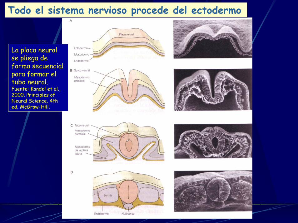
Neural tube formation in Xenopus. The external views are from the dorsal aspect. The cross sections are cut in a plane indicated by the broken lines. Fuente: Alberts et al., 2002.
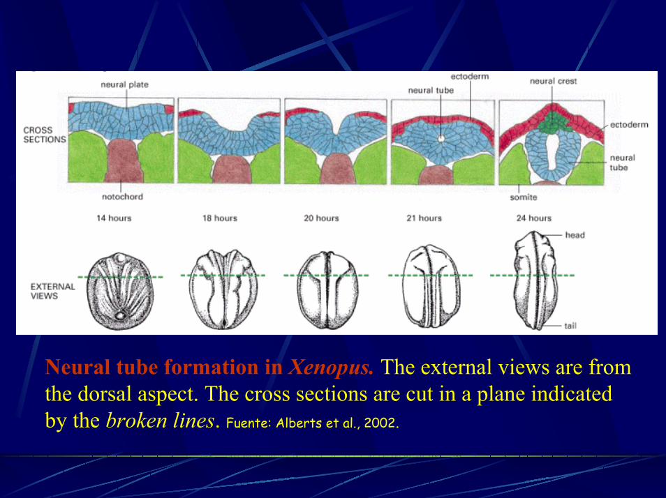
Estadios sucesivos del desarrollo del tubo neural
Fuente: Kandel et al., 2000. Principles of Neural Science, 4th ed. McGraw-Hill.
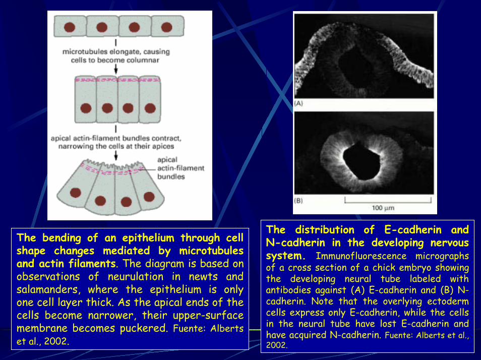
The distribution of E-cadherin andN-cadherin in the developing nervous system. Immunofluorescence micrographs of a cross section of a chick embryo showing the developing neural tube labeled with antibodies against (A) E-cadherin and (B) N-cadherin. Note that the overlying ectoderm cells express only E-cadherin, while the cellsin the neural tube have lost E-cadherin and have acquired N-cadherin. Fuente: Alberts et al., 2002.
The bending of an epithelium through cell shape changes mediated by microtubules and actin filaments. The diagram is based onobservations of neurulation in newts and salamanders, where the epithelium is only one cell layer thick. As the apical ends of the cells become narrower, their upper-surface membrane becomes puckered. Fuente: Alberts et al., 2002.
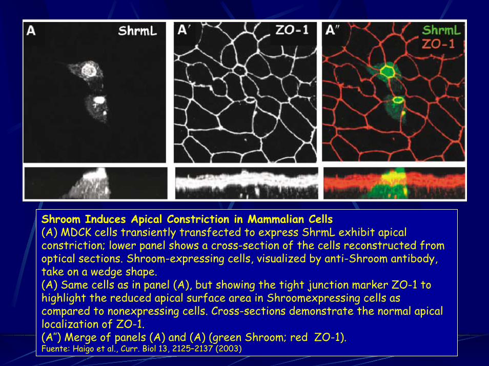
Shroom Induces Apical Constriction in Mammalian Cells(A) MDCK cells transiently transfected to express ShrmL exhibit apicalconstriction; lower panel shows a cross-section of the cells reconstructed from optical sections. Shroom-expressing cells, visualized by anti-Shroom antibody,take on a wedge shape.(A) Same cells as in panel (A), but showing the tight junction marker ZO-1 to highlight the reduced apical surface area in Shroomexpressing cells ascompared to nonexpressing cells. Cross-sections demonstrate the normal apicallocalization of ZO-1.(A″) Merge of panels (A) and (A) (green Shroom; red ZO-1).Fuente: Haigo et al., Curr. Biol 13, 2125–2137 (2003)
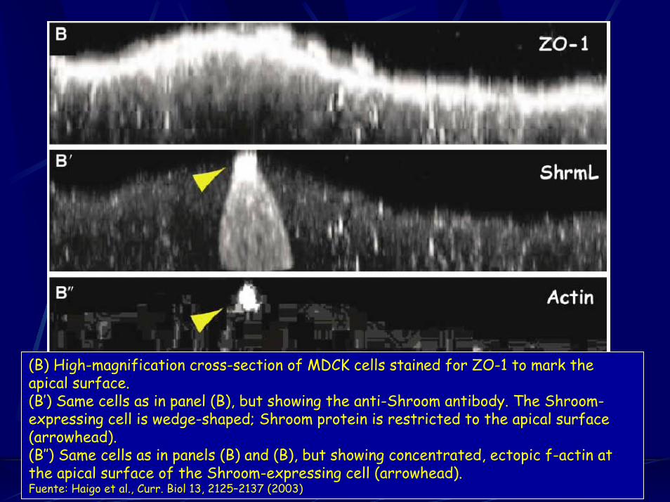
(B) High-magnification cross-section of MDCK cells stained for ZO-1 to mark theapical surface.(B’) Same cells as in panel (B), but showing the anti-Shroom antibody. The Shroom-expressing cell is wedge-shaped; Shroom protein is restricted to the apical surface(arrowhead).(B″) Same cells as in panels (B) and (B), but showing concentrated, ectopic f-actin at the apical surface of the Shroom-expressing cell (arrowhead).Fuente: Haigo et al., Curr. Biol 13, 2125–2137 (2003)
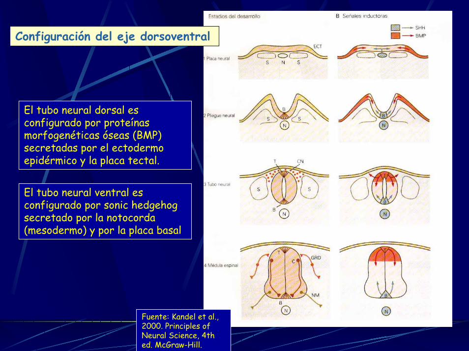
Configuración del eje dorsoventral
El tubo neural ventral es configurado por sonic hedgehogsecretado por la notocorda (mesodermo) y por la placa basal
El tubo neural dorsal es configurado por proteínasmorfogenéticas óseas (BMP) secretadas por el ectodermo epidérmico y la placa tectal.
Fuente: Kandel et al., 2000. Principles of Neural Science, 4th ed. McGraw-Hill.
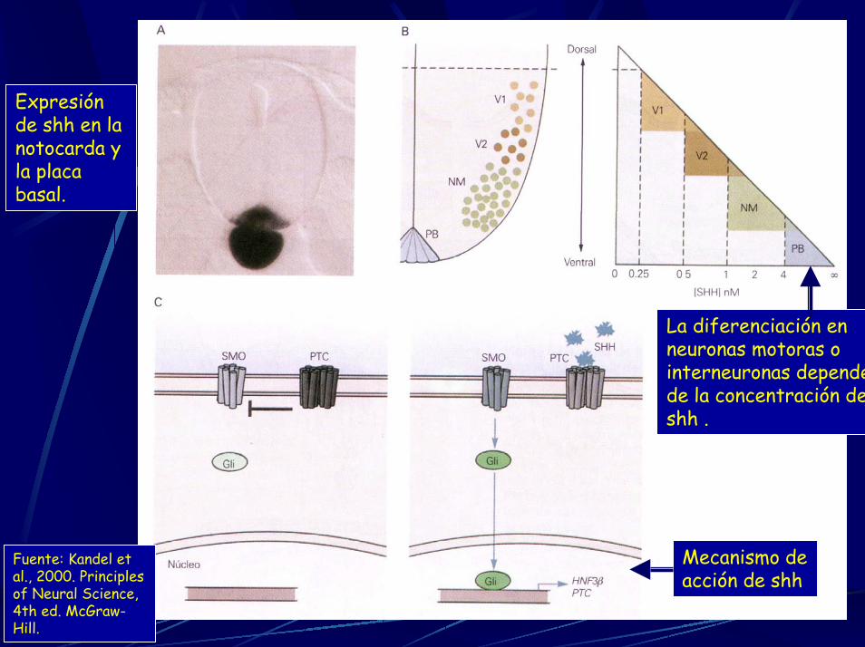
Mecanismo de acción de shh
La diferenciación en neuronas motoras o interneuronas dependede la concentración deshh .
Fuente: Kandel et al., 2000. Principles of Neural Science, 4th ed. McGraw-Hill.
Expresión de shh en la notocarda y la placa basal.
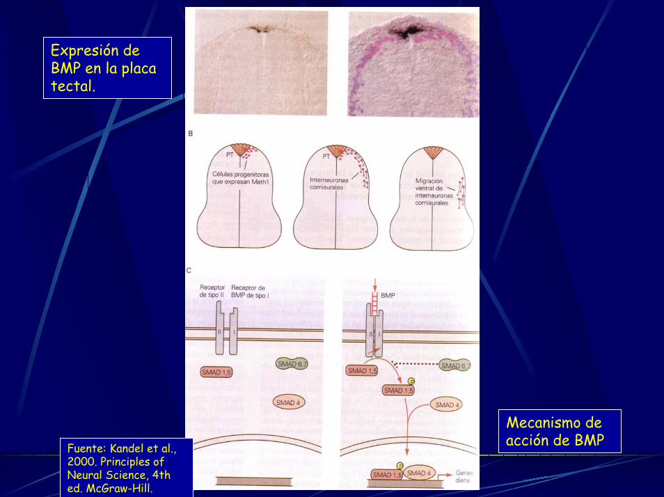
Expresión de BMP en la placa tectal.
Mecanismo de acción de BMPFuente: Kandel et al.,
2000. Principles of Neural Science, 4th ed. McGraw-Hill.