Embed Size (px)
Citation preview
Accepted Manuscript
TOCILIZUMAB IN GIANT CELL ARTERITIS. OBSERVATIONAL,OPEN-LABEL MULTICENTER STUDY OF 134 PATIENTS INCLINICAL PRACTICE
Monica Calderon-Goercke MD , Javier Loricera MD, PhD ,Vicente Aldasoro MD , Santos Castaneda MD, PhD ,Ignacio Villa MD , Alicia Humbrıa MD, PhD , Clara Moriano MD ,Susana Romero-Yuste MD , Javier Narvaez MD, PhD ,Catalina Gomez-Arango MD , Eva Perez-Pampın MD, PhD ,Rafael Melero MD , Elena Becerra-Fernandez MD, PhD ,Marcelino Revenga MD, PhD , Noelia Alvarez-Rivas MD ,Carles Galisteo MD , Francisca Sivera MD, PhD ,Alejandro Olive-Marques MD, PhD , Marıa Alvarez del Buergo MD ,Luisa Marena-Rojas MD , Carlos Fernandez-Lopez MD ,Francisco Navarro MD, PhD , Enrique Raya MD, PhD ,Eva Galindez-Agirregoikoa MD , Beatriz Arca MD ,Roser Solans-Laque MD, PhD , Arantxa Conesa MD ,Cristina Hidalgo MD, PhD , Carlos Vazquez MD ,Jose Andres Roman-Ivorra MD, PhD , Pau Lluch MD ,Sara Manrique-Arija MD, PhD , Paloma Vela MD, PhD ,Eugenio De Miguel MD, PhD , Carmen Torres-Martın MD ,Juan Carlos Nieto MD, PhD , Carmen Ordas-Calvo MD ,Eva Salgado-Perez MD, PhD , Cristina Luna-Gomez MD ,F. Javier Toyos-Saenz de Miera MD ,Nagore Fernandez-Llanio MD , Antonio Garcıa MD ,Carmen Larena MD , Natalia Palmou-Fontana MD, PhD ,Vanesa Calvo-Rıo MD, PhD , Diana Prieto-Pena MD ,Carmen Gonzalez-Vela MD, PhD , Alfonso Corrales MD, PhD ,Marıa Varela-Garcıa MD , Elena Aurrecoechea MD, PhD ,Raquel Dos Santos MD , Angel Garcıa-Manzanares MD ,Norberto Ortego MD, PhD , Sabela Fernandez MD ,Francisco Ortiz-Sanjuan MD, PhD , Montserrat Corteguera MD ,Jose L. Hernandez MD, PhD , Miguel A. Gonzalez-Gay MD, PhD ,Ricardo Blanco MD, PhD
PII: S0049-0172(18)30571-7DOI: https://doi.org/10.1016/j.semarthrit.2019.01.003Reference: YSARH 51436
To appear in: Seminars in Arthritis & Rheumatism
Please cite this article as: Monica Calderon-Goercke MD , Javier Loricera MD, PhD ,Vicente Aldasoro MD , Santos Castaneda MD, PhD , Ignacio Villa MD , Alicia Humbrıa MD, PhD ,Clara Moriano MD , Susana Romero-Yuste MD , Javier Narvaez MD, PhD ,Catalina Gomez-Arango MD , Eva Perez-Pampın MD, PhD , Rafael Melero MD ,
© 2019. This manuscript version is made available under the CC-BY-NC-ND 4.0 license http://creativecommons.org/licenses/by-nc-nd/4.0/
Elena Becerra-Fernandez MD, PhD , Marcelino Revenga MD, PhD , Noelia Alvarez-Rivas MD ,Carles Galisteo MD , Francisca Sivera MD, PhD , Alejandro Olive-Marques MD, PhD ,Marıa Alvarez del Buergo MD , Luisa Marena-Rojas MD , Carlos Fernandez-Lopez MD ,Francisco Navarro MD, PhD , Enrique Raya MD, PhD , Eva Galindez-Agirregoikoa MD ,Beatriz Arca MD , Roser Solans-Laque MD, PhD , Arantxa Conesa MD , Cristina Hidalgo MD, PhD ,Carlos Vazquez MD , Jose Andres Roman-Ivorra MD, PhD , Pau Lluch MD ,Sara Manrique-Arija MD, PhD , Paloma Vela MD, PhD , Eugenio De Miguel MD, PhD ,Carmen Torres-Martın MD , Juan Carlos Nieto MD, PhD , Carmen Ordas-Calvo MD ,Eva Salgado-Perez MD, PhD , Cristina Luna-Gomez MD , F. Javier Toyos-Saenz de Miera MD ,Nagore Fernandez-Llanio MD , Antonio Garcıa MD , Carmen Larena MD ,Natalia Palmou-Fontana MD, PhD , Vanesa Calvo-Rıo MD, PhD , Diana Prieto-Pena MD ,Carmen Gonzalez-Vela MD, PhD , Alfonso Corrales MD, PhD , Marıa Varela-Garcıa MD ,Elena Aurrecoechea MD, PhD , Raquel Dos Santos MD , Angel Garcıa-Manzanares MD ,Norberto Ortego MD, PhD , Sabela Fernandez MD , Francisco Ortiz-Sanjuan MD, PhD ,Montserrat Corteguera MD , Jose L. Hernandez MD, PhD , Miguel A. Gonzalez-Gay MD, PhD ,Ricardo Blanco MD, PhD , TOCILIZUMAB IN GIANT CELL ARTERITIS. OBSERVATIONAL, OPEN-
LABEL MULTICENTER STUDY OF 134 PATIENTS IN CLINICAL PRACTICE, Seminars in Arthritis &Rheumatism (2019), doi: https://doi.org/10.1016/j.semarthrit.2019.01.003
This is a PDF file of an unedited manuscript that has been accepted for publication. As a serviceto our customers we are providing this early version of the manuscript. The manuscript will undergocopyediting, typesetting, and review of the resulting proof before it is published in its final form. Pleasenote that during the production process errors may be discovered which could affect the content, andall legal disclaimers that apply to the journal pertain.
ACCEPTED MANUSCRIPT
ACCEPTED MANUSCRIP
T
TOCILIZUMAB IN GIANT CELL ARTERITIS. OBSERVATIONAL, OPEN-LABEL
MULTICENTER STUDY OF 134 PATIENTS IN CLINICAL PRACTICE
Mónica Calderón-Goercke, MD1*; Javier Loricera, MD, PhD1*; Vicente Aldasoro, MD2; Santos
Castañeda, MD, PhD3; Ignacio Villa, MD4; Alicia Humbría,MD, PhD3; Clara Moriano, MD5;
Susana Romero-Yuste, MD6; Javier Narváez, MD, PhD7; Catalina Gómez-Arango, MD8; Eva
Pérez-Pampín MD, PhD9; Rafael Melero, MD10; Elena Becerra-Fernández, MD, PhD 11;
Marcelino Revenga, MD, PhD 12; Noelia Álvarez-Rivas, MD13; Carles Galisteo MD14;
Francisca Sivera, MD, PhD 15; Alejandro Olivé-Marqués, MD, PhD16; María Álvarez del
Buergo MD17; Luisa Marena-Rojas MD18; Carlos Fernández-López, MD19; Francisco
Navarro, MD, PhD20; Enrique Raya, MD, PhD21; Eva Galindez-Agirregoikoa, MD22; Beatriz
Arca, MD23; Roser Solans-Laqué, MD, PhD 24; Arantxa Conesa MD25; Cristina Hidalgo, MD,
PhD 26; Carlos Vázquez, MD27; José Andrés Román-Ivorra, MD, PhD28; Pau Lluch MD29;
Sara Manrique-Arija, MD, PhD30; Paloma Vela, MD, PhD 31; Eugenio De Miguel, MD, PhD32;
Carmen Torres-Martín, MD33; Juan Carlos Nieto, MD, PhD 34; Carmen Ordas-Calvo, MD35;
Eva Salgado-Pérez, MD, PhD 36; Cristina Luna-Gomez MD37; F. Javier Toyos-Sáenz de
Miera, MD38; Nagore Fernández-Llanio, MD39; Antonio García MD40; Carmen Larena, MD12;
Natalia Palmou-Fontana, MD, PhD1; Vanesa Calvo-Río, MD, PhD1; Diana Prieto-Peña, MD1;
Carmen González-Vela, MD, PhD1; Alfonso Corrales, MD, PhD1; María Varela-García MD2;
Elena Aurrecoechea, MD, PhD4; Raquel Dos Santos, MD9; Ángel García-Manzanares MD11;
Norberto Ortego, MD, PhD21; Sabela Fernández, MD23; Francisco Ortiz-Sanjuán, MD, PhD28;
Montserrat Corteguera, MD33; José L. Hernández, MD, PhD1†; Miguel Á. González-Gay, MD,
PhD1†; Ricardo Blanco, MD, PhD1†.
ACCEPTED MANUSCRIPT
ACCEPTED MANUSCRIP
T
1Departments of Rheumatology, Internal Medicine and Pathology, Hospital Universitario
Marqués de Valdecilla, IDIVAL, Santander. Universidad de Cantabria, Spain. 2Department of
Rheumatology, Complejo Hospitalario de Navarra, Navarra, Spain. 3Department of
Rheumatology, Hospital Universitario de La Princesa, IIS-Princesa, Madrid, Spain.
4Department of Rheumatology, Hospital de Sierrallana, Torrelavega, Spain. 5Department of
Rheumatology, Complejo Asistencial Universitario de León, León, Spain. 6Department of
Rheumatology, Complejo Hospitalario Universitario Pontevedra, Spain. 7Department of
Rheumatology, Hospital de Bellvitge, Barcelona, Spain. 8Department of Rheumatology,
Hospital Alto Deba, Mondragón, Spain. 9Department of Rheumatology, Complejo
Hospitalario Universitario de Santiago, Santiago de Compostela, Spain. 10Department of
Rheumatology, Complexo Hospitalario Universitario de Vigo, Vigo, Spain. 11Department of
Rheumatology, Hospital Universitario de Torrevieja, Alicante, Spain. 12Department of
Rheumatology, Hospital Ramón y Cajal, Madrid, Spain. 13Department of Rheumatology,
Hospital Universitario Lucus Augusti, Lugo, Spain. 14Department of Rheumatology, Hospital
Parc Taulí, Barcelona, Spain. 15Department of Rheumatology, Hospital Universitario de
Elda, Alicante, Spain. 16Department of Rheumatology, Hospital Trías i Pujol, Badalona,
Spain. 17Department of Rheumatology, Hospital Río Carrión, Palencia, Spain. 18Department
of Rheumatology, Hospital La Mancha Centro, Alcázar de San Juan, Spain. 19Department of
Rheumatology, Hospital Universitario Juan Canalejo, A Coruña, Spain. 20Department of
Rheumatology, Hospital General Universitario de Elche, Alicante, Spain. 21Department of
Rheumatology and Internal Medicine, Hospital San Cecilio, Granada, Spain. 22Department of
Rheumatology, Hospital de Basurto, Bilbao, Spain. 23Department of Rheumatology, Hospital
Universitario San Agustín, Avilés, Spain. 24Department of Internal Medicine, Hospital Valle
de Hebrón, Barcelona, Spain. 25Department of Rheumatology, Hospital General
Universitario de Castellón, Spain. 26Department of Rheumatology, Complejo Asistencial
Universitario de Salamanca, Spain. 27Department of Rheumatology, Hospital Miguel Servet,
Zaragoza, Spain. 28Department of Rheumatology, Hospital Universitario y Politécnico La Fe,
Valencia, Spain. 29Department of Rheumatology, Hospital Mateu Orfila, Menorca, Spain.

ACCEPTED MANUSCRIPT
ACCEPTED MANUSCRIP
T
30Department of Rheumatology, Hospital Regional de Málaga, Málaga, Spain. 31Department
of Rheumatology, Hospital General Universitario de Alicante, Alicante, Spain. 32Department
of Rheumatology, Hospital La Paz, Madrid, Spain. 33Department of Rheumatology, Complejo
Asistencial de Ávila, Ávila, Spain. 34Department of Rheumatology, Hospital Gregorio
Marañón, Madrid, Spain. 35Department of Rheumatology, Hospital Cabueñes, Gijón, Spain.
36Department of Rheumatology, Complejo Hospitalario Universitario de Ourense, Ourense,
Spain. 37Department of Rheumatology, Hospital Universitario Nuestra Señora de la
Candelaria, Tenerife, Spain. 38Department of Rheumatology, Hospital Universitario Virgen
Macarena, Sevilla, Spain. 39Department of Rheumatology. Hospital Arnau de Vilanova,
Lérida, Spain. 40Department of Rheumatology. Hospital Virgen de las Nieves, Granada,
Spain.
* Mónica Calderón-Goercke and Javier Loricera shared first authorship.
† Prof. MA González-Gay, R Blanco and J.L. Hernández shared senior authorship.
This study was presented in part at the 2017 American College of Rheumatology Meeting
held in San Diego, CA, USA.
‡ Joint corresponding authors: Prof. Miguel A. González-Gay or Ricardo Blanco,
Rheumatology Division, Hospital Universitario Marqués de Valdecilla, Avda. Valdecilla s/n.,
ES- 39008, Santander. SPAIN.
E-mail address: [email protected]; OR [email protected]
Competing interests:
Disclosures that might be interpreted as constituting of possible conflict(s) of interest for the
study: Dr. MA Gonzalez-Gay received grants/research supports from Abbvie, MSD and
ACCEPTED MANUSCRIPT
ACCEPTED MANUSCRIP
T
Roche, and had consultation fees/participation in company sponsored speaker´s bureau
from Pfizer, Lilly, Roche and Sanofi. Dr. R Blanco received grants/research supports from
Abbvie, MSD and Roche, and had consultation fees/participation in company sponsored
speaker´s bureau from Abbvie, Pfizer, Roche, Bristol-Myers, Janssen and MSD.
No financial disclosures declared: Mónica Calderón-Goercke, Javier Loricera, Vicente
Aldasoro, Santos Castañeda, Ignacio Villa, Alicia Humbría, Clara Moriano, Susana Romero-
Yuste, Javier Narváez, Catalina Gómez-Arango, Eva Pérez-Pampín, Rafael Melero, Elena
Becerra-Fernández, Marcelino Revenga, Carmen Larena, Noelia Álvarez-Rivas, Carles
Galisteo, Francisca Sivera, Alejandro Olivé-Marqués, María Álvarez del Buergo, Luisa
Marena-Rojas, Carlos Fernández-López, Francisco Navarro, Enrique Raya, Eva Galíndez-
Agirregoikoa, Beatriz Arca, Roser Solans-Laqué, Arantxa Conesa, Cristina Hidalgo, Carlos
Vázquez, José Andrés Román-Ivorra, Pau Lluch, Sara Manrique-Arija, Paloma Vela,
Eugenio De Miguel, Carmen Torres-Martín, Juan Carlos Nieto, Carmen Ordás-Calvo, Eva
Salgado-Pérez, Cristina Luna-Gómez, Javier Toyos, Nagore Fernández-Llanio, Antonio
García, Natalia Palmou-Fontana, Vanesa Calvo-Río, Diana Prieto-Peña, Carmen González-
Vela, Alfonso Corrales, José L. Hernández, María Varela-García, Elena Aurrecoechea,
Antonio Mera, Raquel Dos Santos, Ángel García-Manzanares, Norberto Ortego, Sabela
Fernández, Francisco Ortiz-Sanjuán, Montserrat Corteguera.
Running Title: GCA and TCZ in clinical practice.
Key indexing terms: tocilizumab, giant cell arteritis, biological therapy, large-vessel
vasculitis.
ABSTRACT
Objective: Tocilizumab (TCZ) has shown efficacy in clinical trials on giant cell arteritis
(GCA). Real-world data are scarce. Our objective was to assess efficacy and safety of TCZ
in unselected patients with GCA in clinical practice
ACCEPTED MANUSCRIPT
ACCEPTED MANUSCRIP
T
Methods: Observational, open-label multicenter study from 40 national referral centers
of GCA patients treated with TCZ due to inefficacy or adverse events of previous therapy.
Outcomes variables were improvement of clinical features, acute phase reactants,
glucocorticoid-sparing effect, prolonged remission and relapses. A comparative study was
performed: a) TCZ route (SC vs. IV); b) GCA duration (≤6 vs. >6 months); c) serious
infections (with or without); d) ≤15 vs. >15 mg/day at TCZ onset.
Results: 134 patients; mean age, 73.0±8.8 years. TCZ was started after a median [IQR]
time from GCA diagnosis of 13.5 [5.0-33.5] months. Ninety-eight (73.1%) patients had
received immunosuppressive agents. After 1 month of TCZ 93.9% experienced clinical
improvement. Reduction of CRP from 1.7 [0.4-3.2] to 0.11 [0.05-0.5] mg/dL (p<0.0001), ESR
from 33 [14.5-61] to 6 [2-12] mm/1st hour (p<0.0001) and decrease in patients with anemia
from 16.4% to 3.8% (p<0.0001) were observed. Regardless of administration route or
disease duration, clinical improvement leading to remission at 6, 12, 18, 24 months was
observed in 55.5%, 70.4%, 69.2% and 90% of patients. Most relevant adverse side-effect
was serious infections (10.6/100 patients-year), associated with higher doses of prednisone
during the first three months of therapy.
Conclusion: In clinical practice, TCZ yields a rapid and maintained improvement of
refractory GCA. Serious infections appear to be higher than in clinical trials.
ACCEPTED MANUSCRIPT
ACCEPTED MANUSCRIP
T
1. INTRODUCTION
Giant cell arteritis (GCA) is a large-vessel vasculitis (LVV) which affects medium and
large sized arteries. It is common in European people older than 50 years as well as
in North American of European ancestry (1-3). Although blindness is the most feared
complication, other severe manifestations such as stroke and aneurysms can also
occur (4-7).
The mainstay treatment of GCA are glucocorticoids (8,9). However, adverse events
related to these drugs are common (9-11). Besides, relapses are relative frequent in
GCA patients (12,13). Therefore, other medications such as leflunomide,
azathioprine or cyclophosphamide have been used with no efficacy (14-16). For
methotrexate, results are controversial but a metaanalysis confirmed its efficacy (17)
Although the etiology of GCA remains unknown, different proinflammatory cytokines
including TNFα and IL-6 act as soluble pathogenic factors (18). However,
prospective, double-blind studies with monoclonal TNFα inhibitors led to poor results
(19).
In contrast, several case series suggested that tocilizumab (TCZ), a humanized
monoclonal antibody against the IL-6 receptor, may be a good therapeutic option in
GCA (20-27). In this regard, two recent randomized clinical double-blinded Phase II
and III studies showed efficacy of TCZ in patients with GCA (28,29). Consequently,
TCZ has been approved by the European Medicines Agency (EMA) and the Food
and Drug Administration (FDA) for the treatment of patients with GCA. However, it is
not uncommon to see that patients are often excluded from clinical trials due to
comorbidities. Also, many of the individuals included in these two trials were patients
with a recent diagnosis of GCA and some others had a relative short period of follow-
up with TCZ therapy (29). In this regard, 23 of 30 (77%) patients in a Phase II study,
ACCEPTED MANUSCRIPT
ACCEPTED MANUSCRIP
T
and 119 of 251 (47%) in a Phase III study were patients with GCA of recent onset
(28,29). In the GiACTA trial, newly diagnosed GCA was defined if diagnosis was
done ≤ 6 weeks before baseline, with a mean± SD time between diagnosis and TCZ
onset of 0.5±0.5 months (29). However, in a real-world scenario, TCZ will probably
be more frequently used in patients with GCA refractory to conventional treatment or
in those who experience side effects, including also GCA patients with one or
several comorbidities. Therefore, in real life, patients with GCA undergoing TCZ
therapy have longer disease duration, being often relapsing patients who are
refractory to conventional immunosuppressive drugs. In addition, in the phase II
study TCZ was prescribed intravenously (IV) whereas in the phase III GiACTA trial
TCZ was given subcutaneously (SC) (28,29). Moreover, the recommended dose of
prednisone at the onset of TCZ therapy has not clearly established.
Taking all these considerations into account, the aim of the present study was to
assess the usefulness and safety of TCZ in GCA patients from a real clinical setting
with refractory disease and/or with unacceptable side effects due to conventional
treatment.
2. PATIENTS and METHODS
2.1 Patients, Enrollment Criteria and Study protocol
We set up an observational, retrospective open-label multicenter study that included
134 GCA patients treated with TCZ in real clinical practice and followed-up, in some
cases, up to 48 months. Before TCZ onset, all of them had received high-dose
glucocorticoids, and 98 (73.1%) conventional synthetic and/or other biologic
immunosuppressive drugs. To reduce selection bias, in the present study we
included all the patients who had received at least one TCZ dose, regardless of the
ACCEPTED MANUSCRIPT
ACCEPTED MANUSCRIP
T
outcome. Preliminary, partial data on 22 patients were previously reported (21). The
study was approved by the Clinical Research Ethics Committee of University
Hospital Marqués de Valdecilla from Santander (Cantabria, Spain).
Patients were diagnosed with GCA at the Rheumatology or Autoimmune Units of 40
national referrals centers. GCA diagnosis was based on a) American College of
Rheumatology (ACR) criteria (30), b) a positive biopsy of temporal artery, and/or c)
presence of imaging techniques consistent with large vessel vasculitis (LVV) in
patients with cranial symptoms of GCA. The main imaging techniques used for LVV
diagnosis were 18F-fluorodeoxyglucose positron emission tomography/computed
tomography (18F-FDG PET/CT) scan, magnetic resonance imaging angiography
(MRI-A), computed tomography angiography (CT-A), and helical CT scan.
Treatment of GCA was based on the classic pharmacological therapy, starting on
high dose of glucocorticoids, generally an initial dose of prednisone between 40 to
60 mg/day, which was gradually tapered. Conventional immunosuppressive drugs
and biologic therapy were used to spare glucocorticoid dose, mainly in patients with
relapsing disease or in those cases with adverse side effect related to glucocorticoid
therapy. The doses of conventional immunosuppressive drugs and biological agents
used prior to TCZ were as follows: methotrexate (MTX) (7.5-30 mg/SC/week or 7.5-
25 mg/per os/week), leflunomide (LFN) (10-30 mg/per os/day), hydroxychloroquine
(HCQ) (400 mg/per os/day), azathioprine (AZA) (50-150 mg/per os/day),
cyclophosphamide (CYC) (75-125 mg/per os/day), mycophenolate mofetil (MMF) (2
g/ per os/day), etanercept (ETN) (50 mg/SC/week), infliximab (IFX) (3 mg/kg/IV/8
weeks), rituximab (RTX) (2 g/IV/6 months) and abatacept (ABT) (10 mg/kg/IV at 0, 2,
4 weeks and later every 4 weeks).
As indicated by the Spanish National Guidelines for the administration of biologic

ACCEPTED MANUSCRIPT
ACCEPTED MANUSCRIP
T
therapy in patients with rheumatic diseases, the presence of infectious diseases had
to be ruled out, including tuberculosis and hepatitis B or hepatitis C infection, before
the onset of biologic therapy, as previously described (31).
To exclude latent tuberculosis, a tuberculin skin testing (PPD) and/or an interferon
assay (quantiFERON) as well as a chest radiography were performed. In positive
cases, prophylaxis with isoniazid was initiated at least 4 weeks before the onset of
the biological agent and it was maintained for 9 months. The presence of
malignancies was also excluded in all the patients.
TCZ was prescribed IV at standard dose (8 mg/kg/4 weeks) or SC (162 mg/week). It
was given due to lack of efficacy and/or unacceptable adverse side-effects related to
previous therapy. In most cases, TCZ was prescribed off-label since it was indicated
before its approval by the EMA and the FDA for the treatment GCA. Therefore, a
written informed consent was obtained in these cases.
2.2 Clinical definitions and laboratory data
Fever was considered to be present if temperature was ≥ 38ºC. Constitutional
symptoms included asthenia, anorexia and weight loss greater than 5% of the
normal body weight over the last 6 months before disease diagnosis. Headache if it
was of recent development or had different characteristics than usual. Visual
manifestations include blurred vision, diplopia, amaurosis fugax, unilateral or bilateral
hemianopsia and permanent unilateral or bilateral blindness. Polymyalgia
rheumatica (PMR) was defined according to the classification criteria proposed by
EULAR/ACR 2012 (32). Definition of other clinical manifestations were reported
elsewhere (33).
Serum C-reactive protein (CRP), erythrocyte sedimentation rate (ESR), renal and
liver function tests, as well as a full blood cell count were obtained at the time of TCZ
ACCEPTED MANUSCRIPT
ACCEPTED MANUSCRIP
T
onset and then at each visit. A Serum CRP value was considered to be increased
when it was higher than 0.5 mg/dL. An ESR value greater than 20 mm/h in men or >
25 mm/h in women was also considered abnormal. Anemia was defined as a
hemoglobin level ≤11 g/dL.
A serious infection was considered to be present when a life-threatening infection,
fatal, or requiring hospitalization occurred, intravenous antibiotics were required, or
the infectious process led to persistent or significant disability.
Large vessel involvement was considered to be present if vasculitis involvement of
the aorta and/or its major branches was disclosed by imaging techniques such as
18F-FDG PET-CT scan, MRI-A, CT-A, or helical CT-scan. Assessment of large
vessel vasculitis was performed in each of the recruiting centers. Nuclear medicine
radiologists or angiography or angio/ interventional radiology staff members experts
in the field from each hospital confirmed the presence of large vessel vasculitis.
Remission was defined if the patients was free of symptoms and had normalization
of the acute phase reactants (CRP and ESR).
Prolonged remission was defined if the patients persisted asymptomatic with normal
acute phase reactant for at least 6 months.
Relapse was defined as the recurrence of signs or symptoms of GCA along with
ESR >20 mm/h in men or >25 mm/h in women and/or serum CRP >0.5 mg/dL.
2.3 Data collection
Information was retrieved from the patient’s clinical records according to a specific
designed protocol, including data on clinical and laboratory parameters at diagnosis,
drugs used for the management of GCA, presence of relapses and side effect
related to therapy different from TCZ, response to TCZ and development of side
ACCEPTED MANUSCRIPT
ACCEPTED MANUSCRIP
T
effects while patients were undergoing TCZ therapy. To minimize entry error, all the
data were double checked. Information was stored in a computerized database.
2.4 Statistical Analysis
All continuous variables were tested for normality, and results were expressed as
mean± standard deviation (SD) or as median and interquartile range (IQR) as
appropriate. Student's t test or Mann-Whitney U-test were used to compare
continuous variables, and 2-test to compare categorical variables.
To assess the effect of TCZ on serum CRP levels, ESR and dose of prednisone
between TCZ onset and the different follow-up visits we used the Wilcoxon’s signed
rank test.
A comparative study between the following groups was done: a) patients receiving IV
or SC TCZ; b) TCZ efficacy in GCA patients with a disease duration ≤6 months or >6
months; c) TCZ in GCA patients with and without serious infections; and d) patients
receiving prednisone doses ≤15 mg/d or >15 mg/d at TCZ onset.
Analyses were performed by using STATISTICA software (StatSoft Inc. Tulsa,
Oklahoma, USA).
2.5 Role of the Funding source
This study was not funded by any drug company. It was the result of an independent
initiative of the investigators.
3. RESULTS
3.1 Baseline main clinical features prior to TCZ onset
We studied 134 patients (101 women/33 men); mean age 73.0±8.8 years, diagnosed
with GCA and treated with TCZ. Of them, 119 (88%) fulfilled the 1990 ACR
classification criteria for GCA (30). The remaining patients were diagnosed by a

ACCEPTED MANUSCRIPT
ACCEPTED MANUSCRIP
T
positive temporal artery for GCA in 9 cases. Six patients did not fulfil the ACR
classification criteria for GCA and had a negative TAB. All of them were older than
50 years but had an ESR at the time of diagnosis lower than 50 mm/1st hour.
Nevertheless, they presented cranial ischemic manifestations. In these patients, a
diagnosis of GCA was confirmed by a positive imaging technique showing large
vessel vasculitis involvement. In addition, other entities, such as connective-tissue
diseases, IgG4 related disease or any other inflammatory conditions that may be
associated with large-vessel vasculitis, were excluded. In addition, all of them
exhibited a rapid response to prednisone dose (initial dose range between 40-60
mg/day). With respect to this, a positive temporal artery biopsy was positive in 72
(61.5%) of 117 patients in whom it was performed. Also, imaging techniques to
identify the presence of LVV were performed in 75 patients. Of them 58 yielded
positive results. With respect to this, 51 of these 58 patients with LVV also had
cranial features of GCA.
The median [IQR] time from GCA diagnosis to TCZ onset was 13.5 [5.0-33.5]
months. The main clinical features before TCZ onset were PMR (n=73) and
headache (n=70), followed by constitutional symptoms (n=31) and jaw claudication
(n=14). Twenty-eight patients from this series had visual manifestations at the time
of GCA diagnosis. Besides glucocorticoids and before starting TCZ, 98 (73.1%)
patients had received conventional immunosuppressive drugs: MTX (n=94), AZA
(n=14), LFN (n=9), CYC (n=4), HCQ (n=2), MMF (n=1). Five of them had also
received other biologic agents: IFX (n=2), ETN (n=1), RTX (n=1), and ABA (n=1)
(Table 1). The median [IQR] number of immunosuppressive agents per patient,
before TCZ onset was 1 [0.75-1.0].
3.2 Treatment with TCZ and outcome
ACCEPTED MANUSCRIPT
ACCEPTED MANUSCRIP
T
TCZ was initiated due to refractory GCA (n=86) or unacceptable adverse events of
previous therapy (n=48). It was administered IV to 106 (79.1%) patients and SC to
28 (20.9%). The initial dose was 8 mg/kg/IV/4 weeks or 162 mg/SC/week,
respectively. The maintenance dose of TCZ ranged from 8 mg/IV/kg/4 weeks to 4
mg/IV/kg/8 weeks, and from 162 mg/SC/ week to 162 mg/SC/3 weeks. Regardless
of glucocorticoids, TCZ was prescribed as monotherapy to 82 (62.2%) cases and
combined with conventional immunosuppressive agents in 52 (38.8%) patients: MTX
(n=48), AZA (n=3), and LFN (n=1).
As described in Table 2, one month after TCZ onset, most patients (93.9%)
achieved clinical improvement. At that time, significant reduction of the acute phase
reactants was also observed. In this regard, the median serum CRP fell from 1.7
[0.4-3.2] to 0.11 [0.05-0.5] mg/dL (p<0.0001), and the median ESR from 33 [14.5-61]
to 6 [2-12] mm/1st hour (p<0.0001). The percentage of patients with anemia
decreased from 16.4% to 3.8% (p<0.0001), and the mean hemoglobin level
increased from 12.3±1.5 g/dL to 13.1±1.3 g/dL (p<0001). Furthermore, the median
dose of prednisone was reduced from 15 [10-30] to 13.75 [7.5-20] mg/day
(p<0.0001) at month 1. Noteworthy, improvement of the clinical and laboratory
parameters was observed throughout the follow-up. At the same time successful
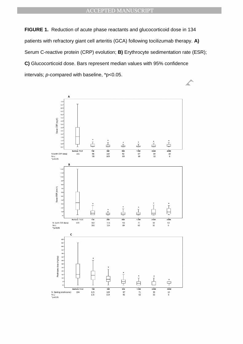
glucocorticoid tapering was achieved (Table 2 and Figure 1, panels A, B and C).
Persistent remission of the disease was observed in 27 (69.2%) of 39 patients
followed-up for at least 2 years. At that time, the median level of the acute phase
reactants in these 39 patients was within the normal range. Interestingly, the median
prednisone dose after 2 years of follow-up in these 39 patients was 0 [0-5] mg/day.
However, 7 of these 39 cases (17.9%) had relapses during the follow-up, usually
mild, that were successfully treated following a small increase of prednisone dose.

ACCEPTED MANUSCRIPT
ACCEPTED MANUSCRIP
T
With respect to these 7 relapsing patients, two of them were still receiving TCZ at 24
months of follow-up. From the remaining 5 patients, TCZ was withdrawn in 3 patients
due to remission and because of severe adverse events in the other 2 patients.
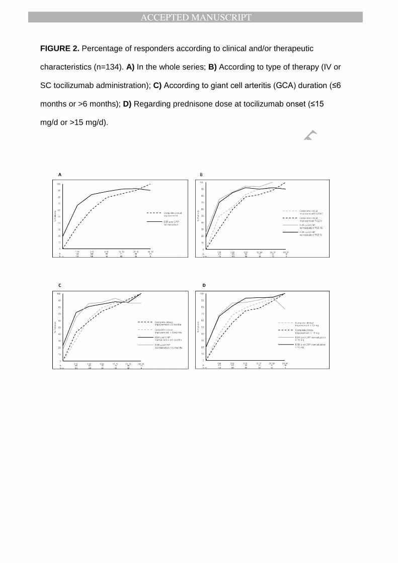
No significant differences in the clinical response or in the acute phase reactants
were observed when patients were stratified according to the duration of the disease
prior to TCZ equal to or less than 6 months or longer than 6 months. It was also the
case when patients were compared according to the route of TCZ administration (IV
or SC) or when patients receiving more than 15 mg/day at the time of TCZ onset
were compared with those receiving 15 mg/day or less (Figure 2 panels B, C and
D).
3.3 Side effects
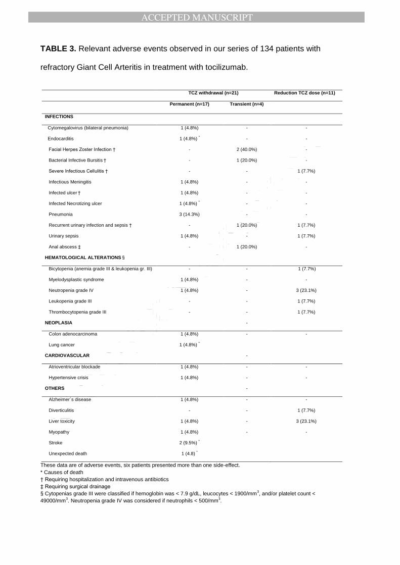
After a median follow-up of 12 [3-24] months, 32 (23.9%) patients developed
relevant adverse events (Table 3). Seventeen of them required permanent
discontinuation of TCZ therapy. The most frequent adverse events were infections
and hematological abnormalities. Serious infections were observed in 16 patients
(11.9%; 10.6 per 100 patients-year).
Five patients died during the follow-up due to stroke (n=2; one of them in the setting
of an infective endocarditis), lung cancer (n=1), necrotizing gluteal ulcer
(hyponatremia and cutaneous infection) (n=1), and of unknown cause (n=1).
We performed a comparative study between patients with serious infections (n=16)
and those without (n=118). At TCZ onset, patients who experienced serious
infections were slightly older (74.3±9.6 vs. 72.9±8.7 years; p=0.55) and had a non-
statistically significant longer course of the disease (20 [4.25-45.8] vs. 13 [5-29.3]
months; p=0.60). They were receiving a significantly greater prednisone dose at TCZ
onset (29.1 [20-40] vs. 15 [10-30] mg/day; p=0.01) and also after 3 months from the
ACCEPTED MANUSCRIPT
ACCEPTED MANUSCRIP
T
onset of TCZ (12.5 [10-20] vs. 7.5 [5.0-12.5] mg/day; p=0.003). Thirteen of the 66
patients that received more than 15 mg/day of prednisone at TCZ onset had serious
infections (16.3 per 100 patients-year) compared to only 3 (4.2 per 100 patients-
year) of 68 from the group on ≤15 mg/day (p=0.006). Nevertheless, clinical and
acute phase reactants improvement was similar in both groups.
To analyze the safety and efficacy of TCZ, we made a comparative study regarding
associated therapies and the duration of the treatment with TCZ. The biologic agent
was prescribed as monotherapy (82, 62.2%) or combined with conventional
immunosuppressants (52, 38.8%). The main immunosuppressants were MTX
(n=48), AZA (n=3), and LFN (n=1). There were not statistically significant differences
in terms of relevant adverse events (28% vs 17.3%, p=0.55) or serious infections
between patients treated with TCZ in monotherapy and those treated with TCZ in
combination with a conventional immunosuppressant (14.6% vs 7.7%, p=0.227).
Likewise, a comparison according to duration of TCZ treatment (< 12 months in 68
[50.7%] patients and ≥ 12 months in 66 [49.3%] patients) was performed. Relevant
adverse events were seen in 14 (20.5%) patients vs 18 (27.2%) patients
respectively, p=0.366. More specifically, regarding serious infections, no significant
differences were observed. In the first group (< 12 months) they occurred in 13.2%
of patients whereas they were observed in 10.6% of the patients from the second
group (≥ 12 months), p=0.640.
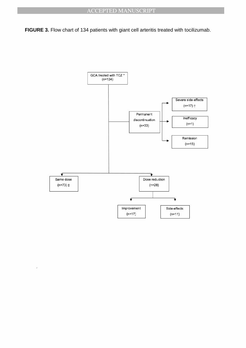
Figure 3 shows the flow-chart summarizing the 134 patients with GCA on TCZ
regarding the dose of the biologic agent and the outcome of the patients.
4. DISCUSSION
ACCEPTED MANUSCRIPT
ACCEPTED MANUSCRIP
T
We present the largest multicenter series of real-life GCA patients in whom short and
long-term efficacy of TCZ was assessed. All of them were refractory and/or had
unacceptable side effects due to conventional therapy. Our results show that TCZ
yields a rapid and maintained clinical and laboratory improvement, regardless of
GCA time course (≤ 6 months or > 6 months), TCZ administration route (IV vs SC) or
prednisone dose at TCZ onset (≤ 15 mg/d or > 15 mg/day). However, the frequency
of serious infections was higher than reported in the GiACTA clinical trial and in
patients with rheumatoid arthritis (RA), especially in those GCA patients on higher
doses of prednisone during the first three months of treatment (29).
Glucocorticoids are the first line of treatment in GCA and they are usually effective at
high doses (9,10). However, relapses are common when the dose is tapered, and
side-effects are also frequent (9,10,12). For that reason, several synthetic
immunosuppressive agents have been used for the treatment of GCA, but only MTX
has shown positive although often contradictory result (10,14). Thus, the biologic
therapy represents a new and promising alternative in the treatment of patients with
GCA, although anti-TNFα agents showed negative results (19).
IL-6 has a wide range of pleiotropic effects, including production of acute phase
reactants by hepatocytes, B lymphocyte differentiation and T lymphocyte subset
differentiation. Th1 and Th17 are involved in GCA, and IL-6 blockade may correct
the imbalance of Th17 and/or Th1 versus Treg lymphocytes (18,34-38).
TCZ is a humanized monoclonal antibody acting against soluble and membrane
bound IL-6 receptor. TCZ has been approved for the treatment of inflammatory and
autoimmune diseases, such as RA, systemic and polyarticular juvenile arthritis and
Castleman’s disease, including also vascular syndromes. A growing body of
ACCEPTED MANUSCRIPT
ACCEPTED MANUSCRIP
T
evidence supports the potential use of TCZ in other autoimmune diseases such as
uveitis and adult-onset Still’s disease (34,39-42).
Several observational studies on the use of TCZ in GCA showed promising results,
with clinical and laboratory data improvement and a glucocorticoid dose sparing (20-
22, 23-27,43,44). Interestingly, a phase II, randomized, double-blind, placebo-
controlled trial showed complete GCA clinical remission at week 12 in 85% of
patients treated with TCZ (8 mg/kg/4 weeks IV), and a relapse-free survival in the
same extent at week 52, with less cumulative glucocorticoid dose (28). Results of the
GiACTA study (29), a phase III, randomized, double-blind placebo-controlled trial,
showed that the use of TCZ at a dose of 162 mg SC weekly or every other week
(eow), plus a 26-week prednisone tapering regimen, allowed sustained remission in
56% and 53% of patients respectively, at 52 weeks. A decrease of the total
cumulative dose of glucocorticoids was also possible (29).
However, clinical trials do not always exemplify a real-world clinical scenario, since
they often include selected patients with very specific inclusion and exclusion criteria
and limited follow-up over time. For example, patients with specific comorbidities are
usually excluded and only a small proportion of patients included in the GiACTA
trials had previously received immunosuppressive drugs (29). On the other hand,
information on the long-term efficacy and safety of TCZ in GCA patients treated in
daily clinical practice is scarce. In this regard, the overall safety profile of TCZ in
GCA is consistent with the known safety in other conditions such as RA (45). In the
GiACTA study (29), serious infections occurred in 7% of the patients that received
TCZ weekly, 4% in the group on TCZ eow and 4% in the group that followed the 26-
week tapering protocol (29). In our series, serious infections occurred in 11.9% of
patients (10.6 per 100 patients-year). In contrast, in clinical practice this incidence in
ACCEPTED MANUSCRIPT
ACCEPTED MANUSCRIP
T
RA patients was 4.7 per 100 patients-year (45). The older age as well as the higher
prednisone dose used in our series of GCA patients at the time of TCZ onset may
explain the increased frequency of serious infections when compared with RA.
Therefore, we feel that several features of the patient should be considered before
using TCZ in the clinical practice. They include the age at the beginning of therapy,
the cumulative dose of corticosteroids and the presence of comorbidities that
predispose to infections.
Our study represents the largest real-world study on refractory GCA treated with
TCZ. However, it has potential limitations derived from its observational and
retrospective nature. In addition, during the follow-up several patients were lost.
Moreover, although the study included 134 cases, only 39 of them reached a follow-
up of 24 months. Also, in our study we did not use TCZ in newly diagnosed patients,
but only in those who were refractory to conventional therapy; being this a limitation
when compared with the GiACTA trial. Another limitation of this study was that the
definition of relapse used by authors might not be accurate for a study assessing the
efficacy of TCZ since this monoclonal antibody acts suppressing CRP, ESR and
fibrinogen. This limitation was also present in the GiACTA trial. However, in an
attempt to compare real life data with the GiACTA trial, the investigators of our study
decided to include in the definition of relapse the presence of an increase of acute
phase proteins, in a similar way to that used in the GiACTA trial.
Our results are consistent with those observed in clinical trials, confirming the
efficacy of TCZ in the treatment of GCA. Interestingly, in our series clinical and
laboratory improvement was unrelated to disease duration, administration route or a
dose of prednisone at TCZ onset greater than or lower than 15 mg/day.
ACCEPTED MANUSCRIPT
ACCEPTED MANUSCRIP
T
5. CONCLUSION
Taken together our findings and the results of the randomized control trials, TCZ
seems to be an excellent therapeutic option in GCA, regardless of the administration
route and GCA duration, helping to minimize the glucocorticoid exposure over time.
The most relevant side effects are serious infections that seem to be higher than in
the GiACTA trial.
In conclusion, TCZ improves clinical manifestations, acute-phase reactants and
imaging findings. However, GCA patients treated with TCZ may have normal levels
of CRP even though active disease may be still present. Therefore; an important
issue to be addressed is how we can be sure that TCZ-treated patients have a “true”
remission of the disease. Therefore, we feel that the definition of relapse in future
studies on patients undergoing biologic therapy, in particular in those treated with
anti-IL-6 receptor agents, should be reconsidered. Because of that, based on
previous observations of our group (46), the definition of relapses in patients
undergoing biologic therapy should be based on the presence of clinical features and
confirmed by an imaging technique (for example a PET/CT-scan).
ACKNOWLEDGEMENTS
To all the members and patients of the participating hospitals.
ACCEPTED MANUSCRIPT
ACCEPTED MANUSCRIP
T
REFERENCES
1. Salvarani C, Cantini F, Hunder GG. Polymyalgia rheumatica and giant-cell
arteritis. Lancet Lond Engl. 2008; 372:234-45.
2. Gonzalez-Gay MA, Vazquez-Rodriguez TR, Lopez-Diaz MJ, Miranda-Filloy JA,
Gonzalez-Juanatey C, Martin J, Llorca J. Epidemiology of giant cell arteritis and
polymyalgia rheumatica. Arthritis Rheum. 2009; 61:1454-61.
3. Hoffman GS. Giant Cell Arteritis. Ann Intern Med. 2016; 165:65-80.
4. Salvarani C, Pipitone N, Versari A, Hunder GG. Clinical features of polymyalgia
rheumatica and giant cell arteritis. Nat Rev Rheumatol. 2012; 8:509-21.
5. Soriano A, Muratore F, Pipitone N, Boiardi L, Cimino L, Salvarani C. Visual loss
and other cranial ischaemic complications in giant cell arteritis. Nat Rev
Rheumatol. 2017; 13:476-84.
6. González-Gay MA, García-Porrúa C, Llorca J, Hajeer AH, Brañas F, Dababneh
A, et al. Visual manifestations of giant cell arteritis. Trends and clinical spectrum
in 161 patients. Medicine (Baltimore). 2000; 79:283-92.
ACCEPTED MANUSCRIPT
ACCEPTED MANUSCRIP
T
7. Gonzalez-Gay MA, Garcia-Porrua C, Piñeiro A, Pego-Reigosa R, Llorca J,
Hunder GG. Aortic aneurysm and dissection in patients with biopsy-proven
giant cell arteritis from northwestern Spain: a population-based study. Medicine
(Baltimore). 2004 Nov;83(6):335-41.
8. Gonzalez-Gay MA, Martinez-Dubois C, Agudo M, Pompei O, Blanco R, Llorca
J. Giant cell arteritis: epidemiology, diagnosis, and management. Curr
Rheumatol Rep. 2010; 12:436-42.
9. Proven A, Gabriel SE, Orces C, O’Fallon WM, Hunder GG. Glucocorticoid
therapy in giant cell arteritis: duration and adverse outcomes. Arthritis Rheum.
2003; 49:703-8.
10. González-Gay MA, Pina T, Prieto-Peña D, Calderon-Goercke M, Blanco R,
Castañeda S. Current and emerging diagnosis tools and therapeutics for giant
cell arteritis. Expert Rev Clin Immunol. 2018; 14:593-605.
11. Petri H, Nevitt A, Sarsour K, Napalkov P, Collinson N. Incidence of giant cell
arteritis and characteristics of patients: data-driven analysis of comorbidities.
Arthritis Care Res. 2015; 67:390-5.
12. Martinez-Lado L, Calviño-Díaz C, Piñeiro A, Dierssen T, Vazquez-Rodriguez
TR, Miranda-Filloy JA, et al. Relapses and recurrences in giant cell arteritis: a
population-based study of patients with biopsy-proven disease from
northwestern Spain. Medicine (Baltimore). 2011; 90:186-93.
13. Kermani TA, Warrington KJ, Cuthbertson D, Carette S, Hoffman GS, Khalidi
NA, et al. Disease Relapses among Patients with Giant Cell Arteritis: A
Prospective, Longitudinal Cohort Study. J Rheumatol. 2015; 42:1213-7.

ACCEPTED MANUSCRIPT
ACCEPTED MANUSCRIP
T
14. Yates M, Loke YK, Watts RA, MacGregor AJ. Prednisolone combined with
adjunctive immunosuppression is not superior to prednisolone alone in terms of
efficacy and safety in giant cell arteritis: meta-analysis. Clin Rheumatol. 2014;
33:227-36.
15. Mahr AD, Jover JA, Spiera RF, Hernández-García C, Fernández-Gutiérrez B,
Lavalley MP, et al. Adjunctive methotrexate for treatment of giant cell arteritis:
an individual patient data meta-analysis. Arthritis Rheum. 2007; 56:2789-97.
16. Diamantopoulos AP, Hetland H, Myklebust G. Leflunomide as a corticosteroid-
sparing agent in giant cell arteritis and polymyalgia rheumatica: a case series.
BioMed Res Int. 2013; 2013:120638.
17. Mahr AD, Jover JA, Spiera RF, Hernández-García C, Fernández-Gutiérrez B,
Lavalley MP, Merkel PA. Adjunctive methotrexate for treatment of giant cell
arteritis: an individual patient data meta-analysis. Arthritis Rheum. 2007; 56:
2789-97.
18. Ciccia F, Rizzo A, Ferrante A, Guggino G, Croci S, Cavazza A, et al. New
insights into the pathogenesis of giant cell arteritis. Autoimmun Rev. 2017;
16:675-83.
19. Hoffman GS, Cid MC, Rendt-Zagar KE, Merkel PA, Weyand CM, Stone JH,
Salvarani C, Xu W, Visvanathan S, Rahman MU; Infliximab-GCA Study Group.
Infliximab for maintenance of glucocorticosteroid-induced remission of giant cell
arteritis: a randomized trial. Ann Intern Med. 2007; 146:621-30.
20. Seitz M, Reichenbach S, Bonel HM, Adler S, Wermelinger F, Villiger PM. Rapid
induction of remission in large vessel vasculitis by IL-6 blockade. A case series.
Swiss Med Wkly. 2011; 141: w13156.
ACCEPTED MANUSCRIPT
ACCEPTED MANUSCRIP
T
21. Loricera J, Blanco R, Hernández JL, Castañeda S, Mera A, Pérez-Pampín E, et
al. Tocilizumab in giant cell arteritis: Multicenter open-label study of 22 patients.
Semin Arthritis Rheum. 2015; 44:717-23.
22. Loricera J, Blanco R, Castañeda S, Humbría A, Ortego-Centeno N, Narváez J,
et al. Tocilizumab in refractory aortitis: study on 16 patients and literature
review. Clin Exp Rheumatol. 2014; 32:79-89.
23. Pazzola G, Padovano I, Boiardi L, Versari A, Pipitone N, Catanoso M, et al.
Tocilizumab in glucocorticoid-naïve large-vessel vasculitis. Clin Exp Rheumatol.
2013; 31:59-61.
24. Beyer C, Axmann R, Sahinbegovic E, Distler JH, Manger B, Schett G, et al.
Anti-interleukin 6 receptor therapy as rescue treatment for giant cell arteritis.
Ann Rheum Dis. 2011; 70:1874-5.
25. Salvarani C, Magnani L, Catanoso M, Pipitone N, Versari A, Dardani L, et al.
Tocilizumab: a novel therapy for patients with large-vessel vasculitis.
Rheumatolgy (Oxford). 2012; 51:151-6.
26. Unizony S, Arias-Urdaneta L, Miloslavsky E, Arvikar S, Khosroshahi A, Keroack
B, et al. Tocilizumab for the treatment of large-vessel vasculitis (giant cell
arteritis, Takayasu arteritis) and polymyalgia rheumatica. Arthritis Care Res.
2012; 64:1720-9.
27. Sciascia S, Rossi D, Roccatello D. Interleukin 6 Blockade as Steroid-sparing
Treatment for 2 Patients with Giant Cell Arteritis. J Rheumatol. 2011; 38:2080-1.
28. Villiger PM, Adler S, Kuchen S, Wermelinger F, Dan D, Fiege V, et al.
Tocilizumab for induction and maintenance of remission in giant cell arteritis: a
ACCEPTED MANUSCRIPT
ACCEPTED MANUSCRIP
T
phase 2, randomised, double-blind, placebo-controlled trial. Lancet. 2016;
387:1921-7.
29. Stone JH, Tuckwell K, Dimonaco S, Klearman M, Aringer M, Blockmans D, et
al. Trial of Tocilizumab in Giant-Cell Arteritis. N Engl J Med. 2017; 377:317-28.
30. Hunder GG, Bloch DA, Michel BA, Stevens MB, Arend WP, Calabrese LH, et al.
The American College of Rheumatology 1990 criteria for the classification of
giant cell arteritis. Arthritis Rheum. 1990; 33:1122-8.
31. Calvo-Río V, Blanco R, Beltrán E, Sánchez-Bursón J, Mesquida M, Adán A, et
al. Anti-TNF-α therapy in patients with refractory uveitis due to Behçet’s
disease: a 1-year follow-up study of 124 patients. Rheumatology (Oxford). 2014;
53:2223-31.
32. Dasgupta B, Cimmino MA, Maradit-Kremers H, Schmidt WA, Schirmer M,
Salvarani C, et al. 2012 provisional classification criteria for polymyalgia
rheumatica: a European League Against Rheumatism/American College of
Rheumatology collaborative initiative. Ann Rheum Dis. 2012; 71:484-92.
33. Gonzalez-Gay MA, Barros S, Lopez-Diaz MJ, Garcia-Porrua C, Sanchez-
Andrade A, Llorca J. Giant cell arteritis: disease patterns of clinical presentation
in a series of 240 patients. Medicine (Baltimore). 2005; 84:269-76.
34. Ogata A, Tanaka T. Tocilizumab for the treatment of rheumatoid arthritis and
other systemic autoimmune diseases: current perspectives and future
directions. Int J Rheumatol. 2012; 2012:946048.
35. Weyand CM, Younge BR, Goronzy JJ. IFN-γ and IL-17: the two faces of T-cell
pathology in giant cell arteritis. Curr Opin Rheumatol. 2011; 23:43-9.
ACCEPTED MANUSCRIPT
ACCEPTED MANUSCRIP
T
36. Manel N, Unutmaz D, Littman DR. The differentiation of human T(H)-17 cells
requires transforming growth factor-beta and induction of the nuclear receptor
RORgammat. Nat Immunol. 2008; 9:641-9.
37. Weyand CM, Goronzy JJ. Immune mechanisms in medium and large-vessel
…...vasculitis. Nat Rev Rheumatol 2013; 9:731-40.
38. Deng J, Younge BR, Olshen RA, Goronzy JJ, Weyand CM. Th17 and Th1 T-cell
…...responses in giant cell arteritis. Circulation. 2010; 121:906-15.
39. Calvo-Río V, Santos-Gómez M, Calvo I, González-Fernández MI, López-
Montesinos B, Mesquida M, et al. Anti-Interleukin-6 Receptor Tocilizumab for
Severe Juvenile Idiopathic Arthritis-Associated Uveitis Refractory to Anti-Tumor
Necrosis Factor Therapy: A Multicenter Study of Twenty-Five Patients. Arthritis
Rheumatol. 2017; 69:668-75.
40. Calvo-Río V, Blanco R, Santos-Gómez M, Díaz-Valle D, Pato E, Loricera J, et
al. Efficacy of Anti-IL6-Receptor Tocilizumab in Refractory Cystoid Macular
Edema of Birdshot Retinochoroidopathy Report of Two Cases and Literature
Review. Ocul Immunol Inflamm. 2017; 25:604-9.
41. Atienza-Mateo B, Calvo-Río V, Beltrán E, Martínez-Costa L, Valls-Pascual E,
Hernández-Garfella M, et al. Anti-interleukin 6 receptor tocilizumab in refractory
uveitis associated with Behçet’s disease: multicentre retrospective study.
Rheumatology (Oxford). 2018; 57: 856-864.
42. Ortiz-Sanjuán F, Blanco R, Calvo-Rio V, Narvaez J, Rubio Romero E, Olivé A,
et al. Efficacy of tocilizumab in conventional treatment-refractory adult-onset
Still’s disease: multicenter retrospective open-label study of thirty-four patients.
Arthritis Rheumatol. 2014; 66:1659-65.
ACCEPTED MANUSCRIPT
ACCEPTED MANUSCRIP
T
43. Régent A, Redeker S, Deroux A, Kieffer P, Ly KH, Dougados M, et al.
Tocilizumab in Giant Cell Arteritis: A Multicenter Retrospective Study of 34
Patients. J Rheumatol. 2016; 43:1547-52.
44. Evans J, Steel L, Borg F, Dasgupta B. Long-term efficacy and safety of
tocilizumab in giant cell arteritis and large vessel vasculitis. RMD Open. 2016;
2:e000137.
45. Morel J, Constantin A, Baron G, Dernis E, Flipo RM, Rist S, et al. Risk factors of
serious infections in patients with rheumatoid arthritis treated with tocilizumab in
the French Registry REGATE. Rheumatology (Oxford). 2017; 56:1746-1754.
46. Prieto-Peña D, Martínez-Rodríguez I, Loricera J, Banzo I, Calderón-Goercke M,
Calvo-Río V, González-Vela C, et al. Predictors of positive (18)F-FDG PET/CT-
scan for large vessel vasculitis in patients with persistent polymyalgia
rheumatica. Semin Arthritis Rheum. 2018 May 18. pii: S0049-0172(18)30215-4.
doi:10.1016/j.semarthrit.2018.05.007. [Epub ahead of print] PubMed PMID:
29903537
FIGURE LEGENDS
ACCEPTED MANUSCRIPT
ACCEPTED MANUSCRIP
T
FIGURE 1. Reduction of acute phase reactants and glucocorticoid dose in 134
patients with refractory giant cell arteritis (GCA) following tocilizumab therapy. A)
Serum C-reactive protein (CRP) evolution; B) Erythrocyte sedimentation rate (ESR);
C) Glucocorticoid dose. Bars represent median values with 95% confidence
intervals; p-compared with baseline, *p<0.05.
ACCEPTED MANUSCRIPT
ACCEPTED MANUSCRIP
T
FIGURE 2. Percentage of responders according to clinical and/or therapeutic
characteristics (n=134). A) In the whole series; B) According to type of therapy (IV or
SC tocilizumab administration); C) According to giant cell arteritis (GCA) duration (≤6
months or >6 months); D) Regarding prednisone dose at tocilizumab onset (≤15
mg/d or >15 mg/d).
ACCEPTED MANUSCRIPT
ACCEPTED MANUSCRIP
T
FIGURE 3. Flow chart of 134 patients with giant cell arteritis treated with tocilizumab.
ACCEPTED MANUSCRIPT
ACCEPTED MANUSCRIP
T
TABLE 1. Main features of 134 patients with refractory Giant Cell Arteritis who were
treated with TCZ.
Overall
n= 134
GCA fulfilling ACR
1990 criteria n= 119
Biopsy-proven
GCA n= 72
Cranial symptoms
plus LVV* n= 51
Age, years, mean± SD 73 ± 8.8 73.4 ± 8.5 75.7 ± 7.1 71.5 ± 8.9
Sex, female/male n (%) 101 / 33 88 / 31 57 / 15 43 / 8
Time from GCA diagnosis to TCZ onset (months), median [IQR] 13.5 [5.0-33.5] 14.0 [5.0-35.0] 14.5 [6.5-44.8] 14.0 [7.0-35.8]
SYSTEMIC MANIFESTATIONS
Fever, n (%) 9 (6.7%) 8 (6.7%) 4 (5.5%) 4 (7.8%)
Constitutional syndrome, n (%) 31 (23.1%) 25 (21%) 14 (19.4%) 11 (21.6%)
PMR, n (%) 73 (54.4%) 73 (61.3%) 40 (55.5%) 29 (56.8%)
ISCHEMIC MANIFESTATIONS
Headache, n (%) 70 (52.2%) 64 (53.7%) 33 (45.8%) 29 (56.8%)
Jaw claudication, n (%) 14 (10.4%) 12 (10.0%) 11 (15.2%) 6 (11.7%))
Stroke, n (%) 1 (0.7%) - - 1 (1.9%)
Visual involvement, n (%) 28 (20.9%) 17 (14.2%) 13 (18%) 7 (13.7%)
AORTITIS AND ANOTHER LVV involvement, n (%) 58 (43.2%) 46 (38.6%) 36 (50.0%) 51 (100%)
ACUTE PHASE REACTANTS at the onset of TCZ
ESR, mm/1st hour, mean (SD) 40.5 ± 31.2 39.3 ± 31.2 39.3 ± 31.2 40.5 ± 33.6
CRP, mg/dL mean (SD) 3 ± 5.3 3.3 ± 5.6 3.2 ± 6.5 4.2 ± 7.6
Hemoglobin, g/dL, mean (SD) 12.3 ± 1.5 12.2 ± 1.4 12.2 ± 1.4 12 ± 1.2
POSITIVE TEMPORAL ARTERY BIOPSY, n (%) 72 (53.7%) 64 (53.7%) 72 (100%) 35 (68.6%)
IMAGING TECHNIQUES
Positive CTA, n/n performed (%) 4/9 (44.4%) 4/9 (44.4%) 2/5 (40.0%) 3/5 (60.0%)
Positive PET/CT, n/n performed (%) 52/57 (91.2%) 43/47 (91.5%) 30/31 (96.8%) 46/46 (100%)
Positive MRA, n/n performed (%) 9/12 (75%) 8/11 (72.7%) 3/5 (60%) 2/4 (50%)
Patients with previous traditional DMARDs, n (%) 98 (73.1%) 85 (71.4%) 51 (70.8%) 42 (82.3%)
MTX, n (%) 94 (70.1%) 82 (68.9%) 51 (70.8%) 32 (62.7%)
AZA, n (%) 14 (10.4%) 14 (11.7%) 4 (5.5%) 6 (11.7%)
LFN, n (%) 9 (6.7%) 8 (6.7%) 2 (2.7%) 2 (3.9%)
CYC, n (%) 4 (2.9%) 3 (2.5 %) 2 (2.7%) 1 (1.9%)
HCQ/CQ, n (%) 2 (1.4%) 2 (1.6%) - 1 (1.9%)
MMF n (%) 1 (0.7%) 1 (0.8%) - -
Patients with previous biologic therapy, n (%) 3 (2.2%) 4 (3.3%) 2 (2.7%) 1 (1.9%)
IFX, n (%) 2 (1.4%) 2 (1.6%) 1 (1.3%) -
ETN, n (%) 1 (0.7%) 1 (0.8%) 1 (1.3%) 1 (1.9%)
RTX, n (%) 1 (0.7%) 1 (0.8%) 1 (1.3%) -
ABA, n (%) 1 (0.7%) 1 (0.8%) 1 (1.3%) -
CORTICOSTEROIDS AT TCZ ONSET
Patients on corticosteroids, n (%) 129 (96.2%) 116 (97.4%) 69 (95.8%) 48 (94.1%)
Prednisone dose, mg/d 21.7±16.1 21.7 ±15.6 22.0 ±15.6 19.2 ±15.5
* Cranial symptoms of giant cell arteritis plus large vessel vasculitis (LVV) by imaging techniques.
Abbreviatios: ABA: abatacept; ACR: American College of Rheumatology; AZA: azathioprine; CQ: cloroquine; CRP: C-reactive
protein; CTA: computed tomography angiography; CYC: cyclophosphamide; ESR: erythrocyte sedimentation rate; ETN:
etanercept; GCA: giant cell arteritis; HCQ: hydroxychloroquine; IFX: infliximab; LFN: leflunomide; LVV: large vessel vasculitis;
MMF: mycophenolate mofetil; MRA: magnetic resonance angiography; MTX: methotrexate; n: number; PET/CT: positron
emission tomography/computed tomography; PMR: polymyalgia rheumatica; RTX: rituximab; SD: standard deviation; TCZ:
tocilizumab.

ACCEPTED MANUSCRIPT
ACCEPTED MANUSCRIP
T
TABLE 2. Main outcome variables of 134 patients with refractory Giant Cell Arteritis
with tocilizumab therapy.
Baseline
n= 134
Month 1
n= 132
Month 3
n= 122
Month 6
n= 99
Month 12
n= 71
Month 24
n= 39
Month 48
n= 10
Clinical improvement, %
(n/n available cases)
93.9%
(124/132)
94.2%
(119/122)
90.9%
(97/99)
92.9%
(66/71)
100%
(39/39)
100%
(10/10)
Laboratory improvement
CRP (mg/dL), median [IQR]
(n/n available cases)
1.7 [0.4-3.2]
(131/134)
0.11 [0.05-0.5]*
(98/132)
0.09 [0.02-0.3]*
(110/122)
0.09 [0.03-0.2]*
(92/99)
0.09 [0.02-0.19]*
(67/71)
0.1 [0.02-0.34]*
(39/39)
0.13 [0.09-0.47]*
(10/10)
ESR (mm/1st/h), median
[IQR]
(n/n available cases)
33 [14.5-61]
(129/134)
6 [2-12]*
(102/132)
4 [2-7.5]*
(116/122)
4 [2-8]*
(93/99)
4 [2-8]*
(71/71)
6 [2-16]*
(39/39)
9 [3-22]*
(10/10)
Hemoglobin (g/dL), mean
(SD)
(n/n available cases)
12.3±1.5
(125/134)
13.1±1.3*
(104/132)
13.3±1.3*
(107/122)
13.4±1.4*
(88/99)
13.3±1.4 *
(64/71)
13.1±1.3*
(36/39)
13.3±1.1*
(9/10)
Anemia (<11.0 g/dL), %
(n/n available cases)
16.4%
(22/134)
3.8%
(5/132)
4.9%
(6/122)
3.0%
(3/99)
4.2%
(3/71)
5.1%
(2/39)
0%
(0/10)
Prolonged remission † %,
(n/n available cases)
-
-
-
55.5%
(55/99)
70.4%
(50/71)
69.2%
(27/39)
90%
(9/10)
Relapses ‡ %,
(n/n available cases)
-
3.0%
(4/132)
5.8%
(7/122)
5.1%
(5/99)
14.1%
(10/71)
17.9%
(7/39)
10%
(1/10)
Prednisone dose, median
[IQR]
(n/n available cases)
15 [10-30]
(134/134)
13.75 [7.5-20]*
(115/132)
8.1 [5-12.5]*
(120/122)
5 [2.5-7.5]*
(97/99)
2.5 [0.0-5]*
(71/71)
0.0 [0.0-5]*
(39/39)
2.5 [1.3-7.5]*
(10/10)
Abbreviations (in alphabetical order): CRP: C-reactive protein (mg/dL); ESR: erythrocyte sedimentation rate (mm/1st
hour); IQR:
interquartile range; n: number;
*p <0.01 vs. baseline (Wilcoxon test).
† Prolonged remission: remission was considered by absence of clinical symptoms and signs and normalization of the acute phase
reactants (CRP and ESR) for at least 6 months. ESR <20 or 25 mm/h (in men and women, respectively) and/or CRP <0.5 mg/dL were
considered normal.
‡ At least one relapse during follow-up.
ACCEPTED MANUSCRIPT
ACCEPTED MANUSCRIP
T
TABLE 3. Relevant adverse events observed in our series of 134 patients with
refractory Giant Cell Arteritis in treatment with tocilizumab.
TCZ withdrawal (n=21) Reduction TCZ dose (n=11)
Permanent (n=17) Transient (n=4)
INFECTIONS
Cytomegalovirus (bilateral pneumonia) 1 (4.8%) - -
Endocarditis 1 (4.8%) * - -
Facial Herpes Zoster Infection † - 2 (40.0%) -
Bacterial Infective Bursitis † - 1 (20.0%) -
Severe Infectious Cellulitis † - - 1 (7.7%)
Infectious Meningitis 1 (4.8%) - -
Infected ulcer † 1 (4.8%) - -
Infected Necrotizing ulcer 1 (4.8%) * - -
Pneumonia 3 (14.3%) - -
Recurrent urinary infection and sepsis † - 1 (20.0%) 1 (7.7%)
Urinary sepsis 1 (4.8%) - 1 (7.7%)
Anal abscess ‡ - 1 (20.0%) -
HEMATOLOGICAL ALTERATIONS §
Bicytopenia (anemia grade III & leukopenia gr. III) - - 1 (7.7%)
Myelodysplastic syndrome 1 (4.8%) - -
Neutropenia grade IV 1 (4.8%) - 3 (23.1%)
Leukopenia grade III - - 1 (7.7%)
Thrombocytopenia grade III - - 1 (7.7%)
NEOPLASIA -
Colon adenocarcinoma 1 (4.8%) - -
Lung cancer 1 (4.8%) *
CARDIOVASCULAR -
Atrioventricular blockade 1 (4.8%) - -
Hypertensive crisis 1 (4.8%) - -
OTHERS -
Alzheimer´s disease 1 (4.8%) - -
Diverticulitis - - 1 (7.7%)
Liver toxicity 1 (4.8%) - 3 (23.1%)
Myopathy 1 (4.8%) - -
Stroke 2 (9.5%) *
Unexpected death 1 (4.8) *
These data are of adverse events, six patients presented more than one side-effect.
* Causes of death
† Requiring hospitalization and intravenous antibiotics
‡ Requiring surgical drainage
§ Cytopenias grade III were classified if hemoglobin was < 7.9 g/dL, leucocytes < 1900/mm3, and/or platelet count <
49000/mm3. Neutropenia grade IV was considered if neutrophils < 500/mm
3.