Embed Size (px)
Citation preview
Tobacco Mosaic Virus Functionalized Alginate Hydrogel Scaffolds forBone Regeneration in Rats with Cranial DefectJittima Amie Luckanagul,†,‡ Kamolrat Metavarayuth,† Sheng Feng,† Phudit Maneesaay,§ Amy Y. Clark,∥
Xiaoming Yang,⊥ Andres J. García,*,∥ and Qian Wang*,†
†Department of Chemistry and Biochemistry, University of South Carolina, 631 Sumter Street, Columbia, South Carolina 29208,United States‡Department of Food and Pharmaceutical Chemistry, Faculty of Pharmaceutical Sciences, Chulalongkorn University, 254 PhayathaiRoad, Wangmai, Pathumwan, Bangkok, Thailand 10330§Department of Pathology, Faculty of Veterinary Medicine, Kasetsart University, 50 Ngamwongwan Road, Lat Yao, Chatuchak,Bangkok, Thailand 10903∥Woodruff School of Mechanical Engineering and Petit Institute for Bioengineering and Bioscience, Georgia Institute of Technology,801 Ferst Drive, Atlanta, Georgia 30332, United States⊥Medical Chronobiology Laboratory and Center for Colon Cancer Research, WJB Dorn VA Medical Center, 6439 Garners FerryRoad, Columbia, South Carolina 29209, United States
*S Supporting Information
ABSTRACT: Plant viruses have been highlighted amongmaterial research due to their well-defined structures innanoscale, monodispersity, stability, and chemical function-alities. Each of the thousands coat protein subunits on a viralnanoparticle can be homogeneously modified, chemically andgenetically, with a functional ligand leading to a high-densityand spatial distribution of ligands on each particle (multi-valency). Previous reports from our group have evidenced thatsubstrates coated with Tobacco mosaic virus (TMV) and itsmutant promote early osteogenesis of mesenchymal stem cells(MSCs). We then fabricated a three-dimensional (3D)biopolymeric scaffold with rod-like TMV in the form of asponge-like hydrogel for tissue engineering purposes. The hydrogel was functionalized with the cellular recognition peptide,arginine−glycine−aspartic acid (RGD), through an incorporation of an RGD mutant of TMV (TMV-RGD). The virus-functionalized hydrogel materials were shown to aid bone differentiation of MSCs in vitro. Herein, we performed an in vivo studybased on the TMV and TMV-RGD hydrogels in Sprague−Dawley rats with cranial bone defects. This report substantiated thehypothesis that TMV-functionalized hydrogel scaffolds did not cause systemic toxicity when implanted in the defect site and thatthe TMV-based hydrogel platform can support cell localization and can be further optimized for bone regeneration and repair.
KEYWORDS: virus nanoparticles, hydrogel, cranial defect, implant, bone regeneration, Tobacco mosaic virus
1. INTRODUCTIONThere is a great need for clinical bone replacement in patientswith bone loss after resection of tumors, bone loss after trauma,and voids created as a result of fractures. Even though bone hasa good healing capacity compared to that of other tissues, theregeneration potential is limited in the case of large defects suchas after tumor resection, major fractures, hip implant revision,or impaired healing capacity of the host.1,2 In such cases, theuse of bone grafts or bone substitutes is indicated to promotehealing and regeneration.2−4 Current clinical methods oftreating skeletal defects are autograft and allograft bonetransplantation. Autograft bone (bone transplanted from onepart of the body to another in the same individual) provides anenvironment that bone cells like to grow in and make bonetissue. However, the supply of autograft bone is limited.
Allograft bone (bone from one person to another includingthose from cadaveric and living relatives) has slower rate ofhealing compared to that of autograft bone and the possibilityof disease transfer. Hence, material scientists introduced theconcept of tissue engineering using biocompatible artificialmaterials that offer an unlimited supply for treating bonedefects.1,5
The translation of therapeutics, including pharmaceuticals,medical devices, and medical strategies, into clinics requiresanimal studies. Despite ethical concerns and efforts to developalternatives to animal experimentation, standardized animal
Received: December 28, 2015Accepted: March 3, 2016
Article
pubs.acs.org/journal/abseba
© XXXX American Chemical Society A DOI: 10.1021/acsbiomaterials.5b00561ACS Biomater. Sci. Eng. XXXX, XXX, XXX−XXX
models are crucial components in translational sciences andmedical technology development.6−9 For bone tissue engineer-ing, the rat calvarium or cranium allows for a reproducibledefect that can be generated quickly and does not requirefixation for stabilization of the skeleton, as is generally requiredwith femoral defects.10,11 However, as an anatomical siteexperiencing less loading than long bones, the functional testingof a bone regeneration strategy intended to withstandbiomechanical forces is not feasible in the cranial defect.12
Thus, taking into consideration the objective of the biomaterialor bone regeneration strategy, the rat calvarial defect can serveas a rapid, high throughput method for in vivo evaluation ofbone regeneration.13 For critical defects, i.e., defects that wouldnot fully heal spontaneously or even to accelerate or guide therepair process, the use of bioactive scaffolding material could beof great advantage. Suitable materials can, in fact, fill the cavityor stabilize the defects while exerting beneficial stimuli thatpromote cell activity and proliferation.5
The goal of this study is to assess the potential for rod-likeplant virus nanoparticles incorporated hydrogels as materialscapable of enhancing bone formation/regeneration. Thehydrogel is composed of Tobacco mosaic viruses (TMVs) asprotein templates with a mixture of a natural hydrogel formingpolymer that is biocompatible and biodegradable, sodiumalginate (a sugar-based polymer derived from algae/seaweed).Alginate has been well established as a hydrogel formingpolymer that is used extensively in biomedical applications.14,15
Therefore, we employed alginate as the supporting hydrogel toincorporate TMV particles. Alginate itself cannot guide cellstoward bone differentiation, unless complex mixtures of proteinmaterials (i.e., growth factors and cell binding sequences) areincorporated.16 Hereby, we rationalized that TMV cansupplement the polymer backbone to promote bone formationbased on the following foundations. First, our group has shownthat TMV and its mutants on two-dimensional substrates canaccelerate the differentiation of bone marrow-derived mesen-chymal stem cells into bone cells through the stimulation ofendogenic bone morphogenic protein-2 (BMP-2) productionin early stage osteogenesis.17−19 Second, a large number offunctionalities (cell anchorage sites and growth factors) can beplaced on the surface of virus particles at the nanometerscale.20,21 The ability to place specific functional groups (cellanchorage sites and growth factors) at nanometer scales withinthe hydrogel has been an important direction for tissueengineering. The virus solution can be purified to be pathogen-free by sterile filtration. In history and at present, there havebeen no reports of TMV infections in mammals. Even in a casewhere the plant virus was to enter the mammalian cells, it doesnot have the proper material to replicate itself insidemammalian cells.18,22−24 By incorporating cell binding ligandson the external surface of TMV, we have shown that TMV canact as nanometer sized building blocks that can supportmesenchymal stem cells and dramatically improve the trans-formation of cells into bone-like cells in vitro.19,25,26 We did notobserve any decrease in cell survival even with very high dosesof TMV. Additionally, from our previous studies reported forbiocompatibility (subcutaneous) of TMV and its mutant(TMV-RGD) incorporated alginate hydrogel scaffold in mice,we did not observe any serious immune responses incomparison with the control group (mice subcutaneouslyimplanted with an alginate-alone scaffold).17 Therefore, theproposed hydrogel will possibly provide a new way to preparebiomaterials for bone repairing applications.
2. MATERIALS AND METHODSAll chemicals were obtained from commercial suppliers and used asreceived unless otherwise noted. Wild-type TMV (TMV) and its RGDmutant (TMV-RGD) were isolated from infected tobacco leavesfollowing previously established protocols.20
Synthesis of Virus-Incorporated Porous Alginate Hydrogels.The incorporation of TMV and its mutant (TMV-RGD) in porousalginate hydrogel was based on previously reported methods.26 Briefly,5% w/v low viscosity sodium alginate (Protanal LF 10/60 FT, 30−60mPas for 1%, kindly provided by FMC Biopolymer UK Ltd.) wasdissolved in 2% w/v sodium bicarbonate (NaHCO3) and 4% w/vpluronic F108 solution. A molar equivalent of citric acid with respectto NaHCO3 was added to the mixture while stirring at 500 rpm usingan overhead stirrer, 115 V. Stirring was continued for 15 min at roomtemperature to allow CO2 to fully develop. For the porous alginatehydrogel (PAH) modified with TMV (TMV-PAH) or PAH modifiedwith mutant TMV-RGD (RGD-PAH), 0.1% w/v of virus was added tothe solution 10 min after adding citric acid. The mixture was stirred foranother 5 min. Afterward, the foamy alginate solution was cast in a6.35 mm (0.25 in.) diameter, 2 mm thick aluminum mold and freeze-dried. The resulting solid foam disc was soaked in 2 M of CaCl2 for 24h to induce the formation of the calcium-based physical gel and thendialyzed against a large volume of 0.1 M CaCl2. Finally, the solid foamwas freeze-dried, resulting in porous alginate hydrogel (PAH) forimplantation in animal studies.
Animals, Lethality Test, and Animal Treatments. Theprocedures were performed in accordance with the guidelines foranimal experimentation and the protocol approved by the InstitutionalAnimal Care and Use Committee, University of South Carolina. NIHguidelines (or similar national regulations for non-U.S. residents) forthe care and use of laboratory animals (NIH Publication #85-23 Rev.1985) have been observed. Twelve-week-old male Sprague−Dawleyrats were housed at 22 ± 2 °C with a 12/12 h light/dark cycle and fedstandard rodent chow and water ad libitum. Nineteen rats wererandomly assigned to experimental groups. Two groups that wereimplanted by PAH and TMV-PAH consisted of 6 rats each,respectively. Another group implanted by RGD-PAH consisted of 7rats. Disk-shaped hydrogels were implanted to fill the cranial bonedefects created in surgery. Rats were given 2−4 mg/kg of carprofen 2−4 h before surgery subcutaneously for analgesia. Procaine penicillin(60,000 IU) was given SQ for infection prophylaxis. Instruments wereautoclaved prior to use to minimize risk of postsurgical infection andcross-contamination. The animal was anesthetized with 3% isofluranein oxygen. After the appropriate plane of anesthesia was obtained, thefur over each animal’s cranium was shaved, and the skin was cleansedwith normal saline to remove loose hair. A dry heating pad was used tokeep the animal warm during surgery. A midline incision was madefrom the middle of the nasofrontal area to the external occipitalprotuberance, utilizing a sterile no. 15 scalpel blade. Full-thickness skinflaps were reflected laterally with a periosteal elevator to expose thecalvaria. An 8 mm craniotomy was performed in all animals utilizing adental handpiece system at low speed and with copious sterile salineirrigation to remove loose debris and to avoid the generation offrictional heat at the surgical site. Bone defects were standardized byusing an 8 mm diameter trephine bur to outline the treatment area. APTFE barrier membrane (kindly provided by Osteogenics Biomedical,Lubbock, TX) was inserted to form a barrier on top of the dura. Next,the hydrogel scaffold was carefully placed into the defect, and thesecond PTFE membrane was placed over the defect and under theperiosteum. The periosteum was repositioned and closed utilizingcontinuous interlocking sutures using nylon suture material. Post-operative analgesic to relieve pain (carprofen (2−4 mg/kg)) was givenorally at least once every 24 h for 48 h. The animals were weighedimmediately before/after surgery and daily afterward for the first 2days. Then they were weighed once a week until the experimentended. Signs of pain or distress were monitored daily after surgery.
Blood withdrawal was performed on day 4, weekly at the first 4weeks, and biweekly until the end point at week 10, after the surgery.Blood samples from all animals were subjected to total blood count
ACS Biomaterials Science & Engineering Article
DOI: 10.1021/acsbiomaterials.5b00561ACS Biomater. Sci. Eng. XXXX, XXX, XXX−XXX
B
and antigen−antibody ELISA assay specific to TMV. Three-hundredmicroliters of blood was withdrawn from the tail vein, while theanimals were restrained using a restraint tube. After iodine scrubbing, abutterfly needle was inserted into the vein. The blood was thencollected into the collection tube.At the 10 week time point, the animals were euthanized. The
implants were recovered together with their surrounding tissues. Theexcised implants with the surrounding tissues were then preserved in10% neutral-buffered formalin for subsequent analyses. The spleen ofevery rat was isolated and weighted.Microcomputed Tomography. The samples and the surround-
ing bone tissues were fixed in a 10% formalin solution formicrocomputed tomography (Micro-CT, Skyscan 1076, Skyscan,Belgium) analysis and histopathology study 1 and 2 months afterimplantation. MicroCT was used to observe the new bone formationin the defected skull site. Each sample was fixed on the object stage,and imaging was performed on the sample for 360° of rotation with anexposure time of 20 min. MicroCT images were reconstructed overthe region of interest (ROI) using CTAn (Skyscan) and CTVol(Skyscan) to make 3D images.Hematological Assay. For complete blood count (CBC), 10−50
μL of blood was collected, transferred into a 1 mL BD MicrotainerTube with K2 EDTA, and analyzed by VetScan HM5 HematologySystem (company, country). The numbers of total white blood cells(WBC), neutrophils (NEU), monocytes (MON), lymphocytes(LYM), red blood cells (RBC), hemoglobin (HGB), and platelets(PLT) and mean platelet volume (MPV) were recorded and analyzedaccording to the calibrated instrumental protocol, which indicated thenormal range of each parameter.Paraffin Embedding and Histological Assays. The formalin
fixed implanted hydrogels/tissues were decalcified using 10% formicacid in formalin for 1 week and then embedded in paraffin blocksindividually with proper orientations. The 5 μm tissue sections wereprepared and stained with Masson’s trichrome and hematoxylin and
eosin (H&E). On the basis of the amount of inflammatory cellsobserved within areas of implants, the degree of tissue inflammationwas scored as 0, 1, and 2 (normal, none or rarely seen; slightlyincreased, 5 to 20 per 40× objective field; and obviously increased, >20/40× objective field). According to the degree of fibrous orconnective tissues and capillary blood vessels observed within the areasof implants, the tissue fibrosis and vascularization were scored as 0, 1,2, and 3 (none; rarely seen, ≤2 vessels/40× field; slightly increased,3−10/40×; and obviously increased, >10 vessels/40× field,respectively). Bone regeneration was also evaluated and given thedegree of new bone formation as follows: none or rarely seen; ≤ 2/40× field, few or moderate; 3−10/40× field, and dense; > 10/40×field. Histological images were taken by a Nikon microscope and anOlympus digital camera.
Enzyme-Linked Immunosorbent Assay (ELISA). The serumsamples for ELISA were obtained after centrifugation (2,000g, 15 min,4 °C) of blood samples collected from every animal and stored at −80°C. ELISA was performed no later than 3 months after the sera werecollected. To coat wells with TMV antigen, a high protein-bindingconical bottomed 96-well plate was used. Wild-type TMV (10 μg/mL,100 mL) in phosphate buffered saline (PBS) was added to themicrowells and incubated for 1 h at room temperature, followed bythree PBS washes. Blocking for nonspecific binding was performed byadding 100 μL of 1% bovine serum albumin (BSA) and incubating for30 min at room temperature, followed by three PBS washes. Bindingwas performed by adding 100 μL of serially diluted antiserum intomicrowells and incubating for 1 h at room temperature, followed bythorough washes. HRP conjugate antimouse IgG (Cayman ChemicalCompany) diluted at 1:20000 ratio was added and incubated for 30min at room temperature. Tetramethyl benzidine (TMB Plus,Amresco) and 1 M H2SO4 were added in sequence to the wellsaccording to the manufacturer’s protocol, and the binding efficiencywas monitored by measuring absorbance at 450 nm.
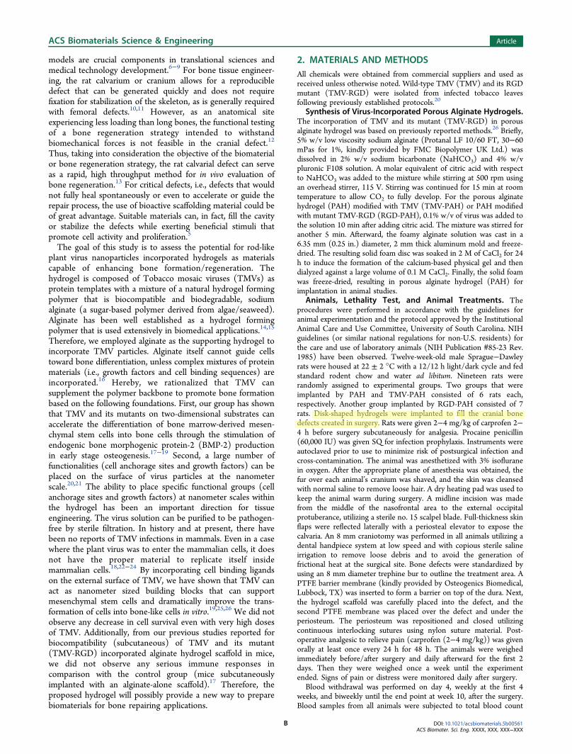
Figure 1. White blood count analysis of all rats with bone substitute implants. The total blood counts were monitored at 8 time points (day 4 andweeks 1, 2, 3, 4, 6, 8, and 10, postoperatively). The histograms show the numbers of total WBC, LYM, MON, and NEU normalized with the valuesmeasured from nontreated rats. Each bar indicates the minimum to maximum with the mean as the middle horizontal bar (n = 6 for PAH and TMV-PAH and n = 7 for RGD-PAH) ± SD. From t test analysis comparing control (nontreatment) and experimental values, the filled bars representstatistical significance, p < 0.05. The red dots represent outliers.
ACS Biomaterials Science & Engineering Article
DOI: 10.1021/acsbiomaterials.5b00561ACS Biomater. Sci. Eng. XXXX, XXX, XXX−XXX
C

Statistical Analysis. Statistical evaluation has been carried outusing Graphpad Prism 6.0 software. For complete blood count, allgroups were compared to each other using multiple t tests to assesswhether there were statistically significant differences among them.Analysis of variance (ANOVA) was performed in all other experimentsin this article.
3. RESULTS AND DISCUSSIONSSurgical Recovery of Animals. The survival surgery of
cranial bone defect creation with subsequent hydrogelimplantation was carried out. Mean surgery time was 10 min(SD = 4). The animal’s weight was measured immediately afterthe cranial defect survival surgery on each animal to monitorpostsurgery weight loss. Figure S1A showed that all rats lostless than 5% of their initial weight measured before theoperation. Blood loss during surgical procedure was alsoestimated to be less than 1 mL in all rats. The animals wereclosely monitored within the first 6 h after surgery and weighedevery day for the first postoperative week and at least onceevery consecutive week, continuously for the entire exper-imental period. Postoperatively, the rats recovered quickly,gaining their baseline weight and returning to routine activitiessuch as grooming, eating, and drinking in less than 48 h. Thesurgical wound healed within 1.5 weeks without postsurgicalbleeding or wound infection. The surgical suture fell offspontaneously from the surgical site after the wound healedcompletely. From gross observations after cranial defectoperation and implantation, all animals were normally activein eating, drinking, and grooming with no observable hair lossand aggressive behavior. The growth rate was evaluated by thebody weight of rats at each time point normalized with theiroriginal weight before implantation. No differences in weight
gained among three groups of animals were observed.Comparing to the normal growth rate of Sprague−Dawleyrats,27 the weight gains are in agreement with unoperated rats(Figure S1B).
Systemic Inflammatory and Immunogenic Responses.Complete blood count was analyzed from whole blood samplesfrom each group of rats at every week for 4 weeks and thenevery other week until termination after 10 postoperativeweeks. Figures 1 and 2 showed different blood counts at varioustime points in normalization to blood counts on day 0 beforethe operation. The host inflammatory responses after thesurgery were evaluated by white blood count including totalWBC, LYM, MON, and NEU counts. However, thecomparison had no statistical difference for each parameter ineach time point except for the PAH group. Rats implanted withPAH had statistically significant lower WBC at weeks 1, 3, 8,and 10, and LYM at weeks 3 and 10 compared to those ofnormal rats (multiple t tests, P value <0.01). MON count fromall rats was comparable with value shown in normal rat’s blood(multiple t tests; no significant difference). We compared thepostoperative WBC count between the viral particle non-exposure group (PAH) and exposure groups (TMV-PAH andRGD-PAH), and found no statistically significant differences ofblood cells count across the groups. There were 2 outliers onday 4 white blood counts: one, from the RGD-PAH implantedgroup, expressed increased numbers of LYM and thecorresponding WBC; another, from PAH implanted group,exhibited high NEU and its corresponding WBC. The elevatedWBC counts could likely reflect the severity of the infectionand associated inflammation. In such cases, the risk for poorwound healing should be highly suspected.28 However, both
Figure 2. Hematology analysis of all rats with bone substitute implants. The total blood counts were monitored at 8 time points (day 4 and weeks 1,2, 3, 4, 6, 8, and 10, postoperatively). The histograms show the numbers of RBC, HGB, PLT, and MPV normalized with the values measured fromnontreated rats. Each bar indicates the minimum to maximum with the mean as the middle horizontal bar (n = 6 for PAH and TMV-PAH and n = 7for RGD-PAH) ± SD. From t test analysis comparing control (nontreatment) and experimental values, the filled bars represent statisticalsignificance, p < 0.05. The red dots represent outliers.
ACS Biomaterials Science & Engineering Article
DOI: 10.1021/acsbiomaterials.5b00561ACS Biomater. Sci. Eng. XXXX, XXX, XXX−XXX
D
rats had WBC counts leveled down to the values comparable tothose of all other rats after 1 postoperative week (Figure 1).Other blood parameters that indicate animal health and
significant loss of blood such as RBC, HGB, PLT, and MPVwere shown to be comparable to the values measured fromnontreated rat’s blood in all animals that were implanted withdifferent types of hydrogels. Even though there were someoutliers that appeared at different time points for eachparameter, those outliers seemed to obtain only temporalchanges in particular blood counts that returned to the normalvalues at the later time point (Figure 2).Organ weight can be the most sensitive indicator of an effect
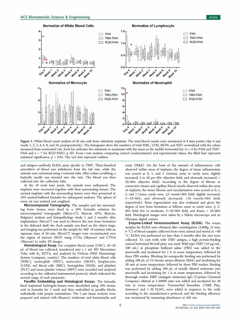
of drug toxicity, as significant differences in organ weightbetween treated and control animals may occur in the absenceof any morphological changes.29 The spleen and liver are themain target organs for various nanomaterials in the circulatorysystem because these organs are part of the reticulo-endothelialsystem (RES) that has removing foreign agents from thecirculation as one of its functions.30−32 In this study, we utilizedviral nanoparticles in providing biofunctionality to the hydrogelto create an extracellular matrix preferable for cell attachmentand growth. It was critical to evaluate the spleen enlargementthat might occur from the immunogenic and/or eliminationreaction of the body toward viral nanoparticles presented in thehydrogel implants. Figure 3 shows that the spleen weightsmeasured after the animal sacrifice (at the age of 22 weeks)were not significantly different among all groups. Thecorresponding mean spleen weight of all rats was 1495 ±236 mg/kg of total body weight, which is close to the valuereported in the literature. The mean normal spleen weight ofmale Sprague−Dawley rat ± SD, used to fit organ weightsversus age in a previous study reported by Schoeffner et al.,equaled to 1029 ± 191 mg/kg of total body weight.33,34 Fromeach animal observed at the end of the experiment, there wereno chronic and/or major inflammatory and toxicity reactions inthe RES that could cause spleen enlargement in any group ofanimals.
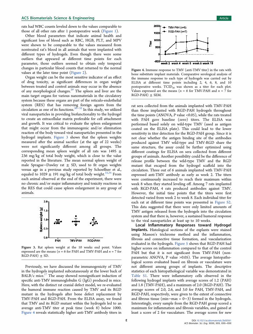
Previously, we have discussed the immunogenicity of TMVin the hydrogels implanted subcutaneously at the lower back ofBALB/c mice.17 The assay showed nonsignificant induction ofspecific anti-TMV immunoglobulin G (IgG) produced in mice.Here, with the distinct rat cranial defect model, we re-evaluatedthe humoral immune reaction caused by TMV and its RGDmutant in the hydrogels after bone defect replacement byTMV-PAH and RGD-PAH. From the ELISA assay, we foundthat TMV and its RGD mutant within the hydrogels led to anaverage anti-TMV titer at peak time (week 8) below 1000.Figure 4 reveals statistically higher anti-TMV antibody titers in
rat sera collected from the animals implanted with TMV-PAHthan those implanted with RGD-PAH hydrogels throughoutthe time points (ANOVA, P value <0.05), while the rats treatedwith PAH gave baseline (zero) titers. The ELISA wasperformed based solely on wild-type TMV (used as antigencoated on the ELISA plate). This could lead to the lowersensitivity in titer detection for the RGD-PAH group. Since it isnot clear whether the antigen binding site of the antibodiesproduced against TMV wild-type and TMV-RGD share thesame structure, the assay could be further optimized usingdifferent coatings for ELISA on sera collected from differentgroups of animals. Another possibility could be the difference ofrelease profile between the wild-type TMV and the RGDmutant that escaped from the hydrogel scaffold into thecirculation. Three out of 6 animals implanted with TMV-PAHexpressed anti-TMV antibody as early as week 2. The titerswere continuously increased to reach their maximum withinweek 8 when they started leveling off. Among 7 rats implantedwith RGD-PAH, 4 rats produced antibodies against TMV.However, the initial time points that the titers were firstdetected varied from week 2 to week 8. Each individual titer foreach rat at different time points was presented in Figure S2.This data suggested that there were only limited amounts ofTMV antigen released from the hydrogels into the circulationsystem and that there is, however, a sustained humoral responseto the viral nanoparticles at least up to 10 weeks.
Local Inflammatory Responses toward HydrogelImplants. Histological sections of the explants were stainedusing Masson’s trichrome method and the inflammation,fibrosis and connective tissue formation, and vascularizationevaluated in the hydrogels. Figure 5 shows that RGD-PAH hadhigher scores on inflammation compared to that of the controlPAH but that it is not significant from TMV-PAH (non-parametric ANOVA, P value >0.05). The average histopatho-logical scores evaluated based on fibrosis or vasculature werenot different among groups of implants. The descriptivestatistics of each histopathological variable was demonstrated inTable S1. There were inflammatory cells observed in thefollowing hydrogel implants with average scores of 1.2 (PAH)and 1.8 (TMV-PAH), and a maximum of 2.0 (RGD-PAH). Theaverage scores of 2.0, 2.6, and 3.0 for PAH, TMV-PAH, andRGD-PAH, respectively, were given to the extent of connectiveand fibrous tissue (min−max = 0−3) formed in the hydrogels.Interestingly, every sample from the RGD-PAH group scored amaximum for inflammation and fibrosis variables, and gained atleast a score of 2 for vasculature. The average scores for new
Figure 3. Rat spleen weight at the 10 weeks end point. Valuesexpressed are the means (n = 6 for PAH and TMV-PAH and n = 7 forRGD-PAH) ± SD.
Figure 4. Immune response to TMV (anti-TMV titer) in the rats withbone substitute implant materials. Comparative serological analysis ofthe immune response to each type of hydrogels was carried out byELISA at different time points including 2, 4, 6, 8, and 10postoperative weeks. TCID50 was shown as a titer for each plot.Values expressed are the means (n = 6 for TMV-PAH and n = 7 forRGD-PAH) ± SEM.
ACS Biomaterials Science & Engineering Article
DOI: 10.1021/acsbiomaterials.5b00561ACS Biomater. Sci. Eng. XXXX, XXX, XXX−XXX
E
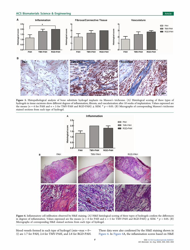
blood vessels formed in each type of hydrogel (min−max = 0−3) are 1.7 for PAH, 2.4 for TMV-PAH, and 2.8 for RGD-PAH.
These data were also confirmed by the H&E staining shown inFigure 6. In Figure 6A, the inflammation scores based on H&E
Figure 5. Histopathological analysis of bone substitute hydrogel implants via Masson’s trichrome. (A) Histological scoring of three types ofhydrogels in tissue excisions show different degrees of inflammation, fibrosis, and vascularization after 10 weeks of implantation. Values expressed arethe means (n = 6 for PAH and n = 5 for TMV-PAH and RGD-PAH) ± SEM. * p < 0.05. (B) Micrographs of corresponding Masson’s trichromestained sections from each type of hydrogel.
Figure 6. Inflammatory cell infiltration observed by H&E staining. (A) H&E histological scoring of three types of hydrogels confirm the differencesin degrees of inflammation. Values expressed are the means (n = 6 for PAH and n = 5 for TMV-PAH and RGD-PAH) ± SEM. * p < 0.05. (B)Micrographs of corresponding H&E stained sections from each type of hydrogel.
ACS Biomaterials Science & Engineering Article
DOI: 10.1021/acsbiomaterials.5b00561ACS Biomater. Sci. Eng. XXXX, XXX, XXX−XXX
F
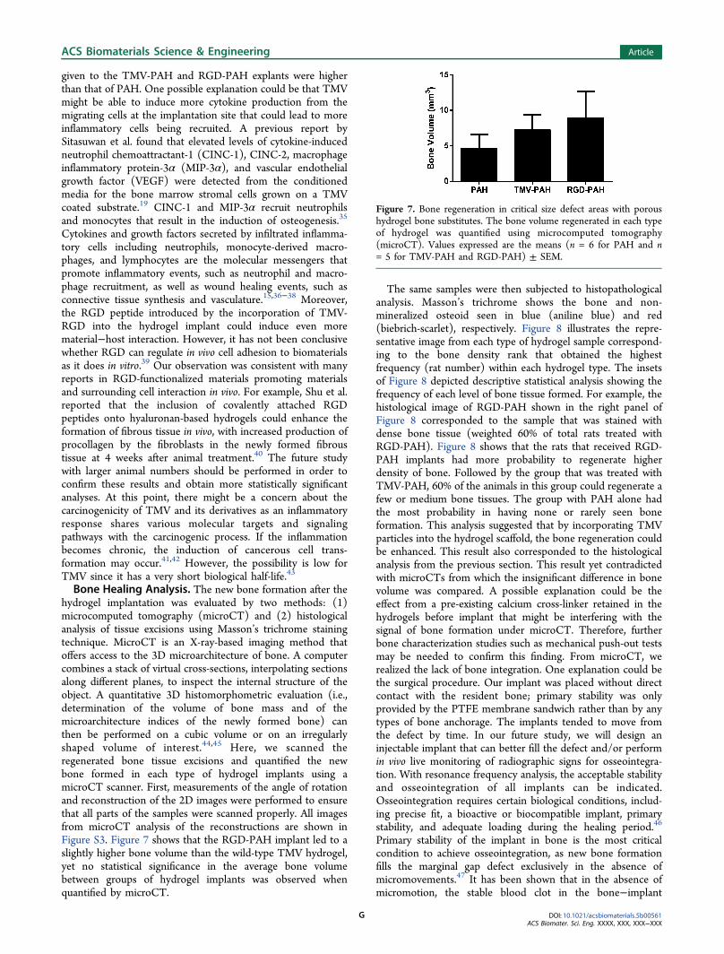
given to the TMV-PAH and RGD-PAH explants were higherthan that of PAH. One possible explanation could be that TMVmight be able to induce more cytokine production from themigrating cells at the implantation site that could lead to moreinflammatory cells being recruited. A previous report bySitasuwan et al. found that elevated levels of cytokine-inducedneutrophil chemoattractant-1 (CINC-1), CINC-2, macrophageinflammatory protein-3α (MIP-3α), and vascular endothelialgrowth factor (VEGF) were detected from the conditionedmedia for the bone marrow stromal cells grown on a TMVcoated substrate.19 CINC-1 and MIP-3α recruit neutrophilsand monocytes that result in the induction of osteogenesis.35
Cytokines and growth factors secreted by infiltrated inflamma-tory cells including neutrophils, monocyte-derived macro-phages, and lymphocytes are the molecular messengers thatpromote inflammatory events, such as neutrophil and macro-phage recruitment, as well as wound healing events, such asconnective tissue synthesis and vasculature.15,36−38 Moreover,the RGD peptide introduced by the incorporation of TMV-RGD into the hydrogel implant could induce even morematerial−host interaction. However, it has not been conclusivewhether RGD can regulate in vivo cell adhesion to biomaterialsas it does in vitro.39 Our observation was consistent with manyreports in RGD-functionalized materials promoting materialsand surrounding cell interaction in vivo. For example, Shu et al.reported that the inclusion of covalently attached RGDpeptides onto hyaluronan-based hydrogels could enhance theformation of fibrous tissue in vivo, with increased production ofprocollagen by the fibroblasts in the newly formed fibroustissue at 4 weeks after animal treatment.40 The future studywith larger animal numbers should be performed in order toconfirm these results and obtain more statistically significantanalyses. At this point, there might be a concern about thecarcinogenicity of TMV and its derivatives as an inflammatoryresponse shares various molecular targets and signalingpathways with the carcinogenic process. If the inflammationbecomes chronic, the induction of cancerous cell trans-formation may occur.41,42 However, the possibility is low forTMV since it has a very short biological half-life.43
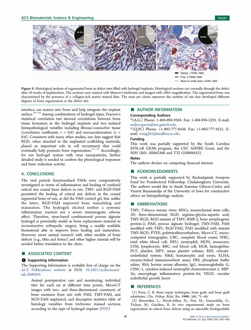
Bone Healing Analysis. The new bone formation after thehydrogel implantation was evaluated by two methods: (1)microcomputed tomography (microCT) and (2) histologicalanalysis of tissue excisions using Masson’s trichrome stainingtechnique. MicroCT is an X-ray-based imaging method thatoffers access to the 3D microarchitecture of bone. A computercombines a stack of virtual cross-sections, interpolating sectionsalong different planes, to inspect the internal structure of theobject. A quantitative 3D histomorphometric evaluation (i.e.,determination of the volume of bone mass and of themicroarchitecture indices of the newly formed bone) canthen be performed on a cubic volume or on an irregularlyshaped volume of interest.44,45 Here, we scanned theregenerated bone tissue excisions and quantified the newbone formed in each type of hydrogel implants using amicroCT scanner. First, measurements of the angle of rotationand reconstruction of the 2D images were performed to ensurethat all parts of the samples were scanned properly. All imagesfrom microCT analysis of the reconstructions are shown inFigure S3. Figure 7 shows that the RGD-PAH implant led to aslightly higher bone volume than the wild-type TMV hydrogel,yet no statistical significance in the average bone volumebetween groups of hydrogel implants was observed whenquantified by microCT.
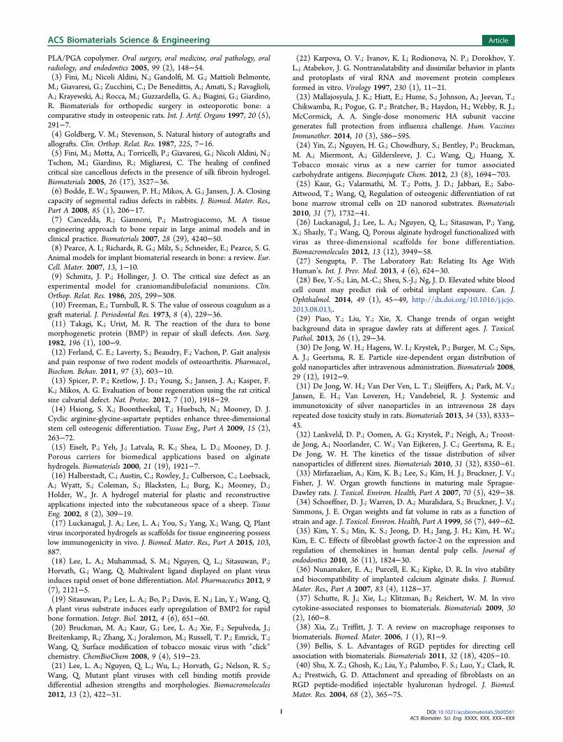
The same samples were then subjected to histopathologicalanalysis. Masson’s trichrome shows the bone and non-mineralized osteoid seen in blue (aniline blue) and red(biebrich-scarlet), respectively. Figure 8 illustrates the repre-sentative image from each type of hydrogel sample correspond-ing to the bone density rank that obtained the highestfrequency (rat number) within each hydrogel type. The insetsof Figure 8 depicted descriptive statistical analysis showing thefrequency of each level of bone tissue formed. For example, thehistological image of RGD-PAH shown in the right panel ofFigure 8 corresponded to the sample that was stained withdense bone tissue (weighted 60% of total rats treated withRGD-PAH). Figure 8 shows that the rats that received RGD-PAH implants had more probability to regenerate higherdensity of bone. Followed by the group that was treated withTMV-PAH, 60% of the animals in this group could regenerate afew or medium bone tissues. The group with PAH alone hadthe most probability in having none or rarely seen boneformation. This analysis suggested that by incorporating TMVparticles into the hydrogel scaffold, the bone regeneration couldbe enhanced. This result also corresponded to the histologicalanalysis from the previous section. This result yet contradictedwith microCTs from which the insignificant difference in bonevolume was compared. A possible explanation could be theeffect from a pre-existing calcium cross-linker retained in thehydrogels before implant that might be interfering with thesignal of bone formation under microCT. Therefore, furtherbone characterization studies such as mechanical push-out testsmay be needed to confirm this finding. From microCT, werealized the lack of bone integration. One explanation could bethe surgical procedure. Our implant was placed without directcontact with the resident bone; primary stability was onlyprovided by the PTFE membrane sandwich rather than by anytypes of bone anchorage. The implants tended to move fromthe defect by time. In our future study, we will design aninjectable implant that can better fill the defect and/or performin vivo live monitoring of radiographic signs for osseointegra-tion. With resonance frequency analysis, the acceptable stabilityand osseointegration of all implants can be indicated.Osseointegration requires certain biological conditions, includ-ing precise fit, a bioactive or biocompatible implant, primarystability, and adequate loading during the healing period.46
Primary stability of the implant in bone is the most criticalcondition to achieve osseointegration, as new bone formationfills the marginal gap defect exclusively in the absence ofmicromovements.47 It has been shown that in the absence ofmicromotion, the stable blood clot in the bone−implant
Figure 7. Bone regeneration in critical size defect areas with poroushydrogel bone substitutes. The bone volume regenerated in each typeof hydrogel was quantified using microcomputed tomography(microCT). Values expressed are the means (n = 6 for PAH and n= 5 for TMV-PAH and RGD-PAH) ± SEM.
ACS Biomaterials Science & Engineering Article
DOI: 10.1021/acsbiomaterials.5b00561ACS Biomater. Sci. Eng. XXXX, XXX, XXX−XXX
G
interface can mature into bone and help integrate the implantsurface.48−50 Among combinations of hydrogel types, Pearson’sstatistical correlation test showed correlations between bonetissue formation in the hydrogel implants and two isolatedhistopathological variables including fibrous/connective tissue(correlation coefficient; r = 0.6) and neovascularization (r =0.6). Consistent with many other studies, our data suggest thatRGD, when attached to the implanted scaffolding materials,played an important role in cell recruitment that couldeventually help promote bone regeneration.51−53 Accordingly,for our hydrogel system with virus nanoparticles, furtherdetailed study is needed to confirm the physiological responsesand bone induction activity.
4. CONCLUSIONS
The viral particle functionalized PAHs were comparativelyinvestigated in terms of inflammation and healing of confinedcritical size cranial bone defects in rats. TMV- and RGD-PAHpermitted the healing of critical size defects in the cranialsegmental bone of rats, as did the PAH control gel, but, unlikethe latter, RGD-PAH improved bone remodeling andmaturation. The hydrogels elicited neither a systemicinflammatory reaction nor a severe immunogenic adverseeffect. Therefore, virus-based combinatorial porous alginatehydrogel is potentially useful as bone replacement material inreconstructive orthopedic surgery, being a readily availablebiomaterial able to improve bone healing and maturation.However, more animal research with other models of bonedefects (e.g., tibia and femur) and other higher animals will beneeded before translation to the clinic.
■ ASSOCIATED CONTENT
*S Supporting InformationThe Supporting Information is available free of charge on theACS Publications website at DOI: 10.1021/acsbiomater-ials.5b00561.
Animal postoperative care and monitoring, individualtiter for each rat at different time points, MicroCTimages with two- and three-dimensional constructs ofbone excisions from rats with PAH, TMV-PAH, andRGD-PAH implanted, and descriptive statistics table ofhistology variables from trichrome stained sectionsaccording to the type of hydrogel implant (PDF)
■ AUTHOR INFORMATIONCorresponding Authors*(A.G.) Phone: 1-404-894-9384. Fax: 1-404-894-2291. E-mail:[email protected].*(Q.W.) Phone: +1-803-777-8436. Fax: +1-803-777-9521. E-mail: [email protected] work was partially supported by the South CarolinaEPSCoR GEAR program, the USC ASPIRE Grant, and theNIH (R01 AR062368 and T32 GM008433).NotesThe authors declare no competing financial interest.
■ ACKNOWLEDGMENTSThis work is partially supported by Rachadapisak SompoteFund for Postdoctoral Fellowship, Chulalongkorn University.The authors would like to thank Katerine Gibson-Corley andVisarut Buranasudja of the University of Iowa for constructiveadvice on histopathology analysis.
■ ABBREVIATIONSTMV, Tobacco mosaic virus; MSCs, mesenchymal stem cells;3D, three-dimensional; RGD, arginine-glycine-aspartic acid;TMV-RGD, RGD mutant of TMV; BMP-2, bone morphogenicprotein-2; PAH, porous alginate hydrogel; TMV-PAH, PAHmodified with TMV; RGD-PAH, PAH modified with mutantTMV-RGD; PTFE, polytetrafluoroethylene; Micro-CT, micro-computed tomography; CBC, complete blood count; WBC,total white blood cell; NEU, neutrophil; MON, monocyte;LYM, lymphocyte; RBC, red blood cell; HGB, hemoglobin;PLT, platelet; MPV, mean platelet volume; RES, reticulo-endothelial system; H&E, hematoxylin and eosin; ELISA,enzyme-linked immunosorbent assay; PBS, phosphate buffersaline; BSA, bovine serum albumin; IgG, immunoglobulin G;CINC-1, cytokine-induced neutrophil chemoattractant-1; MIP-3α, macrophage inflammatory protein-3α; VEGF, vascularendothelial growth factor
■ REFERENCES(1) Perry, C. R. Bone repair techniques, bone graft, and bone graftsubstitutes. Clin. Orthop. Relat. Res. 1999, 360, 71−86.(2) Rimondini, L.; Nicoli-Aldini, N.; Fini, M.; Guzzardella, G.;Tschon, M.; Giardino, R. In vivo experimental study on boneregeneration in critical bone defects using an injectable biodegradable
Figure 8. Histological analysis of regenerated bone at defect sites filled with hydrogel implants. Histological sections cut coronally through the defectafter 10 weeks of implantation. The sections were stained with Masson’s trichrome and imaged with 100× magnification. The regenerated bone wascharacterized by the presence of a collagen-rich matrix stained blue. The inset pie charts represent the number of rats that developed differentdegrees of bone regeneration at the defect site.
ACS Biomaterials Science & Engineering Article
DOI: 10.1021/acsbiomaterials.5b00561ACS Biomater. Sci. Eng. XXXX, XXX, XXX−XXX
H
PLA/PGA copolymer. Oral surgery, oral medicine, oral pathology, oralradiology, and endodontics 2005, 99 (2), 148−54.(3) Fini, M.; Nicoli Aldini, N.; Gandolfi, M. G.; Mattioli Belmonte,M.; Giavaresi, G.; Zucchini, C.; De Benedittis, A.; Amati, S.; Ravaglioli,A.; Krayewski, A.; Rocca, M.; Guzzardella, G. A.; Biagini, G.; Giardino,R. Biomaterials for orthopedic surgery in osteoporotic bone: acomparative study in osteopenic rats. Int. J. Artif. Organs 1997, 20 (5),291−7.(4) Goldberg, V. M.; Stevenson, S. Natural history of autografts andallografts. Clin. Orthop. Relat. Res. 1987, 225, 7−16.(5) Fini, M.; Motta, A.; Torricelli, P.; Giavaresi, G.; Nicoli Aldini, N.;Tschon, M.; Giardino, R.; Migliaresi, C. The healing of confinedcritical size cancellous defects in the presence of silk fibroin hydrogel.Biomaterials 2005, 26 (17), 3527−36.(6) Bodde, E. W.; Spauwen, P. H.; Mikos, A. G.; Jansen, J. A. Closingcapacity of segmental radius defects in rabbits. J. Biomed. Mater. Res.,Part A 2008, 85 (1), 206−17.(7) Cancedda, R.; Giannoni, P.; Mastrogiacomo, M. A tissueengineering approach to bone repair in large animal models and inclinical practice. Biomaterials 2007, 28 (29), 4240−50.(8) Pearce, A. I.; Richards, R. G.; Milz, S.; Schneider, E.; Pearce, S. G.Animal models for implant biomaterial research in bone: a review. Eur.Cell. Mater. 2007, 13, 1−10.(9) Schmitz, J. P.; Hollinger, J. O. The critical size defect as anexperimental model for craniomandibulofacial nonunions. Clin.Orthop. Relat. Res. 1986, 205, 299−308.(10) Freeman, E.; Turnbull, R. S. The value of osseous coagulum as agraft material. J. Periodontal Res. 1973, 8 (4), 229−36.(11) Takagi, K.; Urist, M. R. The reaction of the dura to bonemorphogenetic protein (BMP) in repair of skull defects. Ann. Surg.1982, 196 (1), 100−9.(12) Ferland, C. E.; Laverty, S.; Beaudry, F.; Vachon, P. Gait analysisand pain response of two rodent models of osteoarthritis. Pharmacol.,Biochem. Behav. 2011, 97 (3), 603−10.(13) Spicer, P. P.; Kretlow, J. D.; Young, S.; Jansen, J. A.; Kasper, F.K.; Mikos, A. G. Evaluation of bone regeneration using the rat criticalsize calvarial defect. Nat. Protoc. 2012, 7 (10), 1918−29.(14) Hsiong, S. X.; Boontheekul, T.; Huebsch, N.; Mooney, D. J.Cyclic arginine-glycine-aspartate peptides enhance three-dimensionalstem cell osteogenic differentiation. Tissue Eng., Part A 2009, 15 (2),263−72.(15) Eiselt, P.; Yeh, J.; Latvala, R. K.; Shea, L. D.; Mooney, D. J.Porous carriers for biomedical applications based on alginatehydrogels. Biomaterials 2000, 21 (19), 1921−7.(16) Halberstadt, C.; Austin, C.; Rowley, J.; Culberson, C.; Loebsack,A.; Wyatt, S.; Coleman, S.; Blacksten, L.; Burg, K.; Mooney, D.;Holder, W., Jr. A hydrogel material for plastic and reconstructiveapplications injected into the subcutaneous space of a sheep. TissueEng. 2002, 8 (2), 309−19.(17) Luckanagul, J. A.; Lee, L. A.; You, S.; Yang, X.; Wang, Q. Plantvirus incorporated hydrogels as scaffolds for tissue engineering possesslow immunogenicity in vivo. J. Biomed. Mater. Res., Part A 2015, 103,887.(18) Lee, L. A.; Muhammad, S. M.; Nguyen, Q. L.; Sitasuwan, P.;Horvath, G.; Wang, Q. Multivalent ligand displayed on plant virusinduces rapid onset of bone differentiation. Mol. Pharmaceutics 2012, 9(7), 2121−5.(19) Sitasuwan, P.; Lee, L. A.; Bo, P.; Davis, E. N.; Lin, Y.; Wang, Q.A plant virus substrate induces early upregulation of BMP2 for rapidbone formation. Integr. Biol. 2012, 4 (6), 651−60.(20) Bruckman, M. A.; Kaur, G.; Lee, L. A.; Xie, F.; Sepulveda, J.;Breitenkamp, R.; Zhang, X.; Joralemon, M.; Russell, T. P.; Emrick, T.;Wang, Q. Surface modification of tobacco mosaic virus with ″click″chemistry. ChemBioChem 2008, 9 (4), 519−23.(21) Lee, L. A.; Nguyen, Q. L.; Wu, L.; Horvath, G.; Nelson, R. S.;Wang, Q. Mutant plant viruses with cell binding motifs providedifferential adhesion strengths and morphologies. Biomacromolecules2012, 13 (2), 422−31.
(22) Karpova, O. V.; Ivanov, K. I.; Rodionova, N. P.; Dorokhov, Y.L.; Atabekov, J. G. Nontranslatability and dissimilar behavior in plantsand protoplasts of viral RNA and movement protein complexesformed in vitro. Virology 1997, 230 (1), 11−21.(23) Mallajosyula, J. K.; Hiatt, E.; Hume, S.; Johnson, A.; Jeevan, T.;Chikwamba, R.; Pogue, G. P.; Bratcher, B.; Haydon, H.; Webby, R. J.;McCormick, A. A. Single-dose monomeric HA subunit vaccinegenerates full protection from influenza challenge. Hum. VaccinesImmunother. 2014, 10 (3), 586−595.(24) Yin, Z.; Nguyen, H. G.; Chowdhury, S.; Bentley, P.; Bruckman,M. A.; Miermont, A.; Gildersleeve, J. C.; Wang, Q.; Huang, X.Tobacco mosaic virus as a new carrier for tumor associatedcarbohydrate antigens. Bioconjugate Chem. 2012, 23 (8), 1694−703.(25) Kaur, G.; Valarmathi, M. T.; Potts, J. D.; Jabbari, E.; Sabo-Attwood, T.; Wang, Q. Regulation of osteogenic differentiation of ratbone marrow stromal cells on 2D nanorod substrates. Biomaterials2010, 31 (7), 1732−41.(26) Luckanagul, J.; Lee, L. A.; Nguyen, Q. L.; Sitasuwan, P.; Yang,X.; Shazly, T.; Wang, Q. Porous alginate hydrogel functionalized withvirus as three-dimensional scaffolds for bone differentiation.Biomacromolecules 2012, 13 (12), 3949−58.(27) Sengupta, P. The Laboratory Rat: Relating Its Age WithHuman’s. Int. J. Prev. Med. 2013, 4 (6), 624−30.(28) Bee, Y.-S.; Lin, M.-C.; Sheu, S.-J.; Ng, J. D. Elevated white bloodcell count may predict risk of orbital implant exposure. Can. J.Ophthalmol. 2014, 49 (1), 45−49, http://dx.doi.org/10.1016/j.jcjo.2013.08.013,.(29) Piao, Y.; Liu, Y.; Xie, X. Change trends of organ weightbackground data in sprague dawley rats at different ages. J. Toxicol.Pathol. 2013, 26 (1), 29−34.(30) De Jong, W. H.; Hagens, W. I.; Krystek, P.; Burger, M. C.; Sips,A. J.; Geertsma, R. E. Particle size-dependent organ distribution ofgold nanoparticles after intravenous administration. Biomaterials 2008,29 (12), 1912−9.(31) De Jong, W. H.; Van Der Ven, L. T.; Sleijffers, A.; Park, M. V.;Jansen, E. H.; Van Loveren, H.; Vandebriel, R. J. Systemic andimmunotoxicity of silver nanoparticles in an intravenous 28 daysrepeated dose toxicity study in rats. Biomaterials 2013, 34 (33), 8333−43.(32) Lankveld, D. P.; Oomen, A. G.; Krystek, P.; Neigh, A.; Troost-de Jong, A.; Noorlander, C. W.; Van Eijkeren, J. C.; Geertsma, R. E.;De Jong, W. H. The kinetics of the tissue distribution of silvernanoparticles of different sizes. Biomaterials 2010, 31 (32), 8350−61.(33) Mirfazaelian, A.; Kim, K. B.; Lee, S.; Kim, H. J.; Bruckner, J. V.;Fisher, J. W. Organ growth functions in maturing male Sprague-Dawley rats. J. Toxicol. Environ. Health, Part A 2007, 70 (5), 429−38.(34) Schoeffner, D. J.; Warren, D. A.; Muralidara, S.; Bruckner, J. V.;Simmons, J. E. Organ weights and fat volume in rats as a function ofstrain and age. J. Toxicol. Environ. Health, Part A 1999, 56 (7), 449−62.(35) Kim, Y. S.; Min, K. S.; Jeong, D. H.; Jang, J. H.; Kim, H. W.;Kim, E. C. Effects of fibroblast growth factor-2 on the expression andregulation of chemokines in human dental pulp cells. Journal ofendodontics 2010, 36 (11), 1824−30.(36) Nunamaker, E. A.; Purcell, E. K.; Kipke, D. R. In vivo stabilityand biocompatibility of implanted calcium alginate disks. J. Biomed.Mater. Res., Part A 2007, 83 (4), 1128−37.(37) Schutte, R. J.; Xie, L.; Klitzman, B.; Reichert, W. M. In vivocytokine-associated responses to biomaterials. Biomaterials 2009, 30(2), 160−8.(38) Xia, Z.; Triffitt, J. T. A review on macrophage responses tobiomaterials. Biomed. Mater. 2006, 1 (1), R1−9.(39) Bellis, S. L. Advantages of RGD peptides for directing cellassociation with biomaterials. Biomaterials 2011, 32 (18), 4205−10.(40) Shu, X. Z.; Ghosh, K.; Liu, Y.; Palumbo, F. S.; Luo, Y.; Clark, R.A.; Prestwich, G. D. Attachment and spreading of fibroblasts on anRGD peptide-modified injectable hyaluronan hydrogel. J. Biomed.Mater. Res. 2004, 68 (2), 365−75.
ACS Biomaterials Science & Engineering Article
DOI: 10.1021/acsbiomaterials.5b00561ACS Biomater. Sci. Eng. XXXX, XXX, XXX−XXX
I
(41) Hussain, S. P.; Harris, C. C. Inflammation and cancer: anancient link with novel potentials. Int. J. Cancer 2007, 121 (11), 2373−80.(42) Medzhitov, R. Origin and physiological roles of inflammation.Nature 2008, 454 (7203), 428−35.(43) Bruckman, M. A.; Randolph, L. N.; VanMeter, A.; Hern, S.;Shoffstall, A. J.; Taurog, R. E.; Steinmetz, N. F. Biodistribution,pharmacokinetics, and blood compatibility of native and PEGylatedtobacco mosaic virus nano-rods and -spheres in mice. Virology 2014,449, 163−73.(44) Holdsworth, D. W.; Thornton, M. M. Micro-CT in small animaland specimen imaging. Trends Biotechnol. 2002, 20 (8), S34−S39,http://dx.doi.org/10.1016/S0167-7799(02)02004-8,.(45) Kallai, I.; Mizrahi, O.; Tawackoli, W.; Gazit, Z.; Pelled, G.; Gazit,D. Microcomputed tomography-based structural analysis of variousbone tissue regeneration models. Nat. Protoc. 2011, 6 (1), 105−10.(46) Villa, R.; Polimeni, G.; Wikesjo, U. M. E. Implantosseointegration in the absence of primary bone anchorage: A clinicalreport. J. Prosthet. Dent. 2010, 104 (5), 282−287, http://dx.doi.org/10.1016/S0022-3913(10)00146-0,.(47) Lioubavina-Hack, N.; Lang, N. P.; Karring, T. Significance ofprimary stability for osseointegration of dental implants. Clinical OralImplants Research 2006, 17 (3), 244−250.(48) Berglundh, T.; Abrahamsson, I.; Lang, N. P.; Lindhe, J. De novoalveolar bone formation adjacent to endosseous implants. Clin OralImplants Res. 2003, 14 (3), 251−62.(49) Abrahamsson, I.; Berglundh, T.; Linder, E.; Lang, N. P.; Lindhe,J. Early bone formation adjacent to rough and turned endosseousimplant surfaces. Clinical Oral Implants Research 2004, 15 (4), 381−392.(50) Davies, J. E. Mechanisms of endosseous integration. Int. J.Prosthodont. 1998, 11 (5), 391−401.(51) Chen, J.; Bly, R. A.; Saad, M. M.; AlKhodary, M. A.; El-Backly,R. M.; Cohen, D. J.; Kattamis, N.; Fatta, M. M.; Moore, W. A.; Arnold,C. B.; Marei, M. K.; Soboyejo, W. O. In-vivo study of adhesion andbone growth around implanted laser groove/RGD-functionalized Ti-6Al-4V pins in rabbit femurs. Mater. Sci. Eng., C 2011, 31 (5), 826−832, http://dx.doi.org/10.1016/j.msec.2010.12.019,.(52) Ferris, D. M.; Moodie, G. D.; Dimond, P. M.; Gioranni, C. W.;Ehrlich, M. G.; Valentini, R. F. RGD-coated titanium implantsstimulate increased bone formation in vivo. Biomaterials 1999, 20(23−24), 2323−31.(53) Kroese-Deutman, H. C.; van den Dolder, J.; Spauwen, P. H.;Jansen, J. A. Influence of RGD-loaded titanium implants on boneformation in vivo. Tissue Eng. 2005, 11 (11−12), 1867−75.
ACS Biomaterials Science & Engineering Article
DOI: 10.1021/acsbiomaterials.5b00561ACS Biomater. Sci. Eng. XXXX, XXX, XXX−XXX
J