Embed Size (px)
Citation preview
Tobacco mosaic virus-directed reprogramming ofauxin/indole acetic acid protein transcriptionalresponses enhances virus phloem loadingTamara D. Colluma, Meenu S. Padmanabhana,1, Yi-Cheng Hsiehb,2, and James N. Culverb,c,3
aDepartment of Cell Biology and Molecular Genetics, University of Maryland, College Park, MD 20742; bInstitute for Bioscience and Biotechnology Research,University of Maryland, College Park, MD 20742; and cDepartment of Plant Science and Landscape Architecture, University of Maryland, College Park,MD 20742
Edited by David C. Baulcombe, University of Cambridge, Cambridge, United Kingdom, and approved March 31, 2016 (received for review December 14, 2015)
Vascular phloem loading has long been recognized as an essentialstep in the establishment of a systemic virus infection. In this study, aninteraction between the replication protein of tobacco mosaic virus(TMV) and phloem-specific auxin/indole acetic acid (Aux/IAA) tran-scriptional regulators was found to modulate virus phloem loading inan age-dependent manner. Promoter expression studies show that inmature tissues TMV 126/183-kDa–interacting Aux/IAAs predom-inantly express and accumulate within the nuclei of phloem compan-ion cells (CCs). Furthermore, CC Aux/IAA nuclear localization isdisrupted upon infection with an interacting virus. In situ analysis ofvirus spread shows that the inability to disrupt Aux/IAA CC nuclearlocalization correlates with a reduced ability to load into the vasculartissue. Subsequent systemic movement assays also demonstrate thata virus capable of disrupting Aux/IAA localization is significantly morecompetitive at moving out of older plant tissues than a noninteractingvirus. Similarly, CC expression and overaccumulation of a degrada-tion-resistant Aux/IAA-interacting protein was found to inhibit TMVaccumulation and phloem loading selectively in flowering plants.Transcriptional expression studies demonstrate a role for Aux/IAA-interacting proteins in the regulation of salicylic and jasmonic acidhost defense responses as well as virus-specific movement factors,including pectin methylesterase, that are involved in regulating plas-modesmata size-exclusion limits and promoting virus cell-to-cellmovement. Combined, these findings indicate that TMV directs thereprogramming of auxin-regulated gene expression within the vascu-lar phloem of mature tissues as a means to enhance phloem loadingand systemic spread.
pathogen defense | plant hormone signaling | virus movement |plasmodesmata gating | age-related resistance
To establish a systemic infection, plant viruses must access theirhost’s vascular phloem. The first step in this process involves
cell-to-cell movement through intercellular cytoplasmic and endo-membrane connections called “plasmodesmata” (PD). Virusmovement through the PD is facilitated by viral movement proteins(MP) that function to modulate the size-exclusion limits of the PD,allowing virus-transport forms composed of either nucleoproteincomplexes or virions to pass between cells (1, 2). For systemicmovement viruses must “load” into the vascular phloem. Phloemloading requires passage through specialized branched PD con-nections known as “pore units” that occur between companioncells (CCs) and phloem sieve elements (SE) (3). Once in theanucleate SEs, viruses move following the source-to-sink path ofphotoassimilates to distal plant tissues (4). In addition to thetransport of photoassimilates, the vascular phloem also serves as aconduit for the movement of numerous host components includingproteins, mRNA, microRNAs, and small molecules involved in arange of plant responses including development and flowering aswell as abiotic and biotic stress responses (5–7). It is clear that thevascular phloem functions as a gatekeeper between distal planttissues, controlling the passage of numerous molecules that affectmany aspects of plant physiology as well as responses to outside
stimuli. Thus, to establish a systemic infection, plant viruses mustusurp this gateway. However, how plant viruses appropriate thevascular phloem remains a fundamental question in plant virology.A number of virus and host components have been shown to
impact virus systemic movement (2, 8, 9). For tobacco mosaic virus(TMV), systemic transport via the phloem involves the viral coatprotein (CP), P30 MP, and 126/183-kDa replication proteins. Boththe MP and replication protein are required for cell-to-cell move-ment via the PD (10). Phloem loading and systemic movement alsoinvolve the MP and replication protein, because transit via PD isnecessary to access the SEs. In addition, a functional virus CP isrequired for TMV systemic movement. Mutations that disrupt CPexpression or its ability to form virions are known to inhibit sys-temic movement (11–13). Furthermore, chimeric TMV recombi-nants encoding the MP or CP of the orchid-infecting TobamovirusOndontoglossum ringspot virus display impaired systemic move-ment, and similar chimeric recombinants derived from sun-hempmosaic virus suggest a role for the viral replication proteins invascular movement (14–16). Combined, these findings suggest ahost-selective gating mechanism involving multiple virus–host in-teractions as essential factors in TMV phloem loading.
Significance
For plant viruses a successful infection correlates with the abilityto access the vascular phloem and move systemically into distaltissues. However, how viruses gain access to and usurp vasculartissues is poorly understood. Here we show how tobacco mosaicvirus (TMV) enhances its access to the phloem of mature planttissues through the targeted disruption of auxin/indole aceticacid (Aux/IAA) transcriptional regulators that control expressionof host genes involved in virus cell-to-cell movement, plasmo-desmata gating, and defense. TMV’s ability to disrupt Aux/IAAfunction successfully confers a significant advantage in the sys-temic spread of this virus, allowing it to outcompete non-disrupting viruses. In summary, TMV interacts with Aux/IAAproteins to reprogram the vascular phloem, making it moreconducive to systemic movement.
Author contributions: T.D.C. and J.N.C. designed research; T.D.C., M.S.P., Y.-C.H., and J.N.C.performed research; T.D.C., M.S.P., Y.-C.H., and J.N.C. analyzed data; and T.D.C. and J.N.C.wrote the paper.
The authors declare no conflict of interest.
This article is a PNAS Direct Submission.
Data deposition: The data reported in this paper have been deposited in the Gene Ex-pression Omnibus (GEO) database, www.ncbi.nlm.nih.gov/geo (accession no. GSE75983).1Present address: Department of Plant Biology and Genome Center, University of Califor-nia, Davis, CA 95616.
2Present address: Office of the Texas State Chemist, Texas A&M AgriLife Research, TexasA&M University System, College Station, TX 77843.
3To whom correspondence should be addressed. Email: [email protected].
This article contains supporting information online at www.pnas.org/lookup/suppl/doi:10.1073/pnas.1524390113/-/DCSupplemental.
E2740–E2749 | PNAS | Published online April 26, 2016 www.pnas.org/cgi/doi/10.1073/pnas.1524390113
Dow
nloa
ded
by g
uest
on
Mar
ch 2
6, 2
020
Several host components also have been found to impact thesystemic spread of TMV.Many of these host factors affect PD gatingand have been reviewed in detail (2, 8, 10). However, there is anemerging link between virus systemic movement and transcriptionalreprograming. For example, the crucifer strain of TMV-cg has beenshown to modulate the expression of a WRKY8 transcription factor,and the P30 protein from tomato mosaic virus (ToMV) associates invivo with the transcriptional coactivator KELP (17, 18). WRKYtranscription factors are associated with basal defense responses, andsuppression of WRKY8 during infection correlates with enhancedTMV-cg systemic accumulation (18). Similarly, overexpression ofKELP resulted in the partial relocation of P30 to the nucleus andprevented ToMV cell-to-cell and systemic movement. Furthermore,the P30 protein from the Tobamovirus turnip vein clearing virus(TVCV) encodes a nuclear localization signal required for both cell-to-cell and systemic virus movement (19). Association of the TVCVP30 protein with nuclear F-actin filaments was hypothesized to altergene expression and promote virus infection (19). These studiesprovide evidence that virus interactions with host-associated tran-scriptional regulators can modulate systemic virus movement.In our previous studies we identified an interaction between the
126/183-kDa replication protein of TMV and specific auxin/indoleacetic acid (Aux/IAA) host transcriptional regulators (20–22). Thesestudies show that the helicase domain present within the virus rep-lication protein interacts strongly with Aux/IAAmember IAA26 andmore weakly with IAA27 and IAA18 in Arabidopsis thaliana as wellas an IAA26 homolog in tomato. Members of the Aux/IAA familyencode short-lived nuclear proteins that mediate auxin-dependentgene expression (23–25). Based on current evidence, Aux/IAAproteins interact with auxin-responsive transcription factors (ARFs)that in turn regulate numerous auxin-responsive genes (26, 27).Within Arabidopsis there are 29 Aux/IAA family members and 23ARF members capable of forming an array of hetero- and homo-complexes from which auxin signaling can be orchestrated (28–30).Within the plant, auxin binds TIR1/AFB F-box proteins of the SCFE3 ubiquitin ligase complex, promoting their association withAux/IAA proteins (24, 27, 31, 32). Ubiquitination of Aux/IAAproteins results in their targeted degradation via the 26S protea-some (27, 33). The plant’s auxin gradient thus provides a spatiallysensitive means to regulate Aux/IAA activity via proteolysis.Cellular localization studies demonstrate that the nuclear local-
ization of interacting Aux/IAA proteins is disrupted during TMVinfection (20, 22). In contrast, Aux/IAA proteins remain localizedto the nucleus during infection by noninteracting viruses. Sub-sequent analysis indicates that viruses with reduced ability to in-teract with Aux/IAA proteins are compromised in their ability toaccumulate and move in inoculated tissue (21). Interestingly, effectson virus accumulation are observed only in mature tissues and notin younger, immature tissues (21). This developmental relationshipcorresponds to the accumulation of Aux/IAA proteins in maturetissues and is consistent with the lower auxin levels and reducedauxin-mediated degradation found in older plant tissues (21, 34).In this study we investigated the mechanism through which the
TMV–Aux/IAA interactions affect virus accumulation and spread.Results indicate that interacting Aux/IAA proteins are expressedpredominantly in the CC of mature vascular phloem tissues. Theability to disrupt the nuclear localization of these Aux/IAAs wasfound to correlate with enhanced virus phloem loading and move-ment within the vascular tissues. As a result, TMV, with the ability tointeract with Aux/IAA proteins, gains a significant advantage insystemic movement over a virus defective in this interaction. Tran-scriptomic analysis of plant genes under the control of an interactingAux/IAA protein identified an array of host genes linked to plantdefense responses, virus cell-to-cell movement, and PD regulation.Interestingly, overaccumulation of Aux/IAA proteins in CCs beforevirus infection led to decreased accumulation and phloem loading ofTMV only in mature flowering plants. This correlation with flow-ering has similarities to age-related resistance (ARR) and suggests
that interactions with the identified Aux/IAA proteins may provideTMV with a mechanism to overcome this form of host resistance(35). Based on these studies, we propose that within mature planttissues TMV selectively targets phloem-expressed Aux/IAA proteinsto reprogram functions of the CC–SE complex that contribute tophloem loading.
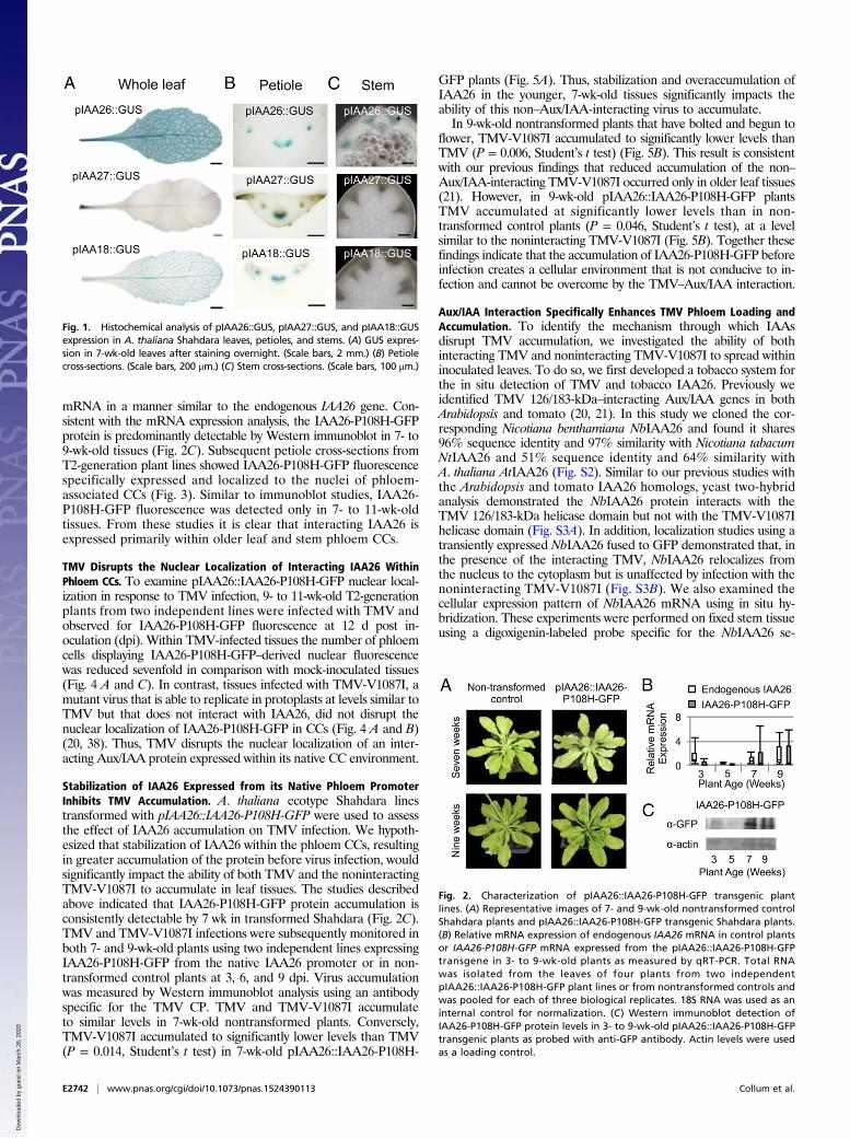
ResultsInteracting Aux/IAA Factors Are Expressed and Localize Within theNucleus of Phloem CCs. To define the importance of the Aux/IAAinteraction on TMV accumulation, we first examined the expres-sion patterns of three known interacting Arabidopsis Aux/IAAproteins. Previously IAA26, IAA27, and IAA18 were identified asinteracting with and displaying varying levels of cytoplasmiccolocalization with the TMV 126-kDa replication protein (22). Thestrength of the interaction corresponded to the level of cytoplasmiccolocalization. IAA26 confers the strongest interaction with thevirus 126-kDa protein and shows the greatest disruption in nuclearlocalization, followed by IAA27 and IAA18. These past studiesused only the cauliflower mosaic virus 35S constitutive promoter todrive the expression of GFP-tagged IAA proteins in whole-leaftissues. However, IAA family members are known to be differen-tially expressed and to confer tissue-specific functions (24, 36). Todetermine the expression pattern of the three TMV 126/183-kDa–interacting IAAs, promoter sequences upstream of their translationstart codons (2,000 nt for IAA26, 1,500 nt for IAA27, and 2,000 ntfor IAA18) were all cloned in front of the β-glucuronidase (GUS)ORF to create pIAA26::GUS, pIAA27::GUS, and pIAA18::GUS.All three promoter–reporter constructs were transformed into thesystemic TMV host A. thaliana ecotype Shahdara (37). Fully ex-panded leaves from T2-generation plants were histochemicallystained for GUS activity. Results from three or four independentplant lines for each of the three interacting IAAs consistentlyshowed the predominant GUS activity in vascular tissues, but withdistinct expression patterns (Fig. 1). pIAA26::GUS displayed themost robust expression with strong levels of GUS staining in all veinclasses (I, II, III, and IV) of the leaves. Conversely, pIAA27::GUSwas expressed predominately in vein class I of the petiole. LikepIAA26::GUS, pIAA18::GUS expression was observed in all veinclasses, but at a markedly lower level, indicating that in leaf tissueIAA18 is not as highly expressed as IAA26 (Fig. 1A). Petiole cross-sections of pIAA26::GUS, pIAA27::GUS, and pIAA18::GUS linesshowed GUS staining predominantly localized to the phloem for allthree interacting IAA family members (Fig. 1B). pIAA26::GUSexpression also was observed in stem and root vascular phloem(Fig. 1C and Fig. S1). In contrast pIAA27::GUS expression was notobserved in stem or root vascular tissues but was observed at thesites of lateral root formation (Fig. S1). pIAA18::GUS expressionwas not observed in either stem or roots (Fig. 1C and Fig. S1).To investigate the localization of Aux/IAA proteins further, we
focused on IAA26 because it displays by far the strongest in-teraction with the TMV 126-kDa protein and, as described above,is the most abundantly expressed in leaf and root vascular tissues.However, the rapid turnover of Aux/IAA proteins can makedetecting or visualizing Aux/IAA proteins difficult. To address thisissue, we used a previously generated IAA26 mutant allele, IAA26-P108H, which is resistant to auxin-mediated degradation but retainsthe ability to interact with the TMV 126/183-kDa protein (22). Thenative IAA26 promoter (2,000 nt upstream of the start codon) wascloned in front of the IAA26-P108H ORF and fused to GFP,creating pIAA26::IAA26-P108H-GFP. This construct was used totransform A. thaliana ecotype Shahdara. Transgenic pIAA26::IAA26-P108H-GFP plant lines grew and developed similarly tonontransformed Shahdara (Fig. 2A). In addition, quantitative RT-PCR (qRT-PCR) analysis for the endogenous IAA26 and transgeneIAA26-P108H-GFP mRNAs show a similar expression pattern ofincreasing transcripts in older plant tissues (Fig. 2B). Thus, transgenicpIAA26::IAA26-P108H-GFP plants express IAA26-P108H-GFP
Collum et al. PNAS | Published online April 26, 2016 | E2741
PLANTBIOLO
GY
PNASPL
US
Dow
nloa
ded
by g
uest
on
Mar
ch 2
6, 2
020
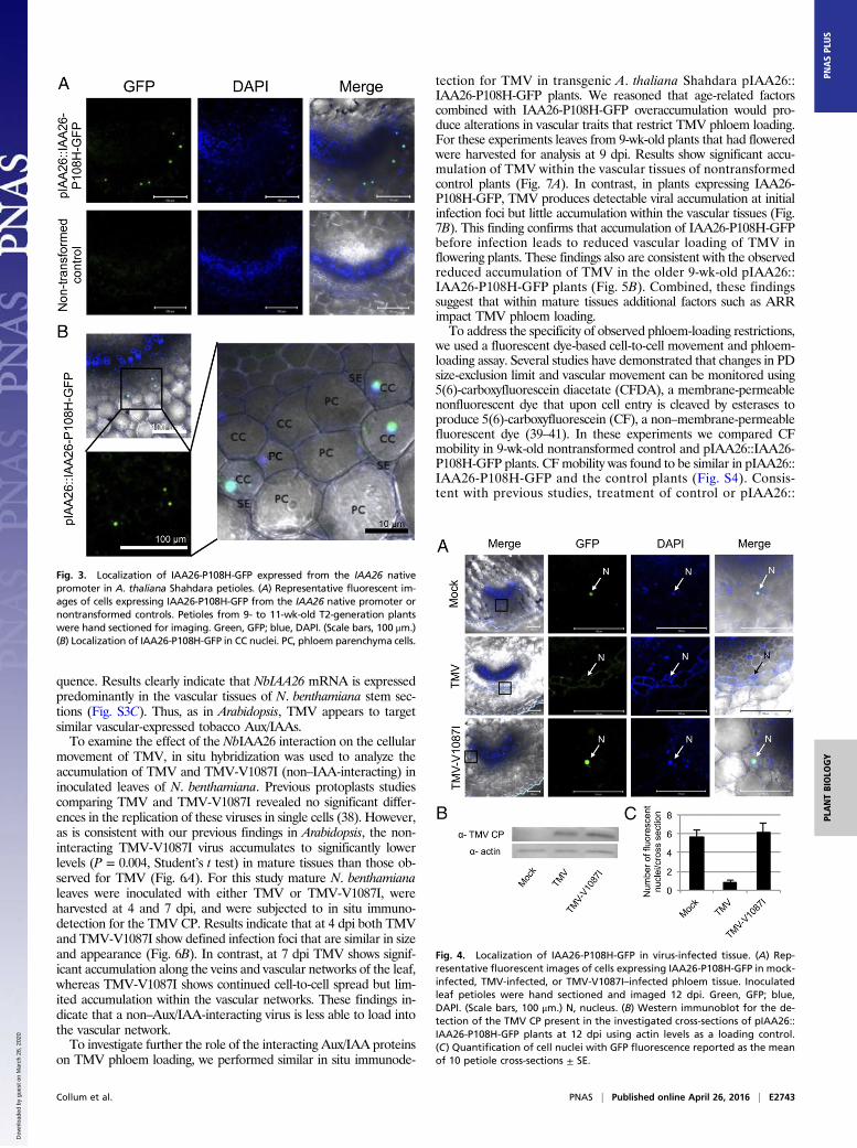
mRNA in a manner similar to the endogenous IAA26 gene. Con-sistent with the mRNA expression analysis, the IAA26-P108H-GFPprotein is predominantly detectable by Western immunoblot in 7- to9-wk-old tissues (Fig. 2C). Subsequent petiole cross-sections fromT2-generation plant lines showed IAA26-P108H-GFP fluorescencespecifically expressed and localized to the nuclei of phloem-associated CCs (Fig. 3). Similar to immunoblot studies, IAA26-P108H-GFP fluorescence was detected only in 7- to 11-wk-oldtissues. From these studies it is clear that interacting IAA26 isexpressed primarily within older leaf and stem phloem CCs.
TMV Disrupts the Nuclear Localization of Interacting IAA26 WithinPhloem CCs. To examine pIAA26::IAA26-P108H-GFP nuclear local-ization in response to TMV infection, 9- to 11-wk-old T2-generationplants from two independent lines were infected with TMV andobserved for IAA26-P108H-GFP fluorescence at 12 d post in-oculation (dpi). Within TMV-infected tissues the number of phloemcells displaying IAA26-P108H-GFP–derived nuclear fluorescencewas reduced sevenfold in comparison with mock-inoculated tissues(Fig. 4 A and C). In contrast, tissues infected with TMV-V1087I, amutant virus that is able to replicate in protoplasts at levels similar toTMV but that does not interact with IAA26, did not disrupt thenuclear localization of IAA26-P108H-GFP in CCs (Fig. 4 A and B)(20, 38). Thus, TMV disrupts the nuclear localization of an inter-acting Aux/IAA protein expressed within its native CC environment.
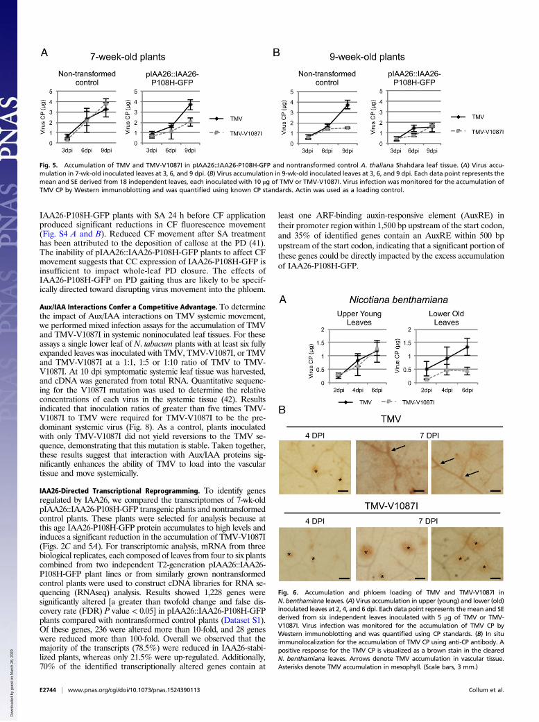
Stabilization of IAA26 Expressed from its Native Phloem PromoterInhibits TMV Accumulation. A. thaliana ecotype Shahdara linestransformed with pIAA26::IAA26-P108H-GFP were used to assessthe effect of IAA26 accumulation on TMV infection. We hypoth-esized that stabilization of IAA26 within the phloem CCs, resultingin greater accumulation of the protein before virus infection, wouldsignificantly impact the ability of both TMV and the noninteractingTMV-V1087I to accumulate in leaf tissues. The studies describedabove indicated that IAA26-P108H-GFP protein accumulation isconsistently detectable by 7 wk in transformed Shahdara (Fig. 2C).TMV and TMV-V1087I infections were subsequently monitored inboth 7- and 9-wk-old plants using two independent lines expressingIAA26-P108H-GFP from the native IAA26 promoter or in non-transformed control plants at 3, 6, and 9 dpi. Virus accumulationwas measured by Western immunoblot analysis using an antibodyspecific for the TMV CP. TMV and TMV-V1087I accumulateto similar levels in 7-wk-old nontransformed plants. Conversely,TMV-V1087I accumulated to significantly lower levels than TMV(P = 0.014, Student’s t test) in 7-wk-old pIAA26::IAA26-P108H-
GFP plants (Fig. 5A). Thus, stabilization and overaccumulation ofIAA26 in the younger, 7-wk-old tissues significantly impacts theability of this non–Aux/IAA-interacting virus to accumulate.In 9-wk-old nontransformed plants that have bolted and begun to
flower, TMV-V1087I accumulated to significantly lower levels thanTMV (P = 0.006, Student’s t test) (Fig. 5B). This result is consistentwith our previous findings that reduced accumulation of the non–Aux/IAA-interacting TMV-V1087I occurred only in older leaf tissues(21). However, in 9-wk-old pIAA26::IAA26-P108H-GFP plantsTMV accumulated at significantly lower levels than in non-transformed control plants (P = 0.046, Student’s t test), at a levelsimilar to the noninteracting TMV-V1087I (Fig. 5B). Together thesefindings indicate that the accumulation of IAA26-P108H-GFP beforeinfection creates a cellular environment that is not conducive to in-fection and cannot be overcome by the TMV–Aux/IAA interaction.
Aux/IAA Interaction Specifically Enhances TMV Phloem Loading andAccumulation. To identify the mechanism through which IAAsdisrupt TMV accumulation, we investigated the ability of bothinteracting TMV and noninteracting TMV-V1087I to spread withininoculated leaves. To do so, we first developed a tobacco system forthe in situ detection of TMV and tobacco IAA26. Previously weidentified TMV 126/183-kDa–interacting Aux/IAA genes in bothArabidopsis and tomato (20, 21). In this study we cloned the cor-responding Nicotiana benthamiana NbIAA26 and found it shares96% sequence identity and 97% similarity with Nicotiana tabacumNtIAA26 and 51% sequence identity and 64% similarity withA. thaliana AtIAA26 (Fig. S2). Similar to our previous studies withthe Arabidopsis and tomato IAA26 homologs, yeast two-hybridanalysis demonstrated the NbIAA26 protein interacts with theTMV 126/183-kDa helicase domain but not with the TMV-V1087Ihelicase domain (Fig. S3A). In addition, localization studies using atransiently expressed NbIAA26 fused to GFP demonstrated that, inthe presence of the interacting TMV, NbIAA26 relocalizes fromthe nucleus to the cytoplasm but is unaffected by infection with thenoninteracting TMV-V1087I (Fig. S3B). We also examined thecellular expression pattern of NbIAA26 mRNA using in situ hy-bridization. These experiments were performed on fixed stem tissueusing a digoxigenin-labeled probe specific for the NbIAA26 se-
Fig. 1. Histochemical analysis of pIAA26::GUS, pIAA27::GUS, and pIAA18::GUSexpression in A. thaliana Shahdara leaves, petioles, and stems. (A) GUS expres-sion in 7-wk-old leaves after staining overnight. (Scale bars, 2 mm.) (B) Petiolecross-sections. (Scale bars, 200 μm.) (C) Stem cross-sections. (Scale bars, 100 μm.)
Fig. 2. Characterization of pIAA26::IAA26-P108H-GFP transgenic plantlines. (A) Representative images of 7- and 9-wk-old nontransformed controlShahdara plants and pIAA26::IAA26-P108H-GFP transgenic Shahdara plants.(B) Relative mRNA expression of endogenous IAA26 mRNA in control plantsor IAA26-P108H-GFP mRNA expressed from the pIAA26::IAA26-P108H-GFPtransgene in 3- to 9-wk-old plants as measured by qRT-PCR. Total RNAwas isolated from the leaves of four plants from two independentpIAA26::IAA26-P108H-GFP plant lines or from nontransformed controls andwas pooled for each of three biological replicates. 18S RNA was used as aninternal control for normalization. (C) Western immunoblot detection ofIAA26-P108H-GFP protein levels in 3- to 9-wk-old pIAA26::IAA26-P108H-GFPtransgenic plants as probed with anti-GFP antibody. Actin levels were usedas a loading control.
E2742 | www.pnas.org/cgi/doi/10.1073/pnas.1524390113 Collum et al.
Dow
nloa
ded
by g
uest
on
Mar
ch 2
6, 2
020
quence. Results clearly indicate that NbIAA26 mRNA is expressedpredominantly in the vascular tissues of N. benthamiana stem sec-tions (Fig. S3C). Thus, as in Arabidopsis, TMV appears to targetsimilar vascular-expressed tobacco Aux/IAAs.To examine the effect of the NbIAA26 interaction on the cellular
movement of TMV, in situ hybridization was used to analyze theaccumulation of TMV and TMV-V1087I (non–IAA-interacting) ininoculated leaves of N. benthamiana. Previous protoplasts studiescomparing TMV and TMV-V1087I revealed no significant differ-ences in the replication of these viruses in single cells (38). However,as is consistent with our previous findings in Arabidopsis, the non-interacting TMV-V1087I virus accumulates to significantly lowerlevels (P = 0.004, Student’s t test) in mature tissues than those ob-served for TMV (Fig. 6A). For this study mature N. benthamianaleaves were inoculated with either TMV or TMV-V1087I, wereharvested at 4 and 7 dpi, and were subjected to in situ immuno-detection for the TMV CP. Results indicate that at 4 dpi both TMVand TMV-V1087I show defined infection foci that are similar in sizeand appearance (Fig. 6B). In contrast, at 7 dpi TMV shows signif-icant accumulation along the veins and vascular networks of the leaf,whereas TMV-V1087I shows continued cell-to-cell spread but lim-ited accumulation within the vascular networks. These findings in-dicate that a non–Aux/IAA-interacting virus is less able to load intothe vascular network.To investigate further the role of the interacting Aux/IAA proteins
on TMV phloem loading, we performed similar in situ immunode-
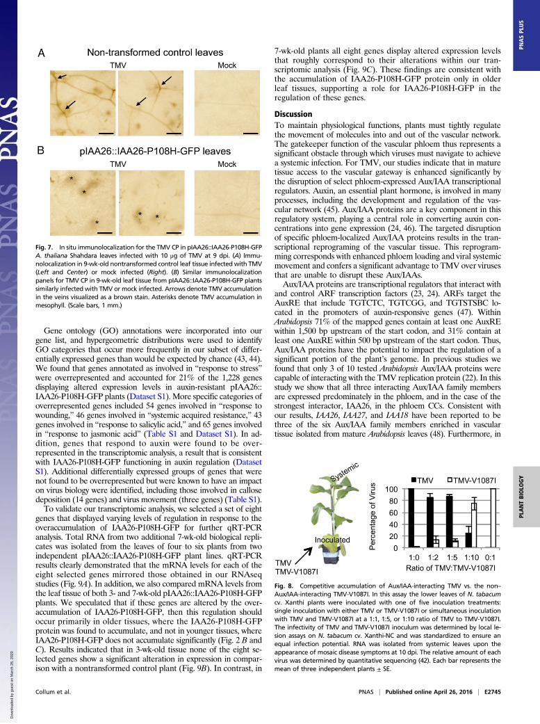
tection for TMV in transgenic A. thaliana Shahdara pIAA26::IAA26-P108H-GFP plants. We reasoned that age-related factorscombined with IAA26-P108H-GFP overaccumulation would pro-duce alterations in vascular traits that restrict TMV phloem loading.For these experiments leaves from 9-wk-old plants that had floweredwere harvested for analysis at 9 dpi. Results show significant accu-mulation of TMV within the vascular tissues of nontransformedcontrol plants (Fig. 7A). In contrast, in plants expressing IAA26-P108H-GFP, TMV produces detectable viral accumulation at initialinfection foci but little accumulation within the vascular tissues (Fig.7B). This finding confirms that accumulation of IAA26-P108H-GFPbefore infection leads to reduced vascular loading of TMV inflowering plants. These findings also are consistent with the observedreduced accumulation of TMV in the older 9-wk-old pIAA26::IAA26-P108H-GFP plants (Fig. 5B). Combined, these findingssuggest that within mature tissues additional factors such as ARRimpact TMV phloem loading.To address the specificity of observed phloem-loading restrictions,
we used a fluorescent dye-based cell-to-cell movement and phloem-loading assay. Several studies have demonstrated that changes in PDsize-exclusion limit and vascular movement can be monitored using5(6)-carboxyfluorescein diacetate (CFDA), a membrane-permeablenonfluorescent dye that upon cell entry is cleaved by esterases toproduce 5(6)-carboxyfluorescein (CF), a non–membrane-permeablefluorescent dye (39–41). In these experiments we compared CFmobility in 9-wk-old nontransformed control and pIAA26::IAA26-P108H-GFP plants. CF mobility was found to be similar in pIAA26::IAA26-P108H-GFP and the control plants (Fig. S4). Consis-tent with previous studies, treatment of control or pIAA26::
Fig. 3. Localization of IAA26-P108H-GFP expressed from the IAA26 nativepromoter in A. thaliana Shahdara petioles. (A) Representative fluorescent im-ages of cells expressing IAA26-P108H-GFP from the IAA26 native promoter ornontransformed controls. Petioles from 9- to 11-wk-old T2-generation plantswere hand sectioned for imaging. Green, GFP; blue, DAPI. (Scale bars, 100 μm.)(B) Localization of IAA26-P108H-GFP in CC nuclei. PC, phloem parenchyma cells.
Fig. 4. Localization of IAA26-P108H-GFP in virus-infected tissue. (A) Rep-resentative fluorescent images of cells expressing IAA26-P108H-GFP in mock-infected, TMV-infected, or TMV-V1087I–infected phloem tissue. Inoculatedleaf petioles were hand sectioned and imaged 12 dpi. Green, GFP; blue,DAPI. (Scale bars, 100 μm.) N, nucleus. (B) Western immunoblot for the de-tection of the TMV CP present in the investigated cross-sections of pIAA26::IAA26-P108H-GFP plants at 12 dpi using actin levels as a loading control.(C) Quantification of cell nuclei with GFP fluorescence reported as the meanof 10 petiole cross-sections ± SE.
Collum et al. PNAS | Published online April 26, 2016 | E2743
PLANTBIOLO
GY
PNASPL
US
Dow
nloa
ded
by g
uest
on
Mar
ch 2
6, 2
020
IAA26-P108H-GFP plants with SA 24 h before CF applicationproduced significant reductions in CF fluorescence movement(Fig. S4 A and B). Reduced CF movement after SA treatmenthas been attributed to the deposition of callose at the PD (41).The inability of pIAA26::IAA26-P108H-GFP plants to affect CFmovement suggests that CC expression of IAA26-P108H-GFP isinsufficient to impact whole-leaf PD closure. The effects ofIAA26-P108H-GFP on PD gaiting thus are likely to be specif-ically directed toward disrupting virus movement into the phloem.
Aux/IAA Interactions Confer a Competitive Advantage. To determinethe impact of Aux/IAA interactions on TMV systemic movement,we performed mixed infection assays for the accumulation of TMVand TMV-V1087I in systemic noninoculated leaf tissues. For theseassays a single lower leaf of N. tabacum plants with at least six fullyexpanded leaves was inoculated with TMV, TMV-V1087I, or TMVand TMV-V1087I at a 1:1, 1:5 or 1:10 ratio of TMV to TMV-V1087I. At 10 dpi symptomatic systemic leaf tissue was harvested,and cDNA was generated from total RNA. Quantitative sequenc-ing for the V1087I mutation was used to determine the relativeconcentrations of each virus in the systemic tissue (42). Resultsindicated that inoculation ratios of greater than five times TMV-V1087I to TMV were required for TMV-V1087I to be the pre-dominant systemic virus (Fig. 8). As a control, plants inoculatedwith only TMV-V1087I did not yield reversions to the TMV se-quence, demonstrating that this mutation is stable. Taken together,these results suggest that interaction with Aux/IAA proteins sig-nificantly enhances the ability of TMV to load into the vasculartissue and move systemically.
IAA26-Directed Transcriptional Reprogramming. To identify genesregulated by IAA26, we compared the transcriptomes of 7-wk-oldpIAA26::IAA26-P108H-GFP transgenic plants and nontransformedcontrol plants. These plants were selected for analysis because atthis age IAA26-P108H-GFP protein accumulates to high levels andinduces a significant reduction in the accumulation of TMV-V1087I(Figs. 2C and 5A). For transcriptomic analysis, mRNA from threebiological replicates, each composed of leaves from four to six plantscombined from two independent T2-generation pIAA26::IAA26-P108H-GFP plant lines or from similarly grown nontransformedcontrol plants were used to construct cDNA libraries for RNA se-quencing (RNAseq) analysis. Results showed 1,228 genes weresignificantly altered [a greater than twofold change and false dis-covery rate (FDR) P value < 0.05] in pIAA26::IAA26-P108H-GFPplants compared with nontransformed control plants (Dataset S1).Of these genes, 236 were altered more than 10-fold, and 28 geneswere reduced more than 100-fold. Overall we observed that themajority of the transcripts (78.5%) were reduced in IAA26-stabi-lized plants, whereas only 21.5% were up-regulated. Additionally,70% of the identified transcriptionally altered genes contain at
least one ARF-binding auxin-responsive element (AuxRE) intheir promoter region within 1,500 bp upstream of the start codon,and 35% of identified genes contain an AuxRE within 500 bpupstream of the start codon, indicating that a significant portion ofthese genes could be directly impacted by the excess accumulationof IAA26-P108H-GFP.
Fig. 5. Accumulation of TMV and TMV-V1087I in pIAA26::IAA26-P108H-GFP and nontransformed control A. thaliana Shahdara leaf tissue. (A) Virus accu-mulation in 7-wk-old inoculated leaves at 3, 6, and 9 dpi. (B) Virus accumulation in 9-wk-old inoculated leaves at 3, 6, and 9 dpi. Each data point represents themean and SE derived from 18 independent leaves, each inoculated with 10 μg of TMV or TMV-V1087I. Virus infection was monitored for the accumulation ofTMV CP by Western immunoblotting and was quantified using known CP standards. Actin was used as a loading control.
Fig. 6. Accumulation and phloem loading of TMV and TMV-V1087I inN. benthamiana leaves. (A) Virus accumulation in upper (young) and lower (old)inoculated leaves at 2, 4, and 6 dpi. Each data point represents the mean and SEderived from six independent leaves inoculated with 5 μg of TMV or TMV-V1087I. Virus infection was monitored for the accumulation of TMV CP byWestern immunoblotting and was quantified using CP standards. (B) In situimmunolocalization for the accumulation of TMV CP using anti-CP antibody. Apositive response for the TMV CP is visualized as a brown stain in the clearedN. benthamiana leaves. Arrows denote TMV accumulation in vascular tissue.Asterisks denote TMV accumulation in mesophyll. (Scale bars, 3 mm.)
E2744 | www.pnas.org/cgi/doi/10.1073/pnas.1524390113 Collum et al.
Dow
nloa
ded
by g
uest
on
Mar
ch 2
6, 2
020
Gene ontology (GO) annotations were incorporated into ourgene list, and hypergeometric distributions were used to identifyGO categories that occur more frequently in our subset of differ-entially expressed genes than would be expected by chance (43, 44).We found that genes annotated as involved in “response to stress”were overrepresented and accounted for 21% of the 1,228 genesdisplaying altered expression levels in auxin-resistant pIAA26::IAA26-P108H-GFP plants (Dataset S1). More specific categories ofoverrepresented genes included 54 genes involved in “response towounding,” 46 genes involved in “systemic acquired resistance,” 43genes involved in “response to salicylic acid,” and 65 genes involvedin “response to jasmonic acid” (Table S1 and Dataset S1). In ad-dition, genes that respond to auxin were found to be over-represented in the transcriptomic analysis, a result that is consistentwith IAA26-P108H-GFP functioning in auxin regulation (DatasetS1). Additional differentially expressed groups of genes that werenot found to be overrepresented but were known to have an impacton virus biology were identified, including those involved in callosedeposition (14 genes) and virus movement (three genes) (Table S1).To validate our transcriptomic analysis, we selected a set of eight
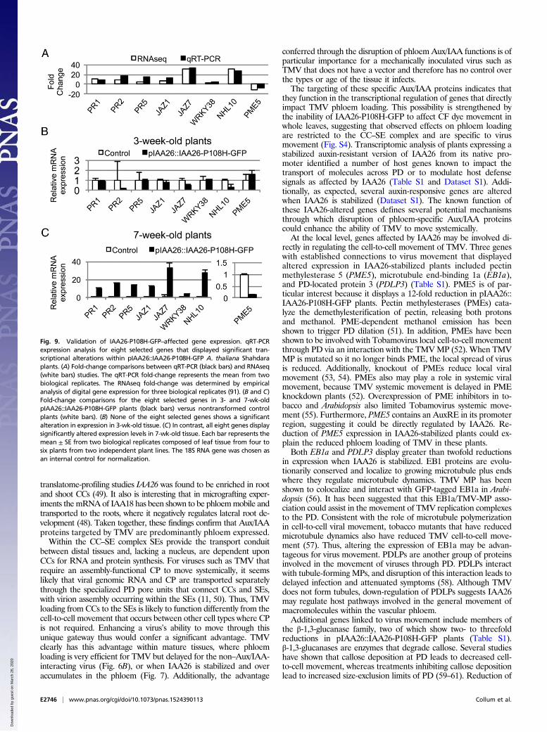
genes that displayed varying levels of regulation in response to theoveraccumulation of IAA26-P108H-GFP for further qRT-PCRanalysis. Total RNA from two additional 7-wk-old biological repli-cates was isolated from the leaves of four to six plants from twoindependent pIAA26::IAA26-P108H-GFP plant lines. qRT-PCRresults clearly demonstrated that the mRNA levels for each of theeight selected genes mirrored those obtained in our RNAseqstudies (Fig. 9A). In addition, we also compared mRNA levels fromthe leaf tissue of both 3- and 7-wk-old pIAA26::IAA26-P108H-GFPplants. We speculated that if these genes are altered by the over-accumulation of IAA26-P108H-GFP, then this regulation shouldoccur primarily in older tissues, where the IAA26-P108H-GFPprotein was found to accumulate, and not in younger tissues, whereIAA26-P108H-GFP does not accumulate significantly (Fig. 2 B andC). Results indicated that in 3-wk-old tissue none of the eight se-lected genes show a significant alteration in expression in compar-ison with a nontransformed control plant (Fig. 9B). In contrast, in
7-wk-old plants all eight genes display altered expression levelsthat roughly correspond to their alterations within our tran-scriptomic analysis (Fig. 9C). These findings are consistent withthe accumulation of IAA26-P108H-GFP protein only in olderleaf tissues, supporting a role for IAA26-P108H-GFP in theregulation of these genes.
DiscussionTo maintain physiological functions, plants must tightly regulatethe movement of molecules into and out of the vascular network.The gatekeeper function of the vascular phloem thus represents asignificant obstacle through which viruses must navigate to achievea systemic infection. For TMV, our studies indicate that in maturetissue access to the vascular gateway is enhanced significantly bythe disruption of select phloem-expressed Aux/IAA transcriptionalregulators. Auxin, an essential plant hormone, is involved in manyprocesses, including the development and regulation of the vas-cular network (45). Aux/IAA proteins are a key component in thisregulatory system, playing a central role in converting auxin con-centrations into gene expression (24, 46). The targeted disruptionof specific phloem-localized Aux/IAA proteins results in the tran-scriptional reprograming of the vascular tissue. This reprogram-ming corresponds with enhanced phloem loading and viral systemicmovement and confers a significant advantage to TMV over virusesthat are unable to disrupt these Aux/IAAs.Aux/IAA proteins are transcriptional regulators that interact with
and control ARF transcription factors (23, 24). ARFs target theAuxRE that include TGTCTC, TGTCGG, and TGTSTSBC lo-cated in the promoters of auxin-responsive genes (47). WithinArabidopsis 71% of the mapped genes contain at least one AuxREwithin 1,500 bp upstream of the start codon, and 31% contain atleast one AuxRE within 500 bp upstream of the start codon. Thus,Aux/IAA proteins have the potential to impact the regulation of asignificant portion of the plant’s genome. In previous studies wefound that only 3 of 10 tested Arabidopsis Aux/IAA proteins werecapable of interacting with the TMV replication protein (22). In thisstudy we show that all three interacting Aux/IAA family membersare expressed predominately in the phloem, and in the case of thestrongest interactor, IAA26, in the phloem CCs. Consistent withour results, IAA26, IAA27, and IAA18 have been reported to bethree of the six Aux/IAA family members enriched in vasculartissue isolated from mature Arabidopsis leaves (48). Furthermore, in
Fig. 8. Competitive accumulation of Aux/IAA-interacting TMV vs. the non–Aux/IAA-interacting TMV-V1087I. In this assay the lower leaves of N. tabacumcv. Xanthi plants were inoculated with one of five inoculation treatments:single inoculation with either TMV or TMV-V1087I or simultaneous inoculationwith TMV and TMV-V1087I at a 1:1, 1:5, or 1:10 ratio of TMV to TMV-V1087I.The infectivity of TMV and TMV-V1087I inoculum was determined by local le-sion assays on N. tabacum cv. Xanthi-NC and was standardized to ensure anequal infection potential. RNA was isolated from systemic leaves upon theappearance of mosaic disease symptoms at 10 dpi. The relative amount of eachvirus was determined by quantitative sequencing (42). Each bar represents themean of three independent plants ± SE.
Fig. 7. In situ immunolocalization for the TMV CP in pIAA26::IAA26-P108H-GFPA. thaliana Shahdara leaves infected with 10 μg of TMV at 9 dpi. (A) Immu-nolocalization in 9-wk-old nontransformed control leaf tissue infected with TMV(Left and Center) or mock infected (Right). (B) Similar immunolocalizationpanels for TMV CP in 9-wk-old leaf tissue from pIAA26::IAA26-P108H-GFP plantssimilarly infected with TMV or mock infected. Arrows denote TMV accumulationin the veins visualized as a brown stain. Asterisks denote TMV accumulation inmesophyll. (Scale bars, 1 mm.)
Collum et al. PNAS | Published online April 26, 2016 | E2745
PLANTBIOLO
GY
PNASPL
US
Dow
nloa
ded
by g
uest
on
Mar
ch 2
6, 2
020
translatome-profiling studies IAA26 was found to be enriched in rootand shoot CCs (49). It also is interesting that in micrografting exper-iments themRNAof IAA18 has been shown to be phloemmobile andtransported to the roots, where it negatively regulates lateral root de-velopment (48). Taken together, these findings confirm that Aux/IAAproteins targeted by TMV are predominantly phloem expressed.Within the CC–SE complex SEs provide the transport conduit
between distal tissues and, lacking a nucleus, are dependent uponCCs for RNA and protein synthesis. For viruses such as TMV thatrequire an assembly-functional CP to move systemically, it seemslikely that viral genomic RNA and CP are transported separatelythrough the specialized PD pore units that connect CCs and SEs,with virion assembly occurring within the SEs (11, 50). Thus, TMVloading from CCs to the SEs is likely to function differently from thecell-to-cell movement that occurs between other cell types where CPis not required. Enhancing a virus’s ability to move through thisunique gateway thus would confer a significant advantage. TMVclearly has this advantage within mature tissues, where phloemloading is very efficient for TMV but delayed for the non–Aux/IAA-interacting virus (Fig. 6B), or when IAA26 is stabilized and overaccumulates in the phloem (Fig. 7). Additionally, the advantage
conferred through the disruption of phloem Aux/IAA functions is ofparticular importance for a mechanically inoculated virus such asTMV that does not have a vector and therefore has no control overthe types or age of the tissue it infects.The targeting of these specific Aux/IAA proteins indicates that
they function in the transcriptional regulation of genes that directlyimpact TMV phloem loading. This possibility is strengthened bythe inability of IAA26-P108H-GFP to affect CF dye movement inwhole leaves, suggesting that observed effects on phloem loadingare restricted to the CC–SE complex and are specific to virusmovement (Fig. S4). Transcriptomic analysis of plants expressing astabilized auxin-resistant version of IAA26 from its native pro-moter identified a number of host genes known to impact thetransport of molecules across PD or to modulate host defensesignals as affected by IAA26 (Table S1 and Dataset S1). Addi-tionally, as expected, several auxin-responsive genes are alteredwhen IAA26 is stabilized (Dataset S1). The known function ofthese IAA26-altered genes defines several potential mechanismsthrough which disruption of phloem-specific Aux/IAA proteinscould enhance the ability of TMV to move systemically.At the local level, genes affected by IAA26 may be involved di-
rectly in regulating the cell-to-cell movement of TMV. Three geneswith established connections to virus movement that displayedaltered expression in IAA26-stabilized plants included pectinmethylesterase 5 (PME5), microtubule end-binding 1a (EB1a),and PD-located protein 3 (PDLP3) (Table S1). PME5 is of par-ticular interest because it displays a 12-fold reduction in pIAA26::IAA26-P108H-GFP plants. Pectin methylesterases (PMEs) cata-lyze the demethylesterification of pectin, releasing both protonsand methanol. PME-dependent methanol emission has beenshown to trigger PD dilation (51). In addition, PMEs have beenshown to be involved with Tobamovirus local cell-to-cell movementthrough PD via an interaction with the TMVMP (52). When TMVMP is mutated so it no longer binds PME, the local spread of virusis reduced. Additionally, knockout of PMEs reduce local viralmovement (53, 54). PMEs also may play a role in systemic viralmovement, because TMV systemic movement is delayed in PMEknockdown plants (52). Overexpression of PME inhibitors in to-bacco and Arabidopsis also limited Tobamovirus systemic move-ment (55). Furthermore, PME5 contains an AuxRE in its promoterregion, suggesting it could be directly regulated by IAA26. Re-duction of PME5 expression in IAA26-stabilized plants could ex-plain the reduced phloem loading of TMV in these plants.Both EB1a and PDLP3 display greater than twofold reductions
in expression when IAA26 is stabilized. EB1 proteins are evolu-tionarily conserved and localize to growing microtubule plus endswhere they regulate microtubule dynamics. TMV MP has beenshown to colocalize and interact with GFP-tagged EB1a in Arabi-dopsis (56). It has been suggested that this EB1a/TMV-MP asso-ciation could assist in the movement of TMV replication complexesto the PD. Consistent with the role of microtubule polymerizationin cell-to-cell viral movement, tobacco mutants that have reducedmicrotubule dynamics also have reduced TMV cell-to-cell move-ment (57). Thus, altering the expression of EB1a may be advan-tageous for virus movement. PDLPs are another group of proteinsinvolved in the movement of viruses through PD. PDLPs interactwith tubule-forming MPs, and disruption of this interaction leads todelayed infection and attenuated symptoms (58). Although TMVdoes not form tubules, down-regulation of PDLPs suggests IAA26may regulate host pathways involved in the general movement ofmacromolecules within the vascular phloem.Additional genes linked to virus movement include members of
the β-1,3-glucanase family, two of which show two- to threefoldreductions in pIAA26::IAA26-P108H-GFP plants (Table S1).β-1,3-glucanases are enzymes that degrade callose. Several studieshave shown that callose deposition at PD leads to decreased cell-to-cell movement, whereas treatments inhibiting callose depositionlead to increased size-exclusion limits of PD (59–61). Reduction of
Fig. 9. Validation of IAA26-P108H-GFP–affected gene expression. qRT-PCRexpression analysis for eight selected genes that displayed significant tran-scriptional alterations within pIAA26::IAA26-P108H-GFP A. thaliana Shahdaraplants. (A) Fold-change comparisons between qRT-PCR (black bars) and RNAseq(white bars) studies. The qRT-PCR fold-change represents the mean from twobiological replicates. The RNAseq fold-change was determined by empiricalanalysis of digital gene expression for three biological replicates (91). (B and C)Fold-change comparisons for the eight selected genes in 3- and 7-wk-oldpIAA26::IAA26-P108H-GFP plants (black bars) versus nontransformed controlplants (white bars). (B) None of the eight selected genes shows a significantalteration in expression in 3-wk-old tissue. (C) In contrast, all eight genes displaysignificantly altered expression levels in 7-wk-old tissue. Each bar represents themean ± SE from two biological replicates composed of leaf tissue from four tosix plants from two independent plant lines. The 18S RNA gene was chosen asan internal control for normalization.
E2746 | www.pnas.org/cgi/doi/10.1073/pnas.1524390113 Collum et al.
Dow
nloa
ded
by g
uest
on
Mar
ch 2
6, 2
020
β-1,3-glucanases could lead to the accumulation of callose at PDand decreased cell-to-cell movement when IAA26 accumulates.Additionally, two PD callose-binding proteins, PDCB2 and PDCB3,were significantly down-regulated in pIAA26::IAA26-P108H-GFPplants, by 6.4- and 2.2-fold, respectively (Table S1). PDCBs specifi-cally bind to callose in vitro and have been proposed to be involvedin callose-mediated regulation within the PD (62). Taken together,the observed down-regulation of genes that positively impact virusmovement suggests that IAA26 functions to regulate these genesnegatively.Host defense pathways, including SA and jasmonic acid (JA),
have been shown to be key factors in the development of systemicresistance against TMV (63–65). Transcriptomic analysis ofpIAA26::IAA26-P108H-GFP plants showed marked changes ingenes associated with these defense pathways. Of particular interestwere genes related to SA defense responses, including the SA de-fense markers pathogenesis-related (PR) genes PR1, PR2, and PR5,which are up-regulated 10.5-, 8.4-, and 3.8-fold, respectively, uponIAA26 stabilization in the phloem (Table S1) (66). The up-regula-tion of SA-mediated defense genes is consistent with previouslypublished results showing that, when the AUX/IAA protein AXR2/IAA7 is stabilized, AvrRpt2, a Pseudomonas syringae effector, is nolonger able to suppress the induction of SA-mediated defense genes(67). Additional genes involved in SA biosynthesis, includingANAC019 and ANAC072, and genes involved in SA-mediated sig-naling also were up-regulated in pIAA26::IAA26-P108H-GFP plants(Table S1 and Dataset S1). Thus, Aux/IAA proteins appear to playimportant roles in the regulation of SA-mediated defense response.We also observed transcriptional alterations in several key genes
associated with cross-communication between the SA and JA de-fense pathways. In particular, we identified nine WRKY transcrip-tion factors that were altered in IAA26-stabilized plants (Table S1).WRKY transcription factors function as regulators between the SAand JA defense pathways (68). Five WRKY family members wereup-regulated (WRKY30, -38, -51, -55, and -58), and four were down-regulated (WRKY12, -14, -35, and -44). Interestingly, the five up-regulated WRKYs have all been shown previously to be induced inresponse to SA treatment, whereas the four that were down-regu-lated were all undetectable (WRKY12, -14, and -35) or repressed(WRKY44) by SA treatment (69). WRKY38 expression has beenlinked to the degradation of NPR1 and in combination withWRKY62 is required for systemic acquired resistance (70), andWRKY51 has been shown to mediate SA-dependent repression ofJA signaling (71). Another important regulator affecting the an-tagonism between SA- and JA-mediated signaling is GRX480,which showed a sixfold increase in IAA26-P108H-GFP plants (72).Additionally, seven JASMONATE ZIM-DOMAIN (JAZ) re-pressor proteins were up-regulated (Table S1). JAZ proteins neg-atively regulate the JA-signaling pathway by repressing transcriptionfactors that control JA-regulated genes (73). It has been proposedthat stabilization of JAZ proteins may be one way that SA exerts anantagonistic effect on JA signaling (74). Taken together, these re-sults indicate that IAA26 likely functions as an important regulatorof these host defense pathways within the phloem of mature tissues.There is growing evidence for auxin involvement in disease re-
sistance through cross-talk with the SA and JA signaling pathways.Treatment of Arabidopsis with the SA analog benzothiadiazoleS-methylester (BTH) resulted in an overall reduction of auxin re-sponses (75). One mechanism by which SA might inhibit auxinsignaling is through transcriptional repression of the auxin-receptorgenes, leading to reduced degradation of Aux/IAA proteins andthus to the repression of auxin responses. Support for this mecha-nism comes from observations that auxin-receptor genes are down-regulated in response to BTH treatment and that detection of Aux/IAA proteins by Western blot is increased after SA treatment (75).Conversely, activation of auxin signaling leads to the suppression ofSA biosynthesis and signaling (76). It also has been reported thatauxin triggers the induction of genes involved in JA biosynthesis
(77). Consistent with these findings are studies that have linked thestabilization of Aux/IAA proteins to bacterial resistance. For ex-ample, when axr2-1 plants, which produce a nondegradable form ofAXR2/IAA7, were infected with the bacterial pathogen P. syringaepv. maculicola, there was a 10-fold reduction in bacterial growth(75). Consistent with these findings, our results also demonstrate arole for auxin as a negative regulator of the SA pathway, perhapsvia activation of JA signaling.Interestingly, the biological advantage of the TMV–Aux/IAA
interaction was observed only in older plant tissues (Figs. 5 and 6).In particular, inhibition of TMV phloem loading occurred in 9-wk-old and not in 7-wk-old pIAA26::IAA26-P108H-GFP plants, eventhough both developmental stages expressed and accumulatedsimilar levels of IAA26-P108H-GFP mRNA and protein (Fig. 2 Band C). This finding suggests that additional factors can impact theability of IAA26 to affect TMV phloem loading. Although no ob-vious phenotypic differences were observed between pIAA26::IAA26-P108H-GFP plants and nontransformed control plants, wedid find that bolting and the initiation of flowering occurred be-tween week 7 and week 9. This finding is of interest, becauseprevious studies have noted that the development of ARR, definedas the development of resistance in mature tissues, is often asso-ciated with transition to flowering (35, 78). Within Arabidopsis,ARR has been shown to impact the virulence of P. syringae and thesystemic movement of cauliflower mosaic virus (79, 80). In bothsystems, mutations in genes associated with the induction andmaintenance of flowering were shown to influence the developmentof this resistance. Resistance against P. syringae has been linked tothe accumulation of SA (81). To examine ARR in pIAA26::IAA26-P108H-GFP plants, we determined whether the expression ofSA-associated genes changed between week 7 and week 9. Resultsindicated that PR1, PR2 and PR5 expression is greater in pIAA26::IAA26-P108H-GFP plants than in nontransformed controls atboth week 7 and week 9. In addition, PR1 and PR2, but not PR5,show significantly increased expression in flowering 9-wk-old plants(Fig. S5). These findings indicated that the developmental age ofthe plant significantly influences the effect of IAA26-P108H-GFPaccumulation on the expression of these SA-associated genes. Al-though additional studies are needed, these findings suggest thatIAA26 is a factor in the regulation of ARR.In summary, IAA26 plays an important role in regulating the
phloem environment within mature plant tissues and appears to bea contributor in ARR. The diverse array of genes impacted throughthe stabilization of IAA26 suggests that this auxin-regulated proteinhas a significant role in modulating both the local environment ofthe CC–SE complex and surrounding and distal tissues. The reg-ulation of genes that impact PD function and the inability of TMVto move into the phloem in older IAA26-stabilized plants suggestthat this protein regulates the transport of macromolecules withinthe phloem. Impacts on SA and JA defense pathways provide an-other mechanism whereby IAA26 could produce wider cellularresponses both in tissues surrounding the vascular phloem andsystemically throughout the plant, providing a means for TMV tomodulate responses such as ARR and systemic acquired resistance.Additional studies directed at understanding the precise impact ofthis TMV-directed vascular reprogramming should provide greaterdetails about the role these mechanisms play in promoting virusphloem loading and systemic movement.
Experimental ProceduresPromoter Constructs and GUS Assays. Promoter fragments of 2 kb for IAA26 andIAA18 and 1.5 kb for IAA27 were amplified from genomic DNA extracted fromA. thaliana ecotype Shahdara using promoter-specific primers (Table S2). Clonedpromoter fragments were moved into pBI101.1 (Clontech) upstream of the GUSreporter ORF via primer-generated restriction sites. Promoter pIAA::GUS con-structs were introduced into the Agrobacterium tumefaciens strain GV3101(82), and a floral dip method was used for plant transformation (83). All plantswere maintained in growth chambers for a 12-h photoperiod at 24 °C.
Collum et al. PNAS | Published online April 26, 2016 | E2747
PLANTBIOLO
GY
PNASPL
US
Dow
nloa
ded
by g
uest
on
Mar
ch 2
6, 2
020
Histochemical staining for GUS activity was performed as previously described(84). After staining, whole leaves or hand-sectioned petioles or stems wereimaged using an Olympus Stereo MVX10 or BX60 Microscope.
pIAA26::IAA26-P108H-GFP Construction and Characterization. To create pIAA26::IAA26-P108H-GFP, the ORF of our previously described 35S::IAA26-P108H-GFP(22) construct was removed by BamHI and SacI digestion and ligated into asimilarly cut pIAA26::GUS (described above), replacing the GUS ORF with that ofIAA26-P108H-GFP. A. thaliana ecotype Shahdara plants were transformed withpIAA26::IAA26-P108H-GFP as described above (85). Plants were maintained asdescribed above, and 3- to 9-wk-old T2-generation plants from two independenttransgenic lines were analyzed for IAA26-P108H-GFP mRNA and protein ex-pression as described in SI Experimental Procedures. GFP fluorescence localiza-tion for IAA26-P108H-GFP was done by hand-sectioning petioles. Sectionedtissues were stained with 1 μg/mL DAPI and were imaged using a Zeiss LSM700lase- scanning confocal microscope (Carl Zeiss, Inc.).
NbIAA26 Cloning and Characterization. Total leaf RNA from N. benthamianaplants was used to amplify the NbIAA26 ORF as described in SI ExperimentalProcedures. Yeast two-hybrid and transient NbIAA26-GFP localization studieswere performed as previously described (20–22). NbIAA26 in situ hybridizationswere done following previously published methods (86). Details for this pro-tocol can be found in the SI Experimental Procedures.
Virus Accumulation, in Situ Localizations, and Systemic Movement Assays.Seven- and nine-week-old Shahdara control and transgenic pIAA26::IAA26-P108H-GFP plantswere used for virus accumulation studies. Rosette leavesweredusted with carborundum and mechanically inoculated with 10 μg of purifiedTMV or TMV-V1087I. Leaf punches were collected from 18 independently in-oculated rosette leaves at 3, 6, and 9 dpi. For studies in N. benthamiana, plantswere inoculated with 5 μg of TMV or TMV-V1087I, and leaf punches werecollected from six inoculated leaves at 2, 4, and 6 dpi. All samples were groundin Laemmli sample buffer (87) and analyzed by Western immunoblot analysisusing TMV-specific antibodies as previously reported (21, 37). Whole-leaf insitu localizations for the detection of the TMV CP were done as previouslydescribed (88).
For systemic movement assays lower leaves of N. tabacum cv. Xanthi plantswere inoculated with 10 ng of virus. Three plants were used for each in-oculation treatment. Plants were randomly assigned to one of five inoculationtreatments: single inoculation with either TMV or TMV-V1087I or simultaneousinoculation with TMV and TMV-V1087I at a 1:1, 1:5, or 1:10 ratio of TMV toTMV-V1087I. The infectivity of TMV and TMV-V1087I inoculum was determinedby local lesion assays on N. tabacum cv. Xanthi-NC and was standardized toensure an equal infection potential. RNA was isolated from systemic leaves at10 dpi, when mosaic disease symptoms were first observed, and cDNA wasprepared as described above. Quantitative sequencing for the detection andquantification of the G-to-A TMV-V1087I point mutation was done as describedin ref. 42. See SI Experimental Procedures for details.
Transcriptomic Analysis and Validation. Total RNA was isolated from 7-wk-oldpIAA26::IAA26-P108H-GFP transgenic plants and nontransformed control Shah-dara plants using the RNeasy kit (Qiagen). Four fully expanded leaves werecollected fromeach test plant, and total RNA fromfour to six individual test plantswaspooled for eachof the threebiological replicates. RNAseq library constructionand sequencing was done by the University of Maryland Institute for Bioscienceand Biotechnology Research (UM-IBBR) Sequencing Core. RNAseq analysis wasdone using CLC Bio genomics workbench as described in SI Experimental Pro-cedures (89, 90). The RNAseq data have been deposited in NCBI’s Gene Ex-pression Omnibus (GEO, www.ncbi.nlm.nih.gov/geo) and are accessible throughGEO series accession number GSE75983.
To validate RNAseq findings, total RNA from 3- or 7-wk-old pIAA26::IAA26-P108H-GFP plants or nontransformed control plants was isolated and used togenerate cDNA. qRT-PCR was performed as described in SI Experimental Pro-cedures for two additional biological replicates, each containing three technicalreplicates. Primer sequences used for the amplification of selected genes areprovided in Table S2. The 18S RNA gene was chosen as an internal controlfor normalization.
ACKNOWLEDGMENTS. This work was supported in part by US Department ofAgriculture National Research Initiative Competitive Grant 2008-35319-19168and National Science Foundation Grant ISO-1120044. T.D.C. was additionallysupported by NIH Institutional Training Grant 5T32AI051967 awarded to theUniversity of Maryland.
1. Heinlein M (2015) Plasmodesmata: Channels for viruses on the move. Methods Mol
Biol 1217:25–52.2. Solovyev AG, Savenkov EI (2014) Factors involved in the systemic transport of plant
RNA viruses: The emerging role of the nucleus. J Exp Bot 65(7):1689–1697.3. Stadler R, et al. (2005) Expression of GFP-fusions in Arabidopsis companion cells re-
veals non-specific protein trafficking into sieve elements and identifies a novel post-
phloem domain in roots. Plant J 41(2):319–331.4. Leisner SM, Turgeon R (1993) Movement of virus and photoassimilate in the phloem:
A comparative analysis. BioEssays 15(11):741–748.5. Ham BK, Lucas WJ (2014) The angiosperm phloem sieve tube system: A role in me-
diating traits important to modern agriculture. J Exp Bot 65(7):1799–1816.6. Kehr J, Buhtz A (2008) Long distance transport and movement of RNA through the
phloem. J Exp Bot 59(1):85–92.7. Turgeon R, Wolf S (2009) Phloem transport: Cellular pathways and molecular traf-
ficking. Annu Rev Plant Biol 60:207–221.8. Harries P, Ding B (2011) Cellular factors in plant virus movement: At the leading edge
of macromolecular trafficking in plants. Virology 411(2):237–243.9. Chisholm ST, Parra MA, Anderberg RJ, Carrington JC (2001) Arabidopsis RTM1 and
RTM2 genes function in phloem to restrict long-distance movement of tobacco etch
virus. Plant Physiol 127(4):1667–1675.10. Liu C, Nelson RS (2013) The cell biology of Tobacco mosaic virus replication and
movement. Front Plant Sci 4:12.11. Ding X, Shintaku MH, Carter SA, Nelson RS (1996) Invasion of minor veins of tobacco
leaves inoculated with tobacco mosaic virus mutants defective in phloem-dependent
movement. Proc Natl Acad Sci USA 93(20):11155–11160.12. Culver JN, Dawson WO (1989) Tobacco mosaic virus coat protein: An elicitor of the
hypersensitive reaction but not required for the development of mosaic symptoms in
Nicotiana sylvestris. Virology 173(2):755–758.13. Dawson WO, Bubrick P, Grantham GL (1988) Modifications of the Tobacco Mosaic-
Virus Coat Protein Gene Affecting Replication, Movement, and Symptomatology.
Phytopathology 78(6):783–789.14. Hilf ME, Dawson WO (1993) The tobamovirus capsid protein functions as a host-
specific determinant of long-distance movement. Virology 193(1):106–114.15. Fenczik CA, Padgett HS, Holt CA, Casper SJ, Beachy RN (1995) Mutational analysis of
the movement protein of odontoglossum ringspot virus to identify a host-range
determinant. Mol Plant Microbe Interact 8(5):666–673.16. Deom CM, Quan S, He XZ (1997) Replicase proteins as determinants of phloem-
dependent long-distance movement of tobamoviruses in tobacco. Protoplasma
199(1-2):1–8.
17. Sasaki N, et al. (2009) Over-expression of putative transcriptional coactivator KELP
interferes with Tomato mosaic virus cell-to-cell movement. Mol Plant Pathol 10(2):161–173.
18. Chen L, Zhang L, Li D, Wang F, Yu D (2013) WRKY8 transcription factor functions inthe TMV-cg defense response by mediating both abscisic acid and ethylene signalingin Arabidopsis. Proc Natl Acad Sci USA 110(21):E1963–E1971.
19. Levy A, Zheng JY, Lazarowitz SG (2013) The tobamovirus Turnip Vein Clearing Virus30-kilodalton movement protein localizes to novel nuclear filaments to enhance virus
infection. J Virol 87(11):6428–6440.20. Padmanabhan MS, Goregaoker SP, Golem S, Shiferaw H, Culver JN (2005) Interaction
of the tobacco mosaic virus replicase protein with the Aux/IAA protein PAP1/IAA26 isassociated with disease development. J Virol 79(4):2549–2558.
21. Padmanabhan MS, Kramer SR, Wang X, Culver JN (2008) Tobacco mosaic virus rep-licase-auxin/indole acetic acid protein interactions: Reprogramming the auxin re-sponse pathway to enhance virus infection. J Virol 82(5):2477–2485.
22. Padmanabhan MS, Shiferaw H, Culver JN (2006) The Tobacco mosaic virus replicaseprotein disrupts the localization and function of interacting Aux/IAA proteins. Mol
Plant Microbe Interact 19(8):864–873.23. Calderon-Villalobos LI, Tan X, Zheng N, Estelle M (2010) Auxin perception–structural
insights. Cold Spring Harb Perspect Biol 2(7):a005546.24. Salehin M, Bagchi R, Estelle M (2015) SCFTIR1/AFB-based auxin perception: Mecha-
nism and role in plant growth and development. Plant Cell 27(1):9–19.25. Reed JW (2001) Roles and activities of Aux/IAA proteins in Arabidopsis. Trends Plant
Sci 6(9):420–425.26. Guilfoyle TJ (2015) The PB1 domain in auxin response factor and Aux/IAA proteins: A
versatile protein interaction module in the auxin response. Plant Cell 27(1):33–43.27. Chapman EJ, Estelle M (2009) Mechanism of auxin-regulated gene expression in
plants. Annu Rev Genet 43:265–285.28. Piya S, Shrestha SK, Binder B, Stewart CN, Jr, Hewezi T (2014) Protein-protein in-
teraction and gene co-expression maps of ARFs and Aux/IAAs in Arabidopsis. FrontPlant Sci 5:744.
29. Nanao MH, et al. (2014) Structural basis for oligomerization of auxin transcriptionalregulators. Nat Commun 5:3617.
30. Korasick DA, et al. (2014) Molecular basis for AUXIN RESPONSE FACTOR protein in-teraction and the control of auxin response repression. Proc Natl Acad Sci USA111(14):5427–5432.
31. Dharmasiri N, Dharmasiri S, Estelle M (2005) The F-box protein TIR1 is an auxin re-ceptor. Nature 435(7041):441–445.
32. Kepinski S, Leyser O (2005) The Arabidopsis F-box protein TIR1 is an auxin receptor.Nature 435(7041):446–451.
E2748 | www.pnas.org/cgi/doi/10.1073/pnas.1524390113 Collum et al.
Dow
nloa
ded
by g
uest
on
Mar
ch 2
6, 2
020
33. Gray WM, Kepinski S, Rouse D, Leyser O, Estelle M (2001) Auxin regulates SCF(TIR1)-dependent degradation of AUX/IAA proteins. Nature 414(6861):271–276.
34. Fleming AJ (2006) Plant signalling: The inexorable rise of auxin. Trends Cell Biol 16(8):397–402.
35. Carella P, Wilson DC, Cameron RK (2015) Some things get better with age: Differencesin salicylic acid accumulation and defense signaling in young and mature Arabidopsis.Front Plant Sci 5:775.
36. Hayashi K (2012) The interaction and integration of auxin signaling components.Plant Cell Physiol 53(6):965–975.
37. Dardick CD, Golem S, Culver JN (2000) Susceptibility and symptom development inArabidopsis thaliana to Tobacco mosaic virus is influenced by virus cell-to-cellmovement. Mol Plant Microbe Interact 13(10):1139–1144.
38. Goregaoker SP, Lewandowski DJ, Culver JN (2001) Identification and functionalanalysis of an interaction between domains of the 126/183-kDa replicase-associatedproteins of tobacco mosaic virus. Virology 282(2):320–328.
39. Oparka K, Duckett C, Prior D, Fisher D (1994) Real‐time imaging of phloem unloadingin the root tip of Arabidopsis. Plant J 6(5):759–766.
40. Knoblauch M, van Bel AJ (1998) Sieve tubes in action. Plant Cell 10(1):35–50.41. Wang X, et al. (2013) Salicylic acid regulates Plasmodesmata closure during innate
immune responses in Arabidopsis. Plant Cell 25(6):2315–2329.42. Hall GS, Little DP (2007) Relative quantitation of virus population size in mixed ge-
notype infections using sequencing chromatograms. J Virol Methods 146(1-2):22–28.43. Berardini TZ, et al. (2004) Functional annotation of the Arabidopsis genome using
controlled vocabularies. Plant Physiol 135(2):745–755.44. Boyle EI, et al. (2004) GO:TermFinder–open source software for accessing Gene On-
tology information and finding significantly enriched Gene Ontology terms associ-ated with a list of genes. Bioinformatics 20(18):3710–3715.
45. Mattsson J, Ckurshumova W, Berleth T (2003) Auxin signaling in Arabidopsis leafvascular development. Plant Physiol 131(3):1327–1339.
46. Shimizu-Mitao Y, Kakimoto T (2014) Auxin sensitivities of all Arabidopsis Aux/IAAs fordegradation in the presence of every TIR1/AFB. Plant Cell Physiol 55(8):1450–1459.
47. Mironova VV, Omelyanchuk NA, Wiebe DS, Levitsky VG (2014) Computational anal-ysis of auxin responsive elements in the Arabidopsis thaliana L. genome. BMCGenomics 15(Suppl 12):S4.
48. Notaguchi M, Wolf S, Lucas WJ (2012) Phloem-mobile Aux/IAA transcripts target tothe root tip and modify root architecture. J Integr Plant Biol 54(10):760–772.
49. Mustroph A, et al. (2009) Profiling translatomes of discrete cell populations resolvesaltered cellular priorities during hypoxia in Arabidopsis. Proc Natl Acad Sci USA106(44):18843–18848.
50. Saito T, Yamanaka K, Okada Y (1990) Long-distance movement and viral assembly oftobacco mosaic virus mutants. Virology 176(2):329–336.
51. Dorokhov YL, et al. (2012) Airborne signals from a wounded leaf facilitate viralspreading and induce antibacterial resistance in neighboring plants. PLoS Pathog 8(4):e1002640–e1002640.
52. Chen MH, Citovsky V (2003) Systemic movement of a tobamovirus requires host cellpectin methylesterase. Plant J 35(3):386–392.
53. Heinlein M (2002) The spread of tobacco mosaic virus infection: Insights into thecellular mechanism of RNA transport. Cell Mol Life Sci 59(1):58–82.
54. Lucas WJ, Gilbertson RL (1994) Plasmodesmata in relation to viral movement withinleaf tissues. Annu Rev Phytopathol 32(1):387–415.
55. Lionetti V, Raiola A, Cervone F, Bellincampi D (2014) Transgenic expression of pectinmethylesterase inhibitors limits tobamovirus spread in tobacco and Arabidopsis. MolPlant Pathol 15(3):265–274.
56. Brandner K, et al. (2008) Tobacco mosaic virus movement protein interacts with greenfluorescent protein-tagged microtubule end-binding protein 1. Plant Physiol 147(2):611–623.
57. Ouko MO, et al. (2010) Tobacco mutants with reduced microtubule dynamics are lesssusceptible to TMV. Plant J 62(5):829–839.
58. Amari K, et al. (2010) A family of plasmodesmal proteins with receptor-like propertiesfor plant viral movement proteins. PLoS Pathog 6(9):e1001119–e1001119.
59. Beffa RS, Hofer R-M, Thomas M, Meins F, Jr (1996) Decreased Susceptibility to ViralDisease of [beta]-1,3-Glucanase-Deficient Plants Generated by Antisense Trans-formation. Plant Cell 8(6):1001–1011.
60. Iglesias VA, Meins F, Jr (2000) Movement of plant viruses is delayed in a β-1,3-glu-canase-deficient mutant showing a reduced plasmodesmatal size exclusion limit andenhanced callose deposition. Plant J 21(2):157–166.
61. Lee J-Y, et al. (2011) A plasmodesmata-localized protein mediates crosstalk betweencell-to-cell communication and innate immunity in Arabidopsis. Plant Cell 23(9):3353–3373.
62. Simpson C, Thomas C, Findlay K, Bayer E, Maule AJ (2009) An Arabidopsis GPI-anchorplasmodesmal neck protein with callose binding activity and potential to regulatecell-to-cell trafficking. Plant Cell 21(2):581–594.
63. White RF (1979) Acetylsalicylic acid (aspirin) induces resistance to tobacco mosaic virusin tobacco. Virology 99(2):410–412.
64. Malamy J, Carr JP, Klessig DF, Raskin I (1990) Salicylic acid: A likely endogenous signalin the resistance response of tobacco to viral infection. Science 250(4983):1002–1004.
65. Zhu F, et al. (2014) Salicylic acid and jasmonic acid are essential for systemic resistanceagainst tobacco mosaic virus in Nicotiana benthamiana. Mol Plant Microbe Interact27(6):567–577.
66. Fu ZQ, Dong X (2013) Systemic acquired resistance: Turning local infection into globaldefense. Annu Rev Plant Biol 64:839–863.
67. Cui F, et al. (2013) The Pseudomonas syringae type III effector AvrRpt2 promotespathogen virulence via stimulating Arabidopsis auxin/indole acetic acid proteinturnover. Plant Physiol 162(2):1018–1029.
68. Caarls L, Pieterse CM, Van Wees SC (2015) How salicylic acid takes transcriptionalcontrol over jasmonic acid signaling. Front Plant Sci 6:170.
69. Dong J, Chen C, Chen Z (2003) Expression profiles of the Arabidopsis WRKY genesuperfamily during plant defense response. Plant Mol Biol 51(1):21–37.
70. Spoel SH, et al. (2009) Proteasome-mediated turnover of the transcription coactivatorNPR1 plays dual roles in regulating plant immunity. Cell 137(5):860–872.
71. Gao Q-M, Venugopal S, Navarre D, Kachroo A (2011) Low oleic acid-derived re-pression of jasmonic acid-inducible defense responses requires the WRKY50 andWRKY51 proteins. Plant Physiol 155(1):464–476.
72. Ndamukong I, et al. (2007) SA-inducible Arabidopsis glutaredoxin interacts with TGAfactors and suppresses JA-responsive PDF1.2 transcription. Plant J 50(1):128–139.
73. Chini A, et al. (2007) The JAZ family of repressors is the missing link in jasmonatesignalling. Nature 448(7154):666–671.
74. Leon-Reyes A, et al. (2010) Salicylate-mediated suppression of jasmonate-responsivegene expression in Arabidopsis is targeted downstream of the jasmonate biosynthesispathway. Planta 232(6):1423–1432.
75. Wang D, Pajerowska-Mukhtar K, Culler AH, Dong X (2007) Salicylic acid inhibitspathogen growth in plants through repression of the auxin signaling pathway. CurrBiol 17(20):1784–1790.
76. Robert-Seilaniantz A, Navarro L, Bari R, Jones JD (2007) Pathological hormone im-balances. Curr Opin Plant Biol 10(4):372–379.
77. Tiryaki I, Staswick PE (2002) An Arabidopsis mutant defective in jasmonate response isallelic to the auxin-signaling mutant axr1. Plant Physiol 130(2):887–894.
78. Develey-Rivière MP, Galiana E (2007) Resistance to pathogens and host de-velopmental stage: A multifaceted relationship within the plant kingdom. NewPhytol 175(3):405–416.
79. Rusterucci C, et al. (2005) Age-related resistance to Pseudomonas syringae pv. tomatois associated with the transition to flowering in Arabidopsis and is effective againstPeronospora parasitica. Physiol Mol Plant Pathol 66(6):222–231.
80. Leisner SM, Turgeon R, Howell SH (1993) Effects of host plant development and ge-netic determinants on the long-distance movement of cauliflower mosaic virus inArabidopsis. Plant Cell 5(2):191–202.
81. Cameron RK, Zaton K (2004) Intercellular salicylic acid accumulation is important forage-related resistance in Arabidopsis to Pseudomonas syringae. Physiol Mol PlantPathol 65(4):197–209.
82. Holsters M, et al. (1978) In vivo transfer of the ti-plasmid of Agrobacterium tume-faciens to Escherichia coli. Mol Gen Genet 163(3):335–338.
83. Clough SJ, Bent AF (1998) Floral dip: A simplified method for Agrobacterium-medi-ated transformation of Arabidopsis thaliana. Plant J 16(6):735–743.
84. Jefferson RA, Kavanagh TA, Bevan MW (1987) GUS fusions: Beta-glucuronidase as asensitive and versatile gene fusion marker in higher plants. EMBO J 6(13):3901–3907.
85. Holsters M, et al. (1978) Transfection and transformation of Agrobacterium tume-faciens. Mol Gen Genet 163(2):181–187.
86. Raikhel NV, Bednarek SY, Lerner DR (1989) In situ RNA hybridization in plant tissues.Plant Molecularl Biology Manual (Springer, New York), pp 371–402.
87. Laemmli UK (1970) Cleavage of structural proteins during the assembly of the head ofbacteriophage T4. Nature 227(5259):680–685.
88. Zachgo S, Perbal MC, Saedler H, Schwarz-Sommer Z (2000) In situ analysis of RNA andprotein expression in whole mounts facilitates detection of floral gene expressiondynamics. Plant J 23(5):697–702.
89. Mortazavi A, Williams BA, McCue K, Schaeffer L, Wold B (2008) Mapping andquantifying mammalian transcriptomes by RNA-Seq. Nat Methods 5(7):621–628.
90. Lamesch P, et al. (2012) The Arabidopsis Information Resource (TAIR): Improved geneannotation and new tools. Nucleic Acids Res 40(Database issue, D1):D1202–D1210.
91. Robinson MD, Smyth GK (2008) Small-sample estimation of negative binomial dis-persion, with applications to SAGE data. Biostatistics 9(2):321–332.
92. Yan T, et al. (2005) PatMatch: A program for finding patterns in peptide and nucle-otide sequences. Nucleic Acids Res 33(Web Server issue, suppl 2):W262-6.
93. Schneider CA, Rasband WS, Eliceiri KW (2012) NIH Image to ImageJ: 25 years of imageanalysis. Nat Methods 9(7):671–675.
94. Roberts AG, et al. (1997) Phloem unloading in sink leaves of Nicotiana benthamiana:Comparison of a fluorescent solute with a fluorescent virus. Plant Cell 9(8):1381–1396.
Collum et al. PNAS | Published online April 26, 2016 | E2749
PLANTBIOLO
GY
PNASPL
US
Dow
nloa
ded
by g
uest
on
Mar
ch 2
6, 2
020


![An Auxin Transport Inhibitor Targets Villin-Mediated · An Auxin Transport Inhibitor Targets Villin-Mediated Actin Dynamics to Regulate Polar Auxin Transport1[OPEN] Minxia Zou,a Haiyun](https://img.pdfslide.us/doc/110x75/5f495bd623de363ead44b1aa/an-auxin-transport-inhibitor-targets-villin-an-auxin-transport-inhibitor-targets.jpg)