Embed Size (px)
Citation preview
TRIGEMINAL NERVE FOUNDATION
Orofacial pain website
’to provide excellence in
education, management and
prevention of trigeminal
chronic orofacial pain’

OFP classification
•Types of pain
•Politics
Differential
diagnosis
•Assessment
•Management
An update
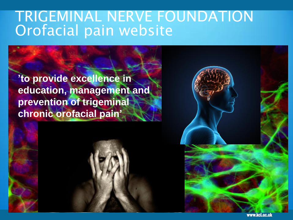
Education
Complex region
Consequences
• Social function
• Eating
• Drinking
• Speaking
• Kissing
• Make up / shaving
• Sleeping
Trigeminal nerve pain
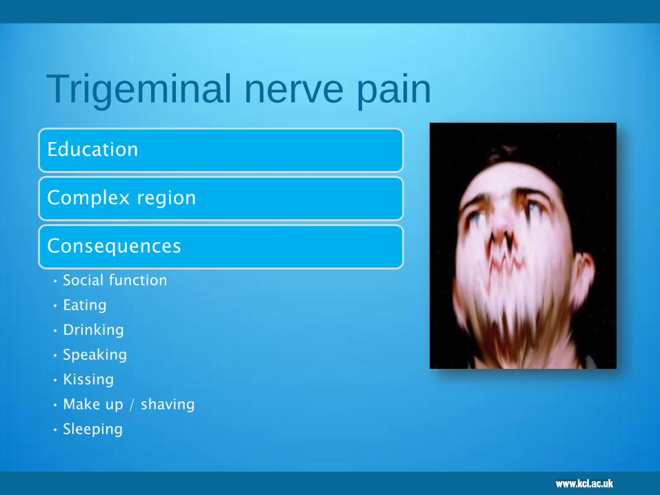
Trigeminal nerve
Sensory supply to
face, scalp and
mouth
Homunculus
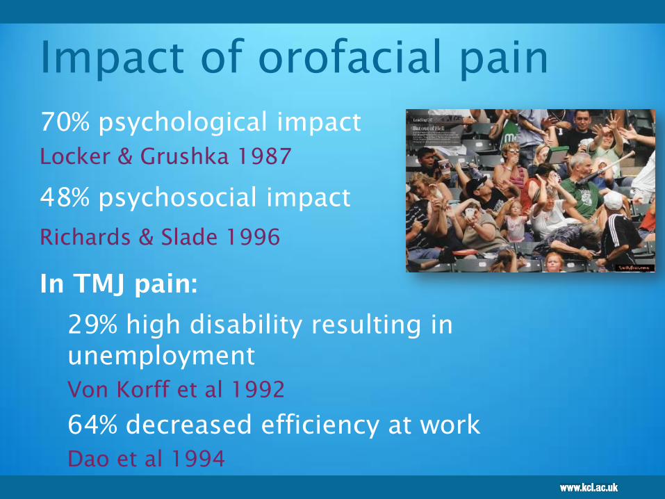
Impact of orofacial pain
70% psychological impact
Locker & Grushka 1987
48% psychosocial impact
Richards & Slade 1996
In TMJ pain:
29% high disability resulting in
unemployment
Von Korff et al 1992
64% decreased efficiency at work
Dao et al 1994
Impact of chronic V pain
7

Type of patient….BSOS
8
9
Pain
“An unpleasant sensory and
emotional experience
associated with actual or
potential tissue damage or
described in terms of such
damage” (IASP, 1979).

10
Pain
“An unpleasant sensory and
emotional experience
associated with actual or
potential tissue damage or
described in terms of such
damage” (IASP, 1979).
Chronic pain
Neuropathic pain Acute pain
inflammatory pain
Acute pain
Nociceptive
Dysfunctional
pain
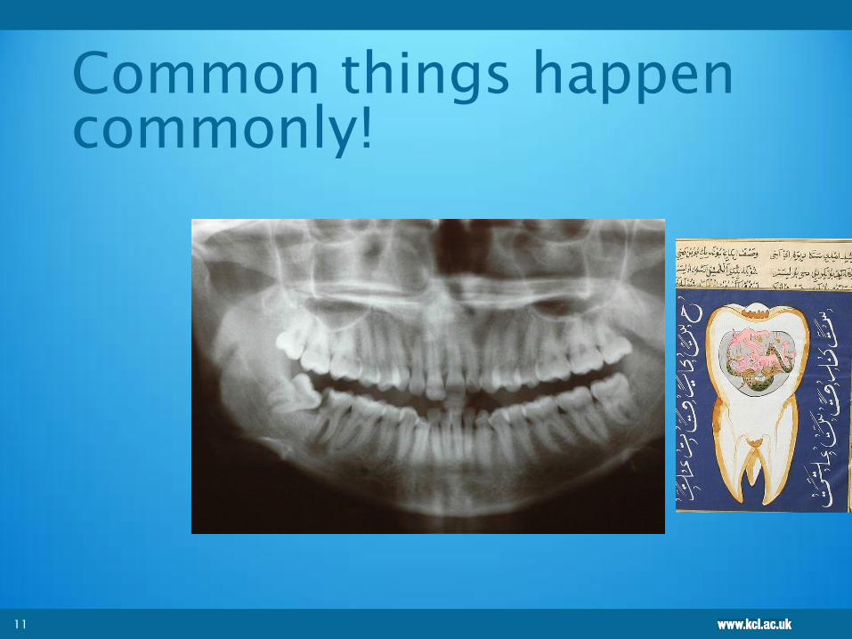
Common things happen
commonly!
11
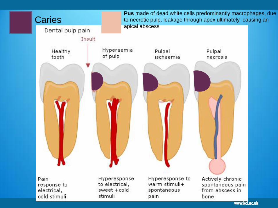
Pus made of dead white cells predominantly macrophages, due
to necrotic pulp, leakage through apex ultimately causing an
apical abscess Caries
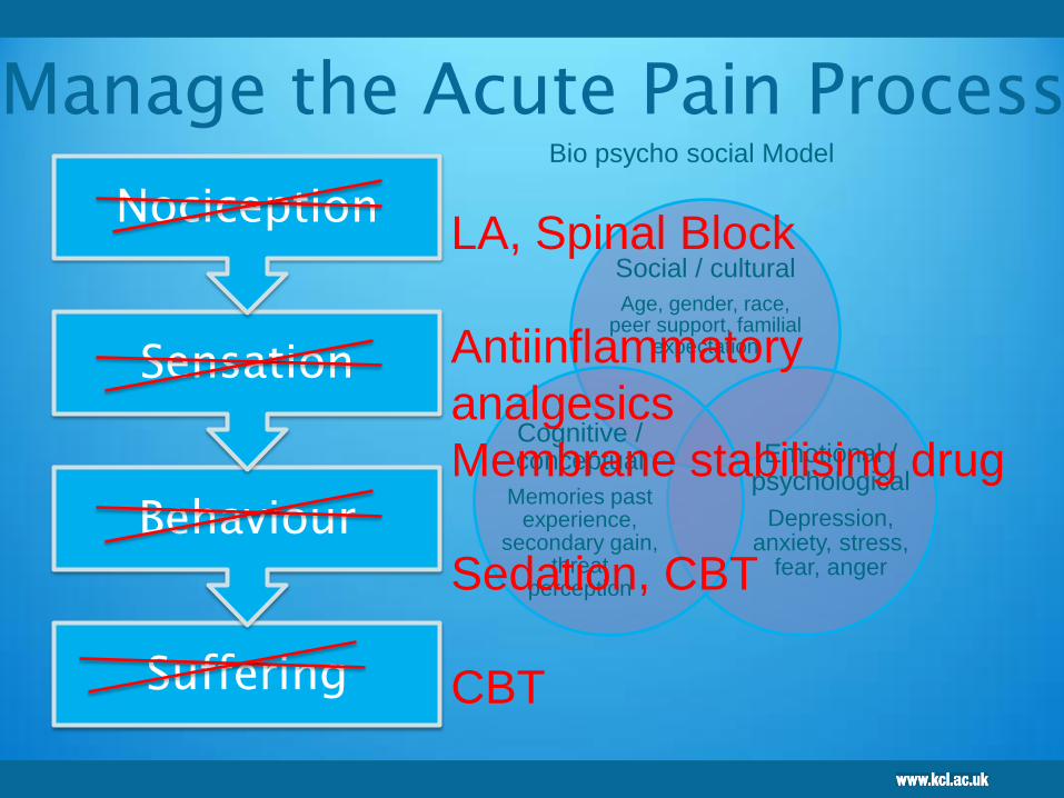
Suffering
Behaviour
Sensation
Nociception
Manage the Acute Pain Process
Social / cultural
Age, gender, race, peer support, familial
expectation
Emotional / psychological
Depression, anxiety, stress,
fear, anger
Cognitive / conceptual
Memories past experience,
secondary gain, threat
perception
Bio psycho social Model
LA, Spinal Block
Antiinflammatory
analgesics
Membrane stabilising drug
Sedation, CBT
CBT
14
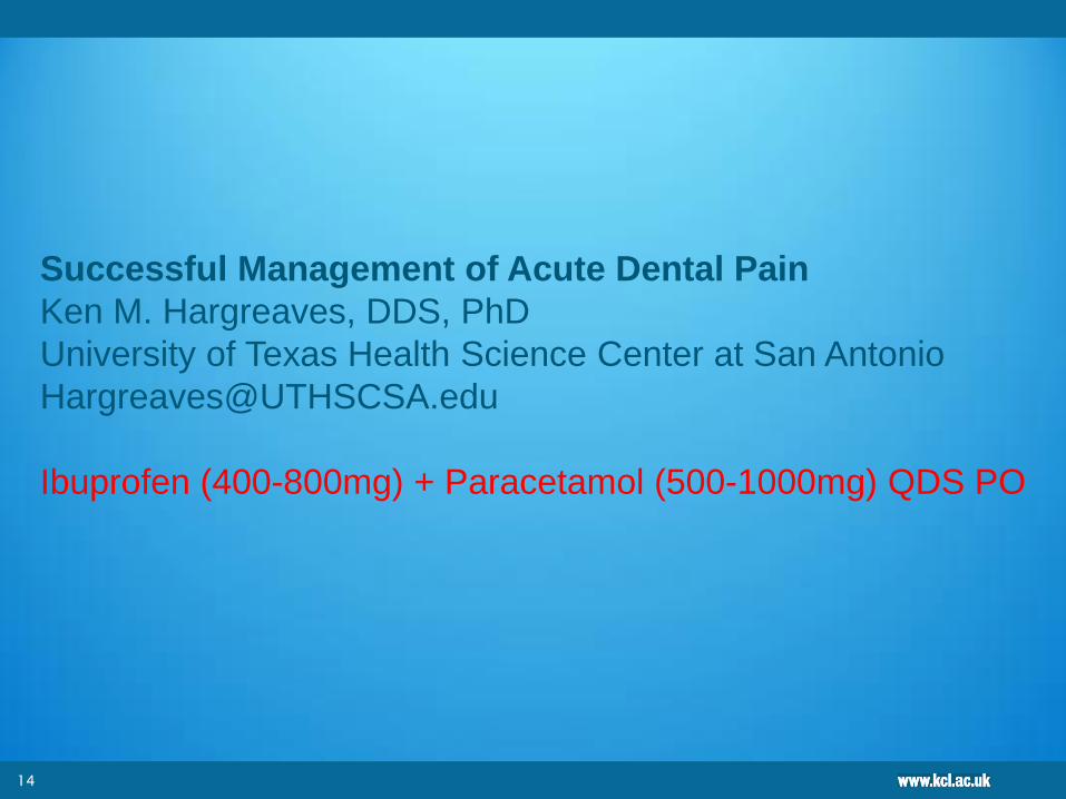
Successful Management of Acute Dental Pain
Ken M. Hargreaves, DDS, PhD
University of Texas Health Science Center at San Antonio
Ibuprofen (400-800mg) + Paracetamol (500-1000mg) QDS PO

Synergism paracetamol + NSAIDs
Miranda HF, Puig MM, Prieto JC, Pinardi G. Synergism
between paracetamol and nonsteroidal anti-inflammatory drugs in
experimental acute pain. Pain. 2006 Mar;121(1-2):22-8. Epub 2006
Merry AF et al. Combined acetaminophen and ibuprofen for pain relief after
oral surgery in adults: a randomized controlled trial. Br J Anaesth. 2010
Jan;104(1):80-8.
Merry AF Eur J Clin Pharmacoz. 2009 Apr;65(4):343-53. Epub 2009 Feb 28.
Onset of analgesia with sodium ibuprofen, ibuprofen acid incorporating
poloxamer andacetaminophen--a single-dose, double-blind, placebo-
controlled study in patients with post-operative dental pain.
15
16
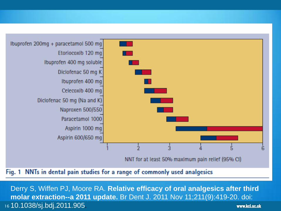
Derry S, Wiffen PJ, Moore RA. Relative efficacy of oral analgesics after third
molar extraction--a 2011 update. Br Dent J. 2011 Nov 11;211(9):419-20. doi:
10.1038/sj.bdj.2011.905.
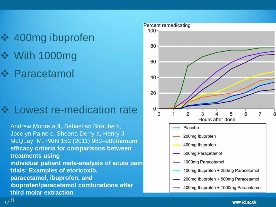
400mg ibuprofen
With 1000mg
Paracetamol
Lowest re-medication rate
17
Andrew Moore a,⇑, Sebastian Straube b,
Jocelyn Paine c, Sheena Derry a, Henry J.
McQuay M. PAIN 152 (2011) 982–989inimum
efficacy criteria for comparisons between
treatments using
individual patient meta-analysis of acute pain
trials: Examples of etoricoxib,
paracetamol, ibuprofen, and
ibuprofen/paracetamol combinations after
third molar extraction
R
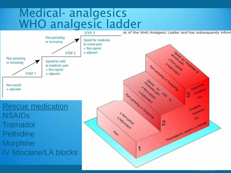
Medical- analgesics
WHO analgesic ladder
Rescue medication
NSAIDs
Tramadol
Pethidine
Morphine
IV lidocaine/LA blocks
Classification of chronic OFP
19
20
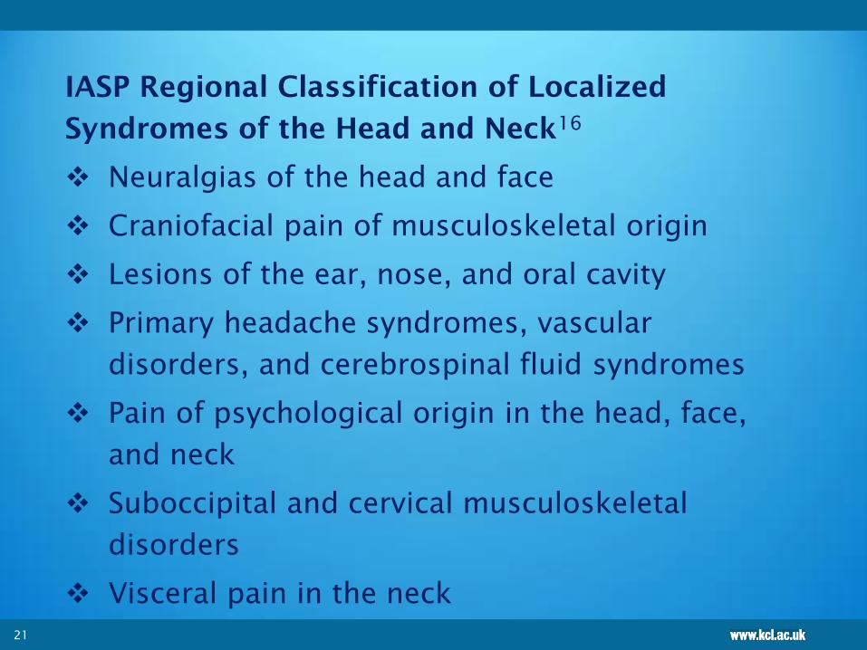
IASP Regional Classification of Localized
Syndromes of the Head and Neck16
Neuralgias of the head and face
Craniofacial pain of musculoskeletal origin
Lesions of the ear, nose, and oral cavity
Primary headache syndromes, vascular
disorders, and cerebrospinal fluid syndromes
Pain of psychological origin in the head, face,
and neck
Suboccipital and cervical musculoskeletal
disorders
Visceral pain in the neck
21

22
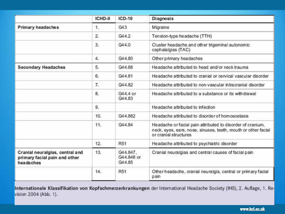
A Hierarchical International headache classification IHCD II 17
Part I: The Primary Headaches
1. Migraine
2. Tension-type headache
3. Cluster headache and other trigeminal autonomic cephalalgias
4. Other primary headaches
Part II: The Secondary Headaches
5. Headache attributed to head and/or neck trauma
6. Headache attributed to cranial or cervical vascular disorder
7. Headache attributed to non-vascular intracranial disorder
8. Headache attributed to a substance or its withdrawal
9. Headache attributed to infection
10. Headache attributed to disorder of homoeostasis
11. Headache or facial pain attributed to disorder of cranium, neck,eyes,
ears, nose, sinuses, teeth, mouth or other facial or cranial structures
12. Headache attributed to psychiatric disorder
Part III: Cranial Neuralgias Central and Primary Facial Pain and Other
Headaches
13. Cranial neuralgias and central causes of facial pain
14. Other headache, cranial neuralgia, central or primary facial pain
23
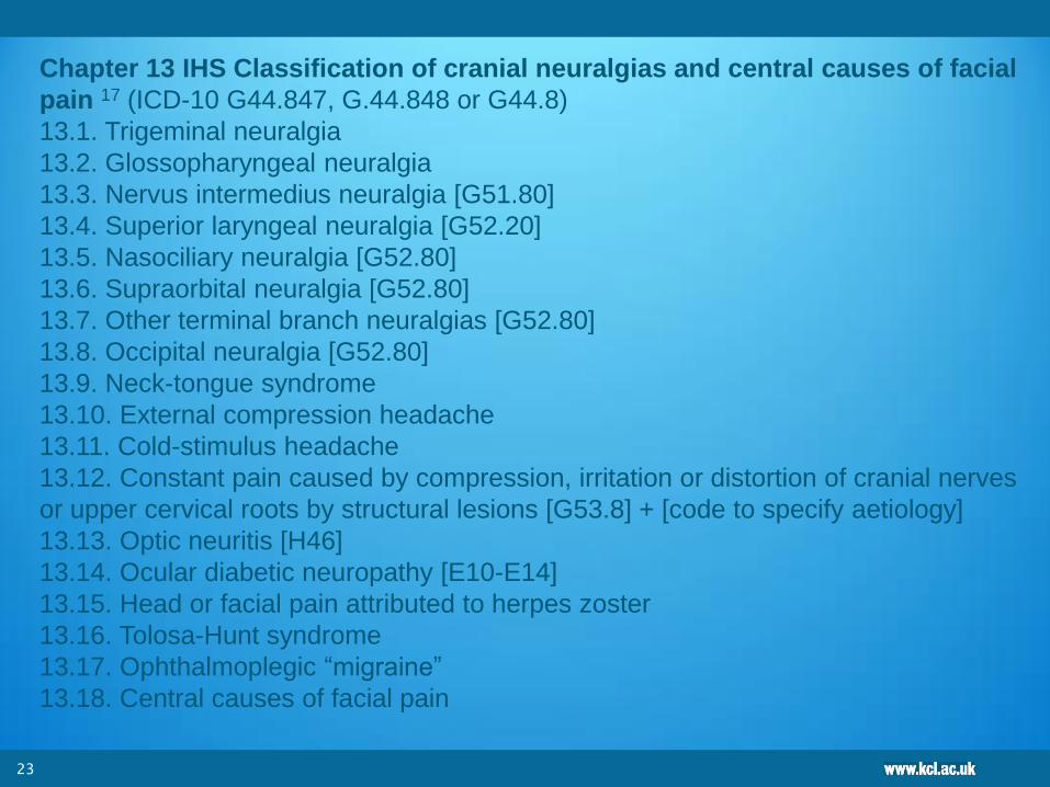
Chapter 13 IHS Classification of cranial neuralgias and central causes of facial
pain 17 (ICD-10 G44.847, G.44.848 or G44.8)
13.1. Trigeminal neuralgia
13.2. Glossopharyngeal neuralgia
13.3. Nervus intermedius neuralgia [G51.80]
13.4. Superior laryngeal neuralgia [G52.20]
13.5. Nasociliary neuralgia [G52.80]
13.6. Supraorbital neuralgia [G52.80]
13.7. Other terminal branch neuralgias [G52.80]
13.8. Occipital neuralgia [G52.80]
13.9. Neck-tongue syndrome
13.10. External compression headache
13.11. Cold-stimulus headache
13.12. Constant pain caused by compression, irritation or distortion of cranial nerves
or upper cervical roots by structural lesions [G53.8] + [code to specify aetiology]
13.13. Optic neuritis [H46]
13.14. Ocular diabetic neuropathy [E10-E14]
13.15. Head or facial pain attributed to herpes zoster
13.16. Tolosa-Hunt syndrome
13.17. Ophthalmoplegic “migraine”
13.18. Central causes of facial pain
24
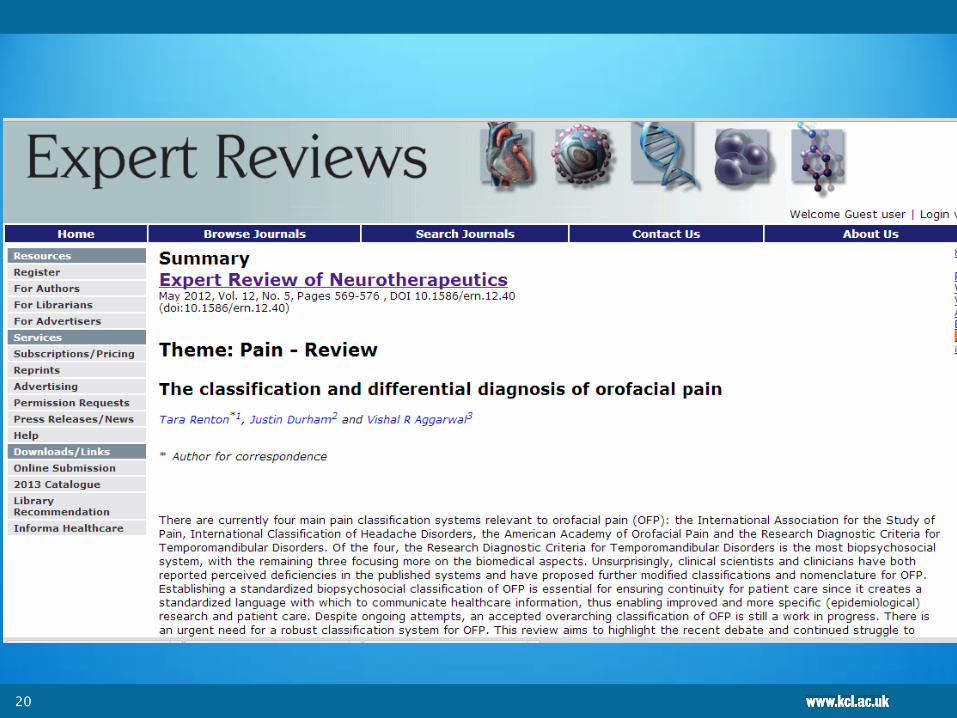
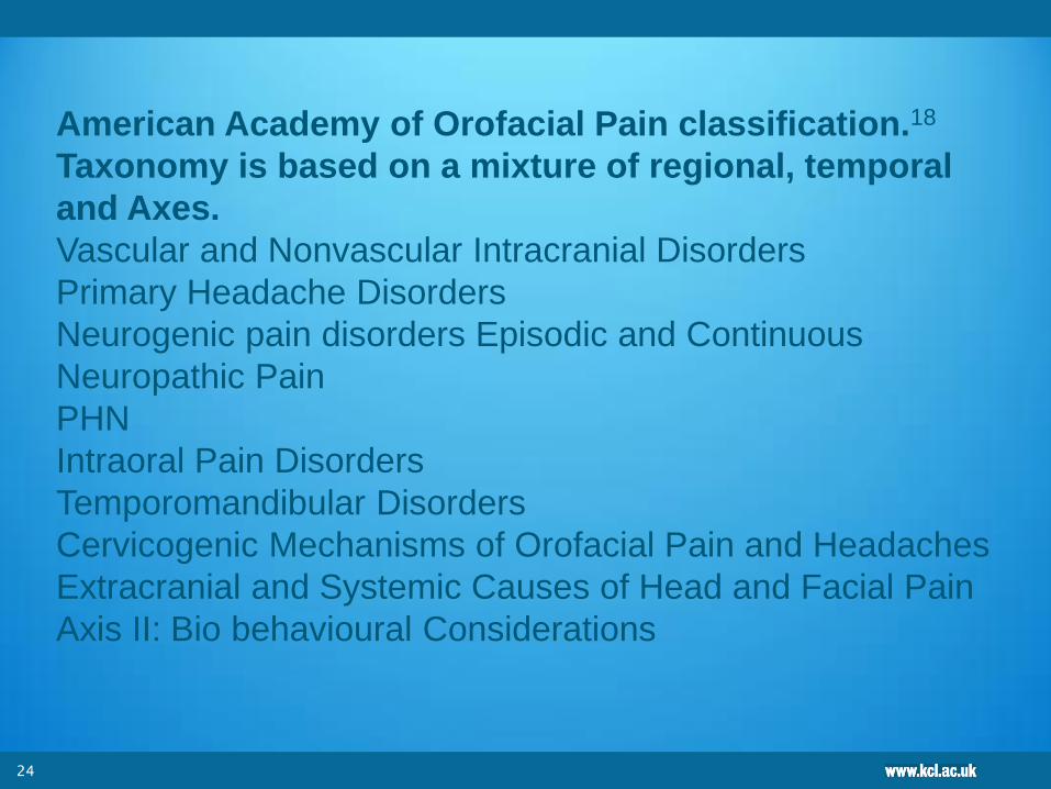
American Academy of Orofacial Pain classification.18
Taxonomy is based on a mixture of regional, temporal
and Axes.
Vascular and Nonvascular Intracranial Disorders
Primary Headache Disorders
Neurogenic pain disorders Episodic and Continuous
Neuropathic Pain
PHN
Intraoral Pain Disorders
Temporomandibular Disorders
Cervicogenic Mechanisms of Orofacial Pain and Headaches
Extracranial and Systemic Causes of Head and Facial Pain
Axis II: Bio behavioural Considerations
25
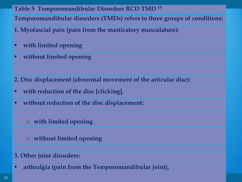
Table 5 Temporomandibular Disorders RCD TMD 19
Temporomandibular disorders (TMDs) refers to three groups of conditions:
1. Myofascial pain (pain from the masticatory musculature):
with limited opening
without limited opening
2. Disc displacement (abnormal movement of the articular disc):
with reduction of the disc [clicking],
without reduction of the disc displacement:
o with limited opening
o without limited opening
3. Other joint disorders:
arthralgia (pain from the Temporomandibular joint),
osteoarthritis (painful crepitus)
osteoarthrosis (an osteoarthritis that is now quiescent)
Other pathology that can affect the Temporomandibular joint complex can include:
Tumours – either primary or secondary
Rheumatoid arthritis
Hyper or hypomobility
26
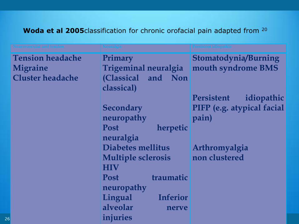
Neurovascular and tension Neuralgia Persistent idiopathic
Tension headache Migraine Cluster headache
Primary Trigeminal neuralgia (Classical and Non classical) Secondary neuropathy Post herpetic neuralgia Diabetes mellitus Multiple sclerosis HIV Post traumatic neuropathy Lingual Inferior alveolar nerve injuries
Stomatodynia/Burning mouth syndrome BMS Persistent idiopathic PIFP (e.g. atypical facial pain) Arthromyalgia non clustered
Woda et al 2005classification for chronic orofacial pain adapted from 20

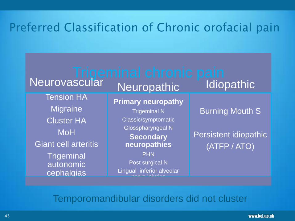
Preferred Classification of Chronic orofacial pain
Trigeminal chronic pain
Neurovascular
Tension HA
Migraine
Cluster HA
MoH
Giant cell arteritis
Trigeminal autonomic cephalgias
Neuropathic Primary neuropathy
Trigeminal N
Classic/symptomatic
Glosspharyngeal N
Secondary neuropathies
PHN
Post surgical N
Lingual inferior alveolar nerve injuries
Idiopathic
Burning Mouth S
Persistent idiopathic
(ATFP / ATO)
27
Temporomandibular disorders did not cluster

Preferred Classification of Chronic orofacial pain
Trigeminal chronic pain
Neurovascular
Tension HA
Migraine
Cluster HA
MoH
Giant cell arteritis
Trigeminal autonomic cephalgias
Neuropathic Primary neuropathy
Trigeminal N
Classic/symptomatic
Glosspharyngeal N
Secondary neuropathies
PHN
Post surgical N
Lingual inferior alveolar nerve injuries
Idiopathic
Burning Mouth S
Persistent idiopathic
(ATFP / ATO)
28
Temporomandibular disorders did not cluster
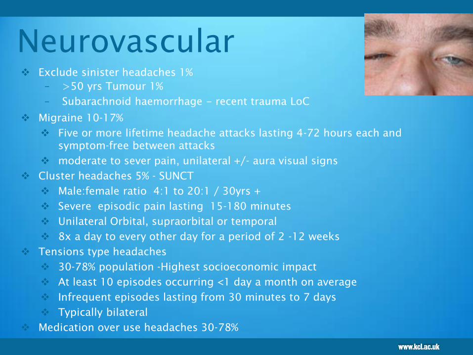
Neurovascular
Exclude sinister headaches 1%
– >50 yrs Tumour 1%
– Subarachnoid haemorrhage - recent trauma LoC
Migraine 10-17%
Five or more lifetime headache attacks lasting 4-72 hours each and
symptom-free between attacks
moderate to sever pain, unilateral +/- aura visual signs
Cluster headaches 5% - SUNCT
Male:female ratio 4:1 to 20:1 / 30yrs +
Severe episodic pain lasting 15-180 minutes
Unilateral Orbital, supraorbital or temporal
8x a day to every other day for a period of 2 -12 weeks
Tensions type headaches
30-78% population -Highest socioeconomic impact
At least 10 episodes occurring <1 day a month on average
Infrequent episodes lasting from 30 minutes to 7 days
Typically bilateral
Medication over use headaches 30-78%
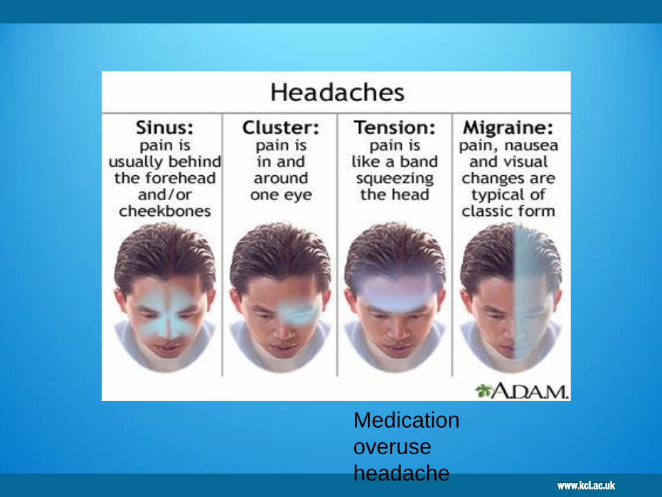
Medication
overuse
headache
31
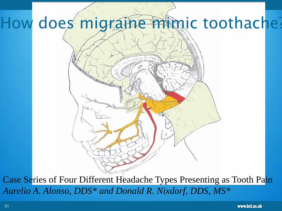
Case Series of Four Different Headache Types Presenting as Tooth Pain
Aurelio A. Alonso, DDS* and Donald R. Nixdorf, DDS, MS*
How does migraine mimic toothache?

32
Chronic migraine, Migraine chronique
D. Valade Review Neurologique May 2013

33
http://www.nice.org.uk/nicemedia/l
ive/13901/60853/60853.pdf

Acute headache
Kids exclude meningitis
Adults 1% sinister causes
Stroke
Sub arrachnoid heamorrhage
Headache -Migraine
Rehydration/ Anxiolysis,
IV Sumatriptan
Primary headache syndromes
• Migraine
• Cluster headache and related syndromes (including
paroxysmal hemicranias, SUNCT)
• Thunderclap headache
• Hypnic headaches
• Benign exertional/sex headache
• Cough headache
• Exploding head syndrome (note this is a sensation, not
headache
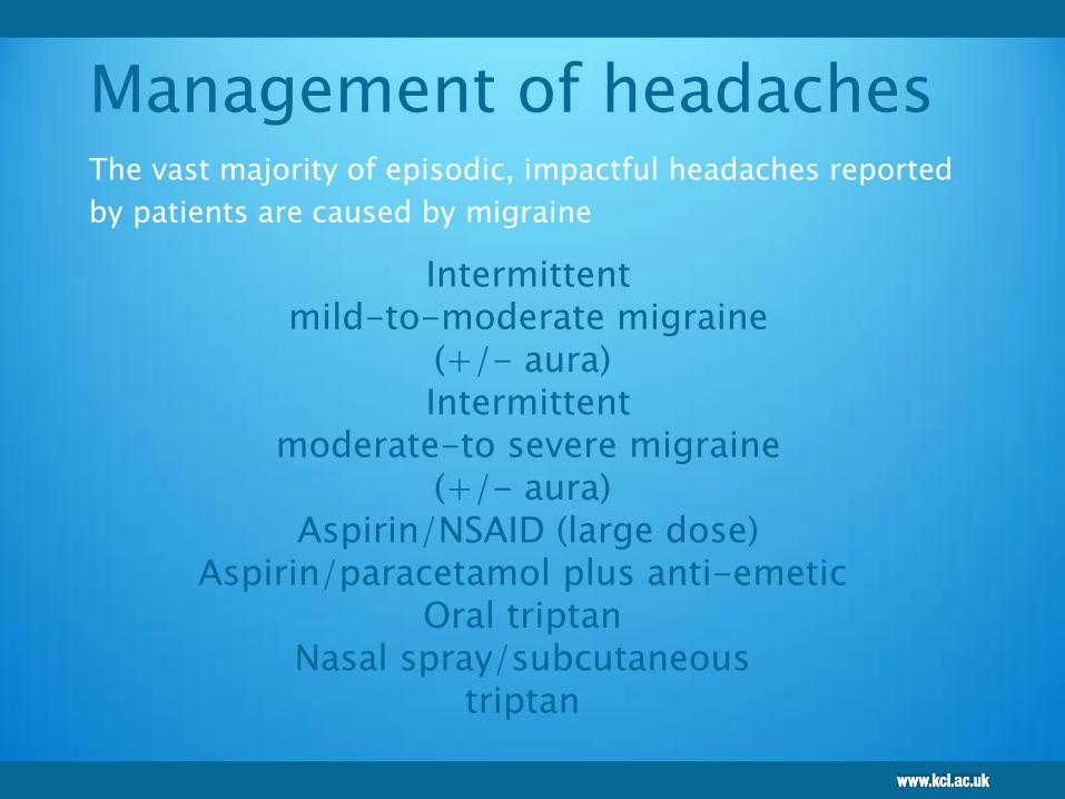
Management of headaches
The vast majority of episodic, impactful headaches reported
by patients are caused by migraine
Intermittent mild-to-moderate migraine
(+/- aura) Intermittent
moderate-to severe migraine (+/- aura)
Aspirin/NSAID (large dose) Aspirin/paracetamol plus anti-emetic
Oral triptan Nasal spray/subcutaneous
triptan

Exclude sinister headaches
Subarachnoid haemorrhage - recent trauma LoC
Cranial arteritis
Tumour 1%
>50 yrs
New-onset, acute headaches associated with other
symptoms
e.g. rash, neurological deficit, vomiting,
pain/tenderness, accident/head injury, hypertension
Neurological change/deficit does not disappear when
the patient is pain-free between attacks
Develop algorithm for sinister headaches
Dowson AJ, Cady RC. Rapid Reference to Migraine
2002.

Giant cell arteritis
Acute temporal onset pain
Palpable temporal artery
May be bilateral
+/- Occular signs
Risk of blindness
Prednisolone 50mg oral
(GMP)
Ophthalmic assessment

Trigeminal autonomic
cephalgias (TACs)
Cluster headache
SUNCT
SUNA
Paroxysmal hemicrania
Hemicrania continua
39

SUNCT sudden onset neuralgiform conjunctival
irritation and tearing
Redness
Ptosis
Tearing
Nasal congestion
V2
40

CH/ SUNCT/SUNA/PH
41
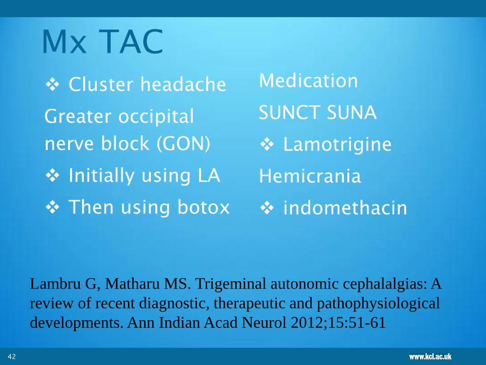
Mx TAC
Cluster headache
Greater occipital
nerve block (GON)
Initially using LA
Then using botox
Medication
SUNCT SUNA
Lamotrigine
Hemicrania
indomethacin
42
Lambru G, Matharu MS. Trigeminal autonomic cephalalgias: A
review of recent diagnostic, therapeutic and pathophysiological
developments. Ann Indian Acad Neurol 2012;15:51-61
Preferred Classification of Chronic orofacial pain
Trigeminal chronic pain
Neurovascular
Tension HA
Migraine
Cluster HA
MoH
Giant cell arteritis
Trigeminal autonomic cephalgias
Neuropathic Primary neuropathy
Trigeminal N
Classic/symptomatic
Glosspharyngeal N
Secondary neuropathies
PHN
Post surgical N
Lingual inferior alveolar nerve injuries
Idiopathic
Burning Mouth S
Persistent idiopathic
(ATFP / ATO)
43
Temporomandibular disorders did not cluster

Preferred Classification of Chronic orofacial pain
Trigeminal chronic pain
Neurovascular
Tension HA
Migraine
Cluster HA
MoH
Giant cell arteritis
Trigeminal autonomic cephalgias
Neuropathic Primary neuropathy
Trigeminal N
Classic/symptomatic
Glosspharyngeal N
Secondary neuropathies
PHN
Post surgical N
Lingual inferior alveolar nerve injuries
Idiopathic
Burning Mouth S
Persistent idiopathic
(ATFP / ATO)
44
Temporomandibular disorders did not cluster

Primary neuralgia
Trigeminal neuralgia (TN) Typical Classic
Atypical symptomatic
Glossopharyngeal neuralgia Acute pain pharynx, tongue base, mastoid regions
Secondary neuralgia
Post herpetic neuralgia (PHN) > 50 yrs 60% likely to develop pain post shingles
Ramsay Hunt syndrome
Diabetes
HIV
PHN
Chemotherapy
MS
Post traumatic V neuralgia Lingual nerve injuries
Inferior alveolar nerve
Neuropathic OFP with ‘neuralgia’
BMS?
Trigeminal Neuralgia
IASP defines trigeminal neuralgia as
“ a sudden, usually unilateral, severe,
brief, stabbing, recurrent pain in the
distribution of one or more branches of
the fifth cranial nerve”.
Classical TN has diagnostic criteria
International Headache Society.

Classical TN
Symptomatic TN
bilateral
neuropathy
younger age
47
Classic TN
Character
Flashing, shooting, sharp, unbearable
Severity
Moderate to severe
Site, radiation
Distribution of trigeminal nerve
Duration, periodicity
Bouts last for seconds, pain free periods
Provoking factors
Elicited -Light touch, eating, talking
Relieving factors
Avoid touch, anticonvulsants
Associated factors
Trigger areas, weight loss
No causative event
Classic TN
interesting features
Never at sleep
Elicited
Unilateral
V2/3
Responds to
tegretol
No neuropathic
area
Exclude;
TACs
MS
SOL
49

TN Investigations
MRI – patients under 40 years to exclude
multiple sclerosis
assess if micro vascular compression
Space occupying lesions (Devor 2010)
CT - tumours of posterior fossa
Haematological tests
Biochemical tests
Neurological – sensory testing and hearing
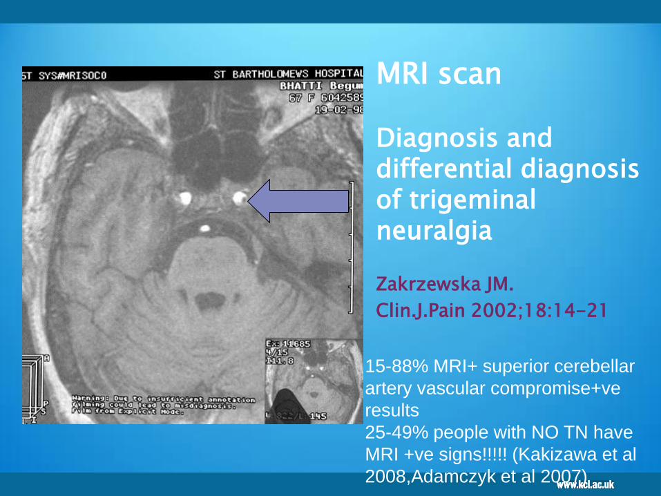
MRI scan Diagnosis and differential diagnosis of trigeminal neuralgia
Zakrzewska JM.
Clin.J.Pain 2002;18:14-21
15-88% MRI+ superior cerebellar
artery vascular compromise+ve
results
25-49% people with NO TN have
MRI +ve signs!!!!! (Kakizawa et al
2008,Adamczyk et al 2007)

Sup cerebellar artery
vascular compromise
52
Green arrow shows retraction of trigeminal vein in contact with but not
compressing V; red arrow shows a branch of the superior cerebellar artery
passing medial to and severely compressing V at the root entry zone
Courtesy Mr Sinan Barazi Neurosurgeon KCH
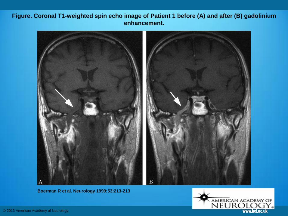
Figure. Coronal T1-weighted spin echo image of Patient 1 before (A) and after (B) gadolinium
enhancement.
Boerman R et al. Neurology 1999;53:213-213
© 2013 American Academy of Neurology
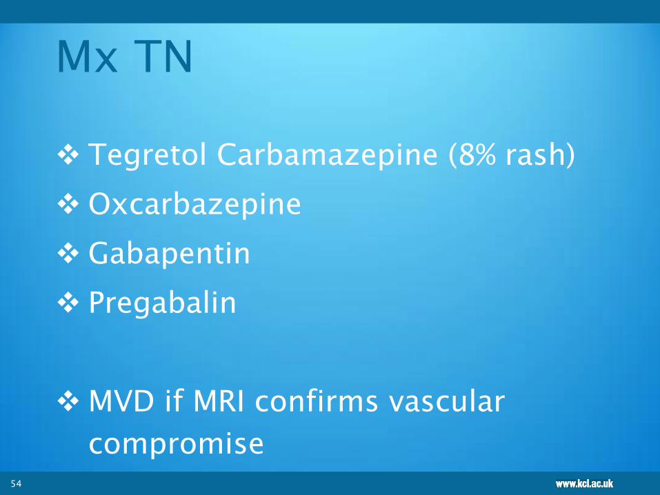
Mx TN
Tegretol Carbamazepine (8% rash)
Oxcarbazepine
Gabapentin
Pregabalin
MVD if MRI confirms vascular
compromise
54
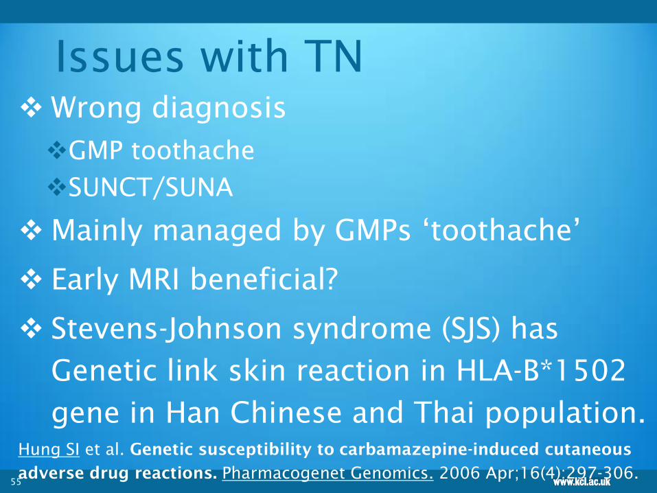
Issues with TN
Wrong diagnosis
GMP toothache
SUNCT/SUNA
Mainly managed by GMPs ‘toothache’
Early MRI beneficial?
Stevens-Johnson syndrome (SJS) has
Genetic link skin reaction in HLA-B*1502
gene in Han Chinese and Thai population.
Hung SI et al. Genetic susceptibility to carbamazepine-induced cutaneous
adverse drug reactions. Pharmacogenet Genomics. 2006 Apr;16(4):297-306.
55
Useful links TN
Information also available on TNA
UK website http://www.tna.org.uk
Brain and spine foundation booklet
on face pain Available on
http://www.brainandspine.org.uk
ISBN 978-1-901893-60-1
56
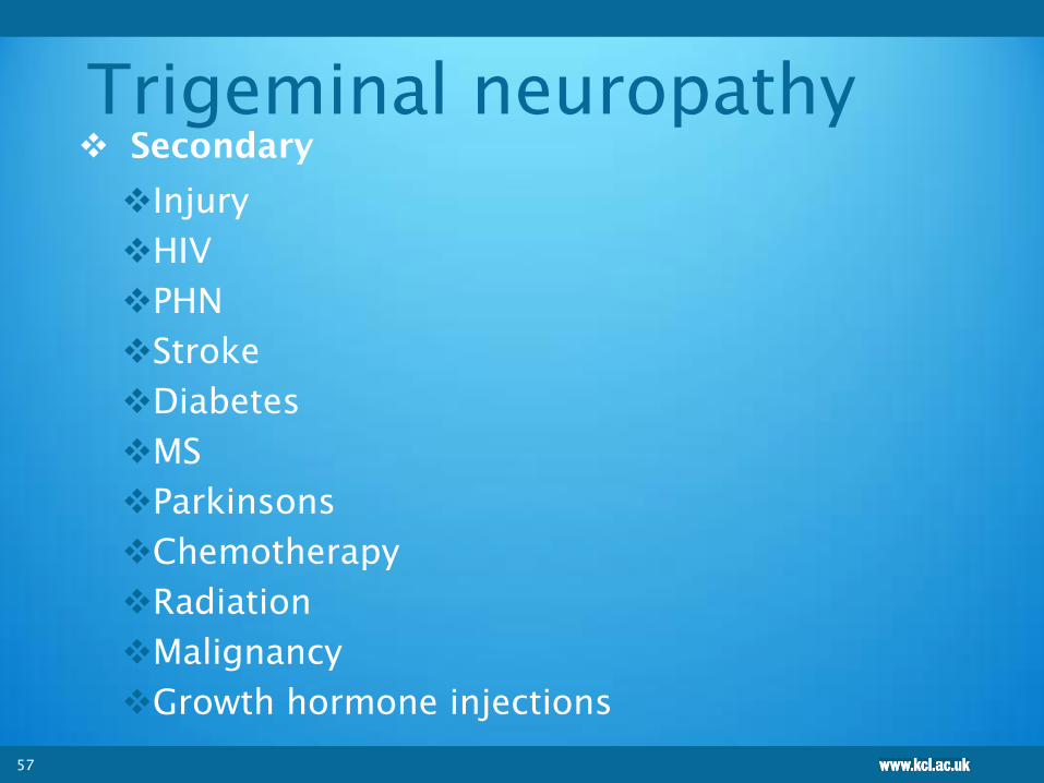
Trigeminal neuropathy Secondary
Injury
HIV
PHN
Stroke
Diabetes
MS
Parkinsons
Chemotherapy
Radiation
Malignancy
Growth hormone injections
57

Post herpetic neuralgia
PHN
Shingles and PHN - Shingles Support
Society
www.shinglessupport.org/faq
58

Post ophthalmic herpes zoster – hyperaemia and corneal scarring
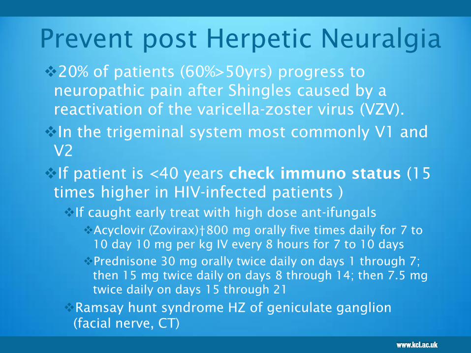
Prevent post Herpetic Neuralgia
20% of patients (60%>50yrs) progress to
neuropathic pain after Shingles caused by a
reactivation of the varicella-zoster virus (VZV).
In the trigeminal system most commonly V1 and
V2
If patient is <40 years check immuno status (15
times higher in HIV-infected patients )
If caught early treat with high dose ant-ifungals
Acyclovir (Zovirax)†800 mg orally five times daily for 7 to
10 day 10 mg per kg IV every 8 hours for 7 to 10 days
Prednisone 30 mg orally twice daily on days 1 through 7;
then 15 mg twice daily on days 8 through 14; then 7.5 mg
twice daily on days 15 through 21
Ramsay hunt syndrome HZ of geniculate ganglion
(facial nerve, CT)
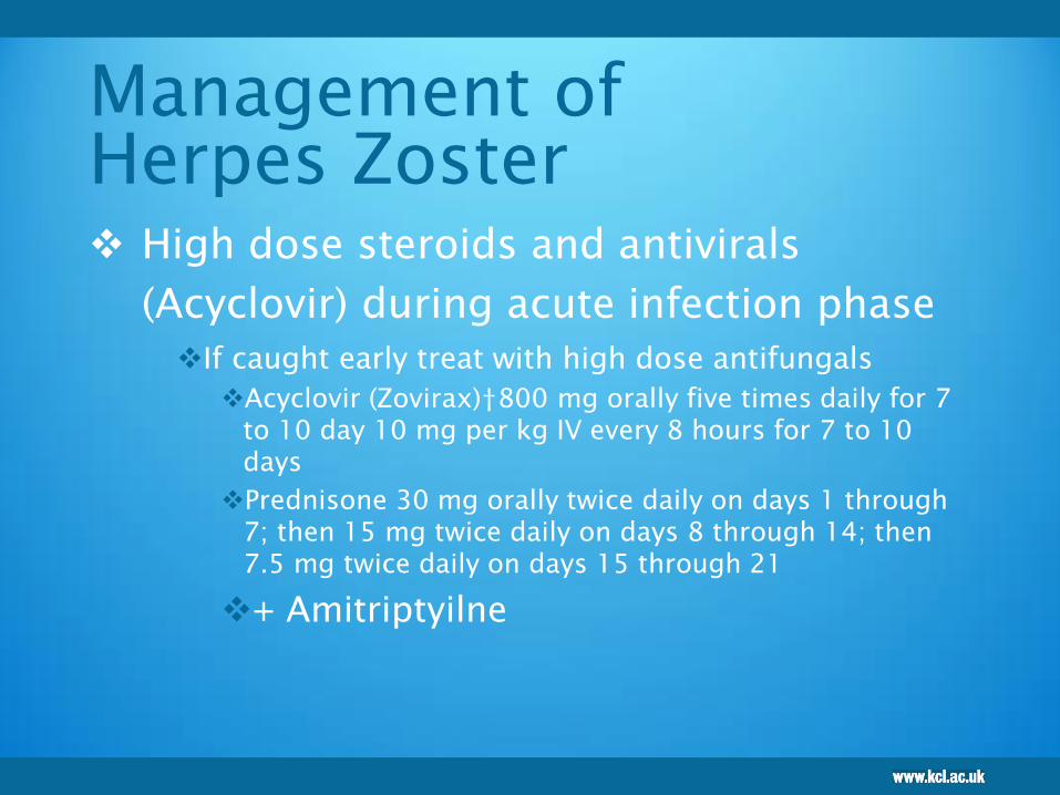
Management of
Herpes Zoster
High dose steroids and antivirals
(Acyclovir) during acute infection phase
If caught early treat with high dose antifungals
Acyclovir (Zovirax)†800 mg orally five times daily for 7
to 10 day 10 mg per kg IV every 8 hours for 7 to 10
days
Prednisone 30 mg orally twice daily on days 1 through
7; then 15 mg twice daily on days 8 through 14; then
7.5 mg twice daily on days 15 through 21
+ Amitriptyilne
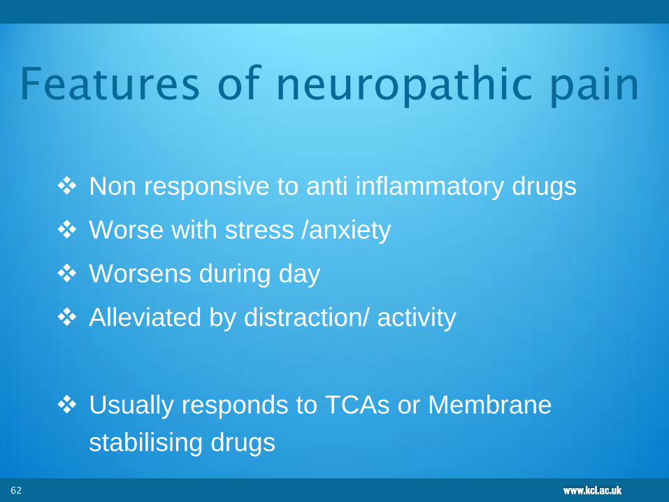
Features of neuropathic pain
Non responsive to anti inflammatory drugs
Worse with stress /anxiety
Worsens during day
Alleviated by distraction/ activity
Usually responds to TCAs or Membrane
stabilising drugs
62

Management painful neuropathy
Counselling
CBT
Medical
Antidepressants
Tricyclic antidepressants
Amitriptyline
Nortriptiyine
Anticonvulsants
Carbamazepine
Gabapentin
Pregabalin
Surgery early repair / late exploration repair
90% patients feel as though surgery is worthwhile (Robinson PP
et al., 2003)

64

65
NICE

Preferred Classification of Chronic orofacial pain
Trigeminal chronic pain
Neurovascular
Tension HA
Migraine
Cluster HA
MoH
Giant cell arteritis
Trigeminal autonomic cephalgias
Neuropathic Primary neuropathy
Trigeminal N
Classic/symptomatic
Glosspharyngeal N
Secondary neuropathies
PHN
Post surgical N
Lingual inferior alveolar nerve injuries
Idiopathic
Burning Mouth S
Persistent idiopathic
(ATFP / ATO)
66
Temporomandibular disorders did not cluster

Preferred Classification of Chronic orofacial pain
Trigeminal chronic pain
Neurovascular
Tension HA
Migraine
Cluster HA
MoH
Giant cell arteritis
Trigeminal autonomic cephalgias
Neuropathic Primary neuropathy
Trigeminal N
Classic/symptomatic
Glosspharyngeal N
Secondary neuropathies
PHN
Post surgical N
Lingual inferior alveolar nerve injuries
Idiopathic
Burning Mouth S
Persistent idiopathic
(ATFP / ATO)
67
Temporomandibular disorders did not cluster
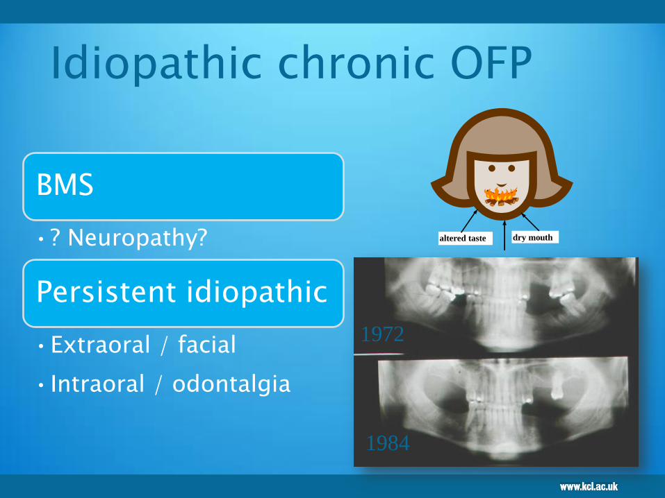
Idiopathic chronic OFP
BMS
•? Neuropathy?
Persistent idiopathic
•Extraoral / facial
• Intraoral / odontalgia
1972
1984
Burning Mouth Syndrome
altered taste dry mouth
tongue thrusting
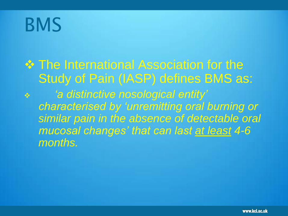
BMS
The International Association for the Study of Pain (IASP) defines BMS as:
‘a distinctive nosological entity’ characterised by ‘unremitting oral burning or similar pain in the absence of detectable oral mucosal changes’ that can last at least 4-6 months.
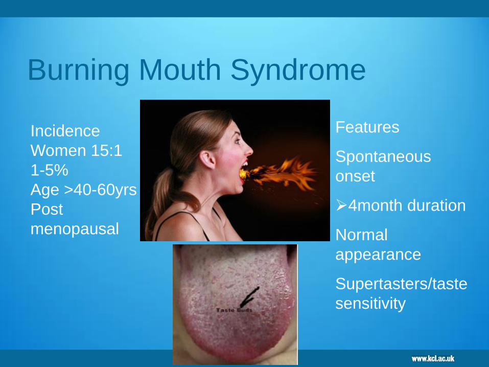
Features
Spontaneous
onset
4month duration
Normal
appearance
Supertasters/taste
sensitivity
Incidence
Women 15:1
1-5%
Age >40-60yrs
Post
menopausal
Burning Mouth Syndrome

BMS causes
Menopausal
Supertasters
Deficiency in Haematinics
Psychometric - increased HADS scores
Diabetes
Neuropathy ??
Aetiology of BMS
An alteration in autonomic innervation and oral blood flow (Heckmann et al., 2001)
Changes in endocrine status during menopause, causing a disruption in sensory pathways (Basker et al., 1978)
A disruption of central sensory and modulatory pathways that include the spinal trigeminal nucleus and striatum (Hagelberg et al., 2003; Gao et al., 2000).
A sensory dysfunction illustrated by changes in QST associated with a small and/or large fibre neuropathy (Forssell et al., 2002)
A trigeminal, peripheral small-fibre sensory neuropathy (Lauria et al., 2005; Lauritano et al., 2005).

BMS
Dr Kiran Beneng PhD St
Prof Praveen Anand
Dr Zehra Yilmaz
Ongoing work TRPM8
CB1, P2X3 and GABA receptors
Imaging central pathways (CNS)
Dr Matt Howard
Control BMS
0.0
0.5
1.0
1.5
*
EP
N/P
ap
illa
NF Bar charts of the mean ± SEM
of epithelial nerve fibres per papilla
in control and BMS tongue. * P
<0.0001.
P =0.0006, Spearman r =0.55
Control BMS0.0
0.5
1.0
1.5
2.0 *
TR
PV
1 %
im
mu
noreactiv
ity
TRPV1
0 1 2 3 40
5
10
15
TRPV1
VA
S P
ain
score
Control BMS0.0
0.5
1.0
1.5
2.0
2.5
3.0
3.5
4.0
4.5*
% N
GF
im
mu
noreactive f
ib
res NGF
NF
Research
TRPV1

BMS update
BMS may encompass three distinct, subclinical neuropathic pain states
that may overlap in individual patients. 11-16
o Subgroup 1 (50-65%) is characterized by peripheral small diameter fibre
neuropathy of intraoral mucosa.
o Subgroup 2 (20-25%) consists of patients with subclinical lingual, mandibular, or
trigeminal system pathology that can be dissected with careful neurophysiologic
examination but is clinically indistinguishable from the other two subgroups.
o Subgroup 3 (20-40%) fits the concept of central pain that may be related to
hypofunction of dopaminergic neurons in the basal ganglia.
The neurogenic factors acting in these subgroups differ, and will
require different treatment strategies. In the future, with proper use
of diagnostic tests, BMS patients may benefit from interventions
specifically targeted at the underlying pathophysiological
mechanisms.
74

Management of BMS
Systematic Review and data in Clinical Evidence
Cognitive behaviour therapy may be beneficial
Reassurance
Notriptyline first line but limited evidence for use
of antidepressants
? Future neuropathic pain blocking agents
Capsaicin lollies
Tabasco sauce

Chronic idiopathic facial pain
Persistent Idiopathic facial pain
PIFP
Character
Intense -Nagging, dull, throbbing, sharp, aching ‘pain
all the time resistant to all interventions usually
>3years’
Severity
Varies, mild to severe though patient can often sleep
and function normally
Site, radiation
no anatomical area
Duration, periodicity Constant >6 months
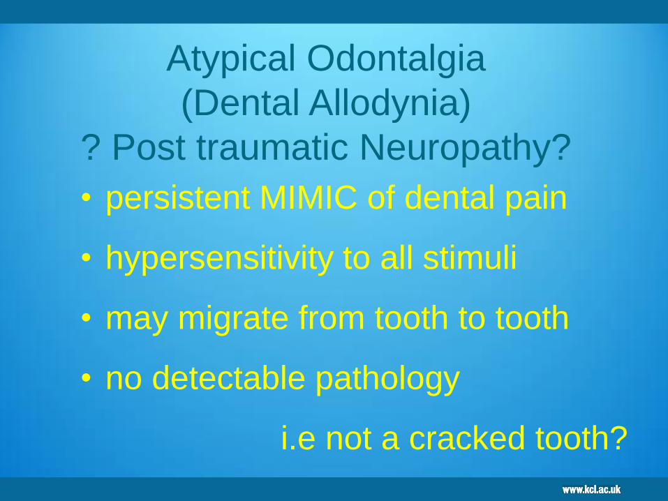
Atypical Odontalgia
(Dental Allodynia)
? Post traumatic Neuropathy?
• persistent MIMIC of dental pain
• hypersensitivity to all stimuli
• may migrate from tooth to tooth
• no detectable pathology
i.e not a cracked tooth?

Exclude the obvious
Cracked tooth……………

1984
1972
Natural history of atypical odontalgia

Prognosis
Chronic idiopathic facial pain – after one year
38% of patients pain free but 39% taking drugs
to prevent relapse
Feinmann and Harris 1984
12/15 required surgery to control their pain

Management of PIFP /AO
Counselling and reassurance
CBT
Medical
Antidepresants
Tricyclic antidepressants
Amitryptiline
Nortryptiline 10mg,20mg,30mg,40mg each week. Maintain
on 40mg nocte for 6 weeks before review
Anticonvulsants
Oxcarbazipine
Carbamazepine
Gabapentin
Pregabalin
Topical local analgesia
Capsaicin
Non clusterable disorder
Temporomandibular disorders (TMD)
Dr Justin Durham Newcastle University
82
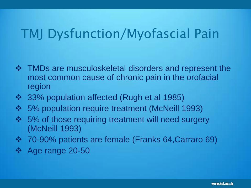
TMJ Dysfunction/Myofascial Pain
TMDs are musculoskeletal disorders and represent the most common cause of chronic pain in the orofacial region
33% population affected (Rugh et al 1985)
5% population require treatment (McNeill 1993)
5% of those requiring treatment will need surgery (McNeill 1993)
70-90% patients are female (Franks 64,Carraro 69)
Age range 20-50
Chronic TMD often does not occur in isolation.
Individuals suffering from chronic pain associated
with a TMD frequently report other chronic pain
conditions including: chronic headache,
fibromyalgia, chronic fatigue syndrome, irritable
bowel syndrome, sleep disturbance and depression
{Dworkin, 2011, #69458; Hoffmann et al., 2011,
#64698; Maixner et al., 2011, #63267}.
84
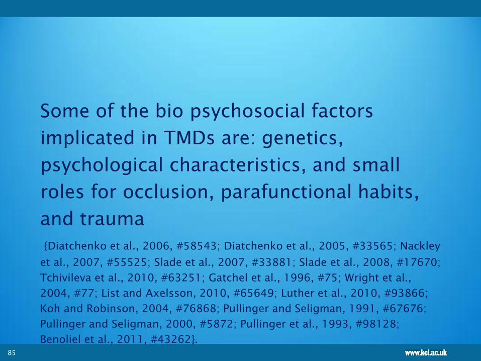
Some of the bio psychosocial factors
implicated in TMDs are: genetics,
psychological characteristics, and small
roles for occlusion, parafunctional habits,
and trauma
{Diatchenko et al., 2006, #58543; Diatchenko et al., 2005, #33565; Nackley
et al., 2007, #55525; Slade et al., 2007, #33881; Slade et al., 2008, #17670;
Tchivileva et al., 2010, #63251; Gatchel et al., 1996, #75; Wright et al.,
2004, #77; List and Axelsson, 2010, #65649; Luther et al., 2010, #93866;
Koh and Robinson, 2004, #76868; Pullinger and Seligman, 1991, #67676;
Pullinger and Seligman, 2000, #5872; Pullinger et al., 1993, #98128;
Benoliel et al., 2011, #43262}.
85
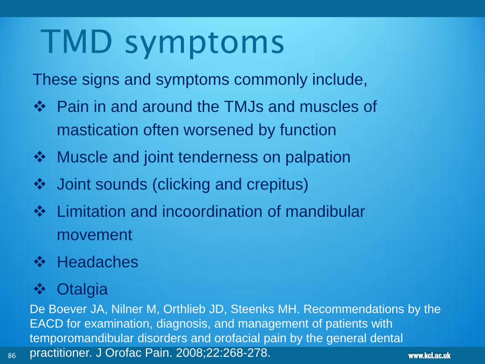
TMD symptoms
These signs and symptoms commonly include,
Pain in and around the TMJs and muscles of
mastication often worsened by function
Muscle and joint tenderness on palpation
Joint sounds (clicking and crepitus)
Limitation and incoordination of mandibular
movement
Headaches
Otalgia
86
De Boever JA, Nilner M, Orthlieb JD, Steenks MH. Recommendations by the
EACD for examination, diagnosis, and management of patients with
temporomandibular disorders and orofacial pain by the general dental
practitioner. J Orofac Pain. 2008;22:268-278.
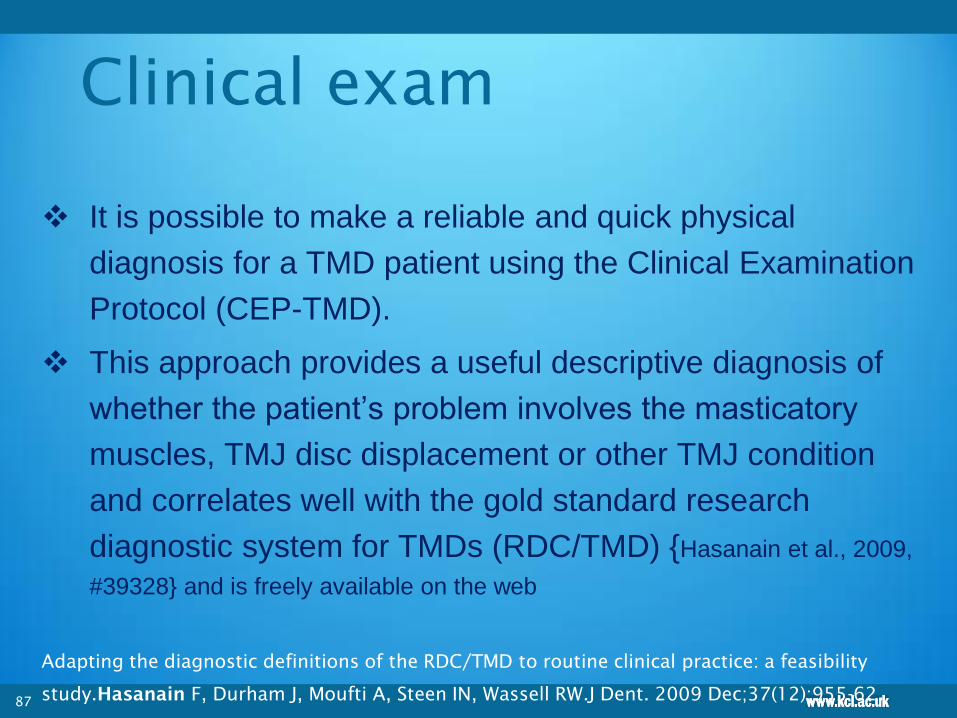
Clinical exam
It is possible to make a reliable and quick physical
diagnosis for a TMD patient using the Clinical Examination
Protocol (CEP-TMD).
This approach provides a useful descriptive diagnosis of
whether the patient’s problem involves the masticatory
muscles, TMJ disc displacement or other TMJ condition
and correlates well with the gold standard research
diagnostic system for TMDs (RDC/TMD) {Hasanain et al., 2009,
#39328} and is freely available on the web
Adapting the diagnostic definitions of the RDC/TMD to routine clinical practice: a feasibility
study.Hasanain F, Durham J, Moufti A, Steen IN, Wassell RW.J Dent. 2009 Dec;37(12):955-62.
87
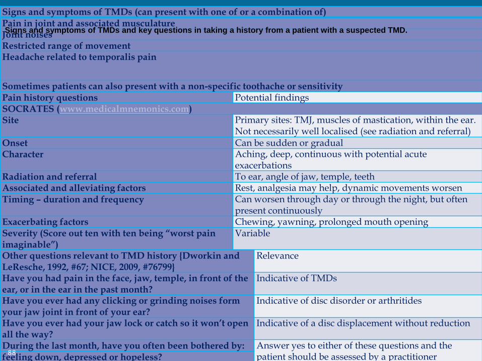
Signs and symptoms of TMDs (can present with one of or a combination of) Pain in joint and associated musculature Joint noises Restricted range of movement Headache related to temporalis pain
Sometimes patients can also present with a non-specific toothache or sensitivity Pain history questions Potential findings SOCRATES (www.medicalmnemonics.com) Site Primary sites: TMJ, muscles of mastication, within the ear.
Not necessarily well localised (see radiation and referral) Onset Can be sudden or gradual Character Aching, deep, continuous with potential acute
exacerbations Radiation and referral To ear, angle of jaw, temple, teeth Associated and alleviating factors Rest, analgesia may help, dynamic movements worsen Timing – duration and frequency Can worsen through day or through the night, but often
present continuously Exacerbating factors Chewing, yawning, prolonged mouth opening Severity (Score out ten with ten being “worst pain imaginable”)
Variable
Other questions relevant to TMD history {Dworkin and LeResche, 1992, #67; NICE, 2009, #76799}
Relevance
Have you had pain in the face, jaw, temple, in front of the ear, or in the ear in the past month?
Indicative of TMDs
Have you ever had any clicking or grinding noises form your jaw joint in front of your ear?
Indicative of disc disorder or arthritides
Have you ever had your jaw lock or catch so it won’t open all the way?
Indicative of a disc displacement without reduction
During the last month, have you often been bothered by: Answer yes to either of these questions and the patient should be assessed by a practitioner competent in mental health assessment
feeling down, depressed or hopeless? having little interest or pleasure in doing things?
88
Signs and symptoms of TMDs and key questions in taking a history from a patient with a suspected TMD.
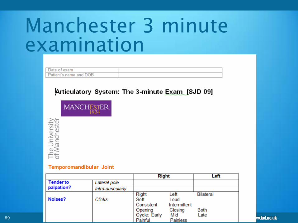
Manchester 3 minute
examination
89

TMJ research Diagnostic
Criteria
TMJ RCD
Arthritides +/-pain
Myalgia Muscle pain
Dysfunction
Internal derangements +/-pain
Dworkin SF, LeResche L. Research diagnostic criteria for temporomandibular disorders: review,
criteria, examinations and specifications, critique. J Craniomandib Disord. 1992;6:301-355
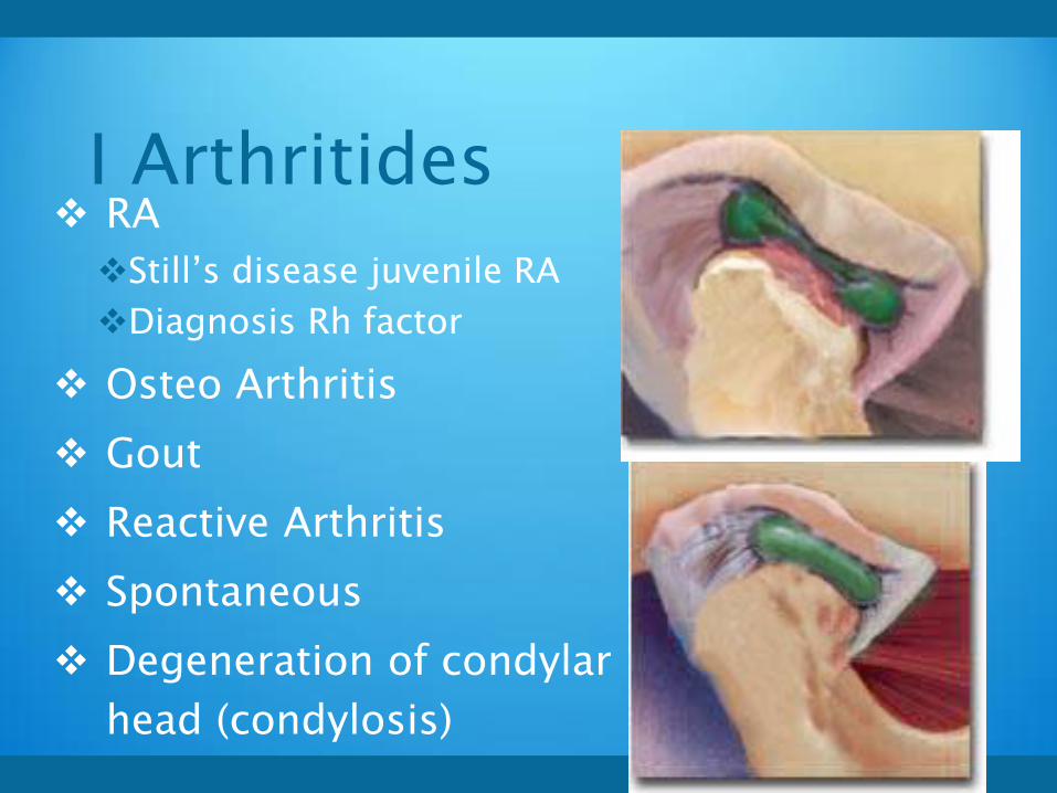
I Arthritides RA
Still’s disease juvenile RA
Diagnosis Rh factor
Osteo Arthritis
Gout
Reactive Arthritis
Spontaneous
Degeneration of condylar
head (condylosis)

Osteoarthritis
Major joints
Older age

Rheumatoid Arthritis
Investigations helpful in
diagnosis of RA
Erythrocyte sedimentation rate (ESR)/
C-reactive protein (CRP) / plasma viscosity Usually elevated in RA but may be normal
Full blood count (FBC) Normochromic, normocytic anaemia and reactive thrombocytosis common in active disease
Urea & electrolytes (U&E), Liver function tests (LFT) Mild elevation of alkaline phosphatase and gamma-GT common in active disease
Uric acid/ synovial fluid analysis Will assist in excluding polyarticular gout
Urinalysis Microscopic haematuria/proteinuria may suggest connective tissue disease
Rheumatoid factor (RF) RF positive in only 60-70% RA patients.
Antinuclear antibody (ANA) Positive in SLE and related conditions. ANA positive in up to 30% of RF-positive RA patients. May
be weakly positive in up to 10% of normal individuals
Radiology May be normal or may show periarticular osteopenia and/or erosions

Differential diagnosis of
arthritis
Viral arthritis (e.g. parvovirus, rubella)
Reactive arthritis (e.g. post-infective: throat, gut, sexually acquired)
Seronegative spondyloarthropathy (e.g. psoriatic, ankylosing spondylitis, inflammatory bowel disease)
Connective tissue disease (e.g. systemic lupus erythematosus (SLE), scleroderma)
Polymyalgia rheumatica
Polyarticular gout
Fibromyalgia
Medical conditions presenting with arthropathy (e.g. sarcoidosis, thyroid disease, infective endocarditis, haemochromatosis, diabetic cheiroarthropathy, paraneoplastic syndromes, multiple myeloma).
II Muscle pain = myalgia
Myositis
Myofascial pain
Myospasm
Hyperkinesia
Hypokinesia
Contracture
Fibromyalgia
Trismus
Limited opening due to muscle spasm
Exclude parafunction
Pain am (night bruxist or sleep position)
Pain late day chewing gum
III TMJ dysfunction
Internal derangement
Painful internal
derangement
Signs Clicking
locking
Mx includes
Non interventional
Interventional Arthroscopy
Surgical discal plication

Other joint problems
Congenital and developmental disorders
Subluxation
Dislocation
Locking
Traumatic injuries
Ankylosis
Neoplasia

Trismus
Progressive worsening
Consider neoplasia
Infratemporal fossa Ca (1:60,000 Ferguson 1986)
Arthritides
Condylosis
Intermittent with normal resolution
TMJ PDS
Permanent
Ankylosis ( true =bony /false=soft tissue)
Most common in third world due to middle ear
infection /mastoiditis

Beware progressive trismus often
painless
100

Neoplasia
Serious pathology of the Temporomandibular complex
and associated musculature is rare.
Primary tumours in the Temporomandibular complex are thought to account for less than 1% of all head and neck tumours and incidental findings on MRI for TMDs occur in less than 1% of TMDs.
Metastases to the Temporomandibular joint can occur from multiple sites but the most likely are breast, lung, thyroid, kidney and prostate.
Red Flags
These ‘red flag’ signs and symptoms should mandate an
urgent referral and they include: New signs and symptoms of TMDs presenting for the first time in
the advanced age group (> 60 years old)
Ipsilateral lymphadenopathy
Previous history of malignancy elsewhere in the body and new onset TMDs
Cranial nerve dysfunction in relation to the complaint especially in the fifth and seventh cranial nerves
Progressive trismus precluding careful oral examination
Recurrent ipsilateral epistaxis
Anosmia
Persistent nasal obstruction or purulent discharge
Objective ipsilateral hearing loss.
Lets not patients
presenting with TMD
end up like this!
103

104
105
The primary goals of any reversible and ion-invasive
therapy should be:
Encouraging self-management of the condition through
education
Reducing the (impact of) pain associated with the
condition
Decreasing functional limitation caused by the condition
Reducing exacerbations and educating individuals in how
to manage any exacerbation of the condition
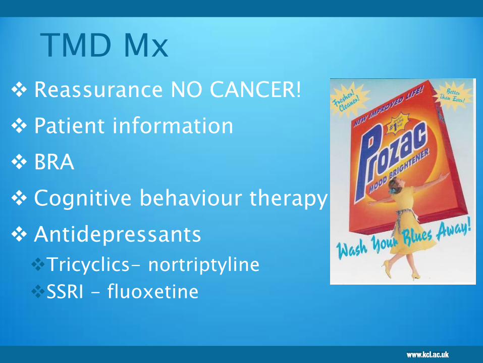
TMD Mx
Reassurance NO CANCER!
Patient information
BRA
Cognitive behaviour therapy
Antidepressants
Tricyclics- nortriptyline
SSRI - fluoxetine
TMD Cochrane reviews
Koh H Robinson PG Occlusal adjustment for treating
and preventing temporomandibular joint disorders. J
Evid Based Dent Pract. 2006.
Al-Ani MZ Stabilisation splint therapy for
temporomandibular pain dysfunction syndrome. Evid
Based Dent. 2004;5(3):65-6.
Koh H, Robinson PG Occlusal adjustment for treating
and preventing temporomandibular joint disorders. J
Oral Rehabil. 2004 Apr;31(4):287-92
Bessa-Nogueira RV, Vasconcelos BC, Niederman R The
methodological quality of systematic reviews
comparing temporomandibular joint disorder
surgical and non-surgical treatment. BMC Oral Health.
2008 Sep 26;8:27
NO EVIDENCE !

More lack of evidence Karppinen K et al., Adjustment of dental occlusion in treatment of
chronic cervicobrachial pain and headache. J Oral Rehabil.1999
Sep;26(9):715-21.
Risk management in clinical practice. Part 8. Temporomandibular
disorders.Gray R, Al-Ani Z.BDJ
NICE on headaches Dental occlusion problems are a major cause of
headache. BMJ 2012; 345 doi:
Badel T, Marotti M, Pavicin IS, Basić-Kes V. Temporomandibular
disorders and occlusion. Acta Clin Croat. 2012 Sep;51(3):419-24.
Lobbezoo F, Ahlberg J, Manfredini D, Winocur E. Are bruxism and
the bite causally related? J Oral Rehabil. 2012 Jul;39(7):489-501.
doi: 10.1111/j.1365-2842.2012.02298.x. Epub 2012 Apr 10.
.
108

109
Cooper BC; International College of Cranio-Mandibular Orthopedics
(ICCMO).Temporomandibular disorders: A position paper of the
International College of Cranio-Mandibular Orthopedics (ICCMO).
Cranio. 2011 Jul;29(3):237-44.
.
Michelotti A Evaluation of the short-term effectiveness of education versus
an occlusal splint for the treatment of myofascialpain of the jaw muscles.
J Am Dent Assoc. 2012 Jan;143(1):47-53
Bales JM, Epstein JB.The role of malocclusion and orthodontics in
temporomandibular disorders. J Can Dent Assoc.1994 Oct;60(10):899-905.
Gebeile-Chauty S et al., Can orthodontic treatment generate
temporomandibular disorders and pain? A review]. Orthod Fr. 2010
Mar;81(1):85-93. doi: 10.1051/orthodfr/2010009. Epub 2010 Apr 1.
Manfredini D et al., Dental occlusion, body posture and temporomandibular
disorders: where we are now and where we are heading for. J Oral
Rehabil. 2012 Jun;39(6):463-71
And more……………..
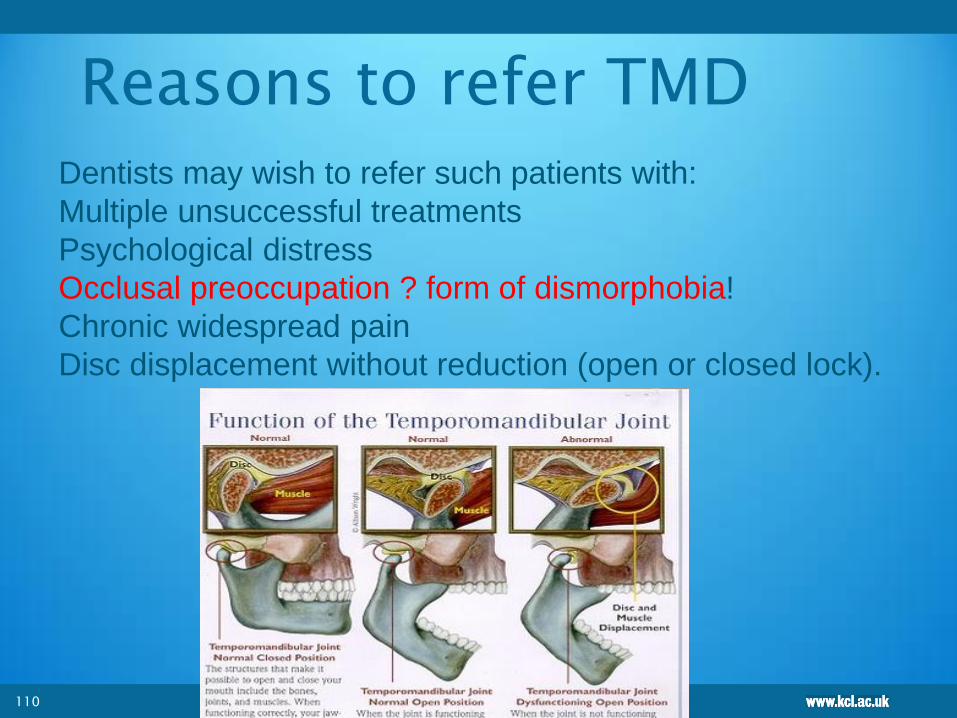
Reasons to refer TMD
110
Dentists may wish to refer such patients with:
Multiple unsuccessful treatments
Psychological distress
Occlusal preoccupation ? form of dismorphobia!
Chronic widespread pain
Disc displacement without reduction (open or closed lock).
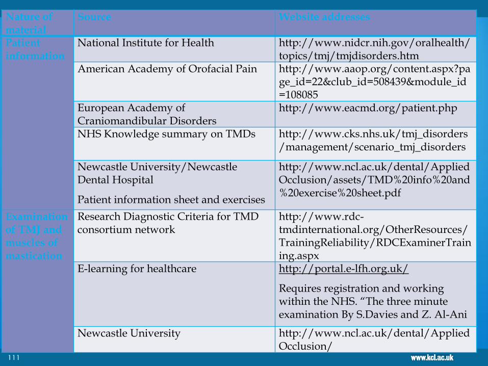
Nature of material
Source Website addresses
Patient information
National Institute for Health http://www.nidcr.nih.gov/oralhealth/topics/tmj/tmjdisorders.htm
American Academy of Orofacial Pain http://www.aaop.org/content.aspx?page_id=22&club_id=508439&module_id=108085
European Academy of Craniomandibular Disorders
http://www.eacmd.org/patient.php
NHS Knowledge summary on TMDs http://www.cks.nhs.uk/tmj_disorders/management/scenario_tmj_disorders
Newcastle University/Newcastle Dental Hospital
Patient information sheet and exercises
http://www.ncl.ac.uk/dental/AppliedOcclusion/assets/TMD%20info%20and%20exercise%20sheet.pdf
Examination of TMJ and muscles of mastication
Research Diagnostic Criteria for TMD consortium network
http://www.rdc-tmdinternational.org/OtherResources/TrainingReliability/RDCExaminerTraining.aspx
E-learning for healthcare http://portal.e-lfh.org.uk/
Requires registration and working within the NHS. “The three minute examination By S.Davies and Z. Al-Ani
Newcastle University http://www.ncl.ac.uk/dental/AppliedOcclusion/
111
ASSESSMENT OF PAIN
112

Pain assessment
Diagnosis of pain
Pain History
Pain thresholds
Subjective measurement of pain
Indirect measurement of pain
Objective assessment of pain

Good history taking
Social history
Medical history
Pain history
LISTEN!
114
Ask the patient!
Pain profiling
Functional profiling (impact on their
life)
Neurological profiling
Psychometric profiling
Clinical examination
Investigations
115

Pain’s multiple components nociception / sensation / suffering / behavior
Disability
lack of mobility, inability to work, difficulty in interpersonal
relationships
Multiple components of pain assessment
physical location of pain, description tools
functional tools: sickness/impact profile, pain disability index
behavioral/cognitive drug use, physician visits
economic
Socio-cultural, litigation, patient independence, quality of
life, family dynamics, patient goals.
116
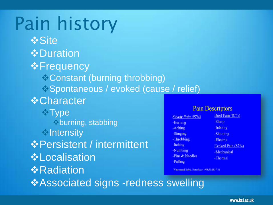
Pain history
Site
Duration
Frequency Constant (burning throbbing)
Spontaneous / evoked (cause / relief)
Character Type
burning, stabbing
Intensity
Persistent / intermittent
Localisation
Radiation
Associated signs -redness swelling
118
Blau44 suggested fifteen questions to facilitate the history taking
process in OFP which cover the following aspects of the presenting
pain: Onset
Frequency
Duration
Provoking factors
Site of initiation of pain
Radiation and referral of pain
Is the pain deep or superficial
Aggravating or exacerbating factors
Relieving factors
Characteristics of the pain
Severity
Other associated features, for example lacrimation or other
autonomic signs and symptoms
Previous management strategies attempted
Patient’s perceived cause(s) of pain
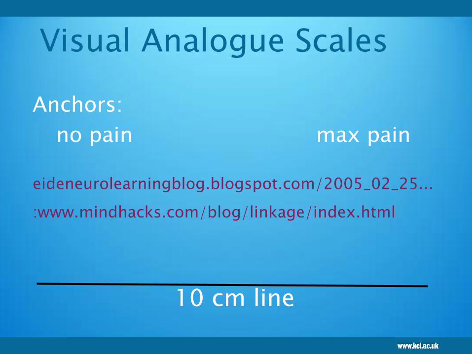
Anchors:
no pain max pain
eideneurolearningblog.blogspot.com/2005_02_25...
:www.mindhacks.com/blog/linkage/index.html
10 cm line
Visual Analogue Scales
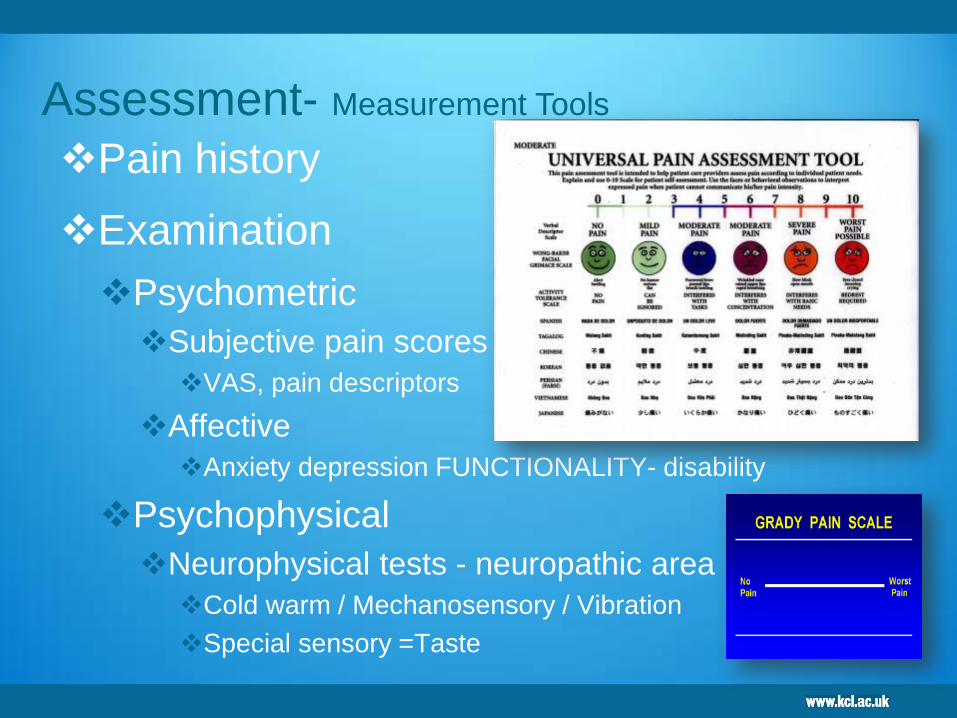
Pain history
Examination
Psychometric
Subjective pain scores
VAS, pain descriptors
Affective
Anxiety depression FUNCTIONALITY- disability
Psychophysical
Neurophysical tests - neuropathic area
Cold warm / Mechanosensory / Vibration
Special sensory =Taste
Assessment- Measurement Tools
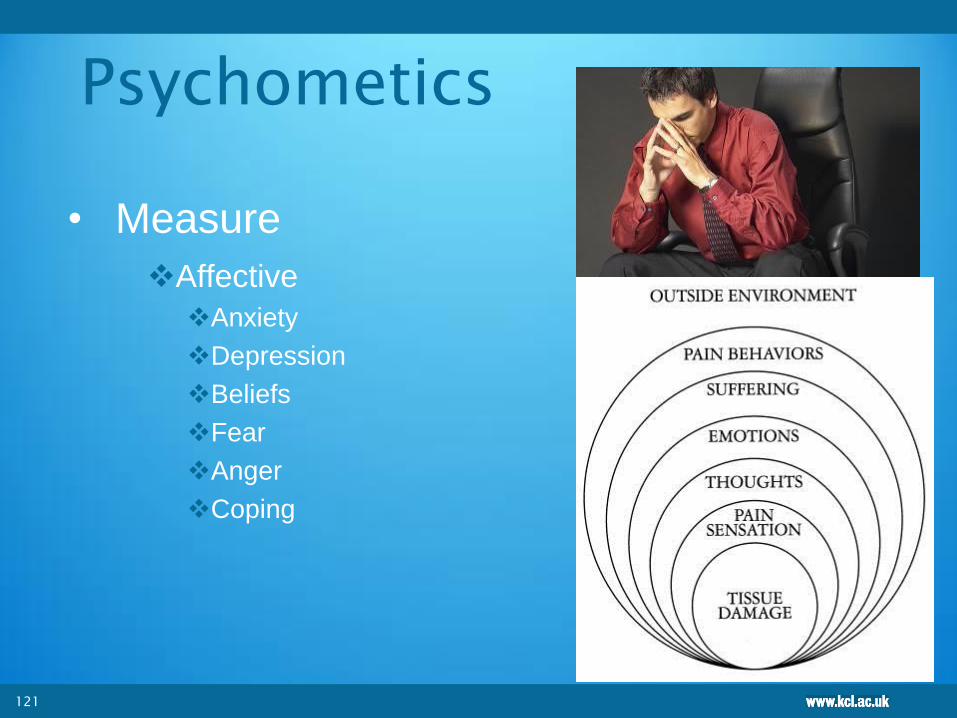
Psychometics
• Measure
Affective
Anxiety
Depression
Beliefs
Fear
Anger
Coping
121

We use………………
PSEQ Patient self efficacy Q
PCS Patient catastrophising scale
HADs Hospital anxiety depression
CPAQ Chronic pain acceptance Q
Euroquol Quality of health
OHIP Oral health impact Q
PCL The Posttraumatic Stress Disorder
Checklist
122

Clinical examination
123
Diagnostic process refines:
1. Inspection of the head and neck, skin, topographic anatomy, and
swelling or other orofacial asymmetry
2. Palpation of the temporomandibular joint and masticatory
muscles, tests for strength and provocation. With assessment and
measurement of the range of mandibular movement
4. Palpation of soft tissue (including lymph nodes)
6. Palpation of cervical muscles and assessment of cervical range
of motion
7. Cranial nerve examination
8. General inspection of the ears, nose, and oropharyngeal areas
9. Examination and palpation of intraoral soft tissue
10. Examination of the teeth and periodontium (including occlusion)
124

125
Systemic Diseases Associated with Headache and Orofacial Pain
Paget’s disease
Metastatic disease
Hyperthyroidism
Multiple myeloma
Hyperparathyroidism
Vitamin B deficiencies
Systemic lupus erythematosus
Vincristine and other chemotherapy for cancer
Folic acid and iron deficiency anaemias
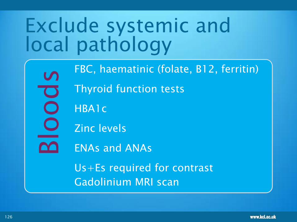
Exclude systemic and
local pathology Blo
ods FBC, haematinic (folate, B12, ferritin)
Thyroid function tests
HBA1c
Zinc levels
ENAs and ANAs
Us+Es required for contrast
Gadolinium MRI scan
126
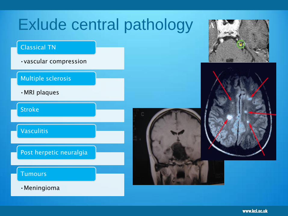
Exlude central pathology
•vascular compression
Classical TN
•MRI plaques
Multiple sclerosis
Stroke
Vasculitis
Post herpetic neuralgia
•Meningioma
Tumours
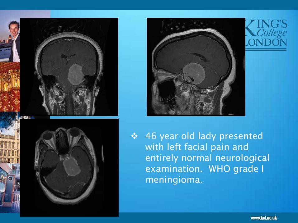
46 year old lady presented
with left facial pain and
entirely normal neurological
examination. WHO grade I
meningioma.
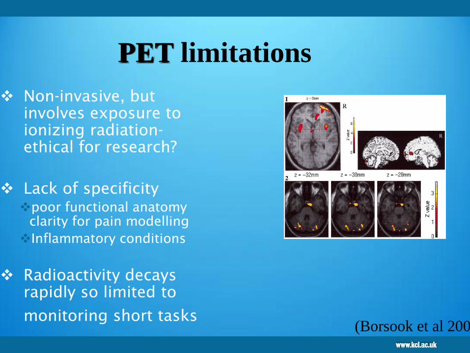
Non-invasive, but
involves exposure to
ionizing radiation-
ethical for research?
Lack of specificity
poor functional anatomy
clarity for pain modelling
Inflammatory conditions
Radioactivity decays
rapidly so limited to
monitoring short tasks
PET limitations
(Borsook et al 2004)
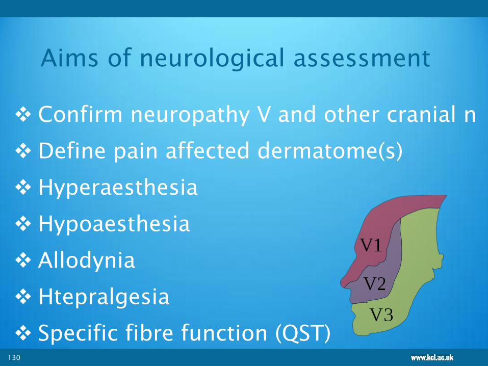
Aims of neurological assessment
Confirm neuropathy V and other cranial n
Define pain affected dermatome(s)
Hyperaesthesia
Hypoaesthesia
Allodynia
Htepralgesia
Specific fibre function (QST)
130

Assessment - neuropathy
• VAS
– At rest
– Dynamic allodynia
– Cold allodynia
– capsaicin
• Mechanosensory
– Von Frey
– Neuropathic area
• Local analgesia
• Thermo sensory
• Biopsy
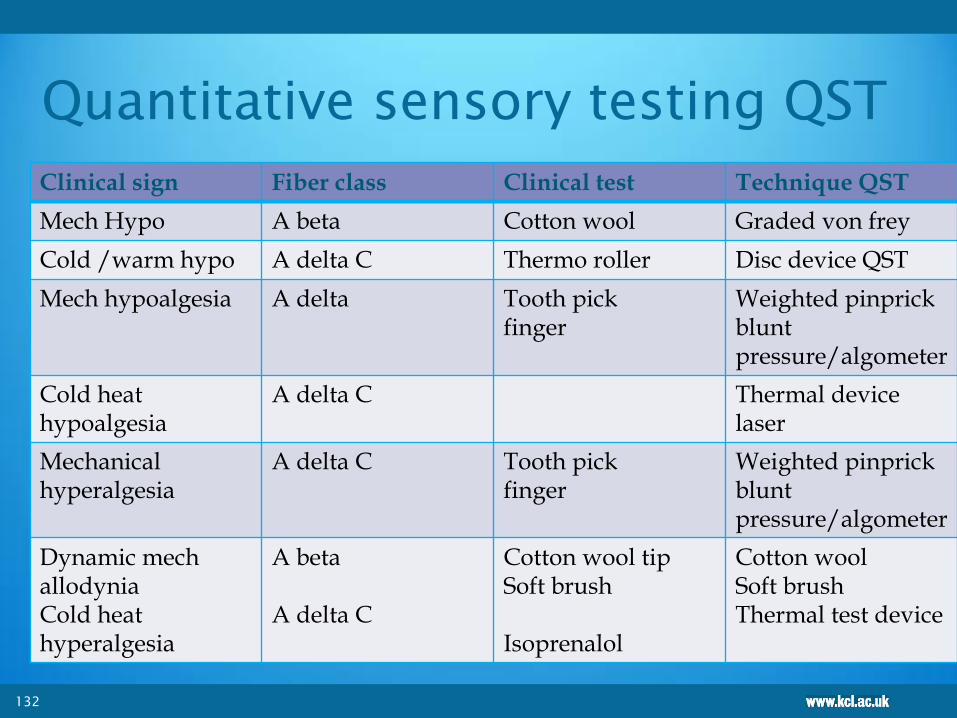
Quantitative sensory testing QST
Clinical sign Fiber class Clinical test Technique QST
Mech Hypo A beta Cotton wool Graded von frey
Cold /warm hypo A delta C Thermo roller Disc device QST
Mech hypoalgesia A delta Tooth pick finger
Weighted pinprick blunt pressure/algometer
Cold heat hypoalgesia
A delta C
Thermal device laser
Mechanical hyperalgesia
A delta C
Tooth pick finger
Weighted pinprick blunt pressure/algometer
Dynamic mech allodynia Cold heat hyperalgesia
A beta A delta C
Cotton wool tip Soft brush Isoprenalol
Cotton wool Soft brush Thermal test device
132

Psycho physical testing Quantitative thermo sensory testing
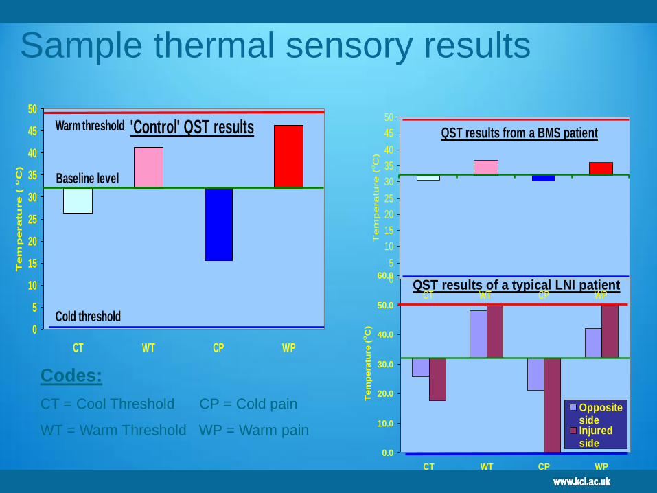
0
5
10
15
20
25
30
35
40
45
50
CT WT CP WP
Tem
peratu
re (
oC
)
'Control' QST results
Baseline level
Warm threshold
Cold threshold
0.0
10.0
20.0
30.0
40.0
50.0
60.0
CT WT CP WP
Te
mp
era
ture
(oC
)
OppositesideInjuredside
QST results of a typical LNI patient
QST results from a BMS patient
0
5
10
15
20
25
30
35
40
45
50
CT WT CP WP
Te
mp
era
ture
(oC
)Codes:
CT = Cool Threshold CP = Cold pain
WT = Warm Threshold WP = Warm pain
Sample thermal sensory results
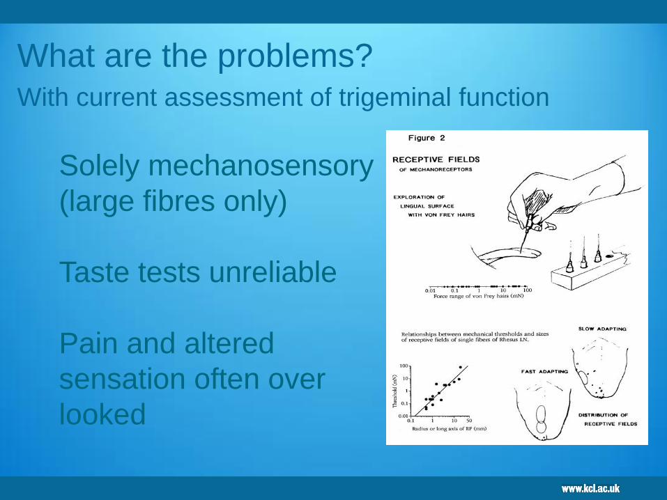
What are the problems? With current assessment of trigeminal function
Solely mechanosensory
(large fibres only)
Taste tests unreliable
Pain and altered
sensation often over
looked
QST may directly link suitable
management
Mechanical allodynia, hyperalgesia
Thermal
Blink reflex
JOR
136
Value of quantitative sensory testing in neurological and pain disorders:
NEUPSIG consensus.
Backonja M, Attal N, Baron R, Bouhassira D, Drangholt M, Dyck PJ, Edwards RR,
Freeman R, Gracely D, Haanpaa MH, Hansson P, Hatem SM, Krumova EK,
Jensen TS, Maier C, Mick G, Rice AS, Rolke R, Serra J, Toelle TR, Treede RD,
Tugnoli V, Walk D, Wallace M, Ware M, Yarnitsky D, Ziegler D.
Pain. 2013 Jun 3. doi:pii: S0304-3959(13)00289-3. 10.1016/j.

Summary
Management chronic OFP
Psychological
Medical
Interventional
KCH OFP
Lead TR
Liaison Psychiatrist Annabel Price
Clinical Psychologist Dr Sarah Barker
Health Psychologist Jared Smith
137
MDT St Thomas InPUT
TR lead OFP clinician
Pain management Tom Smith
Neurologist SamChong, Georgio
Lambru
Neurosurgery Sinan Barazi

Neurosurgical interventions
Microvascular decompression (MVD) for TN remains EB more effective
than thermo-controlled radiofrequency trigeminal rhizotomy
However……
Percutaneous interventions for:
trigeminal neuralgia (pterygoplatine blockade, gasserian ganglion glycerol injection)
cryotherapy
facet joint syndromes,
Most ablative pain surgery procedures
Neurotomy
Rhizotomy
sympathectomy, etc.)
These procedures have been replaced by neuromodulatory approaches
such as electrical stimulation of the central nervous system (CNS)
139

Interventional
Neural blockade (diagnostic or therapeutic)
Epidural injections PHN
Steroid injections Radiculopathy
Sympathetic blocks
GONB cluster headache
Botox Migraines
Superficial simulation – Neurostim/ TENS
Spinal cord stimulation SCS
failed back surgery
CRPS
Intrathecal medication
140
Value of quantitative sensory testing in neurological and pain disorders: NEUPSIG consensus.
Backonja M, Attal N, Baron R, Bouhassira D, Drangholt M, Dyck PJ, Edwards RR, Freeman R,
Gracely D, Haanpaa MH, Hansson P, Hatem SM, Krumova EK, Jensen TS, Maier C, Mick G, Rice
AS, Rolke R, Serra J, Toelle TR, Treede RD, Tugnoli V, Walk D, Wallace M, Ware M, Yarnitsky D,
Ziegler D. Pain. 2013 Jun 3. doi:pii: S0304-3959(13)00289-3. 10.101

141
Good evidence supports the use of neurostimulation for reducing pain
associated with failed back surgery syndrome (FBSS) and CRPS I, CRPS II,
peripheral nerve injury, DPN, PHN, brachial plexus lesion, amputation (stump
and phantom pains), and partial spinal cord injury
Motor cortex stimulation may be useful for central post-stroke pain and
neuropathic facial pain. Additionally, practice parameters issued in the United
States support the use of spinal cord stimulation techniques for treating
neuropathic pain in patients who have failed other forms of therapy [25].
A study published in January 2010 considered the use of spinal cord stimulation for
FBSS, using the outcome of workers’ compensation. A prospective
A task force assembled by the EFNS reviewed the literature published between 1968
and 2006 on neurostimulation therapy for treating neuropathic pain.

All treatments far from satisfactory
CBT and ACT most effective in our
unit
142
TRIGEMINAL NERVE FOUNDATION
Orofacial pain website
’to provide excellence in
education, management and
prevention of trigeminal chronic
orofacial pain’
THANK YOU