Embed Size (px)
Citation preview

AMERICAN ACADEMY OF PEDIATRICSPROCEEDINGS
NEW TECHNIQUES IN THE DIAGNOSIS ANDTREATMENT OF MEGALOURETERS
By Orvar Swenson, M.D., and John Herbert Fisher, M.D.Boston Floating Hospital for Infants and Children
Presented at the Annual Meeting, Chicago, October 5, 1955.
ADDRESS: (0.5.) 20 Ash Street, Boston 11, Massachusetts.
304
PediatricsVOLUME 18 AUGUST 1956 NUMBER 2
M EGALOURETERS is an uncommon condi-
tion that has been the subject of
much medical speculation.’ It has been a
troublesome clinical problem for the physi-
cian inasmuch as tile treatment has not
been uniformly effective. Patients with this
condition develop a gradual dilatation of
the ureters and, as time progresses, the
renal pelves and caliceal system become
dilated. Gradually the renal tissue is com-
pressed, and over a period of time com-
plete destruction of the renal tissue may
occur. Rate of progression of the disease
varies from patient to patient and, un-
doubtedly, infection is a complication which
intensifies kidney damage. In our experi-
ence fatalities have taken place as early as
the second year of life; rarely a patient may
reach middle age before renal failure oc-
curs.
In reviewing a group of 60 patients with
rnegaloureters, we have been impressed
with the variety of complaints and physical
findings which have initiated investigation
of the urinary system. The paucity of corn-
plaints tilese patients may have is illustrated
by the following case history:
A 9-year-old boy was admitted to our
hospital in extremus and died a few hours
after admission. Necropsy demonstrated mega-
loureters and hydronephrosis to be the cause
of renal failure. A retrospect review of the his-tory with this family failed to uncover any def-
mite complaints that would have been refer-
able to the genitourinary system. The child
was smaller than normal for his age but outside
of this the family had no clue which might
have warned them of progressive renal disease.
In this group of 60 patients, 35 came for
medical attention because of urinary tract
infection. In 15, failure to gain prompted
a general physical examination. Seven pa-
tients were found to have leukocytes in the
urine on routine analysis, and 5 children
had either palpable bladder or kidney on
routine examination. This experience has
made us more liberal in our indications for
�Il intravenous urograrn for patients who
are not doing well or who have rather
vague complaints with an otherwise normal
history and physical examination.
PROCEDURE FOR STUDY OF
URINARY SYSTEM
We have been particularly interested in this
condition and have endeavored to perform
studies of the urinary system in an attempt to
classify these patients according to the etiologyof the dilatation of the upper urinary tract.
by guest on April 1, 2019www.aappublications.org/newsDownloaded from

AMERICAN ACADEMY OF PEDIATRICS - PROCEEDINGS 305
Our routine is that the children are admitted
to the hospital and those who prove to have a
normal blood urea nitrogen are subjected to
intravenous urography. Before any other tests
which iilvolve catheterization of the bladder
are done, the residual urine is determined by
catheterizing the child immediately after
voiding. Children normally may have a re-
sidual urine volume as high as 10 to 15 ml.
More than this amount, it is our belief, mdi-
cates some defect in function or an obstruc-
tion which prevents complete emptying of the
bladder. At the time the residual urine is de-
termined, a specimen is obtained for culture
aIld antibiotic sensitivity stndies. The catheter
is then left in place for serial cystograms and
cystometrograms. The cystometrogram has to
be performed under ideal conditions, for if the
patient had been catheterized or had an in-
dwelling catheter, even a few days prior to
the test, the resulting curve may be unreliable.
lil order to standardize the cystometrogram
study we have utilized an ink writing recorder
with a transducer sensitive in the range of
0-100 ml. of water (Fig. 1). This annaratus . . .
. FIG. 2. Voiding urethrogram from patient withmakes a permanent and continuous record of iparisyiiipatI�etic bladder.
intravesical pressure during bladder filling as
well as of the timing and intensity of detrusor
contractions. It is (iifficlilt to obtain good trac-
tugs on a child who is not co-operative. By
amusing the ciliidren we are usually able to
secure good tracings even with youngsters of
2 to 3 years of age. Below 2 �‘ears of age the
cvstonletrogram is difficult to perform and
interpret. To shorten the duration of the test,
bladder filling is augmented with a slow drip
of a solution consisting of 1 part of 30 per cent
Diodrastui� to 2 parts of saline. It is advisable
to oi)tain 3 or 4 roentgenograms during filling
of the l)la(lder and to record the intravesicular
pressure which is present at the time the film
is taken. Such a procedure will not oniv revealthe presence of reflux but also at what stage
of bladder filling it occurs. \Vhen the patieiit’s
bladder is full and there is discomfort, the
exammation is discontinued, the catheter is
removed from the bladder, and a voiding ore-
throgram is obtained (Fig. 2). This is essen-
tial to rule out mechanical obstruction at the
bladder neck or along the course of the
urethra. Such a urethrogram is far more in-
FIG. 1. Recording apparatus for cystometrogram formative than a retrograde urethrogram, since
and peristalsis studies. abnormalities are more readily outlined during
by guest on April 1, 2019www.aappublications.org/newsDownloaded from

306 SWENSON - DIAGNOSIS AND TREATMENT OF MEGALOURETERS
FIG. 3. Cystometrogram froiii normal patient.
voiding tilaIl when the radio-opaque material
is injected retrograde. On the following day
cystoscopy is performed and the posterior
urethra and bladder neck carefully observed
for any form of mechanical obstruction.
RESULTS
Mechanical Obstructions
Applying these diagnostic tests to pa-
tieITLts Witil megaloureter, only a small num-
ber have proved to have mechanical ob-
struction. Of 60 children, 2 had posterior
urethral valves and 1 had a congenital
stricture of the urethra. The diagnosis in
these conditions is evident on voiding ure-
tllrOgrams and on inspection during cystos-
copy. These obstructions produce large
trabeculated bladders and dilated upper
urinary tract. Removal of urethral valves is
most readily accomplished with a trans-
urethral resectoscope. The child with a
strictured urethra was treated by dilatation.
None of the 60 patients proved to have
stricture of the distal part of the ureter.
Four patients had a ureterocele as the cause
of megaloureters; treatment consists of
opening the ureterocele. Six patients with
neurogenic bladders have been observed to
have megaloureters. The etiology is an in-
creased intravesicular pressure rather than
a defect in the ureter itself. Treatment of
these patients is too complex for a review
of the subject in this place.
Aparasympathetic Bladders
Thirty-two of tile sixty patients have been
found to have the following deviations from
normal in their urinary tract: The residual
urine volume is elevated, frequently being
over 100 ml., and in some patients it Ilas
been as high as 600 ml. Ureteral reflux from
the bladder is fairly common but does not
invariably occur. The cystometrogram trac-
ing deviates from the normal considerably.
In a series of patients with normal blad-
ders, in whom we ilave had an opportunity
to perform cystometrograms with the type
of recording apparatus described, the initial
intravesical pressure has been near zero.
During filling, the pressure gradually in-
creases to 10 or 15 cm. (Fig. 3). In a 4- to
6-year-old child, when the bladder contains
150 to 200 ml. of fluid the patient has a
strong urge to void and there are powerful
detrusor contractions. Usually the pressure
does Ilot exceed 15 cm. of water prior to
initiation of powerful detrusor contractions.
Patients with megalobladder and meg-
aloureters have been found to have large
bladder capacity (Fig. 4). A 4- or 5-year-old
child with tilis condition may tolerate 400
to 500 ml. of fluid injected into the bladder
without discomfort. Sucil patients seem to
have a loss of sensation in the bladder,
and it is the generalized abdominal discorn-
fort at the extreme of filling rather than a
specific bladder discomfort wllicil prompts
tilem to attempt to micturate. They empty
the bladder by increasing intra-abdominal
I)ressure; consequently tile urinary stream is
feeble, and voiding consumes more time
than in normal individuals. The intravesicu-
lar pressure is excessive, averaging 20 to
25 cm. of water before abdominal discom-
fort is noted. One patient had an intra-
vesicular pressure of 40 cm. of water. In
view of the fact that the average ureteral
peristaltic wave generates a pressure of 35
cm. of water, the patient with an intra-
by guest on April 1, 2019www.aappublications.org/newsDownloaded from

FIG. 4. Cystometrograrn from patient with aparasympathetic bladder.
AMERICAN ACADEMY OF PEDIATRICS - PROCEEDINGS 307
vesicular Presst1i�e of 20 or 25 cn�. of water
requires niore work of a ureter than when
tile i)ladder is normal, with a pressure of 12
cm. or less. Ill addition such patients have
weak or completely absent detrusor con-
tractiolls. Occasionally there may be a pro-
URETERAL PERISTALSIS
LEFT �
RIGHT -�.--�--�----� -� � -�-�--�
RIGHT �
FIG. 5. Ureteral l)eristaltic tracings showing One of
normal amplitude and others diminished.
longed contraction, developing a pressure
of 10 or 20 cm. of water, compared to a
nornlal detrusor contraction wilich develops
a pressure of 100 cm. of water or nlOre. At
cystoscopy no obstruction ill the urethra,
bladder neck or ureterovesical junction was
found in tilese patients. The bladders were
sfllootil ill outline and showed evidence of
cilrOllic infection, and the urethral orifices
were enlarged if reflux was present. Each
ureter was catheterized and peristalsis re-
corduigs illade employing tile same equip-
nient used for cystometrograms (Fig. 5). In
normal ureters peristalsis occurs at 15- to 20-
Secoild intervals aild each cotitraction de-
velops a flow of urille witil a iresstire of 35
to 40 cm. of water.2 Anesthetic agents and
1)1eoperati\e medication have no influenceOIl this function. In pitti#{128}�11ts \Vitll nieg-
aloureter peristalsis tracings revealed nor-
fll�il contractions in the ureter; in a fe�v
there was low amplitude and fewer contrac-
tions than normal (Fig. 6). Following the
completion of these studies which are diffi-
by guest on April 1, 2019www.aappublications.org/newsDownloaded from

308 SWENSON - DIAGNOSIS AND TREATMENT OF MEGALOURETERS
FIG. 6. Ureteral peristaltic tracing from patient
�s’itI:i aparasympathetic bladder.
cult, tedius lIl(I tulle consuming to 1)erforlll,
retrograde �)velogranls are niade.\Ve ilave had an op�)Ortullitv for iliS-
tologic exaillinatioll of 8 bladders from pa-
ti&�iits who seenied to fit all the clinical
criteria of nlegalobladder and nlegaloure-
ters, of the ty�)e outlined. ‘ It was foulld
111)011 careful studs’ of sections of the whole
i)ld(l(ler that there is a marked (linlinutioll
ill ganglion cells as comparedi to tile normal
concentration �ul(1 distril)ution (Figs. 7 and
8). This suggests tile possibility of a con-
genital I)ar�lS�’ll1I)�1t1letic defect Ill the I)ladl-
der to account for the dysfunction. Gen-
erally it is agreed that detrusor function,
that is i)ladder contraction, is dependent on
intact parasymp��ihetic innervation . �Ve
have teSte(i this ill the laboratory by tile
following experinlents. Ten healthy female
dogs were subjected to catheterization, and
cystometrogram tracings revealed normal
pressure and active detrusor contractions.
Bilateral division of the parasympathetic
nerves were performed, and repeat cysto-
metrograms revealed no bladder contrac-
tions and this absence persisted over a 3-
month period. We do not consider this
experimental lesion to be fully comparable
to the congenital defect, however, it does
indicate the relationship of the parasympa-
thetic system to bladder contractions.
It has been interesting in studying pa-
tients with megacolon to demonstrate that
about 50 per cent have some defect in
bladder function detectable on careful
cystometrogram studies, consisting of large
capacities and absent detrusor contractions.
However, they have normal residual urine
volumes and intravesical pressures are nor-
mal. In about 3 per cent of patients with
megacolon we have studied, the lesion in
the bladder is more pronounced and then
megaloureters are associated with megalo-
bladder. In these patients the bladder has
the same changes that we have noticed in
patients with megaloureters and megalo-
bladders but with normal colon. Tile
changes referred to are: increased capacity
of the bladder; feeble or absent detrusor
contractions; high residual urine volume,
and high intravesical pressure.
It would seem reasonable that in patients
with a congenital defect in the pelvic para-
sympathetic system, and consequently no
ganglion cells in the distal part of the
colon, a few would have a defect in the
parasympathetic innervation of the bladder.
A congenital defect in the pelvic parasym-
pathetic system may produce 3 types of
l)atiellts. If limited to that portion of thepelvic nerves which supply tile distal part
of tile colon, the patient would have con-
genital nlegacolon with a normal urillary
Systeill. A second ty�)e of patient Illight
have a illore extensive malformation and
consequently function of 1)0th the colon
alld urinary bladder might become defec-
tive. We have observed such patients in tile
group of children with congenital mega-
colon and megaloureters we have studied.
by guest on April 1, 2019www.aappublications.org/newsDownloaded from

AMERICAN ACADEMY OF PEDIATRICS - PROCEEDINGS 309
FIG. 7 (Left). The wall of a normal bladder.
FIG. 8 (Right). The wall of an aparasvmpathetic bladder.
Logically there should be a third type of
patient with normal colon and a defect ill
that part of the pelvic parasympathetic
system which supplies the bladder. We
believe that this comprises the group of
patients Witil megalobladder and megalou-
reters, and we have designated them as
having an aparasympathetic bladder. Such
patients have a defect in function which is
less prollouncedl than tilat noted ill cord
bladder, for they are never incontinent.
From these studies we concludedi that pa-
tients ill the group with aparasympathetic
bladder have dilatation of the upper un-
nary tract secondary to a defect in bladder
function. Should this be true, bladder drain-
age would be effective treatment. With this
ill mind, 24 such patients were treated b�
either suprapubic drainage or a catheter ill
the urethra and illlprovelllent occurred in
all.
Prolonged drainage of tile bladder is not
a convenient method of treatment either
for the child or parent. In order to reduce
tile residual urine alld to aid tile children in
emptying the bladder, resection of the blad-
der neck has been performed. Tile ration-
ality of tilis treatment is appreciated 01)011
realization that opening of the internal
sl)hillcter or tile bladder neck is a functionof tile detrusor muscle (Fig. 9). These chil-
dren have a defect ill detrusor contractions
and as a result there appears to be ineffec-
tive opening of the bladder neck; tiliS, 1)ltlS
feeble contractions of the bladder, prob-
ably results in the large residual urine vol-
ume which is COIT1IT1OI1 ill this conditioll.
The internal sphincter has no essential func-
tiOlls and can be eliminated without en-
dangering the child’s continence. Either
transurethral resection of the bladder neck,
or resection through open cystotomy, has
been performed �n 15 patients with an
apanasympathetic bladder.
The most iml)ortallt feature of treatInent
consists in ilaving tile l)atient niake a sos-
tamed effort to empty tile bladder corn-
pletely. Usually this can only be accom-
by guest on April 1, 2019www.aappublications.org/newsDownloaded from

:310 SWENSON - DIAGNOSIS AND TREATMENT OF MEGALOURETERS
FIG. 9. Bladder neck showing detrusor fibers be-
fore and after resection.
1)lished by tile aid of manual suprapubic
pressure. Because these children have no
sense of bladder fullness ulltil tile bladder
colltaills excessive amounts of urine under
high pressure, it is equally important to
have them void at regular intervals despite
the absence of a sense of bladder fullness.
A schedule of 4 or 5 voidings a day and 1
or 2 at night is ideal, for ullder such circuni-
stances the intravesical pressure is never
greatly above normal.
Treatment of urinary infection is also
important and should consist of small doses
of sulfisoxazol for long periods of time.
These patients should be regarded as hay-
ing a chronic disease and regular visits to
the physician Silould be made. At monthly
intervals urinalysis will give an indication
of the amount of infection. A large number
of leukocytes in the urine should prompt
catheterization for residual urine and cul-
tune to determine the organism and test tile
sensitivity to various therapeutic agents.
When exacerbations of infection occur, a
more potent agent than sulfisoxzol is desina-
ble for short periods. Roentgenograrns
silould be made annually to determine the
course of the disease. It has been necessary
to do a second resection on some of these
patients in order to maintain normal re-
sidti�l urine volumes. Our longest follow-up
is on a patient 18 years of age, first seen
at 11 years (7 years ago), with fairly adi-
vanced hydronephrosis, megaloureters and!
megalobladder (Fig. 10). Initially the blood
nonprotein nitrogen was 66 mg./100 ml. A
period of bladder drainage was beneficial.
This was followed by 2 transurethral resec-
tions in order to reduce the large residual
Unille volume to normal. Until this was ac-
coinplished the infection could not be con-
trolled. This co-operative patient ilas voided!
OIl a regular schedule of 5 times a day and
once at night to prevent development of
high intravesical pressure. Tile 1)loodl urea
nitrogen has remained normal and uro-
grams are now essentially normal. Out of a
group of 15 patients subjected to resection
all but 2 have done well. For a period of
time we were resectioning a large part of
the fundus. The 2 patients who have not
done well had more extensive removal of
the bladder wall than the others, and tile
intravesical pressures are higher now than
prior to operation. This experience has
prompted us to discontinue resection of pant
of the bladder. There Ilas been 1 death-
a child who died of monilia infection after
prolonged intensive antibiotic treatment.
In this condition direct operations on the
ureter are not indicated. Actually the ureter
has normal peristalsis without evidence of
ureterovesical obstruction, and attempts to
shorten such ureters or to re-implant them
into the bladder are of no avail; the funda-
mental defect is in bladder function.4 Op-
erations On the lower end of the ureter are
by guest on April 1, 2019www.aappublications.org/newsDownloaded from

AMERICAN ACADEMY OF PEDIATRICS - PROCEEDINGS 311
FIG. 10. Intravenous pyelogranl of patient with aparasympathetic bladder before (left), and 7
�ears after beginning of treatment (rig/it).
contraindicated due to the fact that addi-
tional damage to the innervation of tile
i)ladder niay be sustained during such pro-
cedures. We Ilave seen 1 patient who had
bilateral operations on the lower end of tile
ureter, and tilere was inability to enlpty
the bladder postoperatively, and! tile patient
was committed to permanent catheter
drainage. Our interpretation of this result is
that the patient initially had considerable
defect in parasympathetic innervation of
tile bladder and that inadvertently during
operation Oil the lower end of the ureter, an
area \vilene innervation enters the bladder,
the meager nerve supply was damaged with
the result that the patient was unable to
void postoperatively. In other patients the
residual urine volume has risen consider-
ably after this type of operation, indicating
damage to the bladder innervation. In this
disease it is far more logical and effective to
attack the basic pathology which is in the
bladder. We have observed 2 patients with
agenesis of the abdominal wall associated
�vit1i nlegalobladder and nlegaloureters;
tilese patients have not fared! d!ifferentlv
from other children with this cond!ition cx-
cept that they seem to do better as tile al)-
domirial wall grows more firm later in life.
Perhaps, tiliS is due to the fact that they are
able to empty the bladder more effectively
with increased intra-aI)dominal pressure.
Absence of Ureteral Peristalsis
In the course of studies of patients with
nlegaloureters, 15 have been found to have
essentially normal bladders \vitil no residual
urine. In studying these patients for ureterai
peristalsis we found an absence of, or only
feeble, contractions. Some of tilese children
apparently Ilave a congenital defect ill tile
ureter to account for the absence of pen-
stalsis. It has been suggested that infection
is tile cause of tile absellce of ureteral
penstalsis. However, we have studiedi sonic
Patielits who had quite extensive urinarytract infection with perfectly normal ore-
teral peristalsis. It is true that sonic of the
by guest on April 1, 2019www.aappublications.org/newsDownloaded from
312 SWENSON - DIAGNOSIS AND TREATh’IENT OF MEGALOURETERS
patients have had some slight return of
function after prolonged treatment of the
infection. Tilis may indicate that in certain
instances infection niav play a role ill
dampening or dimiiiishing ureteral penistal-
sis, but where there is complete absence of
l)eniStaltic activity, after repeated studies,we are inclined to believe that it is prob-
ably on a congenital basis. Unfortunately
one cannot gain the answer to this question
from studymg the ureters histologically,
for normally there are few if any ganglion
cells in this structure. :i Therefore, this
method of studying the autonomic innerva-
tion of an organ which has been so useful
in the colon and! bladder is of no avail in
the ureter. Regardless of the etiology of
absent penistalsis, the defect in function
leads to considerable enlargement of the
ureter and! of tile renal pelvis. It has 1)een
extremely difficult to control ilifection ill
these patients. Providing infection is eradi-
cated, these cilild!ren niav go on \vitil ver�’
little �)rogresSioll of the disease and do well
generally. In some installces control of in-
fection has been impossible and in some,
dilatation of the ureters and pelves has oc-
curred. One patient in this group died with
renal failure. The lack of effective therapy
for such patients prompted us to seek a
substitute for ureters deficient in peristalsis.
In dogs, segments of the small intestine
were isolated and the lumen reduced in size
by excising three-quarters of the wall. This
left a narrow strip of intestinal wall at-
tached to the mesentery which could be
sewed into a small tube not much larger
than a normal ureter. At a second stage,
the ureter on one side was removed and
replaced by this previously prepared! nar-
row ileal segment. We were gratified with
the results ill the laboratory. The function
seemed to be ad!equate and infection was
ilot a probleni. Furthermore, there seemed
to be no perceptible reabsorption of dee-
trolytes through the small surface area of
ileal mucosa exposed to urine. In the past,
full-size segments of ileum had been used
to replace the ureter.5 We felt that it would
be impossible to prevent reflux when one
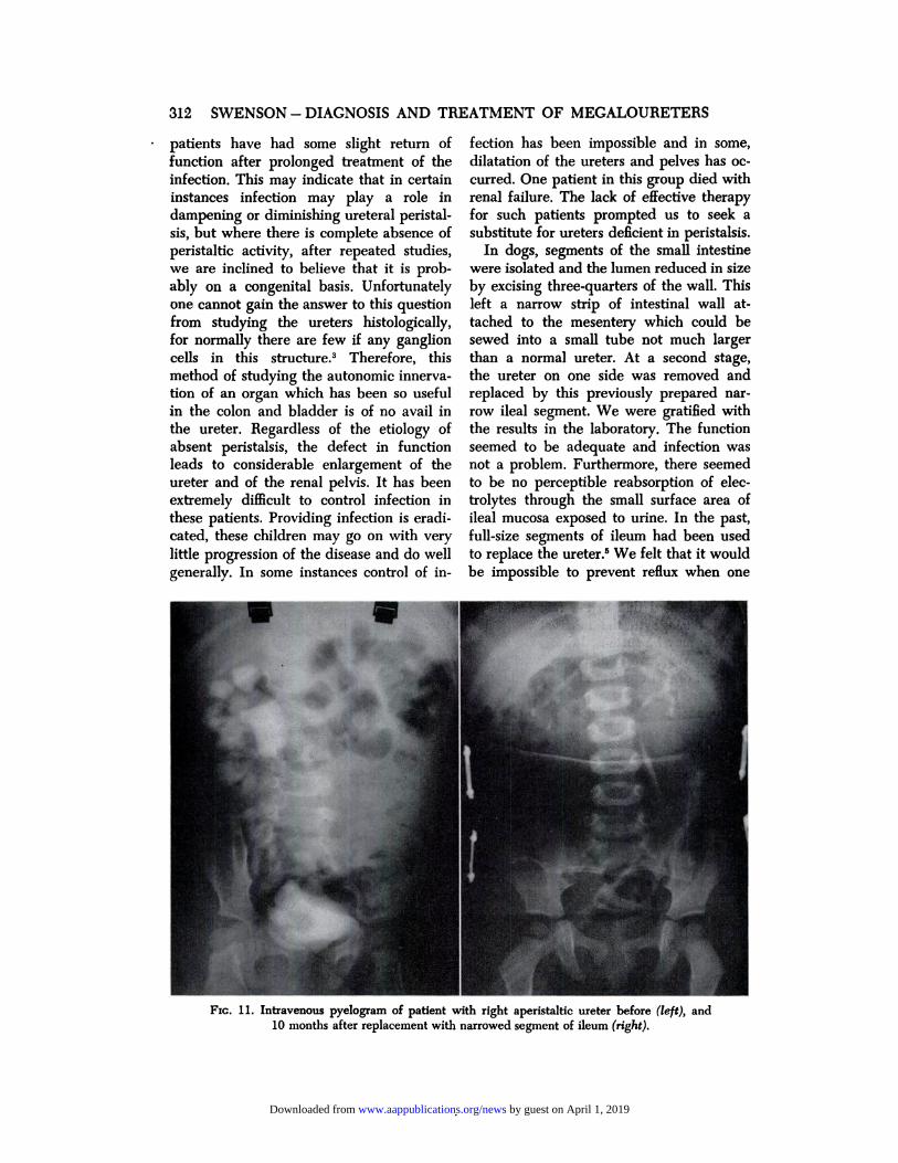
Fic. 1 1 . Intravenous pyelogram of patient with right aperistaltic ureter before (left), and
10 months after replacement with narrowed segment of ileum (right).
by guest on April 1, 2019www.aappublications.org/newsDownloaded from

AMERICAN ACADEMY OF PEDIATRICS - PROCEEDINGS 313
was faced with anastomosing the full-sized
lumen of the small intestine to the bladder,
and chances of reabsorption were greater
because of the enlarged ileal surface area.
We concluded that narrowing the lumen
would overcome all of the objections to a
large extent. Because of our favorable cx-
perience in the laboratory, 6 patients were
subjected to this procedure with gratifying
results. It has been interesting to study the
peristaltic activity of these narrow segments
of small intestine. Peristaltic contractions
with pressures as high as 70 and 80 cm.
of water have been recorded, which mdi-
cate that these “ureters” made from small
intestine have considerably more effective
pumping action than a Ilormal ureter. One
patient has been followed for 10 months
after replacement of the right ureter (Fig.
11). The problem in this particular patient
preoperatively was uncontrolled infection.
There has been no urinary infection since
the apenistaltic ureter was replaced with a
segment of small intestine, and the hydro-
nephrotic kidney has returned to normal. It
would seem that in patients with aperi-
staltic ureters or ureters with defective pen-
stalsis, where infection cannot be controlled
or where there is progression of the dis-
ease, replacement with ileal segments offers
a practical form of treatment.
SUMMARY
Studies on 60 patients with megaloure-
tens are described.
Thirty-two patients proved to have a de-
feet in parasympathic innervation of the
bladder. The defect in bladder function was
the cause of ureteral dilatation. Bladder
neck resection is the treatment of choice.
Fifteen patients proved to have defects
in ureteral penistalsis as the cause of ore-
teral dilatation. Replacement of some of
these defective ureters with narrowed seg-
ments of ileum has proved successful.
REFERENCES
1. Williams, D. I. : The chronically dilated
ureter. Ann. Roy. Coil. Surgeons Eng-land, 14:107, 1954.
2. Trattner, H. R. : A method for recording
contractions of the intact human ureter. J.Urol., 11:477, 1924.
3. Swenson, 0., MacMahon, H. E., Jaques,
W. E., and Campbell, J. S. : A newconcept of the etiology of megalo-ureters. New England J. Med., 246:41,
1952.4. Lewis, E. L., and Kimbrough, J. C. : Megalo-
ureter; new concept in treatment, South.
M. J.,45:171, 1952.5. Pyrah, L. N., and Rapen, F. P. : Some uses
of an isolated loop of ileum in genito-
urinary surgery. Brit. J. Sung., 42:337,
1955.
by guest on April 1, 2019www.aappublications.org/newsDownloaded from

1956;18;304Pediatrics Orvar Swenson and John Herbert Fisher
MEGALOURETERSNEW TECHNIQUES IN THE DIAGNOSIS AND TREATMENT OF
ServicesUpdated Information &
http://pediatrics.aappublications.org/content/18/2/304including high resolution figures, can be found at:
Permissions & Licensing
http://www.aappublications.org/site/misc/Permissions.xhtmlentirety can be found online at: Information about reproducing this article in parts (figures, tables) or in its
Reprintshttp://www.aappublications.org/site/misc/reprints.xhtmlInformation about ordering reprints can be found online:
by guest on April 1, 2019www.aappublications.org/newsDownloaded from

1956;18;304Pediatrics Orvar Swenson and John Herbert Fisher
MEGALOURETERSNEW TECHNIQUES IN THE DIAGNOSIS AND TREATMENT OF
http://pediatrics.aappublications.org/content/18/2/304the World Wide Web at:
The online version of this article, along with updated information and services, is located on
Copyright © 1956 by the American Academy of Pediatrics. All rights reserved. Print ISSN: 1073-0397. American Academy of Pediatrics, 141 Northwest Point Boulevard, Elk Grove Village, Illinois, 60007.been published continuously since 1948. Pediatrics is owned, published, and trademarked by the Pediatrics is the official journal of the American Academy of Pediatrics. A monthly publication, it has
by guest on April 1, 2019www.aappublications.org/newsDownloaded from