Embed Size (px)
Citation preview

Title STUDIES ON THE HELMINTH FAUNA OF JAPAN -PART52. TREMATODES OF FISHES, XI-
Author(s) Yamaguti, Satyu
Citation PUBLICATIONS OF THE SETO MARINE BIOLOGICALLABORATORY (1958), 6(3): 369-384
Issue Date 1958-06-20
URL http://hdl.handle.net/2433/174588
Right
Type Departmental Bulletin Paper
Textversion publisher
Kyoto University

STUDIES ON THE HELMINTH FAUNA OF JAPAN
PART 52. TREMATODES OF FISHES, XI
SATYU YAMAGUTI
Department of Parasitology, Okayama University Medical School, Okayama
With Plates XIV-XV
CONTENTS
AEROBIOTREMA TIDAE n. fam. Page 1. Aerobiotrema muraenesocis n. g., n. sp .............................................. 369
ALLOCREADIIDAE STOSSICH, 1903 2. Helicometra epinepheli YAMAGUTI, 1934 .......................................... 372 3. Opecoelus lateolabracis n. sp. .. .. 000000 ...... 00 ...... oo .... 00 ......................... 372 4. Opecoelus sebastisci n. sp. .. .......... 00 00 ........ 00 .............. 00 00 .. 00 .... 00 00 00 oo373 5. Opecoelus pagrosomi n. sp. oo ................ oo .. oo .... oo .. oooo .. oo ...... OOOOOoOOOOOoOo374 6. Ope gaster cryptocentri n. sp. .. .. 00 00 ........ 00 .......... 00 00 .............. 00 00 .... 00.375
ACANTHOSTOMIDAE PocHE, 1926 7. Biovarium lateolabracis n. sp. oo .. oooo .......... oo .............. oooo ................... 376
ACANTHOCOLPIDAE LtiHE, 1909 8. Adolescaria of Stephanostomum pagrosomi (YAMAGUTI, 1939) ............ 377
CRYPTOGONIMIDAE CruREA, 1933 9. Diplopharyngotrema lateolabracis n. g., n. sp ................... oo .. OOOo .. OOOo .... 379
10. Pseudosiphoderoides hapalogenyos n. g., n. sp ..... 0000 ............................ 381 BUCEPHALIDAE PocHE, 1907
11. Prosorhynchus crucibulum japonicum n. subsp. .. ............................... 382 ACKNOWLEDGMENTS ........................................................................... 383 LITERATURE ...................... OOOOOooo ................ oo ............................... oo••·•oo383 EXPLANATION OF PLATES XIV-XV oo .......... oo ................ oo .... oo .. oo .... oo ...... 384 ABBREVIATIONS USED IN FIGURES ...... oo oo ...... oo oo .. oo .. oo oo oo ...................... 384
AEROBIOTREMATIDAE n. fam.
1. Aerobiotrema muraenesocis n. g., n. sp.
(Pl. XIV, Figs. 4-6; Pl. XV, Fig. 10)
Habitat: Air-bladder of Muraenesox cinereus. Material and locality: Two mature specimens; Tamano Aquarium at Shibukawa,
Tamano City, Okayama Prefecture.
Publ. Seto Mar. Biol. Lab., VI (3), 1958. (Article 19)

370 S. YAMAGUTI
Body plump 13.5 mm long1l, rounded at two extremities; the hemispherical posterior
region covered with smooth cuticle is delimited from the main body by a distinct
circular ridge as shown in the free-hand sketch (Fig. 10). Forebody tapered anteriorly
and curved a little ventrally; cuticle thick, transversely wrinkled ventrally, especially at
the acetabular level. Between the genital pore and the acetabulum is a transversely
elongated shallow depression clearly recognizable with a hand lens. On the sectioned
preparations the body parenchyma consists of an outer layer of comparatively fine
network of connective fibrils and an inner layer of spongy network of coarser fibrils,
the open spaces of which contain very fine granules precipitated by SCHAUDINN's
fixative with glacial acid added. The oral and ventral suckers, pharynx, esophagus,
terminal genitalia and peripheral uterine coils are embedded in the dense peripheral
layer, while the other reproductive organs, intestinal ceca and excretory vesicle occupy
the spongy central parenchyma.
Oral sucker spherical, 1.7-2.1 mm in diameter, subterminal, with overhanging
preoral lip formed by body wall, directly followed by muscular pharynx 0.9 mm long
by 0.96 mm broad. Esophagus constricted at very beginning, but greatly expanded
elsewhere, directed posterodorsally and then turning ventrad to bifurcate behind
pharynx. Ceca very wide throughout their length, with sinuous walls, forming an
abrupt turn in front of testes, and encircling the latter, behind which they form
another acute turn to encircle the ovarian complex, finally terminating blindly at
posterior extremity near dorsal cuticle. Acetabulum spherical, embedded in body paren
chyma about one-third of body length from anterior extremity.
Testes rounded rectangular in lateral view, 3.3mm in greatest dorsoventral diameter,
lying in direct contact with each other behind level of acetabulum between two ceca,
surrounded dorsally by uterine coils, each giving off its own vas efferens from near
anterodorsal corner. Two vasa efferentia running forward convergently, uniting to
form elongate sigmoid vesicula seminalis, which is wider distally than proximally,
attaining maximum width of 0.4 mm. Pars prostatica 0.7 mm long, widest (0.25 mm)
at base, surrounded by flask-shaped compact mass of gland cells, situated dorsoventrally
about midway between pharynx and acetabulum, followed by comparatively short
(0.4 mm long) ejaculatory duct which is lined with thick smooth cuticle and opens
along with the metraterm into the ductus hermaphroditicus. The latter, appearing as
a direct continuation of the metraterm, is 0.7 mm long by 0.15 mm wide and opens
outside in the median line a little behind the mouth aperture.
Ovary small, ovoid, 0.8 X 0.4 mm, situated about middle of posterior portion
demarcated from main body, left of shell gland, with long axis dorsoventral. Shell
gland large, enclosed in fibrous capsule in front of distal end of ceca; ootype large,
surrounded by radiating gland ducts ; receptaculum seminis and Laurer's canal absent.
The uterine duct, winding in the spongy parenchyma on the left of the ovary, proceeds
1) Unless otherwise stated, all measurements are from specimens subjected to coverglass pressure when fixed with SCHAUDINN's solution.
-130-

Studies on the Helminth Fauna of japan, 52 371
toward the periphery; after forming a transverse coil on the right of the ovarian
complex the uterus turns back to the left side and extends longitudinally, occupying
the whole ventral area between the posterior extremity and the testes, then running
dorsad behind the testes forms two longitudinal loops on the dorsal side of the
testes and ceca. Finally it comes to lie in front of the testes, where the terminal
uterus passes between the two vasa efferentia and leads into the metraterm beside
the pars prostatica. Metraterm 1.0 mm long, lined with thick smooth cuticle and
provided with inner circular and outer longitudinal muscles. Eggs round, embryonated,
16-18 p. in diameter. Vitellaria divided into bunches of large rounded follicles, ex
tending longitudinally on each side of ovarian complex, between the latter and ceca,
as well as in intercecal field just posterior to testes.
Excretory vesicle Y -shaped in general pattern ; the stem originating from the
terminal pore ascends in the central region, then on the left of the ovarina complex,
and bifurcates at the level of the anterior end of the vitellaria; two arms apparently
uniting dorsal to pharynx, sending off numerous, long or short, partly anastomosing,
side branches, most of which lie close to the ceca medial, ventral, or lateral to them.
This genus is characterized by the main body being demarcated from the
hemispherical posterior extremity by a circular ridge, and by the vitellaria forming
grape-like bunches of large follicles and being confined to the post-testicular intercecal
field, and by the Y -shaped excretory arms being provided with numerous side branches
running parallel to the winding ceca. In view of these characteristics it undoubtedly
represents a distinct family, though resembling Isoparorchis SoUTHWELL, 1913, in
gross internal anatomy and habitat.
The new family, for which Aerobiotrematidae is proposed, is defined as follows
and placed near the Isoparorchiidae.
AEROBIOTREMATIDAE n. fam.
Family diagnosis.-Digenea with ventral acetabulum. Body robust, plump, with
posterior region marked off from main body by circular ridge. Body parencyma
divided into dense peripheral layer and spongy central layer. Oral sucker and
pharynx well developed, ceca sinuous. Acetabulum near anterior extremity. Testes
juxtaposed, postacetabular. Vesicula seminalis and prostatic complex strongly devel
oped, hermaphroditic duct present. Genital pore median between two suckers. Ovary
compact, near posterior extremity. No Laurer's canal. Vitellaria forming grapelike
bunches of large follicles, extending in posttesticular field. Uterus strongly distended
with eggs, occupying all available space posterior, dorsal and anterior to testes; _
metraterm well differentiated. Excretory vesicle Y -shaped in general pattern, with
arms uniting anteriorly and provided with numerous side branches running parallel
to ceca. Parasitic in air bladder of fishes.
Type genus: Aerobiotrema') n. g.
1) Refers to the habitat.
-131-

372 S. YAMAGUTI
Aerobiotrema n. g.
Generic diagnosis.-Aerobiotrematidae: With characters of family. Oral sucker
subterminal, directly followed by pharynx, esophagus turned back on itself ; ceca very
wide, with sinuous walls, terminating blindly at posterior extremity. Acetabulum
smaller than oral sucker, nearly one third of body length from anterior extremity, not prominent over ventral surface. Testes voluminous, juxtaposed behind acetabulum ;
seminal vesicle elongate, winding behind intestinal bifurcation; pars prostatica surrounded by dense mass of prostate cells ; ductus ejaculatorius joining metraterm to
form hermaphroditic duct. No cirrus pouch. Genital pore about halfway between
two suckers. Ovary situated beside shell gland near posterior extremity. Neither
receptaculum seminis nor Laurer's canal. Uterus strongly distended with eggs, winding
posterior, dorsal and anterior to testes; metraterm in direct continuation of hermaph
roditic duct ; eggs small, round, embryonated. Excretory vesicle Y -shaped ; arms with
numerous side branches, uniting dorsal to pharynx. Parasitic in air bladder of teleosts.
Genotype : A. muraenesocis n. sp.
ALLOCREADIIDAE STOSSICH, 1903
2. Helicometra epinepheli YAMAGUTI, 1934
Ten mature specimens, collected from the small intestine of Epinephelus akaara on September 8 at Sibukawa, Okayama Prefecture, gave under cover glass pressure
the following measurements in mm, which will serve to extend the range of variations
given in my original description. Body 1.5-3.5 X 0.5-1.1 ; oral sucker 0.14-0.19 in diameter, prepharynx 0.03-0.06;
pharynx 0.06-0.13 x 0.05-0.09; esophagus 0.11-0.2 long ; acetabulum 0.2-0.25 in dia
meter ; testes 0.12-0.42 X 0.3-0.61 ; cirrus pouch 0.3-0.6 x 0.05-0.1 ; ovary 0.1-0.25 X 0.25-
0.36; receptaculum seminis 0.07-0.13 wide; eggs 47-54 X 27-30 ,u.
3. Opecoelus lateolabracis n. sp.
(Pl. XIV, Fig. 3)
Habitat: Small intestine of Lateolabrax japonicus. Material, locality and date : Four gravid specimens; Inland Sea; Sept. 7, 1957.
Body slender, 1.6-2.3 mm lo:-~g by 0.14-0.22 mm broad, covered with thin smooth
cuticle; forebody tapering anteriorly, hindbody cylindrical, with uniform width of
0.14-0.22 mm, rounded at posterior extremity. Oral sucker terminal, 54-58 X 47-70 ,u, with subterminal aperture; prepharynx 10-56 ,u long, pharynx barrel-shaped, 31-44
x 26-40 ,u; esophagus 0.065-0.14 mm long, bifurcating just anterior to base of aceta
bulum. Ceca narrow, united posteriorly and opening ventroterminally. Acetabulum
projecting prominently, 0.11-0.156 mm in diameter, situated close to anterior extremity,
-132--

Studies on the Helminth Fauna of Japan, 52 373
with 6, sharp-pointed, solid, horn-like, margiral projections, 3 on anterior margin and
3 on posterior margin.
Testes oval, 0.11-0.15 X 0.07-0.11 mm, separated ::me from the other by vitellaria,
situated in middle third of body or a little more posteriorly. Seminal vesicle elongated
claviform, with maximum width of 33-56 p., extending posterior to acetabulum.
Prostatic cells reduced around attenuated distal portion of seminal vesicle. Cirrus
rouch slender, subcylindrical, 78 p. by 15.6 p. wide in the type, reaching as far backward
2s level of anterior end of acetabulum, enclosing small oval pars prostatica at its
ba,e. Genital pore to left of median line just in front of intestinal bifurcatior.
Ovary reniform cr trile: 1::::1e, with concavity in front, 23-44 p. by 7E-114 p.,
situated tn:msversely in fL:::J of arter:or testis, from which it is separated by vitellaria.
No receptaculum seminis, Lamer's canal orening c:':rs'llly posterosinistral to ovary.
Uterus winding between preovarian shell gland and base of seminal vesicle, whence
it t<Jkes a straight ascending course along with seminal vesicle. Eggs oval, 62-65 p.
in diarreter in whole mounts, comparatively few in number. Vitellaria extending in
lateral fields between level of posterior end of seminal vesicle and posterior extremity,
confluent in posttesticular area. Excretory vesicle simple, tubular, with terminal pore.
This species differs from tl::e most closely related 0. sebastodis YAMAGUTI, 1934,
in the marginal appendages of the acetabulum being sharp-pointed, the seminal vesicle
not being bipartite, and the opening of the Lamer's canal being submedian instead
of median ; the eggs are 75-92 p. long by 45-63 p. broad in sebastodis, but 62-65 p.
long by 41-43 p. broad in the present species.
4. Opecoelus sebastisci n. sp.
(Pl. XV, Fig. 11)
Habitat: Small intestine of Sebastiscus marmoratus. Material and locality: One gravid specimen (April 30, 1957) and three gravid
specimens (Sept. 9, 1957) ; Inland Sea.
Body subcylindrical to fusiform, flattened, blunt-pointed at extremities, unarmed,
1.75-3.5 mm long by 0.3-0.58 mm broad. Oral sucker spherical, 0.1-0.14 mm in dia
meter, with ventroterminal aperture. Prepharynx distinct, pharynx barrel-shaped,
70-83 X 41-80 p. ; esophagus short, 0.065-0.12 mm long, lined with cuticle throughout
its length, bifurcating in front of base of acetabulum; ceca united posteriorly and
opening ventrally at posterior extremity. Acetabulum short-stalked, 0.14-0.21 mm in
diameter, at about middle of anterior third of body, with six blunt digitiform marginal
appendages.
Testes subglobular, more or less indented, 0.11-0.28 x 0.2-0.41 mm; anterior testis
equatorial or postequatorial, separated by vitellaria from posterior testis as well as
from ovary. Vesicula seminalis winding, with maximum width of 52-70 p. at its
cylindrical posterior portion ; prostatic cells surrounding attenuated distal portion of
-133-

374 S. YAMAGUTI
seminal vesicle. Small oval pars prostatica and proximally winding cirrus enclosed
in club- or retort-shaped cirrus pouch 0.12-0.2 mm long by 39-50 fl wide. Genital
pore on the left of esophagus.
Ovary coarsely trilobate, transversely elongated, 0.065'--0.14 mm long by 0.2-0.32 mm
wide, pre-equatorial, nearly median; germiduct sinuous, giving off Laurer's canal in
front of ovary, and uniting with vitelline duct anteromedial to vitelline reservoir.
Laurer's canal originating from germiduct in front of ovary, describing a loop and
opening dorsal to ovary immediately posterior or posteromedial to vitelline reservoir.
Uterus convoluted in intercecal field between shell gland and level of posterior portion
of seminal vesicle, and then running straight forward ; metraterm well differentiated, crossing cirrus pouch dorsally; eggs oval, 56-65 x 30-35 p. in life. Vitellaria occupying
lateral fields between level of posterior end of seminal vesicle and posterior extremity,
confluent in posttesticular region. Excretory vesicle tubular, reaching to ovary; pore
terminal. This species resembles Opecoelus nipponicus YAMAGUTI, 1951, in general body
shape, but differs from it in the length of the cirrus pouch. That the Laurer's canal
opens dorsal to the ovary behind the vitellire re~ervoir is also worth noting.
5. Opecoelus pagrosomi n. sp.
(Pl. XV, Fig. 12)
Habitat: Small intestine of Pagrosomus unicolor. Material, locality and date : 3 gravid specimens; Inland Sea; Sept. 7, 1957.
Body lanceolate 1.65-l. 7 mm long by 0.48-0.52 mm wide, unarmed. Oral sucker
subterminal, spherical, about 0.14 mm in diameter; prepharynx 30-40 fl long, pharynx
barrel-shaped, 70-80 x 60-65 fl. Esophagus 80-100 fl long; ceca united posteriorly and
opening ventrally about 70 fl from posterior tip of body in the type. Acetabulum
0.2 mm in diameter, with six conical marginal appendages interlocking with one
another, situated at junction of anterior with middle third of body.
Testes irregularly lobed or indented, wider than long, 0.11-0.18 X 0.26-0.3 mm,
directly tandem, posterior one at junction of middle with posterior third of body.
Seminal vesicle usually not reaching backward beyond acetabulum, consisting of a
cylindrical proximal portion 50-80 fl wide and a much narrower, winding, distal portion
surrounded by prostatic cell.>. Cirrus pouch claviform, 90-100 X 30-50 fl, with thick
wall of inner circular and outer longitudinal muscle fibers, situated obliquely with
its base ventral to left cecum, enclosing ovoid pars prostatica and proximally winding
cirrus. Genital pore sinistral to esophagus.
Ovary transversely elongated, concave in front, indented behind or not, median
or slightly to right of median line, equatorial or postequatorial, 60-90 fl by 0.2-0.24 mm;
germiduct tortuous, giving off Laurer's canal at the point, where it turns back toward
the left just anterior to the right end of the ovary. Laurer's canal crossing transversely
-134-

Studies on the Helminth Fauna of japan, 52 375
dorsal to the vitelline reservoir and forms a complete loop just before opening dorsally
in front of the left portion of the ovary. Receptaculum seminis uterinum present.
Uterine coils confined to intercecal field between ovary and acetabulum; metraterm
short, along the left side of cirrus pouch ; eggs oval, 56-65 x 32-39 J1. in life. Vitellaria
commencing at level of acetabulum or a little in front of it, occupying whole lateral
field of hirrdbody as well as posttesticular region ; vitelline reservoir anterodorsal to
ovary. Excretory vesicle tubular, median, reaching to ovary; pore terminal.
This species resembles 0. xenistii MANTER, 1940, very closely, but differs from
it in egg size; in MANTER's species the eggs are 50-59 J1. by 29-34 Jl.. Since no
mention is made about the size of the cirrus pouch and the position of the aperture
of the Laurer's canal in 0. xenistii, a further comparison is not possible.
6. Opegaster cryptocentri n. sp.
(Pl. XIV, Fig. 2)
Habitat : Small intestine of Cryptocentrus filifer. Material: A single gravid specimen stained and mounted in toto.
Locality and date: Inland Sea; September 9, 1957.
Body flattened fusiform, with rounded ends, 1.6 x 0.56 mm, widest at level of
midbody. Cuticle smooth. In the shoulder region there is on each side a group of
unicellular gland cells containing fine secretory granules. Oral sucker terminal, with
ventral aperture, 0.18 X 0.15 mm ; prepharynx expanded, with circular and longitudinal
muscles; pharynx muscular, 0.1 X 0.16 mm; esophagus about 0.1 mm long, lined with
thik cuticle, with muscular wall; ceca very wide throughout, united posteriorly and
opening ventrally very close to posterior end of body. Acetabulum 0.21 X 0.24 mm,
with a transverse row of seven papillae along anterior and posterior margins respec
tively, situated at third sixth of body ; its lateral ends projecting only slightly.
Testes transversely elongated, with a shallow notch on posterior border, directly
tandem, at about middle of hind body; anterior testis 0.1 x 0.2 mm, at junction of middle
with posterior third of body ; posterior testis 0.13 X 0.2 mm. Seminal vesicle retort
shaped, 0.1 mm in diameter, with its expanded portion confined to median field between
intestinal bifurcation and acetabulum, and its tapering anterior portion crossing com
mencement of left cecum ventrally ; prostate cells around this portion markedly
reduced. Cirrus pouch fusiform, 0.12 mm long, with maximum width of 20 J1. at level
of pars prostatica which is oval and measures only 16 J1. long by 10 p. wide; cirrus
(or ejaculatory duct) 36 p. by 4.5 J.!., lined with smooth cuticle. Genital pore just to
left of esophagus.
Ovary transversely elongated bean-shaped, 0.07 X 0.2 mm, immediately in front of
anterior testis, slightly to right. The germiduct, originating from the ventral surface
of the ovary near its anterodextral corner, follows a sinuous transverse course toward
the right, and turns back on itself in front of the right end of the vitelline reservoir,
-135--

376 S. YAMAGUTI
and then unites with the vitelline duct coming from behind ; the ootype lies immedi
ately in front of the ovary to the left of the vitelline reservoir. Uterus coiled between ovary and acetabulum and on the left of the latter; metraterm not appreciably
differentiated; eggs oval, 63~73 X 34~39 p. in life. Vitelline follicles extending in lateral
fields of fore- and hindbody, commencing at level of posterior end of esophagus and
occupying whole posttesticular median field; vitelline reservoir oval, 0.05 X 0.1 mm,
anterodorsal to right portion of ovary, giving off its efferent duct at its left end.
Excretory vesicle tubular, reaching to dorsal side of ovary; pore terminal.
This species differs from the most closely related Opegaster beliyai PANDE, 1937,
from Gobius giuris and 0. mehrii HARSHEY, 1937, from Mastacembelus armatus in
the posterior extent of the seminal vesicle, and from 0. synodi MANTER, 1947, from
Synodus foetens in egg size ; in MANTER's species the egge are distinctly smaller,
measuring 50~54 X 30~32 p..
ACANTHOSTOMIDAE PocHE, 1926
7. Biovarium lateolabracis n. sp.
(Pl. XV, Fig. 7)
Habitat: Small intestine of Lateolabrax japonicus. Material, locality and date: A single gravid specimen; Inland Sea; Sept. 10,
1957.
Body lanceolate, 1.6 mm long, with maximum width of of 0.34 mm at middle,
blunt-pointed at two extremities ; cuticle beset all over with very fine spines. Sub
cuticular dermal gland cells scattered throughout body except for two extremities.
Residue of larval eye spots scattered in anterior region of body. Oral sucker spherical, 0.13 mm in diameter, opening ventroterminally. Prepharynx 0.1 mm long; pharynx
barrel-shaped, 63 X 52 p., with wide lumen ; esophagus only 64 1~ long ; ceca terminating
short of posterior extremity, at about middle of posttesticular region. Acetabulum
78 x 86 p., embedded in body parenchyma at about midbody.
Testes flattened elliptical, directly tandem ; anterior testis 0.2 X 0.1 mm, at junction
of middle with posterior third of body, posterior one 0.3 X 0.1 mm. Seminal vesicle
up to 35 f1. wide, winding posterior and dextral to acetabulum ; pars prostatica cylin
drical, curved in form of letter S, about 25 f1. wide ; its distal end turns back on itself
to be continued into an equally wide cirrus, which opens into the genital atrium ventral
to the opening of the metra term. Genital atrium 28 f1. in diameter, covered inside with
cuticular projections, opening ventrally immediately anterolateral to acetabulum.
Ovary divided into subsymmetrical groups of lobes of irregular outline, situated
ventrally about halfway between acetabulum and anterior testis, each group consisting
of three or more lobes overreaching ceca laterally. Receptaculum seminis rounded,
about 50 f1. in diameter, situated immediately behind acetabulum ventral to postacetabular
portion of seminal vesicle, slightly to right of median line, containing yolk cells ; its
-136--

Studies on the Helminth Fauna of japan, 52 377
backwardly directed duct and the Laurer's canal join the germiduct at the same point
just before the latter turns ventrad. Laurer's canal comparatively wide proximally,
narrowed and coiled distally and opening dorsally. Proximal portion of uterus winding
in intercecal field between ovary and anterior testis, ventral to ceca on each side of
anterior and posterior testes, and in posttesticular region, finally ascending straight
in median field ventral to anterior testis and dorsal to medial portion of right lobes
of ovary, passing immediately behind seminal receptacle from right to left and then
ventral and lateral to proximal portion of seminal vesicle to left side of acetabulum ;
metraterm 18 p. long, much narrower than uterus proper, opening into genital atrium
from anterosinistral side dorsal to cirrus; eggs oval, light brown, 14.5-17.5 x 9.5-10 p..
Vitellaria extending diffusely along ceca from behind intestinal bifurcation to level of
anterior part of posterior testis, confluent in intercecal field of fore body; descending
vitelline duct just medial to cecum of its own side ; ascending duct, however, lateral
to cecum of its own side, both meeting mediodorsal to cecum just in front of ovary ;
transverse ducts uniting behind seminal receptacle ; vitelline reservoir opening into
germiduct just distal to point of adrupt turn of latter.
Excretory vesicle Y -shaped, bifurcating dorsal to ovary ; arms ascending just
medial and parallel to ceca and crossing beginning of latter dorsally, terminating one
on each side posterolateral to pharynx, where they are markedly dilated in form of
a club.
This species differs from Biovarium cryptocotyle YAMAGUTI, 1934, from the same
host species in more extensive development of the vitellaria in the forebody and in the
ovary being divided into two groups of lobes instead of two compact masses ; moreover the eggs are definitely smaller in the present species than they are in the genotype.
There is no genitoacetabular pocket as observed in B. cryptocotyle. The saccular organ
indicated as vesicula seminalis in Figure 57 of Biovarium cryptocotyle is undoubtedly
a seminal receptacle, and my previous account that the pars prostatica and cirrus
are lacking is erroneous in view of the present observation made on the HEIDENHAIN's
preparation, on which morphological details can be worked out more accurately than
on hematoxyline-eosin preparations. With the addition of a second species of the
genus the generic diagnosis given in my original description of 1934 as well as that of
1953 and 1958 in Systema Helminthum Part I and Vol. I, respectively, must be emended
so far as the genito-acetabular pocket and male terminal genitalia are concerned.
ACANTHOCOLPIDAE LORE, 1909
8. Adolescaria of Stephanostomm pagrosomi YAMAGUTI, 1939
Habitat: Encysted on inner surface of swim-bladder of Ditrema temmincki. Locality and date: Inland Sea; September 7, 1957.
Large numbers of Ditrema temmincki were examined and almost every one of them
was found heavily infected with this adolescaria folded upon itself in the cyst. The
-~ 137-

378 S. YAMAGUTI
worm was dissected out of the cyst, and straightened out and fixed under a cover slip.
Body retort-shaped, 1.35-1.7 mm in length, 0.64-0.78 mm in maximum width at
level of anterior testis, posterior expanded portion occupied by excretory vesicle.
Cuticle spined all over except behind oral sucker, where the spineless area is very
narrow dorsally but triangular ventrally. A pair of larval eye-spots in neck region.
Oral sucker terminal, saucer-shaped, 0.09-0.11 mm by 0.15-0.16 mm, with two alternat
ing rows of 25-27 spines each; ventral spines 29-34 p. long, without any appreciable
size difference in the two rows ; dorsal spines, especially aboral ones, are somewhat
smaller. Prepharynx 0.2-0.24 mm long, pharynx barrel-shaped, 0.12-0.14 X 0.08-0.11 mm,
esophagus only 30-80 p. long, lined with thick cuticle; ceca wide, pressed against body
wall by swollen excretory vesicle and terminating blindly close to each other at
posterior extremity. Acetabulum 0.22-0.25 mm in transverse diameter, pre-equatorial.
Testes subglobular to oval 0.06-0.18 mm long by 0.1-0.16 mm wide, tandem in
posterior third of body ventral to excretory vesicle, anterior vas efferens pas::>ing
dorsal to ootype, posterior one running along right border of anterior testis, both
meeting at posterior end of cirrus pouch. Cirrus pouch long and slender, reaching to
near ovary ; seminal vesicle cylindrical, about 70 p. long by 25 p. wide, followed by
short pars prostatica of nearly the same diameter ; ejaculatory duct nearly half as
wide as seminal vesicle, winding, surrounded throughout its length by prostate cells,
joining metraterm dorsal to acetabulum and forming hermaphroditic duct, which in
turn opens midventrally immediately in front of the acetabulum.
Ovary subglobular, 41-56 p. by 62-65 p., situated anterodorsal to right end of
anterior testis ; the germiduct arising from the dorsal side of the ovary turns back
on itself and gives off the Laurer's canal near its origin, and then unites with the
duct from the vitelline reservoir; the ootype lies on the left of the ovary; Laurer's
canal opens dorsally in front of the ovary; the initial uterine duct is convoluted
anterodorsal to the left end of anterior testis ; the distal portion of the uterus com
mencing at the level of the posterior end of the cirrus pouch is provided with well
developed longitudinal muscle fibers and a thick coat of glandular accompanying cells.
Vitelline anlagen scattered in cecal and intercecal fields posterior to level of ovary in
form of small irregular masses of yolk cells, but not recognizable as such anterior
to this level.
Excretory vesicle occupying whole intercecal area from posterior extremity to level
of ovary, pushing ceca laterally, testes ventrally and ovary dorsally or anterodorsally.
From marked resemblance in general anatomy, especially in the number of the
circumoral spines and the ovarian complx it is almost certain that this adolescaria
may be referred to Stephanostomum pagrosomi YAMAGUTI, 1939. This assumption is
supported by the fact thet Pagrosomus unicolor, the definitive host of this trematode,
is known to be a voracious fish. The communication between the cecal ends and the
excretory vesicle must take place in the definitive host.
--138~
Studies on the Helminth Fauna of japan, 52
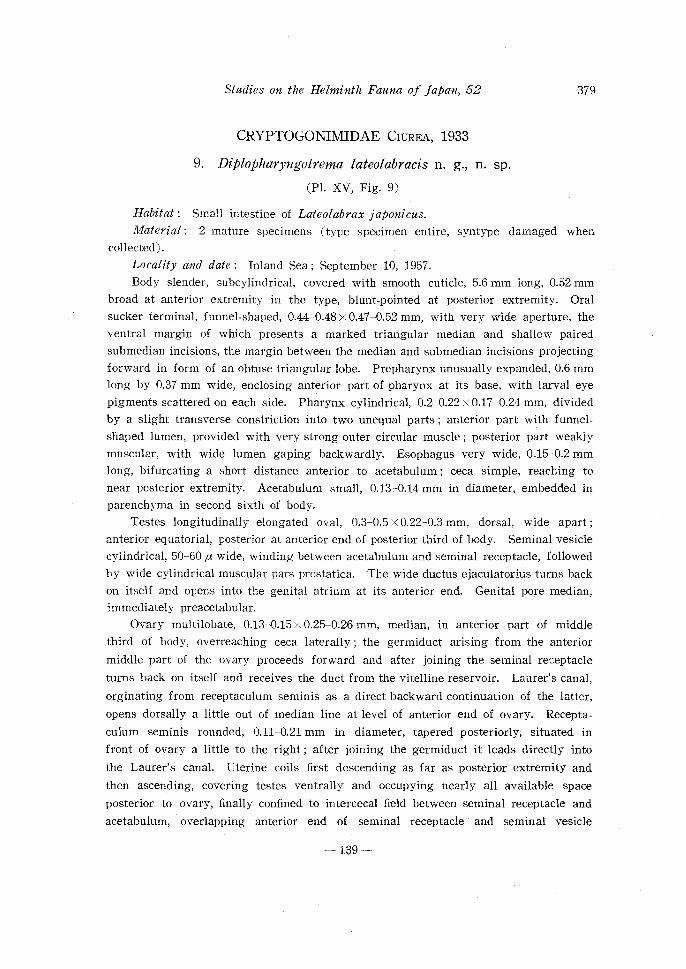
CRYPTOGONIMIDAE CIUREA, 1933
9. Diplopharyngotrema lateolabracis n. g., n. sp.
(Pl. XV, Fig. 9)
Habitat : Small intestine of Lateolabrax japonicus.
379
Material: 2 mature specimens (type specimen entire, syntype damaged when collected).
Locality and date: Inland Sea; September 10, 1957.
Body slender, subcylindrical, covered with smooth cuticle, 5.6 mm long, 0.52 mm
broad at anterior extremity in the type, blunt-pointed at posterior extremity. Oral
sucker terminal, funnel-shaped, 0.44-0.48 x 0.47-0.52 mm, with very wide aperture, the
ventral margin of which presents a marked triangular median and shallow paired
submedian incisions, the margin between the median and submedian incisions projecting
forward in form of an obtuse triangular lobe. Prepharynx unusually expanded, 0.6 mm long by 0.37 mm wide, enclosing anterior part of pharynx at its base, with larval eye
pigments scattered on each side. Pharynx cylindrical, 0.2-0.22 x 0.17-0.24 mm, divided
by a slight transverse constriction into two unequal parts ; anterior part with funnel
shaped lumen, provided with very strong outer circular muscle ; posterior part weakly
muscular, with wide lumen gaping backwardly. Esophagus very wide, 0.15-0.2 mm
long, bifurcating a short distance anterior to acetabulum ; ceca simple, reaching to
near posterior extremity. Acetabulum small, 0.13c-0.14 mm in diameter, embedded in
parenchyma in second sixth of body.
Testes longitudinally elongated oval, 0.3-0.5 X 0.22-0.3 mm, dorsal, wide apart ;
anterior equatorial, posterior at anterior end of posterior third of body. Seminal vesicle
cylindrical, 50-60 p. wide, winding between acetabulum and seminal receptacle, followed
by wide cylindrical muscular pars prostatica. The wide ductus ejaculatorius turns back
on itself and opens into the genital atrium at its anterior end. Genital pore median, immediately preacetabular.
Ovary multilobate, 0.13-0.15 x 0.25-0.26 mm, median, in anterior part of middle
third of body, overreaching ceca laterally ; the germiduct arising from the anterior
middle part of the ovary proceeds forward and after joining the seminal receptacle
turns back on itself and receives the duct from the vitelline reservoir. Laurer's canal,
orginating from receptaculum seminis as a direct backward continuation of the latter,
opens dorsally a little out of median line at level of anterior end of ovary. Recepta
culum seminis rounded, 0.11-0.21 mm in diameter, tapered posteriorly, situated in
front of ovary a little to the right ; after joining the germiduct it leads directly into
the Laurer's canal. Uterine coils first descending as far as posterior extremity and
then ascending, covering testes ventrally and occupying nearly all available space
posterior to ovary, finally confined to intercecal field between seminal receptacle and
acetabulum, overlapping anterior end of seminal receptacle and seminal vesicle
-139-

380 S. YAMAGUTI
ventrally ; metraterm short, narrow, opening into genital atrium at its posterior end ;
eggs oval, very small, 15-18 x 8-10 ,u as mounted in balsam. Vitellaria follicular,
extending in lateral fields from level of pharynx to near posterior testis ; transverse
vitellire ducts united across posterior end of seminal receptacle or Laurer's canal
ventrally. Excretory vesicle obscured by uterine coils.
Although the excretory system was unable to work out, there is no doubt from
general anatomy whether this worm belongs to the Cryptogonimide CruREA, 1933.
Apparently it represents a new subfamily, for which the name Diplopharyngotrematinae
is suggested, and should be placed near Biovariinae YAMAGUTI, 1957. The generic
name refers to the pharynx consisting of two different parts.
DIPLOPHARYNGOTREMATINAE n. subf.
Subfamily diagnosis.-Cryptogonimidae: Body slender. Oral sucker funnel-sh<?ped,
prepharynx very wide and long ; pharynx consisting of two parts of different structure.
Esophagus wide, short ; ceca simple, long. Acetabulum small, embedded in parenchyma,
in anterior region of body. Testes tandem, in hindbody. Vesicula seminalis long,
winding; pars prostatica present. Genital pore preacetabular. Ovary multilobate,
single, pretesticular. Receptaculum seminis and Laurer's canal present. Uterus ex
tending as far back as posterior extremity; eggs numerous, small. Vitellaria follicular,
lateral, moderately extensive. Excretory vesicle ? Intestinal parasites of marine teleosts.
Diplopharyngotrema n. g.
Generic diagnasis.-Diplopharyngotrematinae: Body flattened cylindrical, un
armed, with larval eye pigments scattered in neck region. Oral sucker funnel-shaped,
gaping forward. Prepharynx strongly expanded ; pharynx consisting of anterior part
with funnel-shaped lumen and strong outer circular muscles, and posterior part with
backwardly gaping lumen and without strong outer circular muscles. Esophagus short,
wide; ceca reaching to near posterior extremity. Acetabulum small, postbifurcal.
Testes oval, wide apart, in midregion of hindbody. Seminal vesicle winding spirally
behind acetabulum, followed by cylindrical, muscular pars prostatica and winding
ductus ejaculatorius. Genital pore immediately preacetabular, median. Ovary irregu
larly multilobed, overreaching ceca laterally anterior to fore testis. Receptaculum
seminis voluminous, preovarian, produced backward into Laurer's canal. Uterine coils
extensive, occupying all available space of hindbody ; metraterm short, narrow; eggs
oval, very small, light brown, operculate at narrower end. Vitellaria extending in
lateral field of hindbody and intruding a little into forebody. Excretory vesicle un
known. Parasitic in intestine of marine teleosts.
Genotype: Diptopharygotrema lateolabracis n. g., n. sp.
-140-

Studies on the Helminth Fauna of japan, 52
10. Pseudosiphoderoides hapalogenyos n. g., n. sp.
(Pl. XIV, Fig. 1)
Habitat : Small intestine of Hapalogenys sp.
Material: Three gravid specimens.
Locality and date: Inland Sea; September 10, 1957.
381
Body plump, broadly rounded in front, gradually tapered toward blunt posterior
extremity, 3.15-3.2 X 1.35-1.5 mm. Cuticle thick, unspined, striated perpendicularly.
Oral sucker subterminal, 0.2-0.26 X 0.3-0.35 mm ; prepharynx very short, pharynx 0.18-
0.2 x 0.23-0.27 mm in flattened whole mounts, but longer than wide (0.2 x 0.15 mm) in
a section of unflattened specimen. Esophagus practically absent. Ceca comparatively
wide, each terminating blindly at a short, equal or unequal distance (0.55 and 0.7 mm
respectively in the type) from posterior extremity. Acetabulum 0.23-0.25 X 0.24-0.26
mm, muscular, embedded in body parenchyma a little behind intestinal bifurcation,
and covered up by a fold of body wall which contains circular muscle fibers and has
a large oval opening 0.18-0.26 mm in greater transverse diameter.
Testes rounded, 0.3-0.4 mm in diameter, situated somewhat diagonally dorsal to
ceca in anterior part of posterior half of body. Seminal vesicle twisted, largely
posterolateral to acetabulum, with maximum width of 0.17-0.2 mm. Pars prostatica
well differentiated, about 50 p. in diameter, anterodorsal to acetabulum; short ejacula
tory duct opening with uterus into base of pocket enclosing acetabulum ; prostatic
cells strongly developed in vicinity of pars prostatica, especially on its right side.
Ovary median, ventral, equatorial, divided into numerous claviform lobules; semi
nal receptacle ovoid, 0.25-0.3 mm in diameter, median or slightly submedian, immedi
ately preovarian. Lamer's canal winding, opening dorsally in right submedian line
at level of posterior end of ovary or immediately behind it. Vitellaria forming
bunches of follicles in acetabula-ovarian zone dorsal and lateral to ceca, partly intru
ding mesad dorsally. Uterus winding ventromedial to testes and coiling backward
from side to side, overreaching ceca laterally, and then turning forward near posterior
extremity to take a sinuous ascending course on the left half of the body up to the level of the seminal receptacle or seminal vesicle, where it passes to the right ventral
to the seminal receptacle and is thrown into convolutions on the ventral side anterior
to the right testis ; finally returning to the median field it runs forwards ventromedial
to the seminal vesicle to open into the above mentioned genito-acetabular pouch.
Eggs oval, light to dark brown, 18-21 x 11-13 p..
Excretory vesicle Y -shaped, with terminal pore ; stem very wide, bifurcating at
level of posterior end of ovary ; arms also wide, terminating at level of pharynx.
This genus differs from the most closely related Siphoderoides MAl',TTER, 1940, in the
cuticle not being spined, in the seminal vesicle being tubular and sinuous instead of a
large undivided sac, in the testes lying dorsal to the ce:::a, and in the vitellaria exten
ding largely dorsal and lateral to the ceca, and from Paracryptogonimus YAMAGUTI,
-141-

382 S. YAMAGUTI
1934, in the absence of eye spots, body spines and circumoral spines, though resembling in other respects.
Pseudosiphoderoides n. g.
Generic diagnosis.-Cryptogonimidae, Cryptogoniminae: Body medium-sized,
plump, unspined. No circumoral spines. Oral sucker larger than acetabulum, sub
terminal. Prepharynx very short, pharynx elongate, esophagus practically absent, ceca
terminating short of posterior extremity. Acetabulum small, embedded in parenchyma
in anterior half of body and covered up by a circular fold of body wall (genito
acetabular pouch). Testes two, subsymmetrical or diagonal, postequatorial, dorsal to
ceca. Seminal vesicle tubular, sinuous, largely postacetabular. Prostatic complex well
developed. Neither cirrus nor cirrus pouch. Genital pore median, opening into
genito-acetabular pouch immediately in front of acetabulum. Ovary median, multi
lobed, pretesticular, with seminal receptacle immediately in front. Vitellaria follicular,
extending largely in acetabulo-ovarian zone lateral and dorsal to ceca. Uterine coils
pre- and posttesticular, leaving posterior end of body free; eggs very small. Excretory
vesicle Y -shaped. Intestinal parasites of marine teleosts.
Genotype: P. hapalogenyos n. sp.
BUCEPHALIDAE POCHE, 1907
11. Prosorhynchus crucibulum japonicum n. subsp.
(Pl. XV, Fig. 8)
Habitat : Small intestine of Conger myriaster. Material: A single gravid specimen stained and mounted in toto. Locality and date: Inland Sea; September 8, 1957.
Body shaped like a plump rod, swollen at level of testes, about 2 mm long by
0.7 mm wide. Rhynchus wedge-shaped, 0.38 x 0.28 mm. Pharynx 90 fJ. in diameter,
pre-equatorial. Esophagus 0.15 x 0.03 mm, intestine 0.2 X 0.09 mm, directed straight
forward, with the blind end a little beyond ovary. Testes oval, 0.2-0.22 x 0.15--D.16 mm,
situated obliquely one on each side of esophagus. Cirrus pouch 0.53 mm long by
0.2 mm broad, containing sigmoid, cylindrical, seminal vesicle 80 fJ. wide, well developed
pars prostatica 0.13 mm wide, and a short cirrus only 80 fJ. long by 20 fJ. wide. Genital
atrium 0.2 mm in diameter, with wide ventral aperture 0.3 mm from posterior extremity.
Ovary oval, 0.22 x 0.13 mm, overlapping right testis dorsomedially; shell gland
behind right testis. Uterus winding first between right testis and cirrus pouch, then
convoluted between left testis and left vitellaria, finally descending on the left of
cirrus pouch as far back as beyond genital pore, where it turns forward to open into
the genital atrium; eggs oval, 26-28 fJ. long by 20-21 fJ. broad. Vitelline follicles
-142-

Studies on the Helminth Fauna of Japan, 52 383
forming symmetrical groups of 12 and 14 each in shoulder region between rhynchus
and testes.
Excretory vesicle tubular, extending on the right of cirrus pouch as far forward
as anterior end of cirrus pouch ; pore terminal.
This species resembles Prosorhynchus crucibulum (RuD.) of NICOLL, 1910, or of
OzAKI, 1924, very closely, but differs from it in the shell gland being situated behind
the right testis at the level of the pharynx ; in OzAKI's specimen the shell gland lies
between the ovary and the right testis. The relative position of the testes, ovary and
pharynx is subject to great individual variation in the known members of the genus,
so that much importance cannot be ascribed to differences in this respect. If the shell
gland be also variable in position individually, the present specimen should be referred
to P. crucibulum, but the contrary seems to be true so far as my experience goes.
I would like, therefore, to assign it for the present to a new subspecies, for which the name Prosorhynchus crucibulum japonicum is proposed.
Acknowledgements
I wish to express my sincere appreciation to Mr. S. 0BA, Assistant Professor of
the Marine Biological Laboratory of Okayama University, for his help in collecting
and identifying the host fishes, and also to the Oceanographical Museum of Tamano
City, Okayama Prefecture, for the generous supply of the fishes which died in the
aquarium of the museum.
LITERATURE
MANTER, H. W. 1940. Digenetic trematodes of fishes from the Galapagos Islands and the neighboring Pacific. Rep. Allan Hancock Pacif. Exped. (1932-1938) 2 (14), 325-497.
------- 1947. The digenetic trematodes of marine fishes of Tortugas, Florida. Amer. Mid!. Nat. 38 (2), 257-416.
YAMAGUTI, S. 1934. Studies on the helminth fauna of Japan. Pt. 2. Trematodes of fishes, I. Jap. J. Zoo!. 5 (3), 249-541.
1934. Studies on the helminth fauna of Japan. Pt. 26. Trematodes of fishes, VI. Jap. J. Zoo!. 8 (2), 211-230.
1951. Studies on the helminth fauna of Japan. Pt. 48. Trematodes of fishes, X. Arb. Med. Fak. Okayama 7 (4), 315-334.
1952. Parasitic worms mainly from Celebes. Pt. 1. New digenetic trematodes of fishes. Acta Med. Okayama 8 (2), 146-198.
--------- 1953. Systema helminthum. Part I. Digenetic trematodes of fishes. Tokyo. -------- 1958. Systema helminthum. Vol. I. The digenetic trematodes of vertebrates.
Interscience, New York.
-143--

384 S. YAMAGUTI
EXPLANATION OF PLATES XIV-XV
PLATE XIV
Fig. 1. Pseudosiphoderoides hapalogenyos n. g., n. sp., ventral view.
Fig. 2. Opegaster cryptocentri n. sp., ventral view.
Fig. 3. Opecoelus lateolabracis n. sp. ; forebody in lateral view, hindbody in
ventral view.
Fig. 4. Aerobiotrema muraenesocis n. g., n. sp. strongly flattened, lateral view.
Fig. 5. Longitudinal section of Aerobiotrema muraenesocis through terminal
genitalia.
Fig. 6. Transverse section of Aerobiotrema muraenesocis through shell gland
complex.
Fig. 7.
Fig. 8.
PLATE XV
Biovarium lateolabracis n. sp., ventral view.
Prosorhynchus crucibulum japonicum n. subsp., ventral view.
Fig. 9. Diplopharyngotrema lateolabracis n. g., n. sp., ventral view.
Fig. 10. Unflattened specimen of Aerobiotrema muraenesocis, free-hand ske
tch, ventral view.
Fig. 11. Opecoelus sebastisci n. sp. ; forebody in lateral view, hindbody in
ventral vew.
Fig. 12. Opecoelus pagrosomi n. sp., ventral view.
ABBREVIATIONS USED IN FIGURES
A=acetabulum, AN =anus, CP=cirrus pouch, EA=excretory arm, EP=excretory pore,
EV=excretory vesicle, GA=genital atrium, GP=genital pore, !=intestine, LC=
Laurer's canal, MO=mouth opening, O=ovary, OS=oral sucker, P=pharynx,
PR=prostatic cell, R=rhynchus, RS=receptaculum seminis, SG=shell gland, T=testis,
U =uterus, VR= Vitelline reservoir, VS=vesicula seminalis, VT=vitelline gland.
-144--

Publ. Seto Mar. Bioi. Lab., VI, 3 (1958) PLATE XIV
S. YAMAGUTI: STUDIES ON THE HELMINTH FAUNA OF }APAN, 52.

Publ. Seto Mar. Biol. Lab., VI, 3 (1958) PLATE XV
10
S. YAMAGUTI: STUDIES ON THE HELMINTH FAUN!-\ OF }APAN, 52.


![HELMINTH PARASITES IN MAMMALS - Australian …parasite.org.au/para-site/text/helminth.pdf · HELMINTH PARASITES IN MAMMALS ... Subclass: EUTHERIA [placental mammals] ... NEM:Asc Ascaris](https://img.pdfslide.us/doc/110x75/5b78c38f7f8b9a331e8c41aa/helminth-parasites-in-mammals-australian-helminth-parasites-in-mammals-.jpg)