Embed Size (px)
Citation preview

Title slide
How you should study this module
Learning outcomes
Epidemiology
Pathophysiology
Section one quiz
Assessment
Section two quiz
Management
Section three quiz
Information sources
“Nice pic, but why is this module important?”
Well, in 2007, 246million people aged 20 - 79 years were diagnosed with diabetes mellitus – a global epidemic affecting 6% of the adult population.
25% of these develop foot problems...that’s 61.5 million diabetic feet!
And, worryingly, the prevalence of diabetes mellitus is expected to reach 333 million by 2025.
For information on the authors and reviewers click here
The foot of a diabetic patient showing extensive tissue necrosis and infection
WELCOME TO THE DIABETIC FOOT MODULE!
Page 1 of 67
Diabetic Foot

Title slide
How you should study this module
Learning outcomes
Epidemiology
Pathophysiology
Section one quiz
Assessment
Section two quiz
Management
Section three quiz
Information sources
How should you study this module?
1. We suggest that you start with the learning objectives and try to keep these in mind as you go through the module slide by slide.
2. Print out the mark sheet.3. As you go along, write your answers to the questions on the
mark sheet as best you can before looking at the answers.4. Award yourself marks as detailed on the mark sheet: one
mark for each keyword (shown in the red text) in the short answer questions and for every correct answer in the True/False questions.
5. Repeat the module until you have achieved a mark of > 80% (65/81)
6. Finish with the formative multiple choice questionnaire to assess how well you have covered the materials as a whole.
7. You should research any issue that you are unsure about. Look in your textbooks, access the on-line resources indicated at the end of the module and discuss with your peers and teachers.
8. Finally , enjoy your learning! We hope that this module will be enjoyable to study and complement your learning about diabetic foot from other sources.
Page 2 of 67

Title slide
How you should study this module
Learning outcomes
Epidemiology
Pathophysiology
Section one quiz
Assessment
Section two quiz
Management
Section three quiz
Information sources
By the end of the module, you should be able to:
1. Discuss the global burden of the diabetic foot in both the developing and developed world
2. List the causes of diabetic foot ulceration then fully assess for each one and their complications; using bedside examinations, blood tests, microscopy and radiology
3. Discuss the management of diabetic foot ulcers using
i. mechanical intervention (debridement, dressing and cast application)
ii. invasive treatment (larvae, antimicrobials and amputation)
iii. analgesia
4. Offer advice to diabetic patients on proper foot care and footwear for prevention of foot problems
Page 3 of 67
Learning Outcomes

Title slide
How you should study this module
Learning outcomes
Epidemiology
Pathophysiology
Section one quiz
Assessment
Section two quiz
Management
Section three quiz
Information sources
“So how many people with diabetes are there?”
Here are recent estimates of the disease burden due to diabetes and projections for the future.
2003 2025Europe Africa Europe Africa
Population• Total
• Adult
(20-79 yrs)
872 million
621 million
667 million
295 million
863 million
646 million
1107 million
541 million
Diabetes• No. of people
(20-79 yrs)
• Prevalence
(20-79 yrs)
48.4 million
7.8 %
7.1 million
2.4 %
65 million
7.8 %
19million
4.3 %
Source: International Diabetes Federation and The international Working Group on Diabetes joint publication 2006.
Page 4 of 67
Epidemiology 1

Title slide
How you should study this module
Learning outcomes
Epidemiology
Pathophysiology
Section one quiz
Assessment
Section two quiz
Management
Section three quiz
Information sources
“That’s a lot! How many of these get foot ulcers?” Developed countries: 15% of people with diabetes get ulcers at least once in their lifetime Developing countries: the prevalence is even higher at 20%.
“...and does amputation use vary from place to place?” Yes!...see below;
Incidence of minor and major amputations per 1000 people with diabetes
Incidence per 1000
Population Year
Mauritius 680 Hospital-oriented 1998-2002
Tanzania 400 Hospital-oriented 2002
Croatia 6.8 Hospital-oriented 2002
UK 2.6 Regional 1998
The Netherlands 3.6 Nationwide 1991-2000
Source: International Diabetes Federation and The international Working Group on Diabetes joint publication 2005.
Page 5 of 67
Epidemiology 2

Title slide
How you should study this module
Learning outcomes
Epidemiology
Pathophysiology
Section one quiz
Assessment
Section two quiz
Management
Section three quiz
Information sources
According to the above data;
1. Which region has the most people with diabetes?
2. Which region will see the greatest increase in diabetes prevalence by 2025 ?
3. Which region has the greatest disease burden due to diabetic foot ?
Click the box for the correct answer
1
2
3
Page 6 of 67
Epidemiology Quiz

Title slide
How you should study this module
Learning outcomes
Epidemiology
Pathophysiology
Section one quiz
Assessment
Section two quiz
Management
Section three quiz
Information sources
“How does the diabetic foot affect individualsand society?”
• Diabetic foot ulcers and their complications (explained later) are often painful. Patients often become dependent on others for mobility.As a result, patients suffer a loss of autonomy and reduced social function, making depression common.
• The cost of diabetic foot management is 12-15% of the total healthcare budget for diabetes in developed countries. This figure may as high as 40% in developing countries*. These figures do not account for the cost of the loss of potential working members to the economy and the social costs of the inability to support a family.
*IDF/IWG joint publication on diabetic foot.
Page 7 of 67
Epidemiology 3

Title slide
How you should study this module
Learning outcomes
Epidemiology
Pathophysiology
Section one quiz
Assessment
Section two quiz
Management
Section three quiz
Information sources
“Well those diabetic feet are everywhere and causingchaos! If we’re going to stop them, I would first like toknow how diabetic foot ulcers occur…”
Diabetic foot ulcers may have multiple causes, the prominent ones being;
A. Peripheral neuropathy (nerve damage)
B. Peripheral vascular disease (poor pedal blood supply)
C. Trauma
i. Acute: any injury to the foot such as burns or cuts
ii. Chronic: due to foot deformities (changes of foot shape that lead to ill-fitting shoes and, thereby, ulceration)
Page 8 of 67
Pathophysiology

Title slide
How you should study this module
Learning outcomes
Epidemiology
Pathophysiology
Section one quiz
Assessment
Section two quiz
Management
Section three quiz
Information sources
Neuropathy
Motor Sensory Autonomic
↓ nociception
↓ Proprioception,Unawarenessof foot position A-V Shunt* open
Permanent
Increase footBlood flow
Bulging foot veins,Warm foot
Reduced sweating
Dry skin
Fissures andcracks
Muscle wastingFoot weakness
Postural deviation
Deformities, stressand shear pressures
*Shunts: blood vessels that bypass capillaries and lead directly from arteries to veins
Trauma
Stress on bones & jointsPlantar pressure
Callus formation
InfectionUlcer
Page 9 of 67
Pathophysiology Neuropathy

Title slide
How you should study this module
Learning outcomes
Epidemiology
Pathophysiology
Section one quiz
Assessment
Section two quiz
Management
Section three quiz
Information sources
“Then how are blood vessels affected?”
High blood sugar expeditesartherosclerosis giving peripheral vascular disease (reduction of blood supply to the foot). The delivery of essential nutrientsand oxygen to the foot iscompromised leading to anaerobic infections and tissue necrosis.
Peripheral arterial disease
Artherosclerosis narrows or blocks the arterial lumen
Foot ischaemia
Foot ulcer Necrosis/ Gangrene
Infection
Artheroma plaque narrowing the arterial lumen
Ischaemic toes due to artherosclerosis
Page 10 of 67
Pathophysiology Peripheral Arterial Disease

Title slide
How you should study this module
Learning outcomes
Epidemiology
Pathophysiology
Section one quiz
Assessment
Section two quiz
Management
Section three quiz
Information sources
“Don’t people with diabetes feel trauma before it reaches ulceration stage?” No- that’s the problem!
Acute trauma: abrasions and burns occur often due to the absence of nociception. Poor wound healing makes ulcerations more likely occur.
Chronic trauma: reduced motor function results in a high arch. Together with decreased proprioception, this creates classical deformed foot shapes (explained later). These result in bony prominences which, when coupled with high mechanical pressure on the overlying skin, results in ulceration.
Page 11 of 67
Pathophysiology Trauma

Title slide
How you should study this module
Learning outcomes
Epidemiology
Pathophysiology
Section one quiz
Assessment
Section two quiz
Management
Section three quiz
Information sources
Well done!You have come to the end of the first section
We suggest that you answer Question 1 to 4 to assess your learning so far. Please remember to
write your answers on the mark sheet before looking at the correct answers!
Page 12 of 67
End of Section 1

Title slide
How you should study this module
Learning outcomes
Epidemiology
Pathophysiology
Section one quiz
Assessment
Section two quiz
Management
Section three quiz
Information sources
Question 1: write ‘T’ or ‘F’ on the answer sheet. When you have completed all 5 questions, click on the boxes and mark your answers.
a) Diabetic foot is a global health problem
b) The prevalence of diabetes is falling
c) The incidence of foot ulcers in people with diabetes is higher in developed than developing countries
d) Diabetic foot amputation is commoner in developing countries than developed countries
e) Post amputation mortality is higher in developed countries
a
b
c
d
e
Page 13 of 67
Section 1 Quiz

Title slide
How you should study this module
Learning outcomes
Epidemiology
Pathophysiology
Section one quiz
Assessment
Section two quiz
Management
Section three quiz
Information sources
Question 2: write ‘T’ or ‘F’ on the answer sheet. When you have completed all 4 questions, click on the boxes and mark your answers.
a) Diabetic foot problems result in a higher cost to the economy in developing than developed countries
b) Depression is common in diabetic foot patients
c) Wound healing is slower in diabetes
d) Artherosclerosis is common in diabetes patients
a
b
c
dPage 14 of 67
Section 1 Quiz

Title slide
How you should study this module
Learning outcomes
Epidemiology
Pathophysiology
Section one quiz
Assessment
Section two quiz
Management
Section three quiz
Information sources
Question 3: The 4 main causes of diabetic foot ulcers are; write the answers in your mark sheet.
a) Peripheral neuropathy
b) ………………………
c) Peripheral arterial disease
d) …………………………
Click here for the answers
Section 1 Quiz

Title slide
How you should study this module
Learning outcomes
Epidemiology
Pathophysiology
Section one quiz
Assessment
Section two quiz
Management
Section three quiz
Information sources
Question 4: Study this flow chart and list 4 factors that predispose to diabetic foot ulceration. Write your answer in your mark sheet
Neuropathy
Motor Sensory Autonomic
↓ nociception
↓ Proprioception,Unawarenessof foot position A-V Shunt* open
Permanent
Increase footBlood flow
Bulging foot veins,Warm foot
Reduced sweating
Dry skin
Fissures andcracks
Muscle wastingFoot weakness
Postural deviation
Deformities, stressand shear pressures
*Shunts: blood vessels that bypass capillaries and lead directly from arteries to veins
Trauma
Stress on bones & jointsPlantar pressure
Callus formation
InfectionUlcer
Click here for the answers
Page 16 of 67
Section 1 Quiz

Title slide
How you should study this module
Learning outcomes
Epidemiology
Pathophysiology
Section one quiz
Assessment
Section two quiz
Management
Section three quiz
Information sources
“How do we predict how bad a diabetic foot is then?”
Foot assessment needs to be undertaken in all people with diabetes to evaluate the individual’s risk of foot complications and hence plan management.
It can be undertaken by a podiatrist, junior doctor, specialised diabetes nurse or other trained nurses.
The aim of the assessment is to examine each pathological cause that creates ulcers:
1) peripheral neuropathy 2) peripheral arterial disease3) structural
But how do you assess the diabetic foot? Let me guess. As always start with the history and then the examination for each cause …?”- Bingo!
Page 17 of 67
Assessment

Title slide
How you should study this module
Learning outcomes
Epidemiology
Pathophysiology
Section one quiz
Assessment
Section two quiz
Management
Section three quiz
Information sources
History• burning, tingling, numbness of the foot
and nocturnal leg pain indicate cutaneous sensory deficits
• Note that in ~35% of patients who are asymptomatic, neuropathy can be detected by examination
Examination• Inspect deformities such as claw toes,
hair loss, muscle atrophy and a high medial longitudinal arch (giving prominent metatarsal heads)
• Test for reduced power and reflexes that are evidence of muscular motor deficits.
• Test sensation by skin pinprick (spinothalamic tracts), proprioception and vibration (dorsal columns)
Claw toes
Prominent metatarsal heads and an ulcer
Page 18 of 67
Assessment Peripheral Neuropathy

Title slide
How you should study this module
Learning outcomes
Epidemiology
Pathophysiology
Section one quiz
Assessment
Section two quiz
Management
Section three quiz
Information sources
• Place a 10g nylon Semmes-Weinstein monofilament at a right angle to the skin
• Apply pressure until the monofilament buckles, indicating that a specific pressure has been applied.
• Inability to perceive the 10g of force applied by the monofilament is associated with clinically significant large fibre neuropathy and an increased risk of ulceration (sensitivity of 66 to 91%)
• Test 4 plantar sites on the forefoot (great toe and the base of 1st, 3rd and 5th metatarsals ) to identify 90% of patients with an insensate foot.
Monofilament test
Page 19 of 67
Assessment Monofilament for pressure sensation (pinprick sense)

Title slide
How you should study this module
Learning outcomes
Epidemiology
Pathophysiology
Section one quiz
Assessment
Section two quiz
Management
Section three quiz
Information sources
• Apply a vibrating 128 Hz tuning fork to the bony prominence of the big toe
• If the patient cannot feel the vibration, gradually move the fork upwards
• The sensitivity of this test for demonstrating a deficit is ~53%
• A biothesiometer is a portable device that measures the vibration perception threshold. A vibration threshold of more than 25V has a sensitivity of 83%.
Tuning fork test
Either an abnormal 10g monofilament test or a vibration threshold of more than 25V predicts foot ulceration with a sensitivity of 100% , hence the rationale for combining these two tests in clinical practice.
Page 20 of 67
Assessment Tuning Fork (vibration)

Title slide
How you should study this module
Learning outcomes
Epidemiology
Pathophysiology
Section one quiz
Assessment
Section two quiz
Management
Section three quiz
Information sources
“So how do we know how well the blood is flowing?”• History : claudication (calf pain after walking a specific distance) that
is relieved by rest. However this is uncommon in people with diabetes due the concomitant neuropathy.
• Examination: Palpate the foot for temperature (cool in PVD); palpate the dorsalis pedis pulse and, if absent, the posterior tibial pulse. Test for Bergers angle (at which leg turns white) and reactive hyperaemia (leg turns bright red on declining back to the ground).
Palpation of the dorsalis pedis pulse Palpation of the posterior tibial pulse
Page 21 of 67
Assessment Peripheral Vascular Disease (PVD)

Title slide
How you should study this module
Learning outcomes
Epidemiology
Pathophysiology
Section one quiz
Assessment
Section two quiz
Management
Section three quiz
Information sources
Measure the blood pressure (BP) in the arm using a sphygmanometer
Measure the blood pressure in the foot. Place a BP cuff around the calf and detect the dorsalis pedis pulse using a small hand-held doppler. Inflate the cuff and slowly deflate until the pulse appears.
The ankle brachial pressure index (ABPI) is the ratio of the ankle systolic pressure to brachial systolic pressure.
ABPI is usually >1 but in the presence of peripheral vascular disease is <1. Normal ABPI effectively excludes significant arterial disease in >90% of limbs. Doppler being used to detect
the dorsalis pedis pulse
Absence of pulses and an ABPI of <1 confirms significant ischaemia. An exception is in medial artery calcification, in which the ABPI can be falsely elevated due to the simultaneously lower blood pressure (BP) in the upper limb.
Page 22 of 67
Assessment Investigations: ankle brachial pressure index

Title slide
How you should study this module
Learning outcomes
Epidemiology
Pathophysiology
Section one quiz
Assessment
Section two quiz
Management
Section three quiz
Information sources
Structural abnormalities and deformities lead to bony prominences which are associated with high mechanical pressure on the overlying skin. This results in ulceration, particularly in the absence of a protective pain sensation and when shoes are unsuitable.Ideally, the deformity should be recognised early and accommodated in properly fitting shoes before ulceration occurs.
Common abnormalities / deformities include:i. Callusii. Bunioniii. Hammer toesiv. Claw toesv. Charcot footvi. Nail deformities
Note: It is vital to inspect the patients shoes as part of the assessment!
Callus on plantar surface
Bunion on the medial border of the foot
Page 23 of 67
Assessment Structural Abnormalities and Deformities

Title slide
How you should study this module
Learning outcomes
Epidemiology
Pathophysiology
Section one quiz
Assessment
Section two quiz
Management
Section three quiz
Information sources
Claw toes
Charcot foot deformityNail deformity
Page 24 of 67
Assessment Some Common Foot Deformities

Title slide
How you should study this module
Learning outcomes
Epidemiology
Pathophysiology
Section one quiz
Assessment
Section two quiz
Management
Section three quiz
Information sources
• Several foot ulcer classifications have been proposed although none is universally accepted.
• The simplest classification is based on the underlying pathogenesis: neuropathic, ischaemic or neuroischaemic.
• It is vital to carefully monitor the progress of an ulcer once one has developed.
• The University of Texas system shown on the next slide can be used to predict outcome by grading wound depth and presence of infection and/or ischaemia. However there is no measure of neuropathy.
A neuropathic ulcer on the sole of the foot
“Pre-ulcer assessment all done! What about after an ulcer has developed?”
Page 25 of 67
Assessment Ulcers

Title slide
How you should study this module
Learning outcomes
Epidemiology
Pathophysiology
Section one quiz
Assessment
Section two quiz
Management
Section three quiz
Information sources
Ulcer Grade ( depth )
0 I. II. III.
Ulcer
stage
A Pre / postulcerative lesion completely epethelialised
Superficial lesion, not involving tendon, capsule or bone
Wound penetrating to tendon or capsule
Wound penetrating to bone or joint
B Pre / postulcerative lesion with Infection
Superficial lesion, not involving tendon, capsule or bone with Infection
Wound penetrating to tendon or capsule with Infection
Wound penetrating to bone or joint with Infection
C Pre / postulcerative lesion with ishaemia
Superficial lesion, not involving tendon, capsule or bone with ischaemia
Wound penetrating to tendon or capsule with ishaemia
Wound penetrating to bone or joint with ishaemia
D Pre /postulcerative lesion with infection and ishaemia
Superficial lesion, not involving tendon, capsule or bone with infection and ischaemia
Wound penetrating to tendon or capsule with infection and ishaemia
Wound penetrating to bone or joint with infection and ishaemia
Page 26 of 67
Assessment University of Texas system for classification of ulcers

Title slide
How you should study this module
Learning outcomes
Epidemiology
Pathophysiology
Section one quiz
Assessment
Section two quiz
Management
Section three quiz
Information sources
“How do you know if the ulcer is infected then?”
Assessing foot ulcers for the presence of infection is vital. All open wounds are likely to get colonised with microorganisms, such as Staphylococcus aureus, and not necessarily infected. Therefore, the presence of infection needs to be defined clinically rather than microbiologically.
An infected ulcer
Signs suggesting infection include;
1. purulent secretions
2. presence of friable tissue
3. undermined edges4. foul odour
Page 27 of 67
Assessment Infected Ulcers

Title slide
How you should study this module
Learning outcomes
Epidemiology
Pathophysiology
Section one quiz
Assessment
Section two quiz
Management
Section three quiz
Information sources
Simple investigations include:• Tissue specimens or material obtained from the bottom of a
wound for gram staining and culture for microbial sensitivity. Aspiration of material for culture is better than taking a swab which is prone to contamination.
• Full blood count, urea and electrolytes, inflammatory markers (WCC, ESR and CRP) for assessing severity of infection
• Plain X-ray of the leg for signs of bone damage, presence of foreign body, or gas in soft tissue (gas gangrene)
More advanced radiology involves:• Technetium bone scan and MRIs may be necessary in some
patients to define underlying bony involvement
Invasive investigations include:• Bone biopsy, as the gold test for diagnosing osteomyelitis. • Arteriography using contrast dye can be used to visualise leg
ischaemiaPage 28 of 67
Assessment Infected Ulcers: Investigations

Title slide
How you should study this module
Learning outcomes
Epidemiology
Pathophysiology
Section one quiz
Assessment
Section two quiz
Management
Section three quiz
Information sources
Well done!You have come to the end of the second section
We suggest that you answer Questions 5 to 9 to assess your learning so far. Please remember to
write your answers on the mark sheet before looking at the correct answers!
Page 29 of 67
End of Section 2

Title slide
How you should study this module
Learning outcomes
Epidemiology
Pathophysiology
Section one quiz
Assessment
Section two quiz
Management
Section three quiz
Information sources
1) ………………………..
2) ………………………..
3) ………………………..
Question 5: List the 3 components of diabetic foot assessment. Write your answer in your mark sheet
Click here for the answersPage 30 of 67
Section 2 Quiz

Title slide
How you should study this module
Learning outcomes
Epidemiology
Pathophysiology
Section one quiz
Assessment
Section two quiz
Management
Section three quiz
Information sources
a) A high medial longitudinal arch and prominent metatarsal heads are signs of ischaemia
b) The tuning fork and biothesiometer are used for assessing pressure sensation
c) Ankle brachial pressure index is the ratio of ankle systolic pressure to brachial diastolic pressure
d) A doppler can be used to confirm the presence of pulses but cannot quantify the vascular supply
e) Bone biopsy is the gold standard for diagnosing
osteomyelitis
Question 6: Write ‘T’ or ‘F’ on the answer sheet. After completing all 5 questions, click on the boxes and mark your answers.
a
b
c
d
e
Page 31 of 67
Section 2 Quiz
Title slide
How you should study this module
Learning outcomes
Epidemiology
Pathophysiology
Section one quiz
Assessment
Section two quiz
Management
Section three quiz
Information sources
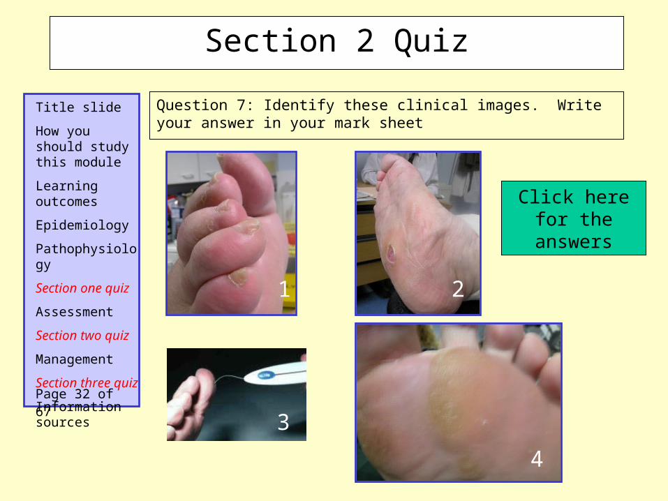
Question 7: Identify these clinical images. Write your answer in your mark sheet
Click here for the answers
1
4
2
Page 32 of 67
Section 2 Quiz
3

Title slide
How you should study this module
Learning outcomes
Epidemiology
Pathophysiology
Section one quiz
Assessment
Section two quiz
Management
Section three quiz
Information sources
I. ………………………………
II. ………………………………
III. ………………………………
IV. ………………………………
V. ………………………………
Question 8: List 5 common foot deformities found in association with diabetic feet. Write your answers on the mark sheet.
Click here for the answers
Page 33 of 67
Section 2 Quiz

Title slide
How you should study this module
Learning outcomes
Epidemiology
Pathophysiology
Section one quiz
Assessment
Section two quiz
Management
Section three quiz
Information sources
Question 9: Fill in the blanks in the University of Texas grading and staging table. Write your answer in your mark sheet
Ulcer Grade ( depth )
0 I. II. III.
Ulcer
stage
A Pre / postulcerative lesion completely epethelialised
Superficial lesion, not involving tendon, capsule or bone
Wound penetrating to tendon or capsule
Wound penetrating to bone or joint
B Superficial lesion, not involving tendon, capsule or bone with Infection
Wound penetrating to bone or joint with Infection
C Pre / postulcerative lesion with Ishaemia
Wound penetrating to tendon or capsule with Ishaemia
D Pre /postulcerative lesion with Infection and Ishaemia
Superficial lesion, not involving tendon, capsule or bone with Infection and Ischaemia
Wound penetrating to tendon or capsule with Infection and Ishaemia
Wound penetrating to bone or joint with Infection and ishaemia
Page 34 of 67
Section 2 Quiz

Title slide
How you should study this module
Learning outcomes
Epidemiology
Pathophysiology
Section one quiz
Assessment
Section two quiz
Management
Section three quiz
Information sources
“Ok, so now we know the extent of the problem, how it occurs and how to assess for it. Now what do we do about it?”
General measuresManaging diabetes and it’s complications requires a
multidisciplinary approach because• optimum glycaemic control is key in reducing all
complications• cardiovascular risk factors such as smoking,
dyslipidaemia and hypertension should be addressed to reduce risks of PVD, acute coronary syndrome and chronic renal failure
• education of patients on proper foot care and on the importance of seeking medical advice early is very important
Page 35 of 67
Management

Title slide
How you should study this module
Learning outcomes
Epidemiology
Pathophysiology
Section one quiz
Assessment
Section two quiz
Management
Section three quiz
Information sources
“If a patient with diabetes has normal feet do we need to worry?...YES!”Your aim is to keep the foot normal. Key elements are:• wearing the correct footwear• the diagnosis and prompt treatment of foot problems that are common in the general
population including people without diabetes.
Good shoe guide: Toe box should be sufficiently long, broad and deep to accommodate the toes without
pressing on them, with a clear space between the apices of the toe the toe box Shoes should be fasten with adjustable lace, strap or Velcro high on the foot in order to hold
foot firmly inside the shoe and thus reduce frictional forces when the patient walks The heel of the shoe should be less than 5 cm to avoid weight being thrown forward into
metatarsal heads The inner lining of shoe should be smooth Stocking or socks should always be worn to avoid blisters
Good pairs of shoes for men and women An example of a bad shoe type
Page 36 of 67
Management The Normal Foot

Title slide
How you should study this module
Learning outcomes
Epidemiology
Pathophysiology
Section one quiz
Assessment
Section two quiz
Management
Section three quiz
Information sources
Most people in this stage will be able to cut their own toe nails. However specific nails and other minor foot problems will need treatment from the podiatrist. These are the most common conditions:
Onychogryphosis (ram’s horn nail); regular debulking by a podiatrist
Onychocryptosis (ingrowing toe nail); removal of the offending nail splinter and filing of the ragged edge by a podiatrist
Involuted toe nail; clearance of the sulcus with a Black’s file (specially design for it)
Onychomycosis; reduce bulk of the nail at regular intervals, treat with antifungals
Tinea pedis (athletes foot); treat with topical antifungals (e.g canesten).
Verrucae (warts); treat by cryotherapy. Most resolve within 2 years.
Corns; removal by a podiatrist.
Nail cutting
Athletes foot
Page 37 of 67
Management Diagnosing and treating common foot problems

Title slide
How you should study this module
Learning outcomes
Epidemiology
Pathophysiology
Section one quiz
Assessment
Section two quiz
Management
Section three quiz
Information sources
Deformities should be accommodated in properly fitting footwear. Special footwear will be needed if the deformity is severe.
Some specific deformities need special management;
Clawed toes need a shoe with a wide, deep, soft toe box to reduce pressure on the dorsum of the toes. Extra depth shoes to protect the apices of the toes
Prominent metatarsal heads: an extra depth stock shoe with a cushioning insole may suffice
Callus: Is the most important pre-ulcerative lesion in this stage. It should be regularly and sufficiently remove by a podiatrician with a scalpel.
Dry skin and fissure: treat with an emolient (E45 or calmurid cream), reduce fissure margins with scalpel
Callus removal
“And if neuropathic or ischaemic and/ or deformities are present?” - This foot is susceptible to ulcers, so...
Page 38 of 67
Management The At-Risk Foot
Title slide
How you should study this module
Learning outcomes
Epidemiology
Pathophysiology
Section one quiz
Assessment
Section two quiz
Management
Section three quiz
Information sources
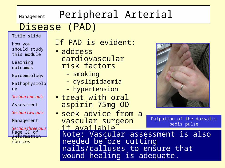
If PAD is evident:• address cardiovascular
risk factors– smoking– dyslipidaemia– hypertension
• treat with oral aspirin 75mg OD
• seek advice from a vascular surgeon if available Palpation of the dorsalis pedis pulse
Note: Vascular assessment is also needed before cutting nails/calluses to ensure that wound healing is adequate.
Page 39 of 67
Management Peripheral Arterial Disease (PAD)

Title slide
How you should study this module
Learning outcomes
Epidemiology
Pathophysiology
Section one quiz
Assessment
Section two quiz
Management
Section three quiz
Information sources
“How should we advise patients that get deformities?”
Provide patients with the following information:
• Never walk bare footed• Visit a podiatrist regularly if you have
callus• Never try to remove corns or callus by
yourself• Prevent dryness in your feet by using
creams• Be careful not to burn your feet• Shake out loose pebbles or grit before
you put on your shoes• Run a hand around the sides of the
shoes to detect rough, worn places• Repair or replace worn out shoes
Claw toes
Page 40 of 67
Management Foot Deformities

Title slide
How you should study this module
Learning outcomes
Epidemiology
Pathophysiology
Section one quiz
Assessment
Section two quiz
Management
Section three quiz
Information sources
“It’s an ulcer..what now!?”-Don’t panic, be methodical.Treatment of diabetic foot ulcers largely depends on the underlying causes: ischaemia, neuropathy or a combination of both. Treatment approaches for ischaemia include:
Ischaemic necrosis of a toe and an extensive
plantar ulcer
Medical: reduce cardiovascular risk factors (see above)
Surgical: revascularisation to achieve timely and durable wound healing is sometimes necessary. Patients with supra-inguinal (aorta-iliac) disease may be amenable to angioplasty (+/- stenting), with good long-term results being achieved at a low risk. Open bypass surgery may be considered for those patients who do not have an endovascular option.
Page 41 of 67
Management Ulcers due to Ischaemia

Title slide
How you should study this module
Learning outcomes
Epidemiology
Pathophysiology
Section one quiz
Assessment
Section two quiz
Management
Section three quiz
Information sources
• The best method is some form of cast (see later) .
• If not available, temporary ready-made shoes with a plastozote insole such as Drushoe can off-load the site of ulceration. Alternatively, weight-relief shoes and felt pads may also be used.
• Other weight-relieving measures such as the use of crutches, wheelchairs and zimmer frames should be encouraged.
• Heeled ulcers also need off-loading by foam wedges, heel protector splints or rings.
The common site for a neuropathic ulcer
The key to treatment here is to redistribute plantar pressure.
When the neuropathic ulcer has healed, it is vital that the patient is fitted with a cradled insole and bespoke shoes to prevent recurrence.
Page 42 of 67
Management Ulcers due to Neuropathy

Title slide
How you should study this module
Learning outcomes
Epidemiology
Pathophysiology
Section one quiz
Assessment
Section two quiz
Management
Section three quiz
Information sources
“These cast things sound useful...what are they?”Various casts are available and all aim to relieve plantar pressure. Their use is governed by local experience and expertise
Air cast (walking brace)A bivalved cast with the halves joined together with Velcro strapping. The cast is lined with 4 air cells which can be inflated with a hand pump to ensure a close fit. The cast can be removed easily by patients to check their ulcers and before going to bed.
Scotch cast bootA simple, removable boot made of stockinette, soffban bandage, felt and fibreglass tape.
Total contact castIt is a close-fitting plaster of paris and fibreglass cast applied over minimum padding. It is very efficient method of redistributing plantar pressure, and should be reserved for plantar ulcers that have not responded to other casting treatments.
An air cast
A scotch cast boot
Page 43 of 67
Management Offloading Pressure: Casts

Title slide
How you should study this module
Learning outcomes
Epidemiology
Pathophysiology
Section one quiz
Assessment
Section two quiz
Management
Section three quiz
Information sources
Casts should be removed every week for wound inspection and then renewed. Once the ulcer is healed, the patient should be assessed for cradled insoles and bespoke shoes.
Cast problems to be aware of:
• Iatrogenic lesions (rubs, pressure sores, infections) which often go undetected
• Cast are often heavy and uncomfortable and reduce the patient’s mobilty
• Patients may not drive a car in a cast
• The leg may develop immobilisation osteoporosis
• Danger of fracture and the development of a Charcot foot when coming out of a cast if patient walks too far too soon
• A few patients develop a cast phobia and will not wear them
Page 44 of 67
Management Casts: Some Precautions
Title slide
How you should study this module
Learning outcomes
Epidemiology
Pathophysiology
Section one quiz
Assessment
Section two quiz
Management
Section three quiz
Information sources
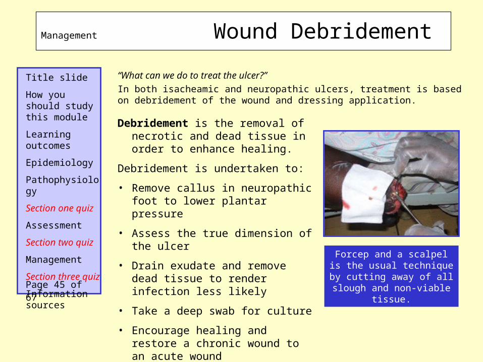
“What can we do to treat the ulcer?”
In both isacheamic and neuropathic ulcers, treatment is based on debridement of the wound and dressing application.
Debridement is the removal of necrotic and dead tissue in order to enhance healing.
Debridement is undertaken to:
• Remove callus in neuropathic foot to lower plantar pressure
• Assess the true dimension of the ulcer
• Drain exudate and remove dead tissue to render infection less likely
• Take a deep swab for culture
• Encourage healing and restore a chronic wound to an acute wound
Forcep and a scalpel is the usual technique by cutting
away of all slough and non-viable tissue.Page 45 of 67
Management Wound Debridement

Title slide
How you should study this module
Learning outcomes
Epidemiology
Pathophysiology
Section one quiz
Assessment
Section two quiz
Management
Section three quiz
Information sources
The larvae of the green bottle fly (which feed on dead flesh) are sometimes used to debride ulcers, especially in the ischaemic foot. Only sterile maggots obtained from a medical maggot farm should be used!
Maggots produce a mixture of proteolytic enzymes that breakdown slough and necrotic tissue which they ingest as a source of nutrients. During this process, they also ingest and kill bacteria including antibiotic resistant strains.
As a result of their wound cleansing activity, the application of maggots has been found to reduce wound odour, and it has also been reported that their presence within a wound stimulates the formation of granulation tissue.
Contra-indications to maggot therapy:• Free range maggots should not be introduced into wounds that
communicate with the body cavity or any internal organ• They should not be applied to wounds that have a tendency to
bleed easily or contain exposed large blood vessels• They should not be applied to patients with clotting disorders, or
individuals receiving anticoagulant therapy unless under constant medical supervision in a health facility.
Page 46 of 67
Management Wound Debridement using maggots (larvaetherapy)

Title slide
How you should study this module
Learning outcomes
Epidemiology
Pathophysiology
Section one quiz
Assessment
Section two quiz
Management
Section three quiz
Information sources
Maggots are available in 2 forms.
1. ‘Free Range’ maggots• applied directly to the wound• roam freely over the surface seeking
out areas of slough or necrotic tissue
• generally left on wound for a maximum of 3 days.
2. BioFOAM Dressing• Maggots enclosed in net pouches
containing pieces of hydrophilic polyurethane foam
• dressing is placed directly upon the wound surface
• BioFOAM Dressing can be left for up to 5 days then the wound is reassessed.
BioFOAM dressing with maggots inside
Page 47 of 67
Management Larvaetherapy Preparations

Title slide
How you should study this module
Learning outcomes
Epidemiology
Pathophysiology
Section one quiz
Assessment
Section two quiz
Management
Section three quiz
Information sources
A sterile, non-adherent dressing should cover all open diabetic foot lesions to protect them from trauma, absorb exudate, reduce infection and promote healing.
Dressings should be lifted every day to ensure that problems or complications are detected quickly, especially in patients who lack nociception.
Additional approaches include
Skin graft:A split-skin graft may be harvested and applied to the ulcer to speeds healing of the ulcer which if has a clean granulating wound bed
Vacuum-Assisted closure (VAC) pump:This is an innovative measure to close diabetic foot wounds. It applies gentle negative pressure to the ulcer via a tube and foam sponge which are applied to the ulcer over a dressing and sealed in place with a plastic film to create a vacuum. Exudate from the wound is sucked along the tube to a disposable collecting chamber. The negative pressure improves the vascularity and stimulates granulation of the wound.
Page 48 of 67
Management Wound Dressings

Title slide
How you should study this module
Learning outcomes
Epidemiology
Pathophysiology
Section one quiz
Assessment
Section two quiz
Management
Section three quiz
Information sources
“Are there any new interesting aids for wound healing?” –Yes, three here; Hyperbaric oxygen therapy: Poor tissue oxygenation with diabetic
microangiopathy reduces wound healing. Therefore hyperbaric oxygen therapy (HBOT) would theoretically aid in faster wound healing, there is however little evidence for this at present.
Growth factor therapy: Recombinant platelet derived growth factor (PDGF) was the first growth factor approved by the Food and Drug Administration (FDA) for the treatment of lower extremity diabetic neuropathic ulcers that extend into the subcutaneous tissue and have adequate blood supply. PDGF, applied as a gel , theoretically acts to enhance granulation tissue formation and facilitate epithelialisation . It may be useful in small, low-grade so may have a role in chronic neuropathic ulcers that are refractory to conventional therapy but there is no evidence to support this theory.
Bioengineered human dermis transplantation: Dermagraft is a cultured human dermis produced by seeding dermal fibroblasts on a biodegradable scaffold. After culture, a living dermal tissue is created which can later support the formation of an epidermis. Furthermore, dermatograft can generate growth factors, cytokines, matrix proteins and glycosaminoglycan, thus aiding the healing process. There have been a limited number of trials have confirmed the efficacy of dermagraft in healing chronic ulcers in a significantly shorter time.
Page 49 of 67
Management New Developments

Title slide
How you should study this module
Learning outcomes
Epidemiology
Pathophysiology
Section one quiz
Assessment
Section two quiz
Management
Section three quiz
Information sources
“It appears infected...which antibiotics to use?”
Treating infected ulcers:• Ensure the previously described physical wound management techniques are used.• The initial antibiotic regime is usually selected empirically based upon clinical experience and local preferences; cover of +cocci is essential as they are the usual culprits of infection as they thrive cutaneously. Antibiotics are modified on the basis of clinical response and and wound culture / sensitivity results. Good examples include;
Oral antibiotics Perenteral antibiotics
Penicillin V OR co-amoxiclav +/- Benzylpenicillin +/-
•Flucloxacillin•Ciprofloxacillin•Cephalexin•clindamycin
•Flucloxacillin•Imipenem-cilastin•Ampicillin-sulbactam•Cefuroxime•Metronidazole ( for anaerobes )
For mild infections, 7-10 day course is usually sufficient. Severe infections may need up to 2-3 weeks of treatment.
Page 50 of 67
Management Infected Ulcers - Antibiotics

Title slide
How you should study this module
Learning outcomes
Epidemiology
Pathophysiology
Section one quiz
Assessment
Section two quiz
Management
Section three quiz
Information sources
“And when the bone gets infected?”
Lastly, treating underlying osteomyelitis is an important therapeutic challenge. Presence of osteomyelitis warrants long-term treatment of at least 4 – 6 weeks duration with antibiotics that penetrate well into bone such as fluoroquinolones, clindamycin or fusidic acid.
Surgical ressection still remains the most definitive treatment for osteomyelitis especially for patients not responding to antibiotics.
Treating Charcot’s neuro-osteoarthropathy“Charcot foot” refers to bone and joint destruction that occurs in the neuropathic foot or rarely just the toe. It can be divided into three phases:
•Acute onset;
•Bony destruction / deformity;
•Stabilistion;
1. Acute onset
Characterised by unilateral erythema and oedema and the foot is at least 2˚C hotter than the contralateral foot. About 30% of patients may complain of pain or discomfort which is rarely severe. X-ray may be normal, but a technnetium methylene diphosphonate bone scan will detect early evidence of bony destruction.
An infected ulcer draining pus
Page 51 of 67
Management The Charcot Foot

Title slide
How you should study this module
Learning outcomes
Epidemiology
Pathophysiology
Section one quiz
Assessment
Section two quiz
Management
Section three quiz
Information sources
Patients awaiting bone scan should be treated as if the diagnosis has been confirmed;•Initially the foot is off-loaded and immobilised in a non-weight-bearing cast to prevent deformity. After 1 month, a total-contact cast is applied and the patient may mobilise for brief period. However, the patient is given crutches and encouraged to keep walking to a minimum. • If given early, these measures can prevent bony destruction. Bisphosphonates are potent inhibitors of osteoclast activation and may also be used in this phase.
2. Bony destruction•Clinical signs are swelling, warmth, a temperature 2˚C greater than the contralateral foot and deformities including the rocker-bottom deformity and medial convexity.
X-ray reveals fragmentation, fracture, new bone formation, subluxation & dislocation.
The aim of treatment is immobilisation until there is no X-ray evidence of continuing bone destruction and the foot temperature is within 2˚C of contra lateral foot.
A photo showing a charcot foot with an ulcer on the sole
Page 52 of 67
Management The Charcot Foot - 2

Title slide
How you should study this module
Learning outcomes
Epidemiology
Pathophysiology
Section one quiz
Assessment
Section two quiz
Management
Section three quiz
Information sources
3. StabilisationThe foot is no longer warm and red. There may still be oedema but the difference in skin temperature between the feet is less than 2˚C. the X-ray shows fracture healing, sclerosis and bone remodelling.
The patient can now progress from a total-contact cast to an orthotic walker, fitted with cradled moulded insoles if necessary to accommodate a rocker-bottom or medial convexity deformity. Cautious rehabilitation should be the rule, beginning with a few short steps in a new footwear.
Finally, the patient may progress to bespoke footwear with moulded insoles as the rocker-bottom charcot foot with plantar bony prominence is a site of very high pressure. Regular reduction of callus can prevent ulceration.
During the acute stage, charcot foot’s foot may be misdiagnosed as;CellulitisOsteomyelitisDeep vein thrombosisInflammatory arthropathy
Therefore a high index of suspicion is very important at this stage!
Page 53 of 67
Management The Charcot Foot - 3

Title slide
How you should study this module
Learning outcomes
Epidemiology
Pathophysiology
Section one quiz
Assessment
Section two quiz
Management
Section three quiz
Information sources
“...if the foot does not stabilise or ulcer is worsening?”- Definitive management
AmputationReferral to vascular surgeons for possible
amputation is made on clinical findings that the
ulceration is not healing/ infection worsening in spite
of intensive antibiotic therapy
Signs include: Extensive tissue loss Unreconstructable ischaemia Failed revascularisation Charcot’s of ankle with instabilityPage 54 of 67
Management Amputation

Title slide
How you should study this module
Learning outcomes
Epidemiology
Pathophysiology
Section one quiz
Assessment
Section two quiz
Management
Section three quiz
Information sources
“What about giving them some analgesia?”
Treating painful diabetic neuropathy:General approach; •Reassure the patient that intense pain improves within 2 years. •Regular appointments to monitor their pain and try new strategies if refractory to previous attempts. •It is essential to optimise diabetic control.
Drugs;Simple analgesics; e.g. aspirin, paracetamol, and mild opiates such as codeine phosphate singly or in combination. Prescribe hypnotics for disturbed sleep.
Trycyclic antidepressants; e.g imipramine, amitriptyline. Commence with low dose and gradually increase according to symptomatic response
Anticonvulsants; e.g carbamazepine, valproate, phenytoin, gabapentin, lamotrigine may be very useful. The latter two may improve sleep in addition to pain relief.
Capsaicin is a very useful topical analgesic
Page 55 of 67
Management Pain
Title slide
How you should study this module
Learning outcomes
Epidemiology
Pathophysiology
Section one quiz
Assessment
Section two quiz
Management
Section three quiz
Information sources
“So that’s where we are at the moment. How about future developments?”
Prophylactic foot surgery:The last decade has a dramatic interest in reconstructive foot surgery for the diabetic foot. The aim of this surgery is to reduce risk of ulceration.
A short Achilles tendon may be associated with elevated forefoot plantar pressure and hence may benefit from Achilles tendon lengthening surgery.
Tenotomy of toe extensors may reduce toe deformities, thus preventing recurrent ulcerations in this group of patients.
Metatarsal osteotomy may reduce the risk of ulcer recurrences in subjects with prominent metatarsal heads.
However, currently there is no randomise control trial evidence comparing these surgical techniques with medical therapy.
Page 56 of 67
Management New Surgical Techniques

Title slide
How you should study this module
Learning outcomes
Epidemiology
Pathophysiology
Section one quiz
Assessment
Section two quiz
Management
Section three quiz
Information sources
Well done!You have come to the end of the last section
We suggest that you answer Question 10 to 18 to assess what you have learnt. Please
remember to write your answers on the mark sheet before looking at the correct answers!
Page 57 of 67
End of Section 3

Title slide
How you should study this module
Learning outcomes
Epidemiology
Pathophysiology
Section one quiz
Assessment
Section two quiz
Management
Section three quiz
Information sources
Good shoe guide:
a) Toe box should be sufficiently long, broad and deep to
accommodate the toes without pressing on them, with a clear
space between the apices of the toe box
b) Shoes should be fasten with adjustable lace, strap or
velcro high on the foot in order to hold foot firmly inside
the shoe and thus reduce frictional forces when the patient
walks
c) The heel of the shoe should be over 5 cm high
to avoid weight being thrown forward into metatarsal heads
d) The inner lining of shoe should be smooth
e) Stocking or socks should not be worn with shoes
Question 10: Write ‘T’ or ‘F’ on the answer sheet. First complete all 5 questions, then click on the boxes and mark your answers.
a
b
c
e
d
Page 58 of 67
Section 3 Quiz

Title slide
How you should study this module
Learning outcomes
Epidemiology
Pathophysiology
Section one quiz
Assessment
Section two quiz
Management
Section three quiz
Information sources
1) ……….
2) ……….
3) ……….
4) ……….
5) ……….
Question 11: List five common foot problems that occur in the population at large.Write your answer in your mark sheet
Click here for the answers
Page 59 of 67
Section 3 Quiz

Title slide
How you should study this module
Learning outcomes
Epidemiology
Pathophysiology
Section one quiz
Assessment
Section two quiz
Management
Section three quiz
Information sources
Question 12: Identify the following photos below. Write your answer in your mark sheet.
Click here for the answers
1
2
3 4
Page 60 of 67
Section 3 Quiz

Title slide
How you should study this module
Learning outcomes
Epidemiology
Pathophysiology
Section one quiz
Assessment
Section two quiz
Management
Section three quiz
Information sources
1) ……….
2) ……….
3) ……….
Question 13: name three cast techniques used for off-loading pressure in neuropathic diabetic foot. Write your answer in your mark sheet
Click here for the answers
Page 61 of 67
Section 3 Quiz

Title slide
How you should study this module
Learning outcomes
Epidemiology
Pathophysiology
Section one quiz
Assessment
Section two quiz
Management
Section three quiz
Information sources
1) ………………..
2) ………………..
3) ………………..
4) ………………..
5) ………………..
Question 14: List five reasons why debridement is important in the treatment of diabetic foot ulcers. Write your answer in your mark sheet
Click here for the answers
Page 62 of 67
Section 3 Quiz

Title slide
How you should study this module
Learning outcomes
Epidemiology
Pathophysiology
Section one quiz
Assessment
Section two quiz
Management
Section three quiz
Information sources
Oral antibiotics;
1) …………..
2) …………..
3) …………..
4) …………..
Parenteral antibiotics;
1) ……………
2) ……………
3) ……………
4) ……………
Question 15: List 4 oral and 4 parenteral antibiotics used in treating infected diabetic foot ulcers.Write your answer in your mark sheet
Click here for the answers
Page 63 of 67
Section 3 Quiz

Title slide
How you should study this module
Learning outcomes
Epidemiology
Pathophysiology
Section one quiz
Assessment
Section two quiz
Management
Section three quiz
Information sources
……………………………………………………
……………………………………………………
……………………………………………………
a) ………….
b) ………….
c) ………….
Question 16: Describe the term charcot foot and mention its three phases of evolution .Write your answer in your mark sheet
Click here for the answers
Page 64 of 67
Section 3 Quiz

Title slide
How you should study this module
Learning outcomes
Epidemiology
Pathophysiology
Section one quiz
Assessment
Section two quiz
Management
Section three quiz
Information sources
Question 17: identify the following photos below. Write your answer in your mark sheet
Click here for the answers
12
3 4
Page 65 of 67
Section 3 Quiz

Title slide
How you should study this module
Learning outcomes
Epidemiology
Pathophysiology
Section one quiz
Assessment
Section two quiz
Management
Section three quiz
Information sources
A. ………………
B. ………………
C. ………………
D. ………………
E. ………………
Question 18: List 5 categories of drugs used in the treatment of painful diabetic neuropathy. Write your answer in your mark sheet.
Click here for the answers
Page 66 of 67
Section 3 Quiz

Title slide
How you should study this module
Learning outcomes
Epidemiology
Pathophysiology
Section one quiz
Assessment
Section two quiz
Management
Section three quiz
Information sources
1. A Clarke (2005). Pathology of the non-ulcerative foot. Diabetes voice; volume 50.
2. http://www.emedicinehealth.com/diabetic_foot_care
3. Time to Act (2005). International Diabetes Federation and the International Working Group on Diabetic Foot.
4. Edmonds ME, Foster AVM (2005). Managing the diabetic foot (2nd edition). Blackwell Science, Oxford.
5. Khanolkar MP, Stephens JW, Bain SC. (2007) The Diabetic Foot. (in press). Morriston Hospital, Swansea, UK.
6. www.zoobiotic.com; LarvE® data card version 2.9 and dressing application version 2.0 (2007).
7. Levin and O’Neal. Eds. John H. Bowker and Michael A. Pfeifer. (2007) The Diabetic Foot. Mosby, Elsevier. 7th edition
8. The 5th International Symposium on the Diabetic Foot. (May 9-12, 2007). International Diabetes Federation. Noordwijkerhout, the Netherlands,.
Page 67 of 67
Sources of Information/Images and References