Embed Size (px)
Citation preview
1
1
Title: Sibling rivalry in Myxococcus xanthus is mediated by kin recognition and a polyploid 2 prophage 3
4
5
Authors: Arup Dey‡, Christopher N. Vassallo, Austin C. Conklin, Darshankumar T. 6
Pathak†, Vera Troselj, and Daniel Wall1* 7
8
9
Affiliations: 1Department of Molecular Biology 10
University of Wyoming 11
1000 E. University Ave. 12
Laramie, WY 82071, U.S.A. 13
14
15
16
*Correspondence to: Email: [email protected] 17
Current addresses: ‡Biology Department, Suffolk University; †Department of Developmental 18 Biology, Stanford University 19
20
21
Running title: Sibling rivalry in myxobacteria 22
23
24
25
Keywords: Competition, gliding motility, Myxococcus xanthus, prophage, kin recognition, toxin 26
27 28
JB Accepted Manuscript Posted Online 19 January 2016J. Bacteriol. doi:10.1128/JB.00964-15Copyright © 2016, American Society for Microbiology. All Rights Reserved.
on March 25, 2018 by guest
http://jb.asm.org/
Dow
nloaded from
2
ABSTRACT 29
Myxobacteria form complex social communities that elicit multicellular behaviors. One such behavior is 30
kin recognition, in which cells identify siblings via their polymorphic TraA cell surface receptor, to 31
transiently fuse outer membranes and exchange their contents. In addition, outer membrane exchange 32
(OME) regulates behaviors, such as inhibition of wild-type Myxococcus xanthus (DK1622) from 33
swarming. Here we monitored the fate of motile cells and surprisingly found they were killed by 34
nonmotile siblings. The kill phenotype required OME (TraA dependent). The genetic basis of killing was 35
traced to ancestral strains used to construct DK1622. Specifically, the kill phenotype mapped to a large 36
‘polyploid prophage,’ Mx alpha. Sensitive strains contained a 200-kb deletion that removed two of three 37
Mx alpha units. To explain these results we suggest that Mx alpha expresses a toxin-antitoxin cassette 38
that uses the OME machinery of M. xanthus to transfer a toxin that makes the population ‘addicted’ to 39
Mx alpha. Thus siblings that lost Mx alpha units (no immunity) are killed by cells that harbor the 40
element. To test this, an Mx alpha-harboring laboratory strain was engineered (traA allele swap) to 41
recognize a closely related species, M. fulvus. As a result, M. fulvus, which lacks Mx alpha, was killed. 42
These TraA mediated antagonisms provide an explanation for how kin recognition specificity might have 43
evolved in myxobacteria. That is, recognition specificity is determined by polymorphisms in traA, which 44
we hypothesize were selected for because OME with non-kin leads to lethal outcomes. 45
46
on March 25, 2018 by guest
http://jb.asm.org/
Dow
nloaded from
3
IMPORTANCE 47
The transition from single cell to multicellular life is considered a major evolutionary event. 48
Myxobacteria have successfully made this transition. For example, in response to starvation individual 49
cells aggregate into multicellular fruiting bodies wherein cells differentiate into spores. To build fruits, 50
cells need to recognize their siblings and, in part, this is mediated by the TraA cell surface receptor. 51
Surprisingly, we report that TraA recognition can also involve sibling killing. We show that killing 52
originates from a prophage-like element that has apparently hijacked the TraA system to deliver a toxin 53
to kin. We hypothesize that this killing system has imposed selective pressures on kin recognition, which 54
in turn has resulted in TraA polymorphisms, and hence many different recognition groups. 55
on March 25, 2018 by guest
http://jb.asm.org/
Dow
nloaded from
4
INTRODUCTION 56
Myxobacteria inhabit the soil and, as such, live in taxonomically diverse environments in which 57
thousands of microbial species and subspecies compete for scarce resources (1). Remarkably, from 58
these heterogeneous populations, myxobacteria assemble collectives that function like tissues. These 59
multicellular behaviors include rhythmic and synchronized movements that culminate in fruiting body 60
formation. To accomplish this, myxobacteria must recognize their neighbors to determine if they are 61
friend, foe, or food. How myxobacteria recognize kin and assemble homogenous populations is an 62
emerging field of study. 63
The most thoroughly described cell-cell recognition system in myxobacteria is mediated by the TraA 64
polymorphic cell surface receptor. This receptor, with its partner protein, TraB, controls the fusion and 65
exchange of outer membrane (OM) material between cells (2). To engage in OM exchange (OME) the 66
partnering cells must express traA alleles that belong to the same recognition group (3). Because OME 67
leads to the transfer of many different proteins and lipids, it can, in principle, result in cooperative or 68
antagonist interactions. In other bacterial transport systems, cargo transfer is typically unidirectional 69
and the outcomes are usually antagonistic to the recipient. For example, the type III, IV, V, and VI 70
secretion systems transfer effectors to target cells that act as toxins or as virulence or selfish elements 71
(4-7). Cooperative bacterial transfer systems have rarely been described. In contrast, OME involves 72
bidirectional cargo transfer, in which both cells must express compatible TraA/B machinery, suggesting 73
that these interactions are mutually sought. 74
Myxobacteria are gliding bacteria that translocate in a smooth motion on solid surfaces along their long 75
axis (8). The movement of cell groups is called swarming and is a cooperative behavior, because their 76
expansion rate increases with cell density (8). In OME, gliding is indirectly required to facilitate 77
membrane fusion and fission (9). Gliding is powered by two separate engines, referred to as A 78
on March 25, 2018 by guest
http://jb.asm.org/
Dow
nloaded from
5
(adventurous) and S (social) motility (8). The S-engine consists of type IV pili and the A-engine is a multi-79
protein complex that includes mobile cell surface adhesins (10). S-motility is proficient for swarming on 80
soft, moist agar and requires cell-cell contact. In contrast, A-motility is adapted for hard and drier 81
surfaces, on which individual or small groups of cells move (11). A nonmotile mutant (A‒S‒) therefore 82
typically requires two mutations, one in each system. 83
Because Myxococcus xanthus is both a social and predatory species, it is a good model system to study 84
the interplay between cooperative and competitive interactions. Its extensive social behaviors suggest 85
that M. xanthus has evolved a means to regulate these interactions. One example is fruiting body 86
development where a sub-population develops into environmentally resilient spores in response to 87
starvation, while other cells lyse or form persister-like cells (12). How cell fates are determined is poorly 88
understood, but may involve competitive interactions interwoven within cooperative behaviors. 89
Likewise, OME appears to involve both cooperative and competitive interactions. Cooperative 90
interactions are suggested by sharing of cellular resources and, in some cases, the ability of cells to 91
repair their damaged sibling cells (13). In contrast, the swarming and developmental behaviors of some 92
motile strains can be antagonized by OME with some nonmotile strains (2). This antagonistic response is 93
potent, as a ratio of 1 nonmotile cell to 50 motile cells blocks the latter from swarming (14). The nature 94
of swarm inhibition is the focus of this study, in which we found that ancestral strains kill siblings that 95
were derived from them. The kill phenotype required OME and was engineered into a laboratory strain 96
to antagonize an environmental isolate. We suggest that the kill phenotype arises from a toxin-antitoxin 97
system that map to a large polyploid prophage-like element that was fortuitously deleted in laboratory 98
strains. We discuss social and evolutionary implications of these findings. 99
100
101
on March 25, 2018 by guest
http://jb.asm.org/
Dow
nloaded from
6
MATERIALS AND METHODS 102
Growth conditions. Bacterial strains used in this study are listed in Table 1. M. xanthus was grown in the 103
dark at 33°C in CTT medium (1% Casitone, 10 mM Tris-HCl [pH 7.6], 8 mM MgSO4, 1 mM KH2PO4) with or 104
without kanamycin (Km; 50 µg ml–1), zeocin (Zm; 50 µg ml–1), oxytetracycline (Tc; 10 µg ml–1) or 105
galactose (Gal, 2%), as needed. For swarm inhibition assays, agar (1.5%) plates consisted of ½ CTT (0.5% 106
Casitone) with 2 mM CaCl2 added after autoclaving, or TPM (10 mM Tris-HCl [pH 7.6], 8 mM MgSO4, 1 107
mM KH2PO4) agar was used. Standard competition assays were done on 1.5% agar plates with ¼ CTT 108
(0.25% Casitone). For competition assays on semi-solid agar, CTT with 0.5% agar was used. To generate 109
micrographs of mixed swarms, ¼ CTT 0.8% agarose pads were made on glass microscope slides. To 110
determine CFU of mixed cultures, CTT agar plates were supplemented with antibiotics to select for a 111
particular strain and colonies were counted after 6 days of incubation. tdTomato expression was 112
induced in liquid and on agar plates with 0.1 mM IPTG. To grow M. fulvus, ½ CTT was supplemented with 113
0.5% yeast extract. For routine cloning, Escherichia coli DH5α pir+ was grown at 37°C in LB and 114
tetracycline (10 µg ml–1) or Km (50 µg ml–1) was used for selection as need. 115
116
Strain constructions. Gene disruptions were made by amplifying internal gene fragments by PCR that 117
were then cloned into pCR-XL-TOPO or pCR2.1-TOPO (Table S1). The Tn5-Ω2213 insertion site was 118
identified by a PCR-based method as previously described (15). For aglB1 (aglQ) rescue, a plasmid was 119
made by amplifying the aglRQS operon and cloning it into pCR2.1-TOPO generating pDP110. The 120
markerless ∆Mx alpha deletion cassette was made by PCR amplification of the corresponding upstream 121
and downstream DNA fragments, and these fragments were placed in pBJ114 by three-piece Gibson 122
cloning (New England BioLabs) to create pCV101. Primers used for PCR are listed in Table S2. Colony PCR 123
and restriction analysis were used to confirm clone construction. Verified plasmids (Table S1) were 124
electroporated into M. xanthus strains, and recombinants were selected on CTT agar with appropriate 125
on March 25, 2018 by guest
http://jb.asm.org/
Dow
nloaded from
7
antibiotics. M. xanthus transformants were then isolated and verified by diagnostic PCR and/or 126
phenotypic analysis. For DW2403 (ΔMx alpha-29) strain verification, we used diagnostic PCR with 127
primers against MXF1DRAFT_07228 and confirmed that a deletion had occurred in Mx alpha. Additional 128
diagnostic PCR reactions confirmed that the Mx alpha region corresponding to the end of contig 48 was 129
also absent; however, a region corresponding to contig 58 was unexpectedly present. From the counter-130
selection step, seven additional Galr Kms clones that showed no antagonistic phenotype were tested by 131
PCR and were all found to contain different types of deletions in Mx alpha, but none of them contained 132
the full deletion as planned. We concluded that the large Mx alpha repeats were inherently unstable 133
and deletions spontaneously occurred at different positions within Mx alpha. See the Discussion for 134
further details. 135
136
Swarm inhibition. Experiments were typically done by mixing motile and nonmotile strains at a 1:1 ratio 137
(~3 × 10⁹ CFU ml–1) and pipetting the mixtures onto the described plates. Unless stated otherwise, the 138
plates were incubated for 72 h at 33°C, and micrographs were taken with a stereomicroscope at 3.2× 139
magnification or with a 10× phase contrast objective on a compound microscope (2). Time-lapse 140
microscopy was done as described (2). 141
142
Competition experiments. Myxococcus strains were grown in CTT overnight, and cells were harvested at 143
mid-log growth (~3 × 10⁸ CFU ml–1). For fluorescent labeling experiments, either one or both strains 144
were labeled with GFP, tdTomato, or mCherry and mixed at the indicated ratios. Strain mixtures were 145
transferred to agarose pads (5 μl, for direct microscopy) or agar plates (30 μl, to harvest cells) and 146
incubated in a humid chamber. At the indicated times, either the colony edge was observed on agarose 147
pads or cells were collected and washed twice in TPM and observed on a glass slide by phase contrast 148
and fluorescence microscopy with a FITC or Texas red filter set. At least 200 cells were counted for each 149
on March 25, 2018 by guest
http://jb.asm.org/
Dow
nloaded from

8
replicate to determine strain ratios. Micrographs were obtained as described (2). To determine CFU 150
from competition experiments, cells with Tc or Km markers were similarly mixed and collected, and 151
viable cells were enumerated by serial dilution onto selective plates. 152
153
on March 25, 2018 by guest
http://jb.asm.org/
Dow
nloaded from
9
RESULTS 154
Swarm inhibition is caused by sibling killing. In earlier work we found that nonmotile strains of M. 155
xanthus inhibit the ability of apparent isogenic motile strains to swarm (Fig. 1A) (2). Swarm inhibition is 156
not caused by physical obstruction of nonmotile cells but instead is TraA/B dependent. To investigate 157
this phenomenon further, the edge of mixed inoculums was observed at higher resolution. After 24 h of 158
incubation, motile cells had migrated beyond the inoculum spot (Fig. 1B). However, by 48 h those 159
individual cells or small groups of cells seen at 24 h had mostly disappeared, although their phase-bright 160
‘slime trails’ remained (Fig. 1B). The disappearance of cells from the swarm edge raised the possibility 161
that cells either returned to the colony or had lysed. To track the fate of such cells, time-lapse 162
microscopy was used 24 h after the cell mixture was plated. . As previously reported (2, 14), many of the 163
cells at the swarm edge moved slowly or not at all (compare Movie S1 to S2), suggesting that those cells 164
were sick or dead. In addition, in two cases isolated cells lysed (Movie S1). 165
Our results suggested that motile cells at the swarm periphery died and lysed following contact with 166
their nonmotile siblings. To investigate the fate of motile cells within the colony center, strains were 167
differentially labeled with fluorescent proteins. Here, motile and nonmotile strains were labeled with 168
GFP (cytoplasm) and mCherry (cytoplasmic membrane), respectively, and their fitness was assessed. As 169
expected, shortly after mixing and plating, there were a substantial number of green- and red-labeled 170
cells (Fig. 2A, first row). Over time, however, the number of GFP-labeled cells decreased, and by 48 h the 171
green motile cells were rarely detected (Fig. 2A, second row). To clearly delineate individual cells, the 172
colony was collected in buffer and cells were viewed on glass microscope slides. Again, the green motile 173
cells were rarely seen by 72 h (Fig. S1 top row, tra+). These results suggested that the motile GFP-labeled 174
cells had lysed after extended contact with nonmotile siblings. 175
on March 25, 2018 by guest
http://jb.asm.org/
Dow
nloaded from
10
To quantify population dynamics, strain mixtures were collected at various times, washed, and 176
microscopically examined on glass slides. The ratio of motile to nonmotile cells was then determined by 177
fluorescence microscopy. As found above, the ratio of motile cells dramatically decreased over time. In 178
this assay, by 72 h the motile cell population was ~100-fold lower (limit of assay) than the nonmotile cell 179
population (Fig. 3A). To assess a wider dynamic range, the cell populations were enumerated by viable 180
colony forming units (CFU). In this assay, the motile and nonmotile cells were differentiated by Km- or 181
Tc-resistant markers that the strains respectively carried. After 72 h, no viable motile cells were 182
detected (≥104-fold decrease) (Fig. 2B). In contrast, the nonmotile population grew. We conclude that 183
the swarm inhibition was caused by nonmotile cells killing their motile siblings. 184
Sibling killing is Tra dependent. We previously showed that swarm inhibition is Tra dependent (Fig. 1A) 185
(2, 14). That is, when either strain in the mixture contains a traA or traB mutation, swarm inhibition is 186
abolished. We tested whether swarm relief correlated with motile cell survival when OME was blocked. 187
As expected, when the motile cells contained a traA mutation, they swarmed out from the inoculum 188
(Fig. 2A, compare the second row with the bottom row). In addition, and in contrast to Tra+ strain 189
mixtures, the isogenic TraA mutant flourished when mixed with the same nonmotile strain (Fig. 2A, 190
compare the second row with the bottom row). To quantify this, the number of CFU in each population 191
was determined. The motile strain with the TraA mutation survived as well as the nonmotile strain (Fig. 192
2B), indicating that the kill phenotype was Tra dependent. 193
Target cells become filamentous. The morphological fate of motile cells was tracked during swarm 194
inhibition at high magnification. Interestingly, by 24 h the surviving GFP-labeled motile cells became 195
filamentous, ranging in length from 12 to 20 µm (Fig. 2C). Filamentation was Tra dependent, as a traA 196
mutant did not elongate (length ~7 µm; Fig. 2C). Attempts to transfer filamentous cells to glass slides for 197
detailed inspection were unsuccessful, suggesting that filamentous cells had lysed following physical 198
manipulation. Because filamentation is a response to stress, including exposure to poisons (16), we 199
on March 25, 2018 by guest
http://jb.asm.org/
Dow
nloaded from
11
hypothesized that a toxin was delivered by a Tra-dependent mechanism from nonmotile to motile cells, 200
which then led to filamentation and death. 201
Semi-solid agar abolishes killing. OME requires TraA/B function on a hard agar surface; it occurs neither 202
in liquid nor on semi-solid agar (9, 17). Susceptibility of the killing effect was thus tested, and, consistent 203
with prior findings, there was no killing on semi-solid agar, whereas killing occurred on hard agar (Fig. 204
3A). This finding suggests that killing, like OME, requires sustained cell-cell contacts on a hard surface 205
and that it is not mediated by a diffusible factor. 206
An omrA mutation confers resistance. omrA was identified from a forward screen for factors required 207
for swarm inhibition. In contrast to TraA/B, OmrA and the co-discovered OmrB proteins are not required 208
for OME but instead are specifically involved in how cells respond to OME (14). Here, OmrA/B were 209
tested for involvement in killing. Importantly, the omrA mutant was not killed and actually outcompeted 210
the nonmotile strain by about five-fold (Fig. 3B). This indicates that the omrA mutation confers 211
resistance to killing and explains how it was discovered in the screen (14). In contrast, a strain containing 212
a mutation in omrB (identified by bioinformatic methods to function in the OmrA pathway) was killed, 213
although there was a modest delay (Fig. 3B). This result correlates with the partial swarm relief 214
phenotype that is conferred by an omrB mutation (14). 215
Sibling antagonism is not correlated to motility phenotypes. We sought to identify the genetic 216
determinant(s) that caused killing; however a feasible forward screen was not apparent to us. As an 217
alternative approach we surveyed interactions between different laboratory strains in order to obtain 218
clues about the genetic basis of killing. Because our initial observation was swarm inhibition (2, 14), we 219
tested whether motility phenotypes might be involved in killing. However, through a series of 220
experiments we determined that phenotypic differences in A- and S-motility were not the cause of the 221
kill phenotype (for details see Supplemental Material and Figs S2, S3 and S4). 222
on March 25, 2018 by guest
http://jb.asm.org/
Dow
nloaded from
12
DK1622-derived strains are sensitive to killing by ancestral strains. To continue the search for genetic 223
factors involved in sibling rivalry we expanded the panel of strains surveyed. From these studies we 224
discovered that swarm inhibition was correlated to an ancestral strain background. Specifically, in a 225
nonmotile DK101 (A–S–) strain background there was swarm inhibition, whereas in a nonmotile DK1622 226
background there was no swarm inhibition. As shown in Figure 4A this result was repeatable between 227
three different sets on nonmotile strains. Here each strain set contained a different A-motility mutation. 228
As outlined in Figure 5, DK1622 was derived from DK101 via two intermediate strains. The construction 229
of DK1622, which occurred four decades ago, was necessitated because the predecessor strains, 230
including DK101, lacked S-motility (18). 231
To test the hypothesis that strain background has a role in antagonism, three different ancestral strains 232
were investigated. Two of these strains, DK101 and YS, were directly derived from FB (18, 19) and were 233
the parental strains used to create DK1622 (18) (Fig. 5). The third strain, DZ2, shares a common ancestor 234
with DK1622 but was stored independently by the Zusman lab (20, 21). When any of these ancestral 235
strains was mixed with a DK1622-derived strain, the former strains readily outcompeted the latter strain 236
(Fig. 4B). Similarly, DK1217, which is the direct parent of DK1622 (Fig. 5), readily outcompeted a DK1622-237
derived strain (Fig. S4). In addition, when these motile ancestral strains were mixed with a labeled 238
DK8601 aggressor strain, they were not killed, whereas the control strain was killed (Fig. 4C). Taken 239
together, we conclude that ancestral strains (DK101, DZ2, YS, and DK1217) antagonize their DK1622 240
sibling. 241
Ancestral strains, including DK1217, contain Mx alpha. A notable difference between DK1622 and YS 242
and DK101 is that the former strain has a ~200-kb deletion (22, 23). The deleted region contains a 243
defective prophage-like element called Mx alpha (22, 24). Work by Zissler and colleagues’ showed that 244
Mx alpha particles only contained a small portion (~35 kb) of the prophage-like region (300 kb) and were 245
not lytic. In our studies we also found no evidence that Mx alpha particles kill. However, because 246
on March 25, 2018 by guest
http://jb.asm.org/
Dow
nloaded from
13
prophages can contain toxins (25, 26), a possible explanation for killing is that a toxin-antitoxin gene 247
cassette resides in the region missing from DK1622 (Note: Table 1 states Mx alpha genotype of all M. 248
xanthus strains by superscript symbols or text). There was, however, an inconsistency with the 249
hypothesis that Mx alpha was the genetic determinant for killing. Namely, the Mx alpha deletion was 250
presumed to have occurred following UV mutagenesis of DK101 to create DK320 and prior to 251
construction of DK1217 (Fig. 5) (18, 23). To investigate whether the Mx alpha region was correlated with 252
the killing phenotype, the ancestral strains were screened for this deletion by PCR. Here, a DNA marker 253
was identified that corresponded to the deleted Mx alpha region by comparing the draft genome 254
sequence of DK101 (aka DZF1) (27) to the complete DK1622 genome (28) (Fig. 6). Importantly, all of the 255
aggressor strains, including DK1217, contained the Mx alpha region that was absent from DK1622 (Fig. 256
4E). Therefore there was a correlation between the aggressor phenotype and the presence of the 257
complete Mx alpha region. 258
A report by Youderian and colleagues suggested that the nonmotile strain DZ1 also contains a deletion 259
in Mx alpha (29). As outlined (Fig. 5), DZ1 was derived from FB independently of DK1622 (30, 31). To test 260
the proposed correlation, DZ1 was screened and found to lack the Mx alpha diagnostic marker (Fig. 4E) 261
and exhibited no antagonism toward DK1622 (Fig. 4D). These findings support the idea that Mx alpha 262
contains a genetic determinant involved in killing. In addition, the finding that DK1622 and DZ1 263
independently and spontaneously deleted part or all of Mx alpha suggests that this element is unstable 264
during laboratory growth. 265
Mx alpha is necessary for the kill phenotype and resistance. To directly test whether Mx alpha is 266
involved in killing, a ΔMx alpha mutation was created in a nonmotile aggressor strain that contained Mx 267
alpha by use of plasmid pCV101 (which contains ΔMx alpha and counter-selectable cassette). 268
Importantly, this strain (DW2403, ΔMx alpha-29) no longer killed nor caused swarm inhibition (Fig. 7A). 269
on March 25, 2018 by guest
http://jb.asm.org/
Dow
nloaded from
14
In addition, DW2403 was susceptible to being killed (Fig. 7B). We conclude that Mx alpha is a necessary 270
for the kill phenotype and for resistance to killing. To explain these results, we suggest that Mx alpha 271
contains a toxin that kills siblings mediated by OME delivery and a cognate antitoxin that confers 272
immunity. 273
Mx alpha is polyploid. 274
Starich and Zissler showed by Southern blot analysis with DNA from purified particles that Mx alpha 275
consists of three large repeat units, two of which are absent from DK1622 (22). Genome comparisons 276
indeed showed that DK1622 contains a single Mx alpha unit that spans a 100-kb region (MXAN_1801 to 277
MXAN_1900; Fig. 6). The DZF1 draft genome, which consists of 75 contigs (27), contains seven contigs 278
that perfectly match MXAN_1801 to MXAN_1900 and nine other contigs that are unique to DZF1 yet 279
share homology to the aforementioned DK1622 region (Fig. 6). These 16 contigs from DZF1 span 300-kb 280
and constitute three imperfect repeats. That is, alleles of some genes are present in all repeats and 281
other genes are unique to a particular copy. In total, 84 ORFs between MXAN_1801 and MXAN_1900 282
have alternative alleles in DZF1 that were absent from DK1622 (Table S2). These alternative alleles 283
typically share 50–99% identity at the amino acid level. Last, we note, that the Mx alpha region contains 284
several candidate toxin and antitoxin ORFs. 285
Engineered laboratory strain kills environmental isolate. Previously we showed that when a laboratory 286
strain heterologously expresses a traAM. fulvus allele it empowers OME with the corresponding M. fulvus 287
HW-1 environmental isolate (3), which was otherwise unable to engage in OME with DK1622. In 288
addition, there is a fitness gain for the laboratory strain in competition experiments with M. fulvus (3). 289
Conversely, when the traADK1622 gene is deleted from a laboratory strain (DK1622-related), which 290
prevents OME with environmental isolates belonging to the TraADK1622 recognition group, its fitness 291
markedly decreases against those isolates (3). Our current findings suggest an explanation for these 292
on March 25, 2018 by guest
http://jb.asm.org/
Dow
nloaded from
15
results. To investigate this, we tested whether the M. fulvus strain was killed in a manner that depended 293
on OME and Mx alpha. Here, two different DK1217-derived lab strains that contained either the 294
traADK1622 or traAM. fulvus alleles were mixed with M. fulvus. Based on the resulting CFU, the M. fulvus 295
strain outcompeted the traADK1622 strain by 1000-fold after 24 h (Fig. 8B). In contrast, there was nearly a 296
100-fold decrease in the relative fitness of M. fulvus when the laboratory strain contained the traAM. fulvus 297
allele. In fact, the survival ratios of M. fulvus and of the engineered M. xanthus with traAM. fulvus were 298
nearly equal, although for both strains their CFU were lower than the CFU in the starting inoculum (Fig. 299
8B). In contrast, M. fulvus outcompeted DK1622 (i.e., ΔMx alpha) expressing TraAM. fulvus (Fig. 8B). The 300
magnitude of the antagonistic interactions was also evident by visual and microscopic inspection of 301
inoculum mixtures (Fig. 8A). Robust colony growth was observed when M. fulvus dominated the 302
laboratory strains (Fig. 8A, left and right colonies). However, when the DK1217 traAM. fulvus strain 303
containing the entire Mx alpha region, was mixed with M. fulvus the inoculum remained translucent 304
after 48 h, indicating intense bidirectional antagonism that blocked either strain from swarming or 305
growing (Fig. 8A, middle panels). From these results, we conclude that a kill phenotype can be activated 306
toward siblings and non-siblings, including between different species, by engineering compatible traA 307
alleles for OME and hence cargo (toxin) delivery. We also note that M. fulvus has an uncharacterized 308
mechanism(s) to kill M. xanthus that does not depend on OME. 309
310
on March 25, 2018 by guest
http://jb.asm.org/
Dow
nloaded from
16
DISCUSSION 311
Here, we surprisingly discovered that M. xanthus cells kill siblings derived from the same parental 312
lineage by a mechanism that involves OME. This finding provides evolutionary insight for why kin 313
recognition is involved in OME (3). Namely, OME among neighboring cells can lead to beneficial 314
outcomes; however it can also have lethal consequences. Therefore, kin recognition provides a 315
mechanism by which cell-cell selectivity reduces the chance of lethal encounters between isolates. 316
Selectivity is derived from polymorphisms within the TraA variable domain; OME occurs only between 317
isolates that have identical or nearly identical traA alleles (3). The kill phenotype also explains why our 318
previous screen to isolate swarm relief mutants was so powerful (14). Indeed, instead of a screen, as 319
originally conceived, a genetic selection was imposed. Thus, in mixed cultures motile cells were 320
annihilated by their nonmotile siblings unless they contained a mutation that blocked killing. The more 321
than 50 mutations isolated all mapped to traAB or omrA (14). 322
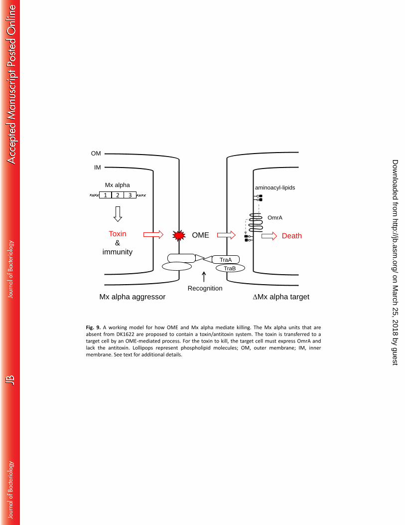
A working model for killing is outlined in Figure 9. In this model, the Mx alpha cell is hypothesized to 323
produce toxin-antitoxin factors. Toxin delivery is mediated by OME, because when OME is blocked by a 324
tra mutation, incompatible traA alleles, or environmental conditions, antagonism is abolished. The 325
finding that an omrA mutation confers resistance provides clues about the toxicity mechanism. Based on 326
sequence similarity to MprF from Staphylococcus aureus, OmrA is predicted to function as an amino-acyl 327
phospholipid flippase (14). In S. aureus, altered MprF function confers resistance to cationic antibiotics, 328
such as daptomycin, that act on the cytoplasmic membrane (32). Thus, by analogy, an omrA mutation 329
will alter the homeostasis of the cytoplasmic membrane and, in turn, may block how a toxin interacts 330
and/or traverses the cytoplasmic membrane (Fig. 9). Alternatively, as was recently described for 331
contact-dependent inhibition (CDI) and type VI secretion (5, 33), OmrA could serve as a receptor to 332
facilitate toxin delivery across the cytoplasmic membrane. Another clue in support of a toxin-mediated 333
interaction is the filamentation response of susceptible cells. Filamentation is a morphological marker of 334
on March 25, 2018 by guest
http://jb.asm.org/
Dow
nloaded from
17
cells stressed by an insult, such as a poison that blocks a core metabolic function (16). In this model, 335
aggressor cells are resistant to toxin-mediated sibling attack, and thus do not show a filamentation 336
phenotype, because they express an antitoxin. Finally, we note that, in ongoing work, the toxin-antitoxin 337
genes in Mx alpha have been identified and we are currently characterizing them. 338
Mx alpha was discovered as a latent prophage involved in specialized transduction of Tn5-marked Mx 339
alpha DNA from strain YS (22, 24). From culture supernatants, low levels of transducing particles were 340
isolated and observed by electron microscopy. When incubated with other Myxococcus strains, these 341
particles do not form plaques and thus are likely to represent defective phage. In support of this, the 342
particles have a small diameter (35 nm) and can package only ~35 kb of DNA, which is insufficient to 343
contain a single Mx alpha unit (22). Mx alpha has similarities to other phage-like elements called genetic 344
transfer agents (GTAs), which package and exchange genomic DNA between cells but do not infect 345
recipients (34). The primary difference between GTAs and Mx alpha is that the latter transfers its own 346
DNA, whereas GTAs conduct generalized transduction. 347
Mx alpha contains 84 ORFs (e.g., Table S2) that are present in multiple alleles. Consequently, Mx alpha 348
has polyploid qualities. To our knowledge this is the first example of a large region in a bacterial genome 349
that is polyploid—a chromosomal segment with allelic variation for a large set of genes. These features 350
imply that allelic differences in Mx alpha provide selective advantages that allow their retention. Given 351
that prophages confer immunity to infection against phage, one possible role for being polyploid is to 352
provide a broad spectrum of phage resistance. Bioinformatically this hypothesis is difficult to assess 353
because many of the Mx alpha ORFs, like other phage genes, contain no predicted functions (Table S2). 354
Our results shed light on how large tandem repeats might have remained stable in M. xanthus. Typically, 355
large DNA repeats are unstable in bacterial genomes because homologous recombination leads to their 356
removal (35). In addition, the Mx alpha units represent >3% of the M. xanthus genome and, 357
on March 25, 2018 by guest
http://jb.asm.org/
Dow
nloaded from
18
consequently, are a burden as selective pressures strive to minimize bacterial genome size. This puzzle is 358
highlighted by the homologs of Mx alpha that are sometimes present in other environmental isolates 359
(22), including in the sequenced genomes of M. virescens DSM 2260 and distantly related Stigmatella 360
aurantiaca DW4/3-1 and Haliangium ochraceum DSM 14365 species (36). A plausible explanation for 361
their presence comes from the discovery of their role in fratricide behavior. That is, cells that lose Mx 362
alpha, or portions of it, become susceptible to killing by siblings that still harbor an intact Mx alpha. Last, 363
it should be noted that in the generation of DK1622 and DZ1, the parental strains were grown in liquid 364
medium, an unnatural environment for this terrestrial bacterium and a condition where OME-365
dependent killing cannot occur. Thus cells that spontaneously delete Mx alpha or portions thereof in 366
liquid medium would escape lethal encounters. Once cured of Mx alpha, isolated DK1622 and DZ1 367
strains could be propagated on agar. 368
During fruiting body formation, ~80% of the cells lyse (12, 37). Lysis has generally been assumed to be 369
the result of a poorly defined programmed cell death pathway. However, our finding of sibling rivalry 370
raises the possibility that cell-cell competition during development might play a role in determining cell 371
fate. Although this idea is speculative, cell competition does lead to sibling killing during Bacillus subtilis 372
development, in a process called cannibalism (38). Like M. xanthus, surviving B. subtilis cells benefit 373
from sibling lysis by the release of their nutrients. Similarly, individual Dictyostelium discoideum 374
amoebae coalesce into fruiting bodies in response to starvation, and those cells compete to become a 375
spore versus terminal differentiation into a stalk cell (39). In M. xanthus, monocultures of traA mutants 376
develop (2, 13), indicating that under laboratory conditions OME is not required. Future studies in M. 377
xanthus will need to test whether developmental lysis is a result of a toxin-antitoxin system, cell 378
competition and/or OME function. 379
Previously it was shown that OME leads to beneficial outcomes (13, 40). Here, OME was found to lead to 380
adversarial interactions, which is a typical response for bacterial cell-cell transfer systems. For instance, 381
on March 25, 2018 by guest
http://jb.asm.org/
Dow
nloaded from
19
CDI (5) and type VI secretion (7) mediate bacterial competition. Interestingly, a fratricide behavior also 382
arises from monocultures of Salmonella. In those cultures, a sub-population of cells undergo a DNA 383
rearrangement that results in the expression of an otherwise silent toxin-antitoxin gene cassette, which 384
in turn blocks sibling growth by a CDI mechanism (41). In addition, clonemate killing was described in 385
Paenibacillus dendritiformis and Streptococcus pneumoniae (42, 43). These findings show that bacteria 386
have evolved systems to compete with not only related strains but also their own progeny. 387
388
on March 25, 2018 by guest
http://jb.asm.org/
Dow
nloaded from
20
FUNDING INFORMATION 389
This work was supported by NIH grant GM101449 and by the USDA National Institute of Food and 390
Agriculture, Hatch project 227896 sub-award WYO-472-12 to D.W. The funders had no role in study 391
design, data collection and interpretation, or the decision to submit the work for publication. 392
ACKNOWLEDGMENTS 393
We thank Jehee Moon for technical assistance. 394
395
on March 25, 2018 by guest
http://jb.asm.org/
Dow
nloaded from
21
FIGURE LEGENDS 396
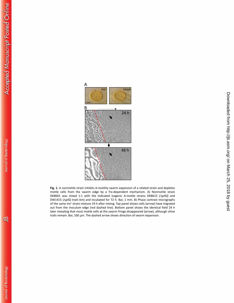
Fig. 1. A nonmotile strain inhibits A-motility swarm expansion of a related strain and depletes motile 397
cells from the swarm edge by a Tra-dependent mechanism. A) Nonmotile strain DK8601 was mixed 1:1 398
with the indicated isogenic A-motile strains DK8615 (ΔpilQ) and DW1415 (ΔpilQ traA::km) and incubated 399
for 72 h. Bar, 1 mm. B) Phase contrast micrographs of the same tra+ strain mixture 24 h after mixing. Top 400
panel shows cells (arrow) have migrated out from the inoculum edge (red dashed line). Bottom panel 401
shows the identical field 24 h later revealing that most motile cells at the swarm fringe disappeared 402
(arrow), although slime trails remain. Bar, 100 μm. The dashed arrow shows direction of swarm 403
expansion. 404
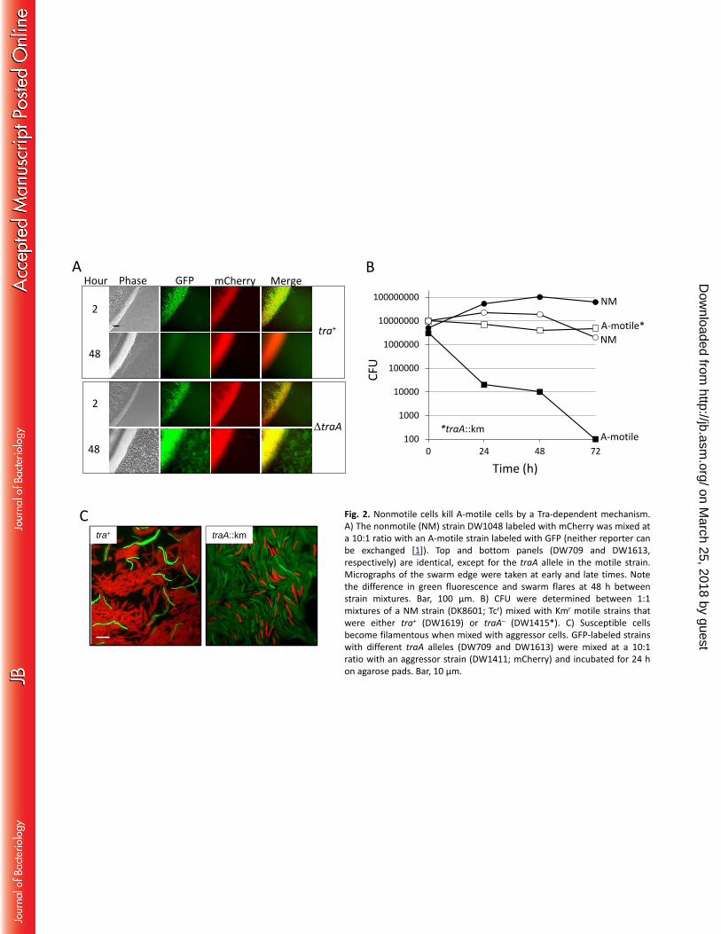
Fig. 2. Nonmotile cells kill A-motile cells by a Tra-dependent mechanism. A) The nonmotile (NM) strain 405
DW1048 labeled with mCherry was mixed at a 10:1 ratio with an A-motile strain labeled with GFP 406
(neither reporter can be exchanged (9)). Top and bottom panels (DW709 and DW1613, respectively) are 407
identical, except for the traA allele in the motile strain. Micrographs of the swarm edge were taken at 408
early and late times. Note the difference in green fluorescence and swarm flares at 48 h between strain 409
mixtures. Bar, 100 μm. B) CFU were determined between 1:1 mixtures of a NM strain (DK8601; Tcr) 410
mixed with Kmr motile strains that were either tra+ (DW1619) or traA– (DW1415*). C) Susceptible cells 411
become filamentous when mixed with aggressor cells. GFP-labeled strains with different traA alleles 412
(DW709 and DW1613) were mixed at a 10:1 ratio with an aggressor strain (DW1411; mCherry) and 413
incubated for 24 h on agarose pads. Bar, 10 µm. 414
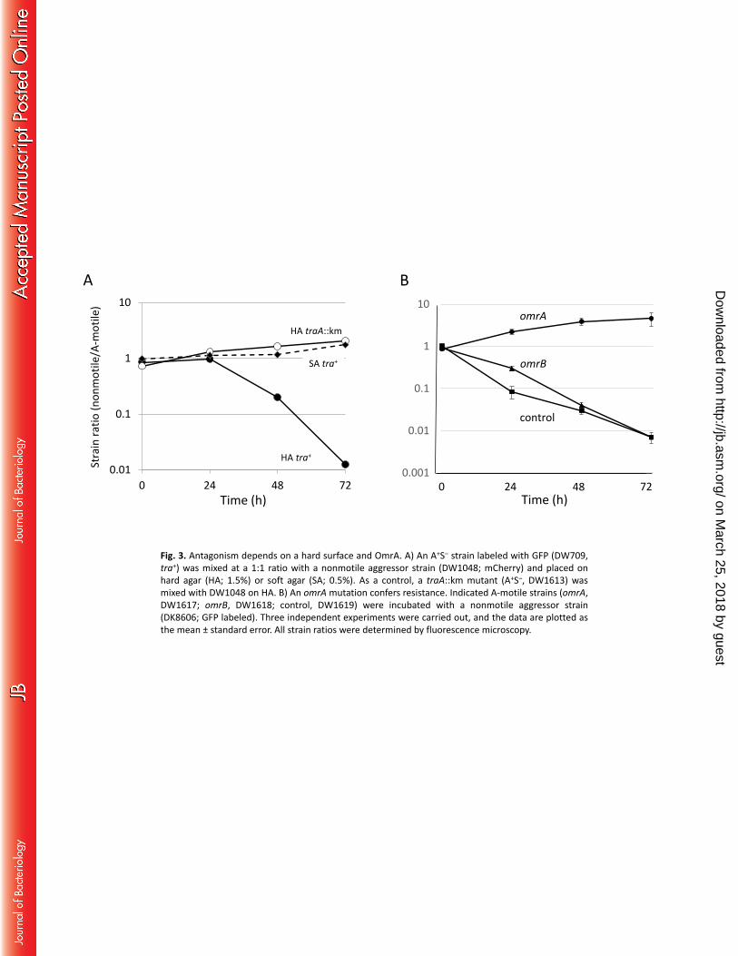
Fig. 3. Antagonism depends on a hard surface and OmrA. A) An A+S‒ strain labeled with GFP (DW709, 415
tra+) was mixed at a 1:1 ratio with a nonmotile aggressor strain (DW1048; mCherry) and placed on hard 416
agar (HA; 1.5%) or soft agar (SA; 0.5%). As a control, a traA::km mutant (A+S‒, DW1613) was mixed with 417
DW1048 on HA. B) An omrA mutation confers resistance. Indicated A-motile strains (omrA, DW1617; 418
on March 25, 2018 by guest
http://jb.asm.org/
Dow
nloaded from
22
omrB, DW1618; control, DW1619) were incubated with a nonmotile aggressor strain (DK8606; GFP 419
labeled). Three independent experiments were carried out, and the data are plotted as the mean ± 420
standard error. All strain ratios were determined by fluorescence microscopy. 421
Fig. 4. Antagonistic behavior is correlated with ancestral strains. A) Swarm inhibition assay at 72 h of an 422
A+S– strain (DK8615) mixed with nonmotile strains that contain three different A-motility mutations 423
placed in either DK1622 (DW1438, DW1443, DW1445) or DK101 (DK1633, DK360, DW704) backgrounds. 424
Bar, 1 mm. B) A susceptible strain labeled with GFP (DW709) was mixed 1:1 with indicated unlabeled 425
ancestral strains and a non-aggressive control (DK8615). All strains were A-motile. After a 48-h 426
incubation, the ratio of cells was determined. Experiments were done in triplicate and the mean ± 427
standard error is shown. C) Same as B, except the competitor was a nonmotile aggressor strain labeled 428
with mCherry (DW1048). D) The nonmotile strain DZ1 was mixed 1:1 with the indicated strains, and no 429
swarm inhibition was observed. Bar, 1 mm. E) DNA agarose gel of diagnostic PCR reactions with primers 430
that are specific to the Mx alpha region absent from DK1622. The locus tag was MXF1DRAFT_07228 431
from DZF1 (contig 40), and the product size was 441 bp. See Table 1 for strain details. 432
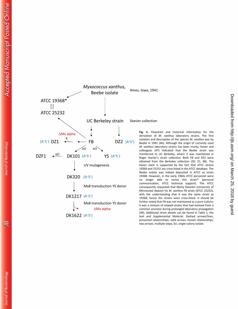
Fig. 5. Flowchart and historical information for the derivation of M. xanthus laboratory strains. The first 433
isolation and description of the species M. xanthus was by Beebe in 1941 (44). Although the origin of 434
currently used M. xanthus laboratory strains has been murky, Kaiser and colleagues (45) indicated that 435
the Beebe strain was transferred to UC Berkeley, where it was maintained in Roger Stanier’s strain 436
collection. Both FB and DZ2 were obtained from the Berkeley collection (20, 21, 46). The Kaiser claim is 437
supported by the fact that ATCC strains 19368 and 25232 are cross-listed in the ATCC database. The 438
Beebe isolate was indeed deposited in ATCC as strain 19368. However, in the early 1960s ATCC 439
personnel were no longer able to revive this strain* (personal communication, ATCC technical support). 440
The ATCC consequently requested that Marty Dworkin (University of Minnesota) deposit his M. xanthus 441
FB strain (ATCC 25232), with the understanding that it was the same strain as 19368; hence the strains 442
on March 25, 2018 by guest
http://jb.asm.org/
Dow
nloaded from
23
were cross-listed. It should be further noted that FB was not maintained as a pure culture; it was a 443
mixture of related strains that had evolved from a common ancestor during prolonged laboratory 444
propagation (47). Additional strain details can be found in Table 1, the text and Supplemental Material. 445
Dashed arrows/lines, presumed relationships; solid arrows, known relationships; two arrows, multiple 446
steps, Sci, single-colony isolate. 447
Fig. 6. Genomic organization of the Mx alpha units in DK1622 and DZF1 (DK101). A) The organization of 448
ORFs found in DK1622 from MXAN_1800 to MXAN_1900. Predicted gene functions are color coded. S/T, 449
serine/threonine. B) The same ORF map as in A with corresponding map position of seven contigs from 450
the DZF1 that perfectly map to this region (top). The DZF1 draft genome has 75 total contigs (27). 451
Regions in nine DZF1 contigs that are homologous to MXAN_1800 to MXAN_1900 and are absent from 452
the DK1622 genome are shown at the bottom. The green and blue contig bars presumably represent 453
two different Mx alpha units. Contig numbers are given at the left. Note that there are gaps in and 454
between some contigs in relation to the DK1622 region. In addition, contig regions that are not 455
homologous to the DK1622 region (insertions) are not shown. In total these nine contigs contain 200 kb 456
of DNA. C) Simplified Mx alpha map illustrating the deleted region in DK1622. See text for additional 457
details. 458
Fig. 7. Deletion of Mx alpha region prevents antagonism. A) Swarm inhibition at 48 h. A-motile strain 459
DW2404 (ΔMx alpha) was mixed 1:1 with isogenic nonmotile strains DK8616 (Mx alpha) or DW2403 460
(ΔMx alpha-29). Bar, 1 mm. B) Fitness experiments in which either an Mx alpha aggressor strain (DK101* 461
labeled with tdTomato [DW1620]; solid lines) or a ΔMx alpha non-aggressor strain (DW2404 tdTomato 462
labeled; dashed lines) was mixed 1:1 with an isogenic Mx alpha strain (DK8616) or a ΔMx alpha-29 strain 463
(DW2403). Strain fitness was microscopically determined by counting labeled and unlabeled cells. 464
on March 25, 2018 by guest
http://jb.asm.org/
Dow
nloaded from
24
Fig. 8. Inter-species antagonism is mediated by traA allele-specific interactions and Mx alpha. A) M. 465
fulvus (Mf) and M. xanthus (Mx) strains with indicated properties (left to right: DW1048, DW1614, and 466
DW1615) were mixed at 1:1 ratios, and after 24 h phase contrast micrographs (bottom) and after 48 h 467
stereo micrographs (top) were taken. Note the middle top panel was translucent. Bars, 1 mm (top); 100 468
μm (bottom). B) The relative fitness of the same strain mixtures as in A was determined by dividing the 469
CFU from 24 h by the CFU from 0 h for each strain. 470
Fig. 9. A working model for how OME and Mx alpha mediate killing. The Mx alpha units that are absent 471
from DK1622 are proposed to contain a toxin/antitoxin system. The toxin is transferred to a target cell 472
by an OME-mediated process. For the toxin to kill, the target cell must express OmrA and lack the 473
antitoxin. Lollipops represent phospholipid molecules; OM, outer membrane; IM, inner membrane. See 474
text for additional details. 475
476
on March 25, 2018 by guest
http://jb.asm.org/
Dow
nloaded from
25
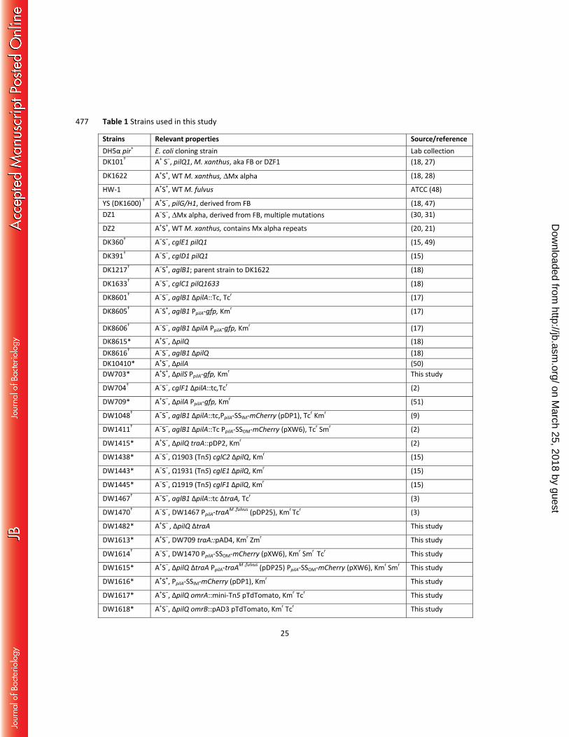
Table 1 Strains used in this study 477
Strains Relevant properties Source/referenceDH5α pir+ E. coli cloning strain Lab collectionDK101† A+ S‒, pilQ1, M. xanthus, aka FB or DZF1 (18, 27)
DK1622 A+S+, WT M. xanthus, ΔMx alpha (18, 28)
HW-1 A+S+, WT M. fulvus ATCC (48)
YS (DK1600) † A+S‒, pilG/H1, derived from FB (18, 47)DZ1 A‒S‒, ΔMx alpha, derived from FB, multiple mutations (30, 31)
DZ2 A+S+, WT M. xanthus, contains Mx alpha repeats (20, 21)
DK360† A‒S‒, cglE1 pilQ1 (15, 49)
DK391† A‒S‒, cglD1 pilQ1 (15)
DK1217† A‒S+, aglB1; parent strain to DK1622 (18)
DK1633† A‒S‒, cglC1 pilQ1633 (18)
DK8601† A‒S‒, aglB1 ΔpilA::Tc, Tcr (17)
DK8605† A‒S+, aglB1 PpilA-gfp, Kmr (17)
DK8606† A‒S‒, aglB1 ΔpilA PpilA-gfp, Kmr (17)
DK8615* A+S‒, ΔpilQ (18) DK8616† A‒S‒, aglB1 ΔpilQ (18) DK10410* A+S‒, ΔpilA (50) DW703* A+S+, ΔpilS PpilA-gfp, Kmr This study
DW704† A‒S‒, cglF1 ΔpilA::tc,Tcr (2)
DW709* A+S‒, ΔpilA PpilA-gfp, Kmr (51)
DW1048† A‒S‒, aglB1 ΔpilA::tc,PpilA-SSIM-mCherry (pDP1), Tcr Kmr (9)
DW1411† A‒S‒, aglB1 ΔpilA::Tc PpilA-SSOM-mCherry (pXW6), Tcr Smr (2)
DW1415* A+S‒, ∆pilQ traA::pDP2, Kmr (2)
DW1438* A‒S‒, Ω1903 (Tn5) cglC2 ∆pilQ, Kmr (15)
DW1443* A‒S‒, Ω1931 (Tn5) cglE1 ∆pilQ, Kmr (15)
DW1445* A‒S‒, Ω1919 (Tn5) cglF1 ∆pilQ, Kmr (15)
DW1467† A‒S‒, aglB1 ΔpilA::tc ΔtraA, Tcr (3)
DW1470† A‒S‒, DW1467 PpilA-traAM .fulvus (pDP25), Kmr Tcr (3)
DW1482* A+S‒ , ΔpilQ ΔtraA This study
DW1613* A+S‒, DW709 traA::pAD4, Kmr Zmr This study
DW1614† A‒S‒, DW1470 PpilA-SSOM-mCherry (pXW6), Kmr Smr Tcr This study
DW1615* A+S‒, ΔpilQ ΔtraA PpilA-traAM .fulvus (pDP25) PpilA-SSOM-mCherry (pXW6), Kmr Smr This study
DW1616* A+S+, PpilA-SSIM-mCherry (pDP1), Kmr This study
DW1617* A+S‒, ΔpilQ omrA::mini-Tn5 pTdTomato, Kmr Tcr This study
DW1618* A+S‒, ΔpilQ omrB::pAD3 pTdTomato, Kmr Tcr This study
on March 25, 2018 by guest
http://jb.asm.org/
Dow
nloaded from
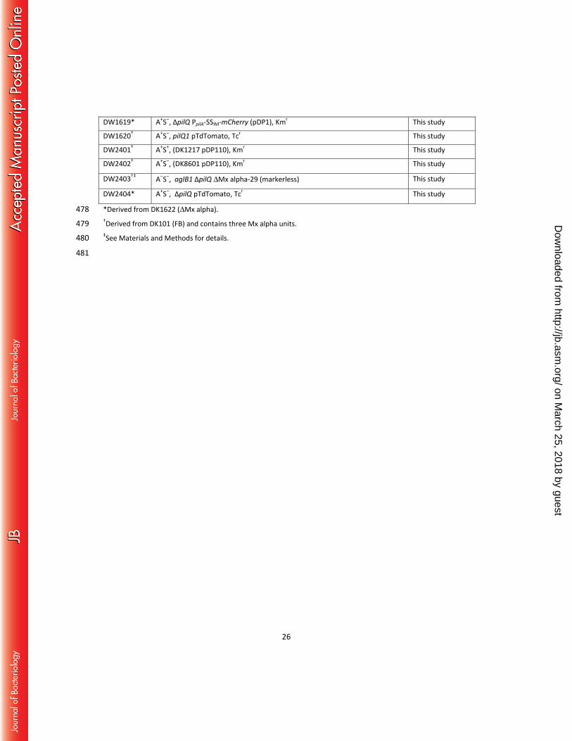
26
DW1619* A+S‒, ΔpilQ PpilA-SSIM-mCherry (pDP1), Kmr This study
DW1620† A+S‒, pilQ1 pTdTomato, Tcr This study
DW2401† A+S+, (DK1217 pDP110), Kmr This study
DW2402† A+S–, (DK8601 pDP110), Kmr This study
DW2403†‡ A‒S‒, aglB1 ΔpilQ ΔMx alpha-29 (markerless) This study
DW2404* A+S‒, ΔpilQ pTdTomato, Tcr This study
*Derived from DK1622 (ΔMx alpha). 478 †Derived from DK101 (FB) and contains three Mx alpha units. 479 ‡See Materials and Methods for details. 480
481
on March 25, 2018 by guest
http://jb.asm.org/
Dow
nloaded from
27
REFERENCES 482
1. Vos M, Velicer GJ. 2009. Social conflict in centimeter-and global-scale populations of the 483 bacterium Myxococcus xanthus. Curr Biol 19:1763-1767. 484
2. Pathak DT, Wei X, Bucuvalas A, Haft DH, Gerloff DL, Wall D. 2012. Cell contact-dependent outer 485 membrane exchange in myxobacteria: Genetic determinants and mechanism. PLoS Genet 486 8:e1002626. 487
3. Pathak DT, Wei X, Dey A, Wall D. 2013. Molecular recognition by a polymorphic cell surface 488 receptor governs cooperative behaviors in bacteria. PLoS Genet 9:e1003891. 489
4. Christie PJ, Cascales E. 2005. Structural and dynamic properties of bacterial type IV secretion 490 systems (review). Mol Membr Biol 22:51-61. 491
5. Willett JL, Gucinski GC, Fatherree JP, Low DA, Hayes CS. 2015. Contact-dependent growth 492 inhibition toxins exploit multiple independent cell-entry pathways. Proc Natl Acad Sci U S A 493 112:11341-11346. 494
6. Mota LJ, Cornelis GR. 2005. The bacterial injection kit: type III secretion systems. Ann Med 495 37:234-249. 496
7. Russell AB, Peterson SB, Mougous JD. 2014. Type VI secretion system effectors: poisons with a 497 purpose. Nat Rev Microbiol 12:137-148. 498
8. Spormann AM. 1999. Gliding motility in bacteria: Insights from studies of Myxococcus xanthus. 499 Microbiol Mol Biol Rev 63:621-641. 500
9. Wei X, Pathak DT, Wall D. 2011. Heterologous protein transfer within structured myxobacteria 501 biofilms. Mol Microbiol 81:315-326. 502
10. Jarrell KF, McBride MJ. 2008. The surprisingly diverse ways that prokaryotes move. Nat Rev 503 Microbiol 6:466-476. 504
11. Shi W, Zusman DR. 1993. The two motility systems of Myxococcus xanthus show different 505 selective advantages on various surfaces. Proc Natl Acad Sci U S A 90:3378-3382. 506
12. Lee B, Holkenbrink C, Treuner-Lange A, Higgs PI. 2012. Myxococcus xanthus developmental cell 507 fate production: heterogeneous accumulation of developmental regulatory proteins and 508 reexamination of the role of MazF in developmental lysis. J Bacteriol 194:3058-3068. 509
13. Vassallo C, Pathak DT, Cao P, Zuckerman DM, Hoiczyk E, Wall D. 2015. Cell rejuvenation and 510 social behaviors promoted by LPS exchange in myxobacteria. Proc Natl Acad Sci U S A 511 112:E2939-2946. 512
14. Dey A, Wall D. 2014. A genetic screen in Myxococcus xanthus identifies mutants that uncouple 513 outer membrane exchange from a downstream cellular response. J Bacteriol 196:4324-4332. 514
15. Pathak DT, Wall D. 2012. Identification of the cglC, cglD, cglE, and cglF genes and their role in 515 cell contact-dependent gliding motility in Myxococcus xanthus. J Bacteriol 194:1940-1949. 516
16. Justice SS, Hunstad DA, Cegelski L, Hultgren SJ. 2008. Morphological plasticity as a bacterial 517 survival strategy. Nat Rev Microbiol 6:162-168. 518
17. Wall D, Kaiser D. 1998. Alignment enhances the cell-to-cell transfer of pilus phenotype. Proc 519 Natl Acad Sci U S A 95:3054-3058. 520
18. Wall D, Kolenbrander PE, Kaiser D. 1999. The Myxococcus xanthus pilQ (sglA) gene encodes a 521 secretin homolog required for type IV pilus biogenesis, social motility, and development. J 522 Bacteriol 181:24-33. 523
19. Hodgkin J, Kaiser D. 1977. Cell-to-cell stimulation of movement in nonmotile mutants of 524 Myxococcus. Proc Natl Acad Sci U S A 74:2938-2942. 525
on March 25, 2018 by guest
http://jb.asm.org/
Dow
nloaded from
28
20. Muller S, Willett JW, Bahr SM, Darnell CL, Hummels KR, Dong CK, Vlamakis HC, Kirby JR. 2013. 526 Draft genome sequence of Myxococcus xanthus wild-type strain DZ2, a model organism for 527 predation and development. Genome Announc 1. 528
21. Campos JM, Zusman DR. 1975. Regulation of development in Myxococcus xanthus: effect of 529 3':5'-cyclic AMP, ADP, and nutrition. Proc Natl Acad Sci U S A 72:518-522. 530
22. Starich T, Zissler J. 1989. Movement of multiple DNA units between Myxococcus xanthus cells. J 531 Bacteriol 171:2323-2336. 532
23. Chen H, Keseler IM, Shimkets LJ. 1990. Genome size of Myxococcus xanthus determined by 533 pulsed-field gel electrophoresis. J Bacteriol 172:4206-4213. 534
24. Starich T, Cordes P, Zissler J. 1985. Transposon tagging to detect a latent virus in Myxococcus 535 xanthus. Science 230:541-543. 536
25. Short FL, Blower TR, Salmond GP. 2012. A promiscuous antitoxin of bacteriophage T4 ensures 537 successful viral replication. Mol Microbiol 83:665-668. 538
26. Canchaya C, Proux C, Fournous G, Bruttin A, Brussow H. 2003. Prophage genomics. Microbiol 539 Mol Biol Rev 67:238-276. 540
27. Muller S, Willett JW, Bahr SM, Scott JC, Wilson JM, Darnell CL, Vlamakis HC, Kirby JR. 2013. 541 Draft genome of a type 4 pilus defective Myxococcus xanthus strain, DZF1. Genome Announc 1. 542
28. Goldman BS, Nierman WC, Kaiser D, Slater SC, Durkin AS, Eisen JA, Ronning CM, Barbazuk WB, 543 Blanchard M, Field C, Halling C, Hinkle G, Iartchuk O, Kim HS, Mackenzie C, Madupu R, Miller 544 N, Shvartsbeyn A, Sullivan SA, Vaudin M, Wiegand R, Kaplan HB. 2006. Evolution of sensory 545 complexity recorded in a myxobacterial genome. Proc Natl Acad Sci U S A 103:15200-15205. 546
29. Magrini V, Storms ML, Youderian P. 1999. Site-specific recombination of temperate Myxococcus 547 xanthus phage Mx8: regulation of integrase activity by reversible, covalent modification. J 548 Bacteriol 181:4062-4070. 549
30. Zusman DR, Krotoski DM, Cumsky M. 1978. Chromosome replication in Myxococcus xanthus. J 550 Bacteriol 133:122-129. 551
31. Zusman D, Rosenberg E. 1970. DNA cycle of Myxococcus xanthus. J Mol Biol 49:609-619. 552 32. Ernst CM, Peschel A. 2011. Broad-spectrum antimicrobial peptide resistance by MprF-mediated 553
aminoacylation and flipping of phospholipids. Mol Microbiol 80:290-299. 554 33. Whitney JC, Quentin D, Sawai S, LeRoux M, Harding BN, Ledvina HE, Tran BQ, Robinson H, Goo 555
YA, Goodlett DR, Raunser S, Mougous JD. 2015. An Interbacterial NAD(P)(+) glycohydrolase 556 toxin requires elongation factor Tu for delivery to target cells. Cell 163:607-619. 557
34. Lang AS, Zhaxybayeva O, Beatty JT. 2012. Gene transfer agents: phage-like elements of genetic 558 exchange. Nat Rev Microbiol 10:472-482. 559
35. Roth JR, Benson N, Galitski T, Haack K, Lawrence JG, Miesel L. 1996. Rearrangements of the 560 Bacterial Chromosome: Formation and Applications, p 2256–2276. In Neidhardt FC, Curtis, R., III, 561 Ingraham, J.L., Lin, E.C.C., Low, K.B., Magasanik, B., Reznikoff, W.S., Riley, M., Schaechter, M. & 562 Umbarger, H.E. (ed), Escherichia coli and Salmonella: Cellular and Molecular Biology. ASM Press, 563 Washington, DC. 564
36. Markowitz VM, Chen IM, Palaniappan K, Chu K, Szeto E, Pillay M, Ratner A, Huang J, Woyke T, 565 Huntemann M, Anderson I, Billis K, Varghese N, Mavromatis K, Pati A, Ivanova NN, Kyrpides 566 NC. 2014. IMG 4 version of the integrated microbial genomes comparative analysis system. 567 Nucleic Acids Res 42:D560-567. 568
37. Boynton TO, McMurry JL, Shimkets LJ. 2013. Characterization of Myxococcus xanthus MazF and 569 implications for a new point of regulation. Mol Microbiol 87:1267-1276. 570
38. Gonzalez-Pastor JE, Hobbs EC, Losick R. 2003. Cannibalism by sporulating bacteria. Science 571 301:510-513. 572
on March 25, 2018 by guest
http://jb.asm.org/
Dow
nloaded from
29
39. Ho HI, Hirose S, Kuspa A, Shaulsky G. 2013. Kin recognition protects cooperators against 573 cheaters. Curr Biol 23:1590-1595. 574
40. Cao P, Dey A, Vassallo CN, Wall D. 2015. How myxobacteria cooperate. J Mol Biol 427:3709-575 3721. 576
41. Koskiniemi S, Garza-Sanchez F, Sandegren L, Webb JS, Braaten BA, Poole SJ, Andersson DI, 577 Hayes CS, Low DA. 2014. Selection of orphan Rhs toxin expression in evolved Salmonella 578 enterica serovar Typhimurium. PLoS Genet 10:e1004255. 579
42. Be'er A, Florin EL, Fisher CR, Swinney HL, Payne SM. 2011. Surviving bacterial sibling rivalry: 580 inducible and reversible phenotypic switching in Paenibacillus dendritiformis. MBio 2:e00069-581 00011. 582
43. Guiral S, Mitchell TJ, Martin B, Claverys JP. 2005. Competence-programmed predation of 583 noncompetent cells in the human pathogen Streptococcus pneumoniae: genetic requirements. 584 Proc Natl Acad Sci U S A 102:8710-8715. 585
44. Beebe JM. 1941. The morphology and cytology of Myxococcus xanthus, N. Sp. J Bacteriol 586 42:193-223. 587
45. Wu Y, Kaiser AD, Jiang Y, Alber MS. 2009. Periodic reversal of direction allows myxobacteria to 588 swarm. Proc Natl Acad Sci U S A 106:1222-1227. 589
46. Dworkin M. 1962. Nutritional requirements for vegetative growth of Myxococcus xanthus. J 590 Bacteriol 84:250-257. 591
47. Wireman JW, Dworkin M. 1975. Morphogenesis and developmental interactions in 592 myxobacteria. Science 189:516-523. 593
48. Li ZF, Li X, Liu H, Liu X, Han K, Wu ZH, Hu W, Li FF, Li YZ. 2011. Genome sequence of the 594 halotolerant marine bacterium Myxococcus fulvus HW-1. J Bacteriol 193:5015-5016. 595
49. Hodgkin J, Kaiser D. 1979. Genetics of gliding molitlity in Myxococcus xanthus (Myxobacterales): 596 Genes controlling movement of single cells. Mol Gen Genet 171:167-176. 597
50. Wu SS, Kaiser D. 1997. Regulation of expression of the pilA gene in Myxococcus xanthus. J 598 Bacteriol 179:7748-7758. 599
51. Wei X, Vassallo CN, Pathak DT, Wall D. 2014. Myxobacteria produce outer membrane-enclosed 600 tubes in unstructured environments. J Bacteriol 196:1807-1814. 601
602
603
on March 25, 2018 by guest
http://jb.asm.org/
Dow
nloaded from
48 h
24 h
A
B
tra+ DtraA
Fig. 1. A nonmotile strain inhibits A-motility swarm expansion of a related strain and depletes motile cells from the swarm edge by a Tra-dependent mechanism. A) Nonmotile strain DK8601 was mixed 1:1 with the indicated isogenic A-motile strains DK8615 (DpilQ) and DW1415 (DpilQ traA::km) and incubated for 72 h. Bar, 1 mm. B) Phase contrast micrographs of the same tra+ strain mixture 24 h after mixing. Top panel shows cells (arrow) have migrated out from the inoculum edge (red dashed line). Bottom panel shows the identical field 24 h later revealing that most motile cells at the swarm fringe disappeared (arrow), although slime trails remain. Bar, 100 μm. The dashed arrow shows direction of swarm expansion.
on March 25, 2018 by guest
http://jb.asm.org/
Dow
nloaded from
100
1000
10000
100000
1000000
10000000
100000000
0 24 48 72
Time (h)
CFU
B A
2
Hour
2
48
48
tra+
DtraA
mCherry GFP Phase Merge
NM
A-motile
A-motile*
NM
*traA::km
traA::km tra+
C Fig. 2. Nonmotile cells kill A-motile cells by a Tra-dependent mechanism. A) The nonmotile (NM) strain DW1048 labeled with mCherry was mixed at a 10:1 ratio with an A-motile strain labeled with GFP (neither reporter can be exchanged [1]). Top and bottom panels (DW709 and DW1613, respectively) are identical, except for the traA allele in the motile strain. Micrographs of the swarm edge were taken at early and late times. Note the difference in green fluorescence and swarm flares at 48 h between strain mixtures. Bar, 100 μm. B) CFU were determined between 1:1 mixtures of a NM strain (DK8601; Tcr) mixed with Kmr motile strains that were either tra+ (DW1619) or traA– (DW1415*). C) Susceptible cells become filamentous when mixed with aggressor cells. GFP-labeled strains with different traA alleles (DW709 and DW1613) were mixed at a 10:1 ratio with an aggressor strain (DW1411; mCherry) and incubated for 24 h on agarose pads. Bar, 10 µm.
on March 25, 2018 by guest
http://jb.asm.org/
Dow
nloaded from
0 24 48 72 Time (h)
omrA
omrB
control
0.001
0.01
0.1
1
10
0.01
0.1
1
10
0 24 48 72
Time (h)
Stra
in r
atio
(n
on
mo
tile
/A-m
oti
le)
HA tra+
SA tra+
HA traA::km
A B
Fig. 3. Antagonism depends on a hard surface and OmrA. A) An A+S‒ strain labeled with GFP (DW709, tra+) was mixed at a 1:1 ratio with a nonmotile aggressor strain (DW1048; mCherry) and placed on hard agar (HA; 1.5%) or soft agar (SA; 0.5%). As a control, a traA::km mutant (A+S‒, DW1613) was mixed with DW1048 on HA. B) An omrA mutation confers resistance. Indicated A-motile strains (omrA, DW1617; omrB, DW1618; control, DW1619) were incubated with a nonmotile aggressor strain (DK8606; GFP labeled). Three independent experiments were carried out, and the data are plotted as the mean ± standard error. All strain ratios were determined by fluorescence microscopy.
on March 25, 2018 by guest
http://jb.asm.org/
Dow
nloaded from
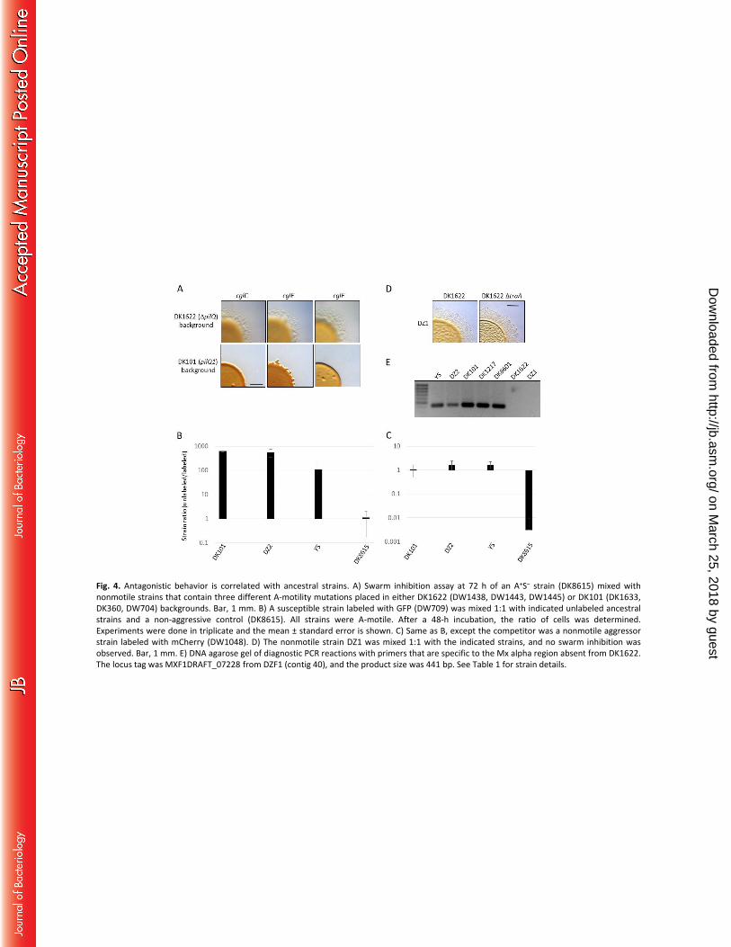
Fig. 4. Antagonistic behavior is correlated with ancestral strains. A) Swarm inhibition assay at 72 h of an A+S– strain (DK8615) mixed with nonmotile strains that contain three different A-motility mutations placed in either DK1622 (DW1438, DW1443, DW1445) or DK101 (DK1633, DK360, DW704) backgrounds. Bar, 1 mm. B) A susceptible strain labeled with GFP (DW709) was mixed 1:1 with indicated unlabeled ancestral strains and a non-aggressive control (DK8615). All strains were A-motile. After a 48-h incubation, the ratio of cells was determined. Experiments were done in triplicate and the mean ± standard error is shown. C) Same as B, except the competitor was a nonmotile aggressor strain labeled with mCherry (DW1048). D) The nonmotile strain DZ1 was mixed 1:1 with the indicated strains, and no swarm inhibition was observed. Bar, 1 mm. E) DNA agarose gel of diagnostic PCR reactions with primers that are specific to the Mx alpha region absent from DK1622. The locus tag was MXF1DRAFT_07228 from DZF1 (contig 40), and the product size was 441 bp. See Table 1 for strain details.
on March 25, 2018 by guest
http://jb.asm.org/
Dow
nloaded from
Myxococcus xanthus, Beebe isolate
Ames, Iowa, 1941
Stanier collection
ATCC 19368*
ATCC 25232
UC Berkeley strain
DZ2 FB
DK101 DZF1
DZ1
YS
DK320
DK1217
DK1622
DMx alpha
UV mutagenesis
Mx8 transduction YS donor
Mx8 transduction YS donor
sci
sci sci
DMx alpha
(A+S–) (A+S–)
(A–S–)
(A+S+)
(A–S+)
(A+S+) (A–S–)
Fig. 5. Flowchart and historical information for the derivation of M. xanthus laboratory strains. The first isolation and description of the species M. xanthus was by Beebe in 1941 (46). Although the origin of currently used M. xanthus laboratory strains has been murky, Kaiser and colleagues (47) indicated that the Beebe strain was transferred to UC Berkeley, where it was maintained in Roger Stanier’s strain collection. Both FB and DZ2 were obtained from the Berkeley collection (20, 21, 48). The Kaiser claim is supported by the fact that ATCC strains 19368 and 25232 are cross-listed in the ATCC database. The Beebe isolate was indeed deposited in ATCC as strain 19368. However, in the early 1960s ATCC personnel were no longer able to revive this strain* (personal communication, ATCC technical support). The ATCC consequently requested that Marty Dworkin (University of Minnesota) deposit his M. xanthus FB strain (ATCC 25232), with the understanding that it was the same strain as 19368; hence the strains were cross-listed. It should be further noted that FB was not maintained as a pure culture; it was a mixture of related strains that had evolved from a common ancestor during prolonged laboratory propagation (49). Additional strain details can be found in Table 1, the text and Supplemental Material. Dashed arrows/lines, presumed relationships; solid arrows, known relationships; two arrows, multiple steps, Sci, single-colony isolate.
on March 25, 2018 by guest
http://jb.asm.org/
Dow
nloaded from
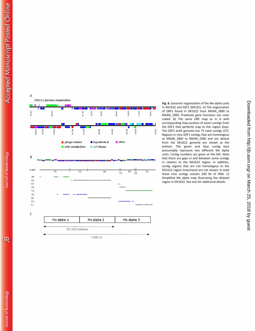
Fig. 6. Genomic organization of the Mx alpha units in DK1622 and DZF1 (DK101). A) The organization of ORFs found in DK1622 from MXAN_1800 to MXAN_1900. Predicted gene functions are color coded. B) The same ORF map as in A with corresponding map position of seven contigs from the DZF1 that perfectly map to this region (top). The DZF1 draft genome has 75 total contigs (27). Regions in nine DZF1 contigs that are homologous to MXAN_1800 to MXAN_1900 and are absent from the DK1622 genome are shown at the bottom. The green and blue contig bars presumably represent two different Mx alpha units. Contig numbers are given at the left. Note that there are gaps in and between some contigs in relation to the DK1622 region. In addition, contig regions that are not homologous to the DK1622 region (insertions) are not shown. In total these nine contigs contain 200 kb of DNA. C) Simplified Mx alpha map illustrating the deleted region in DK1622. See text for additional details.
on March 25, 2018 by guest
http://jb.asm.org/
Dow
nloaded from
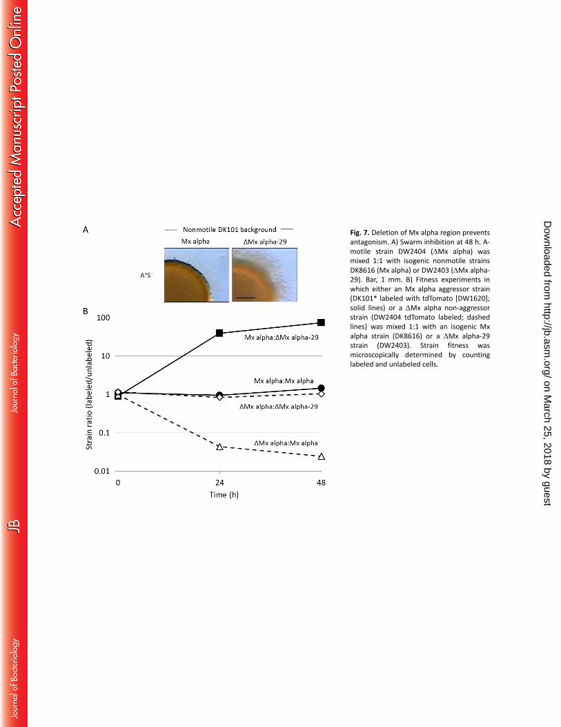
Fig. 7. Deletion of Mx alpha region prevents antagonism. A) Swarm inhibition at 48 h. A-motile strain DW2404 (DMx alpha) was mixed 1:1 with isogenic nonmotile strains DK8616 (Mx alpha) or DW2403 (DMx alpha-29). Bar, 1 mm. B) Fitness experiments in which either an Mx alpha aggressor strain (DK101* labeled with tdTomato [DW1620]; solid lines) or a DMx alpha non-aggressor strain (DW2404 tdTomato labeled; dashed lines) was mixed 1:1 with an isogenic Mx alpha strain (DK8616) or a DMx alpha-29 strain (DW2403). Strain fitness was microscopically determined by counting labeled and unlabeled cells.
on March 25, 2018 by guest
http://jb.asm.org/
Dow
nloaded from
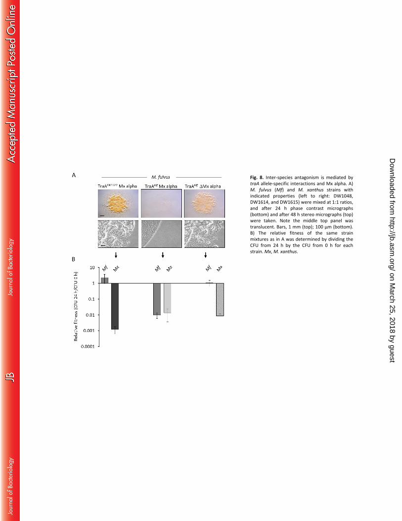
Fig. 8. Inter-species antagonism is mediated by traA allele-specific interactions and Mx alpha. A) M. fulvus (Mf) and M. xanthus strains with indicated properties (left to right: DW1048, DW1614, and DW1615) were mixed at 1:1 ratios, and after 24 h phase contrast micrographs (bottom) and after 48 h stereo micrographs (top) were taken. Note the middle top panel was translucent. Bars, 1 mm (top); 100 μm (bottom). B) The relative fitness of the same strain mixtures as in A was determined by dividing the CFU from 24 h by the CFU from 0 h for each strain. Mx, M. xanthus.
on March 25, 2018 by guest
http://jb.asm.org/
Dow
nloaded from
OmrA
OME Death
TraA
aminoacyl-lipids
OM
IM
TraB
Toxin
&
immunity
Recognition
Mx alpha aggressor
1 2 3
DMx alpha target
Mx alpha
Fig. 9. A working model for how OME and Mx alpha mediate killing. The Mx alpha units that are absent from DK1622 are proposed to contain a toxin/antitoxin system. The toxin is transferred to a target cell by an OME-mediated process. For the toxin to kill, the target cell must express OmrA and lack the antitoxin. Lollipops represent phospholipid molecules; OM, outer membrane; IM, inner membrane. See text for additional details.
on March 25, 2018 by guest
http://jb.asm.org/
Dow
nloaded from