Embed Size (px)
Citation preview

Title Neural correlates of mirth and laughter: a direct electricalcortical stimulation study.
Author(s)
Yamao, Yukihiro; Matsumoto, Riki; Kunieda, Takeharu;Shibata, Sumiya; Shimotake, Akihiro; Kikuchi, Takayuki;Satow, Takeshi; Mikuni, Nobuhiro; Fukuyama, Hidenao;Ikeda, Akio; Miyamoto, Susumu
Citation Cortex (2015), 66: 134-140
Issue Date 2015-05
URL http://hdl.handle.net/2433/210373
Right
© 2015. This manuscript version is made available under theCC-BY-NC-ND 4.0 licensehttp://creativecommons.org/licenses/by-nc-nd/4.0/; The full-text file will be made open to the public on 1 May 2016 inaccordance with publisher's 'Terms and Conditions for Self-Archiving'.
Type Journal Article
Textversion author
Kyoto University
1
Neural correlates of mirth and laughter: a direct electrical cortical stimulation
study
Yukihiro Yamao1) Riki Matsumoto2) Takeharu Kunieda1) Sumiya Shibata1)
Akihiro Shimotake 3) Takayuki Kikuchi1) Takeshi Satow4) Nobuhiro Mikuni5)
Hidenao Fukuyama6) Akio Ikeda2) Susumu Miyamoto1)
1) Department of Neurosurgery, Kyoto University Graduate School of Medicine
2) Department of Epilepsy, Movement Disorders and Physiology, Kyoto University
Graduate School of Medicine
3) Department of Neurology, Kyoto University Graduate School of Medicine
4) Department of Neurosurgery, Nagahama City Hospital
5) Department of Neurosurgery, Sapporo Medical University School of Medicine
6) Human Brain Research Center, Kyoto University Graduate School of Medicine
Corresponding to: Riki Matsumoto & Takeharu Kunieda,
Kyoto University Graduate School of Medicine, Kyoto, Japan
2
54,Shogoin Kawahara-cho, Sakyo-ku, Kyoto, 606-8507, Japan
tel & fax: +81-75-751-3772 & +81-75-751-9416
e-mail: [email protected] (RM), [email protected] (TK)
Highlights:
1) Direct cortical stimulation revealed a close relationship between mirth and language.
2) When mesial temporal structures were intact, electrical cortical stimulation first
elicited contralateral facial movement, followed by bilateral facial movements with
mirth.
3) Emotional facial movements had contralateral dominance.
Abbreviations: ES = electrical cortical stimulation, fMRI = functional magnetic
resonance imaging, HS = hippocampal sclerosis, MEP = motor evoked potential, EMG
= electromyogram, SMA = supplementary motor area, BTLA = basal temporal
language area
3
Abstract
Laughter consists of both motor and emotional aspects. The emotional component,
known as mirth, is usually associated with the motor component, namely, bilateral facial
movements. Previous electrical cortical stimulation (ES) studies revealed that mirth was
associated with the basal temporal cortex, inferior frontal cortex, and medial frontal
cortex. Functional neuroimaging implicated a role for the left inferior frontal and
bilateral temporal cortices in humor processing. However, the neural origins and
pathways linking mirth with facial movements are still unclear. We hereby report two
cases with temporal lobe epilepsy undergoing subdural electrode implantation in whom
ES of the left basal temporal cortex elicited both mirth and laughter-related facial
muscle movements. In one case with normal hippocampus, high-frequency ES
consistently caused contralateral facial movement, followed by bilateral facial
movements with mirth. In contrast, in another case with hippocampal sclerosis, ES
elicited only mirth at low intensity and short duration, and eventually laughter at higher
intensity and longer duration. In both cases, the basal temporal language area was
4
located within or adjacent to the cortex where ES produced mirth. In conclusion, the
present direct ES study demonstrated that 1) mirth had a close relationship with
language function, 2) intact mesial temporal structures were actively engaged in the
beginning of facial movements associated with mirth, and 3) these emotion-related
facial movements had contralateral dominance.
Keywords: mirth; laughter; electrical cortical stimulation; mesial temporal structure;
basal temporal language area

5
1. Introduction
Laughter, an essential part of daily life, consists of motor and emotional components,
the latter of which is known as mirth (Arroyo et al., 1993). Indeed, appreciating or
enjoying humor is associated with a feeling of mirth. Lesion studies have shown that
humor consists of both cognitive and affective processing (Gardner, Ling, Flamm, &
Silverman, 1975). Goel and Dolan (2001), in their pioneer functional magnetic
resonance imaging (fMRI) study, showed that cognitive processing with semantic
components was associated with the left hemisphere (left inferior frontal gyrus and
posterior inferior temporal gyrus), while cognitive processing with semantic
components involved the bilateral temporal cortices (bilateral posterior middle temporal
gyrus and left posterior inferior temporal gyrus), and that affective processing was
associated with reward processing system (medial ventral prefrontal cortex) (Goel &
Dolan, 2001). This notion was confirmed by several lines of evidence using fMRI in
healthy subjects (Amir, Biederman, Wang, & Xu, 2013; Mobbs, Greicius, Abdel-Azim,
Menon, & Reiss, 2003; Moran, Wig, Adams, Janata, & Kelley, 2004; Watson,
6
Matthews, & Allman, 2007). However, despite centuries of inquiry, the neural origins
and pathways linking bilateral facial movements with mirth are still unclear.
Unilateral lower facial motor weakness (contralesional “mimetic palsy” or
emotional facial paresis), which manifests during spontaneous smiling and weeping but
not at all during voluntary muscle contraction, has been reported in patients with lesions
involving mesial temporal structures (amygdala and hippocampus) (Hopf, Muller-Forell,
& Hopf, 1992). This suggests that impairment of contralateral functional connections
originating in mesial temporal structures leads to asymmetric emotional facial
movements.
We report two patients with temporal lobe epilepsy who underwent subdural
electrode implantation, and in whom electrical cortical stimulation (ES) of the cortices
of the left basal temporal lobe elicited mirth, followed by laughter. By analyzing the
features common to the two cases, we postulated that mesial temporal structures directly
bridged mirth and laughter.
2. Materials and Methods

7
2.1. Subjects
We enrolled 13 consecutive patients with medically intractable left temporal lobe
epilepsy who underwent chronic subdural electrode implantation over the basal part of
the temporal lobe for presurgical evaluation between March 2000 and December 2013.
All the patients showed language dominance in the left hemisphere as assessed by the
Wada test (Takayama et al., 2004), with the exception of one who demonstrated
bilateral language representation. We systematically performed high-frequency ES at
the basal part of the temporal lobe in order to map the basal temporal language area
(BTLA), as this area is actively engaged in semantic language processing and
preservation of the BTLA and its white matter connection previously led to preservation
or improvement of verbal memory (Mikuni et al., 2006; Shimotake et al., 2014; Usui et
al., 2003). In two of the 13 patients, mirth was elicited by high-frequency ES. Both
patients had language dominance in the left hemisphere. Postoperative histology
showed an intact hippocampus in one patient (Patient 1) and hippocampal sclerosis
(HS) in the other (Patient 2). Neither laughter nor mirth occurred during the patients’
habitual seizures, and Patient 2 showed mimetic facial palsy on the right side only when

8
smiling and not at all during voluntary upward curving of the lips. The case of Patient 2
was reported preliminarily as a letter (Satow et al., 2003). The demographic details of
Patients 1 and 2 are shown in the supplementary material.
2.2. High-frequency electrical cortical stimulation
High-frequency ES (50 Hz, square-wave pulse of alternating polarity with a pulse width
of 0.3 ms, 1–5 sec, 1–15 mA) was applied to the basal temporal cortices through a pair
of implanted electrodes to define the functional areas, especially those related to
language function (Matsumoto et al., 2011). The amplitude of the electric current was
increased gradually until positive motor symptoms (e.g., contraction of arms or facial
muscles) or subjective perception of epileptic aura or somatosensory, visual and
auditory sensation occurred; patients were instructed, if present, to report all of these
after each stimulation trial was over. When the stimulus intensity was increased >10
mA without any positive motor symptoms or subjective perceptions, the absence of
positive (e.g., tonic contraction) and negative (e.g., impairment of rapid alternating
movements) tongue motor responses was confirmed, and a series of language batteries
9
were administered, each lasting 3–5 sec. This language mapping is described in detail
elsewhere (Matsumoto et al., 2011; Usui et al., 2003). We judged the induced or
impaired behaviors as significant only when the findings were reproducible (at least two
trials) without afterdischarges. In cases of frequent afterdischarges, we decreased the
stimulation intensity by 1−2 mA so that these did not occur. The majority of
high-frequency ES was performed in a bipolar fashion (i.e., stimulation at two adjacent
electrodes). In addition, we occasionally performed monopolar stimulation with
reference to an electrode in the nonfunctional cortex to further localize the language
areas for the clinical purpose.
2.3. Assessments of mirth and laughter-related facial muscle movements
Following every ES trial, if the patient spontaneously reported a feeling of mirth, the
examiners (Y.Y., T.S.) inquired as to its presence and character. Since mirth is solely
subjective, we judged the reported feeling as significant when the findings were
reproducible (in at least two trials) without afterdischarges. In Patient 1, we also
performed sham stimulation (i.e., stimulation at 0 mA intensity) to confirm that mirth
10
was induced only by real ES. In order to reproducibly characterize mirth and its
associated facial movements, additional trials were performed for research purposes.
After the first subjective report of mirth, the stimulation intensity was increased
gradually up to 15 mA in order to assess the language function. This was only possible
in Patient 2, since induced mirth and laughter prevented Patient 1 from properly
completing the language batteries. This high-frequency ES study was approved by the
Ethics Committee, Kyoto University Graduate School of Medicine (No. 79), and both
patients gave written informed consent.
In Patient 1, because asymmetric emotional facial movements were clearly
observed in early trials of high-frequency ES, surface electromyogram (EMG)
electrodes were placed on the bilateral levator labii superior and orbicularis oris muscles
for the following four trials. In addition, single-pulse ES (1 Hz, square-wave pulse of
alternating polarity, 0.3 ms duration, 12 or 15 mA, four trials) was also applied to the
same electrodes in order to record motor evoked potentials (MEPs) from EMG of the
facial muscles (Ikeda et al., 2000; Kikuchi et al., 2012).
11
3. Results
In both patients, mirth was elicited by high-frequency ES at a very restricted area in the
left basal temporal cortex [Fig. 1: a white circle (monopolar stimulation) in Patient 1
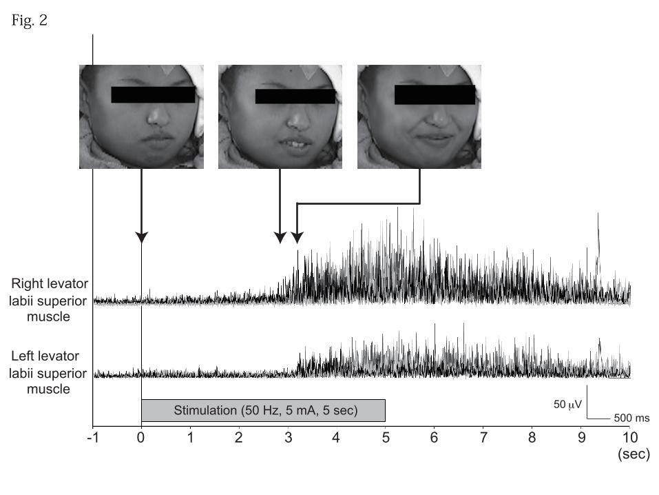
and black circles (bipolar stimulation) in Patient 2]. In Patient 1, high-frequency ES at
low intensity (5 mA, 5 sec) consistently caused lifting of the right side of the mouth,
followed by bilateral facial movements with mirth (Fig. 2). After ES was over, the
patient said, “I do not know why, but something amused me and I laughed.” In Patient 2,
high-frequency ES at low intensity and short duration (5 mA, 1 sec) produced mirth
without laughter. The stimulus intensity was gradually increased, and she eventually
developed laughter at high intensity and long duration (15 mA, 5 sec). Her introspection
was that “A familiar melody that I had heard in a television program in my childhood
came to mind; its tune sounded funny and amused me.” In both cases, mirth was elicited
only during high-frequency ES. In Patient 1, sham stimulation confirmed that the
feeling of mirth was actually induced by high-frequency ES (no specific feeling was
evoked by sham stimulation). These findings were reproducibly elicited by
high-frequency ES without afterdischarges (7/7 trials in Patient 1 and 14/14 trials in
12
Patient 2).
In Patient 1, surface EMG recording during ES (50 Hz, 5 mA, 5 sec, four
trials) showed that the contralateral facial muscle contractions occurred at a mean of 3.3
sec (ranging from 2.8 to 3.7 sec) after the start of 50 Hz stimulation, and the onset of
bilateral facial movements occurred at a mean of 0.3 sec (ranging from 0.2 to 0.4 sec)
after that of unilateral contractions. The onset of laughter in Patient 2 occurred >5 sec
after stimulation as judged from video inspection (2 trials). In Patient 1, single-pulse ES
(1 Hz) was also applied to the electrode at which high-frequency ES caused mirth and
facial muscle contractions (white circle in Fig. 1). Single-pulse ES at the maximum
clinically safe intensity of 15 mA did not produce contralateral facial movements, MEPs,
or mirth.
For clinical purposes, we evaluated language function in the basal temporal
cortex, with the exception of the three (Patient 1) and six (Patient 2) electrodes where
precise language mapping was difficult due to frequent afterdischarges. Stimulation of
the electrodes that produced mirth had different results in the two patients. Mirth and
laughter did not allow Patient 1 to perform language tests properly (white circle in Fig.
13
1). In Patient 2, language impairment was reproducibly induced (15 mA, 5 sec, 4/4 trials,
black circles in Fig.1). In other parts of the left basal temporal cortex, six electrodes
resulted in language impairment (white and black diamonds), 11 resulted in no
impairment (small gray circles), and for nine electrodes the results were inconclusive
due to frequent afterdischarges (“x” marks). Electrodes associated with mirth were
located adjacent to the electrodes related to language function in both patients. In order
to clarify the relationship between mirth and language, the ratio of electrodes that
elicited language impairment was compared between electrodes that were associated
with mirth (“mirth electrode,” 3 electrodes) and those that were not (“no-mirth
electrode,” 17 electrodes) (electrodes with inconclusive results due to afterdischarges
were excluded). The ratio of electrodes eliciting language impairment was higher for the
mirth electrode (2/3 = 0.66) than the no-mirth electrodes (6/17 = 0.32).
4. Discussion
4.1. Mirth and language
The neural basis of mirth has been explored by studying humor, classically via brain
lesions and more recently with fMRI. Humor can be divided into cognitive and affective
14
processing (Gardner et al., 1975). Cognitive processing, i.e., humor detection, is formed
in two stages: 1) a perception of incongruity between the expectation and “punch line”,
and 2) resolution of the incongruity (Suls, 1972). A series of fMRI studies of humor
indicated that cognitive processing has phonological and semantic components. The
phonological component was associated with activation in the left lateral inferior frontal
gyrus (Goel & Dolan, 2001), which was a consistent finding in the previous fMRI
studies (Amir et al., 2013; Mobbs et al., 2003; Moran et al., 2004; Watson et al., 2007).
The semantic component of humor processing was associated with activation in the
temporo-occipital and temporo-parietal junctions; bilateral activation was observed in
tasks involving humor interpretation (Amir et al., 2013), listening to different types of
jokes (Goel & Dolan, 2001), and processing of sight gag-related humor (Watson et al.,
2007), while some studies using funny cartoons (Mobbs et al., 2003) or verbal gags
(Watson et al., 2007) reported the dominance in the left hemisphere. Although the
anterior part of the basal temporal cortex is regarded as one of the core regions in the
semantic network (Binney, Embleton, Jefferies, Parker, & Ralph, 2010; Mion et al.,
2010; Shimotake et al., 2014), only one of these fMRI studies reported activation of this
15
region. This is mainly due to a variety of technical factors including magnetic field
inhomogeneities (for standard gradient echo planar imaging) and limited field of view
(Devlin et al., 2000; Visser, Embleton, Jefferies, Parker, & Ralph, 2010). Moran et al.
(2004) suggested that semantic processing brings stored expectations, and that
phonological processing then resolves the incongruities between these expectations and
punch lines, supporting the incongruity-resolution theory of humor.
Affective processing, i.e., humor appreciation, is thought to be associated with
the mesolimbic dopaminergic reward system. Activation was reported in the bilateral
ventral striatum, nuclei accumbens, ventral tegmenta, hypothalami, and amygdalae
during humor appreciation of funny cartoons (Mobbs et al., 2003), as well as in the right
medial prefrontal cortex while listening to different types of jokes (Goel & Dolan,
2001). These reward-related regions were also activated in other fMRI studies to
varying degrees: the left amygdala, left middle prefrontal cortex and bilateral ventral
striatum (Amir et al., 2013), the bilateral amygdalae and insulae (Moran et al., 2004),
and the left amygdala and nucleus accumbens (Watson et al., 2007). Amir et al. (2013)
hypothesized that the regions associated with humor detection trigger these
16
reward-related regions, producing the hedonic feeling that accompanies humor.
ES has been the gold standard for mapping brain function for functional
neurosurgery, even though it is an invasive technique. Only a handful of studies have
applied this technique to elucidate the cortical network associated with mirth and
laughter. Laughter with mirth was produced during ES of the left basal temporal area
(anterior parahippocampal and fusiform gyri) (Arroyo et al., 1993; Satow et al., 2003),
the left inferior frontal gyrus (pars opercularis) (Fernandez-Baca Vaca, Lüders, Basha,
& Miller, 2011), and the left medial frontal lobe (the rostral part of the supplementary
motor area [SMA]) (Fried, Wilson, MacDonald, & Behnke, 1998; Krolak-Salmon et al.,
2006). Laughter without mirth was elicited by ES of the left or right cingulate gyrus
(Arroyo et al., 1993; Sperli, Spinelli, Pollo, & Seeck, 2006), the right medial frontal
lobe (the rostral part of SMA), and the left lateral dorsal premotor cortex (Schmitt,
Janszky, Woermann, Tuxhorn, & Ebner, 2006). In summary, in our two cases and in
several previously reported cases with definite language dominance (by the Wada test)
or handedness (Arroyo et al., 1993; Fernandez-Baca Vaca et al., 2011; Krolak-Salmon
et al., 2006; Satow et al., 2003), mirth was elicited by ES only at the frontal and

17
temporal cortices of the dominant left hemisphere. Interestingly, when ES was
performed for clinical functional cortical mapping, all of the electrodes producing mirth
were located adjacent to language-related areas: the left SMA (Fried et al. 1998),
Broca’s area (Fernandez-Baca Vaca et al., 2011), and the left BTLA (Arroyo et al.,
1993). Moreover, in our cases the ratio of electrodes eliciting language impairment was
higher for the mirth electrodes than in no-mirth electrodes, suggesting an association
between mirth and language function. Since the BTLA is actively involved in semantic
processing (Shimotake et al., 2014; Usui et al., 2003), this semantic/language area was
likely involved in the semantic aspect of humor detection in our cases.
The stimulation studies cited above demonstrated that the left hemisphere,
which is dominant for language, is more related to mirth, but the findings should be
interpreted carefully, for two reasons. First, since the invasive ES mirth studies were
performed more frequently in the hemisphere dominant for language to identify brain
functions, we currently do not have enough ES findings regarding mirth and laughter in
the hemisphere non-dominant for language, especially in the cortical regions
homologous to those producing mirth, with or without laughter, in the hemisphere

18
dominant for language. Second, humor fMRI studies in healthy right-handed subjects
showed that bilateral temporal cortices were involved in semantic processing while the
left hemisphere was activated during language perception and production. Only a few
brain stimulation studies reported that stimulation of the nucleus accumbens (Okun et
al., 2004) or subthalamic nucleus (Krack et al., 2001) produced laughter and mirth.
Further ES studies are warranted for comprehensive mapping of the cortical and
subcortical regions associated with mirth and laughter by accumulating the ES findings
of the non-dominant hemisphere and of subcortical structures.
4.2. A suggested mechanism for mirth and facial movements
To the best of our knowledge, this is the first report using high-frequency ES with EMG
recordings of the facial muscles to demonstrate concomitant contralateral facial
movements with mirth. In a tracer study in rhesus monkeys, the primary motor,
premotor, and caudal cingulate cortices projected primarily to the contralateral lateral
subnucleus of the facial nucleus, while the supplementary motor and rostral cingulate
cortices projected bilaterally to the dorsal and intermediate subnuclei (Morecraft, Louie,
Herrick, & Stilwell-Morecraft, 2001). Two independent neuronal pathways are

19
proposed for the expression of laughter (Wild, Rodden, Grodd, & Ruch, 2003). The first,
the volitional pathway, runs from motor cortices to the ventral brainstem through the
pyramidal tract. The second, the emotional pathway, involves the limbic system and
leads to the dorsal brainstem. However, it is still unclear which pathways are involved
in the association between facial movements and mirth. In a human lesion study that
investigated a stroke of the primary facial motor cortex, involuntary facial movements
during mirth were preserved and were hypothesized to arise from the cingulate cortex
(Kappos & Mehling, 2010). Contralesional mimetic facial palsy, namely, unilateral
emotional facial paresis, was demonstrated in patients with mesial temporal lobe
epilepsy with HS (Hopf et al., 1992), the latter of which is diagnosed on the basis of
neuronal cell loss and gliosis of mesial temporal structures, including the amygdala
(Wieser, 2004). These studies suggest that the limbic system, especially the anterior
cingulate cortex and mesial temporal structures such as the amygdala, is involved in the
association between facial movements and mirth.
In Patient 1, high-frequency ES (5 mA) evoked contraction of contralateral
facial muscles, and its onset time was shorter than that of laughter with mirth in both
20
patients, while single-pulse ES (15 mA) produced neither facial movements nor MEPs.
This discrepancy implies that the basal temporal cortex is not connected
monosynaptically with a motor pathway such as the corticobulbar tract, but rather
multisynaptically with the contralateral facial motor nucleus through the limbic system.
Indeed, the corresponding region in rhesus monkeys has connections with the amygdala
and the perirhinal cortex (Schmahmann & Pandya, 2006). In Patient 1, the impulse
produced by ES at the anterior basal temporal area was most likely transmitted first to
the normal amygdala and subsequently to the limbic system, leading first to
contralateral facial contraction and then to bilateral facial movements.
In Patient 1, who had a normal hippocampus, high-frequency ES most likely
produced contralateral facial movements through the normal emotional pathway. On the
other hand, in Patient 2, who had HS and contralesional mimetic facial palsy, the
normal emotional pathway was disturbed, and therefore high-frequency ES presumably
produced mirth, followed by facial movements through the volitional pathway. In other
words, we likely observed two sides of the same coin regarding the emotional pathway:
contralateral facial contraction by 50 Hz stimulation through the normal mesial

21
temporal structure in Patient 1, and contralateral emotional facial paresis because of the
pathological mesial temporal structure (HS) in Patient 2. We therefore postulate that
mesial temporal structures play an important role in the onset of facial movements
associated with mirth. We also postulate that these facial movements have contralateral
dominance, as was shown to be the case with the second somatosensory area and
supplementary motor area (Babiloni et al., 2003; Mima et al., 1997). One ES study
showed that ES of the right anterior cingulate cortex produced contralateral facial
movements followed by bilateral facial movements, without mirth (Sperli et al., 2006).
In a deep brain stimulation study, stimulation of the nucleus accumbens produced
contralateral facial movements with euphoria (Okun et al., 2004). These studies further
support our hypothesis.
In summary, the present direct stimulation study showed that 1) mirth had a
close relationship with language function, 2) intact mesial temporal structures actively
engaged at the beginning of facial movements associated with mirth, and 3) these
emotional facial movements had contralateral dominance.
22
ACKNOWLEDGMENTS
We are indebted to Dr. Keiko Usui for providing the patient data. This work was partly
supported by Grants-in-Aid for Scientific Research (B) 26282218 (RM), (C) 24592159
(TK), and Exploratory Research 26560465 (RM) from the Ministry of Education,
Culture, Sports, Science and Technology of Japan. Department of Epilepsy, Movement
Disorders and Physiology, Kyoto University Graduate School of Medicine is an
endowment department, supported with grants by GlaxoSmithKline K.K., NIHON
KOHDEN CORPORATION, Otsuka Pharmaceutical Co., and UCB Japan Co., Ltd.

23
REFERENCES
Amir, O., Biederman, I., Wang, Z., & Xu, X. (2013). Ha Ha! Versus Aha! A Direct
Comparison of Humor to Nonhumorous Insight for Determining the Neural
Correlates of Mirth. Cereb Cortex.
Arroyo, S., Lesser, R. P., Gordon, B., Uematsu, S., Hart, J., Schwerdt, P., et al. (1993).
Mirth, laughter and gelastic seizures. Brain, 116 ( Pt 4), 757-780.
Babiloni, C., Carducci, F., Del Gratta, C., Demartin, M., Romani, G. L., Babiloni, F., et
al. (2003). Hemispherical asymmetry in human SMA during voluntary simple
unilateral movements. An fMRI study. Cortex, 39, 293-305.
Binney, R. J., Embleton, K. V., Jefferies, E., Parker, G. J., & Ralph, M. A. (2010). The
ventral and inferolateral aspects of the anterior temporal lobe are crucial in
semantic memory: evidence from a novel direct comparison of
distortion-corrected fMRI, rTMS, and semantic dementia. Cereb Cortex, 20,
2728-2738.
Devlin, J. T., Russell, R. P., Davis, M. H., Price, C. J., Wilson, J., Moss, H. E., et al.
(2000). Susceptibility-induced loss of signal: comparing PET and fMRI on a
24
semantic task. NeuroImage, 11, 589-600.
Fernandez-Baca Vaca, G., Lüders, H. O., Basha, M. M., & Miller, J. P. (2011). Mirth
and laughter elicited during brain stimulation. Epileptic Disord, 13, 435-440.
Fried, I., Wilson, C. L., MacDonald, K. A., & Behnke, E. J. (1998). Electric current
stimulates laughter. Nature, 391, 650.
Gardner, H., Ling, P. K., Flamm, L., & Silverman, J. (1975). Comprehension and
appreciation of humorous material following brain damage. Brain, 98, 399-412.
Goel, V., & Dolan, R. J. (2001). The functional anatomy of humor: segregating
cognitive and affective components. Nat Neurosci, 4, 237-238.
Hopf, H. C., Muller-Forell, W., & Hopf, N. J. (1992). Localization of emotional and
volitional facial paresis. Neurology, 42, 1918-1923.
Ikeda, A., Ohara, S., Matsumoto, R., Kunieda, T., Nagamine, T., Miyamoto, S., et al.
(2000). Role of primary sensorimotor cortices in generating inhibitory motor
response in humans. Brain, 123 ( Pt 8), 1710-1721.
Kappos, L., & Mehling, M. (2010). Images in clinical medicine. Dissociation of
voluntary and emotional innervation after stroke. N Engl J Med, 363, e25.
25
Kikuchi, T., Matsumoto, R., Mikuni, N., Yokoyama, Y., Matsumoto, A., Ikeda, A., et al.
(2012). Asymmetric bilateral effect of the supplementary motor area proper in
the human motor system. Clin Neurophysiol, 123, 324-334.
Krack, P., Kumar, R., Ardouin, C., Dowsey, P. L., McVicker, J. M., Benabid, A. L., et al.
(2001). Mirthful laughter induced by subthalamic nucleus stimulation. Mov
Disord, 16, 867-875.
Krolak-Salmon, P., Hénaff, M. A., Vighetto, A., Bauchet, F., Bertrand, O., Mauguière, F.,
et al. (2006). Experiencing and detecting happiness in humans: the role of the
supplementary motor area. Ann Neurol, 59, 196-199.
Matsumoto, R., Imamura, H., Inouchi, M., Nakagawa, T., Yokoyama, Y., Matsuhashi,
M., et al. (2011). Left anterior temporal cortex actively engages in speech
perception: A direct cortical stimulation study. Neuropsychologia, 49,
1350-1354.
Mikuni, N., Miyamoto, S., Ikeda, A., Satow, T., Taki, J., Takahashi, J., et al. (2006).
Subtemporal hippocampectomy preserving the basal temporal language area for
intractable mesial temporal lobe epilepsy: preliminary results. Epilepsia, 47,
26
1347-1353.
Mima, T., Ikeda, A., Nagamine, T., Yazawa, S., Kunieda, T., Mikuni, N., et al. (1997).
Human second somatosensory area: subdural and magnetoencephalographic
recording of somatosensory evoked responses. J Neurol Neurosurg Psychiatry,
63, 501-505.
Mion, M., Patterson, K., Acosta-Cabronero, J., Pengas, G., Izquierdo-Garcia, D., Hong,
Y. T., et al. (2010). What the left and right anterior fusiform gyri tell us about
semantic memory. Brain, 133, 3256-3268.
Mobbs, D., Greicius, M. D., Abdel-Azim, E., Menon, V., & Reiss, A. L. (2003). Humor
modulates the mesolimbic reward centers. Neuron, 40, 1041-1048.
Moran, J. M., Wig, G. S., Adams, R. B., Jr., Janata, P., & Kelley, W. M. (2004). Neural
correlates of humor detection and appreciation. NeuroImage, 21, 1055-1060.
Morecraft, R. J., Louie, J. L., Herrick, J. L., & Stilwell-Morecraft, K. S. (2001). Cortical
innervation of the facial nucleus in the non-human primate: a new interpretation
of the effects of stroke and related subtotal brain trauma on the muscles of facial
expression. Brain, 124, 176-208.
27
Okun, M. S., Bowers, D., Springer, U., Shapira, N. A., Malone, D., Rezai, A. R., et al.
(2004). What's in a "smile?" Intra-operative observations of contralateral smiles
induced by deep brain stimulation. Neurocase, 10, 271-279.
Satow, T., Usui, K., Matsuhashi, M., Yamamoto, J., Begum, T., Shibasaki, H., et al.
(2003). Mirth and laughter arising from human temporal cortex. J Neurol
Neurosurg Psychiatry, 74, 1004-1005.
Schmahmann, J. D., & Pandya, D. N. (2006). Fiber Pathways of the Brain: Oxford Univ.
Press.
Schmitt, J. J., Janszky, J., Woermann, F., Tuxhorn, I., & Ebner, A. (2006). Laughter and
the mesial and lateral premotor cortex. Epilepsy Behav, 8, 773-775.
Shimotake, A., Matsumoto, R., Ueno, T., Kunieda, T., Saito, S., Hoffman, P., et al.
(2014). Direct exploration of the role of the ventral anterior temporal lobe in
semantic memory: Cortical stimulation and local field potential evidence from
subdural grid electrodes. Cereb Cortex, http://dx.doi.org/10.1093/cercor/bhu262.
Sperli, F., Spinelli, L., Pollo, C., & Seeck, M. (2006). Contralateral smile and laughter,
but no mirth, induced by electrical stimulation of the cingulate cortex. Epilepsia,
28
47, 440-443.
Suls, J. (1972). A two-stage model for the appreciation of jokes and cartoons:an
information-processing analysis. The Psychology of Humor: Theoretical
Perspectives and Empirical Issues. New York: Academic Press.
Takayama, M., Miyamoto, S., Ikeda, A., Mikuni, N., Takahashi, J. B., Usui, K., et al.
(2004). Intracarotid propofol test for speech and memory dominance in man.
Neurology, 63, 510-515.
Usui, K., Ikeda, A., Takayama, M., Matsuhashi, M., Yamamoto, J., Satoh, T., et al.
(2003). Conversion of semantic information into phonological representation: a
function in left posterior basal temporal area. Brain, 126, 632-641.
Visser, M., Embleton, K. V., Jefferies, E., Parker, G. J., & Ralph, M. A. (2010). The
inferior, anterior temporal lobes and semantic memory clarified: novel evidence
from distortion-corrected fMRI. Neuropsychologia, 48, 1689-1696.
Watson, K. K., Matthews, B. J., & Allman, J. M. (2007). Brain activation during sight
gags and language-dependent humor. Cereb Cortex, 17, 314-324.
Wieser, H. G. (2004). ILAE Commission Report. Mesial temporal lobe epilepsy with
29
hippocampal sclerosis. Epilepsia, 45, 695-714.
Wild, B., Rodden, F. A., Grodd, W., & Ruch, W. (2003). Neural correlates of laughter
and humour. Brain, 126, 2121-2138.
30
Figure legends
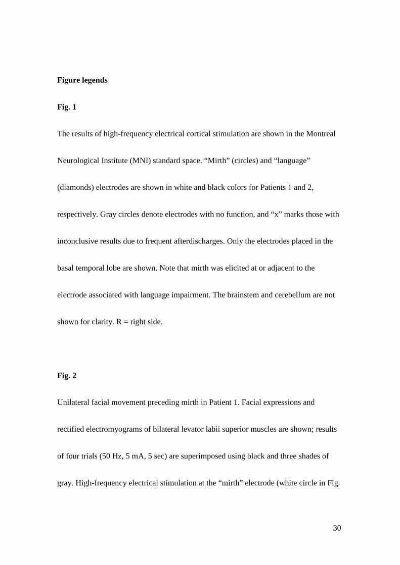
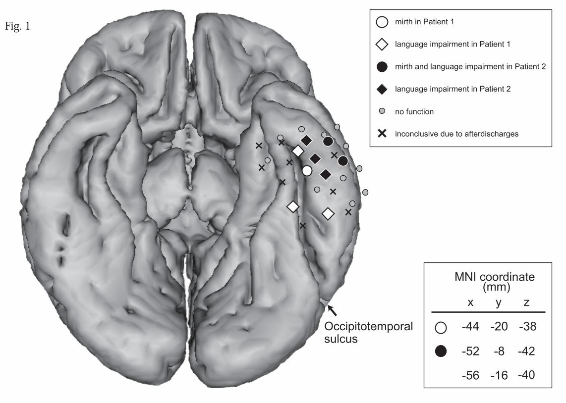
Fig. 1
The results of high-frequency electrical cortical stimulation are shown in the Montreal
Neurological Institute (MNI) standard space. “Mirth” (circles) and “language”
(diamonds) electrodes are shown in white and black colors for Patients 1 and 2,
respectively. Gray circles denote electrodes with no function, and “x” marks those with
inconclusive results due to frequent afterdischarges. Only the electrodes placed in the
basal temporal lobe are shown. Note that mirth was elicited at or adjacent to the
electrode associated with language impairment. The brainstem and cerebellum are not
shown for clarity. R = right side.
Fig. 2
Unilateral facial movement preceding mirth in Patient 1. Facial expressions and
rectified electromyograms of bilateral levator labii superior muscles are shown; results
of four trials (50 Hz, 5 mA, 5 sec) are superimposed using black and three shades of
gray. High-frequency electrical stimulation at the “mirth” electrode (white circle in Fig.
31
1) produced lift of the right mouth via unilateral contraction of the right levator labii
superior muscle (middle photo), which was followed by mirth with bilateral facial
movements (right photo).
Occipitotemporal sulcus
MNI coordinate (mm)
x y z
-44 -38-20
-56 -16 -40
-52 -8 -42R
mirth and language impairment in Patient 2
language impairment in Patient 1
language impairment in Patient 2
mirth in Patient 1
no function
inconclusive due to afterdischarges
Fig. 1
Stimulation (50 Hz, 5 mA, 5 sec)
Right levatorlabii superior
muscle
Left levatorlabii superior
muscle
(sec)-1 0 1 2 3 4 5 6 7 8 9 10
500 ms50 μV
Fig. 2