Embed Size (px)
Citation preview

1
Title: Consideration of the efficacy of non-ionic vesicles in the targeted delivery of oral
vaccines.
Authors: Jitinder S Wilkhu1, Sarah E McNeil1, David E Anderson2, and Yvonne Perrie1*
1School of Life and Health Sciences, Aston University, Birmingham, UK. B4 7ET
2Variation Biotechnologies, 222 Third Street, Suite 2241, Cambridge, MA 02142 USA
Key words: Niosomes, non-ionic vesicles, vaccine delivery, cholesterol, influenza, adjuvants.
*Corresponding author:
Prof Yvonne Perrie
School of Life and Health Sciences,
Aston University,
Aston Triangle,
Birmingham. B4 7ET
Tel: 0121 204 3991
Email address: [email protected].

2
Abstract
The fundamentals of this research were to exploit non-ionic surfactant technology for delivery and
administration of vaccine antigens across the oral route and to gain a better understanding of vaccine
trafficking. Using a newly developed method for manufacture of non-ionic surfactant vesicles
(niosomes and bilosomes) lower process temperatures were adopted thus reducing antigen exposure
to potentially damaging conditions. Vesicles prepared by this method offered high protection to
enzymatic degradation, with only ~10% antigen loss measured when vesicles incorporating antigen
were exposed to enzyme digestion. Interestingly, when formulated using this new production method,
the addition of bile salt to the vesicles offered no advantage in terms of stability within simulated
gastro-intestinal conditions. Considering their ability to deliver antigen to their target site, results
demonstrated that incorporation of antigen within vesicles enhanced delivery and targeting of the
antigen to the Peyer’s Patch, again with niosomes and bilosomes offering similar efficiency. Delivery
to both the Peyer’s patches and mesentery lymphatics was shown to be dose dependent at lower
concentrations, with saturation kinetics applying at higher concentrations. This demonstrates that in
the formulation of vaccine delivery systems the lipid/antigen dose ratio is not only a key factor in
production cost, but equally is a key factor in the kinetics of delivery and targeting of a vaccine system.

3
Introduction
Mucosal sites are primary access points for most human pathogens, therefore the induction of
mucosal immunity plays an important role to prevent pathogen entry and to prevent infection [1-3].
However, despite the oral route being easily accessible and offering good patient compliance, it is not
always feasible to administer vaccines via this route due to problems of degradation of the vaccines
in the gastro-intestinal tract (GIT) [4]. In addition, the residence time of vaccines at the immune
induction sites within the GIT (Peyer's Patches) is short due to GI transit. The target site for mucosal
vaccine delivery systems are the M cells located within Peyer's patches which are randomly distributed
across the mucosa of the GIT, mainly in the jejunum [5]. Due to the short exposure time of the vaccine
to the Peyer's patches, higher doses or an increase in dosing frequency are often required to supply
sufficient antigen delivery to elicit an immune response [6]. However, this strategy of increasing dosing
could potentially lead to reduced secretion of antigen specific IgA levels, due to increased systemic
tolerance to the vaccine [7].
Currently non-ionic surfactant vesicle carrier systems, such as niosomes or bilosomes, are being
employed to encapsulate/associate vaccine antigens and administer via the oral route. The addition
of bile salts (to form bilosomes) has been proposed to enhance antigen delivery by offering increased
protection and retention of the antigen within the bilosomes when subjected to intestinal media
containing bile acids, thus preventing premature release of antigen prior to reaching the target sites
[1]. Commonly, the methods used to form these vesicles involves antigens being heated to 60 °C or
several freeze thaw cycles to prepare the vaccine delivery systems [1, 8] which can be detrimental.
For example, Statens Serum India have shown that after three freeze thaw cycles vaccines such as
Tetanus, Diphtheria and Pertussis lose up to 60% potency [9]. Similarly, the potency of a vaccine can
be related to exposure to excess temperatures, therefore it is essential to maintain vaccine
components at their ideal temperatures for maximum potency [10, 11].

4
Given these issues, it is essential that vaccines are not subjected to freeze thaw cycles or elevated
temperatures as this could diminish the potency of the vaccine. Therefore we have developed a new
protocol for the preparation non-ionic surfactant vesicles (with and without the addition of bile salts)
at a lower process temperature incorporating thermo sensitive antigens.
This work follows on from our previously published work [12] where we studied the influence of
various bilosome formulation parameters on the systemic efficacies of oral vaccine delivery. In these
previously reported studies we demonstrated that the DCP content is a key formulation parameter
for controlling both the zeta potential and pH of suspension and the bile salt content is the major
bilosome size dictating parameter. Furthermore it was demonstrated that the larger bilosome system
of 6 µm diameter lead to enhanced uptake within the target Peyer's patches. Importantly, in an
influenza challenge experiment it was shown that the orally administered bilosome system containing
influenza antigen (rHA) is capable of reducing median temperature differential change and leads to
significant reduction in viral cell load counts. In this paper we demonstrate a new method for bilosome
production which offers an increase in antigen protection/retention within vesicles. Furthermore we
demonstrate that using this method, the addition of bile salts to the formulation offers no notable
advantage in terms of antigen retention or delivery after oral administration.
Materials and Methods
To form the vesicles the surfactants monopalmitoyl glycerol (MPG; Larodan AG, Sweden), synthetic
cholesterol (Chol), dicetyl phosphate (DCP) and sodium deoxycholate (bile salt) (Sigma-Aldrich, UK)
were used. The buffers were made up of sodium bicarbonate (Sigma-Aldrich, UK) at pH 7.6, where
hydrochloric acid and sodium hydroxide (NaOH) (Sigma-Aldrich, UK) was used for pH adjustments. For
the antigen a recombinant H3N2 sub-unit protein (Immune Tech, USA) was used.
Preparation of niosomes and bilosomes

5
Vesicles were prepared based on a modified method [1]. Briefly, a paraffin oil bath was set up at 120
°C and a water bath at 30 °C. A 25 mM sodium bicarbonate buffer (pH 7.6) was prepared containing
the H3N2 antigen and in the case of bilosomes 100 mM sodium deoxycholate (bile salt) was also
added. Appropriate molar ratios of the lipids MPG, Chol and DCP (5:4:1 respectively) were weighed
and placed in a 25 mL flat bottom glass beaker and the mixture was melted by heating at 120 °C for
10 minutes with occasional mixing. The molten lipids (308.5 mg) at 120 °C was immediately removed
from the oil bath and an emulsion was created by the addition of the pre-incubated antigen buffered
stock solution (preheated to 30 °C) and immediately homogenised (using an emulsion head) for 10
minutes at 8000 rpm. Once homogenisation had finished, the bilosome formulation was allowed to
cool to 30 °C, and left for 2 hours in an incubator/shaker at 220 rpm.
Characterisation of vesicles.
The size of the vesicles was determined using laser diffraction on a sympatec 2005 (Helos/BF) analyser.
20 µL aliquots of the vesicle suspension was diluted into the cuvette with 40 mL double distilled water.
The zeta potential was measured in 1.5 mL double distilled water at 25 °C on a Zeta Plus Brookhaven
Instrument. 20 µL of the bilosome suspension was mixed in 1.5 mL double distilled water and then
analysed. The pH of the vesicle suspension was determined using a pH meter where the tip was placed
into the vesicle suspension and left for a few minutes.
Removal of non-incorporated antigen
For quantification of antigen association, ultra-centrifugation of the formulations was required to
isolate vesicles with entrapped antigen, from non-incorporated antigen. To achieve this, 300 µL
aliquots of sample was diluted in a Beckman 3.9 mL Polo-allomer tube and centrifuged twice at
354,000 X g for 45 minutes at 4 °C with the acceleration set at 8 (Slowest) and the deceleration at
coast.

6
Radiolabelling of H3N2 antigen
To quantify antigen incorporation, antigen release and trypsin digestion, the antigen was radio-
labelled using 125I. To achieve this antigen stock solutions were prepared as 100 µg (1mg/mL) in PBS
stock and 40 µL (40 µg) placed into an iodination tube (Pierce Biotechnology) with 2 Mbq for 1 hour
and subsequently separated from non-labelled antigen by column chromatography using sephadex
G75 beads [12].
Trypsin Digestion
Vesicles were prepared with radiolabelled antigen and an initial antigen association was calculated by
centrifugation. Trypsinisation studies were carried out by incubating aliquots of the sample with 100
µg/mL trypsin for a period of 30 and 60 minutes at 37 °C to remove any adsorbed antigen. The samples
were then centrifuged to determine surface adsorption of the antigen [14].
Stability of the vesicles in simulated fasted gastric and intestinal medium
Fasted state simulated gastric media (50 mL) was prepared using a 34.2 mM NaCl solution in 50 mL
HPLC water at pH 1.2 adjusted with 1 M HCl. Pepsin (40 mg) was then added, followed by sodium
taurocholate (2.15 mg) and phosphatidylcholine (0.76 mg) in 50 mL at 37 °C [15].
Fasted state simulated intestinal media (50 mL) was prepared by a 50 mM PBS solution in 50 mL HPLC
water at pH 8.5, adjusted with 1 M NaOH, and sodium glycodeoxycholate (180 mg) and
phosphatidylcholine (34 mg) dissolved in the solution at 37 °C [16]. To establish the effect of these
conditions on vesicle attributes, 400 µL of the vesicle formulations was added to 3.6 mL of Fasted
gastric medium as a 1:10 dilution. The formulations were tested for vesicle size and zeta potential at
specific time intervals. The gastric to intestinal phase was carried out by centrifuging 3.9 mL of the
formulations from the gastric period and then resuspending the pellet in fasted intestinal fluid and
was then tested at the stated time points. To measure antigen retention in these conditions, radio-

7
labelled antigen was incorporated within the formulations and antigen retention tracked in the above
conditions by ultracentrifugation (Beckman, Ultima XP) at 354, 000 X g.
In vivo biodistribution protocol
Inbred female Balb/c (6-10 weeks of age) mice were housed in cages within a laminar flow safety
enclosure and provided with irradiated food and filtered drinking water. Experimentation adhered to
the 1986 Scientific Procedures Act (UK). All protocols have been subject to ethical review and were
carried out in a designated establishment. 200 µL doses of the formulations, which were washed to
remove unentrapped radiolabelled antigen, were administered orally to Balb/c mice (n=4). Animals
were terminated at various time points, organs collected and analysed for both 125I (to quantify
antigen) and 3H (to quantify vesicles), which were used as a radio-active tracker for vesicles by
incorporating 3H-cholesterol in the formulation.
Gamma vials were pre-labelled and individual tissues/organs were weighed and individually placed
into the gamma vials. To the gamma vials 1.5 mL of solvable was added to digest the tissues. Once the
solvable was added to the vials they were then placed onto the gamma counter to record the 125I-
antigen levels. The vials were then placed into an incubator at 50 °C overnight to dissolve the tissues.
Once the tissues had dissolved the contents of the gamma vials were transferred to 20 mL scintillation
vials where 200 µL hydrogen peroxide was added to each vial to bleach the samples. The vials were
left overnight once again to wait for the gas to disappear and 10 mL of scintillation fluid (Ultima Gold)
was added to form an emulsion. The vials were then counted on the scintillation counter which will
represent the counts for the vesicles at each site.
Confocal laser scanning microscopy
Niosome vesicles were prepared incorporating a Dil-C lipid dye which was co-melted during the lipid
melting phase of the method. The antigen was pre-labelled with a fluorescent flamma fluor FPR-648
(Bio Acts) which was carried out by incubating the fluorescent marker with the antigen and then

8
separated using a P-10 (10Kd MWCO) centrifugation tube to remove unbound marker. The niosome
vesicles were prepared and administered orally to Balb-C mice and after 30 minutes the Peyer's Patch
was spliced along with the mesenteric lymph tissue to locate the vesicles and the antigen. Samples
were then analysed on a Leica Confocal microscope using a 63x objective.
Statistical analysis
The results within this study are given as the geometric mean ± S.D. unless stated otherwise. The
statistics were carried out using ANOVA and a probability factor of less than 0.05 (P < 0.05) was
considered to represent statistically significant difference.
Results and Discussion
Physical characteristics of vesicles along the Gastro Intestinal Tract
Non- ionic surfactant based vesicles have provided effective immunity in various animal models such
as mice and ferrets after oral administration [12, 17]. The oral route provides a challenging
environment for delivery of antigens including low pH, digestive/ gastric enzymes the poor absorption
and the rapid transit [18]. As a result, it is important to ensure that the antigen and carrier system
remains intact during transit through the GI tract until it reaches the target site. Therefore to
investigate this, vesicles were subjected to simulated gastric media (pH 1.2) and either transfected
from gastric media to simulated intestinal media (pH 8.6) or placed directly into simulated intestinal
media and at various time points the vesicles were analysed for their characteristics (vesicle size, zeta
potential and antigen retention; Figure 1).
Results (Figure 1A) show that the niosome preparation significantly decreases (p< 0.05) in volume
mean diameter (VMD) from 6.54 ± 0.04 µm (t = 0 h; prior to exposure to gastric media) to 5.46 ± 0.05
µm after 1 h gastric media, down to 3.57 ± 0.03 µm after 4 hours in GSIF medium. In contrast, bilosome
vesicles significantly increase in VMD (p< 0.05) from 6.19 ± 0.04 µm to 9.13 ± 0.31 µm when in gastric

9
medium for 1 h yet they return to their original vesicle size when placed back in SIF (to 6.11 ± 1.27 µm;
t= 4 h; Figure 1A). Directly adding the vesicles into SIF made no significant difference to the size of the
niosomes or the bilosomes (Figure 1A).
Considering the vesicle zeta potential, results (Figure 1B) show that whilst the both niosome and
bilosome vesicles are highly negative in nature at neutral pH (t = 0 h) as would be expected given their
DCP content, in media at pH 1.2 a notable reduction in zeta potential occurs (from ~ -100 mV to -35
mV; Figure 1B) and when in conditions as would be at the site of uptake (intestinal regions containing
Peyer's Patches; pH 8.6), the zeta returns to its original value of -100 mV (Figure 1B). This trend is
observed for both niosomal and bilosome formulations.
These changes in size may be due to differences in osmotic pressure and/or flocculation properties of
vesicles within the various media, particularly given the changes in zeta potential resulting from the
differences in electrolytes present. Similar to these findings, an increase in carrier size has also been
observed by Mane and Muro (2012) where nanocarriers containing IgG incubated in acidic simulated
gastric fluid showed an increase in carrier size which was not observed in neutral simulated intestinal
fluids [19]. The authors attributed the increase in size to be due to the degradation of IgG resulting
from pH changes which caused aggregation. Given that studies have shown the proposed
transparacellular uptake of particles within the Peyer's patches is limited to below 10 µm [20-22] it is
important that the vesicle size and surface characteristics remain appropriate for this route. In
addition, it has been suggested that the retention of particles within the Peyer's patches is optimum
for vesicles 4 -10 µm in size, whilst vesicles below 3 µm translocate into the lymphoid tissue [21, 23,
17]. This is also supported by a study using microspheres by Tabata et al, (1996) which has shown that
particles within the 5-10 µm induced a mucosal immune response whereas smaller vesicles migrate
into the lymphatics resulting in systemic immunity [24]. The results in Fig 1A and B show that whilst
the vesicle size and zeta potential is dependent on the media the vesicles were suspended in, when

10
suspended in conditions simulated to represent the environment of the target area for vaccine uptake
the vesicles fall within an acceptable range for uptake and retention within the Peyer's patches.
Whilst the physico-chemical characteristics were appropriate for particulate uptake by the Peyer’s
patch, it is also pivitol that the vesicles are able to carry antigen to this target site. To consider this,
the H3N2 antigen was radiolabelled and incorporated into the niosome and bilosomes vesicles and
antigen retention studies were carried out. Considering initial antigen (Figure 1C), there was no
notable difference in antigen incorporation between niosomes and bilosomes (39.21 ± 2.72 vs 32.5 ±
2.9 % for niosomes and bilosomes respectively; Figure 1C). Considering the impact of the GI
environment, results in figure 1C indicate that after 15 min in gastric medium (pH 1.2) niosome antigen
retention decreased by approximately 10 % (from 39.21 ± 2.72 to 28.04 ± 0.95 %; Figure 1C) and then
remained around this level for up to 60 min in gastric media. In contrast, bilosomes show no significant
loss in antigen retention when incubated in gastric media for up to 60 min (maintaining their antigen
retention levels of ~32 %; Figure 1C). When the vesicles are taken from gastric to intestinal medium,
both the niosome and bilosome formulations showed reductions in antigen retention (Figure 1C), with
only ~ 10 to 15 % of the antigen being retained. This reduction in antigen loading was a result of the
vesicles being suspended in the higher pH intestinal media, with prior exposure to gastric media
making no notable difference as demonstrated by the fact there was no difference in antigen loading
for vesicles first suspended in gastric media and then transferred to SIF, compared with vesicles
suspended directly into SIF (Figure 1C).
To consider the special location of the antigen and their ability to protect antigen from enzymatic
degradation, both niosomes and bilosomes were subjected to protein (trypsin) digestion. Figure 2
shows that after incubation with trypsin, both formulations show low antigen loss (~5 %) suggesting
that in both systems the antigen is predominately located within the vesicles (and hence protected
from protease digestion). Considering the results of Figure 1C and 2, the choice of bilosome over
niosome gives no advantage with antigen recovery being similar in both formulations. This is in

11
contrast to previous studies by Conacher et al, (2001) where they demonstrated that bilosomes
retained a higher percentage of their entrapped bovine serum albumin compared to the
corresponding niosomes when subjected to 20 mM bile salt concentrations [1]. However, they
confirmed that the presence of bile salts within the formulations did not increase adjuvant activity
and stimulated similar immune responses to niosomes.
In this present study, the method of manufacture of the vesicles has been modified to reduce the
temperatures used in the manufacturing process and also to potentially promote higher loading within
the aqueous core of the vesicles by adding the antigen at an earlier stage during the homogenisation
procedure. Therefore, by modification of the manufacture method (Figure 3), the results in figure 1
and 2 suggest that the addition of bile salts does not improve antigen protection. Indeed in previous
studies from our laboratory [17] using bilosomes formulated by the method of Conacher et al., (2001)
whilst in general vesicle size and zeta potential were similar for the two types of manufacture, we
have measured a sharp decline in antigen recovery when placed into the SIF after gastric exposure; in
these previous studies [17] only ~ 7-8 % antigen was associated with bilosomes (of comparable
surfactant composition), compared to significantly higher (p< 0.05) antigen association of ~ 10-15 %
(Figure 1C) when bilosomes are formulated using the currently reported bilosome production method
(which adopts lower temperatures and includes antigen at the start within the aqueous buffer; Figure
3).
Visualisation of vesicles in GI transit via confocal laser scanning microscopy
Within this study we have shown that the vesicles remain within the desired size range 5-10 µm after
subjecting to simulated gastric and intestinal conditions. To obtain a greater understanding of
antigen/vesicle transport through the GIT, the bilayers of the vesicles were fluorescently labelled with
a lipid dye (Dil-C) and the antigen was fluorescently labelled with a flourophore dye. Figure 4
represents the imaging process from manufacturing of the vesicles to the site of uptake of the vesicles/
antigen in-vivo.
12
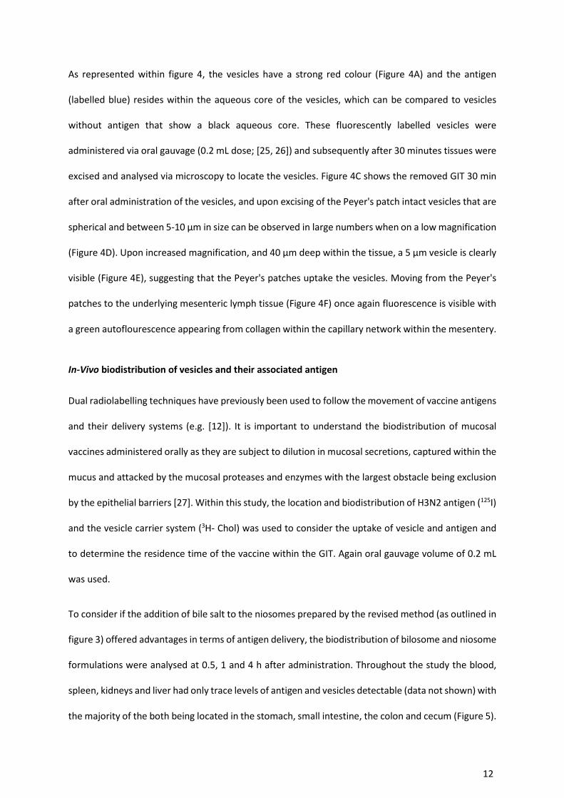
As represented within figure 4, the vesicles have a strong red colour (Figure 4A) and the antigen
(labelled blue) resides within the aqueous core of the vesicles, which can be compared to vesicles
without antigen that show a black aqueous core. These fluorescently labelled vesicles were
administered via oral gauvage (0.2 mL dose; [25, 26]) and subsequently after 30 minutes tissues were
excised and analysed via microscopy to locate the vesicles. Figure 4C shows the removed GIT 30 min
after oral administration of the vesicles, and upon excising of the Peyer's patch intact vesicles that are
spherical and between 5-10 µm in size can be observed in large numbers when on a low magnification
(Figure 4D). Upon increased magnification, and 40 µm deep within the tissue, a 5 µm vesicle is clearly
visible (Figure 4E), suggesting that the Peyer's patches uptake the vesicles. Moving from the Peyer's
patches to the underlying mesenteric lymph tissue (Figure 4F) once again fluorescence is visible with
a green autoflourescence appearing from collagen within the capillary network within the mesentery.
In-Vivo biodistribution of vesicles and their associated antigen
Dual radiolabelling techniques have previously been used to follow the movement of vaccine antigens
and their delivery systems (e.g. [12]). It is important to understand the biodistribution of mucosal
vaccines administered orally as they are subject to dilution in mucosal secretions, captured within the
mucus and attacked by the mucosal proteases and enzymes with the largest obstacle being exclusion
by the epithelial barriers [27]. Within this study, the location and biodistribution of H3N2 antigen (125I)
and the vesicle carrier system (3H- Chol) was used to consider the uptake of vesicle and antigen and
to determine the residence time of the vaccine within the GIT. Again oral gauvage volume of 0.2 mL
was used.
To consider if the addition of bile salt to the niosomes prepared by the revised method (as outlined in
figure 3) offered advantages in terms of antigen delivery, the biodistribution of bilosome and niosome
formulations were analysed at 0.5, 1 and 4 h after administration. Throughout the study the blood,
spleen, kidneys and liver had only trace levels of antigen and vesicles detectable (data not shown) with
the majority of the both being located in the stomach, small intestine, the colon and cecum (Figure 5).

13
When considering carrier (niosomes or bilosomes) distribution, the majority of the dose was located
within the stomach and small intestine (Figure 5A); around 30 % of the initial dose was detected within
the small intestine after 0.5 h with no significant difference between the two formulations. By 4 h the
levels in the small intestine had decreased with higher levels being detected in the cecum and colon,
as would be expected for the formulations as they transit through the GIT. Again there was no
significant difference in the profiles of the two formulations (Figure 5A). Considering the transit of the
associated antigen (Figure 5B), whilst comparable levels of ‘free’ and vesicle-incorporated antigen are
found within the stomach, significantly lower levels (< 10 % of initial dose) for ‘free’ antigen was
detected in the small intestine after 1 h compared to antigen carried by vesicles (Figure 5B). Moreover
these higher levels of antigen recovery correlated with vesicle recovery as it transits from the stomach
and small intestine, and then over time the dose progressed down the GIT to the cecum and colon
(Figure 5A and B) suggesting the antigen has remained associated with the vesicles. Again there was
no significant difference in the profile of the niosome and bilosome delivered antigen across the study
(Figure 5B).
Targeting of the vaccines to the Peyer's Patch and mesenteric lymph tissue
The Peyer’s patches are located at the antimesenteric border of the intestine where they appear as
nodular white masses (1.5 – 3.0 mm) and are key to antigen uptake and induction of immunity [28].
The Peyer's patches are an attractive route for targeted delivery as this provides a direct route for
antigen to reach the lymphatic system where first pass metabolism is avoided and the chances of
cellular rejection are reduced due to the M cells not expressing P-glycoprotein efflux pumps [29, 30].
Furthermore, the Peyer's patches, in comparison to the rest of the GI mucosa, possess minimal
mucosal coating where transcytotic activity is high [31, 32]. To consider targeting of the vaccine system
to the Peyer’s patch approximately 8-11 Peyer's patches recovered (in line with literature a number
of 6-12 patches; [28]). Figure 6A shows that using a vesicle carrier system made no significant
difference to the uptake of antigen within the Peyer’s patch and there was no significant difference in

14
carrier uptake between the niosomes and bilosomes (which was in the range of 1-5 %; Figure 6A).
However in terms of antigen recovery within the mesenteric lymph tissue, the use of a vesicular
delivery system significantly (P < 0.01) increased antigen delivery, yet the choice of carrier (niosomes
versus bilosomes) made no significant difference to either antigen or carrier localisation within the
mesenteric lymph tissue (Figure 6B).
The vesicles used within this study were shown (Figure 1) to remain within the desired size range for
increased uptake and retention within the Peyer’s patches [17]. Figure 6 indicates that the vesicles
and antigen are able to penetrate and reside within the lymph tissue for a period of 4 hours. This is
beneficial as the vesicles which remain in the Peyer's patches offer increased mucosal immunity
whereas the increased antigen and vesicles within the mesentery is more likely to induce systemic
immunity thus, offering both mucosal and systemic immunity [24]. In addition, Neutra and Kozlowski
(2006) determine that antigens delivered to mucosal sites within the small intestine offer greater IgA
secretions which are a characteristic of mucosal immunity compared to antigen delivery via routes
such as nasally where a systemic antibody response is achieved, due to the migration of antigen to the
draining lymph nodes [27].
The impact of carrier dose on antigen update
Based on the biodistribution study a question arose; is uptake within the Peyer’s patches limited in
terms of saturation and uptake? To address this, a study considering varying doses of the vaccine was
carried out which involved four groups of a) double dose (antigen 180 µg/mL, lipid 27 mg/mL), b) a
standard dose from Figure 5 & 6 (antigen 90 µg/mL, lipid 13.5 mg/mL), c) a half dose (antigen 45
µg/mL, lipid 6.75 mg/mL) and d) a quarter dose (antigen 22.5 µg/mL, lipid 3.375 mg/mL) and uptake
of vesicles and antigen at the target site Peyer’s patches and Mesenteric lymph tissue considered.
Based on the biodistribution study the organs removed for the following study focussed specifically
on the GI tract which included the stomach, Mesenteric lymph tissue, Peyer’s patches, small intestine,
colon and cecum (Figure 7).

15
Results from the study (Figure 7) show no significant differences in percentage antigen or vesicle
recovery between different dose concentrations in the organs collected after a 30 min time point and
overall percentage recovery of antigen is comparable (40-70 %) between all doses administered based
on the initial dose (Figure 7A). The vesicle recovery (Figure 7B) between the organs is also comparable,
with no significant differences suggesting that the clearance rate and the gastric emptying time was
not formulation dose dependent over the range tested. Gastric emptying time has been attributed to
the fed or fasted state of the GIT, where the gastric emptying time (T ½) in the fasted state is up to 2
± 1 min compared to a fed state T ½ of 17± 2 min in a mouse model [33]. Whilst both the niosomes
and bilosomes were shown to have similar stability, previous studies have shown that the stability of
lipidic systems can influence gastric clearance. For example, Marciani et al, (2009) study the stability
of lipid emulsions on gastric emptying; they demonstrate variations in gastric emptying times with
500mL of (15% w/w) [13C] palmitate-enriched olive oil-in-water emulsion meals depending on the lipid
composition [34]. Acid-unstable lipid emulsions broke down and rapidly layered in the stomach
compared to acid-stable lipid emulsions which had significantly slower gastric emptying. The acid-
stable emulsion emptied from the stomach linearly compared to the acid-unstable emulsion which
emptied exponentially. The authors attributed this to acid-stable emulsion inducing increased fullness,
decreased hunger and decreased appetite, where it was concluded that it is possible to delay gastric
emptying and increase satiety by stabilising the intragastric distribution of lipid emulsions against the
gastric acid environment [34].
When considering concentrations of lipid and antigen uptake rather than percentage dose (Figure 8),
there is a general trend of increased concentrations of antigen and carrier within both the Peyer’s
Patches and the mesentery lymph tissue as the dose increases up to 90 µg/mL antigen, 13.5 mg/mL
lipid (figure 8). This suggests that increasing the dose of a vaccine can improve delivery to the Peyer’s
patch and mesentery lymphatics. However a saturation point may be reached as shown in figure 8B,
where the increase in the dose to 180 µg/mL antigen / 27 mg/mL lipid did not significantly increase
delivery. Using lipid-based delivery systems has been demonstrated to increase the lymphatic

16
transport and absorption of drugs in other studies. For example, lymphatic uptake of halofantrine was
increased when it was incorporated within a self-microemulsifying drug delivery system based on
structured triglycerides containing medium and long chain fatty acids [35, 36]. The lipid content within
the drug delivery system was able to trigger sufficient lymphatic transport of halofantrine thus,
increasing bioavailability. Saturation of uptake via this route has also been demonstrated by Florence
et al, (2000) using polylysine dendrimer coated with a lipid surface; their results showed maximum
uptake within Peyer's patches of 1 % after a period of 3 hours which was not increased upon increased
dose concentration levels [37].
Conclusions
The proposed method of producing vesicles to protect thermolabile antigens by lowering the process
temperatures shows that antigen can be associated and entrapped within non-ionic surfactant
vesicles. Confocal laser scanning microscopy confirms that the vesicles are spherical in nature and the
vesicles meet the criteria of being within the optimum size range and charge for increased uptake
within the target site of the Peyer's Patches. The biodistribution studies demonstrate that by adopting
the new production method for these non-ionic surfactant vesicles, the necessity of the addition of
bile salts to enhance protection and delivery is circumvented as both antigen retention and antigen
delivery to the target site after oral administration was comparable. However, uptake at the target
site was shown to be dose-rate limited with saturated uptake becoming apparent and therefore
antigen to lipid dose ratio may be a key factor not only in terms of cost effectiveness but also in vaccine
efficacy, and thus requires further consideration in the assessment of the design of carrier-mediated
immunisation.
Acknowledgements

17
Jitinder Singh Wilkhu was funded via a BBSRC Industrial Case Award (BB/ G017948/1) and Variation
Biotechnologies Inc. Sarah McNeil has no conflict of Interest. David E Anderson is the Vice President
of Research at Variation Biotechnologies Inc and provided part funding for the research. Yvonne Perrie
received funding from BBSRC Industrial Case Award (BB/ G017948/1) and Variation Biotechnologies
Inc.
All institutional and national guidelines for the care and use of laboratory animals were followed. The
experiments within this study comply with the current laws within the United Kingdom.

18
References
1. Conacher M, Alexander J, Brewer JM. Oral immunisation with peptide and protein antigens by
formulation in lipid vesicles incorporating bile salts (bilosomes). Vaccine. 2001;19(20-22):2965-74.
doi:10.1016/s0264-410x(00)00537-5.
2. Holmgren J, Czerkinsky C. Mucosal immunity and vaccines. Nat Med. 2005;11(4 Suppl):S45-53.
doi:nm1213 [pii] 10.1038/nm1213.
3. Chadwick S, Kriegel C, Amiji M. Nanotechnology solutions for mucosal immunization. Adv Drug
Deliv Rev. 2010;62(4-5):394-407. doi:S0169-409X(09)00353-6 [pii] 10.1016/j.addr.2009.11.012.
4. Wilkhu J, McNeil SE, Kirby DJ, Perrie Y. Formulation design considerations for oral vaccines.
Therapeutic Delivery. 2011;2(9):1141-64. doi:10.4155/tde.11.82.
5. Cesta MF. Normal structure, function, and histology of mucosa-associated lymphoid tissue. Toxicol
Pathol. 2006;34(5):599-608. doi:X767592561294U17 [pii] 10.1080/01926230600865531.
6. Webster DE, Gahan ME, Strugnell RA, Wesselingh SL. Advances in Oral Vaccine Delivery Options:
What is on the Horizon? American Journal of Drug Delivery. 2003;1(4):227-40.
7. Mowat MA. Anatomical basis of tolerance and immunity to intestinal antigens. Nature.
2003;3:331-41.
8. Mann JFS, Shakir E, Carter KC, Mullen AB, Alexander J, Ferro VA. Lipid vesicle size of an oral
influenza vaccine delivery vehicle influences the Th1/Th2 bias in the immune response and
protection against infection. Vaccine. 2009;27(27):3643-9. doi:10.1016/j.vaccine.2009.03.040.
9. Kartoğlu Ü. Temperature sensitivity of Vaccines. Geneva, Switzerland2006.
10. Plotkin SA, Fletcher MA. Developments in immunization practices and strategies: implications for
vaccine stability. Dev Biol Stand. 1996;87:85-94.
11. Galazka AM, Milstien J, Zaffran M. Thermostability of vaccines: Global programme for Vaccines
and Immunization. In: Organization WH, editor. Geneza, Switzerland1998.
12. Wilkhu JS, McNeil SE, Anderson DE, Perrie Y. Characterization and optimization of bilosomes for
oral vaccine delivery. Journal of Drug Targeting. 2013;21(3):291-9.
doi:doi:10.3109/1061186X.2012.747528.
13. Henriksen-Lacey M, Bramwell V, Perrie Y. Radiolabelling of Antigen and Liposomes for Vaccine
Biodistribution Studies. Pharmaceutics. 2010;2(2):91-104.
14. Kaur R, Bramwell VW, Kirby DJ, Perrie Y. Pegylation of DDA:TDB liposomal adjuvants reduces the
vaccine depot effect and alters the Th1/Th2 immune responses. J Control Release. 2012;158(1):72-7.
doi:10.1016/j.jconrel.2011.10.012 S0168-3659(11)00964-3 [pii].
15. Vertzoni M, Dressman J, Butler J, Hempenstall J, Reppas C. Simulation of fasting gastric
conditions and its importance for the in vivo dissolution of lipophilic compounds. European Journal
of Pharmaceutics and Biopharmaceutics. 2005;60(3):413-7. doi:10.1016/j.ejpb.2005.03.002.

19
16. Pedersen BL, Brondsted H, Lennernas H, Christensen FN, Mullertz A, Kristensen HG. Dissolution
of hydrocortisone in human and simulated intestinal fluids. Pharm Res. 2000;17(2):183-9.
17. Brewer JM, Alexander J. The adjuvant activity of non-ionic surfactant vesicles (niosomes) on the
BALB/c humoral response to bovine serum albumin. Immunology. 1992;75(4):570-5.
18. Singh M, O'Hagan D. The preparation and characterization of polymeric antigen delivery systems
for oral administration. Adv Drug Deliv Rev. 1998;34(2–3):285-304. doi:10.1016/s0169-
409x(98)00044-1.
19. Mane V, Muro S. Biodistribution and endocytosis of ICAM-1-targeting antibodies versus
nanocarriers in the gastrointestinal tract in mice. Int J Nanomedicine. 2012;7:4223-37.
doi:10.2147/IJN.S34105 ijn-7-4223 [pii].
20. O'Hagan DT. The intestinal uptake of particles and the implications for drug and antigen delivery.
J Anat. 1996;189 ( Pt 3):477-82.
21. Eldridge JH, Hammond CJ, Meulbroek JA, Staas JK, Gilley RM, Tice TR. Controlled vaccine release
in the gut-associated lymphoid tissues. I. Orally administered biodegradable microspheres target the
peyer's patches. Journal of Controlled Release. 1990;11(1-3):205-14. doi:10.1016/0168-
3659(90)90133-e.
22. van der Lubben IM, Verhoef JC, van Aelst AC, Borchard G, Junginger HE. Chitosan microparticles
for oral vaccination:: preparation, characterization and preliminary in vivo uptake studies in murine
Peyer's patches. Biomaterials. 2001;22(7):687-94. doi:10.1016/s0142-9612(00)00231-3.
23. Ebel JP. A Method for Quantifying Particle Absorption from the Small Intestine of the Mouse.
Pharmaceutical Research. 1990;7(8):848-51. doi:10.1023/a:1015964916486.
24. Tabata Y, Inoue Y, Ikada Y. Size effect on systemic and mucosal immune responses induced by
oral administration of biodegradable microspheres. Vaccine. 1996;14(17–18):1677-85. doi:10.1016/s0264-410x(96)00149-1.
25. McConnell EL, Basit AW, Murdan S. Measurements of rat and mouse gastrointestinal pH, fluid
and lymphoid tissue, and implications for in-vivo experiments. J Pharm Pharmacol. 2008;60(1):63-70.
doi:10.1211/jpp.60.1.0008.
26. Wolfensohn S, Lloyd M. Handbook of Laboratory Animal Management and Welfare, Third
Edition. Blackwell Publishing Ltd; 2003.
27. Neutra MR, Kozlowski PA. Mucosal vaccines: the promise and the challenge. Nat Rev Immunol.
2006;6(2):148-58. doi:nri1777 [pii] 10.1038/nri1777.
28. Yeh P, Ellens H, Smith PL. Physiological considerations in the design of particulate dosage forms
for oral vaccine delivery. Adv Drug Deliv Rev. 1998;34(2-3):123-33. doi:S0169409X98000362 [pii].
29. Hunter AC, Elsom J, Wibroe PP, Moghimi SM. Polymeric particulate technologies for oral drug
delivery and targeting: a pathophysiological perspective. Nanomedicine: Nanotechnology, Biology
and Medicine. 2012;8, Supplement 1(0):S5-S20. doi:http://dx.doi.org/10.1016/j.nano.2012.07.005.

20
30. Florence A. The Oral Absorption of Micro- and Nanoparticulates: Neither Exceptional Nor
Unusual. Pharmaceutical Research. 1997;14(3):259-66. doi:10.1023/a:1012029517394.
31. Frey A, Giannasca KT, Weltzin R, Giannasca PJ, Reggio H, Lencer WI et al. Role of the glycocalyx in
regulating access of microparticles to apical plasma membranes of intestinal epithelial cells: implications for microbial attachment and oral vaccine targeting. The Journal of Experimental
Medicine. 1996;184(3):1045-59. doi:10.1084/jem.184.3.1045.
32. Plapied L, Duhem N, des Rieux A, Préat V. Fate of polymeric nanocarriers for oral drug delivery.
Current Opinion in Colloid & Interface Science. 2011;16(3):228-37.
doi:http://dx.doi.org/10.1016/j.cocis.2010.12.005.
33. Roda A, Mezzanotte L, Aldini R, Michelini E, Cevenini L. A new gastric-emptying mouse model
based on in vivo non-invasive bioluminescence imaging. Neurogastroenterol Motil.
2010;22(10):1117-e288. doi:NMO1535 [pii] 10.1111/j.1365-2982.2010.01535.x.
34. Marciani L, Faulks R, Wickham MS, Bush D, Pick B, Wright J et al. Effect of intragastric acid
stability of fat emulsions on gastric emptying, plasma lipid profile and postprandial satiety. Br J Nutr.
2009;101(6):919-28. doi:10.1017/S0007114508039986S0007114508039986 [pii].
35. Holm R, Tonsberg H, Jorgensen EB, Abedinpour P, Farsad S, Mullertz A. Influence of bile on the
absorption of halofantrine from lipid-based formulations. Eur J Pharm Biopharm. 2012;81(2):281-7.
doi:10.1016/j.ejpb.2012.03.005S0939-6411(12)00080-X [pii].
36. Holm R, Porter CJ, Edwards GA, Mullertz A, Kristensen HG, Charman WN. Examination of oral
absorption and lymphatic transport of halofantrine in a triple-cannulated canine model after
administration in self-microemulsifying drug delivery systems (SMEDDS) containing structured
triglycerides. Eur J Pharm Sci. 2003;20(1):91-7. doi:S092809870300174X [pii].
37. Florence AT, Sakthivel T, Toth I. Oral uptake and translocation of a polylysine dendrimer with a
lipid surface. Journal of Controlled Release. 2000;65(1–2):253-9.
doi:http://dx.doi.org/10.1016/S0168-3659(99)00237-0.

21
Figure legends:
Figure 1: Characterisation of niosomes and bilosomes when subjected to GI fluids at various time
points for A) Vesicle size, B) zeta potential and C) antigen retention within the vesicles (n=3).

22
Figure 2: Antigen protection from enzyme digestion offered by incorporation within vesicles by
trypsinisation (n=3).

23
Figure 3: Schematic representing method modifications and corresponding characterisation data
between bilosomes produced via the pre-formed vesicle method as outlined Conacher et al., 2001 or
the method outlined within the Method section where antigen is incorporated within the vesicles in-
situ.
24
Figure 4: Confocal laser light microscopy images of niosome vesicles labelled with a lipid Dil-C dye and
antigen labelled with a Flammaflour FPR 648 dye. Stages include A) vesicle emulsion upon formation, B) analysis of vesicles representing vesicles corresponding to vesicle size data showing antigen
location, C) vesicles in GI transit, D) spherical vesicles within Peyer's patches, E) vesicle within Peyer's
patch 40 µm deep and F) mesenteric lymph tissue fluorescence.

25
Figure 5: In vivo trafficking of antigen and vesicles along the GIT after oral gavage where A) represents
the niosomes or bilosomes radiolabelled with 3H-Cholesterol and B) represents H3N2 antigen
radiolabelled with I125 and dosed without a carrier system or in associated with bilosomes or niosomes
(n=4 at each time point).

26
Figure 6: Recovery of antigen and vesicles based on initial dose at t= 1h within A) Peyer's patches and
B) Mesenteric Lymph Tissue (n=4).
Figure 7: In vivo saturation study of various dose concentrations representing antigen and vesicle
recovery within the organs selected; A) antigen recovery, B) vesicle carrier recovery (n=4).

27
Figure 8: Representation of dose recovery per mL at site of uptake for various dose concentrations of
the formulation within Peyer’s patches and mesenteric lymph tissue; A) antigen dose and B) lipid dose
(n=4).


![Recent Advances in Drug Delivery Systems · 41]. Niosomes are a non-ionic surfactant vesicles made up from polyoxyethylene alkyl ethers, polyoxyethylene alkyl esters or saccharose](https://img.pdfslide.us/doc/110x75/5e1d29ad34d3637e3f7b9539/recent-advances-in-drug-delivery-systems-41-niosomes-are-a-non-ionic-surfactant.jpg)