Embed Size (px)
Citation preview

Title A rat model for LGI1-related epilepsies.
Author(s)
Baulac, Stéphanie; Ishida, Saeko; Mashimo, Tomoji; Boillot,Morgane; Fumoto, Naohiro; Kuwamura, Mitsuru; Ohno,Yukihiro; Takizawa, Akiko; Aoto, Toshihiro; Ueda,Masatsugu; Ikeda, Akio; LeGuern, Eric; Takahashi, Ryosuke;Serikawa, Tadao
Citation Human molecular genetics (2012), 21(16): 3546-3557
Issue Date 2012-05-15
URL http://hdl.handle.net/2433/174159
Right
© The Author 2012. Published by Oxford University Press; この論文は出版社版でありません。引用の際には出版社版をご確認ご利用ください。This is not the published version.Please cite only the published version.
Type Journal Article
Textversion author
Kyoto University

1
A RAT MODEL FOR LGI1-RELATED EPILEPSIES
Stéphanie Baulac1,2,3
*#, Saeko Ishida
4*, Tomoji Mashimo
4#, Morgane Boillot
1,2,3, Naohiro
Fumoto4,5
, Mitsuru Kuwamura6, Yukihiro Ohno
7, Akiko Takizawa
4, Toshihiro Aoto
8,
Masatsugu Ueda8, Akio Ikeda
5, Eric LeGuern
1,2,3,9, Ryosuke Takahashi
5 and Tadao Serikawa
4.
*Authors contributed equally to this study.
1. Inserm U975, CRICM, Hôpital de la Pitié-Salpêtrière, Paris F-75013, France
2. Université Pierre & Marie Curie-Paris 6, UMR_S975, Hôpital de la Pitié-Salpêtrière, Paris
F-75013, France
3. CNRS, UMR7225, Hôpital de la Pitié-Salpêtrière, Paris F-75013, France
4. Institute of Laboratory Animals, Graduate School of Medicine, Kyoto University, Kyoto
606-8501, Japan
5. Department of Neurology, Graduate School of Medicine, Kyoto University, Kyoto 606-
8507, Japan
6. Laboratory of Veterinary Pathology, Osaka Prefecture University, Izumisano, Osaka 598-
8531, Japan
7. Laboratory of Pharmacology, Osaka University of Pharmaceutical Sciences, Takatsuki
569-1094, Japan
8. PhoenixBio Co. Ltd., Utsunomiya 321-0973, Japan
9. AP-HP, Département de Génétique et Cytogénétique, centre de génétique moléculaire et
chromosomique, Hôpital de la Pitié-Salpêtrière, Paris F-75013, France
# Correspondence should be addressed to:
Tomoji Mashimo,
Institute of Laboratory Animals, Graduate School of Medicine, Kyoto University,
Yoshidakonoe-cho, Sakyo-ku, Kyoto 606-8501, Japan.
E-mail: [email protected]
Stéphanie Baulac,
Institut du Cerveau et de la Moelle épinière (ICM)
CRICM U975, Hôpital de la Pitié -Salpêtrière - 47, bd de l'hôpital, 75013 Paris, France
Tel: +33-1-5727-4339, E-mail: [email protected]

2
ABSTRACT
Mutations of the leucine-rich glioma-inactivated 1 (LGI1) gene cause an autosomal-dominant
partial epilepsy with auditory features also known as autosomal-dominant lateral temporal
lobe epilepsy (ADLTE). Furthermore, LGI1 is the main antigen present in sera and
cerebrospinal fluids of patients with limbic encephalitis and seizures, highlighting the
importance of LGI1 in a spectrum of epileptic disorders. LGI1 codes for a neuronal secreted
protein, which brain function is still largely unknown. Here, we generated Lgi1-mutant rats
carrying a missense mutation (L385R) by ENU (N-ethyl-N-nitrosourea) mutagenesis. We
found that the L385R mutation prevents secretion of Lgi1 protein by COS7 transfected cells.
However, the L385R-Lgi1 protein was found at low levels in the brains and primary cultured
neuron lysates of Lgi1-mutant rats, suggesting that mutant protein may be destabilized in vivo.
Studies on the behavioral phenotype and intracranial electroencephalographic signals from
homozygous and heterozygous Lgi1-mutant rats recalled several features of the human
genetic disorder. We found that homozygous Lgi1-mutant rats generated early-onset
spontaneous epileptic seizures from P10 and died prematurely. Adult heterozygous Lgi1-
mutant rats were much more susceptible to sound-induced, generalized tonic-clonic seizures,
than control rats. Audiogenic seizures were suppressed by antiepileptic drugs such as
carbamazepine, phenytoin and levetiracetam, that are commonly used to treat partial seizures,
but not by the prototypic absence seizure drug, ethosuximide. Our findings provide the first
rat model for a missense mutation in the Lgi1 gene, a model complementary to knockout mice
in studies on Lgi1-related epilepsies, and reveal a new concept that LGI1 disease-causing
mutations might cause haploinsufficiency, and not only a failure of secretion of the protein.

3
INTRODUCTION
Epilepsy, with a lifetime prevalence of 3%, is a frequent neurological disorder. Studies on
familial idiopathic epilepsies have identified multiple disease-causing genes (1). For example,
mutations in the leucine-rich glioma-inactivated 1 (LGI1) gene cause an inherited epilepsy
syndrome designated either Autosomal Dominant Lateral Temporal Epilepsy (ADLTE) (2) or
Autosomal Dominant Partial Epilepsy with Auditory Features (ADPEAF) (3). Focal seizures,
with prominent auditory auras in about two thirds of patients, emerge in adolescence (4).
Aphasic symptoms and other aberrant perceptions of a visual, vertiginous, epigastric, or
psychogenic nature are also reported, and seizures can be triggered by noises or voices (5, 6).
This seizure semiology tends to indicate a neocortical origin in the lateral temporal lobe. LGI1
mutations (36 published to date (7)) have been found in up to 50% of ADLTE families and
2% of sporadic cases (8). The role of LGI1 in neurological diseases was further expanded
with the recent discovery that a subset of patients with limbic encephalitis (an autoimmune
disorder associated with seizures in the majority of patients) (9, 10) has serum antibodies
against LGI1. It is speculated that antibody-mediated disruption of LGI1 causes increased
excitability, which results in seizures and other symptoms of limbic encephalopathy.
In contrast to other genes linked to idiopathic epilepsies, LGI1 does not encode an ion
channel subunit, but rather a secreted leucine-rich repeat (LRR) protein (11). To the exception
of R407C (12), all tested LGI1 missense mutations tend to suppress protein secretion in in
vitro overexpression systems (11, 13-17), indicating that extracellular levels of LGI1 may be
critical to its pathophysiological effects. Lgi1 protein is expressed in the developing brain
during embryogenesis and increases until adult, suggesting that Lgi1 may have a role during
brain development (18-20). While its functions remain unclear, Lgi1 interacts with the
presynaptic Kv1.1 voltage-gated potassium channel (21), ADAM22/ADAM23 (disintegrin
and metalloproteinase domain 22 and 23)(22), ADAM11 (23) and NogoR1 (24). Insights into
4
the role of Lgi1 in epilepsy have emerged from recent studies on Lgi1 knockout in mice.
There is a consensus that in homozygous Lgi1-/-
mice, the constitutive deletion of Lgi1
induces early-onset spontaneous seizures with a premature death (19, 25, 26). Heterozygous
Lgi1+/ -
mice do not generate seizures spontaneously, but they are more susceptible to seizure
induction by pentylenetetrazole (25) and auditory stimuli (19). Suppression of Lgi1 may be
epileptogenic by modulating signaling in glutamatergic but not GABAergic synaptic
transmissions (25, 26) as well as controlling postnatal maturation and pruning of
glutamatergic synapses in the hippocampus (20) and retinogeniculate afferents in the thalamus
(27).
In the present study, we generated and characterized Lgi1-mutant rats carrying a missense
mutation (L385R). We deciphered the mechanisms by which this mutation led to a loss of
function in mammalian cells and primary neurons in culture. Electroencephalographic (EEG)
monitoring was then used to define how the L385R-Lgi1 protein affected the phenotype of
homozygous and heterozygous Lgi1-mutant rats in vivo. Finally, we examined actions of
antiepileptic drugs on the audiogenic seizures in heterozygous Lgi1+/L385R
rats.

5
RESULTS
Generation of Lgi1-mutant rats
The ENU (N-ethyl-N-nitrosourea)-mutagenized F344/NSlc rat archive (KURMA: Kyoto
University Rat Mutant Archive) was screened for mutations in the Lgi1 gene by a high-
throughput screening assay (28). A missense mutation (c.1154 T>G) in exon 8 of Lgi1 was
found in one DNA sample of KURMA. This mutation resulted in the p.Leu385Arg/L385R
amino acid substitution in the fourth epilepsy-associated-repeat (EAR) (Figure 1A), a residue
highly conserved among vertebrates and invertebrates (Figure 1B). It was predicted to be “not
tolerated” by the SIFT prediction tool of functional effects of variants
(http://blocks.fhcrc.org/sift/SIFT.html). Interestingly, a well-documented ADLTE-causing
mutation, p.Glu383Ala/E383A, was located close to the Rat mutation (3, 11, 13). The
heterozygous Lgi1-mutant rat (Lgi1+/L385R
) was recovered from the corresponding frozen
sperm by intracytoplasmic sperm injection. Nine generations were backcrossed on the
F344/NSIc inbred background to eliminate mutations potentially induced by ENU-
mutagenesis elsewhere in the genome (mean mutation frequency was ~1 in 4 x 10⁶ base pairs).
Backcrossed Lgi1+/L385R
rats were then intercrossed to obtain wild-type (WT), heterozygous
(+/L385R) and homozygous (L385R/L385R) animals. They were born in expected Mendelian
ratios (+/+, n=17; homozygous, n=18; heterozygous, n=29; ² = 0.59, not significant) and sex
ratios (Lgi1L385R/L385R
rats: 28 females, 36 males; ² = 1, not significant).
L385R mutation impairs Lgi1 secretion in COS7 cells and cortical neurons
We asked whether the rat L385R mutation impaired Lgi1 secretion, by transiently transfecting
COS7 cells with Flag-Lgi1-L385R, Flag-Lgi1-E383A or Flag-Lgi1-WT (wild-type Lgi1).
Using an antibody directed against amino acids 200-300 of Lgi1 (ab30868), Western blot
analysis revealed the presence of wild-type Lgi1 and both mutants in the cell lysates.
However, while wild-type Lgi1 protein was predominantly present in the cell culture medium,

6
we did not detect the Flag-Lgi1-L385R or the Flag-Lgi1-E383A mutants (Figure 2A). This
shows that the L385R mutation prevents secretion of Lgi1 into the culture medium of COS7
cells.
We next examined endogenous Lgi1 secretion in isolated neurons of primary cortical
cultures from embryonic day 19 Lgi1L385R/L385R
, Lgi1+/L385R
and Lgi1+/+
littermates. Western
blot analysis revealed only weak signal of 65 kDa in the neuron lysate of homozygous
Lgi1L385R/L385R
rats, as well as in the neuron medium, indicating a low level of L385R-Lgi1
protein in neurons (Figure 2B).
L385R mutation affects in vivo stability of Lgi1 protein
We then compared the endogenous L385R-Lgi1 protein level by Western blot in whole brain
homogenates of P12 Lgi1L385R/L385R
, Lgi1+/L385R
and wild-type (Lgi1+/+
) littermate rats.
Immunoblot revealed a single band of 65 kDa in the lysate of Lgi1+/+
. It was reduced by
about half in Lgi1+/L385R
and was absent in Lgi1L385R/L385R
(n=5; Figure 3A). The low
abundance of L385R-Lgi1 protein in brain was confirmed with a second antibody generated
to the C-terminus of Lgi1 (sc-9583, Santa Cruz) in whole brain lysates from P9 rats (Figure
1S, supplementary data). These findings were replicated in 3 additional litters of wild type,
heterozygous and homozygous rats aged P5 and P9 (before onset of seizures) and P12 (after
onset of seizures), indicating that shortened half-life of L385R-Lgi1 was not due to seizures-
induced damage (data not shown). We next analyzed synaptic fractions of hippocampal and
cortical lysates by preparing Triton X100-soluble crude fractions. In Lgi1+/+
and Lgi1+/L385R
rats, a strong signal was detected, indicating Lgi1 protein was present at the synapse. In
contrast, similarly to whole brain homogenates, L385R-Lgi1 protein was not detected in the
synaptic membrane fraction (Figure 3B), suggesting that it is probably unstable and thus not
delivered to the synapse.
We next asked whether low levels of L385R-Lgi1 protein resulted from a preferential
7
degradation of L385R-Lgi1 transcript. We extracted total RNAs from whole brains of
Lgi1L385R/L385R
(n=5), Lgi1+/L385R
(n=7) and Lgi1+/+
(n=5) littermate rats and analyzed Lgi1
transcript expression by quantitative RT-PCR. Relative levels of the L385R-Lgi1 transcript
were not significantly different from levels of the Lgi1+/+
transcript (Kruskal-Wallis test, P
non significant; Figure 3C). These results suggest that low neuronal levels of L385R-Lgi1
protein may result from a rapid turnover of the protein rather than the transcript.
L385R mutation has no major effect on in vitro neuronal growth
We asked whether this L385R mutation modified the growth of primary neurons plated on
poly-L-lysine coated culture vessels. Cortical and hippocampal neurons from E19 rats were
co-cultured for 3 weeks and examined daily. No differential effect on life span or neurite
outgrowth was detected between Lgi1L385R/L385R
and Lgi1+/+
rats (Image J measurement of total
neurite network length, Kruskal-Wallis test, P non significant) (Figure 4A). No major
morphological differences were detected between hematoxylin-stained brains of P12
Lgi1L385R/L385R
rats and their Lgi1+/L385R
and Lgi1+/+
littermates (Figure 4B).
Homozygous Lgi1-mutant rats are epileptic and die prematurely
At birth, we detected no differences in appearance or behavior of homozygous Lgi1L385R/L385R
rats and heterozygous Lgi1+/L385R
and Lgi1+/+
littermates. During the second postnatal week,
Lgi1L385R/L385R
pups began to exhibit spontaneous seizures (Figure 5A, movie 1). They
occurred at a mean frequency of 8±2.8 per hour (mean± SD, n=8) from P10. Ictal epileptic
discharges (n=11 electro-clinical seizures) were recorded by intracranial
electroencephalography (EEG) in two homozygous Lgi1L385R/L385R
pups (Figure 5B). Seizures
typically consisted of sequences of (i) hypertonic, often asymmetric, trunk, limb and tail
postures, (ii) clonies of all limbs or jerking. EEG records began with rhythmic 5-7 Hz spike
activity which increased in amplitude. It was replaced by polyspike-and-wave complexes at 1
Hz during jerking episodes which slowed (0.5 Hz) as the seizure terminated. Seizures were

8
sometimes associated with motor automatisms, such as chewing. Such spontaneous epileptic
activity was never observed in age-matched heterozygous Lgi1+/L385R
(n=7) or Lgi1+/+
littermates (n=7).
As seizures emerged, Lgi1L385R/L385R
rat pups began to lose body weight. At P15, the body
weight of Lgi1L385R/L385R
rats was significantly (P<0.009, t-test) lower than that of Lgi1+/L385R
or Lgi1+/+
rats (Figure 6A), and development slowed dramatically (Figure 6B). All
homozygous Lgi1L385R/L385R
rats died prematurely and the Kaplan-Meier curve revealed a
mean lifetime of 13 days (n= 10). No homozygous Lgi1L385R/L385R
rat survived beyond P17,
while no Lgi1+/L385R
or Lgi1+/+
littermates had died at this age (Figure 6C). Possibly, this early
mortality results from a failure to feed due to seizures.
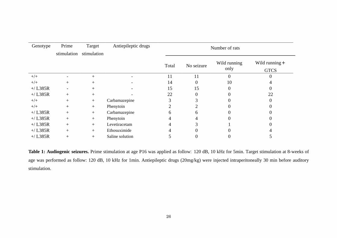
Heterozygous Lgi1-mutant rats display increased audiogenic seizure vulnerability
Heterozygous Lgi1+/L385R
rats appear normal, are fertile and live for at least one year.
Spontaneous clinical seizures have never been observed either in pups or adults. Since partial
seizures can be triggered by audiogenic events in ADLTE patients, we tested the
susceptibility of heterozygous Lgi1-mutant rats to audiogenic seizures (AGS). A single 120
dB sound stimulus at 10 kHz never induced a seizure in Lgi1+/L385R
or Lgi1+/+
rats at 3, 5, 8 or
12 weeks of age, possibly due to this rat strain resistance. Acoustic priming (5 min, 10 kHz,
120 dB) was thus applied to rat pups aged P16, corresponding to the critical period when rats
become seizure-prone (29). Primed rats were then tested for audiogenic seizures at 8 weeks of
age. Auditory stimulus first induced wild running, a typical behavior of audiogenic seizures,
in all Lgi1+/L385R
(n=22) and Lgi1+/+
(n=14) rats at 8 weeks (Table 1). Following wild running,
we noted that auditory stimulation yielded generalized tonic-clonic seizures (GTCS) in all
Lgi1+/L385R
rats, but only in 28 % of Lgi1+/+
rats (100 % versus 28 %, 2=36, P =1.9 10
-9)
(Figure 7A, movie 2). The latency from auditory stimulus to wild running was shorter in
Lgi1+/L385R
than Lgi1+/+
rats (Student’s t test, P=2.7 10-2
; Figure 7B). The duration of wild

9
running was also shorter in Lgi1+/L385R
than in Lgi1+/+
rats, probably since wild running was
more rapidly replaced by a GTCS in Lgi1+/L385R
rats (Student’s t test, P=6.8 10-5
; Figure 7C).
The duration of GTCS did not differ significantly in Lgi1+/L385R
and Lgi1+/+
rats (Student’s t
test, P=4.5 10-1
; Figure 7D).
We compared cortical and hippocampal EEG signals generated by Lgi1+/L385R
(n=3) and
Lgi1+/+
rats (n=1) during auditory stimuli (Figure 8). During wild running, movement artifacts
tended to obscure EEG signals. After running terminated, EEG signals were strongly
suppressed in the tonic phase of Lgi1+/L385R
rats and the immobility phase of Lgi1+/+
rats.
During the clonic seizure phase in Lgi1+/L385R
rats, continuous rhythmic slow activity at 2-3 Hz
was detected in cortex and hippocampus. EEG signals were then suppressed, as Lgi1+/L385R
rats remained immobile until auditory stimuli ceased.
Effect of antiepileptic drugs on audiogenic seizures
Finally, we evaluated the efficacy of several antiepileptic drugs on audiogenic seizures in
primed Lgi1+/+
and Lgi1+/L385R
rats. We administered carbamazepine, phenytoin, levetiracetam
and ethosuximide intraperitoneally (20 mg/kg) at 30 min before the auditory stimuli in 8
week-old rats (Table 1, movie 3). Both wild running and GTCS were completely inhibited by
carbamazepine and phenytoin in Lgi1+/+
and Lgi1+/L385R
rats. Levetiracetam prevented wild
running and GTCS in 3 of 4 Lgi1+/L385R
rats. No ictal EEG activity was detected during
auditory stimulation (not shown). Ethosuximide, a prototypic generalized absence seizure
drug, had no effect on seizures of Lgi1+/L385R
rats (n=4).
DISCUSSION
Here we present the first genetically-engineered animal model to express a missense mutation
in the Lgi1 gene. We generated and characterized a Lgi1-mutant rat with a missense mutation
(L385R) and studied its functional consequences in vivo. We first examined the impact of the
mutation using in vitro overexpression paradigm. Our results showed that this mutation

10
prevented Lgi1 secretion in transiently transfected COS7 cells. This variant is thus deleterious
and apparently shares common effects with ADLTE-causing missense mutations which
nearly all decrease protein secretion, except one (30). Testing endogenous expression levels
of the mutated Lgi1 protein in cultured cortical neurons of Lgi1-mutant animals revealed very
low levels of L385R-Lgi1 protein, both in extracellular medium from cultures and also in
neuron lysates. Moreover, endogenous levels of Lgi1 protein were also substantially lower in
the brain of Lgi1-mutant animals than of wild-type littermates. Probably, in vivo, the L385R
mutation favors misfolding and so reduces Lgi1 protein stability, causing its degradation
through protein quality control mechanisms. This is consistent with in silico computational
models predicting that a number of disease-causing mutations alter protein folding (30). Thus
a physiopathological loss-of-function may emerge not only due to a failure of protein
secretion but also from a lack of correctly folded neuronal Lgi1. This new mechanism must
be considered together with previous suggestions of a defective secreted extracellular Lgi1
(acting as a ligand for ADAM22/23 at the post-synaptic) (22), rather than cytoplasmic
(through the modulation of Kv1.1 channel) (21). Here, the Lgi1-mutant rat carrying a
missense mutation located nearby a well-characterized naturally occurring missense mutation
found in ADLTE patients (3, 11, 13), lacks both cytoplasmic and extracellular Lgi1. While
cortical tissue from patients is not available, we speculate that ADLTE-associated missense
mutations might also lead to instability in vivo, causing a haploinsufficiency. We note such a
deficiency in Lgi1 occurs in patients with limbic encephalitis and seizures, in which immune-
mediated disruption of LGI1 results in hyperexcitability (31).
While focal epilepsies are often associated with brain lesions, we detected no major
abnormality in brain morphology of Lgi1-mutant rats. Since Lgi1 may promote neurite
outgrowth in vitro via ADAM23 (32) and NogoR1 (24), we examined neuron growth in co-
cultures of cortical and hippocampal neurons from Lgi1-mutant rats. We detected no obvious
11
defect in neuritic outgrowth or neuronal life-span suggesting that neuronal network activity,
rather than dendritic architecture may contribute to hyperexcitability in this rat.
The phenotype of mutant rats possessed similarities to the ADLTE syndrome. Epileptic
seizures, associated with cortical and hippocampal ictal epileptiform activity, emerged at P10
in homozygous Lgi1L385R/L385R
pups. Frequent and severe seizures led to death of the
homozygous animals around P13. Lgi1+/L385R
rats, which carry a heterozygous missense
mutation similar to ADLTE patients, did not generate seizures spontaneously but were highly
susceptible to audiogenic seizures, as patients with LGI1 mutations are susceptible to
auditory-induced seizures (seizures may be triggered by noises or voices) (6). In addition, we
showed that rat audiogenic seizures responded to the same drugs as used in the human: they
were suppressed by two antiepileptic drugs, carbamazepine and phenytoin, that target voltage-
gated channels, but also by levetiracetam which anticonvulsant activity is mediated via
interaction with the synaptic vesicle protein 2A (SV2A)(33, 34). Since Lgi1 co-
immunoprecipitates with several other neuronal vesicle-related proteins (35), this latter
pathway involving SV2A might be promising for preventing seizures in this syndrome. As
expected, ethosuximide, a first choice drug for absence seizures, did not prevent audiogenic
seizures. This rat model thus permitted more detailed studies on audiogenic seizures and tests
on anti-epileptic molecules. As SV2A, Lgi1 may point towards novel antiepileptic therapies
for drug-resistant patients.
The Lgi1-mutant rat is therefore a relevant animal model for modeling Lgi1-related
epilepsy. It shares several features with the phenotype of Lgi1 knockout mice (19): (i) a
natural history of severe early onset spontaneous seizures (age at onset, semiology of seizures,
EEG pattern and premature death associated with loss of body weight), (ii) an absence of
morphological abnormalities, (iii) audiogenic seizures in heterozygous rats recapitulating the
genetic cause and mimicking the auditory triggering of seizure in the human. We note the
similar age of seizure onset (around P10) in Lgi1-null mice and Lgi1-mutant rats that might be

12
consistent with the timing of Lgi1 expression and maturation of glutamatergic synapses
similar to both species. We were initially surprised to define a similar phenotype in
homozygous Lgi1-mutant rats (L385R-Lgi1) and Lgi1-null mice (absence of Lgi1). However,
our discovery of a rapid degradation of Lgi1-L385R suggests the point mutation induces a
haploinsufficiency that is equivalent to a gene knockout.
In conclusion, we report a unique and original rat model of Lgi1-related epilepsies, which
is complementary to knockout mice. It gave us the opportunity to better understand the
consequences of missense mutations on the fate of the mutant protein, revealing a major
finding that L385R-Lgi1 protein is unstable in vivo. Thanks to this model, we also
investigated the consequences of Lgi1 deficiency on the neuronal and neurite outgrowth.
Finally, the heterozygous Lgi1+/L385R
rats allowed us to initiate pharmacological studies on
their auditory triggered-seizures which are close to those occurring in ADLTE patients.
MATERIAL AND METHODS
ENU mutagenesis in rats
ENU mutagenesis and screening protocols using MuT-POWER in rats have been described
(28). The sperm archive KURMA has been deposited in the National BioResource Project-Rat
in Japan (NBRP-Rat: www.anim.med.kyoto-u.ac.jp/nbr). Primers were designed to amplify
by PCR the exonic region of the rat Lgi1 gene from ~50 bp flanking each intron
(supplemental Table 1). Sequencing was performed with BigDye terminator mix, followed by
the protocol for the Applied Biosystems 3100 DNA Sequencer. Lgi1-mutant rats were
recovered from frozen sperm by intracytoplasmic sperm injection.
Animals
Lgi1-mutant rats (strain name, F344-Lgi1m1kyo
) were deposited in NBRP-Rat (N° 0656). They
were kept and bred at the Institute of Laboratory of Animals, Graduate School of Medicine,
Kyoto University in air-conditioned rooms under a 14h light/10h dark cycle. Animal care and
13
experiments conformed to the Guidelines for Animal Experiments and were approved by the
Animal Research Committee of Kyoto University.
Genotyping of Lgi1-mutant rats
Exon 8 of Lgi1 was amplified by PCR with Ex8-1 primers (supplemental Table 1) using the
Ampdirect Plus®
PCR buffer (Shimadzu) and FTA®
card for blood samples. PCR products
were then sequenced with BigDye terminators mix.
Western blots
Littermate rat pups aged postnatal day 9 (P9) and P12 were decapitated; whole brains were
quickly removed and lysed in 3M urea, 2.5% SDS, 50 mM Tris, 30 mM NaCl buffer (total
brain homogenates). For synaptic fractions, brains of littermate rats were homogenized in 50
mM Tris, 5 mM EDTA, 120 mM NaCl with complete inhibitor cocktail, spun for 1h at
165,000 × g and pellets resuspended with 1% Triton X-100. Total protein concentrations were
determined by the BCA method (Pierce). Twenty-five g of each sample were separated on
10 % Tris-glycine polyacrylamide gels were analyzed by Western blot with the following
antibodies: rabbit polyclonal anti-Lgi1 antibody (ab30868; 1ug/ml; Abcam), goat polyclonal
anti-Lgi1 antibody (sc-9583; 1ug/ml; Santa Cruz) and rabbit anti-actin antibody (1/1000,
Sigma Aldrich), and detected with the SuperSignal Chemiluminescent Substrate (Pierce).
Cell culture and transfection
Drs. K. Senechal and J. Noebels kindly provided the mouse wild-type Lgi1 cDNA with a Flag
tag at the N-terminus. Lgi1-E383A and Lgi1-L385R were generated using the QuikChange®
Site-Directed Mutagenesis Kit. COS7 cells were cultured in DMEM containing 10% fetal
bovine serum, penicillin and streptomycin. Transient transfections were performed using
LipofectamineTM
2000 according to instructions (Invitrogen), followed by a 14-16 hour
incubation in serum-free media. Cells and media were analyzed 24-36 hours after transfection.

14
Cell lysates and conditioned media were prepared as described (13) and analyzed by Western
blot.
Neuronal cultures
Cortex and hippocampus were removed from ten rat embryos aged embryonic day 19 (E19)
and dissociated using the Nerve-Cell culture system (Sumitomo Bakelite co). Neurons were
plated at 105 cells/ml in neuron culture medium (Sumitomo Bakelite co) on 35mm poly-L-
Lysine coated dishes. They were cultured for 12 days and then lysed as previously described
in COS7 cells. Neuronal outgrowth was imaged and measured automatically using ImageJ.
Quantitative reverse transcription PCR
Whole brains were removed from P9 rats (n=6) and stored in RNAlater® solution (Applied
Biosystems). Total RNA was isolated with RNeasy Miniprep columns (Qiagen) and
contaminating DNA was depleted using RNase-free DNase. First-strand cDNA was
synthesized from 5 µg of total RNA by oligo dT-primed reverse transcription
(ThermoScript™ Reverse Transcriptase, Invitrogen). Quantitative PCR were performed as
triplicates using the QuantiFast Multiplex PCR Kit (Qiagen) using predesigned probes for
Lgi1 (QuantiFast Probe Assays) and peptidyl prolyl isomerase A (PPIA) as a reference gene
included in all multiplex reaction. The error bars of the quantitative PCR represent SDs of
triplicates.
Brain histochemistry
Lgi1+/+
littermates (n=1), Lgi1+/L385R
(n=2) and Lgi1L385R/L385R
(n=1) aged P12 were deeply
anesthetized with sodium pentobarbital (50 mg/kg by intraperitoneal injection). Brains were
removed, fixed in Bouin's fixative and embedded in paraffin. Morphological changes were
evaluated from hematoxylin and eosin stained, 4 µm thick paraffin sections.
Animal surgery and intracranial EEG recordings
Cortical EEG was recorded from homozygous P10 rats (during 3 continuous hours) and
heterozygous rats aged 8 weeks. Rats were anesthetized with an intraperitoneal injection of

15
sodium pentobarbital (40 mg/kg) and the heads were fixed in a stereotaxic instrument. One-
mm-diameter screw electrodes were implanted into the epidural space of the left frontal cortex.
A reference electrode was fixed on the frontal cranium. For hippocampal EEG, 0.2-mm-
diameter stainless-steel electrodes were implanted in the hippocampus (3.8 mm caudal, 2.0
mm lateral to the bregma and 2.2 mm from the cortex surface). A miniature plug was
positioned and fixed on the midline of the skull to provide electrical connections. After 1-hour
recovery period for P10 rats and 1-week recovery period for 8-week old rats, animals were
placed in a shielded box (40×40×40 cm3) and the EEG signals were amplified with a
sampling rate of 0.5–100 Hz with a 8-channel system (MEG-6108; Nihon Kohden) and
recorded (RTA-1100; Nihon Kohden) under free-moving conditions. The signals were stored
in a computer for analysis (ML845; PowerLab). Behavioral changes were simultaneously
observed with video recording.
Acoustic stimulation
The testing apparatus consisted of a 17×25×13 cm plastic cage placed inside a larger sound-
proof box. Acoustic stimulation was administered from a loudspeaker (JBL Professional)
centrally placed on the cover of the cage. Tone bursts were delivered by a sound stimulator
(DPS-725, Dia Medical System Co.) and the signal was amplified using a power amplifier
(D75-A, Amcron). Lgi1+/L385R
and Lgi1+/+
littermate rats were exposed individually to intense
auditory stimulation after 1-minute habituation. Priming stimulation was performed in P16
rats with a sound stimulus of 120 dB at 10 kHz for 5 min. Target stimulation consisted of a
120 dB sound stimulus at 10 kHz for 1 min at 8 weeks (36). The onset, latency, and duration
of wild running and GTCS were measured from video records.
Antiepileptic drugs administration
Antiepileptic drugs (Sigma-Aldrich) were administrated intraperitoneally 30 minutes before
target stimulation with therapeutic range (20 mg/Kg). Carbamazepine and Ethosuximide were
first dissolved in polyethylene glycol 400 then in water. Phenytoin was first dissolved in 0.5 N

16
NaOH and then diluted with saline solution. Levetiracetam was dissolved in saline solution.
FIGURE LEGENDS
Figure 1. The L385R mutation. (A) Schematic representation of the Lgi1 protein showing
its domain organization and location of the L385R mutation. The protein is composed of 2
structural domains: four N-terminal leucine-rich repeats (LRR in blue) and seven epilepsy-
associated repeats (EAR in yellow) in the C-terminal half of the protein. (B) Multiple protein
alignments of Lgi1 protein showing strong conservation of L385 residue in both vertebrates
and invertebrates using the Alamut® Mutation Interpretation Software.
Figure 2. Lack of secretion of L385R-Lgi1 mutant protein in COS7 cells and cortical
neurons. (A) COS7 cells were transiently transfected with the wild-type (WT) or indicated
mutant Flag-Lgi1-expressing plasmids. Cell lysates and cell media were analyzed by Western
blot with anti-Lgi1 antibody (ab30868). Lgi1-WT was detected in both the lysate and the
culture media, while Lgi1-L385R and Lgi1-E383A mutants were only detected in the cell
lysates. (B) Cortical and hippocampal neurons from embryonic day 19 (E19) Lgi1+/+
,
Lgi1+/L385R
and Lgi1L385R/L385R
littermate rats were cultured. Cell lysates and media were
analyzed by Western blot with anti-Lgi1 antibody (ab30868). L385R-Lgi1 mutant protein was
weakly detected in both the lysates and the culture media, in contrast to the wild-type Lgi1
protein. Ponceau staining indicated equal loading in each well.
Figure 3. Instability of the L385R-Lgi1 mutant protein. (A) We assessed Lgi1 protein
expression by Western blot of whole brain lysates of Lgi1+/+
(n=1), Lgi1+/L385R
(n=1) and
Lgi1L385R/L385R
(n=5) littermate rats aged postnatal day 12 (P12). Wild-type Lgi1 protein was
detected, but not L385R-Lgi1. Equal amounts of proteins were loaded as shown by the actin
control. (B) Lysates from synaptic fractions of Lgi1+/+
(n=1), Lgi1+/L385R
(n=1) and
Lgi1L385R/L385R
(n=1) littermate rats aged P12 were loaded. L385R-Lgi1 mutant protein was not

17
detected. (C) Quantitative PCR on total cDNAs of Lgi1+/+
(n=5), Lgi1+/L385R
(n=6) and
Lgi1L385R/L385R
(n=6) rat brains. Data are means +/- SDs of triplicates (P non significant)
corresponding to expression of Lgi1 transcript in relation to the housekeeping gene PPIA.
Figure 4. No major effect of L385R mutation on neuronal growth. (A) Photography of
cortical neurons from E19 Lgi1+/+
(n=2) and Lgi1L385R/L385R
(n=6) littermate rats after 4 or 7
days in culture. We observed no major difference on the length of neurites or survival in
Lgi1L385R/L385R
rats compared to Lgi1+/+
littermates. Background was subtracted using ImageJ.
(B) Hematoxylin -staining of coronal brain sections show similar morphology of dentate
gyrus in Lgi1+/+
(n=1), Lgi1+/L385R
(n=1) and Lgi1L385R/L385R
(n=1) littermates aged P12.
Figure 5. Spontaneous epileptic seizures in homozygous Lgi1L385R/L385R
rats. (A)
Photography during a spontaneous seizure in a P10 Lgi1L385R/L385R
rat showing asymmetric
clonie of the four limbs. (B) Epidural EEG recording in a P10 Lgi1L385R/L385R
rat showing the
onset and end of an electroclinical seizure (trace corresponds to left cortex). Behavioral
modifications are correlated with EEG event; (i) Tonic attack, (ii) Jerking.
Figure 6. Premature death and reduced body weight in homozygous Lgi1L385R/L385R
rats.
(A) Body weight was comparable for Lgi1+/+
(n=6), Lgi1+/L385R
(n=7) and Lgi1L385R/L385R
(n=5)
animals at birth. The body weight of Lgi1L385R/L385R
rats became reduced with respect to
Lgi1+/L385R
and Lgi1+/+
rats after P10. (B) Photography shows Lgi1L385R/L385R
rats smaller than
Lgi1+/L385R
and Lgi1+/+
littermates at P14. (C) Kaplan-Meier survival curves of Lgi1+/+
(n=10),
Lgi1+/L385R
(n=10) and Lgi1L385R/L385R
(n=10) rats from P0 to P17. All Lgi1L385R/L385R
rats had
died at P17.
Figure 7. Susceptibility to audiogenic seizures. (A) Primed rats were tested at 8 weeks for
audiogenic seizures with a stimulus of 10 kHz, 120 dB applied for 1 min. Generalized tonic-
clonic seizures (GTCS) were induced in all Lgi1+/L385R
rats but in only 28% of Lgi1+/+
rats.
18
(B) The time to onset of wild running was shorter in Lgi1+/L385R
rats than in Lgi1+/+
rats. (C)
The duration of wild running was shorter in Lgi1+/L385R
than in Lgi1+/+
rats. (D) The duration
time of GTCS was not significantly different between Lgi1+/L385R
(n=22) and Lgi1+/+
(n=4). **
denotes P <0.01 and * denotes P <0.05 by student’s t-test.
Figure 8. EEG recording in Lgi1+/L385R
rat during audiogenic seizure. Traces of typical
cortical and hippocampal EEG responses to auditory stimuli from an 8-week-old Lgi1+/L385R
rat. Behavioral changes were correlated with EEG events; 1: Wild running, 2: Tonic attack, 3:
Clonic convulsions, 4: Immobility, 5: Alternative knee bending exercise. Movement artefacts
are present on the EEG signal during the wild running phase and mostly suppressed in the
second tonic phase. Rhythmic low-voltage slow activities were observed in the third clonic
phase, in cortex and hippocampus. They decreased in the fourth immobility phase, and were
suppressed in the final phase until the end of the stimulus. Cx, cortex; Hp, hippocampus.
MOVIE LEGENDS
Movie 1. Video recording of a postnatal day 12 Lgi1L385R/L385R
pup during a spontaneous
epileptic seizure.
Movie 2. Video recording of a Lgi1+/L385R
rat aged 8-weeks during an audiogenic seizure.
Movie 3. Video recording during auditory stimulus of a Lgi1+/L385R
rat aged 8-weeks injected
with carbamazepine.
SUPPLEMENTARY FIGURE LEGENDS
Figure 1S. Endogenous Lgi1 expression. We assessed Lgi1 protein expression by Western
blot of lysates from brain samples of Lgi1L385R/L385R
, Lgi1+/L385R
and Lgi1+/+
littermate rats
aged P9. Wild-type Lgi1 protein was detected, but not L385R-Lgi1 using the sc-9583
antibody against the C-terminus of Lgi1 (A) and the ab30868 antibody directed against amino

19
acid 200-300 (B). Asterisk indicates non-specific band. Equal amounts of proteins were
loaded as shown by the actin control.
FUNDINGS
This work was supported by Japan Society of the Promotion of Sciences [to SB and SI]; FP6
Integrated Project EPICURE; Ministry of Education, Culture, Sports, Science and Technology
Grant-in-Aid for Scientific Research (grant N°16200029); and Industrial Technology
Research Grant Program in 2008 from New Energy and the Industrial Technology
Development Organization of Japan [to TM].
ACKNOWLEDGMENTS
We would like to thank Philippe Couarch and Yayoi Kunihiro for technical help, Aurélien
Dauphin for imaging, Sophie Rivaux-Pechoux for statistical analysis, and Richard Miles,
Vincent Navarro and Yukihiro Ohno for helpful discussion.
AUTHOR CONTRIBUTIONS
SB and SI conceived the study and wrote the manuscript. TM designed and coordinated the
study. MB performed Western blot assay to investigate Lgi1 expression level. NF assisted
with EEG recording and animal breeding. MK performed histological studies. YO assisted
with EEG recording and pharmacological trial. AT, TA, MU performed ICSI technology. AI,
RT, TS participated in interpreting the results and revised the manuscript. ELG participated to
the writing and revision of the manuscript. All authors read and approved the final manuscript.

20
REFERENCES
1 Baulac, S. and Baulac, M. (2010) Advances on the genetics of mendelian idiopathic
epilepsies. Clin Lab Med, 30, 911-929.
2 Morante-Redolat, J.M., Gorostidi-Pagola, A., Piquer-Sirerol, S., Saenz, A., Poza, J.J.,
Galan, J., Gesk, S., Sarafidou, T., Mautner, V.F., Binelli, S. et al. (2002) Mutations in the
LGI1/Epitempin gene on 10q24 cause autosomal dominant lateral temporal epilepsy. Hum
Mol Genet, 11, 1119-1128.
3 Kalachikov, S., Evgrafov, O., Ross, B., Winawer, M., Barker-Cummings, C.,
Martinelli Boneschi, F., Choi, C., Morozov, P., Das, K., Teplitskaya, E. et al. (2002)
Mutations in LGI1 cause autosomal-dominant partial epilepsy with auditory features. Nature
genetics, 30, 335-341.
4 Michelucci, R., Poza, J.J., Sofia, V., de Feo, M.R., Binelli, S., Bisulli, F., Scudellaro,
E., Simionati, B., Zimbello, R., D'Orsi, G. et al. (2003) Autosomal dominant lateral temporal
epilepsy: clinical spectrum, new epitempin mutations, and genetic heterogeneity in seven
European families. Epilepsia, 44, 1289-1297.
5 Winawer, M.R., Ottman, R., Hauser, W.A. and Pedley, T.A. (2000) Autosomal
dominant partial epilepsy with auditory features: defining the phenotype. Neurology, 54,
2173-2176.
6 Michelucci, R., Pasini, E. and Nobile, C. (2009) Lateral temporal lobe epilepsies:

21
clinical and genetic features. Epilepsia, 50 Suppl 5, 52-54.
7 Ho, Y.Y., Ionita-Laza, I. and Ottman, R. (2012) Domain-dependent clustering and
genotype-phenotype analysis of LGI1 mutations in ADPEAF. Neurology, 78, 563-568.
8 Nobile, C., Michelucci, R., Andreazza, S., Pasini, E., Tosatto, S.C. and Striano, P.
(2009) LGI1 mutations in autosomal dominant and sporadic lateral temporal epilepsy. Human
mutation, 30, 530-536.
9 Lai, M., Huijbers, M.G.M., Lancaster, E., Graus, F., Bataller, L., Balice-Gordon, R.,
Cowell, J.K. and Dalmau, J. (2010) Investigation of LGI1 as the antigen in limbic encephalitis
previously attributed to potassium channels: a case series. Lancet Neurol, 9, 776-785.
10 Irani, S.R., Alexander, S., Waters, P., Kleopa, K.A., Pettingill, P., Zuliani, L., Peles, E.,
Buckley, C., Lang, B. and Vincent, A. (2010) Antibodies to Kv1 potassium channel-complex
proteins leucine-rich, glioma inactivated 1 protein and contactin-associated protein-2 in limbic
encephalitis, Morvan's syndrome and acquired neuromyotonia. Brain, 133, 2734-2748.
11 Senechal, K.R., Thaller, C. and Noebels, J.L. (2005) ADPEAF mutations reduce levels
of secreted LGI1, a putative tumor suppressor protein linked to epilepsy. Hum Mol Genet, 14,
1613-1620.
12 Striano, P., Busolin, G., Santulli, L., Leonardi, E., Coppola, A., Vitiello, L., Rigon, L.,
Michelucci, R., Tosatto, S.C.E., Striano, S. et al. (2011) Familial temporal lobe epilepsy with
psychic auras associated with a novel LGI1 mutation. Neurology, 76, 1173-1176.
22
13 Chabrol, E., Popescu, C., Gourfinkel-An, I., Trouillard, O., Depienne, C., Senechal, K.,
Baulac, M., LeGuern, E. and Baulac, S. (2007) Two novel epilepsy-linked mutations leading
to a loss of function of LGI1. Archives of neurology, 64, 217-222.
14 de Bellescize, J., Boutry, N., Chabrol, E., Andre-Obadia, N., Arzimanoglou, A.,
Leguern, E., Baulac, S., Calender, A., Ryvlin, P. and Lesca, G. (2009) A novel three base-pair
LGI1 deletion leading to loss of function in a family with autosomal dominant lateral
temporal epilepsy and migraine-like episodes. Epilepsy research, 85, 118-122.
15 Sirerol-Piquer, M.S., Ayerdi-Izquierdo, A., Morante-Redolat, J.M., Herranz-Perez, V.,
Favell, K., Barker, P.A. and Perez-Tur, J. (2006) The epilepsy gene LGI1 encodes a secreted
glycoprotein that binds to the cell surface. Hum Mol Genet, 15, 3436-3445.
16 Striano, P., de Falco, A., Diani, E., Bovo, G., Furlan, S., Vitiello, L., Pinardi, F.,
Striano, S., Michelucci, R., de Falco, F.A. et al. (2008) A novel loss-of-function LGI1
mutation linked to autosomal dominant lateral temporal epilepsy. Archives of neurology, 65,
939-942.
17 Di Bonaventura, C., Operto, F.F., Busolin, G., Egeo, G., D'Aniello, A., Vitello, L.,
Smaniotto, G., Furlan, S., Diani, E., Michelucci, R. et al. (2011) Low penetrance and effect on
protein secretion of LGI1 mutations causing autosomal dominant lateral temporal epilepsy.
Epilepsia.
18 Silva, J., Wang, G. and Cowell, J.K. (2011) The temporal and spatial expression

23
pattern of the LGI1 epilepsy predisposition gene during mouse embryonic cranial
development. BMC neuroscience, 12, 43.
19 Chabrol, E., Navarro, V., Provenzano, G., Cohen, I., Dinocourt, C., Rivaud-Pechoux,
S., Fricker, D., Baulac, M., Miles, R., Leguern, E. et al. (2010) Electroclinical
characterization of epileptic seizures in leucine-rich, glioma-inactivated 1-deficient mice.
Brain, 133, 2749-2762.
20 Zhou, Y.D., Lee, S., Jin, Z., Wright, M., Smith, S.E. and Anderson, M.P. (2009)
Arrested maturation of excitatory synapses in autosomal dominant lateral temporal lobe
epilepsy. Nat Med, 15, 1208-1214.
21 Schulte, U., Thumfart, J.O., Klocker, N., Sailer, C.A., Bildl, W., Biniossek, M., Dehn,
D., Deller, T., Eble, S., Abbass, K. et al. (2006) The epilepsy-linked Lgi1 protein assembles
into presynaptic Kv1 channels and inhibits inactivation by Kvbeta1. Neuron, 49, 697-706.
22 Fukata, Y., Adesnik, H., Iwanaga, T., Bredt, D.S., Nicoll, R.A. and Fukata, M. (2006)
Epilepsy-related ligand/receptor complex LGI1 and ADAM22 regulate synaptic transmission.
Science, 313, 1792-1795.
23 Sagane, K., Ishihama, Y. and Sugimoto, H. (2008) LGI1 and LGI4 bind to ADAM22,
ADAM23 and ADAM11. Int J Biol Sci, 4, 387-396.
24 Thomas, R., Favell, K., Morante-Redolat, J., Pool, M., Kent, C., Wright, M., Daignault,
K., Ferraro, G.B., Montcalm, S., Durocher, Y. et al. (2010) LGI1 is a Nogo receptor 1 ligand

24
that antagonizes myelin-based growth inhibition. J Neurosci, 30, 6607-6612.
25 Fukata, Y., Lovero, K.L., Iwanaga, T., Watanabe, A., Yokoi, N., Tabuchi, K.,
Shigemoto, R., Nicoll, R.A. and Fukata, M. (2010) Disruption of LGI1-linked synaptic
complex causes abnormal synaptic transmission and epilepsy. Proc Natl Acad Sci U S A, 107,
3799-3804.
26 Yu, Y.E., Wen, L., Silva, J., Li, Z., Head, K., Sossey-Alaoui, K., Pao, A., Mei, L. and
Cowell, J.K. (2010) Lgi1 null mutant mice exhibit myoclonic seizures and CA1 neuronal
hyperexcitability. Hum Mol Genet, 19, 1702-1711.
27 Zhou, Y.D., Zhang, D., Ozkaynak, E., Wang, X., Kasper, E.M., Leguern, E., Baulac, S.
and Anderson, M.P. (2012) Epilepsy Gene LGI1 Regulates Postnatal Developmental
Remodeling of Retinogeniculate Synapses. J Neurosci, 32, 903-910.
28 Mashimo, T., Yanagihara, K., Tokuda, S., Voigt, B., Takizawa, A., Nakajima, R.,
Kato, M., Hirabayashi, M., Kuramoto, T. and Serikawa, T. (2008) An ENU-induced mutant
archive for gene targeting in rats. Nature genetics, 40, 514-515.
29 Ross, K.C. and Coleman, J.R. (2000) Developmental and genetic audiogenic seizure
models: behavior and biological substrates. Neuroscience and biobehavioral reviews, 24, 639-
653.
30 Leonardi, E., Andreazza, S., Vanin, S., Busolin, G., Nobile, C. and Tosatto, S.C.E.
(2011) A Computational Model of the LGI1 Protein Suggests a Common Binding Site for

25
ADAM Proteins. PLoS One, 6.
31 Lalic, T., Pettingill, P., Vincent, A. and Capogna, M. (2011) Human limbic
encephalitis serum enhances hippocampal mossy fiber-CA3 pyramidal cell synaptic
transmission. Epilepsia, 52, 121-131.
32 Owuor, K., Harel, N.Y., Englot, D.J., Hisama, F., Blumenfeld, H. and Strittmatter,
S.M. (2009) LGI1-associated epilepsy through altered ADAM23-dependent neuronal
morphology. Molecular and cellular neurosciences, 42, 448-457.
33 Lynch, B.A., Lambeng, N., Nocka, K., Kensel-Hammes, P., Bajjalieh, S.M., Matagne,
A. and Fuks, B. (2004) The synaptic vesicle protein SV2A is the binding site for the
antiepileptic drug levetiracetam. Proc Natl Acad Sci U S A, 101, 9861-9866.
34 Kaminski, R.M., Gillard, M., Leclercq, K., Hanon, E., Lorent, G., Dassesse, D.,
Matagne, A. and Klitgaard, H. (2009) Proepileptic phenotype of SV2A-deficient mice is
associated with reduced anticonvulsant efficacy of levetiracetam. Epilepsia, 50, 1729-1740.
35 Kunapuli, P., Jang, G.F., Kazim, L. and Cowell, J.K. (2009) Mass spectrometry
identifies LGI1-interacting proteins that are involved in synaptic vesicle function in the
human brain. J Mol Neurosci, 39, 137-143.
36 Ross, K.C. and Coleman, J.R. (1999) Audiogenic seizures in the developmentally
primed Long-Evans rat. Developmental psychobiology, 34, 303-313.
26
Genotype Prime Target Antiepileptic drugs Number of rats
stimulation stimulation
Total No seizure Wild running
only
Wild running+
GTCS
+/+ - + - 11 11 0 0
+/+ + + - 14 0 10 4
+/ L385R - + - 15 15 0 0
+/ L385R + + - 22 0 0 22
+/+ + + Carbamazepine 3 3 0 0
+/+ + + Phenytoin 2 2 0 0
+/ L385R + + Carbamazepine 6 6 0 0
+/ L385R + + Phenytoin 4 4 0 0
+/ L385R + + Levetiracetam 4 3 1 0
+/ L385R + + Ethosuximide 4 0 0 4
+/ L385R + + Saline solution 5 0 0 5
Table 1: Audiogenic seizures. Prime stimulation at age P16 was applied as follow: 120 dB, 10 kHz for 5min. Target stimulation at 8-weeks of
age was performed as follow: 120 dB, 10 kHz for 1min. Antiepileptic drugs (20mg/kg) were injected intraperitoneally 30 min before auditory
stimulation.

27
Lgi1 exons Forward Primer Reverse Primer Amplicon size (bp)
Exon1 gctcccatctcagcacctc tccaccagctggaattcttt 552
Exon2 cggattaacgtaagctctgt tccaaatacatgccatatca 272
Exon3 tgagcgtgtaactgttctca aacaacatcctcatttggtc 275
Exon4 ctgtctgaccaaatgaggat tcaattaacccaaatcaacc 275
Exon5 ttgaattttcactacatttttgt agccagtgatttcttaggtc 272
Exon6 tactgcaaatgcagcagac tgctgtagaaatggttttca 370
Exon7 catcttcttttcctatttttgc ccctctgtcaaagcagttta 366
Exon8-1 ccacacatctaatgtctcatctgtt aggatcaaatgaggtgttctgag 495
Exon8-2 tgatgtggaatacctagaaatagcc gaaaattacgcttgttaatggacac 508
Exon8-3 aagtttcaggagttaaatgttcagg aacatgtttataaattatgggaaatca 687
Supplemental Table 1: Primers for Lgi1 sequencing

Homo Sapiens
Pan troglodytes
Pongo pygmaeus
Macaca mulatta
Rattus norvegicus
Mus musculus
Oryctologus cuniculus
Canis familiaris
Felis catus
Bos taurus
Dasypus novemcinctus
Gallus gallus
Xenopus tropicalis
Tetraodon nigroviridis
Drosophila melanogaster
L385
Figure 1
B
A Lgi1 protein
EAR-2 LRR-1 EAR-3 EAR-4 EAR-5 EAR-6 EAR-7 EAR-1 LRR-2 LRR-3 LRR-4 SP
1 557
LRRCT LRRNT
L385R

Figure 2
Flag-Lgi1 64 kDa
COS7 lysates COS7 media A
Neuron lysates Neuron Media
Lgi1
B
64 kDa
Pon
ceau
Sta
inin
g Po
nce
au S
tain
ing

Figure 3
Lgi1 64 kDa
51 kDa
39 kDa
97 kDa
actin
A
C
B
Total brain homogenates
Lgi1
actin
Synaptic fractions
64 kDa
51 kDa
1
1.05
1.1
1.15
1.2
1.25
1.3
1.35
1.4
1.45
1.5
+/+ L385R/+ L385R/L385R
Rela
tive m
RN
A e
xpre
ssio
n

Figure 4
L385R/L385R +/+
4 days
7 days
A
L385R/L385R +/+ +/L385R
B
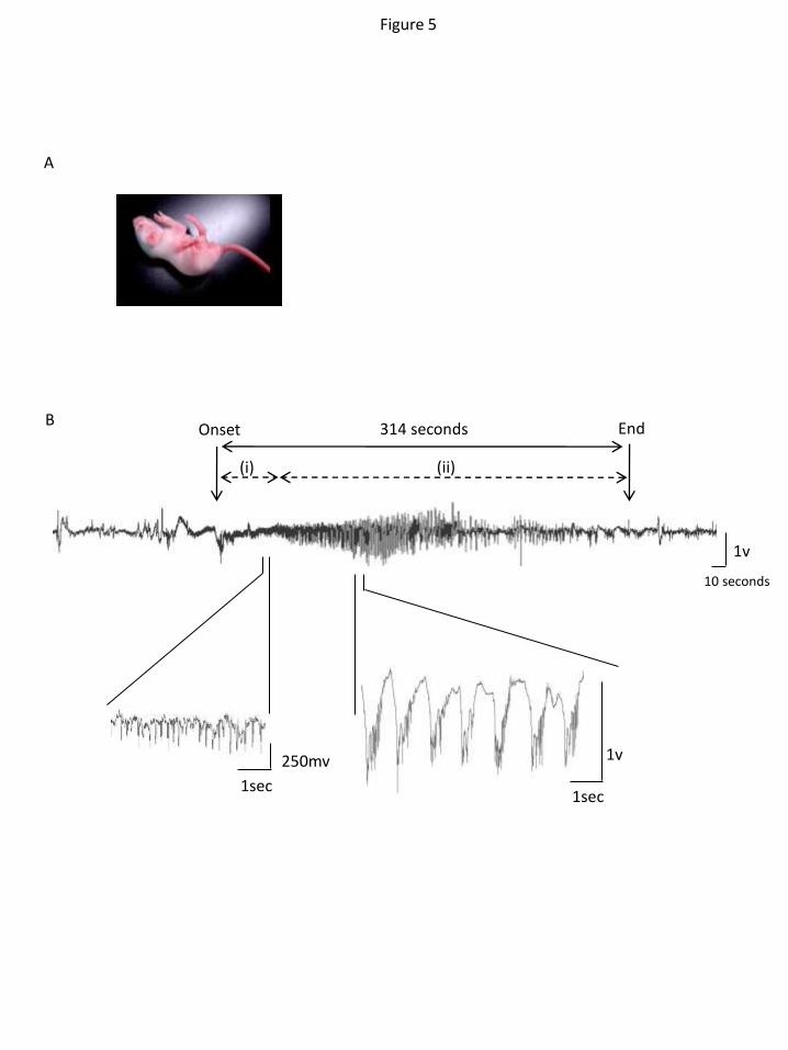
Figure 5
A
B Onset End 314 seconds
(i) (ii)
1v
10 seconds
1sec
250mv
1sec
1v

L385R/+ +/+ L385R/L385R
B
Figure 6
A
C
0
5
10
15
20
25
30
35
8 9 10 11 12 13 14 15 16
Bo
dy
we
igh
t (g
)
Postnatal age (days)
+/+ (n=6)
+/L385R (n=7)
L385R/L385R (n=5)
0
20
40
60
80
100
0 2 4 6 8 10 12 14 16
% a
live
Postnatal age (days)
+/+ & L385R/+ (n=10)
L385R/L385R (n=10)
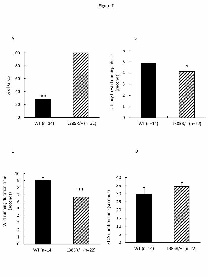
Figure 7
*
A B
C D
0
20
40
60
80
100
WT (n=14) �L385R/+ (n=22)
% o
f G
TCS
0
1
2
3
4
5
6
WT (n=14) L385R/+ (n=22)
Late
ncy
to
wild
ru
nn
ing
ph
ase
(sec
on
ds)
**
0
1
2
3
4
5
6
7
8
9
10
WT (n=14) L385R/+ (n=22)
Wild
ru
nn
ing
du
rati
on
tim
e (s
eco
nd
s)
0
5
10
15
20
25
30
35
40
WT (n=14) L385R/+ (n=22)
GTC
S d
ura
tio
n t
ime
(sec
on
ds)
**

1v
1sec
Cx
Hp
start end
1 2 3 4 5 4
1sec
Cx
Hp
1v 1v
1sec
Cx
Hp
Figure 8

Lgi1 (ab30868)
64 kDa
51 kDa
39 kDa
97 kDa
actin
A
Lgi1 (sc-9583)
*
64 kDa
51 kDa
39 kDa
97 kDa
B
actin
Supplementary figure 1


![· Quick reference guide. The epilepsies : diagnosis and management of the epilepsies in adults in primary and secondary care [homepage on the Internet]. National Institute for C](https://img.pdfslide.us/doc/110x75/5f9d30da2e8f9d72ea258e61/quick-reference-guide-the-epilepsies-diagnosis-and-management-of-the-epilepsies.jpg)