Embed Size (px)
Citation preview
Scientific ArticleISSN 1678-2305 online version
BOLETIM DO INSTITUTO DE PESCA
REZENDE et al. Bol. Inst. Pesca 2018, 44(2): e343. DOI: 10.20950/1678-2305.2018.343 1/11
TITANIUM DIOXIDE NANOPARTICLES ALTER ROUTINE METABOLISM AND CAUSE HISTOPATHOLOGICAL ALTERATIONS IN
Oreochromis niloticus
ABSTRACTTitanium Dioxide Nanoparticles (TiO2NPs) could cause alterations in exposed aquatic species, in terms of oxygen consumption, ammonia excretion and tissues functionality therefore, the aim of the present study is to evaluate the effects of acute exposure to different concentrations of TiO2NPs (0.1, 0.5, 1.0 and 2.5 mgL-1) on routine metabolism (oxygen consumption and ammonia excretion) and histological parameters (branchial and hepatic) in Oreochromis niloticus. After 24 hours, we observed an increase in oxygen consumption of 2.36 and 3.23 times in groups exposed to 1.0 and 2.5 mgL-1 of TiO2NPs respectively, as well as an increase in ammonia excretion of 3.54, 4.0 and 4.82 times higher in groups exposed to 0.5, 1.0 and 2.5 mgL-1 of TiO2NPs respectively, compared to the control group. Histological analysis showed, after 72 hours, moderate to severe alterations in both gills and liver of TiO2 exposed fish at concentrations 1.0 and 2.5 mgL-1, the severity and occurrence of the alteration observed was grade 3 (severe and extensive pathological alterations). We concluded that waterborne exposure of Nile tilapia to TiO2NPs caused alteration in routine metabolism and histological parameters in a dose-dependent manner.Key words: Nile tilapia; nanoparticles; oxygen consumption; ammonia excretion; gills; liver.
EFEITO DE NANOPARTICULAS DE DIÓXIDO DE TITÂNIO SOBRE Oreochromis niloticus: METABOLISMO DE ROTINA E
ALTERAÇÃO HISTOLÓGICA
RESUMONanoparticulas de Dióxido de Titânio (TiO2NPs) podem causar alterações nas espécies aquáticas expostas, em termos de consumo de oxigênio, excreção de amônia e funcionalidade de tecidos, portanto, o objetivo do presente estudo é avaliar os efeitos da exposição aguda a diferentes concentrações de TiO2NPs (0,1, 0,5, 1,0 e 2,5 mgL-1) sobre o metabolismo de rotina (consumo de oxigênio e excreção de amônia) e parâmetros histológicos (branquiais e hepáticos) em Oreochromis niloticus. Após 24 horas, observamos um aumento no consumo de oxigênio de 2,36 e 3,23 vezes em grupos expostos a 1,0 e 2,5 mgL-1 de TiO2NPs respectivamente, bem como um aumento na excreção de amônia de 3,54, 4,0 e 4,82 vezes maior nos grupos expostos a 0,5, 1,0 e 2,5 mgL-1 de TiO2NPs respectivamente, em comparação com o grupo controle. A análise histológica mostrou, após 72 horas, alterações moderadas a severas tanto nas brânquias quanto no fígado de peixes expostos a TiO2NPs em concentrações de 1,0 e 2,5 mgL-1, a gravidade e a ocorrência da alteração observada foram de grau 3 (alterações patológicas graves e extensas). Concluímos que a exposição à TiO2NPs da tilápia do Nilo causaram alteração no metabolismo de rotina e nos parâmetros histológicos de uma maneira dose-dependente.Palavras-chave: Tilapia do Nilo; nanopartículas; consumo de oxigênio; excreção de amônia; brânquias; fígado.
INTRODUCTION
Titanium dioxide nanoparticles (TiO2 NPs), due mainly to their ability to confer opacity and whiteness, are widely used in industrial and consumer products, including cosmetics, paints and food additives (XIONG et al., 2013).
Karina Fernandes Oliveira REZENDE1
Elisa BERGAMI2
Kelison Venício Brito ALVES3
Ilaria CORSI2
Edison BARBIERI3
1 Universidade de São Paulo – USP, Departamento de Biologia Celular e do Desenvolvimento, Av. Prof. Lineu Prestes, 1524, Butantã, CEP 05508-900, São Paulo, SP, Brasil.
2 Department of Physical, Earth and Environmental Sciences, University of Siena, Via Mattioli, 4, 53100, Siena, Italy.
3 Instituto de Pesca, Secretaria de Agricultura e Abastecimento – SAASP, Agência Paulista de Tecnologia dos Agronegócios – APTA, Av. Professor Wladimir Besnard, s/n, CP 43, CEP 11990-000, Cananéia, SP, Brasil. E-mail: [email protected] (corresponding author)
Received: December 11, 2017Approved: March 19, 2018
2/11
TITANIUM DIOXIDE NANOPARTICLES…
REZENDE et al. Bol. Inst. Pesca 2018, 44(2): e343. DOI: 10.20950/1678-2305.2018.343
The growing application and consequent increase of its production results in the release of large quantities of TiO2 NPs into urban and industrial sewage (GONDIKAS et al., 2014) is receiving special attention from the ecotoxicological point of view (MIRANDA et al., 2016), since the Predicted Environmental Concentrations (PECs) for these NPs in surface waters are in the order of 0.7-16 μgL-1 (GOTTSCHALK et al., 2013; MENARD et al., 2011).
Studies suggested that TiO2 NPs could pose toxicity to several aquatic organisms including microbes, algae, invertebrates and fish (CHEN et al., 2012). Due to their nanometric size, these NPs are able to be adsorbed and cross biological barriers, besides entering through mechanisms of endocytosis and binding to cellular receptors, which may represent a danger to aquatic organisms (SHI et al., 2013; MILLER et al., 2010). In fish, NPs can trigger biochemical, physiological and morphological responses. It is known that, TiO2 NPs tend to cross the epithelial cells of the gill tissue during the respiratory cycle, thus spreading through the circulatory system, throughout the organism (BARBER et al., 2009; CHEN et al., 2011). Gills are key tissue for evaluating the action of pollutants, since they have vast surface area and intimate contact with the external environment. Considered as first barrier of the natural mechanisms of resistance to pollution, these organs have cells that play an important role in gas exchange, ion regulation, acid/ base balance and nitrogen excretion (SHEPHARD, 1994; MACHADO and FANTA, 2003; CAMPOS-GARCIA et al., 2016).
Toxicological studies have shown gill histological alteration in fish exposed to different NPs (XIONG et al., 2011). CAMPOS-GARCIA et al. (2016), evaluating gill histological alteration in Nile tilapia (Oreochromis niloticus) exposed to multiwalled carbon nanotubes, observed hypertrophy and hyperplasia of the epithelial cells, dilation of capillaries and aneurism of the lamellae.
JAYASEELAN et al. (2014) noticed hyperplasia of the gill epithelium, lamellar fusion of secondary lamellae, dilated marginal channel, epithelial lifting and epithelial rupture in the gill of Oreochromis mossambicus exposed to nickel NPs.
Gill histological alteration after exposure to NPs can compromise vital functions such as, for example, gas exchange. To estimate the energy expended for the maintenance of its vital processes, the amount of oxygen consumed can be evaluated (CAMPOS-GARCIA et al., 2016; BARBIERI et al., 2016; JAYASEELAN et al., 2014).
The respiratory rate of an organism is a useful and sensitive measure of its daily energy expenditure. The metabolism generally includes three distinct levels: (1) standard metabolism, which is the energy corresponding to the minimum requirements for the fish to remain alive, associated with a state of repose and not fed; (2) routine metabolism, which is the energy fraction used by fish not fed with spontaneous swimming movement or routine activity; (3) active metabolism, which represents the metabolic rate at the maximum level of activity (FRY, 1971).
In the context of environmental studies and biological monitoring, routine metabolism is an efficient tool to measure the toxicity of a chemical substance in an organism (DOI et al., 2012). The evaluation of the metabolism has already been
used, for example, to study toxic effects caused by petroleum (KLOTH and WOHLSCHIAG, 1972), aromatic compounds (LEMAIRE et al., 1996), trace metals (SANTOS et al., 2014) and heavy metal (BARBIERI et al., 2017a).
In addition, xenobiotics, such as TiO2 NPs, after reaching the circulatory system of fish, can lead to a biochemical dysfunction and, consequently, to a physiological dysfunction. These dysfunctions may result in biochemical adaptations that, depending on the time of exposure to NPs, may lead to histological alterations in organs such as the liver (SCHLENK et al., 2008).
The liver is considered a target organ for pollutants because it is responsible for the accumulation, biotransformation and excretion of the xenobiotics that are transported by the blood (REZENDE et al., 2016). In the hepatic tissue, NPs may cause imbalance, with consequent histological alteration. JAYASEELAN et al. (2014) observed irregular shaped nuclei, nuclear hypertrophy, cytoplasmic vacuolation, nuclear degeneration, pyknotic nucleus and necrosis in O. mossambicus exposed to nickel NPs.
SUGANTHI et al. (2015) observed congested portal vein, necrotic hepatocytes and vacuole formation as well as blood cells degradation in Oreochromis mossambicus exposed to ZnO NPs.
As a test organism in the present study, was used Oreochromis niloticus, which according to TAWEEL et al. (2013) are fish ecologically significant, widely edible and of economic importance for aquaculture. This species is also widely studied in ecotoxicity tests due to their high tolerance to stress and disease (KAYA et al., 2016).
However, knowing the importance of clarifying the effect of TiO2 NPs on routine metabolism and histology of gills and liver in Oreochromis niloticus it was hypothesized that exposure to TiO2 NPs would cause dose-dependent toxicity to animals, revealed by increased routine metabolism resulting in increased oxygen consumption and ammonia excretion, as well as causing skeletal damage in gills and liver resulting from overload in these organs targets.
Therefore, the aim of this study was to evaluate the effects of acute exposure to TiO2 NPs on the routine metabolism (oxygen consumption and ammonia excretion) and gills and liver histological parameters as proxy of their functionality of Nile tilapia juveniles.
METHODS
Ethical approval: All applicable international, national, and/or institutional guidelines for the care and use of animals were followed.Preparation and characterization of TiO2 NPs
TiO2 NPs namely Aeroxide© (declared purity of 99.9%) was kindly supplied by Eigenmann & Veronelli (Milan, Italy). Stock suspensions (10 mg mL-1) were prepared in milliQ water (mQw) and tip sonicated at 100 W for 45 min in ice. Final working suspensions were prepared in 0.22 μm filtered fresh water (FW) (pH 8.09).
3/11
TITANIUM DIOXIDE NANOPARTICLES…
REZENDE et al. Bol. Inst. Pesca 2018, 44(2): e343. DOI: 10.20950/1678-2305.2018.343
Primary particle diameter and shape were determined through transmission electron microscopy (TEM), while the average hydrodynamic size and z-potential of the particles in FW suspensions were determined by dynamic light scattering (DLS) analysis, performed on a Malvern Zetasizer 3000 HSa (Malvern Instruments, UK).
Dynamic sedimentation of TiO2 NPs (10 mg L-1) was monitored using a Perkin Elmer Lambda 650 UV-Vis spectrophotometer. Changes in absorbance at 269 nm were measured every minute over a period of 360 min and further at 24, 48 and 72 h.
Animals and treatmentsNile tilapia (Oreochromis niloticus) juveniles, with an average
wet weight of 4.78 g (± 0.84) and average total length of 6.63 cm (±0.5), were obtained from the Instituto de Pesca in Cananeia on the Southern coast of the State of Sao Paulo (Brazil). The animals were acclimated for 7 days in 500 L tanks with constant aeration, daily water exchange and food supplied ad libitum once a day.
For the experiments, TiO2 NP suspensions in FW were prepared as described in section“Preparation and characterization of TiO2 NPs”and immediately added to the tanks in order to reach the tested concentrations, which were based on NIGRO et al. (2015).
Nile tilapias (n5) were exposed for 72 hours at different nominal concentrations of TiO2 NPs (n5): 0.1 mg L-1; 0.5 mg L-1; 1.0 mg L-1 and 2.5 mg L-1. FW was used for exposing controls.
All suspensions were changed daily (24h) and fish were not fed during the experiment. No mortality was observed during the test.
Specific oxygen consumption and specific ammonia excretion
After 24 h of exposure, the animals were acclimated individually in cylindrical acrylic respirometers for 1 h with continuous circulation of water to attenuate the stress caused by handling (DAMATO and BARBIERI, 2012).
To determine the oxygen consumption and ammonia excretion in a known volume of water, the flow of water was interrupted and the respirometers were closed for 1 h.
Subsequently, considering the respirometer volume and weight of the animal, the specific oxygen consumption and ammonia excretion was calculated according to the following formulas:
Specific Oxygen Consumption (mL [gh]-1) = [O2]initial - [O2]final, according to the method by WINKLER (1888).
Specific Ammonia Excretion (mg [gh]-1) = [NH3]initial - [NH3]final, according to the Nessler method (USPHA, 1980).
Histological analysis of gills and liverAfter 72 h, O. niloticus were immediately weighed, measured
and the spinal cord was transversely sectioned. The gill and liver were removed and fixed in Methacarn (methanol: chloroform: acetic acid, 6:3:1) for 12 h.
Posteriorly, gills and liver were dehydrated and included in Historesin (Leica Microsystems Nussloch, Heidelberg, Germany),
and 2 µm thick slices were obtained using the microtome (Spencer, Buffalo, NY, USA) for the preparation of slides. The slides were stained with toluidine blue/fuchsin and Periodic Acid-Schiff (P.A.S.), and images were captured using an ICS-Standard 25 microscope (Zeiss) with a camera AxioCam HRC (Zeiss).
According to the protocol of CAMPOS-GARCIA et al. (2016), five fields of 5 random sections were scanned from each animal. After the digitalization of the slides, the images were submitted to a specific Image J program (version 1.48) for the measurements of the area of epithelial, mucous and chloride cells, in addition to the width of capillaries in the gills, and area of hepatocytes in the liver.
The gills and liver were assessed in accordance with the histological alteration index (HAI) (POLEKSIC and MITROVIC-TUTUNDZIC, 1994) and average value of alteration (AVA) (SCHWAIGER et al., 1997), to analyze the degree of severity and the scale of occurrence of the lesions, respectively.
Statistical analysisAfter verification of the normal distributions (Shapiro-Wilk test)
and homogeneity of variance (Levene test), the data obtained were evaluated as a function of the variations of the means and standard deviations using ANOVA followed by Tukey HSD post-hoc test. Differences were considered significant when p < 0.05.
RESULTS
Transmission electron microscopy confirmed the typical spheroid irregular shape of TiO2 NPs (Aeroxide© P25) and primary size of a mean particle diameter of 24 ± 7 nm.
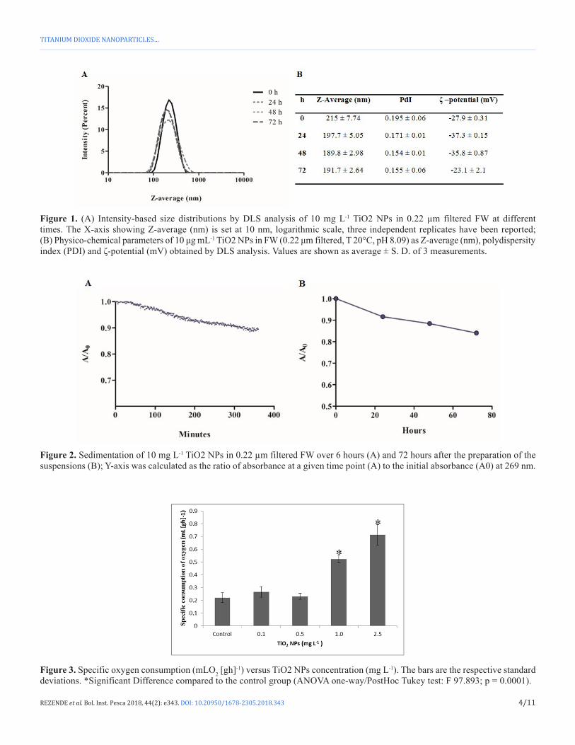
Formation of nano-scale aggregates of 10 mg L-1 TiO2 NPs suspended in FW within the first minutes after sonication was confirmed by DLS analysis, with Z-average values of 215 nm and PDI of 0.195 (Figure 1B). ζ-potential values (-27.9 ± 0.31 at 0 h) confirmed the negative surface charges of the particle.
The aggregation of TiO2 NPs in the medium appeared to be stable over time, as shown by the size distributions in Figure 1A, with values of Z-average ranging from 215 (± 7.74) to 191.7 (± 6.64) nm and PDI ranging from 0.195 (± 0.06) to 0.155 (± 0.06), respectively at 0 and 72 h after the preparation of the suspension (Figure 1B).
TiO2 NPs aggregates in FW showed only little sedimentation over 6 h (Figure 2A) and up to 72 h (Figure 2B).
The specific consumption of oxygen measured after 24 h significantly increased in a dose-dependent manner at the highest concentrations of TiO2 NPs. An increase of 2.36 and 3.23 times was observed in groups exposed to 1.0 and 2.5 mg L-1 of TiO2 NPs respectively, compared to the control group (Figure 3).
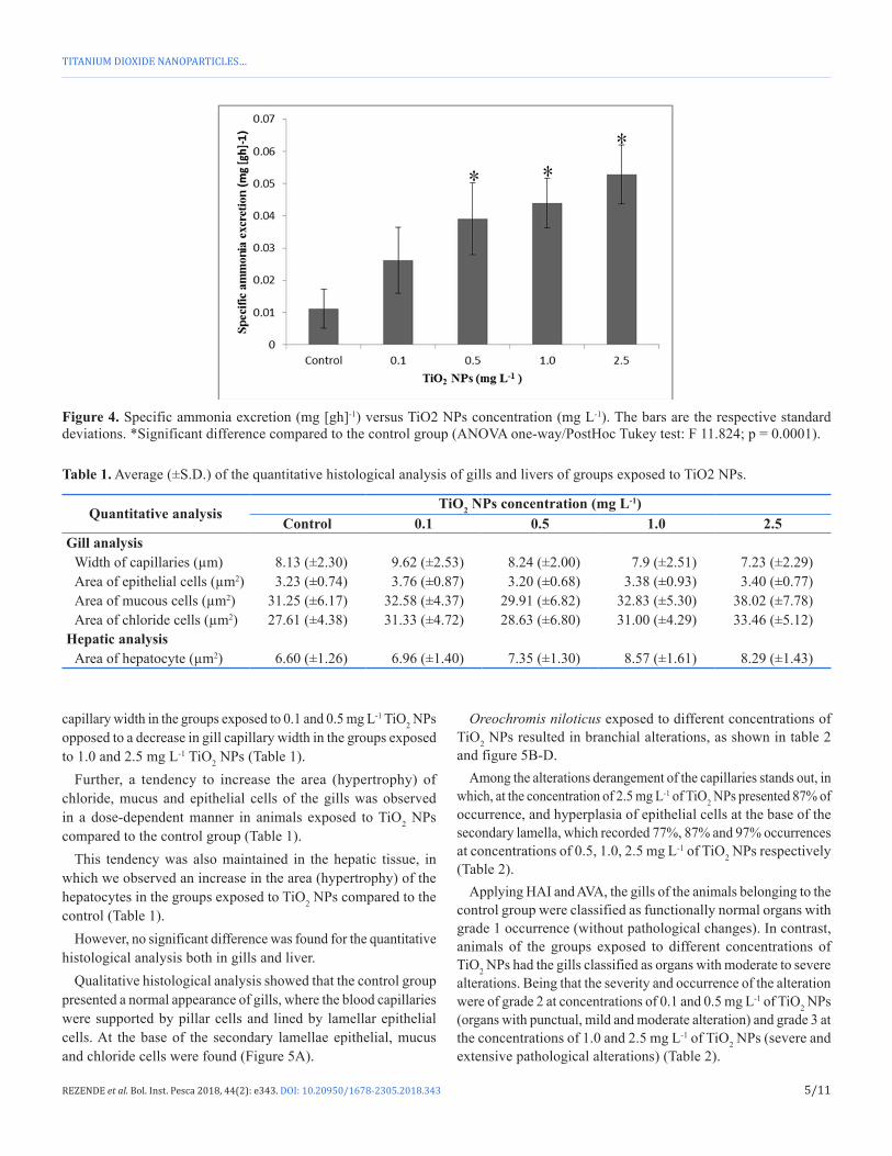
Likely, an increase of 3.54, 4.0 and 4.82 times in the specific ammonia excretion in groups exposed to 0.5, 1.0 and 2.5 mg L-1 of TiO2 NPs respectively, was observed, compared to the control group (Figure 4).
Quantitative histological analysis showed different effects on gills depending to the concentration tested, with an increase in gill
4/11
TITANIUM DIOXIDE NANOPARTICLES…
REZENDE et al. Bol. Inst. Pesca 2018, 44(2): e343. DOI: 10.20950/1678-2305.2018.343
Figure 1. (A) Intensity-based size distributions by DLS analysis of 10 mg L-1 TiO2 NPs in 0.22 µm filtered FW at different times. The X-axis showing Z-average (nm) is set at 10 nm, logarithmic scale, three independent replicates have been reported; (B) Physico-chemical parameters of 10 μg mL-1 TiO2 NPs in FW (0.22 μm filtered, T 20°C, pH 8.09) as Z-average (nm), polydispersity index (PDI) and ζ-potential (mV) obtained by DLS analysis. Values are shown as average ± S. D. of 3 measurements.
Figure 2. Sedimentation of 10 mg L-1 TiO2 NPs in 0.22 µm filtered FW over 6 hours (A) and 72 hours after the preparation of the suspensions (B); Y-axis was calculated as the ratio of absorbance at a given time point (A) to the initial absorbance (A0) at 269 nm.
Figure 3. Specific oxygen consumption (mLO2 [gh]-1) versus TiO2 NPs concentration (mg L-1). The bars are the respective standard deviations. *Significant Difference compared to the control group (ANOVA one-way/PostHoc Tukey test: F 97.893; p = 0.0001).
5/11
TITANIUM DIOXIDE NANOPARTICLES…
REZENDE et al. Bol. Inst. Pesca 2018, 44(2): e343. DOI: 10.20950/1678-2305.2018.343
capillary width in the groups exposed to 0.1 and 0.5 mg L-1 TiO2 NPs opposed to a decrease in gill capillary width in the groups exposed to 1.0 and 2.5 mg L-1 TiO2 NPs (Table 1).
Further, a tendency to increase the area (hypertrophy) of chloride, mucus and epithelial cells of the gills was observed in a dose-dependent manner in animals exposed to TiO2 NPs compared to the control group (Table 1).
This tendency was also maintained in the hepatic tissue, in which we observed an increase in the area (hypertrophy) of the hepatocytes in the groups exposed to TiO2 NPs compared to the control (Table 1).
However, no significant difference was found for the quantitative histological analysis both in gills and liver.
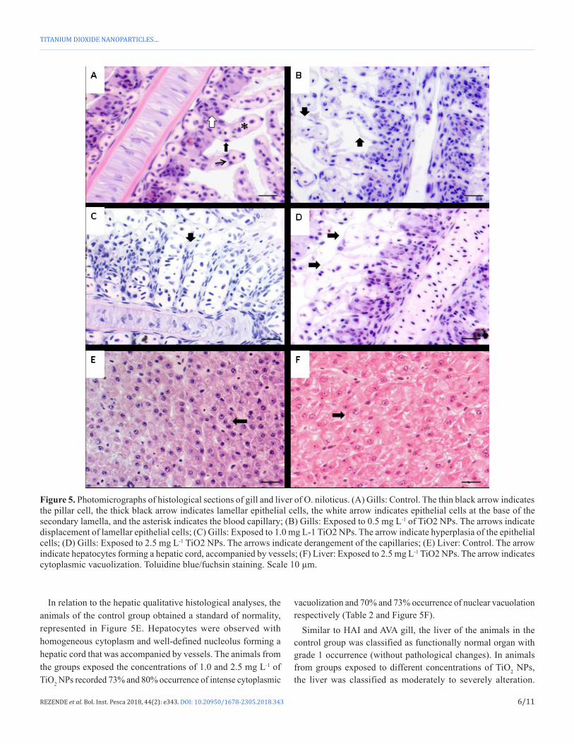
Qualitative histological analysis showed that the control group presented a normal appearance of gills, where the blood capillaries were supported by pillar cells and lined by lamellar epithelial cells. At the base of the secondary lamellae epithelial, mucus and chloride cells were found (Figure 5A).
Oreochromis niloticus exposed to different concentrations of TiO2 NPs resulted in branchial alterations, as shown in table 2 and figure 5B-D.
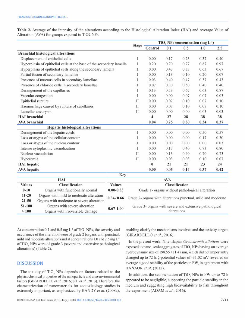
Among the alterations derangement of the capillaries stands out, in which, at the concentration of 2.5 mg L-1 of TiO2 NPs presented 87% of occurrence, and hyperplasia of epithelial cells at the base of the secondary lamella, which recorded 77%, 87% and 97% occurrences at concentrations of 0.5, 1.0, 2.5 mg L-1 of TiO2 NPs respectively (Table 2).
Applying HAI and AVA, the gills of the animals belonging to the control group were classified as functionally normal organs with grade 1 occurrence (without pathological changes). In contrast, animals of the groups exposed to different concentrations of TiO2 NPs had the gills classified as organs with moderate to severe alterations. Being that the severity and occurrence of the alteration were of grade 2 at concentrations of 0.1 and 0.5 mg L-1 of TiO2 NPs (organs with punctual, mild and moderate alteration) and grade 3 at the concentrations of 1.0 and 2.5 mg L-1 of TiO2 NPs (severe and extensive pathological alterations) (Table 2).
Figure 4. Specific ammonia excretion (mg [gh]-1) versus TiO2 NPs concentration (mg L-1). The bars are the respective standard deviations. *Significant difference compared to the control group (ANOVA one-way/PostHoc Tukey test: F 11.824; p = 0.0001).
Table 1. Average (±S.D.) of the quantitative histological analysis of gills and livers of groups exposed to TiO2 NPs.
Quantitative analysisTiO2 NPs concentration (mg L-1)
Control 0.1 0.5 1.0 2.5Gill analysis
Width of capillaries (µm) 8.13 (±2.30) 9.62 (±2.53) 8.24 (±2.00) 7.9 (±2.51) 7.23 (±2.29)Area of epithelial cells (µm2) 3.23 (±0.74) 3.76 (±0.87) 3.20 (±0.68) 3.38 (±0.93) 3.40 (±0.77)Area of mucous cells (µm2) 31.25 (±6.17) 32.58 (±4.37) 29.91 (±6.82) 32.83 (±5.30) 38.02 (±7.78)Area of chloride cells (µm2) 27.61 (±4.38) 31.33 (±4.72) 28.63 (±6.80) 31.00 (±4.29) 33.46 (±5.12)
Hepatic analysisArea of hepatocyte (µm2) 6.60 (±1.26) 6.96 (±1.40) 7.35 (±1.30) 8.57 (±1.61) 8.29 (±1.43)
6/11
TITANIUM DIOXIDE NANOPARTICLES…
REZENDE et al. Bol. Inst. Pesca 2018, 44(2): e343. DOI: 10.20950/1678-2305.2018.343
Figure 5. Photomicrographs of histological sections of gill and liver of O. niloticus. (A) Gills: Control. The thin black arrow indicates the pillar cell, the thick black arrow indicates lamellar epithelial cells, the white arrow indicates epithelial cells at the base of the secondary lamella, and the asterisk indicates the blood capillary; (B) Gills: Exposed to 0.5 mg L-1 of TiO2 NPs. The arrows indicate displacement of lamellar epithelial cells; (C) Gills: Exposed to 1.0 mg L-1 TiO2 NPs. The arrow indicate hyperplasia of the epithelial cells; (D) Gills: Exposed to 2.5 mg L-1 TiO2 NPs. The arrows indicate derangement of the capillaries; (E) Liver: Control. The arrow indicate hepatocytes forming a hepatic cord, accompanied by vessels; (F) Liver: Exposed to 2.5 mg L-1 TiO2 NPs. The arrow indicates cytoplasmic vacuolization. Toluidine blue/fuchsin staining. Scale 10 µm.
In relation to the hepatic qualitative histological analyses, the animals of the control group obtained a standard of normality, represented in Figure 5E. Hepatocytes were observed with homogeneous cytoplasm and well-defined nucleolus forming a hepatic cord that was accompanied by vessels. The animals from the groups exposed the concentrations of 1.0 and 2.5 mg L-1 of TiO2 NPs recorded 73% and 80% occurrence of intense cytoplasmic
vacuolization and 70% and 73% occurrence of nuclear vacuolation respectively (Table 2 and Figure 5F).
Similar to HAI and AVA gill, the liver of the animals in the control group was classified as functionally normal organ with grade 1 occurrence (without pathological changes). In animals from groups exposed to different concentrations of TiO2 NPs, the liver was classified as moderately to severely alteration.
7/11
TITANIUM DIOXIDE NANOPARTICLES…
REZENDE et al. Bol. Inst. Pesca 2018, 44(2): e343. DOI: 10.20950/1678-2305.2018.343
At concentration 0.1 and 0.5 mg L-1 of TiO2 NPs, the severity and occurrence of the alteration were of grade 2 (organs with punctual, mild and moderate alteration) and at concentrations 1.0 and 2.5 mg L-1 of TiO2 NPs were of grade 3 (severe and extensive pathological alterations) (Table 2).
DISCUSSION
The toxicity of TiO2 NPs depends on factors related to the physicochemical properties of the nanoparticle and also environmental factors (GIRARDELLO et al., 2016; SHI et al., 2013). Therefore, the characterization of nanomaterials for ecotoxicology studies is extremely important, as emphasized by HANDY et al. (2008a),
enabling clarify the mechanisms involved and the toxicity targets (GIRARDELLO et al., 2016).
In the present work, Nile tilapias Oreochromis niloticus were exposed to nano-scale aggregates of TiO2 NPs having an average hydrodynamic size of 198.55 ±11.47 nm, which did not importantly changed up to 72 h. ζ-potential values of -31.02 mV revealed on average a good stability of the particles in FW, in agreement with HANAOR et al. (2012).
In addition, the sedimentation of TiO2 NPs in FW up to 72 h appeared to be negligible, supporting the particle stability in the medium and suggesting high bioavailability to fish throughout the experiment (ADAM et al., 2016).
Table 2. Average of the intensity of the alterations according to the Histological Alteration Index (HAI) and Average Value of Alteration (AVA) for groups exposed to TiO2 NPs.
StageTiO2 NPs concentration (mg L-1)
Control 0.1 0.5 1.0 2.5Branchial histological alterations
Displacement of epithelial cells I 0.00 0.17 0.23 0.37 0.40Hyperplasia of epithelial cells at the base of the secondary lamella I 0.20 0.70 0.77 0.87 0.97Hyperplasia of epithelial cells along the secondary lamella I 0.00 0.43 0.33 0.63 0.67Partial fusion of secondary lamellae I 0.00 0.13 0.10 0.20 0.07Presence of mucous cells in secondary lamellae I 0.03 0.40 0.47 0.37 0.43Presence of chloride cells in secondary lamellae I 0.07 0.30 0.50 0.40 0.40Derangement of the capillaries I 0.13 0.53 0.67 0.63 0.87Vascular congestion I 0.00 0.00 0.07 0.07 0.03Epithelial rupture II 0.00 0.07 0.10 0.07 0.10Haemorrhage caused by rupture of capillaries II 0.00 0.07 0.10 0.07 0.10Lamellar aneurysm II 0.00 0.00 0.00 0.03 0.03
HAI branchial 4 27 28 38 38AVA branchial 0.04 0.25 0.30 0.34 0.37
Hepatic histological alterationsDerangement of the hepatic cords I 0.00 0.00 0.00 0.50 0.57Loss or atypia of the cellular contour I 0.00 0.00 0.00 0.17 0.30Loss or atypia of the nuclear contour I 0.00 0.00 0.00 0.00 0.03Intense cytoplasmic vacuolization I 0.00 0.17 0.40 0.73 0.80Nuclear vacuolation II 0.00 0.13 0.40 0.70 0.73Hyperemia II 0.00 0.03 0.03 0.10 0.07
HAI hepatic 0 21 21 23 24AVA hepatic 0.00 0.05 0.14 0.37 0.42
KeyHAI AVA
Values Classification Values Classification0-10 Organs with functionally normal 0.00-0.33 Grade 1- organs without pathological alteration11-20 Organs with mild to moderate alteration 0.34- 0.66 Grade 2- organs with alterations punctual, mild and moderate21-50 Organs with moderate to severe alteration
51-100 Organs with severe alteration 0.67-1.00 Grade 3- organs with severe and extensive pathological alterations> 100 Organs with irreversible damage
8/11
TITANIUM DIOXIDE NANOPARTICLES…
REZENDE et al. Bol. Inst. Pesca 2018, 44(2): e343. DOI: 10.20950/1678-2305.2018.343
Oreochromis niloticus exposed to TiO2 NPs presented increased ammonia specific excretion at concentrations of 0.5, 1.0 and 2.5 mg L-1, when compared to the control group.
Ammonia is the main product of fish excretion (WESTERS, 2001) and the increased excretion observed in this article reflects, according to BARBIERI and FERREIRA (2011), an increase in amino acid catabolism and a dysfunction in ammonia excretion control.
However, no drop in the specific ammonia excretion was observed up to the 2.5 mg L-1 concentration of TiO2 NPs over a period of 24 hours, which may be justified by the increase in protein metabolism, thus maintaining the energy balance of the fish, and by the increase in the number of chloride cells in the gills (BARBIERI et al., 2017b).
Chloride cells are responsible for ionic regulation in freshwater fish and are considered primary site of active absorption of sodium, chloride and calcium ions (EVANS et al., 2005). The presence of chloride cells in the secondary lamella was observed in all groups exposed to TiO2 NPs, with an average occurrence of 40%, leading to the highest metabolic expenditure of the organism for the maintenance of osmoregulation. In addition, exposure to TiO2 NPs caused stress in Oreochromis niloticus, as revealed by increased specific oxygen consumption at 2.36 and 3.23 times in animals exposed to 1.0 and 2.5 mg L-1 of TiO2 NPs respectively, compared to control.
Oxygen consumption, according to PATHIRATNE and GEORGE (1998), provides a sub-stress index and is useful for biomonitoring the toxic effects of xenobiotics. These data demonstrate that TiO2 NPs can affect respiratory processes and increase metabolism aim to utilize other energy sources for detoxification reactions and stabilization of metabolic patterns.
Respiratory processes are probably affected by stress, resulting in branchial alterations, which are responsible for gas exchange (CERQUEIRA and FERNANDES, 2002).
The bioavailable fraction of TiO2 NPs in FW, in contact with Oreochromis niloticus, caused gill alteration from moderate to severe, of degree of occurrence 2 (groups exposed to 0.1 and 0.5 mg L-1 of TiO2 NPs) and 3 (groups exposed to 1.0 and 2.5 mg L-1 of TiO2 NPs).
Gill alterations, such as epithelial cell hyperplasia and epithelial dislocation, were found in all groups exposed to TiO2 NPs and can be considered defense mechanisms, since they increase the distance of blood-water diffusion in an attempt to minimize the deleterious effects of the same (CERQUEIRA and FERNANDES, 2002).
Epithelial cell hyperplasia is the result, according to TAKASHIMA and HIBYA (1995), of functional overload of certain tissue structures. Increased hyperplasia resulted, in all groups exposed to TiO2 NPs, in a partial fusion of the secondary lamella (20% of occurrence in the group exposed to 1.0 mg L-1 of TiO2 NPs) which reduces the total respiratory area (REZENDE et al., 2014).
XIONG et al. (2011) affirm that these defense mechanisms protect the gills but, consequently, they hinder the gas exchange, thus inducing vasodilation of the blood vessels. Vasodilation causes rupture of the pillar cells leading to derangement of the capillaries (87% occurring in the group exposed to 2.5 mg L-1 of TiO2 NPs) and vascular congestion. In more severe cases, vasodilation may
result in capillary aneurysm and hemorrhage (10% occurring in the group exposed to 2.5 mg L-1 of TiO2 NPs). Changes in blood vessels alter blood flow, impairing gas exchange and nutrient distribution (GARCIA-SANTOS et al., 2006).
In addition, the presence of mucous cells along the secondary lamellae was observed at an average of 41.75% occurrence in the groups exposed to TiO2 NPs. These cells are responsible for immunological protection because they have many immunoglobulins and enzymes, branchial damages stimulate cell hyperplasia promoting the increase of the layer of mucus, which acts as a filter, isolating, coagulating and precipitating particles (MONTEIRO et al., 2010).
Studies evaluating the exposure of fish with nanoparticles observed similar branchial alteration. MIRANDA et al. (2016) exposing juveniles of Prochilodus lineatus observed 77.7%, 50% e 77.7% occurrences in groups exposed for 5 days at 0.1, 1 and 10 μg L-1 TiO2 NPs. HAO et al. (2009) observed lamellar displacement in the gills of Cyprinus carpio after exposure to 100 and 200 mg L of TiO2 NPs for 20 days. CHEN et al. (2011) observed in Danio rerio epithelial cell hyperplasia in groups exposed of 1.0 to 7.0 mg L-1 of TiO2 NPs.
Due to its key role in the biotransformation of xenobiotics, the liver is considered a target organ to evaluate the toxicity effects of exposure to TiO2 NPs (HANDY et al., 2008b; DINIZ et al., 2013).
HAO et al. (2009) observed in Cyprinus carpio vacuolization after exposure to 100 and 200 mg L of TiO2 NPs for 20 days. Cytoplasmic and nuclear vacuolation were found in Oreochromis niloticus exposed at all concentrations of TiO2 NPs tested in the present study, with an occurrence of 80% for intense cytoplasmic vacuolization and 73% for nuclear vacuolization at the highest concentration (2.5 mg L-1).
Vacuolization may be indicative of degenerative processes resulting from abnormal lipid metabolism, such as peroxidation (TAKASHIMA and HIBYA, 1995). The intense vacuolization observed probably caused a breakdown of hepatic cords, with an occurrence of 50% and 57% at 1.0 and 2.5 mg L-1 respectively.
Changes in hepatic tissue lead to increased blood flow, aiming to facilitate transport of nutrients and improve oxygenation in areas with injuries (REZENDE et al., 2016), causing hyperemia, as observed in 10% of the cases in the concentration 1.0 mg L-1 of TiO2 NPs.
Gill and hepatic morphologies reflect biochemical responses and histopathologies that resulted in mild and moderate damage in these tissues after exposure to TiO2 NPs for 72 hours can lead to respiratory distress and osmotic and ionic imbalance in the gills and impair liver function.
In the present work, with exposure at 0.1, 0.5, 1.0 and 2.5 mg L-1 concentrations of TiO2 NPs for 72 hours, irreversible gill and liver alterations were not observed, such as necrosis and fibrosis. According to POLEKSIC and MITROVIC-TUTUNDZIC (1994), if animals were transferred to water without xenobiotics, these organs could be restored, in contrast, an increase in the exposure period and/ or increased concentration could lead to irreversible changes and possible bankruptcy of organs.
9/11
TITANIUM DIOXIDE NANOPARTICLES…
REZENDE et al. Bol. Inst. Pesca 2018, 44(2): e343. DOI: 10.20950/1678-2305.2018.343
CONCLUSION
Oreochromis niloticus exposed the highest concentrations of TiO2 NPs showed an increase of the routine metabolism, revealed by increase of the specific consumption of oxygen and specific excretion of ammonia after 24 h.
In addition, TiO2 NPs causes histopathological alterations (mild to moderate) in gills and liver in Oreochromis niloticus, after 72 hours of exposure.
With the increasing application and the consequent increase in the production of TiO2 NPs, this work draws attention to the implications of this nanoparticle on the aquatic environment and the consequences on organisms.
ACKNOWLEDGEMENTS
The authors thanks the Professor Dr. José Roberto Machado Cunha da Silva (Laboratório de Histofisiologia Evolutiva- ICB/USP/ Brazil) for provide the equipment to process the histological materials.
REFERENCES
ADAM, V.; LOYAUX-LAWNICZAK, S.; LABILLE, J.; GALINDO, C.; DEL NERO, M.; GANGLOFF, S.; WEBER, T.; QUARANTA, G. 2016 Aggregation behaviour of TiO2 nanoparticles in natural river water. Journal of Nanoparticle Research, 18(1): 13. http://dx.doi.org/10.1007/s11051-015-3319-4.
BARBER, D.S.; DENSLOW, N.D.; GRIFFITT, R.J.; MARTYNIUK, C.J. 2009 Sources, fate and effects of engineered nanomaterials in the aquatic environment. In: SAHU, S.C.; CASCIANO, D.A. Nanotoxicity, from in vitro models to health risks. Chichester: John Wiley & Sons. p. 227-245. http://dx.doi.org/10.1002/9780470747803.ch12.
BARBIERI, E.; CAMPOS-GARCIA, J.; MARTINEZ, D.S.T.; SILVA, J.R.M.C.; ALVES, O.L.; REZENDE, K.F.O. 2016 Histopathological Effects on Gills of Nile Tilapia (Oreochromis niloticus, Linnaeus, 1758) Exposed to Pb and Carbon Nanotubes. Microscopy and Microanalysis, 22(6): 1162-1169. http://dx.doi.org/10.1017/S1431927616012009. PMid:27998365.
BARBIERI, E.; FERREIRA, L.A.A. 2011 Effects of the organophosphate pesticide Folidol 600® on the freshwater fish, Nile Tilapia (Oreochromis niloticus). Pesticide Biochemistry and Physiology, 99(3): 209-214.
BARBIERI, E.; FERREIRA, A.C.; REZENDE, K.F.O. 2017a Cadmium effects on shrimp ammonia exetion (Farfantepenaeus paulensis) at differents temperatures and levels. Pan-American Journal of Aquatic Sciences, 12(3): 176-183.
BARBIERI, E.; RUIZ-HIDALGO, K.; REZENDE, K.F.O.; LEONARDO, A.F.G.; SABINO, F.P. 2017b Efectos del carbofuran en juveniles de Oreochromis niloticus en la toxicidad, metabólica de rutina y los parámetros hematológicos. Boletim do Instituto de Pesca, 43(4): 513-526. http://dx.doi.org/10.20950/1678-2305.2017v43n4p513.
CAMPOS-GARCIA, J.; MARTINEZ, D.S.T.; REZENDE, K.F.O.; SILVA, J.R.M.C.; ALVES, O.L.; BARBIERI, E. 2016 Histopathological alterations in the gills of Nile tilapia exposed to carbofuran and multiwalled carbon nanotubes. Ecotoxicology and Environmental Safety, 133(1): 481-488. http://dx.doi.org/10.1016/j.ecoenv.2016.07.041. PMid:27543744.
CERQUEIRA, C.C.; FERNANDES, M.N. 2002 Gill tissue recovery after copper exposure and blood parameter responses in the tropical fish, Prochilodus scrofa. Ecotoxicology and Environmental Safety, 52(2): 83-91. http://dx.doi.org/10.1006/eesa.2002.2164. PMid:12061823.
CHEN, G.X.; LIU, X.Y.; SU, C.M. 2012 Distinct effects of humic acid on transport and retention of TiO2 rutile nanoparticles in saturated sand columns. Environmental Science & Technology, 46(13): 7142-7150. http://dx.doi.org/10.1021/es204010g. PMid:22681399.
CHEN, J.; DONG, X.; XIN, Y.; ZHAO, M. 2011 Effects of titanium dioxide nano-particles on growth and some histological parameters of zebrafish (Danio rerio) after a long-term exposure. Aquatic Toxicology, 101(3-4): 493-499. http://dx.doi.org/10.1016/j.aquatox.2010.12.004. PMid:21276475.
DAMATO, M.; BARBIERI, E. 2012 Estudo da Toxicidade aguda e alterações metabólicas provocadas pela exposição do Cádmio sobre o peixe Hyphessobrycon callistus utilizado como indicador de saúde ambiental. O Mundo da Saúde, 36(4): 574-581.
DINIZ, M.S.; DE MATOS, A.P.A.; LOURENÇO, J.; CASTRO, L.; PERES, I.; MENDONÇA, E.; PICADO, A. 2013 Liver alterations in two freshwater fish species (Carassius auratus and Danio rerio) following exposure to different TiO2 nanoparticle concentrations. Microscopy and Microanalysis, 19(5): 1131-1140. http://dx.doi.org/10.1017/S1431927613013238. PMid:23931156.
DOI, S.A.; COLLAÇO, F.L.; STURARO, L.G.R.; BARBIERI, E. 2012 Efeito do chumbo em nível de oxigênio e amônia no camarão rosa (Farfantepeneaus paulensis) em relação à salinidade. O Mundo da Saúde, 36(4): 594-601.
EVANS, D.H.; PIERMARINI, P.M.; CHOE, K.P. 2005 The multifunctional fish gill: dominant site of gas exchange, osmoregulation, acid–base regulation, and excretion of nitrogenous waste. Physiological Reviews, 85(1): 97-177. http://dx.doi.org/10.1152/physrev.00050.2003. PMid:15618479.
FRY, F.E.J. 1971 The effect of environmental factors on the physiology of fish. In: HOAR, W.S.; RANDALL, D.J. Fish physiology. New York: Academic Press. p. 1-98. http://dx.doi.org/10.1016/S1546-5098(08)60146-6.
GARCIA-SANTOS, S.; FONTAÍNHAS-FERNANDES, A.; WILSON, J.M. 2006 Cadmium tolerance in the Nile tilapia (Oreochromis niloticus) following acute exposure: assessment of some ionoregulatory parameters. Environmental Toxicology, 21(1): 33-46. http://dx.doi.org/10.1002/tox.20152. PMid:16463259.
GIRARDELLO, F.; CUSTÓDIO LEITE, C.; VIANNA VILLELA, I.; SILVA MACHADO, M.; LUIZ MENDES JUCHEM, A.; ROESCH-ELY, M.; NEVES FERNANDES, A.; SALVADOR, M.; ANTONIO PÊGAS HENRIQUES, J. 2016 Titanium dioxide nanoparticles induce genotoxicity but not mutagenicity in golden mussel Limnoperna fortunei. Aquatic Toxicology, 170(1): 223-228. http://dx.doi.org/10.1016/j.aquatox.2015.11.030. PMid:26675368.
10/11
TITANIUM DIOXIDE NANOPARTICLES…
REZENDE et al. Bol. Inst. Pesca 2018, 44(2): e343. DOI: 10.20950/1678-2305.2018.343
GONDIKAS, A.P.; KAMMER, F.; REED, R.B.; WAGNER, S.; RANVILLE, J.F.; HOFMANN, T. 2014 Release of TiO2 nanoparticles from sunscreens into surface waters: a one-year survey at the old Danube recreational Lake. Environmental Science & Technology, 48(10): 5415-5422. http://dx.doi.org/10.1021/es405596y. PMid:24689731.
GOTTSCHALK, F.; SUN, T.; NOWACK, B. 2013 Environmental concentrations of engineered nanomaterials: review of modeling and analytical studies. Environmental Pollution, 181(1): 287-300. http://dx.doi.org/10.1016/j.envpol.2013.06.003. PMid:23856352.
HANAOR, D.; MICHELAZZI, M.; LEONELLI, C.; SORRELL, C.C. 2012 The effects of carboxylic acids on aqueous dispersion and eletroforetic deposition of ZrO2. Journal of the European Ceramic Society, 32(1): 235-244. http://dx.doi.org/10.1016/j.jeurceramsoc.2011.08.015.
HANDY, R.D.; HENRY, T.B.; SCOWN, T.M.; JOHNSTON, B.D.; TYLER, C.R. 2008a Manufactured nanoparticles: their uptake and effects on fish - a mechanistic analysis. Ecotoxicology, 17(5): 396-409. http://dx.doi.org/10.1007/s10646-008-0205-1. PMid:18408995.
HANDY, R.D.; OWEN, R.; VALSAMI-JONES, E. 2008b The ecotoxicology of nanoparticles and nanomaterials: current status, knowledge gaps, challenges, and future needs. Ecotoxicology, 17(5): 315-325. http://dx.doi.org/10.1007/s10646-008-0206-0. PMid:18408994.
HAO, L.; WANG, Z.; XING, B. 2009 Effect of sub-acute exposure to TiO2 nanoparticles on oxidative stress and histopathological changes in juvenile carp (Cyprinus carpio). Journal of Environmental Sciences, 21(10): 1459-1466. http://dx.doi.org/10.1016/S1001-0742(08)62440-7. PMid:20000003.
JAYASEELAN, C.; ABDUL RAHUMAN, A.; RAMKUMAR, R.; PERUMAL, P.; RAJAKUMAR, G.; VISHNU KIRTHI, A.; SANTHOSHKUMAR, T.; MARIMUTHU, S. 2014 Effect of sub-acute exposure to nickel nanoparticles on oxidative stress and histopathological changes in Mozambique tilapia, Oreochromis mossambicus. Ecotoxicology and Environmental Safety, 107(1): 220-228. http://dx.doi.org/10.1016/j.ecoenv.2014.06.012. PMid:25011118.
KAYA, H.; AYDIN, F.; GÜRKAN, M.; YILMAZ, S.; ATES, M.; DEMIR, V.; ARSLAN, Z. 2016 A comparative toxicity study between small and large size zinc oxide nanoparticles in tilapia (Oreochromis niloticus): Organ pathologies, osmoregulatory responses and immunological parameters. Chemosphere, 144(1): 571-582. http://dx.doi.org/10.1016/j.chemosphere.2015.09.024. PMid:26398925.
KLOTH, T.C.; WOHLSCHIAG, D.E. 1972 Size-related metabolic responses of the pinfish, Lagodon rhomboides, to salinity variations and sublethal petrochemical pollution. Marketing Science, 16(1): 125-137.
LEMAIRE, P.; STURVE, J.; FORLIN, L.; LIVINGSTONE, D.R. 1996 Studies on aromatic hydrocarbon quinone metabolism and DT-diaphorase function in liver of fish species. Marine Environmental Research, 2(1-4): 317-321. http://dx.doi.org/10.1016/0141-1136(95)00042-9.
MACHADO, M.R.; FANTA, E. 2003 Effects of the organophosphorous methyl parathion on the branchial epithelium of a freshwater fish Metynnis roosevelti. Brazilian Archives of Biology and Technology, 46(3): 361-372. http://dx.doi.org/10.1590/S1516-89132003000300008.
MENARD, A.; DROBNE, D.; JEMEC, A. 2011 Exotoxicity of nanosized TiO2- Review of in vivo data. Environmental Pollution, 159(3): 677-684. http://dx.doi.org/10.1016/j.envpol.2010.11.027. PMid:21186069.
MILLER, R.; LENIHAN, H.; MULLER, E.; TSENG, N.; HANNA, S.; KELLER, A. 2010 Impacts of metal oxide nanoparticles on marine phytoplankton. Environmental Science & Technology, 44(19): 7329-7334. http://dx.doi.org/10.1021/es100247x. PMid:20469893.
MIRANDA, R.R.; DAMASO DA SILVEIRA, A.L.R.; JESUS, I.P.; GRÖTZNER, S.R.; VOIGT, C.L.; CAMPOS, S.X.; GARCIA, J.R.E.; RANDI, M.A.F.; RIBEIRO, C.A.O.; FILIPAK NETO, F. 2016 Effects of realistic concentrations of TiO2 and ZnO nanoparticles in Prochilodus lineatus juvenile fish. Environmental Science and Pollution Research International, 23(6): 5179-5188. http://dx.doi.org/10.1007/s11356-015-5732-8. PMid:26555884.
MONTEIRO, K.M.; CARVALHO, M.O.; ZAHA, A.; FERREIRA, H.B. 2010 Proteomic analysis of the Echinococcus granulosus metacestode during infection of its intermediate host. Proteomics, 10(10): 1985-1999. http://dx.doi.org/10.1002/pmic.200900506. PMid:20217864.
NIGRO, M.; BERNARDESCHI, M.; COSTAGLIOLA, D.; DELLA-TORRE, C.; FRENZILLI, G.; GUIDI, P.; LUCCHESI, P.; MOTTOLA, F.; SANTONASTASO, M.; SCARCELLI, V.; MONACI, F.; CORSI, I.; STINGO, V.; ROCCO, L. 2015 n-TiO 2 and CdCl 2 co-exposure to titanium dioxide nanoparticles and cadmium: genomic, DNA and chromosomal damage evaluation in the marine fish European sea bass (Dicentrarchus labrax). Aquatic Toxicology (Amsterdam, Netherlands), 168(1): 72-77. http://dx.doi.org/10.1016/j.aquatox.2015.09.013. PMid:26448269.
PATHIRATNE, A.; GEORGE, S.G. 1998 Toxicity of malathion to Nile tilapia, Oreochromis niloticus and modulation by other environmental contaminants. Aquatic Toxicology (Amsterdam, Netherlands), 43(4): 261-271. http://dx.doi.org/10.1016/S0166-445X(98)00059-9.
POLEKSIC, V.; MITROVIC-TUTUNDZIC, V. 1994 Fish gills as a monitor of sublethal and chronic effects of pollution. In: MULLER, R.; LLOYD, R. Suletha and chronic effects of pollutants on freshwater fish. Rome: FAO. p. 339-352.
REZENDE, K.F.O.; SANTOS, R.M.; BORGES, J.C.S.; SALVO, L.M.; SILVA, J.R.M.C. 2014 Histopathological and genotoxic effects of pollution on Nile Tilapia (Oreochromis niloticus, Linnaeus, 1758) in the Billings Reservoir (Brazil). Toxicology Mechanisms and Methods, 24(6): 404-411. http://dx.doi.org/10.3109/15376516.2014.925020. PMid:24835316.
REZENDE, K.F.O.; SILVA-NETO, G.M.; PINTO, J.M.; SALVO, L.M.; SEVERINO, D.; MORAES, J.C.T.; SILVA, J.R.M.C. 2016 Hepatic parameters of marine fish Rachycentron canadum (Linnaeus, 1766) exposed to sublethal concentrations of water-soluble fraction of petroleum. Journal of Marine Biology and Oceanography, 5(2): 1-6.
SANTOS, D.B.; BARBIERI, E.; BONDIOLI, A.C.; MELO, C.B. 2014 Effects of Lead in white shrimp (Litopenaeus schmitti) metabolism regarding salinity. O Mundo da Saúde, 38(1): 16-23.
SCHLENK, D.; HANDY, R.; STEINERT, S.; DEPLEDGE, M.H.; BENSON, W. 2008 Biomarkers. In: GIULIO, R.T.; HINTON, D. The toxicology of fishes. Boca Raton: CRC Press. p. 683-731. http://dx.doi.org/10.1201/9780203647295.ch16.
SCHWAIGER, J.; WANKE, R.; ADAM, S.; PAWERT, M.; HONNEN, W.; TRIEBSKORN, R. 1997 The use of histopathological indicators to evaluate contaminant-related stress in fish. Journal of Aquatic Ecosystem Stress and Recovery, 6(1): 75-86. http://dx.doi.org/10.1023/A:1008212000208.
11/11
TITANIUM DIOXIDE NANOPARTICLES…
REZENDE et al. Bol. Inst. Pesca 2018, 44(2): e343. DOI: 10.20950/1678-2305.2018.343
SHEPHARD, K.L. 1994 Functions for fish mucus. Reviews in Fish Biology and Fisheries, 4(4): 401-429. http://dx.doi.org/10.1007/BF00042888.
SHI, H.; MAGAYE, R.; CASTRANOVA, V.; ZHAO, J. 2013 Titanium dioxide nanoparticles: a review of current toxicological data. Particle and Fibre Toxicology, 10(1): 15. http://dx.doi.org/10.1186/1743-8977-10-15. PMid:23587290.
SUGANTHI, P.; MURALI, M.; HE, S.M.; BASU, H.; SINGHAL, R.K. 2015 Morphological and liver histological effects of ZnO nanoparticles on mozambique tilapia. Journal of Advanced Applied Scientific Research, 1(1): 68-83.
TAKASHIMA, F.; HIBYA, T. 1995 An atlas of fish histology: normal and pathological features. 1ª ed. Tokyo: Kodansha. 213 p.
TAWEEL, A.; SHUHAIMI-O, M.; AHMAD, A.K. 2013 In vivo acute toxicity tests of some heavy metals to tilapia fish (Oreochromis niloticus). The Journal of Biological Sciences, 13(5): 365-371. http://dx.doi.org/10.3923/jbs.2013.365.371.
USPHA – United States Public Health Association 1980. Standard Methods for the Examination of Water and Wastewater - 4500-NH3. 15th ed. Washington: USPHA.
WESTERS, H. 2001 Fish hatchery management. Bethesda: American Fisheries Society. 733p.
WINKLER, L. 1888 Die Bestimmung des im Wasser gelösten Sauerstoffes. Berichte der Deutschen Chemischen Gesellschaft, 21(1): 2843-2854. http://dx.doi.org/10.1002/cber.188802102122.
XIONG, D.W.; FANG, T.; YU, L.P.; SIMA, X.F.; ZHU, W.T. 2011 Effects of nano-scale TiO2, ZnO and their bulk counterparts on zebrafish: acute toxicity, oxidative stress and oxidative damage. The Science of the Total Environment, 409(8): 1444-1452. http://dx.doi.org/10.1016/j.scitotenv.2011.01.015. PMid:21296382.
XIONG, S.; GEORGE, S.; JI, Z.; LIN, S.; YU, H.; DAMOISEAUX, R.; FRANCE, B.; NG, K.W.; LOO, S.C.J. 2013 Size of TiO2 nanoparticles influences their phototoxicity: an in vitro investigation. Archives of Toxicology, 87(1): 99-109. http://dx.doi.org/10.1007/s00204-012-0912-5. PMid:22885792.