Embed Size (px)
Citation preview

TISSUE: THE LIVING FABRIC

INTRODUCTION TO TISSUE
• Tissues are groups of cells that are similar in structure and function (tissue=woven)
• There are four primary tissues types:– Epithelial: covering– Connective: support– Nervous: control– Muscular: movement
• Histology: study of tissues

EPITHELIAL TISSUE
• Epithelium (plural: epithelia)• Sheet of cells that covers a body surface or
lines a body cavity (epithe=laid on, covering)• Occurs in the body as:• 1. Covering and lining epithelium:
– Forms the outer layer of the skin, dips into and lines the open cavities of the cardiovascular, digestive, and respiratory systems, and covers the walls and organs of the closed ventral body cavity
• 2. Glandular epithelium:– Fashions the glands of the body

EPITHELIAL TISSUE
• In its role as an interface tissue, epithelium accomplishes many functions, including:
• 1. Protection• 2. Absorption• 3. Filtration• 4. Excretion• 5. Secretion• 6. Sensory Reception

Special Characteristics of Epithelium
• Has many characteristics that distinguish them from other tissue types:• 1. Cellularity:
– Composed of closely packed cells with little extracellular material between• 2. Specialized contacts:
– Adjacent epithelial cells are bound together to form continuous sheets by specialized contacts such as desmosomes and tight junctions
• 3. Polarity:– Exhibits polarity by having an apical surface (upper free surface exposed to the body
exterior or the cavity of an internal organ)) and a lower attached basal surface– All epithelia exhibit polarity, meaning that cell regions near the apical surface differ from
those near the basal surface in both structure and function• Example:
– Some apical surfaces have villi while the basal surface acts as a filter determining which molecules will be allowed to enter the epithelium
• 4. Supported by connective tissue:– Supported by the underlying connective tissue (reticular lamina) containing collagen fibers
• 5. Innervated but avascular:– Nourished by substances diffusing from blood vessels in the underlying connective tissue
• 6. Has a high regeneration capacity:– Replace lost cells rapidly by cell division

Classification of Epithelia• Each epithelial tissue is given two
names:– The first name indicates the number
of layers present:• Simple (one):
– Composed of a single cell layer
– Typically found where absorption and filtration occur and a thin epithelial barrier is desirable
• Stratified (more than one):– Consist of two or more cell
layers stacked one on top of the other
– Common in high-abrasion areas where protection is important, such as the skin surface and the lining of the mouth
– The second name describes the shape of the cells

Classification of Epithelia

Classification of Epithelia• All epithelial cells have six
(somewhat irregular) sides• Apical surface view of an epithelial
sheet looks like a honeycomb– This polyhedral shape allows
the cells to be closely packed• Cells vary in height• Three common shapes of
epithelial cells: nucleus will be the same shape
• 1. Squamous cells are: flattened and scalelike (squam=scale)
• 2. Cuboidal cells are: boxlike– Approximately as tall as they are
wide• 3. Columnar cells are: tall and
column shaped

Classification of Epithelia

Classification of Epithelia
• Simple epithelia are easy to classify by cell shape because all cells in the layer usually have the same shape
• Stratified epithelia:– Cell shapes usually differ among the
different cell layers– Named according the shape of the cells in
the apical layer

Simple Epithelia
• Concerned with absorption, secretion, and filtration
• Consist of a single layer and are usually very thin
• Protection is not one of their specialties

Simple Squamous Epithelium

Simple Cuboidal Epithelium

Simple Columnar Epithelium

Pseudostratied Columnar Epithelium

Stratified Epithelia
• Contains two or more cell layers• Main function is protection• Regenerate from below:
– The basal cells divide and push apically to replace the older surface cells
• Consequently more durable than the simple epithelia– Stratified squamous epithelium is composed of
several layers with the cells on the free surface being squamous-shaped and the underlying cells being cuboidal or columnar in shape
– Transitional epithelium forms the lining of the hollow organs of the urinary system that stretch as they fill

Stratified Squamous Eputhelium

Stratified Cuboidal Epithelium
• Rare
• Found mostly in the ducts of some of the larger glands– Sweat glands– Mammary glands

Stratified Columnar Epithelium
• Found in limited distribution with small amounts in the pharynx, male urethra, and lining some glandular ducts
• Only its apical layer of cells is columnar

Transitional Epithelium

Glandular Epithelia
• A gland consists of one or more cells that make and secrete (export) a particular product:– This product, called a secretion, is an aqueous
(water-based) fluid that usually contains proteins• Some release lipid-rich or steroid-rich secretion
• Secretion is an active process:– Glandular cells obtain needed substances from the
blood and transform them chemically into a product that is then discharged from the cell
– Notice: the term secretion can refer to BOTH the gland’s product and the process of making and releasing that product

Glandular Epithelia
• Classified as to:– Where they release their products:
• Endocrine: internally secreting• Exocrine: externally secreting
– Relative cell numbers making up the gland:– Unicellular: one-celled
» Scattered within epithelial sheets– Multicellular: many-celled
» Form by invagination (inward growth) or evagination (outward growth) from an epithelial sheet

Endocrine Glands
• Ductless glands• Produce hormones: regulatory chemicals
that they secrete by exocytosis directly into the extracellular space– From there the hormones enter the blood or lymphatic
fluid and travel to specific target organs– Each hormone prompts its target organ(s) to respond
in some characteristic way
• Most are complex multicellular organs– Some are individual hormone-producing cells in
organs (intestines/brain)

Exocrine Glands
• More numerous than endocrine glands• Secrete their products onto body
surfaces (skin) or into body cavities:– Unicellular glands directly (exocytosis)– Multicellular glands via an epithelial-walled
duct that transports the secretion to the epithelial surface
• Mucous, sweat, oil, salivary glands• Liver, (bile), pancreas (digestive enzymes)

Unicellular Exocrine Glands• Only important example of a
unicellular (one-celled) gland is the goblet cell
• Shaped like a goblet (drinking glass with a stem)
• (d): Sprinkled in the epithelial linings of the intestinal and respiratory tracts amid columnar cells with other functions– In humans produce mucin:
complex glycoprotein that dissolves in water when secreted
• Once dissolved, mucin forms mucus, a slimy coating that both protects and lubricates surfaces

Pseudostratified Columnar EpitheliumUnicellular Exocrine Glands (Goblet Cells)

Multicellular Exocrine Glands
• Two basic parts:– An epithelium-derived duct– Secretory unit consisting of secretory cells (acini)
• In all BUT the simplest glands, supportive connective tissue surrounds the secretory unit and supplies it with blood vessels and nerve fibers, and forms a fibrous capsule that extends into the gland proper and divides the gland into lobes

Multicellular Exocrine GlandsStructural Classification
• On the basis of their duct structures– Simple:
• Unbranched duct

EXOCRINE GLANDS

Multicellular Exocrine GlandsStructural Classification
• Compound:– Branched duct– Compond: mulitple
branched ducts – Further classified by their
secretory units:• Tubular: if the secretory cells
form tubes• Alveolar: if the secretory
cells form small, flask-like sacs (alveolus=small hollow cavity)
• Tubuloalveolar: if they have BOTH types of secretory units (tubes and alveolar)
– NOTE: acinar is used interchangeably with alveolar

EXOCRINE GLANDS

Multicellular Exocrine GlandsFunctional Classification
• Modes of Secretion:– Merocrine Glands:
• Secrete their products by exocytosis as produced
• Secretory cells are not altered in any way
• Examples:– Sweat glands
– Pancreas
– Salivary glands

Merocrine Gland

Multicellular Exocrine GlandsFunctional Classification
• Modes of Secretion:– Holocrine Glands:
• Accumulate their products within them until they rupture
– They are replaced by the division of underlying cells
• Secretion includes the synthesized product plus dead cell fragments (holo=all)
• Examples:– Sebaceous (oil) glands

Holocrine Gland

Multicellular Exocrine GlandsFunctional Classification
• Modes of Secretion:– Apocrine Glands:
• Present in all animals but questionable in humans• Accumulated their products just beneath the free
surface– Eventually, the apex of the cell pinches off (apo=from
off), releasing the secretory granules and a small amount of cytoplasm
– Example: controversy in humans» Some believe mammary glands are apocrine
while others say merocrine
Connective Tissue
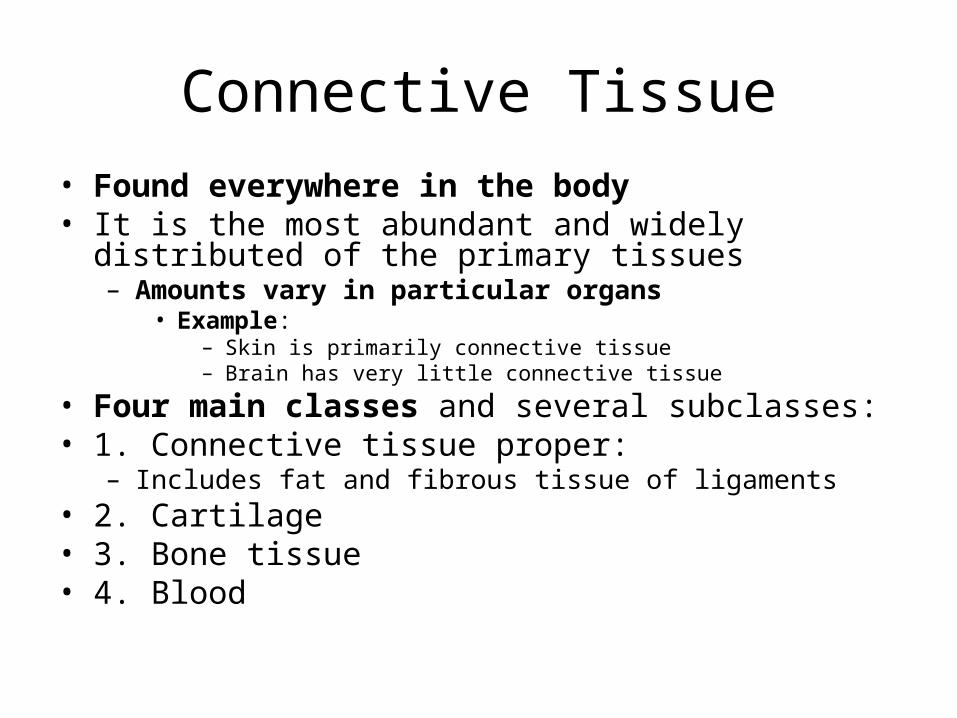
• Found everywhere in the body• It is the most abundant and widely distributed of the
primary tissues– Amounts vary in particular organs
• Example:– Skin is primarily connective tissue– Brain has very little connective tissue
• Four main classes and several subclasses:• 1. Connective tissue proper:
– Includes fat and fibrous tissue of ligaments• 2. Cartilage• 3. Bone tissue• 4. Blood

CONNECTIVE TISSUE
• Does more than just connect body parts– It has many forms and functions
• Major functions include:– Binding and support– Protection– Insulation– Transportation

Common Characteristics of Connective Tissue
• 1. Common origin: All connective tissue arises from an embryonic tissue called mesenchyme
• 2. Degrees of vascularity: Connective tissue ranges from avascular (cartilage) to poorly vascularized (dense connective tissue) to highly vascularized
• 3. Extracellular matrix: Connective tissue is composed mainly of nonliving extracellular matrix that separates the cells of the tissue– Enables connective tissue to withstand physical
trauma

Connective Tissue Origins

EMBRYONIC CONNECTIVE TISSUE

Structural Elements of Connective Tissue
• Three main elements:– Ground substance: extracellular matrix– Fibers: extracellular matrix– Cells
• Properties of the cells and the composition and arrangement of extracellular matrix elements vary tremendously– Resulting in an amazing diversity of connective
tissues– Matrix can be delicate and fragile (soft packing
around an organ) to rope-like (tendons and ligaments)

Structural Elements of Connective Tissue
• Even though there are diverse types they still have a common plan:– Prototype (model) used is areolar
connective tissue• All other subclasses are simply variants of this
common tissue type
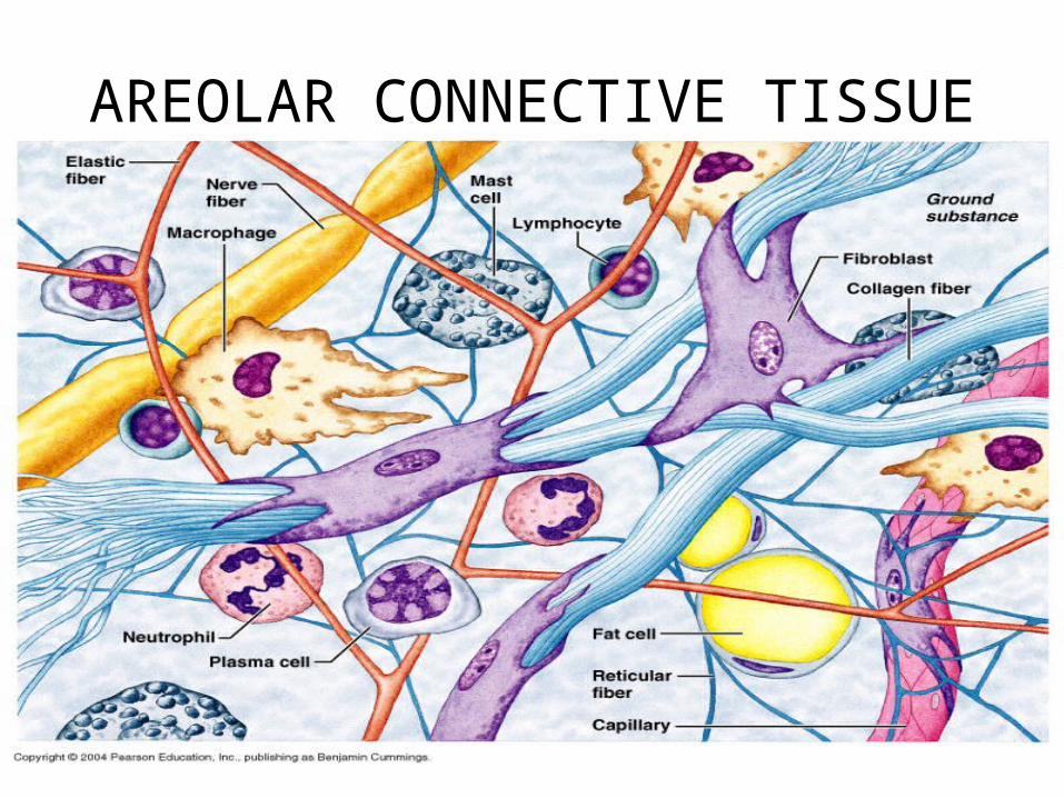
AREOLAR CONNECTIVE TISSUE

Ground Substance• Unstructured material that fills the space
between the cells and contains the fibers• Composed of:
– Interstitial (tissue) fluid– Cell adhesion proteins: serves as
connective tissue glue that allows connective tissue cells to attach themselves to matrix material
• Fibronectin• Laminin
– Proteoglycans:• Consist of a protein core to which
glycosaminoglycans are attached– Strandlike GAGs ( chondroitin
sulfate, keratan sulfate, hyaluronic acid) are large, negatively charged polysaccharides that stick out from the core protein like the fibers of a bottle brush
» Intertwine and trap water, forming a substance that varies from a fluid to a viscous gel

PROTEOGLYCAN

Ground Substance
• Holds large amounts of fluid and functions as a molecular sieve, or medium, through which nutrients and other dissolved substances can diffuse between the blood capillaries and the cells
• Fibers embedded make it less pliable and impede diffusion somewhat

AREOLAR CONNECTIVE TISSUE

Fibers
• Fibers of the connective tissue provide support
• Three types of fibers are found in connective tissue matrix:– Collagen– Elastic– Reticular

AREOLAR CONNECTIVE TISSUE

Collagen Fibers• Strongest and most
abundant• Constructed primarily of the
fibrous protein collagen• Secreted into the extracellular
space, where they assemble spontaneously into cross-linked fibers– Collagen fibers are
extremely strong and provide high tensile strength (ability to resist longitudinal stress) to the matrix
• Stress test show that collagen fibers are stronger than steel fibers of the same size

AREOLAR CONNECTIVE TISSUE

Elastic Fibers• Long, thin fibers that form
branching networks in the extracellular matrix
• Contain a rubberlike protein, elastin, that allows them to stretch and recoil like rubber bands– Connective tissue can
stretch only so much before its thick, ropelike collagen fibers become taut
– When the tension lets up, elastic fibers snap the connective tissue back to its normal length an shape
• Found where elasticity is needed: skin, lungs, blood vessel walls

AREOLAR CONNECTIVE TISSUE

Reticular Fibers• Fine collagenous fibers
(form and chemically different) and are continuous with collagen fibers
• Branch extensively forming delicate networks (reticul=network) that surround small blood vessels and support the soft tissue of organs
• Abundant where connective tissue abuts other tissue types– Example:
• Basement membrane of epithelial tissues
• Around capillaries

AREOLAR CONNECTIVE TISSUE
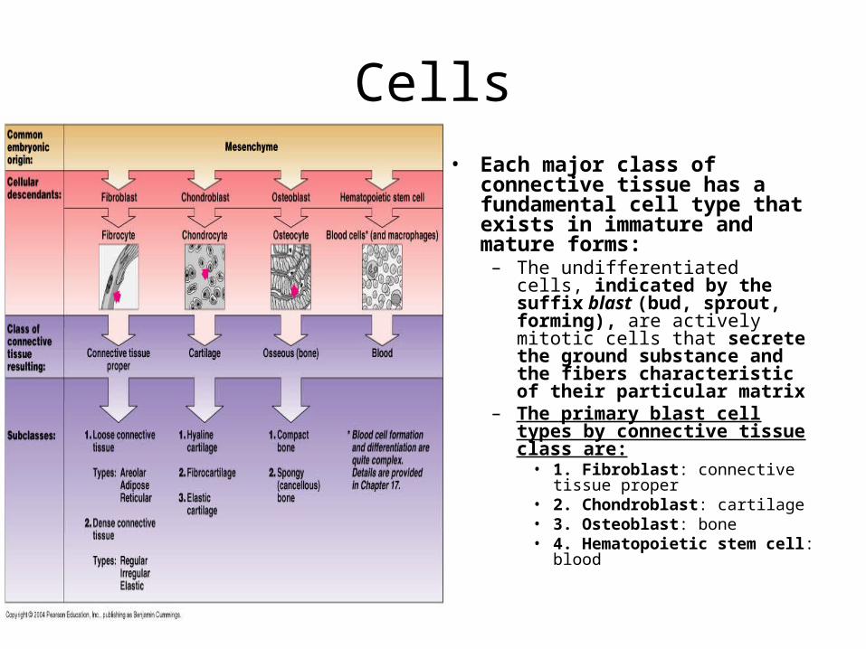
Cells• Each major class of connective
tissue has a fundamental cell type that exists in immature and mature forms:
– The undifferentiated cells, indicated by the suffix blast (bud, sprout, forming), are actively mitotic cells that secrete the ground substance and the fibers characteristic of their particular matrix
– The primary blast cell types by connective tissue class are:
• 1. Fibroblast: connective tissue proper
• 2. Chondroblast: cartilage• 3. Osteoblast: bone• 4. Hematopoietic stem cell:
blood

Connective Tissue Origins

Cells
• Once they synthesize the matrix, the blast cells assume their less active, mature mode, indicated by the suffix cyte
• Mature cells maintain the health of the matrix– If the matrix is injured,
they can easily revert to their more active state to repair and regenerate the matrix

Connective Tissue Origins

Cells
• Additionally, connective tissue is home to an assortment of other cell types:– Fat cells: nutrient-storing cells– Mobile cells that migrate into
the connective tissue matrix from the bloodstream:
• White blood cells: neutrophils, eosinophils, lymphocytes
• Cell types that respond to injury: mast cells, macrophages
• Antibody-producing plasma cells

AREOLAR CONNECTIVE TISSUE

Mast Cells• Mast cells and macrophages are
very important to overall body defense
• Oval mast cells typically cluster along blood vessels
– Act as sensitive sentinels to detect foreign substances (e.g., bacteria, fungi)
– Initiate local inflammatory responses against foreign substances
– Cytoplasm contains conspicuous secretory granules (mast=stuffed full of granules) containing several chemicals that mediate inflammation, especially in severe allergies
• Heparin: anticoagulant chemical that prevents blood clotting when free in the bloodstream
• Histamine: substance that makes capillaries leaky
• Proteases: protein-degrading enzymes

AREOLAR CONNECTIVE TISSUE

Macrophages• Mast cells and macrophages
are very important to overall body defense
• Macro=large; phago=eat• Large, irregularly shaped cells that
avidly phagocytize a broad variety of foreign materials:
– Foreign molecules– Bacteria– Dust particles– Dead tissue cells
• Active in the immune system• May attach to connective tissue
fibers (fixed) or may migrate freely through the matrix

AREOLAR CONNECTIVE TISSUE

Types of Connective Tissue
• Mesenchyme forms during the early weeks of embryonic development from the mesoderm layer and eventually differentiates into other connective tissues

EMBRYONIC CONNECTIVE TISSUE

Connective Tissue Proper
• Two Subclasses:– Loose Connective Tissue:
• Areolar• Adipose• Reticular
– Dense Connective Tissue:• Dense Regular• Dense Irregular• Elastic
– Except for bone, cartilage, and blood, all mature connective tissues belong to this class

Types of Connective Tissue
• Areolar connective tissue serves to bind body parts together while allowing them to move freely over one another, wraps small blood vessels and nerves, surrounds glands, and forms the subcutaneous tissue

AREOLAR CONNECTIVE TISSUE

Types of Connective Tissue
• Adipose (fat) tissue is a richly vascularized tissue that functions in nutrient storage, protection, and insulation

ADIPOSE CONNECTIVE TISSUE

Types of Connective Tissue
• Reticular connective tissue forms the internal framework of the lymph nodes, the spleen, and the bone marrow

RETICULAR CONNECTIVE TISSUE

Types of Connective Tissue
• Dense connective tissue is one of the two subclasses of connective tissue proper;– Dense regular connective tissue contains
closely packed bundles of collagen fibers running in the same direction and makes up tendons and ligaments
– Dense irregular connective tissue contains thick bundles of collagen fibers arranged in an irregular fashion, and is found in the dermis

DENSE REGULAR CONNECTIVE TISSUE
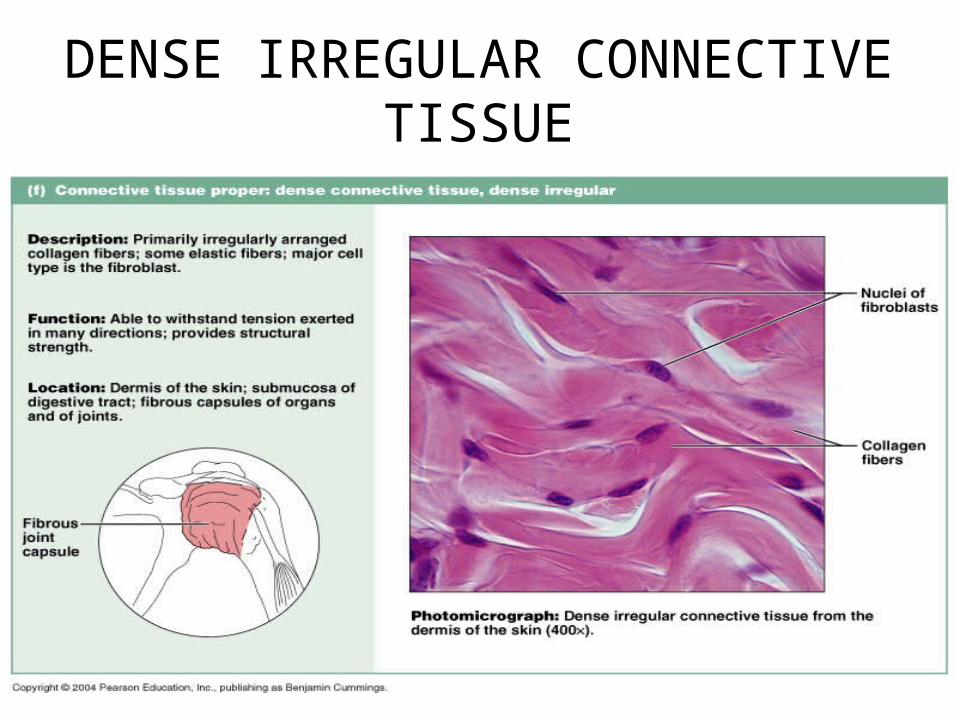
DENSE IRREGULAR CONNECTIVE TISSUE

Cartilage• Stands up to both tension (stretching) and compression• Qualities intermediate between dense connective tissue (very flexible) and
bone– Tough but flexible, providing a resilient rigidity to the structures it supports
• Lacks nerve fibers and is avascular– Receives its nutrients by diffusion from blood vessels located in the connective
tissue membrane (perichondrium) surrounding it• Ground substance contains large amounts of:
– GAGs: glycosaminoglycans (Chrondroitin sulfate, Hyaluronic acid):Intertwine and trap water, forming a substance that varies from a fluid to a viscous gel
• Contains firmly bound collagen fibers and in some cases elastic fibers• Matrix contains an exceptional amount of tissue fluid (up to 80%
water)– Movement of tissue fluid in its matrix enables cartilage to rebound after being
compressed and also helps to nourish the cartilage cells

Cartilage
• Because cartilage is avascular and aging cartilage cells lose their ability to divide, cartilages heal slowly when injured
• During later life, cartilages tend to calcify or even ossify (become bony)– In such cases, the chondrocytes are poorly
nourished and die

Types of Connective Tissue
• Three varieties of cartilage:– Hyaline– Elastic– Fibrocartilage
Types of Connective TissueHyaline Cartilage
• Hyaline cartilage (gristle): is the most abundant cartilage providing firm support with some pliability
• Blue-white color• Hyalin=glass• Articular (ends of long bones)

HYALINE CARTILAGE

Types of Connective TissueElastic Cartilage
• Elastic cartilage is found where strength and exceptional stretchability are needed, such as the external ear– More elastin fibers– Found where strength and exceptional
stretchability are needed

ELASTIC CARTILAGE

Types of Connective TissueFibrocartilage
• Fibrocartilage is found where strong support and the ability to withstand heavy pressure are required, such as the intervertebral disks– Often found where hyaline cartilage meets a true
ligament or a tendon– Perfect intermediate between hyaline cartilage and
dense regular connective tissues– Found where strong support and the ability to
withstand heavy pressure are required

FIBROCARTILAGE

Types of Connective TissueBone
• Bone (osseous tissue) has an exceptional ability to support and protect body structures due to its hardness, which is determined by the additional collagen fibers and calcium salts found in the extracellular matrix– Provides cavities for fat storage and synthesis of blood
cells– Matrix is similar to that of cartilage but is harder and more
rigid because, in addition to its more abundant collagen fibers, bone has an added matrix element—inorganic calcium salts (bone salts)
• Blood is classified as a connective tissue because it developed from mesenchyme, and consists of blood cells and plasma proteins surrounded by blood plasma
Types of Connective TissueBone
• Osteoblasts: immature bone cells– Produce the organic portion of the matrix; then bone
salts are deposited on and between the fibers
• Osteocytes: mature bone cells– Reside in the lacunae (cavity in bone or cartilage)
within the matrix they have made– Unlike cartilage, the next firmest connective tissue
• Vascularized

BONE

Types of Connective TissueBlood
• Fluid within blood vessels• Most atypical connective tissue:
– Does not connect things or give support
• Classified as connective tissue because it develops from mesenchyme and consists of blood cells, surrounded by a nonliving fluid matrix called blood plasma– Fibers of blood are soluble protein molecules that
become visible only during blood clotting

BLOOD

COVERING and LINING MEMBRANES
• Membranes that incorporate BOTH connective tissue and epithelial tissues
• Three types:– Cutaneous– Mucous– Serous
• Essentially they all are continuous multicellular sheets composed of at least two primary tissue types:– An epithelium bound to an underlying layer of connective
tissue proper:• Hence a simple organ
– Synovial membranes: line joint cavities and consist of connective tissue ONLY

COVERING and LINING MEMBRANESCutaneous Membrane (a)
• Cutis=skin• Cutaneous membrane, or skin, is an organ system consisting of
a keratinized squamous epithelium (epidermis) firmly attached to a thick layer of dense irregular connective tissue (dermis)
• Unlike other epithelial membranes, the cutaneous membrane is exposed to the air and is a dry membrane

MEMBRANES

COVERING and LINING MEMBRANESMucous Membranes (b)
• Mucosae • Mucous membranes line body cavities that open to the exterior, such as
those of hollow organs of the digestive, respiratory, and urogenital tracts• They are ALL wet (moist) membranes bathed by secretions or, in the
case of urinary mucosa, urine• Name mucosa refers to the location of the membrane, NOT its cell
composition, which varies

MEMBRANES
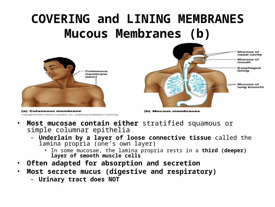
COVERING and LINING MEMBRANESMucous Membranes (b)
• Most mucosae contain either stratified squamous or simple columnar epithelia
– Underlain by a layer of loose connective tissue called the lamina propria (one’s own layer)
• In some mucosae, the lamina propria rests in a third (deeper) layer of smooth muscle cells
• Often adapted for absorption and secretion• Most secrete mucus (digestive and respiratory)
– Urinary tract does NOT

COVERING and LINING MEMBRANESSerous Membranes (c)
• Serosae • Moist membranes found in closed ventral body cavities
– Serosa: moist membrane found in closed ventral body cavities– Visceral serosa: the part of the double-layered membrane that lines the outer surfaces
of organs within the ventral body cavity– Parietal serosa: the part of the double-layered membrane that lines the walls of the
ventral body cavity• Serous membranes consist of simple squamous epithelium (mesothelium) resting on
a thin layer of loose connective (areolar) tissue

MEMBRANES

COVERING and LINING MEMBRANESSerous Membranes (c)
• Mesothelial cells enrich the fluid that filters from the capillaries in the associated connective tissue with hyaluronic acid (GAGs)
– Result is the thin, clear serous fluid that lubricates the facing surfaces of the parietal (lines the walls of the ventral body cavity) and visceral layers (lines outer surface of the organ), so that they slide across each other easily
• Named according to their site and specific organ associations:• Example:
– Pleura: serosa lining the thoracic wall and covering the lungs– Pericardium: serosa lining the heart– Peritoneums: serosa ling the abdominopelvic cavity and viscera

MEMBRANES

NERVOUS TISSUE
• Nervous tissue is the main component of the nervous system (brain, spinal cord, and nerves), which regulates and controls body functions
• Nervous tissue is composed of two types of cells:– Neurons are specialized cells that generate
and conduct electrical impulses– Supporting cells are nonconductive cells that
support, insulate, and protect the neurons

NERVE TISSUE

MUSCLE TISSUE• Muscle tissues are highly cellular, well-vascularized tissues
responsible for movement• Muscle cells (muscle fibers) possess myofilaments:
– Elaborate versions of the actin and myosin filaments that bring about movement or contraction in all muscle cell types
• There are three types of muscular tissue:– Skeletal muscle is packaged by connective tissue sheets into organs
called skeletal muscles that are attached to the skeleton and produces voluntary body movement
– Cardiac muscle is responsible for the involuntary movement of the heart
• Found ONLY in the walls of the heart– Smooth muscle is found in the walls of the hollow organs (digestive
and urinary tract organs, uterus, and blood vessels):• No striations• Acts to squeeze substances through these organs by alternately
contracting and relaxing• Involuntary

SKELETAL MUSCLE

CARDIAC MUSCLE

SMOOTH MUSCLE

TISSUE REPAIR
• When tissue injury occurs, the responses usually take place in connective tissue
• Tissue repair occurs in two ways:– Regeneration:
• Replacement of destroyed tissue with the same kind of tissue
– Fibrosis:• Involves proliferation of fibrous connective
tissue called scar tissue

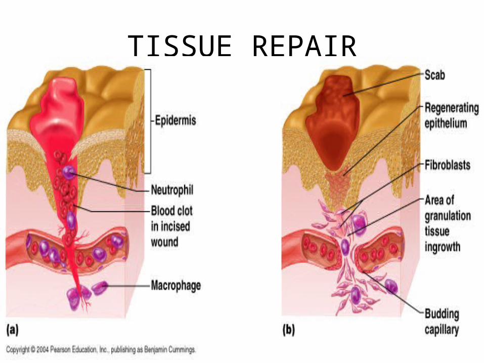
Steps of Tissue Repair• Three steps are involved in the tissue repair process:• 1. Inflammation: (a)
– Tissue trauma causes injured tissue cells, macrophages, mast cells, and others to release inflammatory chemicals, which cause the capillaries to dilate and become very permeable
• Allows white blood cells (neutrophils, monocytes,) and plasma fluid rich in clotting proteins, antibodies, and other substances to seep into the injured area
• The leaked clotting proteins construct a clot:– Stops the loss of blood– Holds the edges of the wound together isolating the injured
area preventing bacteria, toxins, or other harmful substances from spreading to surrounding tissues
– Part exposed to the air quickly dries and hardens forming a scab
– Leaves excess fluid, bits of destroyed cells, and other debris in the area, which are eventually removed via lymphatic vessels or phagocytized by macrophages

TISSUE REPAIR

Steps of Tissue Repair
• 2.Organization restores the blood supply (b):– Blood clot is replaced by granulation tissue:
• A delicate pink tissue composed of several elements:– Contains capillaries that grow in from nearby areas and lay down a
new capillary bed» Granulation tissue is actually named for these capillaries,
which protrude nublike from its surface, giving it a granular appearance
– Proliferating fibroblasts produce growth factors as well as new collagen fibers to bridge the gap
» Some fibroblasts have contractile properties that pull the margins of the wound together
– Macrophages digest the original blood clot– Collagen fiber deposit continues– Granulation tissue, destined to become scar tissue (a
permanent fibrous patch), is highly resistant to infection because it produces bacteria-inhibiting substances

TISSUE REPAIR

Steps of Tissue Repair• 3.Regeneration and fibrosis effect permanent repair (c):
– Surface epithelium begins to regenerate (b)• Growing under the scab, which soon detaches• As the fibrous tissue beneath matures and contracts, the regenerating
epithelium thickens until it finally resembles that of the adjacent skin (c)– End result is a fully regenerated epithelium, and an underlying area of
scar tissue• May be invisible, or visible as a thin white line, depending on the severity of
the wound• The repair process described (1,2,3) follows healing of a wound
(cut, scrape, puncture) that breaches an epithelial barrier• In pure infections (pimple or sore throat), healing is solely by
regeneration– Usually no clot or scarring– Only severe (destructive) infections lead to scarring

TISSUE REPAIR
TISSUE REPAIR

TISSUE REPAIR
Regenerative Capacity of Different Tissues
• The generative capacity of tissues varies widely among the tissue types– Epithelial, bone, areolar connective tissue, dense irregular
tissue, and blood clotting tissue regenerate extremely well– Smooth muscle and dense regular connective tissue have
moderate capacity for regeneration– Skeletal muscle and cartilage have a weak regeneration
capacity– Cardiac muscle and nervous tissue of the brain and spinal cord
have virtually no functional regenerative capacity• Hence, routinely replaced by scar tissue• Scar tissue is strong (mostly collagen fibers) , but it lacks the
flexibility and elasticity of most normal tissue– Cannot perform the normal functions of the tissue it has replaced
DEVELOPMENTAL ASPECTS OF
TISSUES
• Embryonic and Fetal Development of Tissues– Primary germ layer formation is one of the first events
of embryonic development• Ectoderm is the most superficial of the layers• Mesoderm is the middle layer• Endoderm is the deepest layer
– The primary germ layers specialize to form the four primary tissues
• With increasing age, epithelia become thin, the amount of collagen fibers in the body decreases, and bone, muscle, and nervous tissue atrophy

EMBRYO TISSUE

CANCER