Embed Size (px)
Citation preview
Plant Physiol. (1 996) 11 2: 525-535
Tissue-Specific Activity of Two Manganese Superoxide Dismutase Promoters in Transgenic Tobacco’
Wim Van Camp, Didier Hérouart2, Hilde Willekens, Hideki Takahashi, Kazuki Saito, Marc Van Montagu*, and Dirk lnzé
Laboratorium voor Genetica, Department of Genetics, Flanders lnteruniversity lnstitute for Biotechnology (W.V.C., D.H., H.W., M.V.M., D.I.), and Laboratoire Associé de I’lnstitut National de Ia Recherche
Agronomique (France) (D.I.), Universiteit Gent, K.L. Ledeganckstraat 35, B-9000 Gent, Belgium; and Laboratory of Molecular Biology and Biotechnology, Faculty of Pharmaceutical Sciences, Chiba University, Chiba, japan
(H.T., K.S.)
In eukaryotes, manganese superoxide dismutase is a nuclear- encoded protein that scavenges superoxide radicals in the mito- chondrial matrix. We have isolated two manganese superoxide dismutase genes from Nicofiana plumbaginifolia 1. and fused the 5’ upstream regulatory region of these genes to the /3-glucuronidase reporter gene. The two gene fusions displayed a differential tissue specificity in transgenic tobacco (Nicofiana fabacum). Promoter activity of the SodA7 gene fusion was found in the pollen, middle layer, and stomium of anthers, but was usually undetectable in vegetative organs of mature plants. The SodAZ gene fusion was expressed in the leaves, stems, roots, and flowers. SodAZ promoter activity was most prominent in the vascular bundles, stomata, ax- illary buds, pericycle, stomium, and pollen. Histochemical analysis of succinate dehydrogenase activity suggested that the spatial ex- pression of the two gene fusions is generally correlated with mito- chondrial respiratory activity.
Aerobic organisms are constantly exposed to the toxic effects of AOS such as superoxide, H,Oz, and the hydroxyl radical. Cellular protective mechanisms against these AOS consist of AOS-scavenging enzymes and low-molecular- weight antioxidants such as ascorbate, glutathione, and a-tocopherol (Alscher and Hess, 1993; Foyer and Mul- lineaux, 1994). The hydroxyl radical is the most toxic for the cell, but it is too reactive to be controlled enzymically (Halliwell and Gutteridge, 1989). Therefore, the enzymic defense against the hydroxyl radical is directed against its precursors, which are the superoxide radical and H,02 (O-, + H,O, + O, + OH- + OH). Superoxide is con-
‘This work was supported by grants from the Belgian Pro- gramme on Interuniversity Poles of Attraction (Prime Minister‘s Office, Science Policy Programming, no. 38), the Vlaams Actiepro- gramma Biotechnologie (no. 067), the International Atomic Energy Agency (no. 5285), and the International Human Frontier Science Program (IHFSP RG-434/94M). W.V.C. is a Postdoctoral Fellow of the National Fund for Scientific Research and D.I. is a Research Director of the Institut National de la Recherche Agronomique (France).
* Present address: Laboratoire de Biologie Végétale et Microbi- ologie, Université de Nice, F-06108 Nice Cedex 2, France.
* Corresponding author; e-mail mamon8gengenp.rug.ac.be; fax 32-9-2645349.
verted to O, and H,O, by SOD, whereas H,O, is removed by catalases and peroxidases (Foyer and Mullineaux, 1994).
Three classes of SODs are distinguished based on their metal cofactor: MnSOD, FeSOD, and Cu/ZnSOD (Bannis- ter et al., 1987). Each of these classes is found in Nicotiana plumbaginifolia (Van Camp et al., 1990). They are a11 nuclear encoded, but the gene products are present in different subcellular compartments (Bowler et al., 1989a; Van Camp et al., 1990; Tsang et al., 1991). MnSOD is located in the mitochondria, FeSOD in the chloroplasts, and Cu / ZnSOD in both the cytosol and the chloroplasts. The occurrence of SOD in peroxisomes has been reported for a number of plant species, but to date has not been investigated in N . plumbaginifolia (Bowler et al., 1994). The presence of SOD in different subcellular compartments is required to scavenge the superoxide radical efficiently at its site of formation. Three major sites of superoxide production have been iden- tified in plants: mitochondria, chloroplasts, and cytosol; these correspond to the location of SODs in the cell (Bowler et al., 1994). Additional sources of superoxide may reside in the peroxisomes (Sandalio et al., 1988; de1 Rio et al., 1989) and in the plasma membrane (Doke and Ohashi, 1988).
During mitochondrial respiration, part of the O, is re- duced by single-electron transfer (Rich and Bonner, 1978), whereas in the chloroplasts electron leakage to O, occurs primarily at the reducing site of PSI (Asada and Takahashi, 1987). Therefore, expression of organellar SODs in plants may show a high degree of cell specificity, as determined by the metabolic rate of a cell and its dependence on photophosphorylation or respiration for ATP production. The cellular exuression of MnSOD has thus far not been investigated in plants, but a correlation between MnSOD mRNA levels and mitochondrial respiratory activity was suggested by RNA gel blot analysis, which showed high levels of MnSOD mRNA in tissues with elevated respira- tory activity or in response to certain stresses (Bowler et al., 1989a; Tsang et al., 1991; Zhu and Scandalios, 1993).
Abbreviations: AOS, active oxygen species; RT, reverse tran- scription; SDH, succinate dehydrogenase; SOD, superoxide dismutase.
525 www.plantphysiol.orgon February 20, 2018 - Published by Downloaded from
Copyright © 1996 American Society of Plant Biologists. All rights reserved.

526 Van Camp et al. Plant Physiol. Vol. 11 2, 1996
In this study we have analyzed the cell specificity of two MnSOD promoters in tobacco and the relationship between MnSOD promoter activity and mitochondrial respiration. We have isolated both members of the MnSOD gene family from N. plumbaginifolia and determined the transcriptional activity of these genes in different organs and tissues of tobacco by means of reporter gene fusions. This approach has been used previously for other SOD genes, namely a cytosolic Cu / ZnSOD from N. plumbaginifolia (Hérouart et al., 1994) and a chloroplastic Cu/ZnSOD from tomato (Kardish et al., 1994), but to our knowledge it has not yet been applied for a mitochondrial SOD in plants. To inves- tigate a correlation between MnSOD promoter activity and mitochondrial respiration, histochemical localization of SDH activity was performed in parallel with the analysis of the reporter gene fusions.
MATERIALS A N D METHODS
D N A Gel Blot Hybridizations
DNA was isolated from Nicotiana plumbaginifolia L. plants according to the procedure described by Pruitt and Meyerowitz (1986). This DNA was digested with the ap- propriate restriction enzymes in buffers recommended by the manufacturer (Pharmacia). Digested DNA was sepa- rated in 0.8% 40 mM Tris-acetate, 1 mM Tris-acetate/EDTA agarose gels (Sambrook et al., 1989) and transferred to Hybond-N nylon membranes (Amersham) according to the manufacturer’s instructions. Probes were prepared by random-primed labeling (Amersham) from a 0.9-kb HpaI- PstI fragment of pSODl (Bowler et al., 1989a) and a 0.5-kb HpaI-SacII fragment of pGSOD2 (this study). The latter fragment is located upstream from the sequence that en- codes the mature protein. Hybridizations were carried out in 3X SSC (1X SSC = 150 mM NaC1,15 mM sodium citrate, pH 7.0), 0.1% SDS at 68°C. For higher stringency, the salt concentration in the wash buffer (SSC, 0.5% SDS) was gradually reduced from 3X to 0.1X SSC.
Screening of a Genomic Library in Phage A Charon 35
Phages (5 x 10⁵) from a N. plumbaginifolia genomic li- brary in A Charon 35 (De Loose et a]., 1988) were screened with the HpaI-PstI fragment of pSOD1 as a probe according to standard techniques (Maniatis et al., 1982). Seven posi- tive phages were obtained and further analyzed.
Construction and Screening of a Genomic Sublibrary
Fifty micrograms of DNA from N. plumbaginifolia leaves was digested for 10 h with HindIII, and fragments were separated in a 0.8% Tris-acetate/EDTA agarose gel. Frag- ments between 2.2 and 3.0 kb were eluted from the gel, purified using a GeneClean kit (Bio 101, La Jolla, CA), ligated in the dephosphorylated HindIII site of pGEM2 (Promega), and transferred into Esckerichia coli strain MC1061 by electroporation. Recombinant clones (5 X 10⁴) were screened with the random-primed HpaI-PstI fragment of pSODl as a probe. Three positive clones were isolated and further characterized. Restriction analysis showed that
these clones contained identical plasmids. The plasmid pGSODl contained 1126 bp upstream from the initiation codon of SodAl and was used for fusion to the GUS coding sequence.
Specific Detection of SodAl and SodA2 Transcripts by RT-PCR Restriction Fragment Analysis
Total RNA was isolated from young leaves, old leaves, roots, and flowers of mature N. plumbaginifolia plants as described by Logemann et al. (1987). Eventual DNA con- tamination was removed by addition of DNase (Life Tech- nologies) and incubation for 15 min at room temperature. The DNase was subsequently inactivated by adding EDTA to a final concentration of 2.5 mM and incubation for 10 min at 65°C. One microgram of DNA-free RNA was used for RT with an antisense primer (5’-CCAGTTCATAACTTTC- CATATGTTC-3’) that is completely homologous to both SodAl and SodA2. cDNA synthesis was performed for 40 min at 42OC using reverse transcriptase (Moloney Murine Leukemia Virus, Life Technologies) according to the man- ufacturer’s instructions. One-fifth of the synthesized cDNA was taken for PCR with the same antisense primer as for RT and 5’-ATTACAATAAIGCCCTTGAACAGC-3’ as a sense primer. The sense primer has one mismatch with the SodAl and SodA2 sequences. Amplification was done un- der different conditions (30-40 cycles, 54-64°C annealing temperature) in a thermal cycler (Techne PHC-3, New Brunswick Scientific, Edison, NJ) using DNA polymerase (AmpliTaq, Perkin-Elmer). Most of these PCR conditions yielded good results. DNA of the selected amplification reaction (30 cycles of 94”C, 1 min; 62”C, 1 min; 72”C, 2 min) was separated in a 0.8% Tris-acetate/EDTA agarose gel. The fragment of the expected size was eluted from the gel, purified using Micropure Separators (Amicon, Beverly, MA), and digested with restriction enzymes. Restriction fragments (50 ng) were separated on polyacrylamide gels (CleanGel, Pharmacia) and visualized by silver staining according to the manufacturer’s instructions. As a molec- ular weight marker, 100 ng of a 50-bp DNA ladder (Boeh- ringer Mannheim) was included.
Construction of Chimeric Genes
To construct pMnSODGUS1A it was necessary to intro- duce a NcoI site into pGSODl at the initiation codon. Site-specific mutagenesis was performed by PCR (Landt et al., 1990), using a mutated antisense primer (5’-GGTTCG- TAGTGCCATGGTTGAGATATTCC-3’) and the T7 primer as the sense primer. The amplified product was double- digested with NcoI and HindIII, ligated into NcoI-HindIII- digested pGUSl (Peleman et al., 1989), and checked by sequence analysis. The chimeric construct was then cloned as a HindIII-XbaI fragment into the HindIII-XbaI-digested binary vector pGSV4 (a gift of Dr. J. Botterman, Plant Genetic Systems, Gent, Belgium). Thus, pMnSODGUSlA contained between the T-DNA borders (a) the GUS coding sequence under control of the 5’ region (1126 bp) of the SodAl gene and fused at the 3’ end to the polyadenylation signal of the octopine synthase gene (ocs), and (b) the
www.plantphysiol.orgon February 20, 2018 - Published by Downloaded from Copyright © 1996 American Society of Plant Biologists. All rights reserved.
Tissue-Specific Activity of Two SodA Promoters 527
neomycin phosphotransferase I1 (nptl1)-coding sequence under control of the nopaline synthase (nos) promoter and with the ocs polyadenylation signal. Because the nos pro- moter is in the downstream orientation with respect to the chimeric gus construct, the activity from this promoter is unlikely to influence the gus expression in MnSODGUSlA transformants.
For the construction of pMnSODGUSlB, the SodAZ 5’ region (1193 bp) was released from pGSODl by consecu- tively digesting it with SacII, flushing the open ends with T4 polymerase, and digesting with HindIII. This HindIII- flushed SacII fragment was ligated into HindIII-SmaI- digested pHW9 (a gift from Dr. J. Botterman, Plant Genetic Systems) to create an in-frame translational fusion located at the start of the MnSOD mature protein. The chimeric construct was transferred into the binary vector pGSV4 as described for MnSODGUSlA.
Because the position of the SaclI site is conserved be- tween both MnSOD genes, MnSODGUS2 was constructed using a strategy similar to that used for MnSODGUSlB. In the case of SodA2, most of the gene is composed of a 3.2-kb HindIII fragment that was subcloned from a phage into pGEM2, generating pGSOD2. A 1610-bp HindIII-SacII fragment from pGSOD2 was fused in frame with the GUS coding sequence and cloned into pHW9. The chi- meric gene was subsequently ligated as a HindIII-XbnI fragment into the HindIII-XbaI-digested binary vector pGSC1706 (Peleman et al., 1989). The resulting construct, denoted MnSODGUS2, contained between the T-DNA borders, besides the chimeric gus gene, also the nptII- coding sequence under control of the 35s promoter. The 35s promoter in this construct is downstream in position and orientation to the chimeric gus gene.
For the construction of rbc-SS-TP-GUS, BamHI sites were introduced by PCR at the ends of the 1.8-kb fragments encoding the GUS protein. After digestion with BamHI, this fragment was inserted into the dephosphorylated BamHI site of pKAH5 (Teeri et al., 1989), creating a fusion of the chloroplastic transit peptide of the small subunit of Rubisco from pea to GUS (rbc-SS-TP-GUS). The resultant intermediate expression vector was mobilized to the non- oncogenic Ti plasmid pGV2260 of Agrobacterium tumefa- ciens C58C1RifR (Deblaere et al., 1985) by electrotransfor- mation. Co-integration of the intermediate vectors with pGV2260 was confirmed by PCR analysis of total DNA extracted from Agrobacteuium.
Transformation and Propagation of Plant Material
The various gene fusions were mobilized into A. tumefa- ciens C58C1RifR(pGV2260) (Deblaere et al., 1985) with the use of the E. coli helper strain HBlOl(pRK2013) and trans- ferred to leaf discs of Nicotiann tabacum cv Petit Havana SR1 according to De Block et al. (1987). Cuttings taken from regenerated plants were transferred to soil and grown under standard greenhouse conditions. S, and S, plants were obtained by self-pollination of the primary transfor- mants and S, plants, respectively.
Subcellular Fractionation
A11 experiments were carried out at 4°C. Tobacco leaves were cut into small pieces and ground gently with a pestle and mortar in buffer (0.5 M D-sorbitol, 1 mM EDTA-2Na, 0.1% BSA, 2 mM sodium isoascorbate, 50 niM Hepes-KOH, pH 7.2). Crude extracts were obtained by passing homog- enized cells through nylon cloths. Filtrates were centri- fuged at 2,OOOg for 2 min to give crude plastidic pellets. Supernatants were centrifuged again at 8,OOOg for 5 min to give crude mitochondrial pellets. Crude organelle pellets were suspended into the grinding buffer and layered on Percoll (Pharmacia) continuous gradients formed by ultra- centrifugation at 50,OOOg for 40 min. The initial density of the Percoll solution was adjusted to 1.065 g/mL. Subcellu- lar particles in the crude organelle fraction were separated by centrifugation of the gradients at 8,OOOg for 20 min, and equivalent volumes were collected from the bottom.
Protein Extraction and Determination of Enzyme Activities
Protein extraction and quantitative kinetic analysis of GUS activity was carried out by a fluorimetric assay as described by Breyne et al. (1993). One unit of GUS was defined as the amount of enzyme that produces 1 nmol of product per min at 37°C. Fluorescence values of samples were determined in the presence and absence of a known and similar amount of commercial GUS enzyme (Boeh- ringer Mannheim). The difference between them gives the fluorescence value of the known amount of commercial GUS in each extract, which was used to express the sample values in units of GUS.
Fumarase activities were determined spectrophotometri- cally by increase of A240 (2530 M-’ cm-’) as described by Cooper and Beevers (1969). Subcellular fractions prepared by Percoll continuous gradients were used for enzyme reactions carried out at 25°C in 50 mM potassium phos- phate buffer (pH 7.2) and 5 mM sodium L-malate as a substrate in a final volume of 600 pL. Cyt c oxidase activ- ities were assayed spectrophotometrically by decrease of A,,, using dithionite-reduced horse heart Cyt c as a sub- strate at 25°C in 50 mM potassium phosphate buffer (pH 7.2) as described by Storrie and Madden (1990). Oxidized Cyt c was reduced by dithionite in the reaction buffer and reduced Cyt c was diluted 10-fold with the buffer and degassed to keep the ratio A550/A565 between 6 and 9. The enzyme reaction was initiated by the addition of reduced Cyt c solution (50 pL) to the reaction mixture in a final volume of 500 pL. Protein concentration was determined by the method of Bradford (1976) using Bio-Rad’s kit.
Histochemical Analysis of CUS Activity
Sections (80-200 Fm) of plant tissue were cut with a vibrotome (Campden Instruments, Sileby, Loughborough, UK) after embedding in 7% agarose. Histochemical local- ization of GUS activity was performed according to Hér- ouart et al. (1994).
www.plantphysiol.orgon February 20, 2018 - Published by Downloaded from Copyright © 1996 American Society of Plant Biologists. All rights reserved.
528 Van Camp et al. Plant Physiol. Vol. 112, 1996
Histochemical Analysis of SDH Activity
Histochemical determination of SDH activity was per-formed as described by Gahan and Kalina (1968) withminor modifications. Staining was performed for 15 min to1 h at 37°C in 100 mM potassium phosphate buffer (pH 7.8)containing 10 mM sodium succinate and 1 mM nitrobluetetrazolium salt. SDH activity is visualized as a purple toblack staining. Sodium malonate (100 mM) was used as aninhibitor of SDH activity to confirm the specificity of theassay.
Immunodetection of MnSOD in Leaf Tissue ofN. plumbaginifolia
Leaf tissue was prepared and sectioned as describedby Marrison and Leech (1994). Tissue sections (7 /j,m)were placed onto Superfrost Plus microscope slides(BDH Laboratory Supplies, Poole, Dorset, UK) and left todry on a hot plate overnight at 40°C. Immunolocalizationwas performed with a 1:100 dilution of MnSOD anti-serum (Bowler et al., 1991) according to Marrison andLeech (1994). MnSOD was visualized using fluoresceinisothiocyanate isomer I-conjugated goat anti-rabbit anti-sera (Sigma). The sections were mounted in mountingmedium (Vectashield, Vector Laboratories, Burlingame,CA), sealed with nail polish, and viewed using an epi-fluorescence microscope (Axioskop 50, Zeiss) with filtersBP 450-490 for fluorescein isothiocyanate excitation, FT510 for beam splitting, and LP 520 for emission (filter set9, Zeiss).
RESULTS
Isolation of Two MnSOD Genes from N. plumbaginifolia
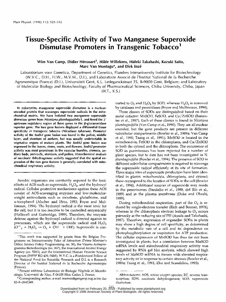
To determine the number of MnSOD genes in N. plum-baginifolia, DNA gel blot analysis was performed with aMnSOD cDNA probe (Bowler et al., 1989a). In addition toa strongly hybridizing fragment that presumably corre-sponds to the previously isolated MnSOD sequence(SodAl; Bowler et al., 1989a), a second band of weakerintensity was reproducibly observed, suggesting that asecond MnSOD gene (SodA2) is present in the haploidgenome of N. plumbaginifolia. This is exemplified forBamHI-restricted DNA in Figure 1A, lane 1.
Subsequently, a genomic library of N. plumbaginifolia inphage A Charon 35 was screened with the MnSOD cDNAprobe. Restriction analysis of seven positive candidatesshowed that they all correspond to one gene, which isdifferent from SodAl. A 3.2-kb Hindlll fragment containingthe 5' region of the gene was subcloned. Sequence analysisconfirmed that the isolated gene does not correspond toSodAl cDNA; therefore, this gene was designated SodA2.Southern blot analysis with a gene-specific SodA2 probeshowed that the SodA2 gene is located on an 11.5-kb BamHIfragment, which corresponds to the weakly hybridizingband previously observed with the SodAl cDNA probe(Fig. 1A, lane 2).
B1 2
bp290-
222-
160-
78-
Figure 1. A, DNA gel blot analysis of BamHI-digested DNA from N.plumbaginifolia, probed with SodA 1 cDNA Hpal-Psfi fragment (com-plete coding sequence) (lane 1) and probed with Sod/42 Hpal-Sacllfragment (5' region) (lane 2). B, RT-PCR restriction fragment analysisof the relative abundance of SodAl and SodA2 mRNA in differentorgans of N. plumbaginifolia. RT-PCR products were digested withCrbl, separated by PAGE, and visualized by silver staining. The sizesof the fragments (SodAl: 78, 160, and 222 bp; Sod/42: 160 and 290bp) are indicated on the left.
Because the SodAl gene was apparently not representedin the genomic phage library, a genomic sublibrary inpGEM2 was constructed that contains 2.2- to 3.0-kb Hmdlllfragments of N. plumbaginifolia DNA (see "Materials andMethods"). The 5' region of SodAl gene is located on a2.6-kb Hmdlll fragment (data not shown) and thus shouldbe present in this genomic sublibrary. Screening with aSodAl cDNA probe yielded three positive clones with a2.6-kb insert and a similar restriction pattern. Sequenceanalysis confirmed that the isolated gene corresponds toSodAl.
Specific Detection of SodAl and SodA2 Transcripts byRT-PCR Restriction Fragment Analysis
The relative amounts of SodAl and SodAl mRNA indifferent plant organs were determined by a semiquantita-tive RT-PCR approach. This strategy was chosen becauseRNA gel blot hybridizations with SodAl- and SodA2-specific probes did not give satisfactory results. Usingprimers that are highly homologous to both SodAl andSodA2, SodA transcripts were specifically amplified byRT-PCR. RT-PCR products from both SodA genes werediscriminated by restriction fragment analysis. Digestionwith C/oI gave fragments of 78, 160, and 222 bp in thecase of SodAl, and fragments of 160 and 290 bp forSodA2. Figure IB shows that the RT-PCR product ofSodAl is more abundant than that of SodA2 in all organsanalyzed. www.plantphysiol.orgon February 20, 2018 - Published by Downloaded from
Copyright © 1996 American Society of Plant Biologists. All rights reserved.
Tissue-Specific Activity of Two SodA Promoters
A GUS
529
3‘0cS
Construction of Chimeric Genes Containing the 5’ Region of the MnSOD Genes Fused to the Coding Region of the uidA Cene and Transfer to Tobacco
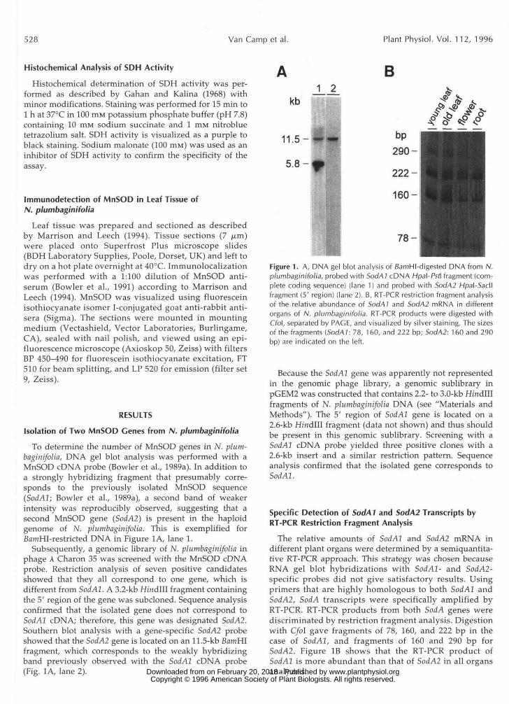
To identify the tissue specificity of MnSOD expression, gene fusions were made between the 5‘ region of the MnSOD genes and the coding region of the GUS gene ( u i d A ) of E. coli (Fig. 2). In the case of the SodAl gene, two translational fusions with the GUS coding sequences were constructed. A translational fusion at the initiation codon was made after introduction of an NcoI restriction site in the SodAl gene using site-directed mutagenesis by PCR (MnSODGUSlA). A second fusion was made at the start of the mature MnSOD protein (MnSODGUSlB).
Since both MnSODGUSl constructs gave similar expres- sion patterns (see below), only the translational fusion that contains the mitochondrial transit sequence was con- structed for the SodA2 gene (MnSODGUS2). The three chimeric constructs were cloned in a binary vector and transferred by triparental crossing into Agrobacterium tume-
ATG
/-J< mnsodl TCA AA4 ATG GCA
ATG
B
mnsod-GUS fusion CTC CGG GTC CGT CCT GTA
L R V L P V mnsod ’ G t V L Q T
2
Figure 2. Structure of SodA-GUS fusions. SodA noncoding region is indicated by a hatched box, SodA sequence coding for a mitochondrial transit peptide i s indicated by a dotted box. 3’nos and 3’0cs are the polyadenylation sites of the nopaline synthase gene and the octopine synthase gene. ATG, lnitiation codon. A, Translational fusion at the MnSODl initiation codon. The con- struct MnSODGUSlA contains a 11 30-bp Hindlll-Ncol fragment from SodA1 fused to the coding region of uidA. The Ncol restriction site (underlined) in SodA1 was created by PCR with a mutated 3’ primer. The original SodAl sequence and the MnSODGUSlA se- quence around the initiation codon are indicated. B, Translational fusion at the start of the mature MnSOD protein. The constructs MnSODGUSl B and MnSODGUS2 contain, respectively, a 11 93-bp HindIIl-Sacll fragment from SodA1 and a 1610-bp Hindlll-Sacll frag- ment from SodAZ fused to the coding region of the GUS gene (uidA). In these constructs GUS is expressed with a 21 -amino acid extension at the NH, terminus that has the properties of a mitochondrial transit peptide. The sequence and the corresponding amino acids at the fusion site are shown. The amino terminus of GUS is indicated by a horizontal arrow. The amino acid sequences of MnSODl and Mn- SOD2 at the processing site are shown at the bottom of the figure. The processing site is indicated by a vertical arrow. For details of the constructions, see “Materials and Methods.”
A MnSOD-TP-GUS CP MT
B Mitochondrion Marker Enzyme
c rbc-SS-TP-GUS -=‘ 20 I I
1 5 10 15 20
Fraction Number
-
Figure 3. Subcellular distribution of GUS activities in MnSOD- GUSlB and MnSODGUS2 plants (A), of fumarate and Cyt c oxidase activities (B), and of G U S activity in rbc-SS-TP-GUS plants (C). CP, Chloroplast; MT, mitochondria.
faciens. Subsequently, leaf discs of Nicotiana tabacum cv Petit Havana SR1 were infected and transformed callus cells were selected on kanamycin-containing medium. Histo- chemical analysis of GUS activity was performed on leaf, stem, and root tissue of regenerated plantlets to select transgenic plants for cultivation in the greenhouse. No aberrant expression patterns were observed within the pri- mary transformants of each construct. The results presented here for SodAl and SodA2 were mostly ob- tained with self-fertilized T, and T, plants of at least five independent transformants. MnSODGUSlA and MnSODGUSlB plants gave a similar expression pattern (data not shown); therefore, only results obtained with MnSODGUSlB plants are presented here.
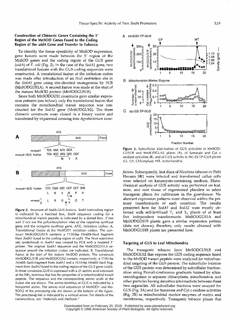
Targeting of CUS to Leaf Mitochondria
The transgenic tobacco lines MnSODGUSlB and MnSODGUS2 that express the GUS coding sequence fused to the MnSOD transit peptide were analyzed for mitochon- drial targeting of the GUS protein. The subcellular location of the GUS protein was determined by subcellular fraction- ation using Percoll-continuous gradients formed by ultra- centrifugation to separate chloroplasts, mitochondria, and other particles having densities intermediate between these two organelles. A11 subcellular fractions were assayed for GUS (Fig. 3A) and for fumarase and Cyt c oxidase activities (Fig. 38) as mitochondrial marker enzymes of matrix and membranes, respectively. Transgenic tobacco plants that
www.plantphysiol.orgon February 20, 2018 - Published by Downloaded from Copyright © 1996 American Society of Plant Biologists. All rights reserved.
530 Van Camp et al. Plant Physiol. Vol. 112, 1996
OP-— PP
ABAD X
*9
B
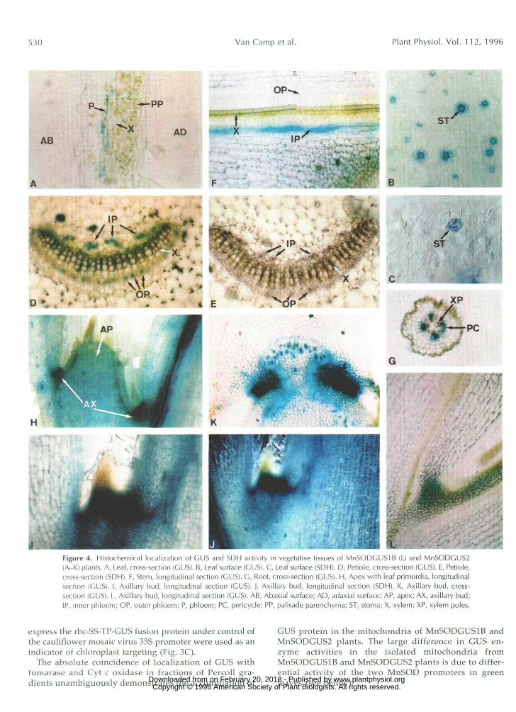
'LFigure 4. Histochemical localization of GUS and SDH activity in vegetative tissues of MnSODGUSIB (L) and MnSODCUS-i(A-K) plants. A, Leaf, cross-section (GUS). B, Leaf surface (GUS). C, Leaf surface (SDH). D, Petiole, cross-section (GUS). E, Petiole,cross-section (SDH). F, Stem, longitudinal section (GUS). G, Root, cross-section (GUS). H, Apex with leaf primordia, longitudinalsection (GUS). I, Axillary bud, longitudinal section (GUS). ), Axillary bud, longitudinal section (SDH). K, Axillary bud, cross-section (GUS). L, Axillary bud, longitudinal section (GUS). AB, Abaxial surface; AD, adaxial surface; AP, apex; AX, axillary bud;IP, inner phloem; OP, outer phloem; P, phloem; PC, pericycle; PP, palisade parenchyma; ST, stoma; X, xylem; XP, xylem poles.
express the rbc-SS-TP-GUS fusion protein under control ofthe cauliflower mosaic virus 35S promoter were used as anindicator of chloroplast targeting (Fig. 3C).
The absolute coincidence of localization of GUS withfumarase and Cyt c oxidase in fractions of Percoll gra-dients unambiguously demonstrates the accumulation of
GUS protein in the mitochondria of MnSODGUSIB andMnSODGUS2 plants. The large difference in GUS en-zyme activities in the isolated mitochondria fromMnSODGUSIB and MnSODGUS2 plants is due to differ-ential activity of the two MnSOD promoters in greenleaves (see below). www.plantphysiol.orgon February 20, 2018 - Published by Downloaded from
Copyright © 1996 American Society of Plant Biologists. All rights reserved.
Tissue-Specific Activity of Two SodA Promoters 531
Activity of the Chimeric SotM::GUS Genes inVegetative Tissues
Histochemical analysis of GUS activity in soil-grownMnSODGUSIB plants showed almost no expression of theSodAl::GUS chimeric gene in mature leaves. Only in a fewcases could very faint staining be observed in the vasculartissue. In mature leaves of MnSODGUSl plants, GUS ac-tivity was found in the vascular tissue (Fig. 4A) and instomatal guard cells (Fig. 4B). Moreover, levels of GUSactivity were much higher than in MnSODGUSIB plants.Using fluorimetric GUS assays, it was estimated that forthe So<M2::GUS chimeric gene activity levels in leaves areas much as 500-fold higher than for the SodAlr.GUS con-struct (1 unit versus 2 milliunits GUS per mg protein).
Staining of cross-sections through the petiole ofMnSODGUS2 plants indicated that the vascular expres-sion was localized mainly in phloem cells and in the cellsadjacent to the xylem vessels (Fig. 4D). Whereas in thepetiole no clear difference was observed between expres-sion levels in inner and outer phloem, GUS expression inthe stem was most prominent in the inner phloem (Fig.4F). In roots, staining was seen mainly in the pericyclecells that are adjacent to the xylem poles (Fig. 4G). GUSactivity of the SodAl chimeric construct was absent orrarely detectable in stem and root (data not shown).
In the growing parts of MnSODGUS2 plants, histochem-ically detectable GUS activity was highest in newly formedaxils near the apex (Fig. 4H) and in axillary buds with leafprimordia (Fig. 4, I and K). Figure 41 shows that the stron-gest staining is observed at the base of the emerging bud.Some staining in the axillary bud is also observed for theSodAl::GUS chimeric gene (Fig. 4L).
Using immunofluorescent techniques, we also investi-gated the presence of MnSOD protein in different cell typesof the leaf. Labeling with MnSOD antibody and fluoresceinisothiocyanate isomer I was most pronounced in vasculartissue (Fig. 5) and stomata (data not shown) and hardlydetectable in mesophyll or palisade parenchyma cells.Thus, the expression pattern of immunodetectable MnSODcorroborates the histochemical localization of GUS activityin the leaf.
Comparison with SDH Activity
Respiratory activity is thought to be the major sourceof superoxide radicals in mitochondria (Rich and Bon-ner, 1978). Therefore, cells or tissues with high respira-tory activity may require the highest expression of Mn-SOD, provided that the relative amount of oxygen that isreduced by monovalent electron addition during respi-ration is constant in different tissues. To investigate therelation between MnSOD expression and mitochondrialrespiration, SDH activity was analyzed in different tis-sues. In the green parts of tobacco, the highest levels ofSDH activity were found in the stomata (Fig. 4C), in thevascular tissue (Fig. 4E), and in the axillary zone (Fig. 4J).In these tissues SDH and GUS activity are localizedwithin the same cells. However, SDH activity was alsovery high in meristematic cells, particularly in the root
Figure 5. Immunoloc.ili/ation ol MnSOD protein in tobacco leafsections with MnSOD antibody and fluorescein isothiocyanate iso-mer I, showing specific expression in vascular tissue. Immunofluo-rescence is not equally distributed over the cytoplasm, but is con-fined to cellular bodies, which, in view of the mitochondrial locationof MnSOD, may tentatively be identified as mitochondria.
apex, which is in contrast to GUS activity (data notshown).
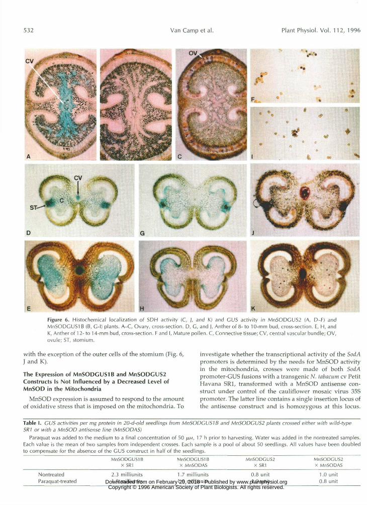
GUS and SDH Activity in the Flower Parts of Tobacco
In sepals, petals, carpels, and style, GUS activity waslocalized in the vascular tissue in MnSODGUS2 plants andwas absent in MnSODGUSIB plants. Also, SDH activitywas seen mainly in the vascular tissue of these organs, butit was also found in ovules (Fig. 6, A-C). In anthers, ex-pression of the MnSOD chimeric genes changed duringflower development. In flower buds of 8 to 10 mm, theSorf/42::GUS chimeric construct was expressed in the cen-tral vascular bundle and in a cluster of cells between thestomium and the connective tissue (Fig. 6D). In later stages(flower bud > 12 mm), GUS activity near the stomiumdisappeared due to degeneration of this tissue, but at thesame time GUS activity was developed by the maturingpollen grains (Fig. 6E). Finally, staining in the vasculartissue declined concurrently with the degradation of theconnective tissue, whereas GUS activity in pollen wasmaintained at least until pollen dehiscence (Fig. 6F).
Contrary to mature vegetative tissues, the SorfA7::GUSchimeric gene was expressed to well detectable levels inanthers. The observed expression pattern was differentfrom that of the SodA2::GUS construct. In anthers of 8- to10-mm buds, GUS activity was seen between the tapetumand the connective tissue (Fig. 6G). This tissue most likelycorresponds to the middle layer of the microsporangiumwall. In addition, staining was sometimes observed in theouter cells of the stomium (Fig. 6G). Staining in thesetissues declined during further anther development,whereas GUS activity in pollen appeared when flowerbuds reached approximately 12 mm, which is similar toSodAl (Fig. 6H). Expression of the SodAlr.GUS chimericgene was also observed in mature pollen (Fig. 61).
SDH activity was seen in all anther tissues that displayexpression of SodAl::GUS and/or SodA2::GUS constructs, www.plantphysiol.orgon February 20, 2018 - Published by Downloaded from
Copyright © 1996 American Society of Plant Biologists. All rights reserved.
532 Van Camp et al. Plant Physiol. Vol. 112, 1996
ife«2fe*l«* 4
Figure 6. Histochemical localization of SDH activity (C, ), and K.) and CDS activity in MnSODGUS2 (A, D-F) andMnSODGUSl B (B, C-l) plants. A-C, Ovary, cross-section. D, C, and ], Anther of 8- to 10-mm bud, cross-section. E, H, andK, Anther of 12-to 14-mm bud, cross-section. F and I, Mature pollen. C, Connective tissue; CV, central vascular bundle; OV,ovule; ST, stomium.
with the exception of the outer cells of the stomium (Fig. 6,J and K).
The Expression of MnSODCUSl B and MnSODGUS2Constructs Is Not Influenced by a Decreased Level ofMnSOD in the Mitochondria
MnSOD expression is assumed to respond to the amountof oxidative stress that is imposed on the mitochondria. To
investigate whether the transcriptional activity of the Sod Apromoters is determined by the needs for MnSOD activityin the mitochondria, crosses were made of both SodApromoter-GUS fusions with a transgenic N. tabacum cv PetitHavana SRI, transformed with a MnSOD antisense con-struct under control of the cauliflower mosaic virus 35Spromoter. The latter line contains a single insertion locus ofthe antisense construct and is homozygous at this locus.
Table I. GUS activities per mg protein in 20-d-old seedlings from MnSODGUSIB and MnSODGUS2 plants crossed either with wild-typeSRI or with a MnSOD antisense line (MnSODAS)
Paraquat was added to the medium to a final concentration of 50 /LIM, 17 h prior to harvesting. Water was added in the nontreated samples.Each value is the mean of two samples from independent crosses. Each sample is a pool of about 50 seedlings. All values have been doubledto compensate for the absence of the GUS construct in half of the seedlings.
MnSODGUSIBx SRI
MnSODGUSIBX MnSODAS
MnSODGUS2X SRI
MnSODGUS2X MnSODAS
NontreatedParaquat-treated
2.3 milliunits1.9 milliunits
1.7 milliunits1.9 milliunits
0.8 unit1.0 unit
1.0 unit0.8 unit www.plantphysiol.orgon February 20, 2018 - Published by Downloaded from
Copyright © 1996 American Society of Plant Biologists. All rights reserved.
Tissue-Specific Activity of Two SodA Promoters 533
The residual MnSOD activity in leaves from this line is less than 10% of the level in untransformed SR1 (W. Van Camp, unpublished data). The MnSODGUSlB and MnSODGUS2 lines used for this cross have a single insertion locus of the transgene and are heterozygous at this locus. Since both the promoter-GUS fusion constructs and the MnSOD antisense constructs contain the nptII gene as selectable marker, no selection can be performed for the presence of both foreign genes in the progeny. Thus, whereas a11 the progeny will express the MnSOD antisense gene, only half will contain the promoter-GUS construct. As controls, back-crosses of the MnSODGUSlB and MnSODGUS2 lines to wild-type SR1 were performed.
The effect of MnSOD suppression on the activity of the promoter-GUS fusions was analyzed in 20-d-old seedlings grown on agar medium (one-half Murashige and Skoog salts, 1% SUC, 0.8% agar, pH 5.7), to which paraquat (50 p~ final concentration) or water was added 17 h prior to harvesting. As shown in Table I, promoter activities of the MnSODGUSlB and MnSODGUS2 constructs were not sig- nificantly different in plants with normal or reduced levels of MnSOD; neither did the paraquat treatment cause any induction of the SodAl and SodA2 promoters in the chi- meric constructs.
DISCUSSION
In mature leaves of N. plumbaginifolia and N . tabacum, MnSOD activity levels are generally much lower than those of FeSOD or Cu/ZnSOD (Van Camp et al., 1990, 1994), which suggests that mitochondrial superoxide pro- duction in leaves is marginal compared with that in the cytosol and chloroplasts. Similarly, it was observed that mitochondrial overproduction of SOD in transgenic to- bacco was less effective than chloroplastic overproduction in protecting plants against leaf damage caused by para- quat (Bowler et al., 1991) or ozone (Van Camp et al., 1994). However, this does not exclude the fact that MnSOD could play a crucial role in the oxidative stress response in tissues other than leaves, or in other developmental stages.
To obtain a more complete picture of MnSOD expression in different tissues, it was first necessary to characterize the MnSOD gene family. We have identified two MnSOD- encoding genes in N. plumbaginifolia, denoted SodAl and SodA2, and we have isolated the 5’ ends of both genes. The two MnSOD genes show extensive homology in the coding sequence (88% at the amino acid level), but only two stretches of homology were found in the 5’ upstream re- gion. In the 5‘ untranslated leader, a sequence of 38 nucle- otides (shortest sequence, not counting gaps) was found that shows 71% sequence identity (taking gaps as mis- matches). A second stretch of 54 nucleotides is 79% iden- tical between both SodA genes. This is located at positions -826 to -773 (distance to the translation start) in SodAZ, and at positions -911 to -857 in SodA2.
By RT-PCR restriction fragment analysis, it was esti- mated that SodAl expression in leaves, flowers, and roots was stronger than that of SodA2. The tissue specificity of MnSOD expression was further investigated in transgenic
tobacco containing gene fusions of the 5’ upstream regions of SodAl and SodA2 with the GUS reporter gene. Two translational fusions have been made for the SodAl gene, one at the initiation codon and a second at the start of the mature protein. In the latter construct, a 21-amino acid terminal extension was fused to the reporter protein GUS that has the characteristics of a mitochondrial transit pep- tide (Bowler et al., 1989b). Using subcellular fractionation techniques, we demonstrated that the MnSODl amino- terminal extension can target GUS to mitochondria and that the GUS protein retained its enzymic activity after transport through the mitochondrial membrane. The fact that no significant GUS activity was found in the cytosolic fraction suggests that most of the GUS fusion protein was imported into mitochondria. This result is in accord with the data from Schmitz and Lonsdale (1989), who showed that a fusion of a yeast mitochondrial transit sequence to GUS is efficiently imported to plant mitochondria. Both translational fusions gave the same spatial expression pat- tern, suggesting that the MnSOD transit peptide or the mitochondrial import do not function as posttranslational control mechanisms of expression. For this reason, it was decided to make only one chimeric construct for the SodA2 gene that produces a fusion protein of the MnSOD2 transit peptide and GUS. As with MnSOD1, the amino-terminal part of MnSOD2 was active in targeting GUS to the mi tochondria.
SodA2::GUS and SodA2::GUS promoter-GUS fusions dis- played distinct patterns of expression in transgenic to- bacco. In leaves, SodA2 promoter activity was as much as 500-fold higher than that of SodAl. Expression of the SodA2::GUS construct was strongest in phloem cells and stomatal guard cells. In the case of the SodA1::GUS con- struct, expression was barely detectable by histochemical GUS staining, but, when observed, it was always found in the same tissues where the SodA2 gene was expressed. The large difference in promoter activity of both SodA genes in leaves is at variance with the RT-PCR data on relative SodAl and SodA2 mRNA abundance. The most probable explanation is that some important enhancer sequences are not contained in the SodAl ::GUS construct. Regulatory el- ements for spatial expression of plant genes are usually located in the first 500 bp upstream of the transcription initiation site, but enhancer sequences are often found at more dista1 positions. To confirm the results on the spatial expression of MnSOD in leaves, we have localized MnSOD protein on leaf sections by immunodetection with MnSOD- specific antibodies. MnSOD protein was most abundant in phloem and stomata, which is in accord with the expres- sion data obtained with the promoter-GUS constructs.
SodAl ::GUS and SodA2::GUS fusions showed distinct spa- tia1 expression pattems in anthers. SodAZ ::GUS expression was observed without concurrent expression of the SodA2 gene in the stomium and the middle layer between connec- tive tissue and tapetum. Although severa1 mRNAs have been identified that are present either exclusively or at elevated levels in tobacco anthers (Koltunow et al., 1990), to our knowledge SodAZ is the first example of a gene that is highly transcribed in the middle layer of the anther. Only the
www.plantphysiol.orgon February 20, 2018 - Published by Downloaded from Copyright © 1996 American Society of Plant Biologists. All rights reserved.
534 Van Camp et al. Plant Physiol. Vol. 11 2, 1996
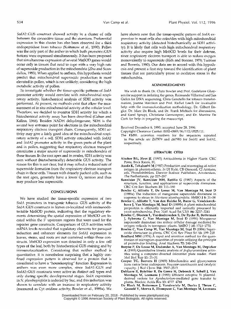
SodA2::GUS construct showed activity in a cluster of cells between the connective tissue and the stomium. Preferential expression in this cluster has also been observed for a thiol endopeptidase from tobacco (Koltunow et al., 1990). Pollen was the only part of the anther in which both promoter::GUS fusions were expressed simultaneously. It has been proposed that simultaneous expression of severa1 MnSOD genes would occur only in tissues that need to cope with a very high rate of superoxide production in the mitochondria (Zhu and Scan- dalios, 1993). When applied to anthers, this hypothesis would predict that mitochondrial superoxide production is most elevated in pollen, which is not unlikely, considering the high metabolic activity of pollen.
To investigate whether the tissue-specific pattems of SodA promoter activity would correlate with mitochondrial respi- ratory activity, histochemical analysis of SDH activity was performed. At present, no methods exist that allow the mea- surement of in situ mitochondrial activity at the cellular level. Therefore, we decided to monitor SDH activity for which a histochemical activity assay has been described (Gahan and Kalina, 1968). Besides NADH dehydrogenase, SDH is the second key entrance point for electrons in the mitochondrial respiratory electron transport chain. Consequently, SDH ac- tivity may give a fairly good idea of the mitochondrial respi- ratory activity of a cell. SDH activity coincided with SodAl and SodA2 promoter activity in the green parts of the plant and in pollen, suggesting that respiratory electron transport constitutes a major source of superoxide in mitochondria of these tissues. In the root apex and in ovules, SDH activity was seen without (histochemically) detectable GUS activity. The reason for this is not clear, but it may reflect a reduced rate of superoxide formation from the respiratory electron transport chain in these cells. Tissues with densely packed cells, such as the root apex, generally have a lower O, tension and thus may produce less superoxide.
CONCLUSIONS
We have studied the tissue-specific expression of two SodA promoters in transgenic tobacco. GUS activity of the SodA::GUS constructs in leaves co-localized with immunode- tectable MnSOD protein, indicating that the regulatory ele- ments determining the spatial expression of MnSOD are lo- cated within the 5' upstream regions that were used for the reporter gene constructs. Comparison of GUS activities with mRNA levels revealed that regulatory elements for paraquat induction and enhancer elements for SodAl expression in leaves, stems, and roots are not contained within these con- structs. MnSOD expression was detected in only a few cell types of the leaf, both by histochemical GUS staining and by immunolocalization. Considering that neither method is quantitative, it is nonetheless surprising that a highly con- fined expression pattern is observed for a protein that is considered to have a "housekeeping" function. Expression in anthers was even more complex, since SodAl::GUS and SodA2::GUS constructs were active in distinct cell types and only during specific developmental stages. SodA expression in N. plumbaginifolia is induced by SUC, and this induction was shown to correlate with an increase in respiratory activity (measured as Cyt oxidase activity; Bowler et al., 1989a). We
have shown now that the tissue-specific pattern of SodA ex- pression in most cells also coincides with high mitochondrial respiratory activity (detected histochemically as SDH activi- ty). It is likely that cells with high mitochondrial respiratory activity also require high MnSOD levels for their defense, since respiratory electron transport is able to reduce oxygen monovalently to superoxide (Rich and Bonner, 1978; Turrens and Boveris, 1980). Our data are in accord with th s hypoth- esis and present a first step toward the identification of plant tissues that are particularly prone to oxidative stress in the mitochondria.
ACKNOWLEDCMENTS
We wish to thank Dr. Chris Bowler and Prof. Godelieve Ghey- sen for support in isolating the genes, Raimundo Villarroel and Jan Gielen for DNA sequencing, Chris Genetello for tobacco transfor- mation, Joanne Marrison and Prof. Rache1 Leech for invaluable help with the immunolocalization methodology, Dr. Gilbert En- gler, Dr. Marc De Block, and Dr. Frank Michiels for discussions, and Karel Spruyt, Christiane Germonprez, and Dr. Martine De Cock for help in preparing the manuscript.
Received December 8, 1995; accepted June 28, 1996. Copyright Clearance Center: 0032-0889/96/ 112/0525/ 11 The EMBL accession numbers for the sequences reported
in this article are 267979 and 267985 for SodAl and SodA2, respectively .
LITERATURE ClTED
Alscher RG, Hess JL (1993) Antioxidants in Higher Plants. CRC Press, Boca Raton, FL
Asada K, Takahashi M (1987) Production and scavenging of active oxygen in photosynthesis. In DJ Kyle, CB Osmond, CJ Arntzen, eds, Photoinhibition. Elsevier Science Publishers, Amsterdam, The Netherlands, pp 227-287
Bannister JV, Bannister WH, Rotilio G (1987) Aspects of the structure, function, and applications of superoxide dismutase. CRC Crit Rev Biochem 22: 111-180
Bowler C, Alliotte T, De Loose M, Van Montagu M, Inzé D (1989a) The induction of manganese superoxide dismutase in response to stress in Nicotiuna plumbaginifolia. EMBO J 8: 31-38
Bowler C, Alliotte T, Van den Bulcke M, Bauw G, Vandekerck- hove J, Van Montagu M, InzC D (1989b) A plant mitochondrial preprotein is efficiently imported and correctly processed by yeast mitochondria. Proc Natl Acad Sci USA 86: 3237-3241
Bowler C, Slooten L, Vandenbranden S, D e Rycke R, Botterman J, Sybesma C, Van Montagu M, Inzé D (1991) Manganese superoxide dismutase can reduce cellular damage mediated by oxygen radicals in transgenic plants. EMBO J 10: 1723-1732
Bowler C, Van Camp W, Van Montagu M, Inzé D (1994) Super- oxide dismutase in plants. CRC Crit Rev Plant Sci 13: 199-218
Bradford MM (1976) A rapid and sensitive method for the quan- titation of microgram quantities of protein utilizing the principle of protein-dye binding. Ana1 Biochem 7 2 248-254
Breyne P, D e Loose M, Dedonder A, Van Montagu M, Depicker A (1993) Quantitative kinetic analysis of P-glucuronidase activ- ities using a computer-directed microtiter plate reader. Plant Mo1 Biol Rep 11: 21-31
Cooper TG, Beevers H (1969) Mitochondria and glyoxysomes from castor bean endosperm. Enzyme constituents and catalytic capacity. J Biol Chem 244: 3507-3513
Deblaere R, Bytebier B, De Greve H, Deboeck F, Schell J, Van Montagu M, Leemans J (1985) Efficient octopine Ti plasmid- derived vectors for Agrobucterium-mediated gene transfer to plants. Nucleic Acids Res 13: 47774788
De Block M, Botterman J, Vandewiele M, Dockx J, Thoen C, Gosselé V, Movva R, Thompson C, Van Montagu M, Leemans
www.plantphysiol.orgon February 20, 2018 - Published by Downloaded from Copyright © 1996 American Society of Plant Biologists. All rights reserved.

Tissue-Specific Activi ty o f Two SodA Promoters 535
J (1987) Engineering herbicide resistance in plants by expression of a detoxifying enzyme. EMBO J 6: 2513-2518
De Loose M, Alliotte T, Gheysen G, Genetello C, Gielen J, Soetaert P, Van Montagu M, Inzé D (1988) Primary structure of a hormonally regulated P-glucanase of Nicotiana plumbaginifolia. Gene 70: 13-23
de1 Rio LA, Fernández VM, Rupérez FL, Sandalio LM, Palma JM (1989) NADH induces the generation of superoxide radicals in leaf peroxisomes. Plant Physiol 89: 728-731
Doke N, Ohashi Y (1988) Involvement of a superoxide anion generating system in the induction of necrotic lesions on tobacco leaves infected with tobacco mosaic virus. Physiol Mo1 Plant Pathol 32: 163-175
Foyer CH, Mullineaux PM (1994) Causes of Photooxidative Stress and Amelioration of Defense Systems in Plants. CRC Press, Boca Raton, FL
Gahan PB, Kalina M (1968) The use of tetrazolium salts in the histochemical demonstration of succinic dehydrogenase activity in plant tissues. Histochemie 14: 81-88
Halliwell B, Gutteridge JMC (1989) Free Radicals in Biology and Medicine. Clarendon Press, Oxford, UK
Hérouart D, Van Montagu M, Inzé D (1994) Developmental and environmental regulation of the Nicotiana plumbaginifolia cytoso- lic Cu / Zn-superoxide dismutase promoter in transgenic to- bacco. Plant Physiol 104: 873-880
Kardish N, Magal N, Aviv D, Galun E (1994) The tomato gene for the chloroplastic Cu,Zn superoxide dismutase: regulation of expression imposed in transgenic tobacco plants by a short promoter. Plant Mo1 Biol 25: 887-897
Koltunow AM, Truettner J, Cox KH, Wallroth M, Goldberg RB (1990) Different temporal and spatial gene expression patterns occur during anther development. Plant Cell 2: 1201-1224
Landt O, Grunert H-P, Hahn U (1990) A general method for rapid site-directed mutagenesis using the polymerase chain reaction. Gene 96: 125-128
Logemann J, Schell J, Willmitzer L (1987) Improved method for the isolation of RNA from tissues. Ana1 Biochem 163: 16-20
Maniatis T, Fritsch EF, Sambrook J (1982) Molecular Cloning: A Laboratory Manual. Cold Spring Harbor Laboratory Press, Cold Spring Harbor, NY
Marrison JL, Leech RM (1994) The subcellular and intra-organelle recognition of nuclear and chloroplast transcripts in developing leaf cells. Plant J 6: 605-614
Peleman J, Boerjan W, Engler G, Seurinck J, Botterman J, Alliotte T, Van Montagu M, Inzé D (1989) Strong cellular preference in the expression of a housekeeping gene of Arnbidopsis thaliana encoding 5'-adenosylmethionine synthetase. Plant Cell 1: 81-93
Pruitt RE, Meyerowitz EM (1986) Characterization of the genome of Arabidopsis thaliana. J Mo1 Biol 187: 169-183
Rich PR, Bonner WD Jr (1978) The sites of superoxide anion generation in higher plant mitochondria. Arch Biochem Biophys
Sambrook J, Fritsch EF, Maniatis T (1989) Molecular Cloning: A Laboratory Manual, Ed 2. Cold Spring Harbor Laboratory Press, Cold Spring Harbor, NY
Sandalio LM, Fernández VM, Rupérez FL, de1 Rio LA (1988) Superoxide free radicals are produced in glyoxysomes. Plant Physiol 87: 1-4
Schmitz UK, Lonsdale DM (1989) A yeast mitochondrial prese- quence functions as a signal for targeting to plant mitochondria in vivo. Plant Cell 1: 783-791
Storrie B, Madden EA (1990) Cytochrome c oxidase (EC 1.9.3.1): mitochondrial markers. Methods Enzymol 182: 214-215
Teeri TH, Patel GK, Aspegren K, Kauppinen V (1989) Chloro- plast targeting of neomycin phosphotransferase II with a pea transit peptide in electroporated barley mesophyll protoplasts. Plant Cell Rep 8: 187-190
Tsang EWT, Bowler C, Hérouart D, Van Camp W, Villarroel R, Genetello C, Van Montagu M, Inzé D (1991) Differential regu- lation of superoxide dismutases in plants exposed to environ- mental stress. Plant Cell 3: 783-792
Turrens JF, Boveris A (1980) Generation of superoxide anion by the NADH dehydrogenase of bovine heart mitochondria. Bio- chem J 191: 421-427
Van Camp W, Bowler C, Villarroel R, Tsang EWT, Van Montagu M, Inzé D (1990) Characterization of iron superoxide dismutase cDNAs from plants obtained by genetic complementation in Escherichia coli. I'roc Natl Acad Sci USA 87: 9903-9907
Van Camp W, Willekens H, Bowler C, Van Montagu M, Inzé D, Reupold-Popp P, Sandermann H Jr, Langebartels C (1994) Elevated levels of superoxide dismutase protect transgenic plants against ozone damage. Bio/Technology 12: 165-168
Zhu D, Scandalios JG (1993) Maize mitochondrial manganese superoxide dismutases are encoded by a differentially expressed multigene family. Proc Natl Acad Sci USA 90: 9310-9314
188: 206-213
www.plantphysiol.orgon February 20, 2018 - Published by Downloaded from Copyright © 1996 American Society of Plant Biologists. All rights reserved.