Embed Size (px)
Citation preview
Trends in Pharmaceutical Sciences 2017: 3(3).
TIPS...................................Carnosine Supplementation Mitigates Brain Tissue Markers of Oxidative Stress in a Rat Model of Fulminant Hepatic Failure
Akram Jamshidzadeh1,2, Narges Abdoli3, Hossein Niknahad1,2, Negar Azarpira4, Somayeh Mousavi2, Elnaz Mardani2, Mojgan Abasvali2, Reza Heidari1,*
1Pharmaceutical Sciences Research Center, Shiraz University of Medical Sciences, Shiraz, Iran.2Department of Pharmacology and Toxicology, School of Pharmacy, Shiraz University of Medical Sciences, Shiraz,
Iran.3Iran Food and Drug Administration (IFDA), Ministry of Health, Tehran, Iran.4Transplant Research Center, Shiraz University of Medical Sciences, Shiraz, Iran.
Trends in Pharmaceutical Sciences 2017: 3(3): 149-160.
.................................................................................................................................
Abstract Fulminant hepatic failure is a deleterious clinical complication, which leads to hyperammonemia. Ammonia is a noxious neurotoxic agent, which affects brain tissue through different mechanisms. On the other hand, it is well-known that oxidative stress and its consequences play a major role in the pathogenesis of ammonia-induced brain injury. Carnosine is a dipeptide abundantly found in the human central nervous system (CNS). This peptide is widely investigated for its neuroprotective properties. The current study aimed to evaluate the effect of carnosine supplementation on oxidative stress markers in the brain tissue of a rat model of fulminant hepatic failure and hyperammonemia. Animals received thioacetamide (400 mg/kg, i.p, for three consecutive days at 24-hr intervals) as a model of acute liver failure and hyperam-monemia. Several serum biochemical parameters, in addition to plasma and brain ammonia level, were monitored. On the other hand, brain tissue markers of oxidative stress including reactive oxygen species (ROS) formation, lipid peroxidation, tissue glutathione content, and total antioxidant capacity were mea-sured. It was found that plasma and brain ammonia was increased, and serum markers of liver injury were significantly elevated in the thioacetamide-treated group. On the other hand, an increase in markers of oxidative stress, including ROS formation, lipid peroxidation, glutathione depletion, and decreased tissue antioxidant capacity, was evident in the brain of thioacetamide-treated animals. It was found that carnosine supplementation (250, 500, and 1000 mg/kg, i.p) decreased serum markers of liver injury, mitigated brain, and plasma ammonia level, and alleviated brain tissue markers of oxidative stress. These data suggest carnosine as a potential neuroprotective agent with therapeutic capability against ammonia-induced CNS injury during hepatic encephalopathy.
Keywords: Hepatic encephalopathy, Hyperammonemia, Neurotoxin, Oxidative stress, Peptide, Protective .................................................................................................................................
...........................................................................................................................
Corresponding Author: Reza Heidari, Pharmaceutical Sciences Research Center, Shiraz University of Medical Sciences, Shiraz, Iran. Email: [email protected]; [email protected]
Recieved: 11/08/2017; Accepted: 26/08/2017
Original Article
1. Introduction Acute or chronic liver failure with dif-ferent etiologies might lead to hepatic encepha-lopathy (HE) and hyperammonemia (1). Although
the exact cause of HE-induced organ injury is not known, there is agreement on the critical role of ammonia in this complication (2). Normally, am-monia is metabolized to urea by the liver. When the liver is damaged, this organ is not able to me-tabolize ammonia. Hence, plasma level of am-
Trends in Pharmaceutical Sciences 2017: 3(3): 149-160.
Akram Jamshidzadeh et al.
monia is elevated in the systemic circulation; and finally, this toxic chemical reaches the brain. Ammonia is a well-known neurotoxin, which af-fects the CNS by several mechanisms (3, 4). Am-monia has several direct toxic effects on neurons and astrocytes (4). It has been found that ammonia causes brain edema, oxidative stress, and neuroin-flammation when its level is raised during HE (5). Consequently, a suppression of the brain function occurs in patients with HE (5). Oxidative stress and its associated biolog-ical consequences are known to be implicated in ammonia neurotoxicity (3, 6). It has been report-ed that ammonia caused severe oxidative stress, lipid peroxidation, and defect in cellular antioxi-dant mechanisms in the CNS (7-12). Hence, anti-oxidants that easily reaches the brain might have therapeutic value in HE-associated CNS compli-cations. Carnosine (b-alanyl-L-histidine) is an endogenously-synthesized dipeptide found at high concentrations in different tissues of mammals (13, 14). Numerous biological roles are attributed to carnosine (15, 16). Several pharmacological properties also have been identified for this pep-tide (17, 18). It has been found that carnosine sig-nificantly mitigated xenobiotics-induced injury in different experimental models (19-23). Carnosine supplementation also was shown to have protec-tive effects against several pathological conditions (24-30). Reactive species scavenging ability and antioxidant properties of carnosine are tightly at-tributed to its protective properties in different bio-logical systems (19, 21, 31, 32). Carnosine is also known as a neuropeptide (33, 34). Several studies mentioned the importance of carnosine in the CNS (15, 33-35). Carnosine ad-ministration has been shown to be effective against a wide range of CNS disorders including Parkin-son and Alzheimer disease (36, 37). Furthermore, it has been found that carnosine serves as a protec-tive agent against a number of neurotoxic chemi-cals (35, 38-40). Interestingly, the neuroprotective effects of this peptide against brain ischemia and stroke are clinically investigated, and there is sub-stantial evidence suggesting carnosine as a protec-tive agent against brain ischemia (41-43). All these findings indicate that this peptide might also act
as an effective protective agent against ammonia-induced CNS complications. The current investigation was designed to evaluate the neuroprotective capability of carnosine in a rat model of acute liver failure and hyperammonemia. Rats received thioacetamide as an animal model of fulminant hepatic failure. Then, plasma and brain ammonia level along with serum biomarkers of liver injury were monitored. Moreover, brain markers of oxidative stress were measured to investigate the effect of carnosine supplementation on HE-induced CNS injury.
2. Material and methods2.1. Chemicals Carnosine, 4,2 Hydroxyethyl,1-piperazine ethane sulfonic acid (HEPES), 6-hydroxy-2,5,7,8-tetramethyl chroman-2-carboxylic acid (Trolox), thiobarbituric acid (TBA), fatty acid-free bo-vine serum albumin (BSA) fraction V, dithiobis-2-nitrobenzoic acid (DTNB), glutathione (GSH), 2′,7′ dichlorofluorescein diacetate (DCFH-DA), malondialdehyde (MDA), sucrose, KCl, Na2H-PO4, MgCl2 , dithiothreitol, Coomassie brilliant blue, ethylene gycol-bis (2-aminoethyl ether)-N,N,N′,N′-tetraacetic acid (EGTA), and ethylene-diaminetetraacetic acid (EDTA) were purchased from Sigma Chemical Co. (St. Louis, MO, USA). Trichloroacetic acids (TCA), ammonium chloride, and hydroxymethyl amino methane hydrochloride (Tris-HCl) were purchased from Merck (Darm-stadt, Germany). All salts for preparing buffer so-lutions were of analytical grade and obtained from Merck (Darmstadt, Germany).
2.2. Animals Male Sprague-Dawley rats (n=48; 200-250 g weight) were obtained from the Animal Breeding Center, Shiraz University of Medical Sciences, Shiraz, Iran. Animals were housed in plastic cages over hardwood bedding. There was an environmental temperature of 23±1 ºC and a 12L: 12D photoschedule along with a 40% of relative humidity. Rats were allowed free access to a normal standard chow diet and tap water. All the experiments were performed in conformity with the guidance for care and use of experimen-tal animals approved by a local ethics committee
150
Trends in Pharmaceutical Sciences 2017: 3(3): 149-160.
Carnosine neuroprotection in rats
in Shiraz University of Medical Sciences, Shiraz, Iran (#95-01-36-12054).
2.3. Animal model of fulminant hepatic failure Thioacetamide is extensively used as a model of acute hepatic failure (44). In the cur-rent study, thioacetamide-induced fulminant hepatic failure was achieved by three consecu-tive intraperitoneal (i.p) injections of thioacet-amide (400 mg/kg) to rats (n=48; 6 rats/group) at 24-hr intervals (45). Carnosine was admin-istered for three consecutive days, two hr after each dose of thioacetamide. The treatments were as follow: 1) control (vehicle-treated); 2) thio-acetamide; 3) thioacetamide+carnosine 250 mg/kg; 4) thioacetamide+carnosine 500 mg/kg; 5) thioacetamide+carnosine 1000 mg/kg; 6) carno-sine 1000 mg/kg. Animals were anesthetized (thiopental, 80 mg/kg, i.p) 24 hr after the last dose of thioacet-amide, and their blood, brain, and liver samples were collected. As mentioned in previous studies, supportive therapy by administering 5% dextrose (2.5 ml/kg body weight, S.C) containing 0.45% sodium chloride and 0.2% potassium chloride, was given to avoid weight loss, hypoglycemia, and re-nal failure in thioacetamide-induced hepatic fail-ure model (45). Control animals (vehicle-treated) received normal saline as the thioacetamide sol-vent. The sole carnosine (1000 mg/kg, i.p) was ad-ministered to ensure its safety.
2.4. Blood biochemistry and tissue histopathology Standard commercial kits and a Min-drayBS-200® auto analyzer were used to measure serum aspartate aminotransferase (AST), alanine aminotransferase (ALT), and lactate dehydroge-nase (LDH) (46). Plasma ammonia was measured with standard kits based on the absorbance pho-tometry method of phenate-hypochlorate reaction (47). To determine brain ammonia content, sam-ples (100 mg) of the forebrain (cerebral cortex) were dissected, homogenized, and deproteinized in 3 ml of ice-cooled lysis solution (Trichloroace-tic acid, 6%, w/v). After centrifugation (12000g, 10 min, 4 °C), the supernatant was collected and neutralized with potassium carbonate (KHCO3; 2 mol/l, pH=7). Afterward, brain ammonia content
was measured using standard kits (47). For histo pathological evaluation, samples of liver were fixed in phosphate-buffered formalin solution (0.4% sodium phosphate monobasic, NaH2PO4, 0.64% sodium phosphate dibasic, Na2HPO4, and 10% formaldehyde in distilled water) (48, 49). Paraffin-embedded sections of liver were prepared and stained with haematoxylin and eosin (H&E) before light microscope viewing.
2.5. Reactive oxygen species formation Reactive oxygen species (ROS) formation in the brain tissue was estimated as previously de-scribed (50, 51). Briefly, samples of brain tissue were homogenized in 5 ml of ice-cooled (4 ºC) Tris-HCl buffer (40 mM, pH=7.4). Samples of the resulted tissue homogenate (100 µl) were mixed with Tris-HCl buffer (1 ml; pH=7.4 4 ºC) and 2′, 7′ dichlorofluorescein diacetate (Final concentra-tion 10 µM) (52, 53). The mixture was incubated at 37 ºC (15 min, in dark). Finally, the fluorescence intensity (FI) of the samples was assessed using a FLUOstar Omega® multi functional microplate reader at λ excitation=485 nm and λ emission=525 nm (50, 54).
2.6. Brain tissue glutathione content Tissue samples were homogenized in 4 ml of ice-cooled EDTA (20 mM; 4 ºC). Then, 2.5 ml of the prepared homogenate were added to 2 ml of distilled water (4 ºC) and 1 ml of trichloroacetic acid (50% w/v; 4 ºC). Samples were mixed well and centrifuged (10,000 g, 4 °C, 25 min). After-ward, 1 ml of the supernatant was mixed with 4ml of Tris buffer (pH=8.9; 4 ºC), and 100 µl of DTNB (10 mM in methanol) (52,53). The absorbance of the developed color was measured at λ=412 nm using an Ultrospec 2000®UV spectrophotometer (Pharmacia Biotech, Uppsala, Sweden) (55).
2.7. Lipid peroxidation The thiobarbituric acid reactive substanc-es (TBARS) test was used as a method to assess lipid peroxidation in the brain tissue (55). The reaction mixture was consisted of 500 µl of tis-sue homogenate (10% w/v in KCl, 1.15%), 1 ml of thiobarbituric acid (0.375%, w/v), and 3 ml of metaphosphoric acid (1% w/v, pH=2). Samples
151
Trends in Pharmaceutical Sciences 2017: 3(3): 149-160.
Akram Jamshidzadeh et al.
were mixed well and heated (100 °C; 45 min). Then, the mixture was cooled, and 2 ml of n-buta-nol was added. Samples were vigorously vortexed and centrifuged (10,000 g for 5 min). Finally, the absorbance of developed color in n-butanol phase was read at λ=532 nm using an Ultrospec 2000®UV spectrophotometer (Pharmacia Biotech, Uppsala, Sweden) (55).
2.8. Ferric reducing antioxidant power (FRAP) of the brain tissue FRAP assay is a method to measure the formation of a blue colored Fe2+-tripyridyl-tri-azine compound from the colorless oxidized Fe3+, which is formed by the action of electron-donat-ing antioxidants (56, 57). In the current study, the working FRAP reagent was prepared by mixing 10 volumes of acetate buffer (300 mmol/L, pH=3.6), with 1 volume of TPTZ (10 mmol/L in 40 mmol/L hydrochloric acid) and 1 volume of ferric chloride (20 mmol/L). All solutions were freshly prepared.
Tissue was homogenized in an ice-cooled Tris-HCl buffer (250 mM Tris-HCl, 200 mM sucrose, and 5 mM DTT, pH=7.4 ºC) (57). Then, 50 µl of tissue homogenate and 150 µl of deionized water was added to 1.5 ml of the FRAP reagent (58). The reaction mixture was incubated at 37ºC for 5 min in dark. Finally, the absorbance of developed color was measured at λ=595 nm by an Ultrospec 2000®UV spectrophotometer (Pharmacia Biotech, Uppsala, Sweden) (51, 59).
2.9. Statistical analysis Data are given as Mean ± SD. Comparison of data sets was performed by the one-way analy-sis of variance (ANOVA) with Tukey’s multiple comparisons as the post hoc test. Values of P<0.05 were considered statistically significant.
3. Resuls Thioacetamide treatment (400 mg/kg, i.p, 24 hr intervals for three consecutive days) caused
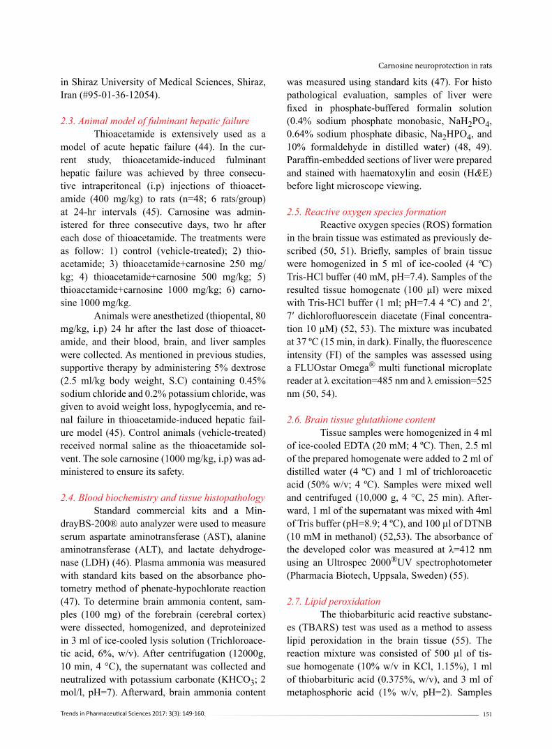
Figure 1. Serum biochemical measurements. TAA: Thioacetamide; Carn.: Carnosine.Data are given as Mean±SD (n=8).***Indicates significantly different as compared with the control group (P<0.001).aIndicates significantly different as compared with TAA group (P<0.001).
Ser
umA
LT(U
/l)
Con
trol
TAA
TAA
+C
arn.
250
mg/
kg
TAA
+C
arn.
500
mg/
kg
TAA
+C
arn.
1000
mg/
kg
0
2 0 0
4 0 0
6 0 0
8 0 0
1 0 0 0***
a a
a
Ser
umA
ST
(U/l)
Con
trol
TAA
TAA
+C
arn.
250
mg/
kg
TAA
+C
arn.
500
mg/
kg
TAA
+C
arn.
1000
mg/
kg
0
5 0 0
1 0 0 0
1 5 0 0
2 0 0 0
***
a
a a
Ser
umLD
H(U
/l)
Con
trol
TAA
TAA
+C
arn.
250
mg/
kg
TAA
+C
arn.
500
mg/
kg
TAA
+C
arn.
1000
mg/
kg
0
2 0 0 0
4 0 0 0
6 0 0 0
***
aa
a
Ser
umto
talb
iliru
bin
(mg/
dl)
Con
trol
TAA
TAA
+C
arn.
250
mg/
kg
TAA
+C
arn.
500
mg/
kg
TAA
+C
arn.
1000
mg/
kg
0
1
2
3
4
a
aa
***
152
Trends in Pharmaceutical Sciences 2017: 3(3): 149-160.
Carnosine neuroprotection in rats
acute hepatic failure as judged by drastic eleva-tion in serum markers of liver injury (Figure 1). On the other hand, liver tissue histopathological changes in the thioacetamide-treated group con-
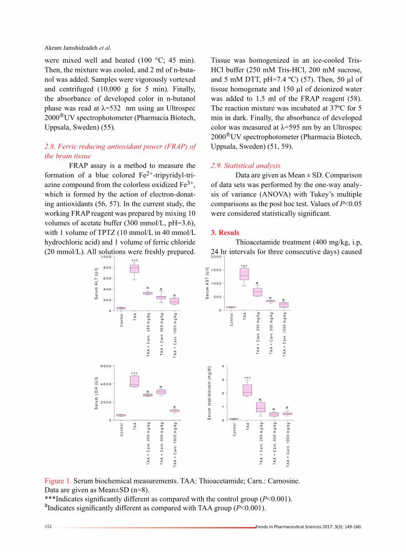
sisted of severe necrosis, ballooning degenera-tion, fatty changes, and inflammation (Figure 2). It was found that carnosine (250, 500, and 1000 mg/kg, i.p) decreased serum biomarkers of liver
Figure 2. Photomicrographs for liver tissue histopathology in thioacetamide and carnosine-treated groups. A: Control. B: Thioacetamide treated rats. C: Thioacetamide+Carnosine 250 mg/kg. D: Thioacetamide + Carnosine 500 mg/kg. E: Thioacetamide+Carnosine 1000 mg/kg. Signs of fatty changes and ballooning degeneration (red arrow) and inflammation (green arrow), developed in thioacetamide-administered ani-mals (B). Carnosine administration significantly alleviated thioacetamide-induced lesions (C, D & E) and no sign of tissue necrosis was observed in carnosine-treated (1000 mg/kg, i.p) animals (E).
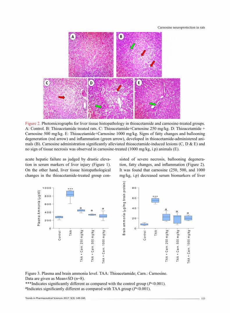
Figure 3. Plasma and brain ammonia level. TAA: Thioacetamide; Carn.: Carnosine.Data are given as Mean±SD (n=8).***Indicates significantly different as compared with the control group (P<0.001).aIndicates significantly different as compared with TAA group (P<0.001).
Plas
ma
Amm
onia
( g/
dl)
Con
trol
TAA
TAA
+C
arn.
250
mg/
kg
TAA
+C
arn.
500
mg/
kg
TAA
+C
arn.
1000
mg/
kg
0
2 0 0
4 0 0
6 0 0
8 0 0
1 0 0 0
a aa
***
Brai
nam
mon
ia(
g/m
gbr
ain
prot
ein)
Con
trol
TAA
TAA
+C
arn.
250
mg/
kg
TAA
+C
arn.
500
mg/
kg
TAA
+C
arn.
1000
mg/
kg
0
2 0
4 0
6 0
8 0
aaa
***
153
Trends in Pharmaceutical Sciences 2017: 3(3): 149-160.
Akram Jamshidzadeh et al.
injury (Figure 1). This peptide (250, 500, and 1000 mg/kg, i.p) also alleviated liver histopathological changes in thioacetamide-treated animals (Figure 2), and no sign of tissue necrosis was found in 1000 mg/k carnosine-treated animals (Figure 2). A high-level of ammonia was detected in the plasma of thioacetamide-treated animals (Fig-ure 3). Brain tissue ammonia level was also higher in thioacetamide-treated rats in comparison with the control group (Figure 3). It was found that car-nosine supplementation (250, 500, and 1000 mg/
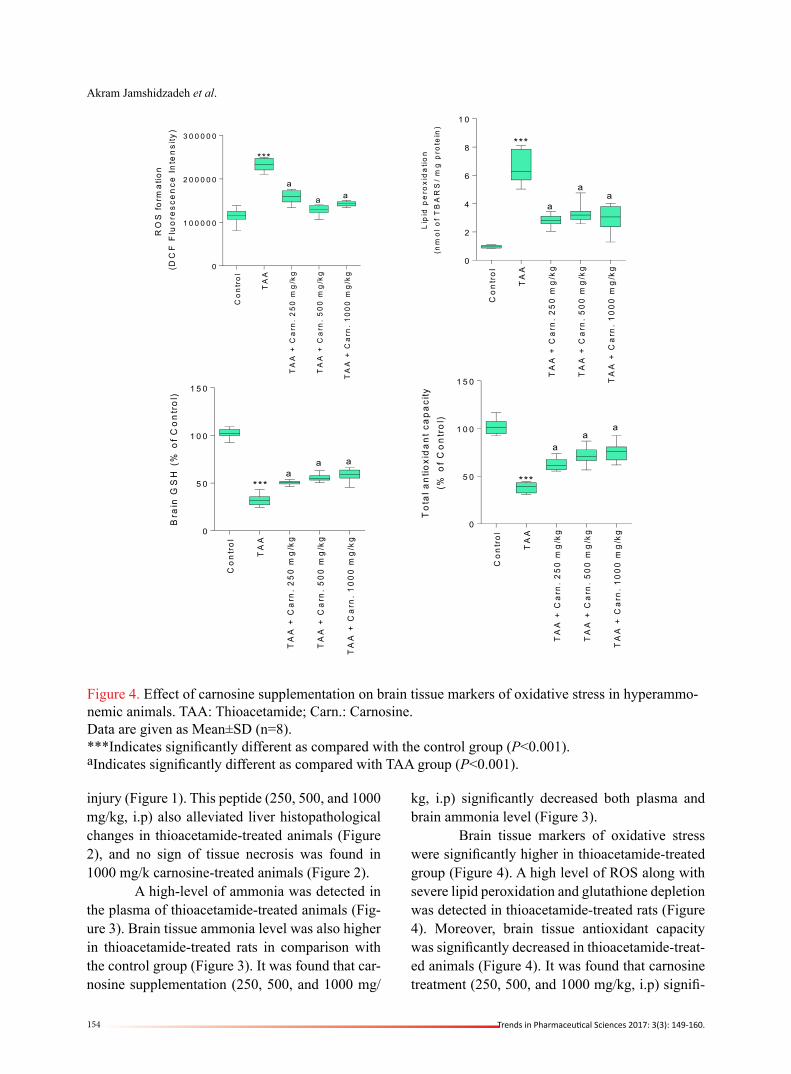
kg, i.p) significantly decreased both plasma and brain ammonia level (Figure 3). Brain tissue markers of oxidative stress were significantly higher in thioacetamide-treated group (Figure 4). A high level of ROS along with severe lipid peroxidation and glutathione depletion was detected in thioacetamide-treated rats (Figure 4). Moreover, brain tissue antioxidant capacity was significantly decreased in thioacetamide-treat-ed animals (Figure 4). It was found that carnosine treatment (250, 500, and 1000 mg/kg, i.p) signifi-
Lip
idp
ero
xid
atio
n(n
mo
lof
TB
AR
S/
mg
pro
tein
)
Co
ntr
ol
TA
A
TA
A+
Ca
rn.
25
0m
g/k
g
TA
A+
Ca
rn.
50
0m
g/k
g
TA
A+
Ca
rn.
10
00
mg
/kg
0
2
4
6
8
1 0
aa
a
***
Bra
inG
SH
(%o
fC
on
tro
l)
Co
ntr
ol
TA
A
TA
A+
Ca
rn.
25
0m
g/k
g
TA
A+
Ca
rn.
50
0m
g/k
g
TA
A+
Ca
rn.
10
00
mg
/kg
0
5 0
1 0 0
1 5 0
aaa
***
To
tala
ntio
xid
an
tca
pa
city
(%o
fC
on
tro
l)
Co
ntr
ol
TA
A
TA
A+
Ca
rn.
25
0m
g/k
g
TA
A+
Ca
rn.
50
0m
g/k
g
TA
A+
Ca
rn.
10
00
mg
/kg
0
5 0
1 0 0
1 5 0
a
aa
***
RO
Sfo
rma
tion
(DC
FF
luo
resc
en
ceIn
ten
sity
)
Co
ntr
ol
TA
A
TA
A+
Ca
rn.
25
0m
g/k
g
TA
A+
Ca
rn.
50
0m
g/k
g
TA
A+
Ca
rn.
10
00
mg
/kg
0
1 0 0 0 0 0
2 0 0 0 0 0
3 0 0 0 0 0
aaa
***
Figure 4. Effect of carnosine supplementation on brain tissue markers of oxidative stress in hyperammo-nemic animals. TAA: Thioacetamide; Carn.: Carnosine.Data are given as Mean±SD (n=8).***Indicates significantly different as compared with the control group (P<0.001).aIndicates significantly different as compared with TAA group (P<0.001).
154
Trends in Pharmaceutical Sciences 2017: 3(3): 149-160.
Carnosine neuroprotection in rats
cantly mitigated brain tissue markers of oxidative stress in hyperammonemic animals (Figure 4). Moreover, lower level of ROS and lipid peroxida-tion was detected in carnosine-supplemented rats (Figure 4). Carnosine treatment also preserved brain antioxidant capacity and prevented tissue glutathione depletion (Figure 4).
4. Discussion Oxidative stress and its associated compli-cations are known to be involved in the pathogen-esis of hyperammonemia-induced brain injury (7-9, 60-62). Therefore, antioxidant molecules might play a role in attenuating ammonia neurotoxicity. Carnosine is an endogenously-found peptide, with a high concentration in the human brain. Several pharmacological properties including antioxidant and radical scavenging activities are attributed to this peptide. In the current study, it was found that carnosine supplementation (250, 500, and 1000 mg/kg, i.p) to rats with acute liver failure and hyperammonemia, attenuated brain tissue mark-ers of oxidative stress. Furthermore, carnosine treatment decreased brain and plasma level of ammonia. It is well-known that ammonia-induced oxidative stress in the brain tissue and its delete-rious consequences play a major role in the neu-
rotoxicity induced by this chemical (9, 63). In accordance with previous studies, we found that acute liver failure and its associated hyperammo-nemia caused significant lipid peroxidation and a decrease in glutathione content of the brain tis-sue (Figures 4). On the other hand, carnosine is a well-known antioxidant and scavenger of reactive intermediates (35, 64-66). This peptide was also reported to boost antioxidant defense mechanisms and preserves cellular glutathione reservoirs (43, 67). In our experiments, carnosine (250, 500, and 1000 mg/kg, i.p) effectively alleviated oxidative stress and its consequences in hyperammonemic animals. Hence, the antioxidant capacity of carno-sine might play a major role in its protective prop-erties against ammonia-induced neurotoxicity. Mitochondrial dysfunction and brain energy crisis is a major mechanism involved in ammonia-induced neurotoxicity. It was found that ammonia caused a significant increase in brain mitochondrial ROS (68-70). Previous stud-ies indicated that carnosine effectively preserves mitochondrial membrane potential (71, 72). Mito-chondrial pH gradient and membrane potential are important factors for mitochondrial function (71). Hence, chemicals that are able to localize in the mitochondrial matrix and regulate matrix pH are capable of preserving mitochondrial membrane
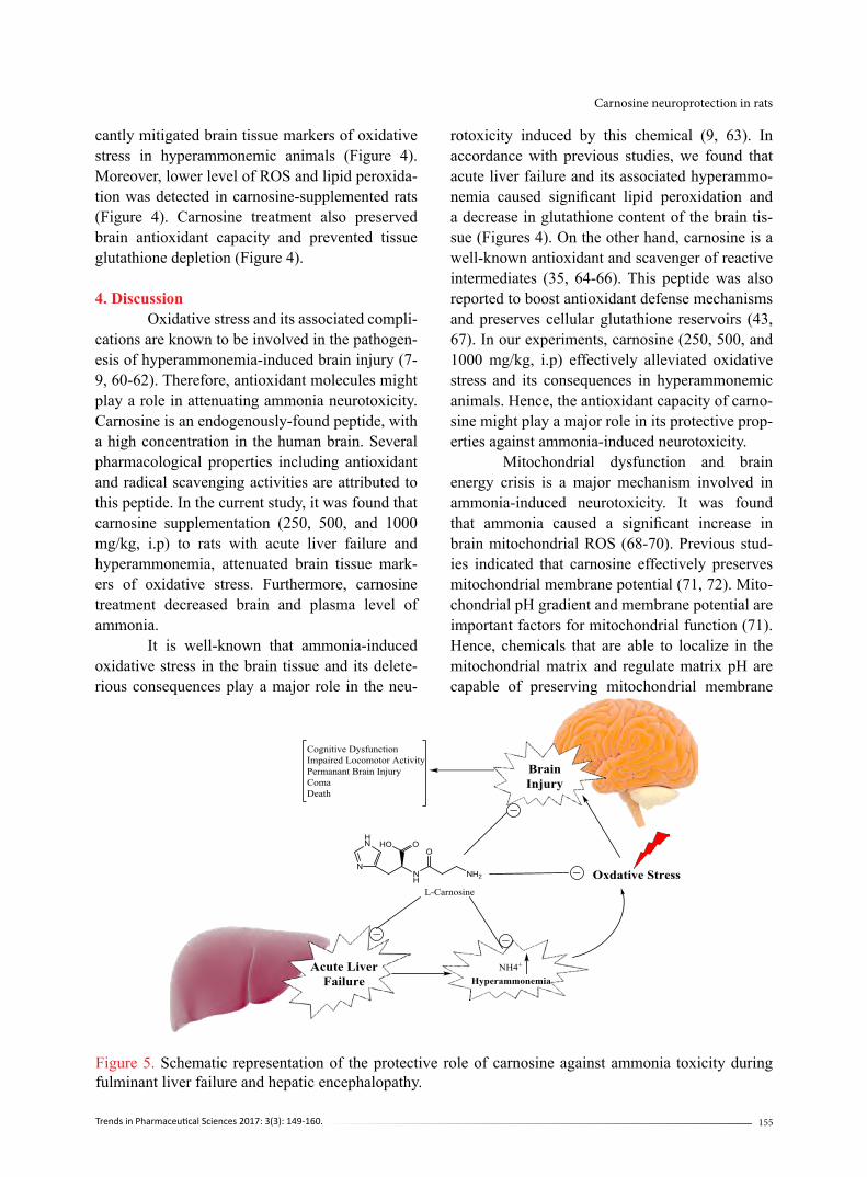
Figure 5. Schematic representation of the protective role of carnosine against ammonia toxicity during fulminant liver failure and hepatic encephalopathy.
155
Trends in Pharmaceutical Sciences 2017: 3(3): 149-160.
Akram Jamshidzadeh et al.
.................................................................................................................................
5. References1. Felipo V. Hepatic encephalopathy: effects of liver failure on brain function. Nature Rev Neu-rosci. 2013;14;851-8.2. Shawcross D, Jalan R. The pathophysio-logic basis of hepatic encephalopathy: central role for ammonia and inflammation. Cell Mol Life Sci.
2005;62;2295-304.3. Norenberg M. Oxidative and nitrosative stress in ammonia neurotoxicity. Hepatology. 2003;37;245-8.4. Bosoi CR, Rose CF. Identifying the direct effects of ammonia on the brain. Metab Brain Dis. 2008;24;95-102.
potential. Furthermore, the collapse of mitochon-drial membrane potential, along with impaired mi-tochondrial defense mechanisms were document-ed in ammonia-exposed mitochondria (68-70). Previously, we found that carnosine significantly mitigated ammonia-induced mitochondrial dys-function in vitro (69). Hence, mitochondrial pro-tecting properties of carnosine might be involved, at least in part, in the neuroprotective properties of this peptide in brain tissue of hyperammonemic models. Interestingly, it was reported that carno-sine significantly improved behavioral disturbanc-es in an in vivo model of acute liver failure (73). Previously, we found that carnosine supplemen-tation effectively improved locomotor activity in cirrhotic rats (74). Milewski et al. proposed that the beneficial effects of carnosine in the brain tis-sue of acute liver failure animal model might be mediated through its antioxidant capacity (73). In the investigation by Milewski et al., only one dose of carnosine was administered (100 mg/kg, i.p, 2 hr before thioacetamide challenge). On the other hand, they didn’t observe significant changes in some antioxidant parameters in carnosine-treated animals (73). In the current study, which is actually a part of a greater investigation on the role of car-nosine supplementation on the brain bioenergetics failure during HE, we found that higher doses of carnosine (250, 500, and 1000 mg/kg) effectively alleviated brain markers of oxidative stress in HE. Moreover, we detected lower plasma and brain ammonia level in carnosine supplemented animals (Figure 3). On the other hand, this peptide effec-tively preserved brain mitochondrial function dur-ing HE episodes (Data not shown). Furthermore, in an in vitro investigation on the role of carnosine supplementation on ammonia-induced mitochon-drial dysfunction, we found that carnosine could significantly preserve mitochondrial function in
a hyperammonemic environment (69). Tremor, rigidity, akinesia, athetosis, impaired locomo-tor activity, and cognitive dysfunction are major HE-associated neuropsychiatric symptoms (1) (Figure 5). It was shown that carnosine supple-mentation effectively mitigated ammonia-induced neuropsychiatric symptoms both in acute and chronic HE models (70, 73). All these data indi-cate that carnosine could be an eligible therapeutic option against ammonia neurotoxicity (Figure 5). The beneficial effects of carnosine in HE could be due to its direct effects on brain tis-sue, where this peptide encounters oxidative stress and its associated complications. On the other hand, carnosine could affect the liver and prevent an increase in plasma and brain ammonia (70) (Figure 5). Hence, the hepatoprotective effects of carnosine might also play a role in the neuropro-tection provided by this peptide (Figure 5). In conclusion, our data suggest that car-nosine exhibits a robust protective effect against ammonia-induced oxidative damage in the CNS. Hence, carnosine might be an ideal therapeutic option against HE. However, further research in different animal models of hepatic failure and hy-perammonemia is needed for understanding the precise mechanism of action of carnosine against ammonia-induced CNS injury.
Acknowledgment The authors thank Pharmaceutical Scienc-es Research Center (PSRC) and the Vice-Chan-cellor of Research Affairs of Shiraz University of Medical Sciences for providing technical and fi-nancial support of the current investigation (Grant number 12054/9741/9823).
Conflict of Interest None declared.
156
Trends in Pharmaceutical Sciences 2017: 3(3): 149-160.
Carnosine neuroprotection in rats
5. Albrecht J, Jones EA. Hepatic encepha-lopathy: molecular mechanisms underlying the clinical syndrome. J Neurol Sci. 1999;170;138-46.6. Felipo V, Butterworth RF. Mitochondrial dysfunction in acute hyperammonemia. Neuro-chem Int. 2002;40;487-91.7. Braissant O, McLin VA, Cudalbu C. Am-monia toxicity to the brain. J Inher Metab Dis. 2013;36;595-612.8. Görg B, Qvartskhava N, Bidmon H-J, Palomero-Gallagher N, Kircheis G, Zilles K, Häussinger D. Oxidative stress markers in the brain of patients with cirrhosis and hepatic en-cephalopathy. Hepatology. 2010;52;256-65.9. Norenberg MD, Jayakumar AR, Rao KVR. Oxidative Stress in the Pathogenesis of Hepatic Encephalopathy. Metab Brain Dis. 2004;19;313-29.10. Rama Rao KV, Reddy PVB, Tong X, No-renberg MD. Brain Edema in Acute Liver Failure. Am J Pathol. 2010;176;1400-8.11. Shawcross DL, Shabbir SS, Taylor NJ, Hughes RD. Ammonia and the neutrophil in the pathogenesis of hepatic encephalopathy in cirrho-sis. Hepatology. 2010;51;1062-9.12. Skowrońska M, Albrecht J. Oxidative and nitrosative stress in ammonia neurotoxicity. Neu-rochem Int. 2013;62;731-7.13. Crush KG. Carnosine and related sub-stances in animal tissues. Comp Biochem Physiol. 1970;34;3-30.14. Boldyrev AA, Aldini G, Derave W. Physi-ology and pathophysiology of carnosine. Physiol Rev. 2013;93;1803-45.15. Budzeń S, Rymaszewska J. The biological role of carnosine and its possible applications in medicine. Adv Clin Exp Med. 2013;22;739-44.16. Hipkiss AR, Preston JE, Himsworth DTM, Worthington VC, Keown M, Michaelis J, Law-rence J, Mateen A, Allende L, Eagles PAM, oth-ers. Pluripotent Protective Effects of Carnosine, a Naturally Occurring Dipeptidea. Ann New York Acad Sci. 1998;854;37-53.17. Prokopieva VD, Yarygina EG, Bokhan NA, Ivanova SA. Use of Carnosine for Oxidative Stress Reduction in Different Pathologies. Oxida-tive Med Cell Long. 2016;2016;2939087.18. Boldyrev AA. Carnosine: new concept for the function of an old molecule. Biochemistry (Mosc). 2012;77:313-26.
19. Aydın AF, Küçükgergin C, Özdemirler-Erata G, Koçak-Toker N, Uysal M. The effect of carnosine treatment on prooxidant–antioxidant balance in liver, heart and brain tissues of male aged rats. Biogerontology. 2010;11;103-9.20. Heidari R, Niknahad H, Jamshidzadeh A, Azarpira N, Bazyari M, Najibi A. Carbonyl traps as potential protective agents against methima-zole-induced liver injury. J Biochem Mol Toxicol. 2015;29;173-81.21. Kohen R, Yamamoto Y, Cundy KC, Ames BN. Antioxidant activity of carnosine, homocar-nosine, and anserine present in muscle and brain. Proc Nat Acad Sci. 1988;85;3175-9.22. Soliman KM, Abdul-Hamid M, Othman AI. Effect of carnosine on gentamicin-induced nephrotoxicity. Med Sci Technol. 2007;13;BR73-BR83.23. Akram J, Reza H, Farzaneh A, Maral R, Forouzan K, Mohammad Mehdi O, Maryam A, Roya F, Arastoo S, Negar A, Asma N. Antima-larial Drugs-Induced Hepatic Injury in Rats and the Protective Role of Carnosine. Pharm Sci. 2016;22;170-80.24. Fouad AA, El-Rehany MA-A, Maghra-by HK. The hepatoprotective effect of carnosine against ischemia/reperfusion liver injury in rats. Europ J Pharmacol. 2007;572;61-8.25. Fouad AA, Morsy MA, Gomaa W. Protective effect of carnosine against cisplatin-induced nephrotoxicity in mice. Environ Toxicol Pharmacol. 2008;25;292-7.26. Lee Y-t, Hsu C-c, Lin M-h, Liu K-s, Yin M-c. Histidine and carnosine delay diabetic dete-rioration in mice and protect human low density lipoprotein against oxidation and glycation. Europ J Pharmacol. 2005;513;145-50.27. Fouad AA, Qureshi HA, Yacoubi MT, Al-Melhim WN. Protective role of carnosine in mice with cadmium-induced acute hepatotoxicity. Food Chem Toxicol. 2009;47;2863-70.28. Kuloglu N, Sönmez MF. A biochemical and immunohistochemical study of the protec-tive effects of carnosine for carbon tetrachloride induced liver injury in rats. Biotech Histochem. 2015;90:608-1429. Yan S-l, Wu S-t, Yin M-c, Chen H-t, Chen H-c. Protective Effects from Carnosine and His-tidine on Acetaminophen-Induced Liver Injury. J Food Sci. 2009;74;H259-H265.
157
Trends in Pharmaceutical Sciences 2017: 3(3): 149-160.
Akram Jamshidzadeh et al.
43. Rajanikant GK, Zemke D, Senut M-C, Frenkel MB, Chen AF, Gupta R, Majid A. Carno-sine Is Neuroprotective Against Permanent Focal Cerebral Ischemia in Mice. Stroke. 2007;38;3023-31.44. Tuñón MJ, Alvarez M, Culebras JM, González-Gallego J. An overview of animal mod-els for investigating the pathogenesis and thera-peutic strategies in acute hepatic failure. World J Gastroenterol. 2009;15:3086-98.45. Bruck R, Aeed H, Shirin H, Matas Z, Zaid-el L, Avni Y, Halpern Z. The hydroxyl radical scav-engers dimethylsulfoxide and dimethylthiourea protect rats against thioacetamide-induced fulmi-nant hepatic failure. J Hepatology. 1999;31;27-38.46. Heidari R, Jamshidzadeh A, Keshavarz N, Azarpira N. Mitigation of Methimazole-Induced Hepatic Injury by Taurine in Mice. Sci Pharm. 2014;83;143-51.47. Chatauret N, Desjardins P, Zwingmann C, Rose C, Rao KVR, Butterworth RF. Direct mo-lecular and spectroscopic evidence for increased ammonia removal capacity of skeletal muscle in acute liver failure. J Hepatol. 2006;44:1083-8.48. Moezi L, Heidari R, Amirghofran Z, Nekooeian AA, Monabati A, Dehpour AR. En-hanced anti-ulcer effect of pioglitazone on gastric ulcers in cirrhotic rats: The role of nitric oxide and IL-1b. Pharmacol Report. 2013;65;134-43.49. Heidari R, Babaei H, Roshangar L, Egh-bal MA. Effects of Enzyme Induction and/or Glutathione Depletion on Methimazole-Induced Hepatotoxicity in Mice and the Protective Role of N-Acetylcysteine. Adv Pharm Bull. 2014;4;21-8.50. Gupta R, Dubey DK, Kannan GM, Flora SJS. Concomitant administration of Moringa ole-ifera seed powder in the remediation of arsenic-induced oxidative stress in mouse. Cell Biol Int. 2007;31;44-56.51. Heidari R, Jamshidzadeh A, Niknahad H, Safari F, Azizi H, Abdoli N, Ommati MM, Khodaei F, Saeedi A, Najibi A. The Hepatopro-tection Provided by Taurine and Glycine against Antineoplastic Drugs Induced Liver Injury in an Ex Vivo Model of Normothermic Recirculating Isolated Perfused Rat Liver. Trend Pharm Sci. 2016;2;59-76.52. Heidari R, Taheri V, Rahimi HR, Shirazi Yeganeh B, Niknahad H, Najibi A. Sulfasala-zine-induced renal injury in rats and the pro-
30. Boldyrev A, Gallant S, Sukhich G. Car-nosine, the protective, anti-aging peptide. Biosci Report. 1999;19;581-7.31. Fu H, Katsumura Y, Lin M, Muroya Y, Hata K, Fujii K, Yokoya A, Hatano Y. Free radical scavenging and radioprotective effects of carnosine and anserine. Rad Physic Chem. 2009;78;1192-1197.32. Guiotto A, Calderan A, Ruzza P, Borin G. Carnosine and carnosine-related antioxidants: a review. Curr Med Chem. 2005;12;2293-315.33. Bonfanti L, Peretto P, De Marchis S, Faso-lo A. Carnosine-related dipeptides in the mamma-lian brain. Progress Neurobiol. 1999;59;333-53.34. Hipkiss AR. Carnosine and its possible roles in nutrition and health. Adv Food Nut Res. 2009;57;87-154.35. Bellia F, Vecchio G, Cuzzocrea S, Cal-abrese V, Rizzarelli E. Neuroprotective features of carnosine in oxidative driven diseases. Mol Asp Med. 2011;32;258-66.36. Hipkiss AR. Could Carnosine or Related Structures Suppress Alzheimer’s Disease? J Al-zheimer’s Dis. 2007;11;229-40.37. Boldyrev A, Fedorova T, Stepanova M, Dobrotvorskaya I, Kozlova E, Boldanova N, Bagyeva G, Ivanova-Smolenskaya I, Illarioshkin S. Carnisone Increases Efficiency of DOPA Thera-py of Parkinson’s Disease: A Pilot Study. Rejuven Res. 2008;11;821-7.38. Trombley PQ, Horning MS, Blakemore LJ. Interactions between carnosine and zinc and copper: implications for neuromodulation and neuroprotection. Biochemistry. 2000;65;807-16.39. Masahiro K, Keiko K, Tetsuya N, Yutaka S. Protective Substances Against Zinc-Induced Neuronal Death after Ischemia:Carnosine as a Tar-get for Drug of Vascular Type of Dementia. Recent Pat CNS Drug Discov. 2007;2:145-9.40. Brownrigg TD, Theisen CS, Fibuch EE, Seidler NW. Carnosine Protects Against the Neu-rotoxic Effects of a Serotonin-Derived Melanoid. Neurochem Res. 2010;36;467-75.41. Boldyrev AA. Carnosine: new concept for the function of an old molecule. Biochemistry. 2012;77;313-326.42. Gallant S, Kukley M, Stvolinsky S, Bu-lygina E, Boldyrev A. Effect of carnosine on rats under experimental brain ischemia. Tohoku J Exp Med. 2000;191;85-99.
158
Trends in Pharmaceutical Sciences 2017: 3(3): 149-160.
Carnosine neuroprotection in rats
tective role of thiol-reductants. Renal Failure. 2016;38;137-41.53. Akram J, Hossein N, Reza H, Maryam A, Forouzan K, Mohammad Reza A, Omid F. Propyl-thiouracil-induced mitochondrial dysfunction in liver and its relevance to drug-induced hepatotox-icity. Pharm Sci. 2017;23;95-102.54. Socci DJ, Bjugstad KB, Jones HC, Pat-tisapu JV, Arendash GW. Evidence that oxidative stress is associated with the pathophysiology of in-herited hydrocephalus in the H-Tx rat model. Exp Neurol. 1999;155;109-17.55. Heidari R, Babaei H, Roshangar L, Egh-bal MA. Effects of Enzyme Induction and/or Glutathione Depletion on Methimazole-Induced Hepatotoxicity in Mice and the Protective Role of N-Acetylcysteine. Adv Pharm Bull. 2014;4;21-8.56. Katalinic V, Modun D, Music I, Boban M. Gender differences in antioxidant capacity of rat tissues determined by 2,2’-azinobis (3-ethylbenzo-thiazoline 6-sulfonate; ABTS) and ferric reducing antioxidant power (FRAP) assays. Comp Biochem Physiol Toxicol Pharmacol. 2005;140;47-52.57. Hossein N, Akram J, Reza H, Narges A, Mohammad Mehdi O, Faezeh J, Mahdi Z, Behnam A. The Postulated Hepatotoxic Metabolite of Me-thimazole Causes Mitochondrial Dysfunction and Energy Metabolism Disturbances in Liver. Pharm Sci. 2016;22;217-26.58. Heidari R, Jafari F, Khodaei F, Shirazi Ye-ganeh B, Niknahad H. The Mechanism of Valproic Acid-Induced Fanconi Syndrome Involves Mito-chondrial Dysfunction and Oxidative Stress in Rat Kidney. Nephrology (Carlton, Vic). 2017; In-Press. 59. Alía M, Horcajo C, Bravo L, Goya L. Ef-fect of grape antioxidant dietary fiber on the total antioxidant capacity and the activity of liver anti-oxidant enzymes in rats. Nut Res. 2003;23;1251-67.60. Túnez I, Muñoz MC, Medina FJ, Salcedo M, Feijóo M, Montilla P. Comparison of mela-tonin, vitamin E and L-carnitine in the treatment of neuro- and hepatotoxicity induced by thioacet-amide. Cell Biochem Func. 2007;25;119-27.61. Häussinger D, Schliess F. Pathogenetic mechanisms of hepatic encephalopathy. Gut. 2008;57;1156-65.62. Kosenko E, Kaminsky M, Kaminsky A, Valencia M, Lee L, Hermenegildo C, Felipo V. Superoxide Production and Antioxidant Enzymes
in Ammonia Intoxication in Rats. Free Rad Res. 1997;27;637-44.63. Lemberg A, Fernández MA. Hepatic en-cephalopathy, ammonia, glutamate, glutamine and oxidative stress. Ann Hepatol. 2009;8;95-102.64. Stvolinsky SL, Kukley ML, Dobrota D, Matejovicova M, Tkac I, Boldyrev AA. Carno-sine: an endogenous neuroprotector in the isch-emic brain. Cell Mol Neurobiol. 1999;19;45-56.65. Lopachev AV, Lopacheva OM, Abaimov DA, Koroleva OV, Vladychenskaya EA, Erukhi-movich AA, Fedorova TN. Neuroprotective effect of carnosine on primary culture of rat cerebellar cells under oxidative stress. Biochemistry (Mos-cow). 2016;81;511-20.66. Baye E, Ukropcova B, Ukropec J, Hipkiss A, Aldini G, Courten Bd. Physiological and thera-peutic effects of carnosine on cardiometabolic risk and disease. Amino Acids. 2016;48;1131-49.67. Kim J, Padanilam BJ. Loss of poly (ADP-ribose) polymerase 1 attenuates renal fibrosis and inflammation during unilateral ureteral obstruc-tion. Am J Physiol Renal Physiol. 2011;301;F450-F459.68. Jamshidzadeh A, Heidari R, Abasvali M, Zarei M, Ommati MM, Abdoli N, Khodaei F, Ye-ganeh Y, Jafari F, Zarei A, Latifpour Z, Mardani E, Azarpira N, Asadi B, Najibi A. Taurine treat-ment preserves brain and liver mitochondrial func-tion in a rat model of fulminant hepatic failure and hyperammonemia. Biomed Pharmacother. 2017;86;514-20.69. Jamshidzadeh A, Niknahad H, Heidari R, Zarei M, Ommati MM, Khodaei F. Carnosine pro-tects brain mitochondria under hyperammonemic conditions: Relevance to hepatic encephalopathy treatment. PharmaNutrition. 2017;5;58-63.70. Niknahad H, Jamshidzadeh A, Heidari R, Zarei M, Ommati MM. Ammonia-induced mito-chondrial dysfunction and energy metabolism dis-turbances in isolated brain and liver mitochondria, and the effect of taurine administration: relevance to hepatic encephalopathy treatment. Clin Exp Hepatol. 2017;3.71. Hansen SH, Andersen ML, Cornett C, Gradinaru R, Grunnet N. A role for taurine in mi-tochondrial function. J Biomed Sci. 2010;17;1-8.72. Shen Y, He P, Fan Y-y, Zhang J-x, Yan H-j, Hu W-w, Ohtsu H, Chen Z. Carnosine pro-tects against permanent cerebral ischemia in his-
159
Trends in Pharmaceutical Sciences 2017: 3(3): 149-160.
Akram Jamshidzadeh et al.
tidine decarboxylase knockout mice by reduc-ing glutamate excitotoxicity. Free Rad Biol Med. 2010;48;727-35.73. Milewski K, Hilgier W, Fręśko I, Polowy R, Podsiadłowska A, Zołocińska E, Grymanowska AW, Filipkowski RK, Albrecht J, Zielińska M. Carnosine Reduces Oxidative Stress and Reverses Attenuation of Righting and Postural Reflexes in Rats with Thioacetamide-Induced Liver Failure.
Neurochem Res. 2016;41;376-84.74. Jamshidzadeh A, Heidari R, Latifpour Z, Ommati MM, Abdoli N, Mousavi S, Azarpira N, Zarei A, Zarei M, Asadi B, Abasvali M, Ye-ganeh Y, Jafari F, Saeedi A, Najibi A, Mardani E. Carnosine ameliorates liver fibrosis and hyper-ammonemia in cirrhotic rats. Clin Res Hepatol Gastroenterol. 2017; In-Press. DOI: 10.1016/j.phanu.2017.02.004
160