Embed Size (px)
Citation preview
JCB: Article
The Rockefeller University Press $30.00J. Cell Biol. Vol. 193 No. 7 1181–1196www.jcb.org/cgi/doi/10.1083/jcb.201006114 JCB 1181
L. Qian and J.D. Wythe contributed equally to this paper.Correspondence to Rolf Bodmer: [email protected]; or Li Qian: lqian@gladstone .ucsf.eduAbbreviations used in this paper: CHD, congenital heart disease; UAS, upstream activation sequence.
IntroductionCardiovascular disease is the leading cause of mortality and morbidity in industrialized nations. Since the discovery of the first key determinant of heart development—tinman—in Drosophila (Azpiazu and Frasch, 1993; Bodmer, 1993), numerous regula-tors, including transcription factors, cell signaling molecules, extracellular matrix proteins, and microRNAs have provided an understanding of an increasingly complex network that guides
cardiac specification, differentiation, function, and maturation in all organisms that possess a heart (Bier and Bodmer, 2004; Olson, 2006; Srivastava, 2006; Qian et al., 2008a).
The fruit fly Drosophila is an attractive model system to study various human diseases, including cardiac disease (St Johnston, 2002; Bier, 2005). The Drosophila heart is a simple linear tube comprised of myocardial and pericardial cells, which is reminiscent of the primitive vertebrate embryonic heart.
Unraveling the gene regulatory networks that gov-ern development and function of the mammalian heart is critical for the rational design of thera-
peutic interventions in human heart disease. Using the Drosophila heart as a platform for identifying novel gene interactions leading to heart disease, we found that the Rho-GTPase Cdc42 cooperates with the cardiac transcrip-tion factor Tinman/Nkx2-5. Compound Cdc42, tinman heterozygous mutant flies exhibited impaired cardiac out-put and altered myofibrillar architecture, and adult heart–specific interference with Cdc42 function is sufficient to cause these same defects. We also identified K+ channels,
encoded by dSUR and slowpoke, as potential effectors of the Cdc42–Tinman interaction. To determine whether a Cdc42–Nkx2-5 interaction is conserved in the mamma-lian heart, we examined compound heterozygous mutant mice and found conduction system and cardiac output defects. In exploring the mechanism of Nkx2-5 interaction with Cdc42, we demonstrated that mouse Cdc42 was a target of, and negatively regulated by miR-1, which itself was negatively regulated by Nkx2-5 in the mouse heart and by Tinman in the fly heart. We conclude that Cdc42 plays a conserved role in regulating heart function and is an indirect target of Tinman/Nkx2-5 via miR-1.
Tinman/Nkx2-5 acts via miR-1 and upstream of Cdc42 to regulate heart function across species
Li Qian,1,2,3 Joshua D. Wythe,2,3 Jiandong Liu,1,4 Jerome Cartry,1 Georg Vogler,1 Bhagyalaxmi Mohapatra,5,12 Robyn T. Otway,6 Yu Huang,2 Isabelle N. King,2,3 Marjorie Maillet,14 Yi Zheng,14 Timothy Crawley,1 Ouarda Taghli-Lamallem,1 Christopher Semsarian,10 Sally Dunwoodie,7,8 David Winlaw,11 Richard P. Harvey,7,8 Diane Fatkin,6,8,9 Jeffrey A. Towbin,5,13 Jeffery D. Molkentin,14 Deepak Srivastava,2,3 Karen Ocorr,1 Benoit G. Bruneau,2,3 and Rolf Bodmer1
1Development and Aging Program, NASCR Center, Sanford-Burnham Medical Research Institute, La Jolla, CA 920372Gladstone Institute for Cardiovascular Disease, 3Department of Pediatrics and Cardiovascular Research Institute, and 4Department of Biochemistry and Biophysics, University of California, San Francisco, San Francisco, CA 94158
5Pediatric Cardiology, Baylor College of Medicine, Texas Children’s Hospital, Houston, TX 770306Division of Molecular Cardiology, 7Division of Developmental Biology, Victor Chang Cardiac Research Institute, 8Faculty of Medicine, and 9St. Vincent’s Hospital, University of New South Wales, Sydney, Australia
10Department of Cardiology, Agnes Ginges Centre for Molecular Cardiology, Centenary Institute, Faculty of Medicine, Royal Prince Alfred Hospital, University of Sydney, Sydney NSW 2006, Australia
11Adolph Basser Cardiac Research and Kids Heart Research, Children’s Hospital at Westmead, Faculty of Medicine, University of Sydney, Sydney, Australia12Department of Zoology, BHU, Varanasi-221005, India13Pediatric Cardiology and 14Molecular Cardiovascular Biology, The Heart Institute, Cincinnati Children’s Hospital Medical Center, Cincinnati, OH 45229
© 2011 Qian et al. This article is distributed under the terms of an Attribution–Noncommercial–Share Alike–No Mirror Sites license for the first six months after the pub-lication date (see http://www.rupress.org/terms). After six months it is available under a Creative Commons License (Attribution–Noncommercial–Share Alike 3.0 Unported license, as described at http://creativecommons.org/licenses/by-nc-sa/3.0/).
TH
EJ
OU
RN
AL
OF
CE
LL
BIO
LO
GY
on April 10, 2019jcb.rupress.org Downloaded from http://doi.org/10.1083/jcb.201006114Published Online: 20 June, 2011 | Supp Info:
JCB • VOLUME 193 • NUMBER 7 • 2011 1182
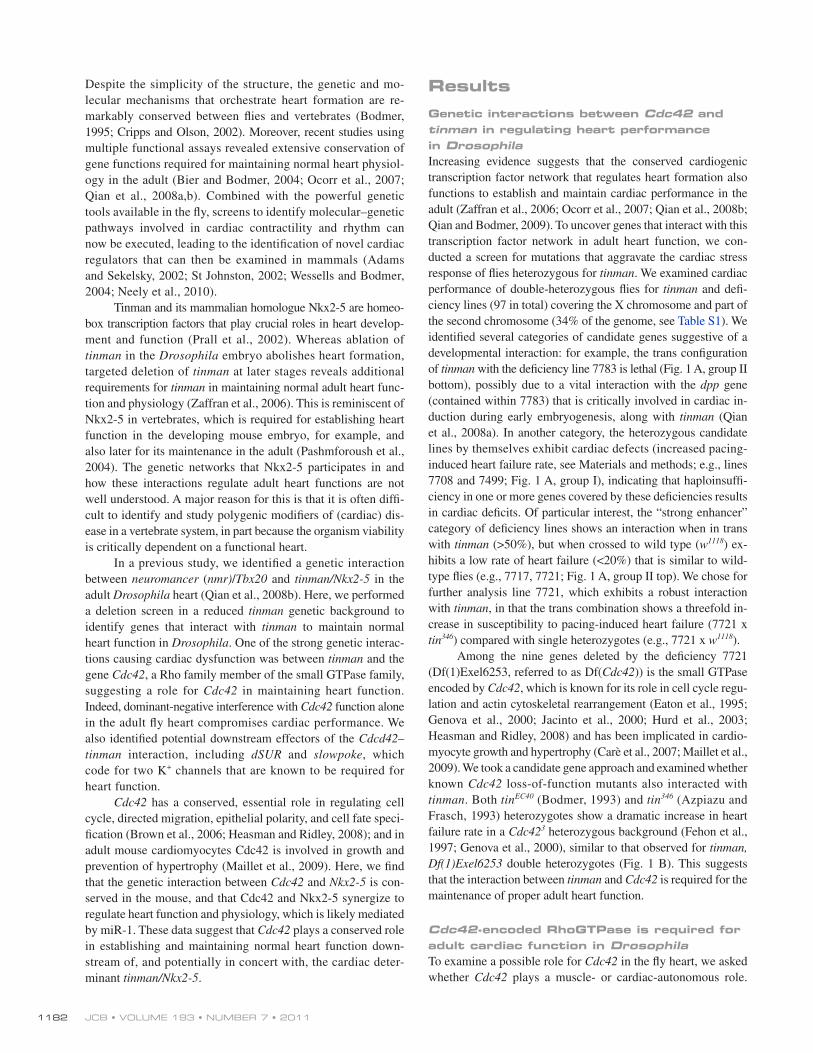
ResultsGenetic interactions between Cdc42 and tinman in regulating heart performance in DrosophilaIncreasing evidence suggests that the conserved cardiogenic transcription factor network that regulates heart formation also functions to establish and maintain cardiac performance in the adult (Zaffran et al., 2006; Ocorr et al., 2007; Qian et al., 2008b; Qian and Bodmer, 2009). To uncover genes that interact with this transcription factor network in adult heart function, we con-ducted a screen for mutations that aggravate the cardiac stress response of flies heterozygous for tinman. We examined cardiac performance of double-heterozygous flies for tinman and defi-ciency lines (97 in total) covering the X chromosome and part of the second chromosome (34% of the genome, see Table S1). We identified several categories of candidate genes suggestive of a developmental interaction: for example, the trans configuration of tinman with the deficiency line 7783 is lethal (Fig. 1 A, group II bottom), possibly due to a vital interaction with the dpp gene (contained within 7783) that is critically involved in cardiac in-duction during early embryogenesis, along with tinman (Qian et al., 2008a). In another category, the heterozygous candidate lines by themselves exhibit cardiac defects (increased pacing- induced heart failure rate, see Materials and methods; e.g., lines 7708 and 7499; Fig. 1 A, group I), indicating that haploinsuffi-ciency in one or more genes covered by these deficiencies results in cardiac deficits. Of particular interest, the “strong enhancer” category of deficiency lines shows an interaction when in trans with tinman (>50%), but when crossed to wild type (w1118) ex-hibits a low rate of heart failure (<20%) that is similar to wild-type flies (e.g., 7717, 7721; Fig. 1 A, group II top). We chose for further analysis line 7721, which exhibits a robust interaction with tinman, in that the trans combination shows a threefold in-crease in susceptibility to pacing-induced heart failure (7721 x tin346) compared with single heterozygotes (e.g., 7721 x w1118).
Among the nine genes deleted by the deficiency 7721 (Df(1)Exel6253, referred to as Df(Cdc42)) is the small GTPase encoded by Cdc42, which is known for its role in cell cycle regu-lation and actin cytoskeletal rearrangement (Eaton et al., 1995; Genova et al., 2000; Jacinto et al., 2000; Hurd et al., 2003; Heasman and Ridley, 2008) and has been implicated in cardio-myocyte growth and hypertrophy (Carè et al., 2007; Maillet et al., 2009). We took a candidate gene approach and examined whether known Cdc42 loss-of-function mutants also interacted with tinman. Both tinEC40 (Bodmer, 1993) and tin346 (Azpiazu and Frasch, 1993) heterozygotes show a dramatic increase in heart failure rate in a Cdc423 heterozygous background (Fehon et al., 1997; Genova et al., 2000), similar to that observed for tinman, Df(1)Exel6253 double heterozygotes (Fig. 1 B). This suggests that the interaction between tinman and Cdc42 is required for the maintenance of proper adult heart function.
Cdc42-encoded RhoGTPase is required for adult cardiac function in DrosophilaTo examine a possible role for Cdc42 in the fly heart, we asked whether Cdc42 plays a muscle- or cardiac-autonomous role.
Despite the simplicity of the structure, the genetic and mo-lecular mechanisms that orchestrate heart formation are re-markably conserved between flies and vertebrates (Bodmer, 1995; Cripps and Olson, 2002). Moreover, recent studies using multiple functional assays revealed extensive conservation of gene functions required for maintaining normal heart physiol-ogy in the adult (Bier and Bodmer, 2004; Ocorr et al., 2007; Qian et al., 2008a,b). Combined with the powerful genetic tools available in the fly, screens to identify molecular–genetic pathways involved in cardiac contractility and rhythm can now be executed, leading to the identification of novel cardiac regulators that can then be examined in mammals (Adams and Sekelsky, 2002; St Johnston, 2002; Wessells and Bodmer, 2004; Neely et al., 2010).
Tinman and its mammalian homologue Nkx2-5 are homeo-box transcription factors that play crucial roles in heart develop-ment and function (Prall et al., 2002). Whereas ablation of tinman in the Drosophila embryo abolishes heart formation, targeted deletion of tinman at later stages reveals additional requirements for tinman in maintaining normal adult heart func-tion and physiology (Zaffran et al., 2006). This is reminiscent of Nkx2-5 in vertebrates, which is required for establishing heart function in the developing mouse embryo, for example, and also later for its maintenance in the adult (Pashmforoush et al., 2004). The genetic networks that Nkx2-5 participates in and how these interactions regulate adult heart functions are not well understood. A major reason for this is that it is often diffi-cult to identify and study polygenic modifiers of (cardiac) dis-ease in a vertebrate system, in part because the organism viability is critically dependent on a functional heart.
In a previous study, we identified a genetic interaction between neuromancer (nmr)/Tbx20 and tinman/Nkx2-5 in the adult Drosophila heart (Qian et al., 2008b). Here, we performed a deletion screen in a reduced tinman genetic background to identify genes that interact with tinman to maintain normal heart function in Drosophila. One of the strong genetic interac-tions causing cardiac dysfunction was between tinman and the gene Cdc42, a Rho family member of the small GTPase family, suggesting a role for Cdc42 in maintaining heart function. Indeed, dominant-negative interference with Cdc42 function alone in the adult fly heart compromises cardiac performance. We also identified potential downstream effectors of the Cdcd42–tinman interaction, including dSUR and slowpoke, which code for two K+ channels that are known to be required for heart function.
Cdc42 has a conserved, essential role in regulating cell cycle, directed migration, epithelial polarity, and cell fate speci-fication (Brown et al., 2006; Heasman and Ridley, 2008); and in adult mouse cardiomyocytes Cdc42 is involved in growth and prevention of hypertrophy (Maillet et al., 2009). Here, we find that the genetic interaction between Cdc42 and Nkx2-5 is con-served in the mouse, and that Cdc42 and Nkx2-5 synergize to regulate heart function and physiology, which is likely mediated by miR-1. These data suggest that Cdc42 plays a conserved role in establishing and maintaining normal heart function down-stream of, and potentially in concert with, the cardiac deter-minant tinman/Nkx2-5.
1183Cdc42-Tin/Nkx2-5 in the heart • Qian et al.
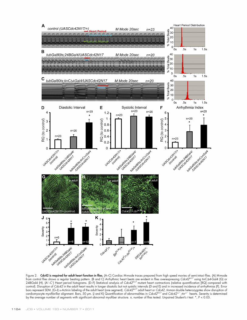
suggesting that Cdc42 is essential for maintaining normal heart function. These Cdc42N17 mutant hearts, when assessed for their beating pattern, also show increased incidence of arrhythmias and an overall lower heart rate, due to lengthening of diastolic intervals, compared with controls (Fig. 2, A–F). Using the mean standard deviation of heart period as an indicator of heart rhythm disturbance (“arrhythmia index”; Ocorr et al., 2007; Fink et al., 2009), we found a dramatic increase in arrhythmias in Cdc42N17 hearts (Fig. 2 F). These effects are more dramatic than in Cdc42 heterozygotes and together strongly support a crucial require-ment for Cdc42 in regulating adult heart function.
The defective Cdc42N17 adult hearts also showed abnor-malities in the cellular architecture of the cardiomyocytes, as manifest by severely misaligned myofibrils and irregularities in the repeated Z-line pattern (Fig. 2, G–I). These structural abnor-malities in Cdc42 mutant hearts are consistent with Cdc42’s role in actin filament assembly, also observed in Drosophila nurse
For this purpose, we expressed a dominant-negative form of Cdc42 (UAS-Cdc42N17; Luo et al., 1994) specifically in the adult heart using tinC4Gal4 (a UAS-Gal4 system that drives gene expression only in myocardial cells; Qian et al., 2005a), or in all mesoderm using 24BGal4 (drives gene expression pan-mesodermally; Brand and Perrimon, 1993). The UAS-GAL4 system has two parts: the GAL4 gene that encodes the yeast transcriptional activator protein Gal4, and the upstream activa-tion sequence (UAS), which is a short section of the promoter region to which Gal4 specifically binds to activate gene tran-scription (Brand and Perrimon, 1993). By using the TARGET system (Gal4/Gal80, see Materials and methods; McGuire et al., 2004) in combination with UASCdc42N17, we specifically inter-fered with Cdc42 function in the adult heart without disturbing its function in the embryo. Interference with Cdc42 function specifically in the heart within the first week of adult life causes a dramatic increase in pacing-induced heart failure (Fig. 1 C),
Figure 1. Genetic screen identifying Cdc42. (A) Candidate deficiency lines interacting with tin346 (susceptibility to pacing-induced heart arrest or fibrilla-tion). (B) Genetic interaction between tinman, Cdc42, and deficiency 7721. Cdc42+/;tin+/ flies show dramatic increase in pacing-induced heart arrest or fibrillation rate compared with Cdc42+/ or tin+/ (Chi square analysis: *, P < 0.01). (C) TARGET-mediated overexpression (see Materials and methods) of dominant-negative Cdc42 (Cdc42N17) in the adult heart results in elevated heart dysfunction (*, P < 0.01). Each bar in B and C represents a single experiment. n, number of flies tested.
JCB • VOLUME 193 • NUMBER 7 • 2011 1184
Figure 2. Cdc42 is required for adult heart function in flies. (A–C) Cardiac M-mode traces prepared from high speed movies of semi-intact flies. (A) M-mode from control flies shows a regular beating pattern. (B and C) Arrhythmic heart beats are evident in flies overexpressing Cdc42N17 using tinC4-Gal4 (G) or 24BGal4 (H). (A–C) Heart period histograms. (D–F) Statistical analysis of Cdc42N17 mutant heart contractions (relative quantification [RQ] compared with control). Disruption of Cdc42 in the adult heart results in longer diastolic but not systolic intervals (D and E) and in increased incidence of arrhythmias (F). Error bars represent SEM. (G–I) -Actinin labeling of the adult heart (one segment). Cdc42N17 adult heart or Cdc42, tinman double heterozygotes show disruption of cardiomyocyte myofibrillar alignment. Bars, 50 µm. (J and K) Quantification of abnormalities in Cdc42N17 and Cdc42+/;tin+/ hearts. Severity is determined by the average number of segments with significant abnormal myofiber structure. n, number of flies tested. Unpaired Student’s t test: *, P < 0.05.
1185Cdc42-Tin/Nkx2-5 in the heart • Qian et al.
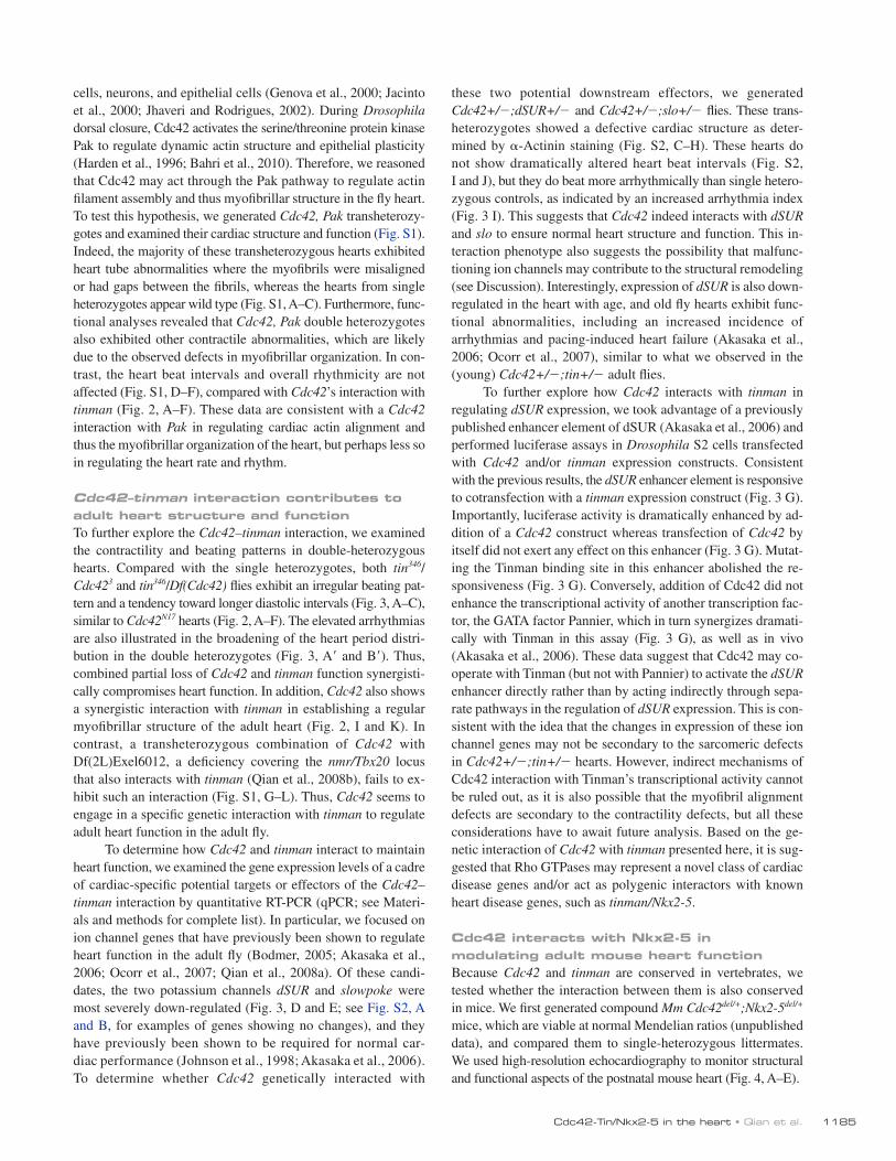
these two potential downstream effectors, we generated Cdc42+/;dSUR+/ and Cdc42+/;slo+/ flies. These trans-heterozygotes showed a defective cardiac structure as deter-mined by -Actinin staining (Fig. S2, C–H). These hearts do not show dramatically altered heart beat intervals (Fig. S2, I and J), but they do beat more arrhythmically than single hetero-zygous controls, as indicated by an increased arrhythmia index (Fig. 3 I). This suggests that Cdc42 indeed interacts with dSUR and slo to ensure normal heart structure and function. This in-teraction phenotype also suggests the possibility that malfunc-tioning ion channels may contribute to the structural remodeling (see Discussion). Interestingly, expression of dSUR is also down-regulated in the heart with age, and old fly hearts exhibit func-tional abnormalities, including an increased incidence of arrhythmias and pacing-induced heart failure (Akasaka et al., 2006; Ocorr et al., 2007), similar to what we observed in the (young) Cdc42+/;tin+/ adult flies.
To further explore how Cdc42 interacts with tinman in regulating dSUR expression, we took advantage of a previously published enhancer element of dSUR (Akasaka et al., 2006) and performed luciferase assays in Drosophila S2 cells transfected with Cdc42 and/or tinman expression constructs. Consistent with the previous results, the dSUR enhancer element is responsive to cotransfection with a tinman expression construct (Fig. 3 G). Importantly, luciferase activity is dramatically enhanced by ad-dition of a Cdc42 construct whereas transfection of Cdc42 by itself did not exert any effect on this enhancer (Fig. 3 G). Mutat-ing the Tinman binding site in this enhancer abolished the re-sponsiveness (Fig. 3 G). Conversely, addition of Cdc42 did not enhance the transcriptional activity of another transcription fac-tor, the GATA factor Pannier, which in turn synergizes dramati-cally with Tinman in this assay (Fig. 3 G), as well as in vivo (Akasaka et al., 2006). These data suggest that Cdc42 may co-operate with Tinman (but not with Pannier) to activate the dSUR enhancer directly rather than by acting indirectly through sepa-rate pathways in the regulation of dSUR expression. This is con-sistent with the idea that the changes in expression of these ion channel genes may not be secondary to the sarcomeric defects in Cdc42+/;tin+/ hearts. However, indirect mechanisms of Cdc42 interaction with Tinman’s transcriptional activity cannot be ruled out, as it is also possible that the myofibril alignment defects are secondary to the contractility defects, but all these considerations have to await future analysis. Based on the ge-netic interaction of Cdc42 with tinman presented here, it is sug-gested that Rho GTPases may represent a novel class of cardiac disease genes and/or act as polygenic interactors with known heart disease genes, such as tinman/Nkx2-5.
Cdc42 interacts with Nkx2-5 in modulating adult mouse heart functionBecause Cdc42 and tinman are conserved in vertebrates, we tested whether the interaction between them is also conserved in mice. We first generated compound Mm Cdc42del/+;Nkx2-5del/+ mice, which are viable at normal Mendelian ratios (unpublished data), and compared them to single-heterozygous littermates. We used high-resolution echocardiography to monitor structural and functional aspects of the postnatal mouse heart (Fig. 4, A–E).
cells, neurons, and epithelial cells (Genova et al., 2000; Jacinto et al., 2000; Jhaveri and Rodrigues, 2002). During Drosophila dorsal closure, Cdc42 activates the serine/threonine protein kinase Pak to regulate dynamic actin structure and epithelial plasticity (Harden et al., 1996; Bahri et al., 2010). Therefore, we reasoned that Cdc42 may act through the Pak pathway to regulate actin filament assembly and thus myofibrillar structure in the fly heart. To test this hypothesis, we generated Cdc42, Pak transheterozy-gotes and examined their cardiac structure and function (Fig. S1). Indeed, the majority of these transheterozygous hearts exhibited heart tube abnormalities where the myofibrils were misaligned or had gaps between the fibrils, whereas the hearts from single heterozygotes appear wild type (Fig. S1, A–C). Furthermore, func-tional analyses revealed that Cdc42, Pak double heterozygotes also exhibited other contractile abnormalities, which are likely due to the observed defects in myofibrillar organization. In con-trast, the heart beat intervals and overall rhythmicity are not affected (Fig. S1, D–F), compared with Cdc42’s interaction with tinman (Fig. 2, A–F). These data are consistent with a Cdc42 interaction with Pak in regulating cardiac actin alignment and thus the myofibrillar organization of the heart, but perhaps less so in regulating the heart rate and rhythm.
Cdc42–tinman interaction contributes to adult heart structure and functionTo further explore the Cdc42–tinman interaction, we examined the contractility and beating patterns in double-heterozygous hearts. Compared with the single heterozygotes, both tin346/Cdc423 and tin346/Df(Cdc42) flies exhibit an irregular beating pat-tern and a tendency toward longer diastolic intervals (Fig. 3, A–C), similar to Cdc42N17 hearts (Fig. 2, A–F). The elevated arrhythmias are also illustrated in the broadening of the heart period distri-bution in the double heterozygotes (Fig. 3, A and B). Thus, combined partial loss of Cdc42 and tinman function synergisti-cally compromises heart function. In addition, Cdc42 also shows a synergistic interaction with tinman in establishing a regular myofibrillar structure of the adult heart (Fig. 2, I and K). In contrast, a transheterozygous combination of Cdc42 with Df(2L)Exel6012, a deficiency covering the nmr/Tbx20 locus that also interacts with tinman (Qian et al., 2008b), fails to ex-hibit such an interaction (Fig. S1, G–L). Thus, Cdc42 seems to engage in a specific genetic interaction with tinman to regulate adult heart function in the adult fly.
To determine how Cdc42 and tinman interact to maintain heart function, we examined the gene expression levels of a cadre of cardiac-specific potential targets or effectors of the Cdc42–tinman interaction by quantitative RT-PCR (qPCR; see Materi-als and methods for complete list). In particular, we focused on ion channel genes that have previously been shown to regulate heart function in the adult fly (Bodmer, 2005; Akasaka et al., 2006; Ocorr et al., 2007; Qian et al., 2008a). Of these candi-dates, the two potassium channels dSUR and slowpoke were most severely down-regulated (Fig. 3, D and E; see Fig. S2, A and B, for examples of genes showing no changes), and they have previously been shown to be required for normal car-diac performance (Johnson et al., 1998; Akasaka et al., 2006). To determine whether Cdc42 genetically interacted with
JCB • VOLUME 193 • NUMBER 7 • 2011 1186
Figure 3. Cdc42 and tinman interact to maintain normal cardiac contraction. (A and B) Representative M-mode traces showing arrhythmic heart contrac-tions in Df(Cdc42)/+;tin346/+ (B), compared with single heterozygotes Df(Cdc42)/+ (A). (A–B) Heart period histograms. (C) Statistical analysis of heart contraction in controls (w, tin346/+, Cdc423/+, Df(Cdc42)/+) and Cdc423/+;tin346/+ and Df(Cdc42)/+;tin346/+ flies, shown as relative quantification (RQ). Error bars represent SEM; *, P < 0.05 (one-way ANOVA). (D and E) RQ of dSUR (A) and slowpoke (B) mRNA in 1-wk-old adult hearts (normalized to rp49, ribosomal protein 49) relative to control (w). Double heterozygotes Df(Cdc42)/+;tin346/+, Df(Cdc42)/+;tinEC40/+, and Cdc423/+;tin346/+ showed lower expression than tinman or Cdc42 heterozygotes, or w controls. (F) Functional analyses of Cdc42,dSUR and Cdc42,slo double heterozygotes showed
1187Cdc42-Tin/Nkx2-5 in the heart • Qian et al.
as in the fly. This could be due to compensatory or redundant mechanisms that may be more prevalent in mammals. Neverthe-less, our data show that Mm Cdc42 interacts strongly with Nkx2-5 in the mouse heart and that this genetic interaction is es-sential for proper cardiac contraction, electrical conduction, and rhythmicity.
Screening for CDC42 gene variants in human cohorts with heart diseaseThe strong evidence in fly and mouse that implicates Cdc42 as an important determinant of normal heart function points to CDC42 as a candidate gene for human heart disease. Mutations in the human homologue of tinman, NKX2-5, or other partici-pants in the cardiogenic transcriptional network, TBX5, TBX20, and GATA4, have been associated with a variety of congenital and adult-onset heart disease. In humans, the CDC42 locus maps to chromosome 1p36. Monosomy 1p36 is the most com-mon terminal deletion syndrome and results in mental retarda-tion and multiple congenital anomalies, including atrial septal defects (ASD), ventricular septal defects (VSD), patent ductus arteriosus (PDA), and dilated cardiomyopathy (DCM). Loss of CDC42 could possibly account for the cardiac defects of this syndrome, as well as for cases in which these defects occur in isolation. We performed mutation screening of the CDC42 gene in two heart disease patient populations from Houston (J. Towbin) and Sydney (D. Fatkin).
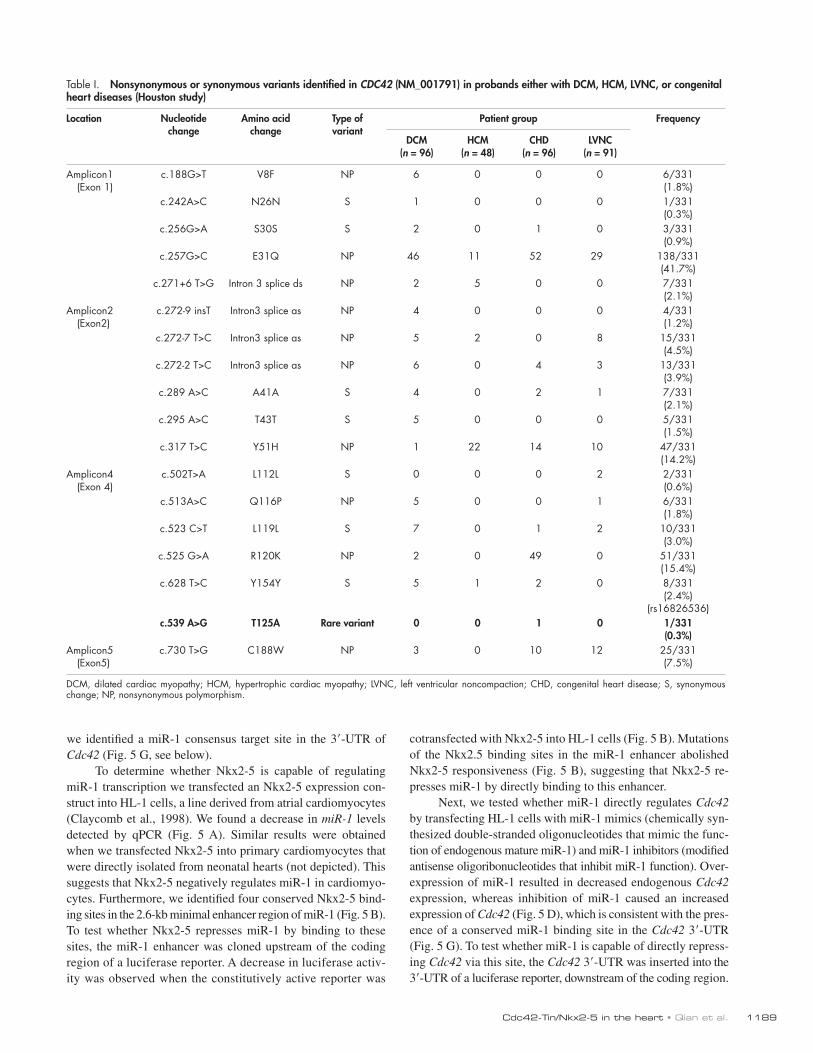
In the Houston study, 331 subjects with heart disease, in-cluding 96 probands with dilated cardiomyopathy (DCM), 48 with hypertrophic cardiomyopathy (HCM), 91 with left ven-tricular noncompaction (LVNC), and 96 with congenital heart disease (CHD), as well as over 400 controls (over 800 chro-mosomes), were examined for potential variants in the human CDC42 gene (GenBank/EMBL/DDBJ accession no. NM_001791; Table I). Seven nonsynonymous and seven synonymous vari-ants and four intronic variants were identified. Six out of the seven nonsynonymous variants were polymorphic and identi-fied in subjects and controls. A single rare nonsynonymous vari-ant, c.539A>G, which is predicted to result in an amino acid change of T125A, was not detected in over 300 Caucasian and over 100 Hispanic controls (Table I). This variant was found in a sporadic CHD patient with an atrial septal defect (ASD) and a patent ductus arteriosus (PDA). The parents did not participate in the study. In the Sydney study, over 300 heart disease patients (AF, CHD, LVNC) and 100 controls were examined (Table II), with no apparent disease-causing mutations identified. Some X synonymous variants were identified and no nonsynonymous variants were found.
These data indicate that despite the clear biological signif-icance of Cdc42 in the heart, coding sequence variants in the CDC42 gene are not a common cause of CHD or adult heart disease. A possible reason is that this small GTPase is extremely
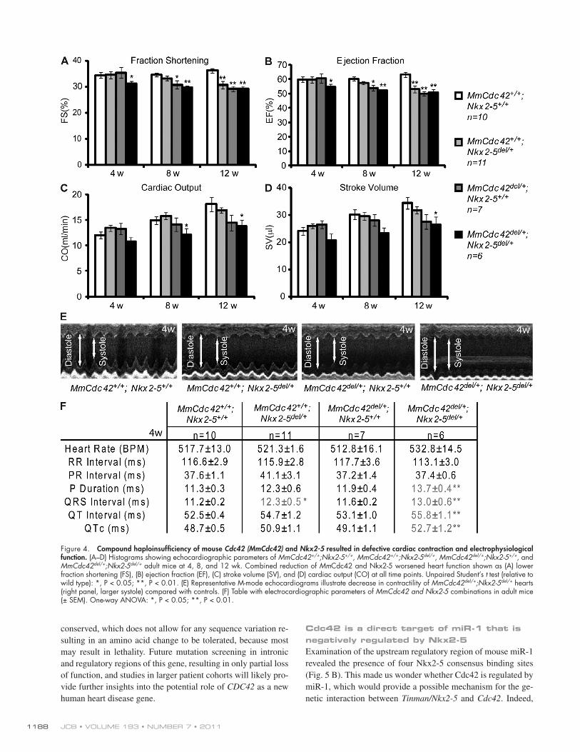
Mm Cdc42+/+;Nkx2-5+/+, Mm Cdc42+/+;Nkx2-5del/+, Mm Cdc42del/+;Nkx2-5+/+, and Mm Cdc42del/+;Nkx2-5del/+ adult mice were subjected to serial imaging at 4–12 wk after birth. At 4 wk of age, cardiac function in single heterozygotes (MmCdc42del/+ or Nkx2-5del/+) was not significantly different from wild-type lit-termates, whereas double heterozygotes showed declining heart function as indicated by decreased fraction of blood ejected from the left ventricle with each contraction (ejection fraction, EF), the fractional shortening of the ventricular chamber (frac-tional shortening, FS), volume of blood ejected with each heart beat (stroke volume, SV), and cardiac output (CO; see Fig. 4, A–D for quantification and Fig. 4 E for representative M-modes of the four genotypes). As the mice aged, single heterozygotes progressively exhibited weakened pumping ability, whereas double heterozygotes always performed worse than their single-heterozygous littermates (Fig. 4, A–D). Collectively, these re-sults demonstrate that cardiac output, as assessed by multiple imaging parameters, is perturbed in Cdc42 heterozygous mice, and as expected in Nkx2-5 heterozygotes. Compellingly, these defects are markedly worse when the two alleles are combined in trans, suggesting that Cdc42 and Nkx2-5 synergistically reg-ulate cardiac output and function in the mouse adult heart.
Mouse models of Nkx2-5 deficiency revealed a critical role for Nkx2-5 in establishing and maintaining proper cardiac conduction system function (Pashmforoush et al., 2004; Briggs et al., 2008; Takeda et al., 2009), and human mutations in Nkx2-5 cause conduction-system dysfunction (Schott et al., 1998). Given the interaction between tinman and Cdc42 that we identified in the fly, we hypothesized that Nkx2-5 also interacts with Cdc42 in regulating the electrical activity of adult mouse heart. Therefore, we performed surface electrocardiography (ECG) and found that the average heart rate of single and double hetero-zygotes is not significantly different from that of wild-type mice, nor is there any difference in the delay between atrial and ventricular depolarization as indicated by the PR interval (Fig. 4 F). However, combined reduction of Cdc42 and Nkx2-5 resulted in prolonged atrial depolarization manifested by increased P dura-tion (Fig. 4 F). The QRS complex, the portion of an ECG that corresponds to the depolarization of the right and left ventricles, was also significantly prolonged in double heterozygotes and was associated with prolongation of the QT and QTc intervals (Fig. 4 F). We also noticed that the double heterozygotes showed signs of right bundle branch block based upon S wave depres-sion and the delays, as calculated in Fig. 4 F, which was not ob-served in single heterozygotes or wild-type controls. We then examined cardiac structure and fibrosis of these adult mice by performing the standard hematoxylin and eosin (H&E) and Masson Trichrome stainings, but did not observe any consistent differences among different genotypes (unpublished data and Fig. S3). Thus, compound heterozygosity for Nkx2-5 and Cdc42 does not alter cardiac morphology in the mouse as significantly
a significant increase in the arrhythmia index. (G) Luciferase assay using the dSUR T3 enhancer (Akasaka et al., 2006). Note the increased relative lu-ciferase activity (RLA) by transfection of Tin was further enhanced by cotransfection with Cdc42 or Pnr, but not when Pnr and Cdc42 were cotransfected. Mutation of the Tin site abolished the responsiveness. Experiments were repeated three times. Error bars represent SEM. Unpaired Student’s t test: *, P < 0.05; **, P < 0.01.
JCB • VOLUME 193 • NUMBER 7 • 2011 1188
Cdc42 is a direct target of miR-1 that is negatively regulated by Nkx2-5Examination of the upstream regulatory region of mouse miR-1 revealed the presence of four Nkx2-5 consensus binding sites (Fig. 5 B). This made us wonder whether Cdc42 is regulated by miR-1, which would provide a possible mechanism for the ge-netic interaction between Tinman/Nkx2-5 and Cdc42. Indeed,
conserved, which does not allow for any sequence variation re-sulting in an amino acid change to be tolerated, because most may result in lethality. Future mutation screening in intronic and regulatory regions of this gene, resulting in only partial loss of function, and studies in larger patient cohorts will likely pro-vide further insights into the potential role of CDC42 as a new human heart disease gene.
Figure 4. Compound haploinsufficiency of mouse Cdc42 (MmCdc42) and Nkx2-5 resulted in defective cardiac contraction and electrophysiological function. (A–D) Histograms showing echocardiographic parameters of MmCdc42+/+;Nkx2-5+/+, MmCdc42+/+;Nkx2-5del/+, MmCdc42del/+;Nkx2-5+/+, and MmCdc42del/+;Nkx2-5del/+ adult mice at 4, 8, and 12 wk. Combined reduction of MmCdc42 and Nkx2-5 worsened heart function shown as (A) lower fraction shortening (FS), (B) ejection fraction (EF), (C) stroke volume (SV), and (D) cardiac output (CO) at all time points. Unpaired Student’s t test (relative to wild type): *, P < 0.05; **, P < 0.01. (E) Representative M-mode echocardiograms illustrate decrease in contractility of MmCdc42del/+;Nkx2-5del/+ hearts (right panel, larger systole) compared with controls. (F) Table with electrocardiographic parameters of MmCdc42 and Nkx2-5 combinations in adult mice (± SEM). One-way ANOVA: *, P < 0.05; **, P < 0.01.
1189Cdc42-Tin/Nkx2-5 in the heart • Qian et al.
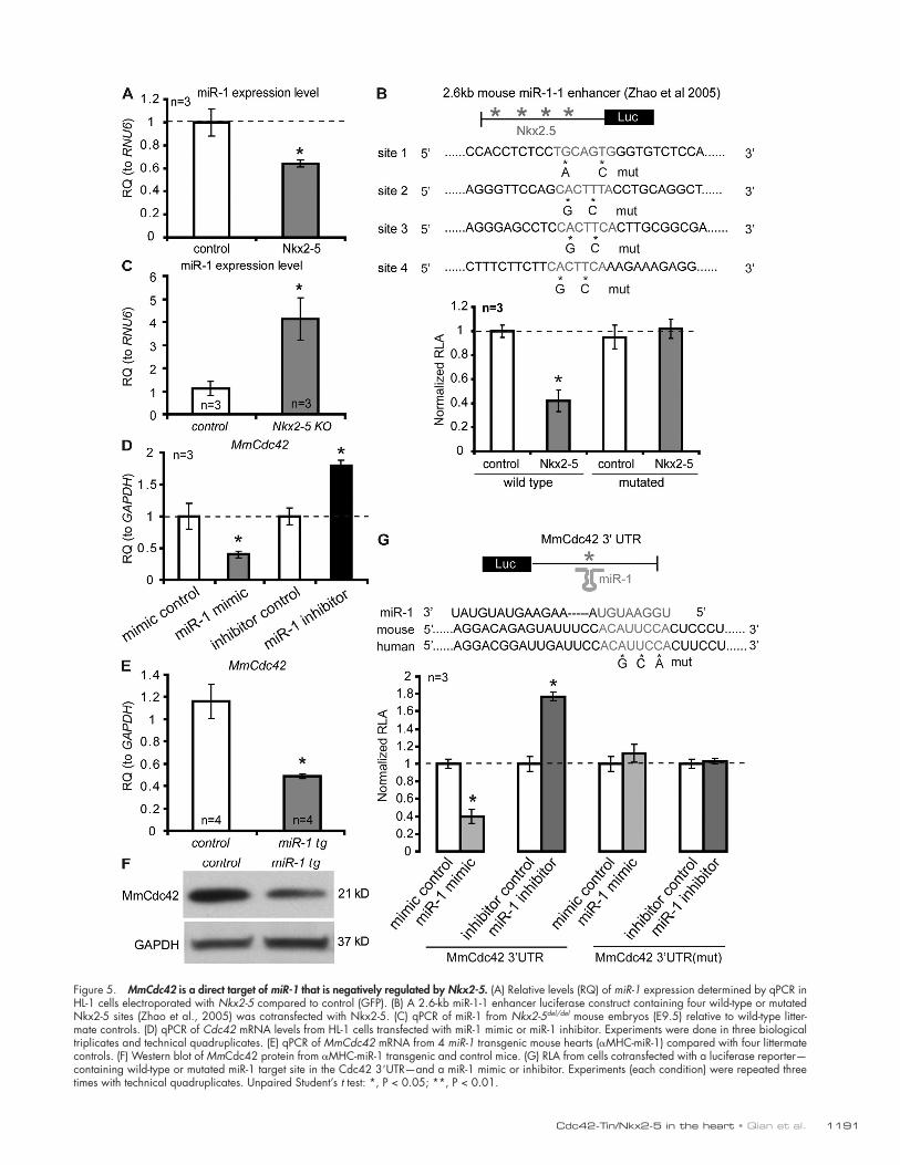
cotransfected with Nkx2-5 into HL-1 cells (Fig. 5 B). Mutations of the Nkx2.5 binding sites in the miR-1 enhancer abolished Nkx2-5 responsiveness (Fig. 5 B), suggesting that Nkx2-5 re-presses miR-1 by directly binding to this enhancer.
Next, we tested whether miR-1 directly regulates Cdc42 by transfecting HL-1 cells with miR-1 mimics (chemically syn-thesized double-stranded oligonucleotides that mimic the func-tion of endogenous mature miR-1) and miR-1 inhibitors (modified antisense oligoribonucleotides that inhibit miR-1 function). Over-expression of miR-1 resulted in decreased endogenous Cdc42 expression, whereas inhibition of miR-1 caused an increased expression of Cdc42 (Fig. 5 D), which is consistent with the pres-ence of a conserved miR-1 binding site in the Cdc42 3-UTR (Fig. 5 G). To test whether miR-1 is capable of directly repress-ing Cdc42 via this site, the Cdc42 3-UTR was inserted into the 3-UTR of a luciferase reporter, downstream of the coding region.
we identified a miR-1 consensus target site in the 3-UTR of Cdc42 (Fig. 5 G, see below).
To determine whether Nkx2-5 is capable of regulating miR-1 transcription we transfected an Nkx2-5 expression con-struct into HL-1 cells, a line derived from atrial cardiomyocytes (Claycomb et al., 1998). We found a decrease in miR-1 levels detected by qPCR (Fig. 5 A). Similar results were obtained when we transfected Nkx2-5 into primary cardiomyocytes that were directly isolated from neonatal hearts (not depicted). This suggests that Nkx2-5 negatively regulates miR-1 in cardiomyo-cytes. Furthermore, we identified four conserved Nkx2-5 bind-ing sites in the 2.6-kb minimal enhancer region of miR-1 (Fig. 5 B). To test whether Nkx2-5 represses miR-1 by binding to these sites, the miR-1 enhancer was cloned upstream of the coding region of a luciferase reporter. A decrease in luciferase activ-ity was observed when the constitutively active reporter was
Table I. Nonsynonymous or synonymous variants identified in CDC42 (NM_001791) in probands either with DCM, HCM, LVNC, or congenital heart diseases (Houston study)
Location Nucleotide change
Amino acid change
Type of variant
Patient group Frequency
DCM (n = 96)
HCM (n = 48)
CHD (n = 96)
LVNC (n = 91)
Amplicon1 (Exon 1)
c.188G>T V8F NP 6 0 0 0 6/331 (1.8%)
c.242A>C N26N S 1 0 0 0 1/331 (0.3%)
c.256G>A S30S S 2 0 1 0 3/331 (0.9%)
c.257G>C E31Q NP 46 11 52 29 138/331 (41.7%)
c.271+6 T>G Intron 3 splice ds NP 2 5 0 0 7/331 (2.1%)
Amplicon2 (Exon2)
c.272-9 insT Intron3 splice as NP 4 0 0 0 4/331 (1.2%)
c.272-7 T>C Intron3 splice as NP 5 2 0 8 15/331 (4.5%)
c.272-2 T>C Intron3 splice as NP 6 0 4 3 13/331 (3.9%)
c.289 A>C A41A S 4 0 2 1 7/331 (2.1%)
c.295 A>C T43T S 5 0 0 0 5/331 (1.5%)
c.317 T>C Y51H NP 1 22 14 10 47/331 (14.2%)
Amplicon4 (Exon 4)
c.502T>A L112L S 0 0 0 2 2/331 (0.6%)
c.513A>C Q116P NP 5 0 0 1 6/331 (1.8%)
c.523 C>T L119L S 7 0 1 2 10/331 (3.0%)
c.525 G>A R120K NP 2 0 49 0 51/331 (15.4%)
c.628 T>C Y154Y S 5 1 2 0 8/331 (2.4%)
(rs16826536)c.539 A>G T125A Rare variant 0 0 1 0 1/331
(0.3%)Amplicon5
(Exon5)c.730 T>G C188W NP 3 0 10 12 25/331
(7.5%)
DCM, dilated cardiac myopathy; HCM, hypertrophic cardiac myopathy; LVNC, left ventricular noncompaction; CHD, congenital heart disease; S, synonymous change; NP, nonsynonymous polymorphism.
JCB • VOLUME 193 • NUMBER 7 • 2011 1190
may not always be conserved (Hyun et al., 2009; unpublished data), even though the pathway overall is likely conserved, as in this case and in a recent report (King et al., 2011). Thus, it remains to be determined how microRNAs regulate down-stream effectors (such as Cdc42; Fig. 6 E), directly by cryptic sites or via an unknown possibly indirect mechanism. However, we did find two consensus binding sites for Tinman within the enhancer of fly miR-1, and luciferase assays in S2 cells revealed that these two sites are responsible for the regulation of fly miR-1 by Tinman (Fig. S5). Taken together, these data sug-gest a functionally conserved regulatory pathway involving tinman–miR-1–Cdc42 in establishing cardiac functionality, al-though some aspects of the molecular mechanisms involved may be different between flies and vertebrates, which could also explain the different severity in phenotypes between flies and mice in our study.
DiscussionDrosophila has been successfully used to study regulatory ge-netic networks of inductive signals and transcription factors that determine cardiac specification and morphogenesis, but the genetic control of cardiac physiology is only beginning to be investigated. Using a cardiac performance-based genetic screen, we discovered a novel genetic interaction between Cdc42 and the NK homeobox gene tinman in regulating cardiac function in the fly. We have identified a heretofore-unknown requirement for the Rho GTPase Cdc42 in regulating the establishment of cardiac function in the adult fly. Significantly, this interaction is conserved in mice, where combined reduction of Cdc42 and Nkx2-5 caused defects in heart contraction, rhythm, and electro-physiological function. Based on the discovery of the interac-tion between this Rho GTPase with a cardiogenic transcription factor in maintaining proper cardiac function, it is conceivable that other genetic interactors and potential polygenic contribu-tors to heart disease traits can be identified using the fly as a screening platform.
Cdc42 is a well-characterized GTPase, and has been widely studied in different organs and tissues, ranging from worms to humans. Because Cdc42 was shown to be involved in regulation of the actin/myosin cytoskeleton in the cells of various organ-isms, we reasoned that Cdc42 might also control cardiac cell morphogenesis. In Drosophila and mouse we found that complete
Transfection of the Cdc42 3-UTR luciferase reporter with miR-1 mimic into HL-1 cells results in a decrease in luciferase activity, whereas cotransfection with miR-1 inhibitor increases luciferase activity (Fig. 5 G). A gradient of miR-1 was used to con-firm the luciferase activity was dose responsive (Fig. S4 A). Muta-tions of the miR-1 binding site in the Cdc42 3-UTR abolished miR-1 responsiveness (Fig. 5 G), suggesting that miR-1 represses Cdc42 by physically binding to its 3-UTR.
We extracted RNA and protein from transgenic mice in which miR-1 was overexpressed in the adult heart via the -MHC promoter (-MHC–miR-1; unpublished mice from the Srivastava laboratory) and examined Cdc42 mRNA and protein levels. Consistently, miR-1 transgenic hearts showed signifi-cantly reduced Cdc42 mRNA and protein levels (Fig. 5, E and F), suggesting a repressive relationship between miR-1 and Cdc42 in vivo. We did not observe increased Cdc42 expression in miR-1-2 knockout hearts, which might be due to the incomplete de-pletion of miR-1 expression because of the intact miR-1-1 locus (Zhao et al., 2007; unpublished data). Furthermore, we deter-mined miR-1 levels in Nkx2.5 knockout embryos (E9.5) by qPCR and found an increase in miR-1 levels compared with litter-mate controls (Fig. 5 C). These data provide further evidence that the genetic interaction between Nkx2-5 and Cdc42 is medi-ated by miR-1.
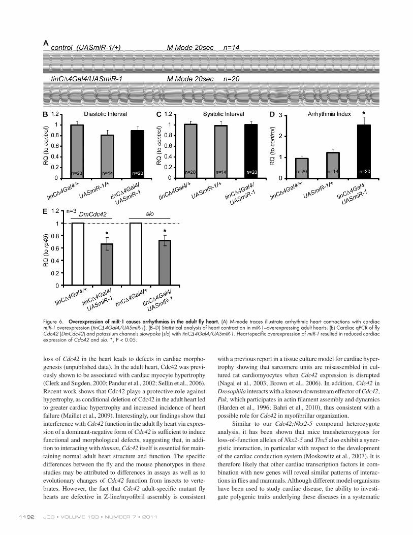
We then wondered whether the genetic interaction be-tween tinman and Cdc42 in Drosophila is also governed by a double-inhibitory mechanism involving the fly homologue of miR-1. We found that cardiac-specific overexpression of miR-1 causes an increase in arrhythmias (Fig. 6, A–D), comparable to cardiac expression of dominant-negative Cdc42N17 (Fig. 2). In addi-tion, fly hearts with excessive cardiac miR-1 expression exhibit reduced levels of Cdc42 and slo mRNA in the heart (Fig. 6 E), suggesting that miR-1’s influence on heart function includes inhibition of Cdc42 and one or more potassium channels. In con-trast to the mouse, however, we did not identify any miR-1 binding sites in the 3-UTR of fly Cdc42. To test whether fly Cdc42 regulation by miR-1 is through binding to “seedless” 3’-UTR sequences that lack predicted microRNA recognition elements (Bamshad et al., 1997), we cloned fly Cdc42 3-UTR into the 3-UTR of a luciferase reporter and performed luciferase assays in Drosophila S2 cells. However, we did not observe consistent and reproducible repression in the presence of miR-1 (Fig. S4 B). Recent evidence suggests that microRNA target sites
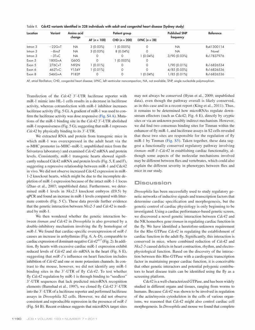
Table II. Cdc42 variants identified in 328 individuals with adult and congenital heart disease (Sydney study)
Location Variant Amino acid change
Patient group Published SNP frequency
Reference
AF (n = 100) CHD (n = 200) LVNC (n = 28)
Intron 3 22G>T NA 3 (0.03%) 1 (0.005%) 0 NA Rs41300114Intron 3 8insT NA 3 (0.03%) 8 (0.04%) 0 NA NovelIntron 3 3T>C NA 0 0 1 (0.04%) 3/90 (0.03%) Rs17837976Exon 5 180G>A G60G 0 1 (0.005%) 0Exon 5 276C>T N92N 1 (0.01%) 0 0 1/90 (0.01%) Rs16826534Exon 6 462T>C Y154Y 1 (0.01%) 0 0 4/85 (0.05%) Rs16826536Exon 8 546G>A P182P 0 0 1 (0.04%) 1/85 (0.01%) Rs16826536
AF, atrial fibrillation; CHD, congenital heart disease; LVNC, left ventricular noncompaction; NA, not available; SNP, single nucleotide polymorphism.
1191Cdc42-Tin/Nkx2-5 in the heart • Qian et al.
Figure 5. MmCdc42 is a direct target of miR-1 that is negatively regulated by Nkx2-5. (A) Relative levels (RQ) of miR-1 expression determined by qPCR in HL-1 cells electroporated with Nkx2-5 compared to control (GFP). (B) A 2.6-kb miR-1-1 enhancer luciferase construct containing four wild-type or mutated Nkx2-5 sites (Zhao et al., 2005) was cotransfected with Nkx2-5. (C) qPCR of miR-1 from Nkx2-5del/del mouse embryos (E9.5) relative to wild-type litter-mate controls. (D) qPCR of Cdc42 mRNA levels from HL-1 cells transfected with miR-1 mimic or miR-1 inhibitor. Experiments were done in three biological triplicates and technical quadruplicates. (E) qPCR of MmCdc42 mRNA from 4 miR-1 transgenic mouse hearts (MHC-miR-1) compared with four littermate controls. (F) Western blot of MmCdc42 protein from MHC-miR-1 transgenic and control mice. (G) RLA from cells cotransfected with a luciferase reporter—containing wild-type or mutated miR-1 target site in the Cdc42 3UTR—and a miR-1 mimic or inhibitor. Experiments (each condition) were repeated three times with technical quadruplicates. Unpaired Student’s t test: *, P < 0.05; **, P < 0.01.
JCB • VOLUME 193 • NUMBER 7 • 2011 1192
with a previous report in a tissue culture model for cardiac hyper-trophy showing that sarcomere units are misassembled in cul-tured rat cardiomyocytes when Cdc42 expression is disrupted (Nagai et al., 2003; Brown et al., 2006). In addition, Cdc42 in Drosophila interacts with a known downstream effector of Cdc42, Pak, which participates in actin filament assembly and dynamics (Harden et al., 1996; Bahri et al., 2010), thus consistent with a possible role for Cdc42 in myofibrillar organization.
Similar to our Cdc42;Nkx2-5 compound heterozygote analysis, it has been shown that mice transheterozygous for loss-of-function alleles of Nkx2-5 and Tbx5 also exhibit a syner-gistic interaction, in particular with respect to the development of the cardiac conduction system (Moskowitz et al., 2007). It is therefore likely that other cardiac transcription factors in com-bination with new genes will reveal similar patterns of interac-tions in flies and mammals. Although different model organisms have been used to study cardiac disease, the ability to investi-gate polygenic traits underlying these diseases in a systematic
loss of Cdc42 in the heart leads to defects in cardiac morpho-genesis (unpublished data). In the adult heart, Cdc42 was previ-ously shown to be associated with cardiac myocyte hypertrophy (Clerk and Sugden, 2000; Pandur et al., 2002; Sellin et al., 2006). Recent work shows that Cdc42 plays a protective role against hypertrophy, as conditional deletion of Cdc42 in the adult heart led to greater cardiac hypertrophy and increased incidence of heart failure (Maillet et al., 2009). Interestingly, our findings show that interference with Cdc42 function in the adult fly heart via expres-sion of a dominant-negative form of Cdc42 is sufficient to induce functional and morphological defects, suggesting that, in addi-tion to interacting with tinman, Cdc42 itself is essential for main-taining normal adult heart structure and function. The specific differences between the fly and the mouse phenotypes in these studies may be attributed to differences in assays as well as to evolutionary changes of Cdc42 function from insects to verte-brates. However, the fact that Cdc42 adult-specific mutant fly hearts are defective in Z-line/myofibril assembly is consistent
Figure 6. Overexpression of miR-1 causes arrhythmias in the adult fly heart. (A) M-mode traces illustrate arrhythmic heart contractions with cardiac miR-1 overexpression (tinC4Gal4/UASmiR-1). (B–D) Statistical analysis of heart contraction in miR-1–overexpressing adult hearts. (E) Cardiac qPCR of fly Cdc42 (DmCdc42) and potassium channels slowpoke (slo) with tinC4Gal4/UASmiR-1. Heart-specific overexpression of miR-1 resulted in reduced cardiac expression of Cdc42 and slo. *, P < 0.05.
1193Cdc42-Tin/Nkx2-5 in the heart • Qian et al.
In conclusion, we have used the power of Drosophila ge-netics to uncover a new, conserved genetic pathway, linking genes with critical heart functions, tinman/Nkx2-5, miR-1, and Cdc42, to a novel network regulating adult heart physiology and perfor-mance. With the fly as a screening platform, it is thus possible to identify new polygenic contributors that ensure normal cardiac function relevant to human heart disease.
Materials and methodsDrosophila stocksThe following mutant stocks were used: Cdc423/FM6 (Boutros et al., 1998); tin346/TM3-ftz-lacZ (Azpiazu and Frasch, 1993); tinEC40/TM3, Df(3R)tinGC14 (Bodmer, 1993); Pak6/TM3,Sb (Pehlivan et al., 1999); Df(2L)Exel9032/Cyo (indicated as dSUR/+), Df(3R)BSC397/TM3,Sb (indicated as slo/+), Df(2L)Exel6012 (Qian et al., 2005b; Exelixis deficiency deleting both nmr1 and nmr2, also indicated as Df(nmr)), and Df(1)Exel6253 (Exelixis deficiency deleting the whole genomic region of Cdc42, also indicated as Df(Cdc42)). Wild-type control flies are w1118. Overexpression of transgenes was achieved using the UAS-Gal4 system (Brand and Perrimon, 1993). The following Gal4 and UAS lines were used: twi-Gal4 (twi>; Greig and Akam, 1993), 24B-Gal4 (24B>; Brand and Perrimon, 1993), the double combination twi-Gal4;24B-Gal4 (twi24B>; pan-mesodermal expression; Lockwood and Bodmer, 2002), tinC4-Gal4 (Lo and Frasch, 2001), and UAS-Cdc42N17 (Luo et al., 1994). We used TARGET to temporally and spatially activate gene ex-pression (McGuire et al., 2004). In brief, we raised flies (with Gal4, Gal80-ts, and UAS constructs) at 18°C before eclosion; after eclosion we raised adult flies at 29°C for 1 wk and then performed heart function assays.
Mouse stocksMice harboring a loss-of-function allele for Nkx2-5 (Nkx2-5del/+/Nkx2.5tm1Siz) have been described previously (Tanaka et al., 1999). Mice harboring a floxed allele of Cdc42 (Cdc42tm1Yizh) have also been described previously (Chen et al., 2006). Cdc42 males were bred to Mef2c-AHF-cre (Verzi et al., 2005) females to enable germ line deletion of Cdc42 (Cdc42del/+), and off-spring that did not inherit the Cre allele were then mated to Nkx2.5 hetero-zygous animals to generate compound heterozygous and single heterozygous offspring. For all functional studies detailed below, littermates of each indi-cated genotype were analyzed.
ImmunohistochemistryFor fly heart immunohistochemistry, antibody staining was performed as described previously (Han et al., 2002). In brief, after micro-dissection, fly hearts were fixed in 4% PFA overnight at 4°C. After blocking in 10% BSA (Sigma-Aldrich)/PBS for 1 h, fly hearts were incubated in primary antibodies for 1 h, washed three times (15 min each) with PBT (PBS + 0.5% Triton), and then incubated in secondary antibodies for 1 h. After washing 3 × 15 min in PBT, fly hearts were mounted in VectaShield (Vector Laboratories). Cy3- or FITC-conjugated secondary antibodies (Jackson ImmunoResearch Laborato-ries, Inc.) were used for fluorescent confocal microscopy. All the secondary antibodies were used at 1:200. Fly heart preparations were analyzed using a confocal microscope (model LSM510; Carl Zeiss). Mouse anti–-Actinin (Saide et al., 1989) was used in this study. For mouse heart immunohisto-chemistry, after perfusion fix the heart was removed and fixed by immersion in 4% PFA in PBS (diluted from 20% PFA stock; Electron Microscopy Sciences) and routinely processed and paraffin embedded. 5-µm sections were stained with hematoxylin and eosin (H&E) and Masson Trichrome and analyzed for regular morphology and fibrosis. Images were acquired by Zeiss LSM soft-ware (for confocal images) or Image-Pro Plus 5.1 (for regular IHC images). Adobe Photoshop was used for image processing; contrast and brightness were adjusted for better visualization.
Electrical pacing in fliesThe electrical pacing was conducted as described previously (Wessells and Bodmer, 2004; Wessells et al., 2004). In brief, 50–100 flies were paced with a square wave stimulator at 40 V and 6 Hz for 30 s and scored for heart failure rate, defined as the percentage of flies that either exhibit cardiac arrest or continuous fibrillation immediately after pacing.
Image analysis and M-mode traces on semi-intact fly preparationsProcedures used were as described previously (Ocorr et al., 2007; Fink et al., 2009). In brief, flies were anesthetized and the head, ventral thorax, and
fashion in the adult is limited. Here, we provide evidence that rigorous screening for modifiers of heart disease can be achieved using Drosophila, leading to identification of similar polygenic trait interactions in mammals.
The genetic relationship between fly Cdc42 with the car-diac determinant tinman in regulating adult heart contraction may occur, in part, via modulation of potassium channel gene expression, dSUR and slo, which have previously been shown to be critical regulators of heart function (Johnson et al., 1998; Akasaka et al., 2006). Indeed, Cdc42-dSUR or –slo transhetero-zygotes show a strong interaction reflected in increased ar-rhythmias. Interestingly, these transheterozygotes also exhibit disturbances in myofibrillar organization, suggesting these ion channel abnormalities somehow also produce sarcomeric de-fects, perhaps via a secondary structural remodeling effect. Although there is increasing evidence of such remodeling pro-cesses being affected by ion channel defects (unpublished data), the underlying mechanism is not yet understood.
A strong interaction was also observed between the mouse homologues of Cdc42 and Nkx2-5 in adult heart function. Most interestingly, double haploinsufficiency of Cdc42 and Nkx2-5 led to elongated QRS intervals, which is typical of abnormal conduction along the atrio-ventricular bundle, bundle branches, and Purkinje fibers. The observed elongated QT intervals in compound heterozygotes are also characteristic of long-QT syndrome in humans. These findings suggest a possible contri-bution of compound haploinsufficiency of Cdc42 and Nkx2-5 in human cardiac conduction diseases.
While attempting to elucidate the mechanistic link be-tween Cdc42 and Nkx2-5, we found that miR-1 directly re-presses Cdc42 and that miR-1 itself can be repressed by Nkx2.5 expression. miR-1 is the first microRNA identified in the heart and plays a conserved role in cardiac morphogenesis, cell cycle, and conduction (Zhao et al., 2005, 2007). miR-1 null mutant mice showed cardiac conduction defects including widened QRS complex and prolonged QT intervals (Zhao et al., 2007), which is also affected in Cdc42;Nkx2-5 double-heterozygous mice. We speculate that loss or gain of miR-1 function causes an imbalance in gene expression in the heart, including that of Cdc42. Decreasing the copy number of the direct miR-1 target Cdc42, and that of a direct transcriptional miR-1 repressor, Nkx2-5, apparently destroys a delicate balance in the adult heart, tipping it toward deregulated cardiac function. This does not rule out that other mechanisms are also involved in the ge-netic interaction between Cdc42 and Nkx2-5. Because Cdc42 is a direct target of miR-1 and has a crucial function in modulating hypertrophic responses in the mouse, it will be interesting to test whether miR-1 and other microRNAs play a direct role in adult cardiac hypertrophy. Interestingly, Cdc42 was also found to be a target of miR-133, which is involved in cardiac hyper-trophy (Carè et al., 2007). Thus, the RhoGTPase Cdc42 appears to be directly regulated by two major cardiac microRNAs that are involved in controlling cardiac function and hypertrophy. The synergistic transcriptional activation of the dSUR enhancer by Tinman and Cdc42 in vitro (Fig. 3 G) is consistent with the idea that Cdc42 can act in a coactivating mechanism with Tinman, but this remains to be further investigated.
JCB • VOLUME 193 • NUMBER 7 • 2011 1194
Cell culture, transfection, and luciferase assayHL-1 cells were maintained in Calycomb Medium supplemented with 100 µM norepinephrine, 10% FBS, and 4 mM l-glutamine. Cells were main-tained at 37°C in a humidified atmosphere of 5% CO2/95% air. Lipo-fectamine 2000 (Invitrogen)–mediated transfection was performed according to Invitrogen’s protocol. miR-1 mimic and inhibitor were pur-chased from Thermo Fisher Scientific. For each transfection in one well of a six-well plate, 40 pmol of mimic or inhibitor was used. For plasmid trans-fection, 100 ng of plasmid was transfected in one well of a six-well plate using the Amaxa Nucleofection kit (Lonza) per the manufacturer’s protocol. Luciferase assays were performed as described previously (Zhao et al., 2005) with the Dual-Luciferase Reporter Assay System (Promega). Cdc42 3-UTR was cloned from mouse genomic DNA with the following primers: 5-ACTAGTACGCATCTCCAGAGCCCTTTCTGC-3 and 5-AAGCTTTAG-CCCTGACTGGTCCCCATGTTGG-3. Amplified DNA was cloned into pCRII vector and subsequently cloned into pMIR-REPORT vector (Invitro-gen). Site-directed PCR-mediated mutagenesis was performed using the QuickChange II XL Site-Directed Mutagenesis kit (Agilent Technologies). Plasmids containing Tinman and Nkx2.5 have been described previously (Akasaka et al., 2006; Takeuchi and Bruneau, 2009). Tinman cDNA was finally inserted in pUAST vector; Nkx2-5 cDNA was finally inserted in pCDNA3.1 vector. Luciferase constructs containing miR-1 enhancer and dSUR enhancer were described previously (Zhao et al., 2005; Akasaka et al., 2006).
Western blotsWestern blots were performed as described previously (Zhao et al., 2005). Rabbit anti-Cdc42 (Cell Signaling Technology) and rabbit anti-GAPDH (Santa Cruz Biotechnology, Inc.) were both used at a 1:1,000 dilution for Western blots. All Western blots were quantified using AlphaImager soft-ware from Alpha Innovations.
StatisticsMean, standard deviation, and standard error of the mean were calcu-lated. Error bars are represented as standard error of the mean. Sample numbers were indicated in corresponding figures. Comparisons between groups were made by one-way ANOVA or unpaired two-tails assuming equal variance t test, as applicable. For fly pacing experiments, 2 test was applied.
Human subjects and DNA sequence analysisHouston study. The study population from Baylor College of Medicine in-cluded 331 unrelated pediatric probands either sporadic or familial cases of heart disease including 96 individuals with dilated cardiomyopathy (DCM), 48 individuals with hypertrophic cardiomyopathy (HCM), 96 indi-viduals with congenital heart disease (CHD), and 91 individuals with either isolated or nonisolated left ventricular noncompaction (LVNC; Table I). All patients were evaluated by physical examination, chest radiography, electro-cardiography, echocardiography, cardiac magnetic resonance imaging (cMRI), and angiography. Left ventricular size and function was evaluated by M-mode and two-dimensional echocardiography, Doppler, and color Doppler analyses. Further details on heart function measures are as de-scribed in Qian et al. (2008b). All subjects or their guardians provided written informed consent. Blood for EBV-transformed lymphoblastoid cell lines and DNA extraction was obtained, as regulated by the Baylor College of Medicine Institutional Review Board. DNA was extracted either directly from blood or from cultured cells using the Puregene DNA Isolation kit (Genetra Systems) according to the manufacturer’s protocol. Primers were designed in to the intron sequences flanking coding exons of CDC42 (Genbank/EMBL/DDBJ accession no. NM_001791). Protein encoding amplicons were amplified by polymerase chain reaction using genomic DNA, as described previously (Qian et al., 2008b). PCR products were purified and sequenced by automated sequencing using a DNA sequencer (ABI 3730; Applied Biosystems) and Big Dye terminator chemistry (version 3.1) according to the manufacturer’s instructions. The only rare variant we found was T125A in a CHD patient. Over 300 Caucasian and >100 His-panic controls were analyzed for all variants identified.
Sydney study. The population study in Australia was comprised of 100 individuals with adult-onset familial atrial fibrillation (AF), 200 individ-uals with CHD, comprised of ventricular septal defect (VSD), n = 113; atrial septal defect (ASD), n = 59; patent ductus arteriosis (PDA), n = 28, and 28 individuals with isolated or syndromic LVNC. Seven sequence vari-ants were identified, including four synonymous amino acid substitutions and three intronic variants. Five of these variants have been reported previ-ously and two were novel (Table II). Of note, the T125A variant from the
ventral abdominal cuticle were removed. All internal organs except the heart were removed as well as any abdominal fat. Dissections were done under oxygenated saline (108 mM Na+, 5 mM K+, 2 mM Ca2+, 8 mM MgCl2, 1 mM NaH2PO4, 4 mM NaHCO3, 10 mM sucrose, 5 mM trehalose, and 5 mM Hepes, pH 7.1). These semi-intact preparations (Ocorr et al., 2009; Vogler and Ocorr, 2009) were allowed to equilibrate with oxygenation for 10–20 min before filming. These procedures were performed at room temperature. Image capture of heart contractions was performed using an EM-CCD digital camera (Hamamatsu Photonics) on a microscope (model DM LFSA; Leica) using direct immersion lenses. High-speed movies taken at rates of 100–200 frames per second were acquired and contrast enhanced using Simple PCI imaging software (Compix, Inc.). Movie analysis and M-mode generation was performed using a MatLab-based image analysis program (Ocorr et al., 2007, 2009; Fink et al., 2009; Vogler and Ocorr, 2009).
Mouse echocardiographyThe mouse protocol was approved by institutional guidelines (UCSF). All analyses were performed in a blinded fashion for genotype and intervention. Mice were anesthetized with 2.4% isoflurane/97.6% oxygen and placed in a supine position on a heating pad (37°C). Echocardiography was per-formed by a High-Resolution Micro-Imaging System (Vevo 770; VisualSonics) with a 15-MHz linear array ultrasound transducer. The left ventricle (LV) was assessed in both parasternal long-axis and short-axis views at a frame rate of 120 Hz. End-systole or end-diastole was defined as the phase in which the smallest or largest area of LV, respectively, was obtained. Left ventricular end-systolic diameter (LVESD) and left ventricular end-diastolic diameter (LVEDD) were measured from the LV M-mode tracing with a sweep speed of 50 mm/s at the papillary muscle level.
Mouse electrocardiographyFor surface electrocardiography, mice were anesthetized with 1.75% isoflu-rane at a core temperature of 37–38°C. Four needle electrodes (AD Instru-ments) were placed subcutaneously in standard limb lead configurations. For each mouse, 10–20 s of continuous signals were sampled at 10 KHz in each lead configuration using a PowerLab4/30 interface (AD Instruments). Data analysis was performed offline using electronic calipers on averaged beats (Chart5Pro v5.4.2; AD Instruments). The QRS interval was measured from the onset of the Q-wave to the isoelectric point preceding the first, rapid repolar-ization wave. The QT interval was measured from the onset of the Q-wave to the end of the second, slower repolarization wave. The measured QT interval was corrected for heart rate using the rodent correction formula, QTc = QT/(RR/100)0.5.
Quantitative real-time PCR (qPCR)For fly qPCR, total RNA was extracted from 20 dissected hearts of 1-wk-old adult females by using TRIzol (Invitrogen). After lysis with TRIzol followed by phase separation, the aqueous phase was subjected to RNA extraction (Mini RNA Isolation kit; Zymo Research) with on-column DNaseI treatment (QIAGEN). After RNA extraction with RNase-free water, the first-strand cDNA was immediately transcribed with SuperScript III (Invitrogen) by using oligo(dT) primer, followed by the second-strand synthesis with DNA polymerase I, E. coli DNA ligase, and RNase H. Quantitative PCR was performed using the LightCycler FastStart DNA Master PLUS SYBR Green I kit (Roche). The primer sets were as follows: for rp49, 5-GACGCTTCAAG-GGACAGTATCTG-3 and 5-AAACGCGGTTCTGCATGAG-3; for dSUR, 5-CATCACCATTGCCCATCGTCT-3 and 5-CGCCCTTCTCCAGTAAACC-3; for slowpoke, 5-GCCAACAGATCAGGTATTCGT-3 and 5-GCGCTACA-CAGTAACAATCA-3; for cdc42, 5-CTACACACTGGGCCTGTTC-3 and 5-TTTGGCAATGGTGTGTAATC-3. The following gene expressions did not show any consistent and/or significant difference among the geno-types we tested: potassium channel genes: shaker, shaker-like, seizure, Inwardly rectifying potassium channel (Ir), and KCNQ; calcium channel and exchanger genes: cacophony, Na/Ca-exchange protein (calx), Inositol 1,4,5,-tris-phosphate receptor (Itp-r83A), and Na+-driven anion exchanger 1 (Ndae1).
For mouse qPCR, RNA was extracted by TRizol method (Invitrogen) from individual hearts. RT-PCR was performed using the Superscript III First-Strand Synthesis System (Invitrogen). qPCR was performed using the ABI 7900HT system (TaqMan; Applied Biosystems), per the manufacturer’s pro-tocols. Optimized primers from Taqman Gene Expression Array were used. miRNA RT was conducted using the Taqman MicroRNA Reverse Transcrip-tion kit (Applied Biosystems). miRNA real-time PCR (qRT-PCR) was per-formed per the manufacturer’s protocols by using primers from Taqman MicroRNA Assays (Applied Biosystems). Expression levels were normal-ized to GAPDH expression and miR-16 (microRNA qPCR).
1195Cdc42-Tin/Nkx2-5 in the heart • Qian et al.
Bier, E., and R. Bodmer. 2004. Drosophila, an emerging model for cardiac dis-ease. Gene. 342:1–11. doi:10.1016/j.gene.2004.07.018
Bodmer, R. 1993. The gene tinman is required for specification of the heart and visceral muscles in Drosophila. Development. 118:719–729.
Bodmer, R. 1995. Heart development in Drosophila and its relation-ship to vertebrates. Trends Cardiovasc. Med. 5:21–28. doi:10.1016/ 1050-1738(94)00032-Q
Bodmer, R. 2005. Development of the Cardiac Musculature. In Muscle Development in Drosophila. H. Sink, editor.
Boutros, M., N. Paricio, D.I. Strutt, and M. Mlodzik. 1998. Dishevelled activates JNK and discriminates between JNK pathways in planar polarity and wing-less signaling. Cell. 94:109–118. doi:10.1016/S0092-8674(00)81226-X
Brand, A.H., and N. Perrimon. 1993. Targeted gene expression as a means of altering cell fates and generating dominant phenotypes. Development. 118:401–415.
Briggs, L.E., M. Takeda, A.E. Cuadra, H. Wakimoto, M.H. Marks, A.J. Walker, T. Seki, S.P. Oh, J.T. Lu, C. Sumners, et al. 2008. Perinatal loss of Nkx2-5 results in rapid conduction and contraction defects. Circ. Res. 103:580–590. doi:10.1161/CIRCRESAHA.108.171835
Brown, J.H., D.P. Del Re, and M.A. Sussman. 2006. The Rac and Rho hall of fame: a decade of hypertrophic signaling hits. Circ. Res. 98:730–742. doi:10.1161/01.RES.0000216039.75913.9e
Carè, A., D. Catalucci, F. Felicetti, D. Bonci, A. Addario, P. Gallo, M.L. Bang, P. Segnalini, Y. Gu, N.D. Dalton, et al. 2007. MicroRNA-133 controls cardiac hypertrophy. Nat. Med. 13:613–618. doi:10.1038/nm1582
Chen, L., G. Liao, L. Yang, K. Campbell, M. Nakafuku, C.Y. Kuan, and Y. Zheng. 2006. Cdc42 deficiency causes Sonic hedgehog-independent holopros-encephaly. Proc. Natl. Acad. Sci. USA. 103:16520–16525. doi:10.1073/ pnas.0603533103
Claycomb, W.C., N.A. Lanson Jr., B.S. Stallworth, D.B. Egeland, J.B. Delcarpio, A. Bahinski, and N.J. Izzo Jr. 1998. HL-1 cells: a cardiac muscle cell line that contracts and retains phenotypic characteristics of the adult cardiomyocyte. Proc. Natl. Acad. Sci. USA. 95:2979–2984. doi:10.1073/pnas.95.6.2979
Clerk, A., and P.H. Sugden. 2000. Small guanine nucleotide-binding proteins and myocardial hypertrophy. Circ. Res. 86:1019–1023.
Cripps, R.M., and E.N. Olson. 2002. Control of cardiac development by an evolutionarily conserved transcriptional network. Dev. Biol. 246:14–28. doi:10.1006/dbio.2002.0666
Eaton, S., P. Auvinen, L. Luo, Y.N. Jan, and K. Simons. 1995. CDC42 and Rac1 control different actin-dependent processes in the Drosophila wing disc epithelium. J. Cell Biol. 131:151–164. doi:10.1083/jcb.131.1.151
Fehon, R.G., T. Oren, D.R. LaJeunesse, T.E. Melby, and B.M. McCartney. 1997. Isolation of mutations in the Drosophila homologues of the human Neurofibromatosis 2 and yeast CDC42 genes using a simple and efficient reverse-genetic method. Genetics. 146:245–252.
Fink, M., C. Callol-Massot, A. Chu, P. Ruiz-Lozano, J.C. Izpisua Belmonte, W. Giles, R. Bodmer, and K. Ocorr. 2009. A new method for detection and quantification of heartbeat parameters in Drosophila, zebrafish, and embryonic mouse hearts. Biotechniques. 46:101–113. doi:10.2144/ 000113078
Genova, J.L., S. Jong, J.T. Camp, and R.G. Fehon. 2000. Functional analysis of Cdc42 in actin filament assembly, epithelial morphogenesis, and cell signaling during Drosophila development. Dev. Biol. 221:181–194. doi:10.1006/dbio.2000.9671
Greig, S., and M. Akam. 1993. Homeotic genes autonomously specify one aspect of pattern in the Drosophila mesoderm. Nature. 362:630–632. doi:10.1038/362630a0
Han, Z., M. Fujioka, M. Su, M. Liu, J.B. Jaynes, and R. Bodmer. 2002. Transcriptional integration of competence modulated by mutual repres-sion generates cell-type specificity within the cardiogenic mesoderm. Dev. Biol. 252:225–240. doi:10.1006/dbio.2002.0846
Harden, N., J. Lee, H.Y. Loh, Y.M. Ong, I. Tan, T. Leung, E. Manser, and L. Lim. 1996. A Drosophila homolog of the Rac- and Cdc42-activated ser-ine/threonine kinase PAK is a potential focal adhesion and focal complex protein that colocalizes with dynamic actin structures. Mol. Cell. Biol. 16:1896–1908.
Heasman, S.J., and A.J. Ridley. 2008. Mammalian Rho GTPases: new insights into their functions from in vivo studies. Nat. Rev. Mol. Cell Biol. 9:690–701. doi:10.1038/nrm2476
Hurd, T.W., L. Gao, M.H. Roh, I.G. Macara, and B. Margolis. 2003. Direct in-teraction of two polarity complexes implicated in epithelial tight junction assembly. Nat. Cell Biol. 5:137–142. doi:10.1038/ncb923
Hyun, S., J.H. Lee, H. Jin, J. Nam, B. Namkoong, G. Lee, J. Chung, and V.N. Kim. 2009. Conserved MicroRNA miR-8/miR-200 and its target USH/FOG2 control growth by regulating PI3K. Cell. 139:1096–1108. doi:10.1016/j.cell.2009.11.020
Houston study was not detected in any DNA samples from the 328 heart disease patients or in samples from 100 healthy Caucasian volunteers (Table II). These patient populations were recruited from St. Vincent’s Hos-pital, Darlinghurst, the Kids Heart Research DNA Bank at The Children’s Hospital at Westmead, the Royal Prince Alfred Hospital, Camperdown, and the National Australian Childhood Cardiomyopathy Study. 100 healthy Caucasian volunteers comprised a control group. All participants gave informed written consent and study protocols were approved by the relevant institutional Human Research Ethics Committees.
DNA sequencingDNA was isolated from patient blood samples using conventional tech-niques. Protein-coding sequences and adjacent intronic sequences of the CDC42 gene were amplified by polymerase chain reaction from genomic DNA using primers derived from intron sequences. Amplimers were puri-fied and sequenced using the Big Dye terminator and were analyzed on a DNA Analyzer (ABI PRISM 3700; Applied Biosystems) at the Ramaciotti Centre for Gene Function Analysis (University of New South Wales, Kens-ington, New South Wales, Australia).
Online supplemental materialFig. S1 describes how Cdc42 interacts with Pak to regulate actin filament alignment and cardiac function in fly adult heart and Cdc42 does not interact with nmr/Tbx20 in maintaining proper heart function. Fig. S2 shows regulation of ion channel genes dSUR and slowpoke by Cdc42 and tinman. Fig. S3 shows cardiac morphology and fibrosis of MmCdc42+/+; Nkx2-5+/+, MmCdc42+/+;Nkx2-5del/+, MmCdc42del/+;Nkx2-5+/+, and MmCdc42del/+;Nkx2-5del/+ adult mice. Fig. S4 shows additional luciferase assays to test regulation of Cdc42 by miR-1. Fig. S5 shows regulation of fly miR-1 by Tinman. Table S1 shows the cardiac arrest or fibrillation rate for deficiency lines covering chromosome X and part of chromo-some II. Online supplemental material is available at http://www.jcb .org/cgi/content/full/jcb.201006114/DC1.
We thank the Bloomington stock center and Developmental Studies Hybrid-oma Bank for sending fly-stocks and antibodies. We are grateful for technique support from T. Akasaka and L. Elmén, and to Dr. Philip Ursell (Anatomical Pa-thology, UCSF) for assistance with interpretation of mouse histology.
L. Qian and J. Liu were supported by a predoctoral fellowship from the American Heart Association. L. Qian is supported by a postdoctoral scholar-ship from the California Institute for Regenerative Medicine and a fellowship from the Lynda and Stewart Resnick Foundation. K. Ocorr is supported by the American Heart Association. J.D. Wythe is supported by a postdoctoral fellow-ship from the National Institutes of Health (T32HL007731). This work was funded by grants from the NHLBI of the National Institutes of Health to J.A. Towbin, J.D. Molkentin, B.G. Bruneau, D. Srivastava, and R. Bodmer, and from the National Health and Medical Research Council (Australia) to D. Fatkin, R.P. Harvey, and C. Semsarian.
Submitted: 23 June 2010Accepted: 24 May 2011
ReferencesAdams, M.D., and J.J. Sekelsky. 2002. From sequence to phenotype: reverse
genetics in Drosophila melanogaster. Nat. Rev. Genet. 3:189–198. doi:10 .1038/nrg752
Akasaka, T., S. Klinedinst, K. Ocorr, E.L. Bustamante, S.K. Kim, and R. Bodmer. 2006. The ATP-sensitive potassium (KATP) channel-encoded dSUR gene is required for Drosophila heart function and is regulated by tinman. Proc. Natl. Acad. Sci. USA. 103:11999–12004. doi:10.1073/pnas.0603098103
Azpiazu, N., and M. Frasch. 1993. tinman and bagpipe: two homeo box genes that determine cell fates in the dorsal mesoderm of Drosophila. Genes Dev. 7(7B):1325–1340. doi:10.1101/gad.7.7b.1325
Bahri, S., S. Wang, R. Conder, J. Choy, S. Vlachos, K. Dong, C. Merino, S. Sigrist, C. Molnar, X. Yang, et al. 2010. The leading edge during dorsal closure as a model for epithelial plasticity: Pak is required for recruitment of the Scribble complex and septate junction formation. Development. 137:2023–2032. doi:10.1242/dev.045088
Bamshad, M., R.C. Lin, D.J. Law, W.C. Watkins, P.A. Krakowiak, M.E. Moore, P. Franceschini, R. Lala, L.B. Holmes, T.C. Gebuhr, et al. 1997. Mutations in human TBX3 alter limb, apocrine and genital development in ulnar-mammary syndrome. Nat. Genet. 16:311–315. doi:10.1038/ng0797-311
Bier, E. 2005. Drosophila, the golden bug, emerges as a tool for human genetics. Nat. Rev. Genet. 6:9–23. doi:10.1038/nrg1503
JCB • VOLUME 193 • NUMBER 7 • 2011 1196
Jacinto, A., W. Wood, T. Balayo, M. Turmaine, A. Martinez-Arias, and P. Martin. 2000. Dynamic actin-based epithelial adhesion and cell matching during Drosophila dorsal closure. Curr. Biol. 10:1420–1426. doi:10.1016/S0960- 9822(00)00796-X
Jhaveri, D., and V. Rodrigues. 2002. Sensory neurons of the Atonal lineage pio-neer the formation of glomeruli within the adult Drosophila olfactory lobe. Development. 129:1251–1260.
Johnson, E., J. Ringo, N. Bray, and H. Dowse. 1998. Genetic and pharmacologi-cal identification of ion channels central to the Drosophila cardiac pace-maker. J. Neurogenet. 12:1–24. doi:10.3109/01677069809108552
King, I.N., L. Qian, J. Liang, Y. Huang, J.T. Shieh, C. Kwon, and D. Srivastava. 2011. A genome-wide screen reveals a role for microRNA-1 in modu-lating cardiac cell polarity. Dev. Cell. 20:497–510. doi:10.1016/j.devcel .2011.03.010
Lo, P.C., and M. Frasch. 2001. A role for the COUP-TF-related gene seven-up in the diversification of cardioblast identities in the dorsal vessel of Drosophila. Mech. Dev. 104:49–60. doi:10.1016/S0925-4773(01)00361-6
Lockwood, W.K., and R. Bodmer. 2002. The patterns of wingless, decapentaple-gic, and tinman position the Drosophila heart. Mech. Dev. 114:13–26. doi:10.1016/S0925-4773(02)00044-8
Luo, L., Y.J. Liao, L.Y. Jan, and Y.N. Jan. 1994. Distinct morphogenetic func-tions of similar small GTPases: Drosophila Drac1 is involved in axonal outgrowth and myoblast fusion. Genes Dev. 8:1787–1802. doi:10.1101/ gad.8.15.1787
Maillet, M., J.M. Lynch, B. Sanna, A.J. York, Y. Zheng, and J.D. Molkentin. 2009. Cdc42 is an antihypertrophic molecular switch in the mouse heart. J. Clin. Invest. 119:3079–3088. doi:10.1172/JCI37694
McGuire, S.E., Z. Mao, and R.L. Davis. 2004. Spatiotemporal gene expression targeting with the TARGET and gene-switch systems in Drosophila. Sci. STKE. 2004:pl6. doi:10.1126/stke.2202004pl6
Moskowitz, I.P., J.B. Kim, M.L. Moore, C.M. Wolf, M.A. Peterson, J. Shendure, M.A. Nobrega, Y. Yokota, C. Berul, S. Izumo, et al. 2007. A molecular pathway including Id2, Tbx5, and Nkx2-5 required for cardiac con-duction system development. Cell. 129:1365–1376. doi:10.1016/j.cell .2007.04.036
Nagai, T., M. Tanaka-Ishikawa, R. Aikawa, H. Ishihara, W. Zhu, Y. Yazaki, R. Nagai, and I. Komuro. 2003. Cdc42 plays a critical role in assembly of sarcomere units in series of cardiac myocytes. Biochem. Biophys. Res. Commun. 305:806–810. doi:10.1016/S0006-291X(03)00838-6
Neely, G.G., K. Kuba, A. Cammarato, K. Isobe, S. Amann, L. Zhang, M. Murata, L. Elmén, V. Gupta, S. Arora, et al. 2010. A global in vivo Drosophila RNAi screen identifies NOT3 as a conserved regulator of heart function. Cell. 141:142–153. doi:10.1016/j.cell.2010.02.023
Ocorr, K., N.L. Reeves, R.J. Wessells, M. Fink, H.S. Chen, T. Akasaka, S. Yasuda, J.M. Metzger, W. Giles, J.W. Posakony, and R. Bodmer. 2007. KCNQ po-tassium channel mutations cause cardiac arrhythmias in Drosophila that mimic the effects of aging. Proc. Natl. Acad. Sci. USA. 104:3943–3948. doi:10.1073/pnas.0609278104
Ocorr, K., M. Fink, A. Cammarato, S. Bernstein, and R. Bodmer. 2009. Semi-automated optical heartbeat analysis of small hearts. J. Vis. Exp. pii: 1435. doi:10.3791/1435
Olson, E.N. 2006. Gene regulatory networks in the evolution and development of the heart. Science. 313:1922–1927. doi:10.1126/science.1132292
Pandur, P., M. Läsche, L.M. Eisenberg, and M. Kühl. 2002. Wnt-11 activation of a non-canonical Wnt signalling pathway is required for cardiogenesis. Nature. 418:636–641. doi:10.1038/nature00921
Pashmforoush, M., J.T. Lu, H. Chen, T.S. Amand, R. Kondo, S. Pradervand, S.M. Evans, B. Clark, J.R. Feramisco, W. Giles, et al. 2004. Nkx2-5 pathways and congenital heart disease; loss of ventricular myocyte lineage speci-fication leads to progressive cardiomyopathy and complete heart block. Cell. 117:373–386. doi:10.1016/S0092-8674(04)00405-2
Pehlivan, T., B.R. Pober, M. Brueckner, S. Garrett, R. Slaugh, R. Van Rheeden, D.B. Wilson, M.S. Watson, and A.V. Hing. 1999. GATA4 haploinsuffi-ciency in patients with interstitial deletion of chromosome region 8p23.1 and congenital heart disease. Am. J. Med. Genet. 83:201–206. doi:10.1002/(SICI)1096-8628(19990319)83:3<201::AID-AJMG11>3.0.CO;2-V
Prall, O.W., D.A. Elliott, and R.P. Harvey. 2002. Developmental paradigms in heart disease: insights from tinman. Ann. Med. 34:148–156.
Qian, L., and R. Bodmer. 2009. Partial loss of GATA factor Pannier impairs adult heart function in Drosophila. Hum. Mol. Genet. 18:3153–3163. doi:10.1093/hmg/ddp254
Qian, L., J. Liu, and R. Bodmer. 2005a. Neuromancer Tbx20-related genes (H15/midline) promote cell fate specification and morphogenesis of the Drosophila heart. Dev. Biol. 279:509–524. doi:10.1016/j.ydbio.2005.01.013
Qian, L., J. Liu, and R. Bodmer. 2005b. Slit and Robo control cardiac cell polar-ity and morphogenesis. Curr. Biol. 15:2271–2278. doi:10.1016/j.cub.2005 .10.037
Qian, L., J. Liu, and R. Bodmer. 2008a. Heart development in Drosophila. In Advances in Developmental Biology. Vol. 18. R. Bodmer, editor. Elsevier. 1–29.
Qian, L., B. Mohapatra, T. Akasaka, J. Liu, K. Ocorr, J.A. Towbin, and R. Bodmer. 2008b. Transcription factor neuromancer/TBX20 is required for cardiac function in Drosophila with implications for human heart disease. Proc. Natl. Acad. Sci. USA. 105:19833–19838. doi:10.1073/pnas.0808705105
Saide, J.D., S. Chin-Bow, J. Hogan-Sheldon, L. Busquets-Turner, J.O. Vigoreaux, K. Valgeirsdottir, and M.L. Pardue. 1989. Characterization of components of Z-bands in the fibrillar flight muscle of Drosophila melanogaster. J. Cell Biol. 109:2157–2167. doi:10.1083/jcb.109.5.2157
Schott, J.J., D.W. Benson, C.T. Basson, W. Pease, G.M. Silberbach, J.P. Moak, B.J. Maron, C.E. Seidman, and J.G. Seidman. 1998. Congenital heart dis-ease caused by mutations in the transcription factor NKX2-5. Science. 281:108–111. doi:10.1126/science.281.5373.108
Sellin, J., S. Albrecht, V. Kölsch, and A. Paululat. 2006. Dynamics of heart differ-entiation, visualized utilizing heart enhancer elements of the Drosophila melanogaster bHLH transcription factor Hand. Gene Expr. Patterns. 6:360–375. doi:10.1016/j.modgep.2005.09.012
Srivastava, D. 2006. Making or breaking the heart: from lineage determination to morphogenesis. Cell. 126:1037–1048. doi:10.1016/j.cell.2006.09.003
St Johnston, D. 2002. The art and design of genetic screens: Drosophila melano-gaster. Nat. Rev. Genet. 3:176–188. doi:10.1038/nrg751
Takeda, M., L.E. Briggs, H. Wakimoto, M.H. Marks, S.A. Warren, J.T. Lu, E.O. Weinberg, K.D. Robertson, K.R. Chien, and H. Kasahara. 2009. Slow progressive conduction and contraction defects in loss of Nkx2-5 mice after cardiomyocyte terminal differentiation. Lab. Invest. 89:983–993. doi:10.1038/labinvest.2009.59
Takeuchi, J.K., and B.G. Bruneau. 2009. Directed transdifferentiation of mouse mesoderm to heart tissue by defined factors. Nature. 459:708–711. doi:10 .1038/nature08039
Tanaka, M., S.B. Wechsler, I.W. Lee, N. Yamasaki, J.A. Lawitts, and S. Izumo. 1999. Complex modular cis-acting elements regulate expression of the cardiac specifying homeobox gene Csx/Nkx2.5. Development. 126:1439–1450.
Verzi, M.P., D.J. McCulley, S. De Val, E. Dodou, and B.L. Black. 2005. The right ventricle, outflow tract, and ventricular septum comprise a restricted expression domain within the secondary/anterior heart field. Dev. Biol. 287:134–145. doi:10.1016/j.ydbio.2005.08.041
Vogler, G., and K. Ocorr. 2009. Visualizing the beating heart in Drosophila. J. Vis. Exp. pii: 1425. doi:10.3791/1425
Wessells, R.J., and R. Bodmer. 2004. Screening assays for heart function mutants in Drosophila. Biotechniques. 37:58–60: 62: 64 passim.
Wessells, R.J., E. Fitzgerald, J.R. Cypser, M. Tatar, and R. Bodmer. 2004. Insulin regulation of heart function in aging fruit flies. Nat. Genet. 36:1275–1281. doi:10.1038/ng1476
Zaffran, S., I. Reim, L. Qian, P.C. Lo, R. Bodmer, and M. Frasch. 2006. Cardioblast-intrinsic Tinman activity controls proper diversification and differentiation of myocardial cells in Drosophila. Development. 133:4073–4083. doi:10.1242/dev.02586
Zhao, Y., E. Samal, and D. Srivastava. 2005. Serum response factor regulates a muscle-specific microRNA that targets Hand2 during cardiogenesis. Nature. 436:214–220. doi:10.1038/nature03817
Zhao, Y., J.F. Ransom, A. Li, V. Vedantham, M. von Drehle, A.N. Muth, T. Tsuchihashi, M.T. McManus, R.J. Schwartz, and D. Srivastava. 2007. Dysregulation of cardiogenesis, cardiac conduction, and cell cycle in mice lacking miRNA-1-2. Cell. 129:303–317. doi:10.1016/j.cell.2007.03.030