Embed Size (px)
Citation preview
Clinical Biomechanics xxx (2013) xxx–xxx
JCLB-03695; No of Pages 8
Contents lists available at ScienceDirect
Clinical Biomechanics
j ourna l homepage: www.e lsev ie r .com/ locate /c l inb iomech
Timing sequence of multi-planar knee kinematics revealed by physiologic cadavericsimulation of landing: Implications for ACL injury mechanism
Ata M. Kiapour a,b, Carmen E. Quatman c,d, Vijay K. Goel b, Samuel C. Wordeman c,e,Timothy E. Hewett c,d,e,f, Constantine K. Demetropoulos g,⁎a Sports Medicine Research Laboratory, Department of Orthopaedic Surgery, Boston Children's Hospital, Harvard Medical School, Boston, MA, United Statesb Engineering Center for Orthopaedic Research Excellence (ECORE), Departments of Orthopaedics and Bioengineering, University of Toledo, Toledo, OH, United Statesc Sports Health and Performance Institute, The Ohio State University, Columbus, OH, United Statesd Department of Orthopaedic Surgery, The Ohio State University, Columbus, OH, United Statese Department of Biomedical Engineering, The Ohio State University, Columbus, OH, United Statesf Departments of Physiology and Cell Biology, Family Medicine and the School of Health and Rehabilitation Sciences, The Ohio State University, Columbus, OH, United Statesg Biomechanics & Injury Mitigation Systems, Research & Exploratory Development Department, The Johns Hopkins University Applied Physics Laboratory, Laurel, MD, United States
⁎ Corresponding author at: Biomechanics & Injury MExploratory Development Department, The Johns HopkLaboratory, 11100 Johns Hopkins Rd, Mail Stop: MP2-NStates.
E-mail address: [email protected]
0268-0033/$ – see front matter © 2013 Elsevier Ltd. All rihttp://dx.doi.org/10.1016/j.clinbiomech.2013.10.017
Please cite this article as: Kiapour, A.M., et allanding: Implications for ACL injury mechan
a b s t r a c t
a r t i c l e i n f oArticle history:
Received 25 March 2013Accepted 22 October 2013Keywords:Anterior cruciate ligamentBiomechanicsCadaveric experimentsLandingInjury
Background: Challenges in accurate, in vivo quantification of multi-planar knee kinematics and relevant timingsequence during high-risk injurious tasks pose challenges in understanding the relative contributions of jointloads in non-contact injury mechanisms. Biomechanical testing on human cadaveric tissue, if properly designed,offers a practical means to evaluate joint biomechanics and injury mechanisms. This study seeks to investigatethe detailed interactions between tibiofemoral joint multi-planar kinematics and anterior cruciate ligamentstrain in a cadaveric model of landing using a validated physiologic drop-stand apparatus.Methods: Sixteen instrumented cadaveric legs, mean 45(SD 7) years (8 female and 8 male) were tested. Eventtiming sequence, change in tibiofemoral kinematics (position, angular velocity and linear acceleration) andchange in anterior cruciate ligament strain were quantified.
Findings: The proposed cadaveric model demonstrated similar tibiofemoral kinematics/kinetics as reportedmeasurements obtained from in vivo studies. While knee flexion, anterior tibial translation, knee abductionand increased anterior cruciate ligament strain initiated and reached maximum values almost simultaneously,internal tibial rotation initiated and peaked significantly later (P b 0.015 for all comparisons). Further, internaltibial rotation reached mean 1.8(SD 2.5)°, almost 63% of its maximum value, at the time that peak anterior cru-ciate ligament strain occurred, while both anterior tibial translation and knee abduction had already reachedtheir peaks.Interpretation: Together, these findings indicate that although internal tibial rotation contributes to increasedanterior cruciate ligament strain, it is secondary to knee abduction and anterior tibial translation in its effecton anterior cruciate ligament strain and potential risk of injury.© 2013 Elsevier Ltd. All rights reserved.
1. Introduction
Over 125,000 anterior cruciate ligament (ACL) injuries occur annuallyin theUnited States (Kimet al., 2011),mainly affecting theyoung athleticpopulation. Non-contact injuries are reported to be the predominantmechanism of ACL injury (N70% of ACL injuries) (Griffin et al., 2000;Henrichs, 2004). These injuries often occur during landing with highground reaction forces, muscle forces and segmental inertia (Bodenet al., 2000; Olsen et al., 2004). Injury prevention strategies are an
itigation Systems, Research &ins University Applied Physics143, Laurel, MD 20723, United
u (C.K. Demetropoulos).
ghts reserved.
., Timing sequence of multi-pism, Clin. Biomech. (2013), ht
appealing option to avoid long-term joint instability, pain, and early de-velopment of osteoarthritis associated with ACL injury (Agel et al., 2005;Arendt and Dick, 1995; Hewett et al., 1999; Malone et al., 1993), as wellas potential loss of sports participation (Maquirriain and Megey, 2006;van Lent et al., 1994) and high costs associated with surgicalreconstruction (de Loes et al., 2000).
Noncontact ACL injury mechanisms are multi-planar in nature, in-volving tibiofemoral joint articulation in all three anatomical planes(Kiapour, 2013; Koga et al., 2010; Quatman et al., 2010). Despite consid-erable efforts to characterize ACL injury mechanisms (Agel et al., 2005;Arendt and Dick, 1995; Boden et al., 2000; Chappell et al., 2002; Deckeret al., 2003; Ford et al., 2003; Griffin et al., 2000; Hewett et al., 1999,2005; Joseph et al., 2011; Kiapour et al., 2013a,b; Koga et al., 2010;Krosshaug et al., 2007; Malone et al., 1993; Moran and Marshall, 2006;Olsen et al., 2004), the relative contribution of each loading axis in the
lanar knee kinematics revealed by physiologic cadaveric simulation oftp://dx.doi.org/10.1016/j.clinbiomech.2013.10.017
2 A.M. Kiapour et al. / Clinical Biomechanics xxx (2013) xxx–xxx
multi-axial (multi-planar) injury mechanism during landing is unclear.Due to the high-rate dynamic environment of injurious events, precisein vivo measurements of tibiofemoral joint six-degrees of freedomkinematics, its interaction with ACL tension and the associated timingsequence remain a challenge.
While clinical studies ultimately represent the gold standard for theevaluation of ACL injuries, studies of cadaveric biomechanics (ex vivo)under controlled laboratory conditions complement and often precedesuch work. Biomechanical testing of human cadaveric tissue offers apractical means for the investigation of various disorders, and can eval-uate associated conservative and non-conservative treatments. Ex vivotechniques serve to enhance our knowledge of joint biomechanics andligament functions, and generate direct measurements of mechanicalparameters (i.e. force and strain) that are challenging, if not impossibleto obtain in vivo. Further, these techniques provide a standard frame-work in which to conduct robust parametric studies.
Over the past three decades, extensive efforts have been undertakento study ACL biomechanics utilizing ex vivo approaches (Bach and Hull,1998; Berns et al., 1992; Butler et al., 1980; Csintalan et al., 2006;DeMorat et al., 2004; Draganich and Vahey, 1990; Durselen et al.,1995; Fukubayashi et al., 1982; Gabriel et al., 2004; Hashemi et al.,2010; Kiapour et al., 2012a; Markolf et al., 2004; Mazzocca et al.,2003; Meyer and Haut, 2008; Oh et al., 2012; Renstrom et al., 1986;Romero et al., 2002; Wall et al., 2012; Wu, 2010; Yeow et al., 2009;Zantop et al., 2007). The majority of these studies simulate low-rate,sub-injurious tasks through the application of static and/or quasi-static loading conditions (Bach and Hull, 1998; Berns et al., 1992; Butleret al., 1980; Csintalan et al., 2006; Draganich and Vahey, 1990; Durselenet al., 1995; Fukubayashi et al., 1982; Gabriel et al., 2004; Kiapour et al.,2012a;Markolf et al., 2004;Mazzocca et al., 2003; Renstrom et al., 1986;Romero et al., 2002; Wu, 2010; Zantop et al., 2007). Reported findingsfrom such studies help to understand ACL biomechanics and overalljoint function. However, they are not strong representations of high-rate (dynamic) injurious conditions that occur during high-risk activi-ties (i.e. landing and cutting maneuvers).
Experimental strategies have been developed to replicate high-risk,potentially injurious conditions and reproduce ACL injury (DeMoratet al., 2004; Hashemi et al., 2010; Meyer and Haut, 2008; Oh et al.,2012; Wall et al., 2012; Withrow et al., 2006; Yeow et al., 2009). Suchexperiments have focused on a variety of causative factors includingmuscle loading (DeMorat et al., 2004; Hashemi et al., 2010; Wall et al.,2012; Withrow et al., 2008), axial compression (Meyer and Haut,2008; Wall et al., 2012; Yeow et al., 2009), and off-axis external loads(Meyer andHaut, 2008; Oh et al., 2012;Withrowet al., 2006) to simulatelanding. Yet, such models are primarily limited by non-physiologic sim-ulation of dynamic loading conditions (i.e. sharp impact peaks generatedby a small mass, lack of muscle forces and insufficient magnitudes of off-axis external loads), unlike those experienced during actual ACL injuries.
Due to the complex, multi-factorial dynamic nature of knee injuries,validated experimental models that simulate realistic inciting eventsleading to consistent physiologic injuries are essential. Such modelscan be utilized to study the overall interaction between knee joint kine-matics/kinetics with ACL tension and further investigate the relativecontribution of each loading axis in the overall risk of ACL injury.Hence, this study aims to develop a novel, physiologic cadavericmodel of landing (as a well-established high-risk task in non-contactACL injury (Olsen et al., 2004; Boden et al., 2000)) in order to investigatedetailed interactions between tibiofemoral joint multi-planar kinemat-ics and ACL strain. We hypothesized that there are significant differ-ences in temporal knee joint kinematics in different planes such thatthe peak knee sagittal and frontal plane motions coincide with peakACL strain, while knee axial rotation peaks significantly later.Detailed understanding of knee joint dynamic motion during high-risk activities can lead to improved knowledge of ACL injury mecha-nisms and associated risk factors. This may in turn help clinicians tooptimize current prevention and rehabilitation strategies in an effort
Please cite this article as: Kiapour, A.M., et al., Timing sequence of multi-planding: Implications for ACL injury mechanism, Clin. Biomech. (2013), h
to minimize the high incidence of ACL injury and early-onset post-traumatic osteoarthritis.
2. Methods
2.1. Specimen preparation
Sixteen unembalmed fresh frozen cadaveric lower limbs, mean45(SD 7) years (8 female and 8 male), were acquired. Each specimenwas inspected visually, and by computed tomography (CT) andmagnet-ic resonance imaging (MRI) for signs of soft or hard tissue pathologyincluding indications of prior surgery, mal-alignment deformities andACL disruption. Specimens were stored at −20 °C. Specimens wereslowly thawed to room temperature 24 h prior to testing. Thawed spec-imens were sectioned at the mid-femoral shaft (30 cm above the jointline) and all soft tissue up to 15 cm proximal to the joint line weredissected. Subsequently, the remaining segment of the proximal femurof each specimen was potted in a 3.8 cm (1.5 in.) diameter polyvinylchloride (PVC) tube with polyester resin for rigid attachment to the test-ing frame.
The quadriceps (rectus femoris) and hamstring (semitendinosus,biceps femoris and semimembranosus) tendons were then isolatedand clamped inside metal tendon grips to allow for the application ofsimulated muscle loads. The remaining musculatures along with theskin were maintained intact. The foot and ankle were also maintainedintact to provide a realistic load transfer interface. The exposed tissuearound the knee joint was keptmoist with 0.9% buffered saline solutionat all times.
2.2. Testing apparatus
A novel testing apparatus was designed tomaintain specimens in anorientation that simulates lower extremity posture during ground strikewhile landing from a jump (Fig. 1) (Kiapour et al., 2013c; Levine et al.,2013). The unconstrained nature of this experimental setup allows fora broad range of loading conditions (i.e. anterior shear force, knee ab-duction and tibial axial rotation) to be applied during simulated landing(Levine et al., 2013; Quatman et al., 2013). Each specimen was rigidlyfixed at the proximal femur to a fixture with an embedded custom-designed six-axis load cell (B9401, Denton, Rochester Hills, MI, USA).Specimens were positioned inverted with the tibia orientated verticallyand the foot positioned above the tibia. The knee was positioned at 25°of flexion to simulate the orientation of this joint during injuriousevents, as reported by video analyses of ACL injuries (Koga et al.,2010). The femoral fixture was able to rotate and translate about fiveaxes (no translation in the Z-direction) in order to orient the tibia inlinewith the axis of the impactor, whilemaintaining 25° of knee flexion.
As shown in Fig. 1, the drop stand apparatus is comprised oftwo independent platforms (floor and impactor). The lower platform(floor platform) acts to simulate floor contact, while the upper platform(impactor platform) imparts a simulated ground reaction force (GRF)during landing. Six vertically aligned linear bearings (three on eachplatform)were used tomaintain platform alignment and guide themo-tion of each platform during the simulated landing. A second six-axisload cell (2586, Denton, Rochester Hills, MI, USA) incorporated intothe floor platform captured all forces andmoments applied to the spec-imen during simulated landing representing the GRF.
Muscle forces were simulated by multiple cable-pulley systemsalong with static weights that served to apply constant forces to thequadriceps and hamstring tendons. Adjustable pulley systems wereused to maintain the physiologic line of action of each muscle group(Fig. 2). In order to simulate different postures during landing, an exter-nal fixation framewith an integrated pulley systemwas rigidly attachedto the tibia. Additional cable-pulley systems along with static weightswere designed to produce forces to generate anterior tibial shear,and force couples to generate pure abduction/adduction and internal/
lanar knee kinematics revealed by physiologic cadaveric simulation ofttp://dx.doi.org/10.1016/j.clinbiomech.2013.10.017
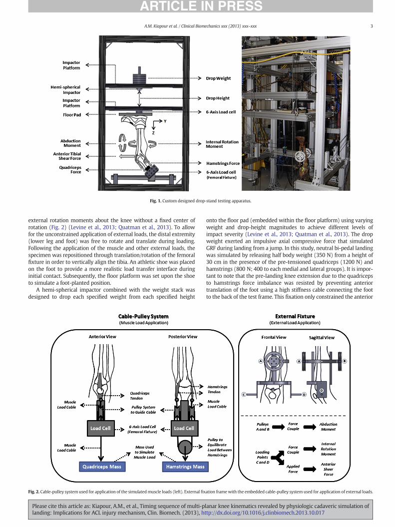
Fig. 1. Custom designed drop-stand testing apparatus.
3A.M. Kiapour et al. / Clinical Biomechanics xxx (2013) xxx–xxx
external rotation moments about the knee without a fixed center ofrotation (Fig. 2) (Levine et al., 2013; Quatman et al., 2013). To allowfor the unconstrained application of external loads, the distal extremity(lower leg and foot) was free to rotate and translate during loading.Following the application of the muscle and other external loads, thespecimen was repositioned through translation/rotation of the femoralfixture in order to vertically align the tibia. An athletic shoe was placedon the foot to provide a more realistic load transfer interface duringinitial contact. Subsequently, the floor platform was set upon the shoeto simulate a foot-planted position.
A hemi-spherical impactor combined with the weight stack wasdesigned to drop each specified weight from each specified height
Fig. 2. Cable-pulley systemused for application of the simulatedmuscle loads (left). External fix
Please cite this article as: Kiapour, A.M., et al., Timing sequence of multi-planding: Implications for ACL injury mechanism, Clin. Biomech. (2013), ht
onto the floor pad (embedded within the floor platform) using varyingweight and drop-height magnitudes to achieve different levels ofimpact severity (Levine et al., 2013; Quatman et al., 2013). The dropweight exerted an impulsive axial compressive force that simulatedGRF during landing from a jump. In this study, neutral bi-pedal landingwas simulated by releasing half body weight (350 N) from a height of30 cm in the presence of the pre-tensioned quadriceps (1200 N) andhamstrings (800 N; 400 to each medial and lateral groups). It is impor-tant to note that the pre-landing knee extension due to the quadricepsto hamstrings force imbalance was resisted by preventing anteriortranslation of the foot using a high stiffness cable connecting the footto the back of the test frame. This fixation only constrained the anterior
ation framewith the embedded cable-pulley systemused for application of external loads.
lanar knee kinematics revealed by physiologic cadaveric simulation oftp://dx.doi.org/10.1016/j.clinbiomech.2013.10.017
4 A.M. Kiapour et al. / Clinical Biomechanics xxx (2013) xxx–xxx
translation of the foot while preserving the other five-degrees of free-dom (2 translations and 3 rotations) within the ankle joint.
2.3. Instrumentation
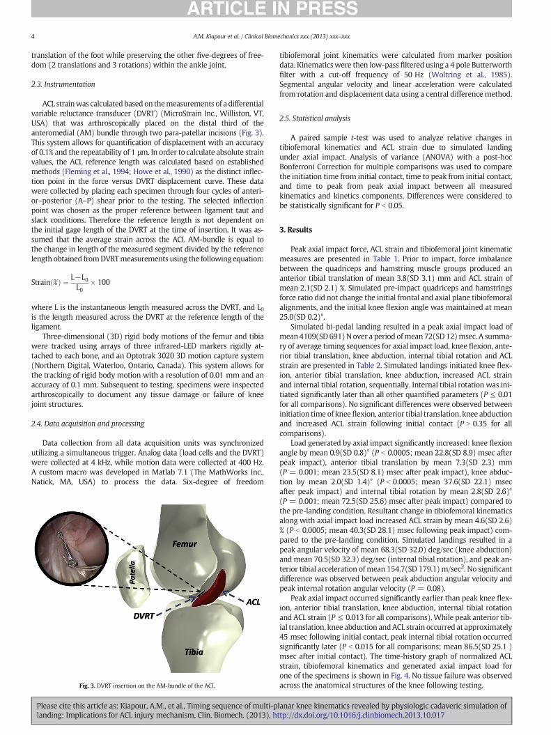
ACL strainwas calculated based on themeasurements of a differentialvariable reluctance transducer (DVRT) (MicroStrain Inc., Williston, VT,USA) that was arthroscopically placed on the distal third of theanteromedial (AM) bundle through two para-patellar incisions (Fig. 3).This system allows for quantification of displacement with an accuracyof 0.1% and the repeatability of 1 μm. In order to calculate absolute strainvalues, the ACL reference length was calculated based on establishedmethods (Fleming et al., 1994; Howe et al., 1990) as the distinct inflec-tion point in the force versus DVRT displacement curve. These datawere collected by placing each specimen through four cycles of anteri-or–posterior (A–P) shear prior to the testing. The selected inflectionpoint was chosen as the proper reference between ligament taut andslack conditions. Therefore the reference length is not dependent onthe initial gage length of the DVRT at the time of insertion. It was as-sumed that the average strain across the ACL AM-bundle is equal tothe change in length of the measured segment divided by the referencelength obtained fromDVRTmeasurements using the following equation:
Strain %ð Þ ¼ L−L0L0
� 100
where L is the instantaneous length measured across the DVRT, and L0is the length measured across the DVRT at the reference length of theligament.
Three-dimensional (3D) rigid body motions of the femur and tibiawere tracked using arrays of three infrared-LED markers rigidly at-tached to each bone, and an Optotrak 3020 3D motion capture system(Northern Digital, Waterloo, Ontario, Canada). This system allows forthe tracking of rigid body motion with a resolution of 0.01 mm and anaccuracy of 0.1 mm. Subsequent to testing, specimens were inspectedarthroscopically to document any tissue damage or failure of kneejoint structures.
2.4. Data acquisition and processing
Data collection from all data acquisition units was synchronizedutilizing a simultaneous trigger. Analog data (load cells and the DVRT)were collected at 4 kHz, while motion data were collected at 400 Hz.A custom macro was developed in Matlab 7.1 (The MathWorks Inc.,Natick, MA, USA) to process the data. Six-degree of freedom
Fig. 3. DVRT insertion on the AM-bundle of the ACL.
Please cite this article as: Kiapour, A.M., et al., Timing sequence of multi-planding: Implications for ACL injury mechanism, Clin. Biomech. (2013), h
tibiofemoral joint kinematics were calculated from marker positiondata. Kinematicswere then low-pass filtered using a 4 pole Butterworthfilter with a cut-off frequency of 50 Hz (Woltring et al., 1985).Segmental angular velocity and linear acceleration were calculatedfrom rotation and displacement data using a central difference method.
2.5. Statistical analysis
A paired sample t-test was used to analyze relative changes intibiofemoral kinematics and ACL strain due to simulated landingunder axial impact. Analysis of variance (ANOVA) with a post-hocBonferroni Correction for multiple comparisons was used to comparethe initiation time from initial contact, time to peak from initial contact,and time to peak from peak axial impact between all measuredkinematics and kinetics components. Differences were considered tobe statistically significant for P b 0.05.
3. Results
Peak axial impact force, ACL strain and tibiofemoral joint kinematicmeasures are presented in Table 1. Prior to impact, force imbalancebetween the quadriceps and hamstring muscle groups produced ananterior tibial translation of mean 3.8(SD 3.1) mm and ACL strain ofmean 2.1(SD 2.1) %. Simulated pre-impact quadriceps and hamstringsforce ratio did not change the initial frontal and axial plane tibiofemoralalignments, and the initial knee flexion angle was maintained at mean25.0(SD 0.2)°.
Simulated bi-pedal landing resulted in a peak axial impact load ofmean4109(SD691)Nover a period ofmean72(SD12)msec. A summa-ry of average timing sequences for axial impact load, knee flexion, ante-rior tibial translation, knee abduction, internal tibial rotation and ACLstrain are presented in Table 2. Simulated landings initiated knee flex-ion, anterior tibial translation, knee abduction, increased ACL strainand internal tibial rotation, sequentially. Internal tibial rotation was ini-tiated significantly later than all other quantified parameters (P ≤ 0.01for all comparisons). No significant differences were observed betweeninitiation time of knee flexion, anterior tibial translation, knee abductionand increased ACL strain following initial contact (P N 0.35 for allcomparisons).
Load generated by axial impact significantly increased: knee flexionangle by mean 0.9(SD 0.8)° (P b 0.0005; mean 22.8(SD 8.9) msec afterpeak impact), anterior tibial translation by mean 7.3(SD 2.3) mm(P = 0.001; mean 23.5(SD 8.1) msec after peak impact), knee abduc-tion by mean 2.0(SD 1.4)° (P b 0.0005; mean 37.6(SD 22.1) msecafter peak impact) and internal tibial rotation by mean 2.8(SD 2.6)°(P = 0.001; mean 72.5(SD 25.6) msec after peak impact) compared tothe pre-landing condition. Resultant change in tibiofemoral kinematicsalong with axial impact load increased ACL strain by mean 4.6(SD 2.6)% (P b 0.0005; mean 40.3(SD 28.1) msec following peak impact) com-pared to the pre-landing condition. Simulated landings resulted in apeak angular velocity of mean 68.3(SD 32.0) deg/sec (knee abduction)and mean 70.5(SD 32.3) deg/sec (internal tibial rotation), and peak an-terior tibial acceleration of mean 154.7(SD 179.1) m/sec2. No significantdifference was observed between peak abduction angular velocity andpeak internal rotation angular velocity (P = 0.08).
Peak axial impact occurred significantly earlier than peak knee flex-ion, anterior tibial translation, knee abduction, internal tibial rotationand ACL strain (P ≤ 0.013 for all comparisons).While peak anterior tib-ial translation, knee abduction andACL strain occurred at approximately45 msec following initial contact, peak internal tibial rotation occurredsignificantly later (P b 0.015 for all comparisons; mean 86.5(SD 25.1 )msec after initial contact). The time-history graph of normalized ACLstrain, tibiofemoral kinematics and generated axial impact load forone of the specimens is shown in Fig. 4. No tissue failure was observedacross the anatomical structures of the knee following testing.
lanar knee kinematics revealed by physiologic cadaveric simulation ofttp://dx.doi.org/10.1016/j.clinbiomech.2013.10.017

Table 1Summary of the peak axial impact load, change in tibiofemoral kinematics and ACL strain.
Specimens Peak impact Knee flexion ATTa Peak abduction Peak int. rotation ACL strain
ID Sex Age Side Impact induced Peak Impact induced Peak Impact induced Peak
1-C080044 F 52 L 3468 N 1.1° 25.8° 5.8 mm 10.9 mm −0.9° 1.6° 1.4% 3.4%2-C090105 M 38 L 3188 N 0.1° 24.8° 3.1 mm 8.0 mm 0.6° 0.8° 1.0% 6.9%3-C080033 M 51 L 3561 N −0.1° 24.8° 8.6 mm 14.6 mm 1.3° 2.5° 5.6% 8.5%4-S090574 F 49 R 4050 N 0.8° 26.0° 12.0 mm 16.5 mm 1.7° 4.4° 7.6% 9.4%5-C090155 M 46 L 4400 N 0.4 25.4° 6.6 mm 5.2 mm 0.9° 1.0° 5.7% 4.4%6-C090105 M 38 R 4155 N 1.0° 25.9° 4.3 mm 5.3 mm 0.9° 1.9° 9.1% 13.3%7-C090155 M 46 R 3394 N 0.7° 25.5° 5.4 mm 10.8 mm 0.9° 2.9° 1.7% 6.2%8-C090361 M 34 R 2869 N 1.6° 26.5° 6.6 mm 7.6 mm 3.8° 5.1° 4.7% 5.1%9-C090508 F 45 L 5076 N −0.2° 25.1° 4.9 mm 15.2 mm 2.8° 1.3° 5.7% 9.3%10-C090508 F 45 R 5036 N 0.7° 25.8° 8.4 mm 8.8 mm 3.6° 8.4° 8.9% 8.5%11-C080044 F 52 R 3616 N 0.1° 25.1° 6.9 mm 8.5 mm 2.4° 2.3° 2.2% 6.0%12-C090552 M 45 R 4751 N 0.6° 25.3° 8.9 mm 13.8 mm 3.6° 4.4° 6.4% 7.4%13-1007889 F 29 R 4483 N 1.3° 26.6° 9.1 mm 12.1 mm 3.9° 6.8° 2.9% 6.4%14-1008352 F 54 R 4274 N 2.3° 27.3° 9.8 mm 17.6 mm 4.0° 4.7° 2.6% 3.9%15-C090552 M 45 L 4453 N 1.6° 26.6° 6.4 mm 11.2 mm 2.0° −0.7° 4.4% 5.7%16-S090706 F 50 L 4962 N 3.1° 28.2° 9.4 mm 9.9 mm 1.3° −1.8° 3.7% 2.2%
a ATT: Anterior tibial translation.
5A.M. Kiapour et al. / Clinical Biomechanics xxx (2013) xxx–xxx
4. Discussion
Challenges in accurate, in vivo quantification ofmulti-planar knee ki-nematics and timing sequence during injury hinder the understandingof the relative contributions of each loading axis to the overall injurymechanisms. Biomechanical testing of human cadaveric tissue, if prop-erly designed, offers a practical means to evaluate joint biomechanicsand injury mechanisms. The purpose of this study was to investigatethe interaction between tibiofemoral joint kinematics and ACL strainin addition to their timing sequence using a novel, physiologic cadavericmodel of landing.
A unique, custom-designed drop-stand apparatus with physiologi-cally relevant drop weights and drop heights was employed. Simulatedlandings from a jump were conducted on sixteen instrumentedcadaveric specimens. Event timing sequence, change in tibiofemoralkinematics and change in ACL strain during a simulated bi-pedal land-ing taskwere quantified. The proposed cadavericmodel of landingdem-onstrated similar tibiofemoral kinematics and kinetics as reported byin vivo biomechanical and video analysis studies, Table 3. Comparisons(in vivo validation) were conducted on: landing duration (landingstance) (Chappell et al., 2002; Joseph et al., 2011), time to peak axialimpact load (GRF) (Decker et al., 2003), peak knee abduction angularvelocity (Joseph et al., 2011), time to peak knee abduction (as percentlanding stance) (Joseph et al., 2011), peak anterior tibial acceleration(Moran and Marshall, 2006) and time to peak ACL strain (ACL rupture)(Koga et al., 2010; Krosshaug et al., 2007). These comparisons are com-pelling, especially in light of the lack of complete active neuromuscularcontrol in cadaveric model, intra-specimen variability in joint geometryand tissuemechanical properties, and the limited sample size comparedto in vivo biomechanical studies. Moreover, this cadaveric model hasbeen reported to consistently reproduce clinically relevant injury
Table 2Summary of the average (SD) timing sequences.
Parameter Initiation timefrom ICa
Time to peak
From IC From peak axialimpact
Axial impact load – 13.7 (2.4) msec –
Knee flexion 7.1 (3.5) msec 36.6 (7.8) msec 22.8 (8.9) msecAnterior tibial translation 7.7 (1.9) msec 37.5 (7.0) msec 23.5 (8.1) msecKnee abduction 10.8 (6.2) msec 51.5 (22.6) msec 37.6 (22.1) msecInternal tibial rotation 21.0 (11.7) msec 86.5 (25.1) msec 72.5 (25.6) msecACL strain 12.4 (6.3) msec 54.2 (27.0) msec 40.3 (28.1) msec
a IC: Initial contact.
Please cite this article as: Kiapour, A.M., et al., Timing sequence of multi-planding: Implications for ACL injury mechanism, Clin. Biomech. (2013), ht
patterns to the ACL (ACL failure in almost 90% of the specimens) andsurrounding soft tissue structures (Levine et al., 2013) under injuriousconditions. Finally, the resultant tibial plateau injury patterns (botharticular cartilage and subchondral bone) were shown to be similar toclinically observed bone bruise patterns across the tibial plateau duringactual cases of non-contact ACL injury (Kiapour et al., 2012c; Levineet al., 2013). As a result, the current cadaveric model can be considereda valid approach in simulating landing biomechanics.
The results of this study demonstrate an increase in both anteriortibial translation and ACL strain due to A-P imbalance in simulatedknee muscle loads prior to impact. This is in agreement with previousfindings demonstrating the anterior translation of the tibia with respectto the femur and increased levels of ACL strain/force or risk of ACL injuryunder aggressive quadriceps force (Berns et al., 1992; Beynnon et al.,1995; DeMorat et al., 2004; Draganich and Vahey, 1990; Durselenet al., 1995; Hashemi et al., 2010; Li et al., 1999; Quatman et al., 2012;Wall et al., 2012). Simulated landings in this study sequentially resultedin increased knee flexion, anterior tibial translation, knee abduction,ACL strain and internal tibial rotation. This is in agreement with our hy-pothesis that temporal differences exist inmulti-planar knee kinematicsduring dynamic landing. Previous clinical, video analysis and in vivobiomechanical studies indicate that knee flexion, anterior tibial transla-tion, knee abduction and internal rotation of the tibia are associatedwith landing (Ford et al., 2003; Hewett et al., 2005; Koga et al., 2010;Krosshaug et al., 2007; Moran and Marshall, 2006). Additionally, thesefactors have been shown to contribute to non-contact ACL injuries atshallow knee flexion angles (Kiapour et al., 2013c; Levine et al., 2013;Meyer and Haut, 2008; Oh et al., 2012).
It was further noted that while knee flexion, anterior tibial transla-tion, knee abduction and increased ACL strainwere initiated and reachedtheir maximum values almost simultaneously, internal tibial rotationwas initiated (P ≤ 0.01 for all comparisons) and peaked (P b 0.015for all comparisons) significantly later (Fig. 4). This observed timingsequence highlights the primary role of the anterior tibial translationalong with knee abduction in ACL loading and potential risk of injuryduring landing, as suggested by others (Boden et al., 2000; Ford et al.,2003; Kiapour et al., 2012d; Koga et al., 2010; Krosshaug et al., 2007;Olsen et al., 2004; Shin et al., 2009;Withrow et al., 2006). The concurrentincrease in both ACL strain and internal tibial rotation during simulatedlandings supports internal rotation as a potential risk factor for ACL inju-ry as previously indicated (Kiapour et al., 2012b,d; Meyer and Haut,2008; Oh et al., 2012). Further, it was demonstrated that internal tibialrotation reached an average ofmean 1.8(SD 2.5)°, almost 63% of its max-imumvalue, by the timepeakACL strain occurs,while both anterior tibialtranslation and knee abduction have already reached their peaks
lanar knee kinematics revealed by physiologic cadaveric simulation oftp://dx.doi.org/10.1016/j.clinbiomech.2013.10.017
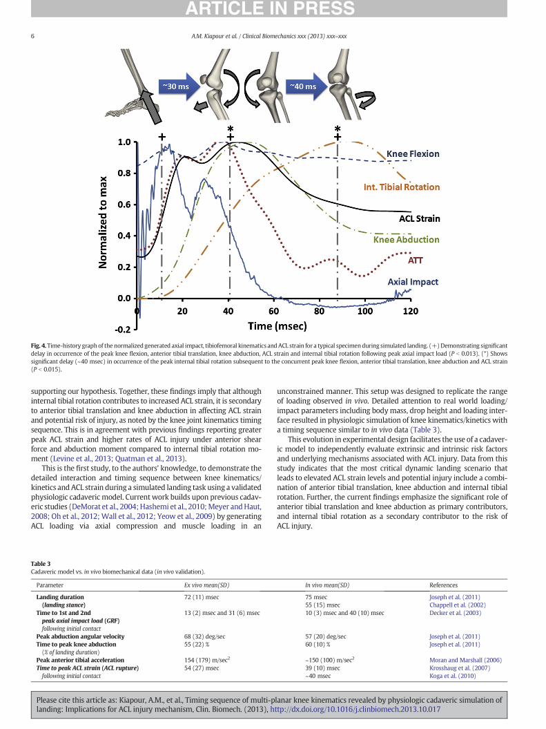
Fig. 4. Time-history graph of the normalized generated axial impact, tibiofemoral kinematics andACL strain for a typical specimenduring simulated landing. (+)Demonstrating significantdelay in occurrence of the peak knee flexion, anterior tibial translation, knee abduction, ACL strain and internal tibial rotation following peak axial impact load (P b 0.013). (*) Showssignificant delay (~40 msec) in occurrence of the peak internal tibial rotation subsequent to the concurrent peak knee flexion, anterior tibial translation, knee abduction and ACL strain(P b 0.015).
6 A.M. Kiapour et al. / Clinical Biomechanics xxx (2013) xxx–xxx
supporting our hypothesis. Together, these findings imply that althoughinternal tibial rotation contributes to increased ACL strain, it is secondaryto anterior tibial translation and knee abduction in affecting ACL strainand potential risk of injury, as noted by the knee joint kinematics timingsequence. This is in agreement with previous findings reporting greaterpeak ACL strain and higher rates of ACL injury under anterior shearforce and abduction moment compared to internal tibial rotation mo-ment (Levine et al., 2013; Quatman et al., 2013).
This is the first study, to the authors' knowledge, to demonstrate thedetailed interaction and timing sequence between knee kinematics/kinetics andACL strain during a simulated landing task using a validatedphysiologic cadaveric model. Currentwork builds upon previous cadav-eric studies (DeMorat et al., 2004;Hashemi et al., 2010;Meyer andHaut,2008; Oh et al., 2012;Wall et al., 2012; Yeow et al., 2009) by generatingACL loading via axial compression and muscle loading in an
Table 3Cadaveric model vs. in vivo biomechanical data (in vivo validation).
Parameter Ex vivo mean(SD)
Landing duration(landing stance)
72 (11) msec
Time to 1st and 2ndpeak axial impact load (GRF)following initial contact
13 (2) msec and 31 (6) msec
Peak abduction angular velocity 68 (32) deg/secTime to peak knee abduction(% of landing duration)
55 (22) %
Peak anterior tibial acceleration 154 (179) m/sec2
Time to peak ACL strain (ACL rupture)following initial contact
54 (27) msec
Please cite this article as: Kiapour, A.M., et al., Timing sequence of multi-planding: Implications for ACL injury mechanism, Clin. Biomech. (2013), h
unconstrained manner. This setup was designed to replicate the rangeof loading observed in vivo. Detailed attention to real world loading/impact parameters including bodymass, drop height and loading inter-face resulted in physiologic simulation of knee kinematics/kinetics witha timing sequence similar to in vivo data (Table 3).
This evolution in experimental design facilitates the use of a cadaver-ic model to independently evaluate extrinsic and intrinsic risk factorsand underlying mechanisms associated with ACL injury. Data from thisstudy indicates that the most critical dynamic landing scenario thatleads to elevated ACL strain levels and potential injury include a combi-nation of anterior tibial translation, knee abduction and internal tibialrotation. Further, the current findings emphasize the significant role ofanterior tibial translation and knee abduction as primary contributors,and internal tibial rotation as a secondary contributor to the risk ofACL injury.
In vivo mean(SD) References
75 msec Joseph et al. (2011)55 (15) msec Chappell et al. (2002)10 (3) msec and 40 (10) msec Decker et al. (2003)
57 (20) deg/sec Joseph et al. (2011)60 (10) % Joseph et al. (2011)
~150 (100) m/sec2 Moran and Marshall (2006)39 (10) msec Krosshaug et al. (2007)~40 msec Koga et al. (2010)
lanar knee kinematics revealed by physiologic cadaveric simulation ofttp://dx.doi.org/10.1016/j.clinbiomech.2013.10.017
7A.M. Kiapour et al. / Clinical Biomechanics xxx (2013) xxx–xxx
4.1. Study limitations
Aswith any study, inherent limitations exist in the current cadavericstudy. First, ACL strain was represented by local strain measurementsacross the AM-bundle. However, the attachment of a second DVRTto the posterolateral bundle of the ACL would have been associatedwith the compromise of the posterior joint capsule and potential mea-surement artifacts (Bach and Hull, 1998). The choice to place a singleDVRT on the ACL AM-bundle was based on a previous work thatfound AM-bundle strain to be a good representation of overall ACLstrain (Markolf et al., 1990). Another limitation is the potential differ-ences in tissue properties associated with cadaveric specimens com-pared with the in vivo tissue properties of young athletes, which canaffect the accuracy of the absolute reported values. We have tried tominimize this artifact by testing relatively young specimens. Moreover,the effect of change in knee flexion angle was not evaluated as all thespecimens were tested at 25° of knee flexion, since this flexion anglehas been reported during real cases of ACL injury. Additionally, landingwas simulated with the foot in a flat position with the ankle joint beingsemi-constrained to a limited range of dorsi flexion, which does notreplicate ankle motion during landing. Finally, the primary and second-ary roles of loading factors on the risk of ACL injury have been identifiedsolely based on the temporal characteristics of knee multi-planar kine-matics. Despite strong agreement with previous findings, further para-metric and sensitivity analyses are required to better characterize theindependent role of each loading axis in ACL injury risk.
Current findings are least likely to be affected by this limitation asthis study was intended to replicate/investigate the isolated knee jointbiomechanical response during the inciting event not the wholemulti-joint landing phenomenon. We believe that the qualitative find-ings and relative comparisons presented in this work minimize suchartifacts. Considering the strengths and limitations of this experimentalmodel, the authors believe that it is well suited and able to evaluate themechanisms of ACL injury.
Acknowledgements
The authors acknowledge funding support from the National Insti-tutes of Health/National Institute of Arthritis and Musculoskeletal andSkin Diseases grants R01-AR049735 and R01-AR056259. The authorswould also like to thank Dr. Jason Levine for his assistance.
References
Agel, J., Arendt, E.A., Bershadsky, B., 2005. Anterior cruciate ligament injury in nationalcollegiate athletic association basketball and soccer: a 13-year review. Am. J. SportsMed. 33, 524–530.
Arendt, E., Dick, R., 1995. Knee injury patterns among men and women in collegiatebasketball and soccer. NCAA data and review of literature. Am. J. Sports Med. 23,694–701.
Bach, J.M., Hull, M.L., 1998. Strain inhomogeneity in the anterior cruciate ligament underapplication of external and muscular loads. J. Biomech. Eng. 120, 497–503.
Berns, G.S., Hull, M.L., Patterson, H.A., 1992. Strain in the anteromedial bundle of theanterior cruciate ligament under combination loading. J. Orthop. Res. 10, 167–176.
Beynnon, B.D., Fleming, B.C., Johnson, R.J., Nichols, C.E., Renstrom, P.A., Pope, M.H., 1995.Anterior cruciate ligament strain behavior during rehabilitation exercises in vivo.Am. J. Sports Med. 23, 24–34.
Boden, B.P., Dean, G.S., Feagin, J.A., Garrett, W.E., 2000. Mechanisms of anterior cruciateligament injury. Orthopedics 23, 573–578.
Butler, D.L., Noyes, F.R., Grood, E.S., 1980. Ligamentous restraints to anterior–posteriordrawer in the human knee. A biomechanical study. J. Bone Joint Surg. Am. 62,259–270.
Chappell, J.D., Yu, B., Kirkendall, D.T., Garrett, W.E., 2002. A comparison of knee kineticsbetween male and female recreational athletes in stop–jump tasks. Am. J. SportsMed. 30, 261–267.
Csintalan, R.P., Ehsan, A., Mcgarry, M.H., Fithian, D.F., Lee, T.Q., 2006. Biomechanical andanatomical effects of an external rotational torque applied to the knee: a cadavericstudy. Am. J. Sports Med. 34, 1623–1629.
De Loes, M., Dahlstedt, L.J., Thomee, R., 2000. A 7-year study on risks and costs of kneeinjuries in male and female youth participants in 12 sports. Scand. J. Med. Sci. Sports10, 90–97.
Please cite this article as: Kiapour, A.M., et al., Timing sequence of multi-planding: Implications for ACL injury mechanism, Clin. Biomech. (2013), ht
Decker, M.J., Torry, M.R., Wyland, D.J., Sterett, W.I., Richard Steadman, J., 2003. Genderdifferences in lower extremity kinematics, kinetics and energy absorption duringlanding. Clin. Biomech. 18, 662–669.
Demorat, G., Weinhold, P., Blackburn, T., Chudik, S., Garrett, W., 2004. Aggressive quadri-ceps loading can induce noncontact anterior cruciate ligament injury. Am. J. SportsMed. 32, 477–483.
Draganich, L.F., Vahey, J.W., 1990. An in vitro study of anterior cruciate ligament straininduced by quadriceps and hamstrings forces. J. Orthop. Res. 8, 57–63.
Durselen, L., Claes, L., Kiefer, H., 1995. The influence ofmuscle forces and external loads oncruciate ligament strain. Am. J. Sports Med. 23, 129–136.
Fleming, B.C., Beynnon, B.D., Tohyama, H., Johnson, R.J., Nichols, C.E., Renstrom, P., et al.,1994. Determination of a zero strain reference for the anteromedial band of theanterior cruciate ligament. J. Orthop. Res. 12, 789–795.
Ford, K.R., Myer, G.D., Hewett, T.E., 2003. Valgus knee motion during landing in highschool female and male basketball players. Med. Sci. Sports Exerc. 35, 1745–1750.
Fukubayashi, T., Torzilli, P.A., Sherman, M.F., Warren, R.F., 1982. An in vitro biomechanicalevaluation of anterior–posterior motion of the knee. Tibial displacement, rotation,and torque. J. Bone Joint Surg. Am. 64, 258–264.
Gabriel, M.T., Wong, E.K., Woo, S.L., Yagi, M., Debski, R.E., 2004. Distribution of in situ forcesin the anterior cruciate ligament in response to rotatory loads. J. Orthop. Res. 22, 85–89.
Griffin, L.Y., Agel, J., Albohm, M.J., Arendt, E.A., Dick, R.W., Garrett, W.E., et al., 2000.Noncontact anterior cruciate ligament injuries: risk factors and prevention strategies.J. Am. Acad. Orthop. Surg. 8, 141–150.
Hashemi, J., Breighner, R., Jang, T.H., Chandrashekar, N., Ekwaro-Osire, S., Slauterbeck, J.R.,2010. Increasing pre-activation of the quadriceps muscle protects the anterior cruci-ate ligament during the landing phase of a jump: an in vitro simulation. Knee 17,235–241.
Henrichs, A., 2004. A review of knee dislocations. J. Athl. Train. 39, 365–369.Hewett, T.E., Lindenfeld, T.N., Riccobene, J.V., Noyes, F.R., 1999. The effect of neuromuscular
training on the incidence of knee injury in female athletes. A prospective study. Am.J. Sports Med. 27, 699–706.
Hewett, T.E., Myer, G.D., Ford, K.R., Heidt Jr., R.S., Colosimo, A.J., Mclean, S.G., et al., 2005.Biomechanical measures of neuromuscular control and valgus loading of the kneepredict anterior cruciate ligament injury risk in female athletes: a prospectivestudy. Am. J. Sports Med. 33, 492–501.
Howe, J.G., Wertheimer, C., Johnson, R.J., Nichols, C.E., Pope, M.H., Beynnon, B., 1990.Arthroscopic strain gauge measurement of the normal anterior cruciate ligament.Arthroscopy 6, 198–204.
Joseph, M.F., Rahl, M., Sheehan, J., MacDougall, B., Horn, E., Denegar, C.R., et al., 2011.Timing of lower extremity frontal plane motion differs between female and maleathletes during a landing task. Am. J. Sports Med. 39, 1517–1521.
Kiapour, A.M., 2013. Non-Contact ACL Injuries during Landing: Risk Factors andMechanisms. (Doctor of Philosophy Degree in Biomedical Engineering Dissertation)The University of Toledo.
Kiapour, A.M., Quatman, C.E., Levine, J.W., Wordeman, S.C., Hewett, T.E., Goel, V.K., et al.,2012a. Coupled Valgus Collapse Due to Internal Rotation: an Important Factor inthe ACL Injury Mechanism. Proceedings of 59th ACSM Annual Meeting. LippincottWilliams & Wilkins, San Francisco, CA.
Kiapour, A.M., Quatman, C.E., Ditto, R.C., Levine, J.W., Wordeman, S.C., Hewett, T.E., et al.,2012b. Global quasi-static mechanical characterization of the human knee undersingle- and multi-axis unconstrained loading conditions. Proceedings of 2012 ASMESummer Bioengineering Conference. American Society of Mechanical Engineers(ASME), Fajardo, Puerto Rico.
Kiapour, A.M., Quatman, C.E., Ditto, R.C., Levine, J.W., Wordeman, S.C., Hewett, T.E., et al.,2012c. Influence of axial rotation moments on ACL strain: a cadaveric study of single-and multi-axis loading of the knee. Proceedings of 37th ASB Annual Meeting.American Society of Biomechanics (ASB), Long Beach, CA.
Kiapour, A.M., Quatman, C.E., Goel, V.K., Ditto, R.C., Wordeman, S.C., Levine, J.W., et al.,2012d. Knee articular cartilage pressure distribution under single- and multi-axisloading conditions: implications for ACL injury mechanism. Proceedings of the 38thASB Annual Meeting, Gainesville, pp. 15–18.
Kiapour, A.M., Kaul, V., Kiapour, A., Quatman, C.E., Wordeman, S.C., Hewett, T.E., et al.,2013a. The effect of ligament modeling technique on knee joint kinematics: a finiteelement study. Appl. Math. 4, 91–97.
Kiapour, A.M., Wordeman, S.C., Paterno, M.V., Quatman, C.E., Levine, J.W., Goel, V.K., et al.,2013b. Diagnostic value of knee arthrometry in the prediction of ACL strain duringlanding. Am. J. Sports Med. 42.
Kiapour, A., Kiapour, A.M., Kaul, V., Quatman, C.E., Hewett, T.E., Demetropoulos, C.K., et al.,2013c. Finite element model of the knee for investigation of injury mechanisms:development and validation. J. Biomech. Eng. http://dx.doi.org/10.1115/1.4025692.
Kim, S., Bosque, J., Meehan, J.P., Jamali, A., Marder, R., 2011. Increase in outpatient kneearthroscopy in the United States: a comparison of National Surveys of AmbulatorySurgery, 1996 and 2006. J. Bone Joint Surg. Am. 93, 994–1000.
Koga, H., Nakamae, A., Shima, Y., Iwasa, J., Myklebust, G., Engebretsen, L., et al., 2010.Mechanisms for noncontact anterior cruciate ligament injuries: knee joint kinematicsin 10 injury situations from female team handball and basketball. Am. J. Sports Med.38, 2218–2225.
Krosshaug, T., Nakamae, A., Boden, B.P., Engebretsen, L., Smith, G., Slauterbeck, J.R., et al.,2007. Mechanisms of anterior cruciate ligament injury in basketball: video analysis of39 cases. Am. J. Sports Med. 35, 359–367.
Levine, J.W., Kiapour, A.M., Quatman, C.E., Wordeman, S.C., Goel, V.K., Hewett, T.E., et al.,2013. Clinically relevant injury patterns after an anterior cruciate ligament injuryprovide insight into injury mechanisms. Am. J. Sports Med. 41, 385–395.
Li, G., Rudy, T.W., Sakane, M., Kanamori, A., Ma, C.B., Woo, S.L., 1999. The importance ofquadriceps and hamstring muscle loading on knee kinematics and in-situ forces inthe ACL. J. Biomech. 32, 395–400.
lanar knee kinematics revealed by physiologic cadaveric simulation oftp://dx.doi.org/10.1016/j.clinbiomech.2013.10.017
8 A.M. Kiapour et al. / Clinical Biomechanics xxx (2013) xxx–xxx
Malone, T.R., Hardaker, W.T., Garrett, W.E., J.A.F., Bassett, F.H., 1993. Relationship ofgender to anterior cruciate ligament injuries in intercollegiate basketball players.J. South. Orthop. Assoc. 2, 36–39.
Maquirriain, J., Megey, P.J., 2006. Tennis specific limitations in players with an ACLdeficient knee. Br. J. Sports Med. 40, 451–453.
Markolf, K.L., Gorek, J.F., Kabo, J.M., Shapiro, M.S., 1990. Direct measurement of resultantforces in the anterior cruciate ligament. An in vitro study performed with a newexperimental technique. J. Bone Joint Surg. Am. 72, 557–567.
Markolf, K.L., O'Neill, G., Jackson, S.R., McAllister, D.R., 2004. Effects of applied quadricepsand hamstrings muscle loads on forces in the anterior and posterior cruciate liga-ments. Am. J. Sports Med. 32, 1144–1149.
Mazzocca, A.D., Nissen, C.W., Geary, M., Adams, D.J., 2003. Valgus medial collateralligament rupture causes concomitant loading and damage of the anterior cruciateligament. J. Knee Surg. 16, 148–151.
Meyer, E.G., Haut, R.C., 2008. Anterior cruciate ligament injury induced by internal tibialtorsion or tibiofemoral compression. J. Biomech. 41, 3377–3383.
Moran, K.A., Marshall, B.M., 2006. Effect of fatigue on tibial impact accelerations and kneekinematics in drop jumps. Med. Sci. Sports Exerc. 38, 1836–1842.
Oh, Y.K., Lipps, D.B., Ashton-Miller, J.A., Wojtys, E.M., 2012. What strains the anteriorcruciate ligament during a pivot landing? Am. J. Sports Med. 40, 574–583.
Olsen, O.E., Myklebust, G., Engebretsen, L., Bahr, R., 2004. Injury mechanisms for anteriorcruciate ligament injuries in team handball: a systematic video analysis. Am. J. SportsMed. 32, 1002–1012.
Quatman, C.E., Quatman-Yates, C.C., Hewett, T.E., 2010. A ‘plane’ explanation of anteriorcruciate ligament injury mechanisms: a systematic review. Sports Med. 40, 729–746.
Quatman, C.E., Kiapour, A.M., Kiapour, A., Levine, J.W., Wordeman, S.C., Demetropoulos, C.K.,et al., 2012. Effects of quadriceps and hamstrings ratio on ACL strain during landingactivities. Proceedings of 2012 ASME Summer Bioengineering Conference. AmericanSociety of Mechanical Engineers (ASME), Fajardo, Puerto Rico.
Quatman, C.E., Kiapour, A.M., Demetropoulos, C.K., Kiapour, A., Wordeman, S.C., Levine, J.W.,et al., 2013. Preferential loading of the ACL compared with the MCL during landing: anovel in sim approach yields the multiplanar mechanism of dynamic valgus duringACL injuries. Am. J. Sports Med. http://dx.doi.org/10.1177/0363546513506558 (October11, published online before print October 11, 2013).
Please cite this article as: Kiapour, A.M., et al., Timing sequence of multi-planding: Implications for ACL injury mechanism, Clin. Biomech. (2013), h
Renstrom, P., Arms, S.W., Stanwyck, T.S., Johnson, R.J., Pope, M.H., 1986. Strain within theanterior cruciate ligament during hamstring and quadriceps activity. Am. J. SportsMed. 14, 83–87.
Romero, J., Duronio, J.F., Sohrabi, A., Alexander, N., Macwilliams, B.A., Jones, L.C., et al.,2002. Varus and valgus flexion laxity of total knee alignment methods in loadedcadaveric knees. Clin. Orthop. Relat. Res. 243–253.
Shin, C.S., Chaudhari, A.M., Andriacchi, T.P., 2009. The effect of isolated valgusmoments on ACL strain during single-leg landing: a simulation study. J. Biomech.42, 280–285.
Van Lent, M.E., Drost, M.R., Vd Wildenberg, F.A., 1994. EMG profiles of ACL-deficientpatients during walking: the influence of mild fatigue. Int. J. Sports Med. 15,508–514.
Wall, S.J., Rose, D.M., Sutter, E.G., Belkoff, S.M., Boden, B.P., 2012. The role of axialcompressive and quadriceps forces in noncontact anterior cruciate ligament injury:a cadaveric study. Am. J. Sports Med. 40, 568–573.
Withrow, T.J., Huston, L.J., Wojtys, E.M., Ashton-Miller, J.A., 2006. The effect of an impul-sive knee valgus moment on in vitro relative ACL strain during a simulated jumplanding. Clin Biomech 21, 977–983.
Withrow, T.J., Huston, L.J., Wojtys, E.M., Ashton-Miller, J.A., 2008. Effect of varying ham-string tension on anterior cruciate ligament strain during in vitro impulsive kneeflexion and compression loading. J. Bone Joint Surg. Am. 90, 815–823.
Woltring, H.J., Huiskes, R., De Lange, A., Veldpaus, F.E., 1985. Finite centroid and helicalaxis estimation from noisy landmark measurements in the study of human jointkinematics. J. Biomech. 18, 379–389.
Wu, J.-L., 2010. In-Situ forces in the anteromedial and posterolateral bundles of the ante-rior cruciate ligament under simulated functional loading conditions. ORS Conf. 35,1995.
Yeow, C.H., Ng, K.S., Cheong, C.H., Lee, P.V., Goh, J.C., 2009. Repeated application of incre-mental landing impact loads to intact knee joints induces anterior cruciate ligamentfailure and tibiofemoral cartilage deformation and damage: a preliminary cadavericinvestigation. J. Biomech. 42, 972–981.
Zantop, T., Herbort, M., Raschke, M.J., Fu, F.H., Petersen, W., 2007. The role of theanteromedial and posterolateral bundles of the anterior cruciate ligament in anteriortibial translation and internal rotation. Am. J. Sports Med. 35, 223–227.
lanar knee kinematics revealed by physiologic cadaveric simulation ofttp://dx.doi.org/10.1016/j.clinbiomech.2013.10.017