Embed Size (px)
Citation preview

October 15, 2004 / Vol. 29, No. 20 / OPTICS LETTERS 2405
Time-resolved spectrophotometer for turbid media based onsupercontinuum generation in a photonic crystal fiber
Andrea Bassi, Johannes Swartling, Cosimo D’Andrea, Antonio Pifferi,Alessandro Torricelli, and Rinaldo Cubeddu
National Laboratory for Ultrafast and Ultraintense Optical Science— Istituto Nazionale per la Fisica della Materia,Istituto di Fotonica e Nanotecnologie— Consiglio Nazionale delle Ricerche, Dipartimento di Fisica, Politecnico di Milano,
Piazza L. da Vinci 32, I-20133 Milan, Italy
Received June 8, 2004
We describe an instrument for the time-resolved spectroscopy of turbid media that is based on supercontinuumgeneration in a photonic crystal fiber. The light injected into the sample consists of subpicosecond pulsesthat cover 550–1000 nm at 85 MHz at an average power of as much as 40 mW. A spectrometer coupled to amultianode photomultiplier tube is used to detect the light simultaneously in 16 wavelength channels, with aresolution of 5–20 nm�channel, depending on the grating. Time-correlated single-photon counting is used toproduce time-dispersion curves, which one fits to the diffusion equation to determine absorption and reducedscattering coefficients. We tested the instrument by measuring the time-resolved diffuse ref lectance of epoxyphantoms and by performing in vivo measurements on volunteers. The results were similar to those obtainedwith previous discrete wavelength systems, whereas the full spectrum (610–810 nm) acquisition time was asshort as 1 s owing to the parallel acquisition. © 2004 Optical Society of America
OCIS codes: 120.6200, 170.6510, 170.7050, 300.6500.
Spectroscopic analysis of turbid media facilitatesquantification of the constituents of a diverse range ofmaterials, including plastics, paper, inorganic materi-als, pharmaceuticals, and foods. The foremost f ieldof applications is in biomedicine, with the developmentof techniques for noninvasive analysis of physiologicalparameters of interest for medical diagnostics aswell as monitoring of administered drugs and of theefficacy of treatments. A few techniques for truewide-spectrum analysis of turbid media in the visibleand near-infrared regions have been developed.1 –5 Ofthese, time-resolved spectroscopy (TRS) on picosecondto nanosecond time scales yields the possibility ofdetermining directly the absorption coeff icient maand the reduced scattering coefficient ms
0 from asingle measurement by fitting the measured data to asuitable light-propagation model such as the diffusionequation.2 – 6
The TRS technique may be used for single measure-ments of the local optical properties between the sourceand the detection points2; alternatively, it can be partof a system for diffuse optical tomography in whichmultiple source and detector point combinations areused for the imaging of optical properties.7 Previouslya system based on tunable solid-state and dye laserswas developed to select wavelengths.2 This systemprovides highly accurate measurements; however, toprovide broad-spectrum measurements it requiressequential wavelength scanning. When TRS mea-surements are performed in vivo, it is especiallyimportant to keep the acquisition time short, for thepatient’s comfort and to be able to detect fast changes,for example, in blood f low or oxygen saturation. Aninstrument that permits parallel acquisition at manywavelengths simultaneously over a broad spectrum isthus desirable.
Early efforts to produce broad-spectrum shortpulses for TRS used high-power laser systems based
0146-9592/04/202405-03$15.00/0
on regenerative amplif iers, in which the output pulsesinteracted with media such as water and sapphire.1
Nonlinear effects produced a supercontinuum (SC) ofwavelengths about the center wavelength. Becauseof the low pulse repetition rates of these systems,streak cameras were necessary for detection. The useof low-power, less-complex laser equipment as lightsources and of relatively cheap photomultiplier tubes(PMTs) for detection would clearly be advantageous.In this Letter we present what is to our knowledge thefirst instrument for TRS that uses broad-spectrumlight based on SC generation in highly nonlinearphotonic crystal f iber (PCF) and a multichannel PMTfor simultaneous detection at different wavelengthsby time-correlated single-photon counting. This tech-nology has advantages compared with streak camerasin terms of higher sensitivity and a greater dynamicrange.1,2,6
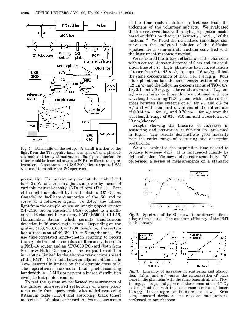
In our system, a passively mode-locked Ti:sapphireoscillator (Tissa 50, CDP Systems, Russian Federa-tion) pumped by an Ar:ion laser (Innova 200, Coherent,USA) provides 50–100-fs pulses at an 85-MHz repe-tition rate near 800 nm and at an average power of400–500 mW. The setup is depicted in Fig. 1. Thelight is focused into an 80-cm-long PCF (NL-2.4-800,Blaze Photonics, UK), with a 2.4-mm core. The cou-pling efficiency to the fiber is �40%. The SC is gen-erated by a combination of several nonlinear effects inthe PCF.8 We shape the SC spectrum by (empirically)adjusting the laser power and wavelength, the lengthof the pulses, and the polarization of the light in rela-tion to the hexagonal symmetry of the fiber core. Theresult is a relatively smooth spectrum covering the re-gion 550–1000 nm (see Fig. 2), which is an appropri-ate range for tissue analysis.2 – 4
The light is guided from the PCF by a 50-mm-coregraded-index (GRIN) fiber and can be coupled to anyof several fiber-optic probe heads that we developed
© 2004 Optical Society of America
2406 OPTICS LETTERS / Vol. 29, No. 20 / October 15, 2004
Fig. 1. Schematic of the setup. A small fraction of thelight from the Ti:sapphire laser was split off to a photodi-ode and used for synchronization. Bandpass interferencefilters could be inserted after the PCF to calibrate the spec-trometer. A spectrometer (USB 2000, Ocean Optics, USA)was used to monitor the SC spectrum.
previously. The maximum power at the probe headis �40 mW, and we can adjust the power by means ofvariable neutral-density (ND) filters (Fig. 1). Partof the light is split off by fused splitters (OZ Optics,Canada) to facilitate diagnostics of the SC and toserve as a reference signal. To detect the diffuselight from the sample we use an imaging spectrometer(SP-2150, Acton Research, USA) coupled to a multi-anode 16-channel linear array PMT (R5900U-01-L16,Hamamatsu, Japan), which permits simultaneousdetection in 16 wavelength bands. Depending on thegrating (150, 300, 600, or 1200 lines�mm), the systemhas a resolution of 40, 20, 10, or 5 nm�channel. Weuse time-correlated single-photon counting to recordthe signals from all channels simultaneously, based ona PML-16 router and an SPC-630 PC card (both fromBecker & Hickl, Germany). The temporal resolutionis �160 ps, limited by the electron transit time spreadof the PMT. Cross talk between adjacent channels is,5%, essentially limited by the electronic cross talk.The operational maximum total photon-countingbandwidth is �2 MHz to prevent a biased distributionowing to lost photon counts.
To test the system we performed measurements ofthe diffuse time-resolved ref lectance of tissue phan-toms made from epoxy resin with added scattering[titanium oxide (TiO2)] and absorbing (black toner)materials.6 We also performed in vivo measurements
of the time-resolved diffuse ref lectance from theabdomens of the volunteer subjects. We evaluatedthe time-resolved data with a light-propagation modelbased on diffusion theory, to extract ma and ms
0 of themedium.2,6 We fitted the normalized time-dispersioncurves to the analytical solution of the diffusionequation for a semi-inf inite medium convolved withthe instrument response function.
We measured the diffuse ref lectance of the phantomswith a source–detector distance of 2 cm and an acqui-sition time of 5 s. Eight phantoms had concentrationsof toner from 0 to 42 mg�g in steps of 6 mg�g; all hadthe same concentration of TiO2, i.e., 1.4 mg�g. Fourother phantoms had the same concentration of toner(12 mg�g) and the following concentrations of TiO2: 0.7,1.4, 2.1, and 2.9 mg�g. The resultant values of ma andms
0 were similar to those that we obtained with ourwavelength-scanning TRS system, with median differ-ences between the systems of 4% for ma and 3% forms
0 and with standard deviations of the differencesof 0.014 cm21 for ma and 0.76 cm21 for ms
0 over thewavelength range of 610–810 nm and a resolution of20 nm�channel.
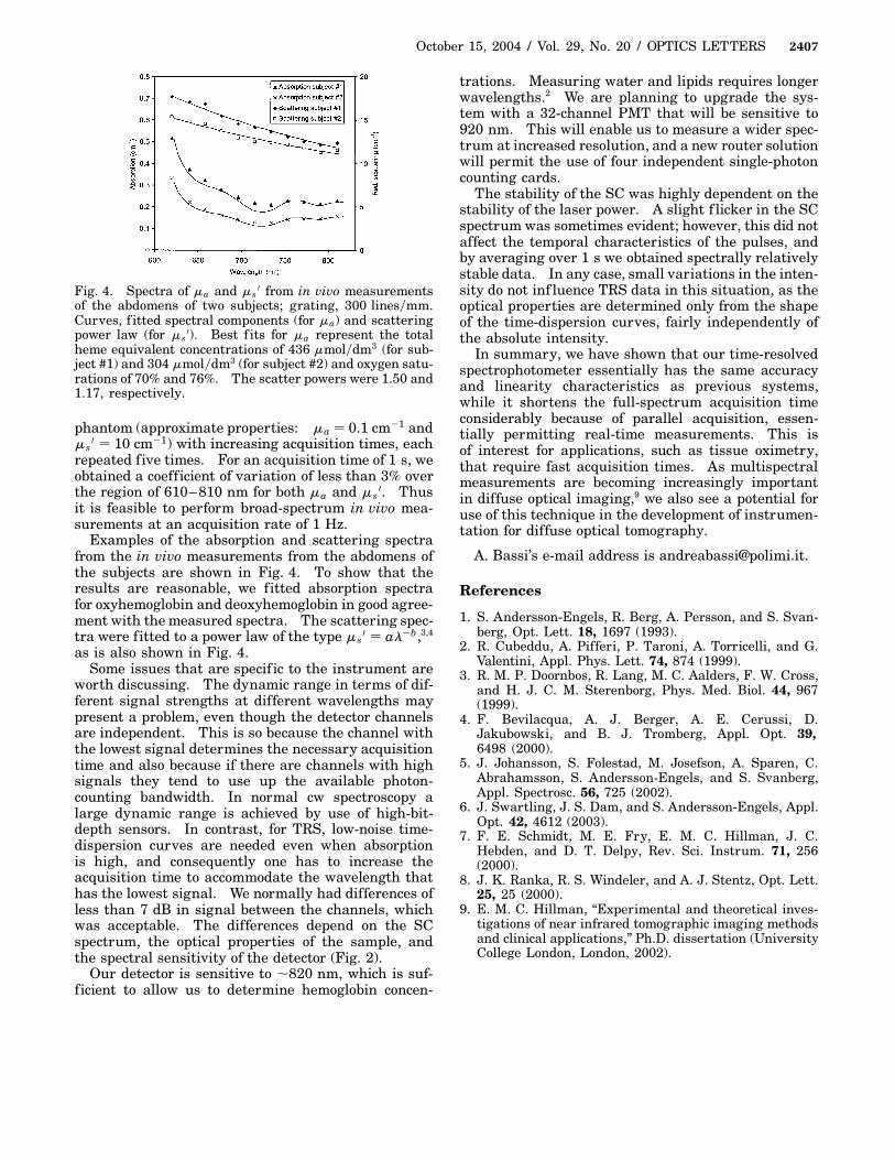
Graphs showing the linearity of increases inscattering and absorption at 695 nm are presentedin Fig. 3. The results demonstrate good linearityover the entire range of scattering and absorptioncoefficients.
We also evaluated the acquisition time needed toproduce low-noise data. It is inf luenced mainly bylight-collection efficiency and detector sensitivity. Weperformed a series of measurements on a standard
Fig. 2. Spectrum of the SC, shown in arbitrary units ona logarithmic scale. The quantum eff iciency of the PMTis also shown.
Fig. 3. Linearity of increases in scattering and absorp-tion: (a) ma and ms
0 versus the concentration of blacktoner in the phantoms with the same concentration of TiO2:1.4 mg�g. (b) ma and ms
0 versus the concentration of TiO2in the phantoms with the same concentration of toner:12 mg�g. Linear regression lines are also shown. Errorbars, standard deviations for repeated measurementsperformed on one phantom.
October 15, 2004 / Vol. 29, No. 20 / OPTICS LETTERS 2407
Fig. 4. Spectra of ma and ms0 from in vivo measurements
of the abdomens of two subjects; grating, 300 lines�mm.Curves, f itted spectral components (for ma) and scatteringpower law (for ms
0). Best f its for ma represent the totalheme equivalent concentrations of 436 mmol�dm3 (for sub-ject #1) and 304 mmol�dm3 (for subject #2) and oxygen satu-rations of 70% and 76%. The scatter powers were 1.50 and1.17, respectively.
phantom (approximate properties: ma � 0.1 cm21 andms
0 � 10 cm21) with increasing acquisition times, eachrepeated five times. For an acquisition time of 1 s, weobtained a coefficient of variation of less than 3% overthe region of 610–810 nm for both ma and ms
0. Thusit is feasible to perform broad-spectrum in vivo mea-surements at an acquisition rate of 1 Hz.
Examples of the absorption and scattering spectrafrom the in vivo measurements from the abdomens ofthe subjects are shown in Fig. 4. To show that theresults are reasonable, we fitted absorption spectrafor oxyhemoglobin and deoxyhemoglobin in good agree-ment with the measured spectra. The scattering spec-tra were fitted to a power law of the type ms
0 � al2b,3,4
as is also shown in Fig. 4.Some issues that are specif ic to the instrument are
worth discussing. The dynamic range in terms of dif-ferent signal strengths at different wavelengths maypresent a problem, even though the detector channelsare independent. This is so because the channel withthe lowest signal determines the necessary acquisitiontime and also because if there are channels with highsignals they tend to use up the available photon-counting bandwidth. In normal cw spectroscopy alarge dynamic range is achieved by use of high-bit-depth sensors. In contrast, for TRS, low-noise time-dispersion curves are needed even when absorptionis high, and consequently one has to increase theacquisition time to accommodate the wavelength thathas the lowest signal. We normally had differences ofless than 7 dB in signal between the channels, whichwas acceptable. The differences depend on the SCspectrum, the optical properties of the sample, andthe spectral sensitivity of the detector (Fig. 2).
Our detector is sensitive to �820 nm, which is suf-ficient to allow us to determine hemoglobin concen-
trations. Measuring water and lipids requires longerwavelengths.2 We are planning to upgrade the sys-tem with a 32-channel PMT that will be sensitive to920 nm. This will enable us to measure a wider spec-trum at increased resolution, and a new router solutionwill permit the use of four independent single-photoncounting cards.
The stability of the SC was highly dependent on thestability of the laser power. A slight f licker in the SCspectrum was sometimes evident; however, this did notaffect the temporal characteristics of the pulses, andby averaging over 1 s we obtained spectrally relativelystable data. In any case, small variations in the inten-sity do not inf luence TRS data in this situation, as theoptical properties are determined only from the shapeof the time-dispersion curves, fairly independently ofthe absolute intensity.
In summary, we have shown that our time-resolvedspectrophotometer essentially has the same accuracyand linearity characteristics as previous systems,while it shortens the full-spectrum acquisition timeconsiderably because of parallel acquisition, essen-tially permitting real-time measurements. This isof interest for applications, such as tissue oximetry,that require fast acquisition times. As multispectralmeasurements are becoming increasingly importantin diffuse optical imaging,9 we also see a potential foruse of this technique in the development of instrumen-tation for diffuse optical tomography.
A. Bassi’s e-mail address is [email protected].
References
1. S. Andersson-Engels, R. Berg, A. Persson, and S. Svan-berg, Opt. Lett. 18, 1697 (1993).
2. R. Cubeddu, A. Pifferi, P. Taroni, A. Torricelli, and G.Valentini, Appl. Phys. Lett. 74, 874 (1999).
3. R. M. P. Doornbos, R. Lang, M. C. Aalders, F. W. Cross,and H. J. C. M. Sterenborg, Phys. Med. Biol. 44, 967(1999).
4. F. Bevilacqua, A. J. Berger, A. E. Cerussi, D.Jakubowski, and B. J. Tromberg, Appl. Opt. 39,6498 (2000).
5. J. Johansson, S. Folestad, M. Josefson, A. Sparen, C.Abrahamsson, S. Andersson-Engels, and S. Svanberg,Appl. Spectrosc. 56, 725 (2002).
6. J. Swartling, J. S. Dam, and S. Andersson-Engels, Appl.Opt. 42, 4612 (2003).
7. F. E. Schmidt, M. E. Fry, E. M. C. Hillman, J. C.Hebden, and D. T. Delpy, Rev. Sci. Instrum. 71, 256(2000).
8. J. K. Ranka, R. S. Windeler, and A. J. Stentz, Opt. Lett.25, 25 (2000).
9. E. M. C. Hillman, “Experimental and theoretical inves-tigations of near infrared tomographic imaging methodsand clinical applications,” Ph.D. dissertation (UniversityCollege London, London, 2002).