Embed Size (px)
Citation preview
JOURNAL OF TISSUE ENGINEERING AND REGENERATIVE MEDICINE R E S E A R C H A R T I C L EJ Tissue Eng Regen Med 2007; 1: 350–359.Published online 30 August 2007 in Wiley InterScience (www.interscience.wiley.com) DOI: 10.1002/term.38
Time-course study of histological and geneticpatterns of differentiation in human engineeredoral mucosa
M. Alaminos1,2*, I. Garzon1, M. C. Sanchez-Quevedo1, G. Moreu3, M. Gonzalez-Andrades1,A. Fernandez-Montoya1,4 and A. Campos1
1Department of Histology, University of Granada, Spain2Fundacion FIBAO and University Hospital San Cecilio, Granada, Spain3Department of Stomatology, University of Granada, Spain4Transfusion Center and Tissue Bank of Granada (CRTS), Spain
Abstract
The lack of sufficient oral mucosa available for intra-oral grafting is a major surgical problem,and new sources of oral tissues for clinical use are needed. In this regard, some models ofengineered oral mucosa have been reported to date, but little is known about the structural andgenetic mechanisms that occur during the process of development and maturation of these tissuesubstitutes. We have carried out a time-course study of the genes and morphological patterns ofcell and tissue differentiation that develop in oral mucosa constructs after 3, 7, 11 and 21 days ofdevelopment. Our electron microscopy and microarray analyses demonstrated that the oral mucosaconstructs generated by tissue engineering undergo a progressive process of cell differentiationwith the sequential formation and maturation of several layers of epithelium (with expression ofstratifin, sciellin, involucrin, trichohyalin and kallikrein 7), intercellular junctions (with expressionof plakophilin, desmocollin, desmoglein and cadherins), cytokeratins, a basement membrane(laminins, collagen IV) and the extracellular matrix (biglycan, matrix metalloproteinases). Inconclusion, although the level and type of keratinization developed in vitro could be different, theoral mucosa substitutes were very similar to the native tissues. Copyright 2007 John Wiley &Sons, Ltd.
Received 9 May 2007; Accepted 13 June 2007
Keywords tissue engineering; oral mucosa; histodifferentiation; microarray; electron microscopy;development
1. Introduction
The lack of sufficient oral mucosa available for intra-oralgrafting has been dealt with so far by using split-thickness skin or oral mucosa grafts harvested from donorsites (Izumi et al., 2000). These procedures, however,often require more than one surgical procedure and areassociated with morbidity at the donor and the recipientsites.
*Correspondence to: M. Alaminos, Department of Histology,University of Granada, Avenida de Madrid 11, E-18012Granada, Spain. E-mail: [email protected]
Construction of artificial organs by tissue engineering(TE) is one of the research fields that has experiencedmajor progress during recent years (Atala, 2000). Byusing TE techniques, different researchers have developedefficient substitutes of different organs and tissues fortherapeutic use, including, among others, human skin(Meana et al., 1998; Llames et al., 2004), cornea (Nishida,2003; Reichl et al., 2004; Alaminos et al., 2006), bone(Mohammadi et al., 2007) and blood vessels (Pascualet al., 2004). Regarding the oral mucosa, several groupshave developed different models of artificial tissues thatcould eventually be used as organotypic substitutesof the human oral mucosa for reconstruction of oraland maxillofacial tissues (Lauer and Schimming, 2001;
Copyright 2007 John Wiley & Sons, Ltd.
Differentiation of engineered human oral mucosa 351
Schultze-Mosgau et al., 2004; Sanchez-Quevedo et al.,2007). Clinical uses for a tissue-engineered oral mucosawould mainly include intra-oral defects, such as repairof acquired or congenital oral mucosal defects, but also,and strikingly, extra-oral defects, e.g. reconstruction ofthe cornea (Nakamura et al., 2006), eyelids, conjunctiva,oesophagus, trachea, bladder, urethra or vagina (Feinberget al., 2005). Other potential uses of artificial oral mucosaconstructs are in vitro models to study the biology andpathology of mucosa, and use of the mucosa as a vehiclefor delivery and expression of transduced genes (Feinberget al., 2005). Most oral mucosa substitutes use three-dimensional (3D) co-cultures of the main cell types ofthe oral mucosa, embedded in different biomaterials. Inthis regard, our group has recently developed a stromalsubstitute made of a mixture of fibrin and agarosethat demonstrated good biomechanical and structuralproperties when used for TE purposes (Alaminos et al.,2006; Sanchez-Quevedo et al., 2007).
In designing a TE oral mucosa, it is critical that theconstructed tissues have the innate functions seen withnatural oral mucosa (especially, acting as a protectivecovering for the underlying chorion), and that thesestructures be interactive with their environment, thatis, they communicate with the surrounding cells viasignalling mechanisms. For these reasons, it is necessarythat the oral mucosa constructs emulate the anatomyand the structure of the native organ, and that thelevel of differentiation is similar in the constructs andin the damaged tissues to be replaced (Feinberg et al.,2005). Hence, evaluation of the degree of cell andtissue differentiation of the constructs developed byTE is mandatory before the artificial tissues can beused clinically. Therefore, it will be necessary to verifythat the developed tissues reproduce the structuralpatterns of differentiation and gene expression thatare linked to keratinocyte maturation in native normaloral mucosa, where up to five different patterns ofmaturation have been described (Moreu et al., 1993;Sanchez-Quevedo et al., 1994). These patterns, initiallyreported by Dourov (1984) and Kullaa-Mikkonen (1986,1987) in the gingival epithelium of normal donors, arestrongly related to the degree of differentiation of thekeratinocytes (Southgate et al., 1987; Moreu et al., 1993;Sanchez-Quevedo et al., 1994). Thus, keratinocytes withsurface microvilli (pattern type I) correspond to cells withthe lowest level of differentiation, whereas cells with pits(pattern type V) are at the last stages of keratinocyticdifferentiation (Moreu et al., 1993; Sanchez-Quevedoet al., 1994). Patterns type II, III and IV are usuallyfound in cells with intermediate degrees of differentiation.Many of these patterns are associated with the cell–celladhesions that exist in the cells and, for example, thedeepest layers of the oral mucosa epithelium display ahigh number of desmosomes and are covered exclusivelyby microvilli (Hodgkins et al., 1978).
In this study, we have correlated the morphostructuralpatterns of differentiation, as determined by electronmicroscopy (EM), and the gene expression profiles in
a model of bioengineered human oral mucosa usingfibrin–agarose scaffolds.
2. Materials and methods
2.1. Generation of primary cultures of oralmucosa fibroblasts and keratinocytes
Twenty-five small biopsies corresponding to normalhuman oral mucosa were obtained from healthy donorsundergoing minor oral surgery under local anaesthesia.All tissues were washed and transported in Dulbecco’sModified Eayle’s Medium (DMEM) medium with antibi-otics and antimycotics (500 U/ml penicillin G, 500 mg/mlstreptomycin, and 1.25 mg/ml amphotericin B) and pro-cessed in the following 24 h. This work was approved bythe institutional research committee.
To obtain primary cultures of human oral fibroblasts, allbiopsies were washed in phosphate buffered saline (PBS)and incubated overnight at 37 ◦C in a solution of 2 mg/mlClostridium histolyticum collagenase I (Gibco BRL LifeTechnologies, Karlsruhe, Germany) in DMEM. Detachedfibroblasts were collected by centrifugation and expandedin culture flasks containing DMEM medium supplementedwith antibiotics (100 U/ml penicillin G, 100 mg/mlstreptomycin, and 0.25 mg/ml amphotericin B) and 10%fetal bovine serum (FBS). To establish primary culturesof oral keratinocytes, undigested oral epithelium waswashed in PBS, cut into small explant pieces and co-cultured with a layer of mitomycin C-treated (10 µg/ml)3T3 feeder cells (8–10 × 103 cell/cm2; Rheinwald andGreen, 1975). Keratinocytes culture medium was a 3 : 1mixture of DMEM and Ham’s F12 supplemented with10% fetal calf serum, 1% antiobiotics, 24 µg/ml adenine,0.4 mg/ml hydrocortisone, 5 mg/ml insulin, 10 ng/mlepidermal growth factor, 1.3 ng/ml triiodothyronine and8 ng/ml cholera toxin.
All cells were incubated at 37 ◦C in 5% carbon dioxideunder standard culture conditions. The medium waschanged every 3 days and subcultivation of the culturedcells was carried out using a trypsin 0.5 g/l–EDTA0.2 g/l solution at 37 ◦C for 10 min. All cells used forexperimentation were at passages 1–4 (Alaminos et al.,2007).
2.2. Construction of human oral mucosasubstitutes by TE
Development of human oral mucosa constructs inthe laboratory was carried out using the followingpreviously published methods (Sanchez-Quevedo et al.,2006). Briefly, a stromal substitute made of human fibrinand 0.1% agarose, with fibroblasts immersed within,was developed using Transwell culture inserts with0.4 µm porous membrane (Costar, Corning Inc., Corning,NY, USA). Twenty-four hours after the stromal matrixsubstitute had solidified, human oral keratinocytes were
Copyright 2007 John Wiley & Sons, Ltd. J Tissue Eng Regen Med 2007; 1: 350–359.DOI: 10.1002/term
352 M. Alaminos et al.
seeded on top of the constructed stroma (approximately1 000 000 keratinocytes/25 ml construct), and culturedfor 21 days submerged in keratinocyte culture medium.Specimens corresponding to oral mucosa substituted wereanalysed at different times after keratinocyte seeding (3,7, 11 and 21 days). In this study, a total of 15 human oralmucosa constructs were analysed.
2.3. Microscopic evaluation of the human oralmucosa substitutes
Samples for scanning EM (SEM) were fixed in 2.5%glutaraldehyde and postfixed in 1% osmium tetroxidefor 90 min. After fixation, the samples were dehydratedin increasing concentrations of acetone (30%, 50%,70%, 95% and 100%), critical point-dried, mounted onaluminium stubs, sputter-coated with gold according toroutine procedures (Sanchez-Quevedo et al., 1994) andexamined in a Quanta 200 scanning electron microscope(FEI, Eindhoven, The Netherlands), using a high vacuummode. For transmission EM (TEM), samples were fixed,postfixed and dehydrated as described above for SEM,embedded in Spurr’s resin and cut into ultrathin sectionsusing an ultramicrotrome. Then, the sections werestained with aqueous uranyl acetate and lead citrate andexamined with a EM902 transmission electron microscope(Carl Zeiss Meditec Inc., Oberkochen, Germany).
2.4. Genome-wide gene expression analysisusing oligonucleotide microarrays
Total RNA corresponding to oral mucosa constructs wasextracted using the Qiagen RNeasy System (Qiagen,Mississauga, Ontario, Canada), according to the manufac-turers’ recommendations. RNA concentration was deter-mined by absorbency at 260 nm, and quality was verifiedby using a Bioanalyser (Agilent). Total cDNA was syn-thesized with a T7-polyT primer and reverse transcriptase(Superscript II, Life Technologies Inc., Carlsbad, CA, USA)before in vitro transcription with biotinylated UTP andCTP (Enzo Diagnostics, Farmingdale, NY, USA). Labellednucleic acid target was hybridized (45 ◦C for 16 h) toAffymetrix Human Genome U133 plus 2.0 oligonucleotidearrays. After automated washing and staining, absolutevalues of expression were calculated and Normalized fromthe scanned array, using Affymetrix Microarray Suite.
For the analysis of the microarray data, averageexpression corresponding to each group of comparisonwas calculated for each probe set. The mean expressionratio (fold-change) was then calculated by dividing themean expression of one group of comparison by that ofthe other group (Alaminos et al., 2003), and genes with aminimum fold-change of 10 were selected. Gene Ontologyanalysis of the selected genes was performed using BiNGO(http://www.psb.ugent.be/cbd/papers/BiNGO/), a plug-in for the program Cytoscape (Maere et al., 2005). Theset of selected genes was tested for enrichment of any
GO category relating to ‘Biological Process’, as comparedto all annotated genes represented on the array. Scoreswere evaluated based on the hypergeometric distributionand Bonferroni correction for multiple testing. Thus, thep value reflects the likelihood that one observes suchan enrichment or higher by chance alone (Boyer et al.,2006).
To identify genes whose expression was statisticallysignificantly associated with specific groups of samples,we used the significance analysis for microarrays (SAM)function of the program TIGR MeV (MultiExperimentViewer 3.1; Institute for Genomic Research, Rockville,MD, USA; Saeed et al., 2003). We used a δ valuethat allowed us a false discovery rate of <1 (i.e.< one gene is falsely named). The program isavailable at http://www.tigr.org/software/tm4. TIGRMeV was also used for hierarchical cluster analysisof the samples by standardizing each expression levelof each gene and each sample to mean = 0 andvariance = 1.
3. Results
3.1. EM evaluation of the oral mucosa constructs
Construction of oral mucosa substitutes by TE wasefficiently carried out by using the methods andtechniques descibed above.
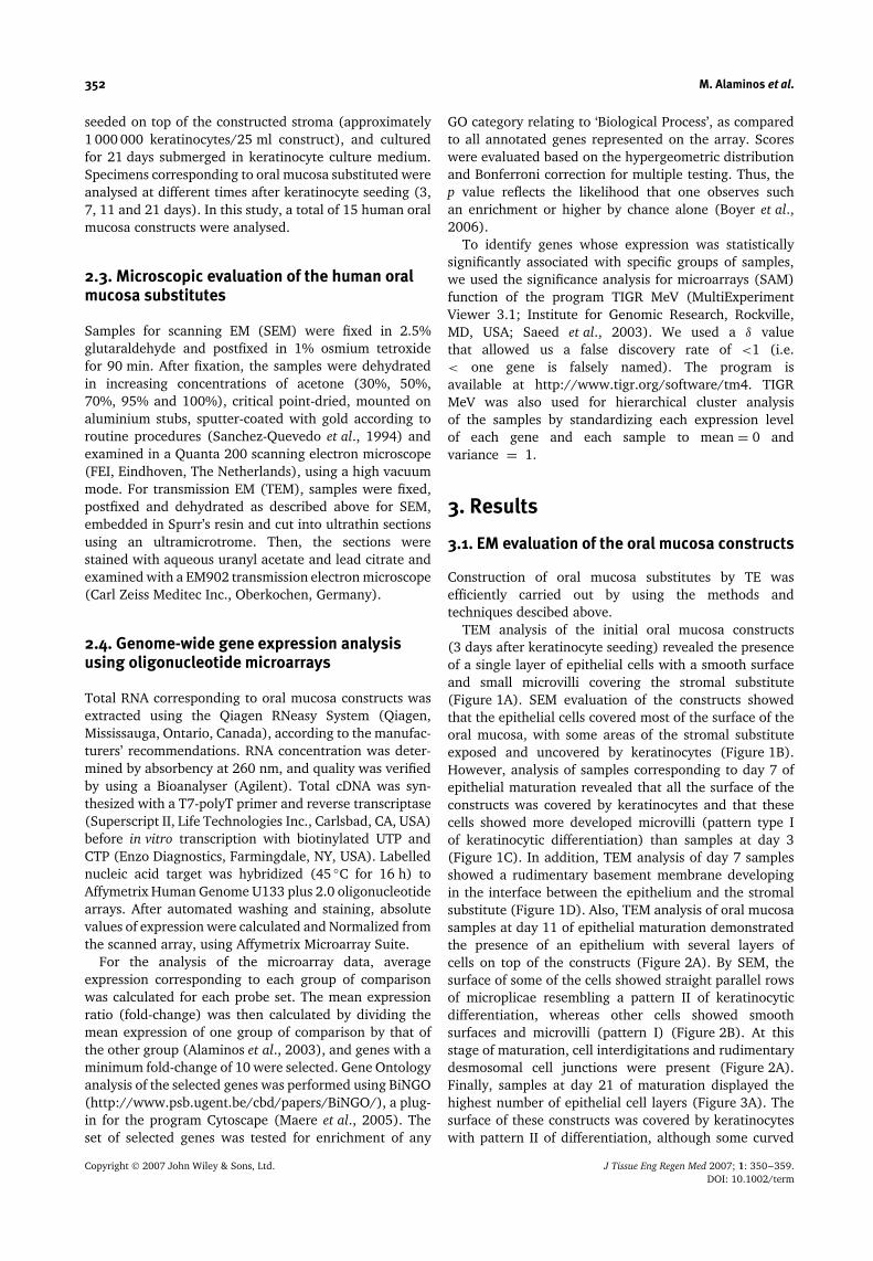
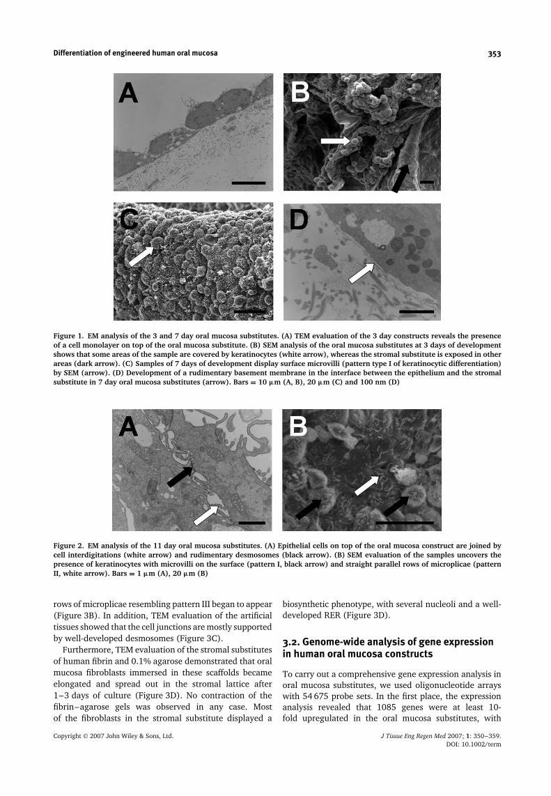
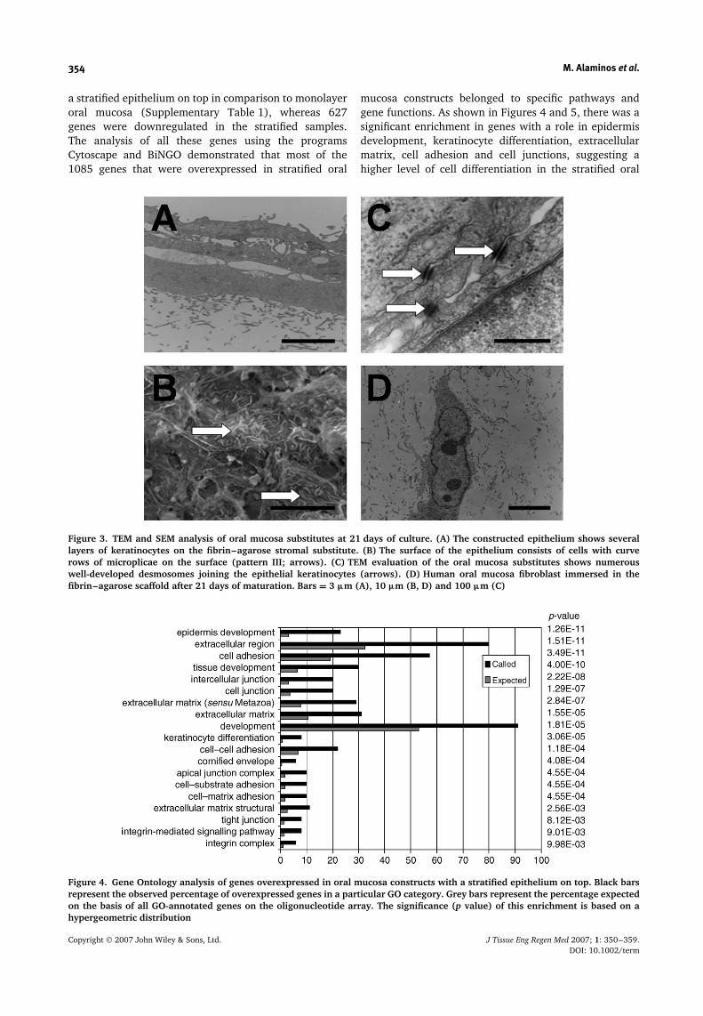
TEM analysis of the initial oral mucosa constructs(3 days after keratinocyte seeding) revealed the presenceof a single layer of epithelial cells with a smooth surfaceand small microvilli covering the stromal substitute(Figure 1A). SEM evaluation of the constructs showedthat the epithelial cells covered most of the surface of theoral mucosa, with some areas of the stromal substituteexposed and uncovered by keratinocytes (Figure 1B).However, analysis of samples corresponding to day 7 ofepithelial maturation revealed that all the surface of theconstructs was covered by keratinocytes and that thesecells showed more developed microvilli (pattern type Iof keratinocytic differentiation) than samples at day 3(Figure 1C). In addition, TEM analysis of day 7 samplesshowed a rudimentary basement membrane developingin the interface between the epithelium and the stromalsubstitute (Figure 1D). Also, TEM analysis of oral mucosasamples at day 11 of epithelial maturation demonstratedthe presence of an epithelium with several layers ofcells on top of the constructs (Figure 2A). By SEM, thesurface of some of the cells showed straight parallel rowsof microplicae resembling a pattern II of keratinocyticdifferentiation, whereas other cells showed smoothsurfaces and microvilli (pattern I) (Figure 2B). At thisstage of maturation, cell interdigitations and rudimentarydesmosomal cell junctions were present (Figure 2A).Finally, samples at day 21 of maturation displayed thehighest number of epithelial cell layers (Figure 3A). Thesurface of these constructs was covered by keratinocyteswith pattern II of differentiation, although some curved
Copyright 2007 John Wiley & Sons, Ltd. J Tissue Eng Regen Med 2007; 1: 350–359.DOI: 10.1002/term
Differentiation of engineered human oral mucosa 353
Figure 1. EM analysis of the 3 and 7 day oral mucosa substitutes. (A) TEM evaluation of the 3 day constructs reveals the presenceof a cell monolayer on top of the oral mucosa substitute. (B) SEM analysis of the oral mucosa substitutes at 3 days of developmentshows that some areas of the sample are covered by keratinocytes (white arrow), whereas the stromal substitute is exposed in otherareas (dark arrow). (C) Samples of 7 days of development display surface microvilli (pattern type I of keratinocytic differentiation)by SEM (arrow). (D) Development of a rudimentary basement membrane in the interface between the epithelium and the stromalsubstitute in 7 day oral mucosa substitutes (arrow). Bars = 10 µm (A, B), 20 µm (C) and 100 nm (D)
Figure 2. EM analysis of the 11 day oral mucosa substitutes. (A) Epithelial cells on top of the oral mucosa construct are joined bycell interdigitations (white arrow) and rudimentary desmosomes (black arrow). (B) SEM evaluation of the samples uncovers thepresence of keratinocytes with microvilli on the surface (pattern I, black arrow) and straight parallel rows of microplicae (patternII, white arrow). Bars = 1 µm (A), 20 µm (B)
rows of microplicae resembling pattern III began to appear(Figure 3B). In addition, TEM evaluation of the artificialtissues showed that the cell junctions are mostly supportedby well-developed desmosomes (Figure 3C).
Furthermore, TEM evaluation of the stromal substitutesof human fibrin and 0.1% agarose demonstrated that oralmucosa fibroblasts immersed in these scaffolds becameelongated and spread out in the stromal lattice after1–3 days of culture (Figure 3D). No contraction of thefibrin–agarose gels was observed in any case. Mostof the fibroblasts in the stromal substitute displayed a
biosynthetic phenotype, with several nucleoli and a well-developed RER (Figure 3D).
3.2. Genome-wide analysis of gene expressionin human oral mucosa constructs
To carry out a comprehensive gene expression analysis inoral mucosa substitutes, we used oligonucleotide arrayswith 54 675 probe sets. In the first place, the expressionanalysis revealed that 1085 genes were at least 10-fold upregulated in the oral mucosa substitutes, with
Copyright 2007 John Wiley & Sons, Ltd. J Tissue Eng Regen Med 2007; 1: 350–359.DOI: 10.1002/term
354 M. Alaminos et al.
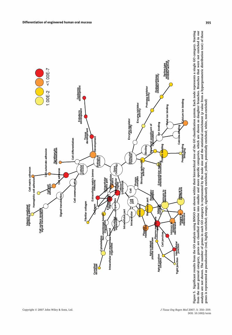
a stratified epithelium on top in comparison to monolayeroral mucosa (Supplementary Table 1), whereas 627genes were downregulated in the stratified samples.The analysis of all these genes using the programsCytoscape and BiNGO demonstrated that most of the1085 genes that were overexpressed in stratified oral
mucosa constructs belonged to specific pathways andgene functions. As shown in Figures 4 and 5, there was asignificant enrichment in genes with a role in epidermisdevelopment, keratinocyte differentiation, extracellularmatrix, cell adhesion and cell junctions, suggesting ahigher level of cell differentiation in the stratified oral
Figure 3. TEM and SEM analysis of oral mucosa substitutes at 21 days of culture. (A) The constructed epithelium shows severallayers of keratinocytes on the fibrin–agarose stromal substitute. (B) The surface of the epithelium consists of cells with curverows of microplicae on the surface (pattern III; arrows). (C) TEM evaluation of the oral mucosa substitutes shows numerouswell-developed desmosomes joining the epithelial keratinocytes (arrows). (D) Human oral mucosa fibroblast immersed in thefibrin–agarose scaffold after 21 days of maturation. Bars = 3 µm (A), 10 µm (B, D) and 100 µm (C)
Figure 4. Gene Ontology analysis of genes overexpressed in oral mucosa constructs with a stratified epithelium on top. Black barsrepresent the observed percentage of overexpressed genes in a particular GO category. Grey bars represent the percentage expectedon the basis of all GO-annotated genes on the oligonucleotide array. The significance (p value) of this enrichment is based on ahypergeometric distribution
Copyright 2007 John Wiley & Sons, Ltd. J Tissue Eng Regen Med 2007; 1: 350–359.DOI: 10.1002/term
Differentiation of engineered human oral mucosa 355
Figu
re5.
Sign
ifica
nt
resu
lts
from
the
GO
anal
ysis
usi
ng
BiN
GO
are
show
nw
ithi
nth
eir
hier
arch
ical
tree
ofth
eG
Ocl
assi
fica
tion
syst
em.
Each
nod
ere
pres
ents
asi
ngl
eG
Oca
tego
ry.S
tart
ing
from
the
mos
tge
ner
alca
tego
ry,
gen
esar
ecl
assi
fied
step
wis
ein
tosm
alle
ran
dm
ore
spec
ific
cate
gori
es,
whi
char
esh
own
asda
ugh
ter
bran
ches
.B
ran
ches
that
wer
en
oten
rich
edin
our
anal
ysis
are
not
show
n.
The
nu
mbe
rof
gen
esin
each
GO
cate
gory
isre
pres
ente
dby
the
nod
esi
zean
dth
est
atis
tica
len
rich
men
t(p
valu
efr
oma
hype
rgeo
met
ric
dist
ribu
tion
test
)of
thes
ege
nes
isre
pres
ente
das
pseu
doco
lour
(red
,hig
hly
enri
ched
;or
ange
,sig
nifi
can
tly
enri
ched
;ye
llow
,pot
enti
ally
enri
ched
;w
hite
,non
-en
rich
ed)
Copyright 2007 John Wiley & Sons, Ltd. J Tissue Eng Regen Med 2007; 1: 350–359.DOI: 10.1002/term
356 M. Alaminos et al.
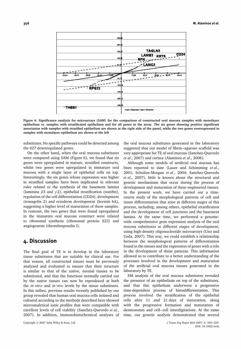
Figure 6. Significance analysis for microarrays (SAM) for the comparison of constructed oral mucosa samples with monolayerepithelium vs. samples with stratificated epithelium and for all genes in the array. The six genes showing positive significantassociation with samples with stratified epithelium are shown at the right side of the panel, while the two genes overexpressed insamples with monolayer epithelium are shown at the left
substitutes. No specific pathways could be detected amongthe 627 downregulated genes.
On the other hand, when the oral mucosa substituteswere compared using SAM (Figure 6), we found that sixgenes were upregulated in mature, stratified constructs,whilst two genes were upregulated in immature oralmucosa with a single layer of epithelial cells on top.Interestingly, the six genes whose expression was higherin stratified samples have been implicated in relevantroles related to the synthesis of the basement lamina(laminins β3 and γ 2), epithelial stratification (statifin),regulation of the cell differentiation (CD24), development(transgelin 2) and ectoderm development (keratin 6A),suggesting a higher level of maturation of these samples.In contrast, the two genes that were found upregulatedin the immature oral mucosa construct were relatedto ribosomal synthesis (ribosomal protein S23) andangiogenesis (thrombospondin I).
4. Discussion
The final goal of TE is to develop in the laboratorytissue substitutes that are suitable for clinical use. Forthat reason, all constructed tissues must be previouslyanalysed and evaluated to ensure that their structureis similar to that of the native, normal tissues to besubstituted, and that the functions normally carried outby the native tissues can now be reproduced at boththe in vitro and in vivo levels by the tissue substitutes.In this milieu, previous results recently published by ourgroup revealed that human oral mucosa cells isolated andcultured according to the methods described here showedmicroanalytical ionic profiles that were compatible withexcellent levels of cell viability (Sanchez-Quevedo et al.,2007). In addition, immunohistochemical analyses of
the oral mucosa substitutes generated in the laboratorysuggested that our model of fibrin–agarose scaffold wasvery appropriate for TE of oral mucosa (Sanchez-Quevedoet al., 2007) and cornea (Alaminos et al., 2006).
Although some models of artificial oral mucosa hasbeen reported to date (Lauer and Schimming et al.,2001; Schultze-Mosgau et al., 2004; Sanchez-Quevedoet al., 2007), little is known about the structural andgenetic mechanisms that occur during the process ofdevelopment and maturation of these engineered tissues.In the present work, we have carried out a time-course study of the morphological patterns of cell andtissue differentiation that arise at different stages of thisprocess, including, among others, epithelial stratificationand the development of cell junctions and the basementlamina. At the same time, we performed a genome-wide comprehensive gene expression analysis of the oralmucosa substitutes at different stages of development,using high-density oligonucleotide microarrays (Uno andUeda, 2007). This way, we could establish a relationshipbetween the morphological patterns of differentiationfound in the tissues and the expression of genes with a rolein the development of those patterns. This informationallowed us to contribute to a better understanding of theprocesses involved in the development and maturationof the artificial oral mucosa tissues generated in thelaboratory by TE.
EM analysis of the oral mucosa substitutes revealedthe presence of an epithelium on top of the substitutes,and that this epithelium underwent a progressivetime-dependent process of histodifferentiation. Thisprocess involved the stratification of the epithelialcells after 11 and 21 days of maturation, alongwith the progressive formation and maturation ofdesmosomes and cell–cell interdigitations. At the sametime, our genetic analysis demonstrated that several
Copyright 2007 John Wiley & Sons, Ltd. J Tissue Eng Regen Med 2007; 1: 350–359.DOI: 10.1002/term
Differentiation of engineered human oral mucosa 357
genes with a role in epidermis development andkeratinocyte differentiation were upregulated in oralmucosa constructs at the final stages of maturation,including sciellin, involucrin, trichohyalin and kallikrein7. First, sciellin is a precursor protein that is normallyexpressed by terminally differentiated keratinocytes ofhuman keratinizing tissues (Kvedar et al. 1992), and itspresence can be associated with mature keratinocytesin our multilayered oral mucosa substitutes. Second,involucrin is expressed by several layers of the stratifiedepithelium of native human oral mucosa (Barrett et al.2005) and it has previously been detected in humanoral mucosa substitutes developed by TE using collagenscaffolds (Imaizumi et al. 2004). Different researchershave previously reported that involucrin is crosslinkedinto the cornified envelope of oral mucosa keratinocytesduring terminal differentiation and, thus, can be usedas a marker of epithelial differentiation (Presland andDale, 2000). Similarly, epidermal and oral keratinocytesexpress the differentiation marker trichohyalin, whichassociates with the keratin cytoskeleton during terminaldifferentiation (Presland and Dale, 2000). Finally,kallikrein 7 (stratum corneum chymotryptic enzyme)is thought to be involved in the physiological processof epithelial desquamation through the proteolysis ofintercellular adhesion molecules, such as desmoglein(Ekholm et al., 2000). The overexpression of all thesedifferentiation markers in the mature oral mucosasubstitutes suggests that these susbstitutes display geneticsimilarities with native normal oral mucosa, and couldprobably exert the same physiological functions in vivo.In contrast, immature oral mucosa constructs with amonolayer epithelium on top tended to express a numberof genes related to cell proliferation (ribosomal andDNA synthesis proteins, cell membrane components,etc.), rather than genes with a role in mature epithelia.Furthermore, the presence of a stratified epithelium inthe oral mucosa constructs coincided with the expressionof the gene encoding for stratifin, which has beenassociated with the stratification of different kinds ofepithelia, including the oral mucosa epithelium (Katz andTaichman, 1999).
All these features correlated very well with the pro-gressive development of different types of keratinocyticdifferentiation patterns in our oral mucosa constructs.Hence, SEM analysis revealed that the keratinocytes of theoral mucosa substitutes initially presented microvilli ontheir surface (pattern type I), suggesting that the degreeof differentiation of these cells was very low (Moreu et al.,1993; Sanchez-Quevedo et al., 1994). However, samplescorresponding to 11 and 21 days of maturation tended toshow other patterns of keratinocyte differentiation thatindicated that these cells were more differentiated. Thedevelopment of pattern types II and III correlated verywell with the expression of genes with a role in epithelialdifferentiation, discussed above. However, the absence ofepithelial cells with pattern types IV and V in the oralmucosa substitutes suggests that the constructs do not
reach in vitro the degree of maturation found in normalnative oral mucosa in vivo.
In order to function correctly, stratified epithelia such asthe oral mucosa have to maintain tight cell–cell adhesionsin the living cells and retain the dead, keratinized squamesas a protective sheath prior to being sloughed (Preslandand Jurevic, 2002). Intercellular adherens junctions,including desmosomes, are cell adhesion complexes thatlink epithelial cells to each other and attach intermediatefilaments to the cell surface (Presland and Jurevic,2002). Desmosomes consist of two principal groupsof proteins: the desmosomal cadherins (desmogleinsand desmocollins), and the cytoplasmic proteins of thedesmosomes (which link the desmosomal cadherins to thecytoplasmic keratin filaments; Green and Jones, 1996).In this study, we demonstrated the formation of differenttypes of cell junctions in the oral mucosa substitutes,especially in the mature ones. In these samples, theidentification of intercellular junctions by TEM overlappedwith the expression of a great number of genes with arole in the synthesis of proteins involved in cell–celljunctions. In short, the presence of well-developeddesmosomes in the constructs corresponding to 21 daysof maturation was simultaneous with the expressionof genes encoding for plakophilin 1, desmocollins 2and 3, desmoglein 3, plakoglobin, corneodesmosin andcadherins (E-cadherin, CDH3, CDH26, protocadherinβ4, protocadherin γ A3), suggesting that the cells wereactively synthesizing and forming this essential kindof intercellular unions. In addition, the expression ofthe gene CD24, with a role in regulation of the celldifferentiation (Jevsek et al., 2006), has recently beenimplicated in epithelial hemidesmosomes formation (Liet al., 2007). The overexpression of different integrins(α6, β2, β3, β4 and β6 and integrin β-like 1) couldbe in relationship with the adhesion of the basal layerof keratinocytes and with hemidesmosomes formation.Furthermore, and although our EM analysis did notreveal the presence or other types of cell–cell junctions,the overexpression of genes related to tight junctions(claudin 1, cingulin, tight junction protein 3, CXADR,INADL, AMOTL1) and gap junctions (connexins 30 and31), suggests that these kinds of intercellular unions mightbe developing in our constructs. All these results implythat the epithelium of the artificial tissues generated byTE could form a tight barrier that would mimic the in vivoproperties of the native oral mucosa.
On the other hand, cytokeratins are a family of cyto-plasmic proteins that are the predominant cytoskeletalproteins in all epithelia. These filaments function as stress-bearing structures within epithelial cells and are criticalfor the maintenance of cell shape and viability (Preslandand Jurevic, 2002). For those reasons, the expression ofthe genes encoding for these proteins is strongly necessaryfor the keratinocytes to exert their functions as a protec-tive barrier of the structures in the oral cavity. In ourcase, the microarray analysis showed that the oral sub-stitutes at the 21st day of maturation strongly expressedcytokeratins 1, 4, 5, 6, 14, 16, 17 and 23. Expression
Copyright 2007 John Wiley & Sons, Ltd. J Tissue Eng Regen Med 2007; 1: 350–359.DOI: 10.1002/term
358 M. Alaminos et al.
of the keratin pair K5–K14 is typical of most stratifiedepithelia (Presland and Jurevic, 2002), whereas keratin 4is expressed by stratified, non-keratinized epithelia (Mon-tenegro et al., 1998). However, keratin 1 is expressed bykeratinized epithelia like the human epidermis (Lessinet al., 1988). Other cytokeratins, such as keratins 17 and23, are expressed upon induction in different types ofcells. In this regard, Kim et al. (2006) showed that keratin17 is rapidly induced in wounded stratified epithelia andthat it regulates cell growth through binding to the adap-tor protein stratifin. All these findings suggest that theepithelial cells of the oral mucosa substitutes are synthe-sizing in vitro a high number of cytokeratins of differenttype, and that these cytokeratins might differ from thoseexpressed by the native normal oral mucosa in vivo. Fur-ther studies are in need to show which cytokeratins areexpressed by the oral mucosa substitutes in vivo.
Moreover, ultrastructural analysis of the tissuesgenerated by TE revealed the presence of a basementlamina in the interface between the epithelium and thestromal substitute. The basement lamina is the anchoringcomplex joining the epithelium and the subjacentconnective tissue of skin, oral mucosa and other organsand structures, and is essential for a proper attachmentof the epithelium to the lamina propria. Structurally,the basement lamina consists of a lamina densa and alamina lucida, which can be clearly identified using TEMtechniques. The lamina densa layer is mainly formed bytype IV collagen, which is organized in a net-like fashion(Timpl et al., 1981). The non-collagenous constituents ofthe basement lamina mainly comprise laminin, entactinand proteoglycans (Kallioinen et al., 1984). In our study,we detected that the formation of a rudimentary basementlamina in the oral mucosa substitutes was simultaneouswith the expression of genes encoding for collagen IV,laminin γ 2, laminin α3 and other proteins, such asdystonin and hemicentin 1. These findings suggest that abasement lamina is developing between the oral mucosaepithelium and the artificial fibrin–agarose connectivetissue with fibroblasts immersed within.
Regarding the stromal substitute of the oral mucosagenerated by TE, different kinds of collagens were foundoverexpressed in the artificial tissues, including collagensI, III, IV, V and XI, along with other genes encoding forproteins of the extracellular matrix [biglycan (Schaeferet al., 2005), matrix metallopeptidase 16 and 28, ADAMmetallopeptidase, fibroblast growth factor 1, 18, fibroblastgrowth factor binding protein 1, etc.]. The expressionof these genes suggests that the stromal substitute offibrin–agarose is likely being progressively modified bythe fibroblasts immersed within to generate a structurechemically similar to the native, normal oral mucosa.Noticeably, some genes with a role in angiogenesis[thrombospondin I (de Fraipont et al., 2000; Thakar et al.,2005) in the immature samples and JAG1 (Li et al., 2006)and CEACAM1 (Ergun et al., 2000) in mature oral mucosaconstructs] were also overexpressed in the oral mucosasubstitutes, implying that the artificial tissues could be
trying to attract blood vessels from peripheral tissues,even when the tissues are kept in vitro.
Altogether, our results suggest that the artificial oralmucosa substitutes generated in the laboratory by TEdisplay numerous structural and genetic similarities withhuman native oral mucosa in both the epithelium andthe stroma. Our EM and microarray analyses demonstratethat the tissues undergo a process of cell differentiationwith the progressive formation and maturation of severallayers of epithelium, intercellular junctions, basementmembrane and extracellular matrix. As a result of thisprocess of cell and tissue development and maturation,and although the level and type of keratinization could bedifferent, the oral mucosa substitutes appear to be verysimilar to the native tissues. Although our findings appearto satisfy the criteria for utilization of the oral constructs inthe oral cavity, in vivo studies are still needed to uncoverthe potential utility of these fibrin–agarose oral mucosaconstructs for clinical purposes.
Supplementary material
Supplementary electronic material for this paper isavailable in Wiley InterScience at: http://www.mrw.interscience.wiley.com/suppmat/1932-6254/suppmat/
Acknowledgements
This work was supported by FIS 04/1306 from the SpanishMinistry of Health (Instituto de Salud Carlos III) and byCM0011/2005 and P06-CTS-02191 from Junta de Andalucia.There is no conflict of interest for any of the authors.
References
Alaminos M, Mora J, Cheung NK, et al. 2003; Genome-wideanalysis of gene expression associated with MYCN in humanneuroblastoma. Cancer Res 63: 4538–4546.
Alaminos M, Sanchez-Quevedo MC, Munoz-Avila JI, et al. 2007;Evaluation of the viability of cultured corneal endothelial cellsby quantitative electron probe X-ray microanalysis. J Cell Physiol211: 692–698.
Alaminos M, Sanchez-Quevedo MC, Munoz-Avila JI, et al. 2006;Construction of a complete rabbit cornea substitute using afibrin–agarose scaffold. Invest Ophthalmol Vis Sci 47: 3311–3317.
Atala A. 2000; Tissue engineering of artificial organs. J Endourol 14:49–57.
Barrett AW, Morgan M, Nwaeze G, Kramer G, Berkovitz BK. 2005;The differentiation profile of the epithelium of the human lip. ArchOral Biol 50: 431–438.
Boyer LA, Plath K, Zeitlinger J, et al. 2006; Polycomb complexesrepress developmental regulators in murine embryonic stem cells.Nature 441: 349–353.
de Fraipont F, El Atifi M, Gicquel C, et al. 2000; Expression ofthe angiogenesis markers vascular endothelial growth factor -A,thrombospondin-1, and platelet-derived endothelial cell growthfactor in human sporadic adrenocortical tumors: correlation withgenotypic alterations. J Clin Endocr Metab 85: 4734–4741.
Dourov N. 1984; Scanning electron microscopy contribution in oralpathology. Scan Electron Microsc 1: 243–248.
Ekholm IE, Brattsand M, Egelrud T. 2000; Stratum corneum trypticenzyme in normal epidermis: a missing link in the desquamationprocess? J Invest Dermatol 114: 56–63.
Copyright 2007 John Wiley & Sons, Ltd. J Tissue Eng Regen Med 2007; 1: 350–359.DOI: 10.1002/term
Differentiation of engineered human oral mucosa 359
Ergun S, Kilic N, Ziegeler G, et al. 2000; CEA-related cell adhesionmolecule 1: a potent angiogenic factor and a major effector ofvascular endothelial growth factor. Mol Cell 5: 311–320.
Feinberg SE, Aghaloo TL, Cunningham LL Jr. 2005; Role of tissueengineering in oral and maxillofacial reconstruction: findings ofthe 2005 AAOMS Research Summit. J Oral Maxillofac Surg 63:1418–1425.
Green KJ, Jones JC. 1996; Desmosomes and hemidesmosomes:structure and function of molecular components. FASEB J 10:871–881.
Hodgkins JFW, Watkins R, Walker DM. 1978; Correlated scanningand transmission electron microscopy of cell surfaces at variouslevels in human gingival epithelium. Arch Oral Biol 23: 355–357.
Imaizumi F, Asahina I, Moriyama T, Ishii M, Omura K. 2004;Cultured mucosal cell sheet with a double layer of keratinocytesand fibroblasts on a collagen membrane. Tissue Eng 10: 657–664.
Izumi K, Terashi H, Marcelo CL, Feinberg SE. 2000; Developmentand characterization of a tissue-engineered human oral mucosaequivalent produced in a serum-free culture system. J Dent Res79: 798–805.
Jevsek M, Jaworski A, Polo-Parada L, et al. 2006; CD24 is expressedby myofiber synaptic nuclei and regulates synaptic transmission.Proc Natl Acad Sci USA 103: 6374–6379.
Kallioinen M, Autio-Harmainen H, Dammert K, Risteli J, Risteli L.1984; Basement membrane laminin and type IV collagen invarious benign and malignant adnexal tumors of the skin: animmunohistochemical study. J Invest Dermatol 83: 276–280.
Katz AB, Taichman LB. 1999; A partial catalog of proteins secretedby epidermal keratinocytes in culture. J Invest Dermatol 112:818–821.
Kim S, Wong P, Coulombe PA. 2006; A keratin cytoskeletal proteinregulates protein synthesis and epithelial cell growth. Nature 441:362–365.
Kullaa-Mikkonen A. 1986; Scanning electron microscopic study ofsurface of human oral mucosa. Scand J Dent Res 94: 50–56.
Kullaa-Mikkonen A. 1987; Scanning electron microscopy in oralmucosal research: a review. Scanning Microsc 1: 1145–1155.
Kvedar JC, Manabe M, Phillips SB, Ross BS, Baden HP. 1992;Characterization of sciellin, a precursor to the cornified envelopeof human keratinocytes. Differentiation 49: 195–204.
Lauer G, Schimming R. 2001; Tissue-engineered mucosa graft forreconstruction of the intra-oral lining after freeing of the tongue:a clinical and immunohistologic study. J Oral Maxillofac Surg 59:169–175.
Lessin SR, Huebner K, Isobe M, Croce CM, Steinert PM. 1988;Chromosomal mapping of human keratin genes: evidence of non-linkage. J Invest Dermatol 91: 572–578.
Li H, Yu B, Zhang Y, et al. 2006; Jagged1 protein enhances thedifferentiation of mesenchymal stem cells into cardiomyocytes.Biochem Biophys Res Commun 341: 320–325.
Li Y, Lin X, Kilani RT, Jones JC, Ghahary A. 2007; 14–3–3sigma isoform interacts with the cytoplasmic domain of thetransmembrane BP180 in keratinocytes. J Cell Physiol [Epub aheadof print].
Llames SG, Del Rio M, Larcher F, et al. 2004; Human plasma as adermal scaffold for the generation of a completely autologousbioengineered skin, Transplantation 77: 350–355.
Maere S, Heymans K, Kuiper M. 2005; BiNGO: a Cytoscape plug-in to assess overrepresentation of gene ontology categories inbiological networks. Bioinformatics 21: 3448–3449.
Meana A, Iglesias J, Del Rio M, et al. 1998; Large surface of culturedhuman epithelium obtained on a dermal matrix based on livefibroblast-containing fibrin gels. Burns 24: 621–630.
Mohammadi Y, Soleimani M, Fallahi-Sichani M, et al. 2007;Nanofibrous poly(ε-caprolactone)/poly(vinyl alcohol)/chitosanhybrid scaffolds for bone tissue engineering using mesenchymalstem cells. Int J Artif Organs 30: 204–211.
Montenegro MA, Ibarra GC, Rojas M. 1998; Cytokeratin expressionin human and mouse gingival epithelia. Rev Chil Anat 16:211–217.
Moreu G, Sanchez-Quevedo MC, Lopez-Escamez JA, et al. 1993; Cellsurface patterns in normal human oral gingival epithelium.A quantitative scanning electron microscopy approach. HistolHistopathol 8: 47–50.
Nakamura T, Ang LP, Rigby H, et al. 2006; The use of autologousserum in the development of corneal and oral epithelial equivalentsin patients with Stevens–Johnson syndrome. Invest Ophthalmol VisSci 47: 909–916.
Nishida K. 2003; Tissue engineering of the cornea. Cornea 22:S28–34.
Pascual G, Rodriguez M, Corrales C, et al. 2004; New approachto improving endothelial preservation in cryopreserved arterialsubstitutes. Cryobiology 48: 62–71.
Presland RB, Dale BA. 2000; Epithelial structural proteins of the skinand oral cavity: function in health and disease. Crit Rev Oral BiolMed 11: 383–408.
Presland RB, Jurevic RJ. 2002; Making sense of the epithelial barrier:what molecular biology and genetics tell us about the functions oforal mucosal and epidermal tissues. J Dent Educ 66: 564–574.
Reichl S, Bednarz J, Muller-Goymann CC. 2004; Human cornealequivalent as cell culture model for in vitro drug permeationstudies. Br J Ophthalmol 88: 560–565.
Rheinwald JG, Green H. 1975; Serial cultivation of strains of humanepidermal keratinocytes: the formation of keratinizing coloniesfrom single cells. Cell 6: 331–343.
Saeed AI, Sharov V, White J, et al. 2003; TM4: a free, open-source system for microarray data management and analysis.Biotechniques 34: 374–378.
Sanchez-Quevedo MC, Alaminos M, Capitan LM, et al. 2007;Histological and histochemical evaluation of human oral mucosaconstructs developed by tissue engineering. Histol Histopathol 22:631–640.
Sanchez-Quevedo MC, Moreu G, Campos A, Garcia JM, Gonzalez-Jaranay M. 1994; Regional differences in cell surface patterns innormal human sulcular epithelium. Histol Histopathol 9: 149–153.
Schaefer L, Babelova A, Kiss E, et al. 2005; The matrix componentbiglycan is proinflammatory and signals through Toll-like receptors4 and 2 in macrophages. J Clin Invest 115: 2223–2233.
Schultze-Mosgau S, Lee BK, Ries J, Amann K, Wiltfang J. 2004;In vitro cultured autologous pre-confluent oral keratinocytes forexperimental prefabrication of oral mucosa. Int J Oral MaxillofacSurg 33: 476–485.
Southgate J, Williams HK, Trejdosiewicz LK, Hodges GM. 1987;Primary culture of human oral epithelial cells. Growthrequirements and expression of differentiated characteristics. LabInvest 56: 211–223.
Thakar CV, Zahedi K, Revelo MP, et al. 2005; Identification ofthrombospondin 1 (TSP-1) as a novel mediator of cell injuryin kidney ischemia. J Clin Invest 115: 3451–3459.
Timpl R, Wiedemann H, van Delden V, Furthmayr H, Kuhn K. 1981;A network model for the organization of type IV collagen moleculesin basement membranes. Eur J Biochem 120: 203–211.
Uno K, Ueda HR. 2007; Microarrays: quality control andhybridization protocol. Methods Mol Biol 362: 225–243.
Copyright 2007 John Wiley & Sons, Ltd. J Tissue Eng Regen Med 2007; 1: 350–359.DOI: 10.1002/term