Embed Size (px)
Citation preview
TISSUE ENGINEERINGVolume 10, Number 7/8, 2004© Mary Ann Liebert, Inc.
Time- and Concentration-Dependent Effects of DissolutionProducts of 58S Sol–Gel Bioactive Glass on Proliferation and
Differentiation of Murine and Human Osteoblasts
ROBERT C. BIELBY, B.A., D.Phil.,1 IOANNIS S. CHRISTODOULOU, B.Sc., M.Sc.,1RUSSELL S. PRYCE, M.Phys., M.Sc.,2
WARWICK J.P. RADFORD, M.B., Ch.B., F.R.C.S.(Ed.)Orth.,3LARRY L. HENCH, B.Sc., Ph.D.,2 and JULIA M. POLAK, M.D., D.Sc.1
ABSTRACT
Bone loss is a significant clinical problem, and treatments utilizing donated graft material are lim-ited. To meet future demands in the healthcare industry, there has been a shift of outlook towardthe use of bioactive materials for tissue regeneration. A number of in vivo and in vitro studies havehighlighted the potential of the bioactive glass ceramic 45S5 Bioglass as a synthetic regenerativescaffold. The application of sol–gel processing techniques has led to the synthesis of mesoporousbioactive glasses with greater textural and compositional variety. In this study, we evaluated the ef-fects of supplemented tissue culture medium containing up to 203 ppm silica prepared by staticsoaking of particles of 58S sol–gel bioactive glass (58% SiO2, 33% CaO, 9% P2O5) on the in vitroproliferation and differentiation of murine and human primary osteoblasts. These extracts had ahigher silica content than those used previously in studies of 45S5 Bioglass, because of the fasterrates of ion exchange permitted by the higher surface area-to-volume ratio of mesoporous glass. Wefound that osteoblasts from both species increased their proliferation in response to the glass-con-ditioned medium. In addition, the extent to which supplemented medium could alter cell differen-tiation varied with time in culture. Proliferation induced by supplemented medium paralleled ef-fects induced by treatment with basic fibroblast growth factor, a known mitogenic growth factorfor osteoblasts. Bone nodule formation was also increased by exposure to the glass-conditionedmedium and this effect was positively correlated with the dose of glass used to prepare the medium.Apoptosis was stimulated by glass-conditioned medium in murine osteoblasts, but inhibited in hu-man osteoblasts. These data demonstrate the bioactive effects of dissolution products derived fromsol–gel materials on primary osteoblasts and complements in vivo studies that indicate the suitabil-ity of this material as a bone graft substitute.
1Tissue Engineering and Regenerative Medicine Centre, Imperial College London, Faculty of Medicine, Chelsea and West-minster Hospital, London, UK.
2Tissue Engineering and Regenerative Medicine Centre, Department of Materials, Imperial College London, London, UK.3Department of Orthopaedic Surgery, Chelsea and Westminster Healthcare NHS Trust, Chelsea and Westminster Hospital,
London, UK.
1018
INTRODUCTION
BONE LOSS DUE TO TRAUMATIC INJURY, orthopaedic sur-gery, or tumor removal is a significant clinical prob-
lem. The limitations of donor sources of natural bone tis-sue for both autografts and allografts have led to thesearch for synthetic alternatives. Although bioinert ma-terials have been implanted into patients for more than 3decades with excellent longevity, of up to 15 years, thereis an ever-increasing percentage of the population that re-quires implanted devices with enhanced (.30 years) sur-vivability. The discovery of a four-component melt-de-rived bioactive glass, composed of SiO2, CaO, Na2O, andP2O5, by Hench led to 45S5 Bioglass and other bioactiveglasses being applied clinically and established as suc-cessful materials to regenerate bone.1,2 The success ofbioactive glasses to stimulate an osteogenic response isbased on two features. First, they have an ability to gen-erate a hydroxycarbonate apatite (HCA) layer on the sur-face of the glass that forms a strong bond to bone andalso to soft tissues. The mechanism underlying the de-velopment of the HCA involves the formation of a silicahydrogel surface layer when exposed to aqueous solutioneither in vitro or in vivo. A series of chemical reactionsresults in the formation of a silica layer on the surface onwhich the nucleation of amorphous calcium phosphateoccurs, resulting in HCA on crystallization. Formation ofthe surface silica layer involves network dissolution ofthe glass, releasing ions from its molecular constituents.This resorption that occurs in vivo results in the releaseof soluble ions (Na, Ca, P, and Si) that can stimulate os-teoblasts to produce bone. Numerous in vivo studies haveshown that 45S5 can stimulate bone regeneration.Wheeler et al. demonstrated accelerated bone healing inrabbit models brought about by the application of 45S5particles into the defect site.3,4 In vitro studies haveshown that the material itself and the soluble ionic speciesreleased by network dissolution may participate in pro-ducing an osteoinductive effect.5–11 Xynos et al. foundenhanced osteogenic effects of both the solid Bioglassmaterial and the soluble products released during glassdissolution.9,10 Significantly increased numbers of pri-mary human osteoblasts were found when cells were cul-tured on disks of 45S5 Bioglass compared with normaltissue culture plastic. Expression of mRNA and proteinfor insulin-like growth factor II (IGF-II) was increasedwhen the cells were exposed to extracts prepared by soak-ing culture medium with particles of the glass. Microar-ray techniques were also utilized to identify a number ofbone-associated genes that were upregulated in responseto treatment with the glass-conditioned medium.11
Previous studies have suggested the use of sol–gel pro-cessing methods to make bioactive glasses.12–17 The useof sol–gel processing techniques has led to the produc-tion of new types of bioactive glasses of different chem-
58S SOL–GEL BIOACTIVE GLASS AND OSTEOBLAST GROWTH
ical compositions. Li et al.12 devised an alkoxide-basedsystem for synthesizing bioactive glasses composed ofSiO2-CaO-P2O5, which was further refined by Pereira etal.13 Zhong and Greenspan found that it was possible toproduce homogeneous, crack-free monoliths usingsol–gel synthesis and high-humidity drying conditions.14
Bioactivity (as assessed by HCA formation in vitro) oc-curred for these materials at much higher silica compo-sitions—up to 77%. The increased bioactivity of sol–gelglasses is dictated by both glass composition and texturalfeatures.15–17 Sol–gel glasses typically have a meso-porous structure (pore size range, 2–50 nm) giving risein turn to a high surface area (on the order of 150 m2/g).When implanted in vivo, sol–gel monoliths were bio-compatible and nontoxic, and underwent resorption.18
HCA formation on the surface of sol–gel glasses is fasterthan on the melt-derived 45S5 bioactive glass because ofthe much greater surface area-to-volume ratio of thesol–gel materials.15 All these observations suggest thatsol–gel glasses may be suitable as a synthetic bone graftmaterial. In this study, we evaluated the effects of solu-ble extracts prepared from 58S bioactive gel-glass com-posed of, by mole percent: 60% SiO2, 4% P2O5, and 36%CaO. The gel-glasses were milled into powders (rangingin particle size from less than 90 mm to greater than 710mm) that were used to prepare soluble extracts of the glassdissolution products by soaking the particles in cell cul-ture medium. These extracts were applied to osteoblastsin combination with the usual growth supplements(serum, glutamine, antibiotics) and buffered normally ina humidified CO2 incubator. Colorimetric 96-well-basedassays were used to measure cell proliferation and celldeath by apoptosis in response to the glass-conditionedmedium and also to assess the conditioned medium ef-fects on osteoblast differentiation, using a histochemicalbone nodule assay.19 Apoptosis (“programmed” celldeath) is an important process in tissue morphogenesis,20
embryonic patterning,21 and tissue homeostasis.22–24
Controlling the balance between cell proliferation andcell death is an important physiological mechanism.
MATERIALS AND METHODS
Standard laboratory chemicals were obtained fromSigma-Aldrich (Poole, Dorset, UK). Cell culture mediaand supplements were obtained from Invitrogen (Paisley,UK).
Cell isolation and culture
Primary murine osteoblasts were obtained by sequen-tial collagenase digestion of 2-day-old mouse calvariae.25
Briefly, after harvesting, tissue was washed with phos-phate-buffered saline (PBS) and finely chopped with scis-sors. The tissue fragments were then digested five times
1019
with collagenase (type II, 1.5 mg/mL) for 10 min at 37°C.After each digest the supernatant was removed into fetalbovine serum (FBS). Populations from the third, fourth,and fifth digests were pooled, centrifuged at 1500 rpmfor 5 min, and then resuspended in basic culture medium(a-modified Eagle’s medium with 15% FBS, penicillinG [50 U/mL], and streptomycin [50 mg/mL]). Cells weremaintained in basic medium and passaged up to threetimes before being used for experiments in supplementedmedium.
Primary human osteoblasts were isolated by outgrowthfrom trabecular bone chips.26 Tissue was obtained fromfemoral heads removed during total hip arthroplasty pro-cedures with full local ethics committee permission(Riverside Research Ethics Committee, London, UK).Cells were isolated from a 68-year-old male patient. Thebone chips were maintained in basic medium (Dulbecco’smodified Eagle’s medium [DMEM] with 10% fetal calfserum [FCS], 2 mM L-glutamine, penicillin G [50 U/mL],and streptomycin [50 mg/mL]) for 2 to 4 weeks to allowoutgrowth of cells, after which the cells were retrievedby trypsinization. The cells were allowed to proliferatein culture in basic medium and were passaged up to threetimes before being used for experiments with supple-mented medium.
To examine the effects of bioactive glass extracts,cells of both species were trypsinized, centrifuged,quantitated by trypan blue viability staining in a he-mocytometer, and replated at 2 3 104/well in 96-welltissue culture treated plates (Nalge Nunc International,Rochester, NY).
Glass extract preparation
58S bioactive gel-glass frit (60% SiO2, 36% CaO, and4% P2O5, in mole percent) was prepared as described pre-viously13 and then ground in a jar mill and sieved to ob-tain a particle size range of 90–710 mm. Glass-condi-tioned medium was prepared by static soaking of glassparticles (between 0.1 and 0.5 g per 100 mL) in unsup-plemented medium for 24 h at 37°C. The extract was thensterilized by vacuum filtration through a 0.2-mm pore sizenylon membrane (Nalgene; Nalge Nunc International).The conditioned medium was then supplemented with10–15% FBS, penicillin–streptomycin (100 U/mL), 2mM L-glutamine, ascorbate 2-phosphate (50 mg/mL), and10 mM b-glycerophosphate. Filtered extracts were ana-lyzed by inductively coupled plasma optical emissionspectroscopy (ICP-OEP). An applied research laboratory3580 B ICP analyzer along with ICP Manager software(provided by Micro-Active Australia, Chermside, Aus-tralia) was used to obtain the elemental concentrations ofSi, Ca, P, and Na. The detection limits were as follows:0.05 ppm for Si, 0.10 ppm for Ca, 0.20 ppm for P, and0.10 ppm for Na.
BIELBY ET AL.
Proliferation assays
Cells were grown in 96–well plates as described9 for2, 4, or 6 days in either glass-conditioned medium withsupplements or in nonconditioned medium with supple-ments. Some cells were additionally treated with eithermitomycin C (100 mg/mL) or human recombinant basicfibroblast growth factor (bFGF, 10 ng/mL). Cell prolif-eration was assessed by a colorimetric bromodeoxyuri-dine (BrdU) incorporation ELISA (Roche Diagnostics,Indianapolis, IN). Cells were labeled with BrdU in 100mL of fresh medium for 2 h before being assayed ac-cording to the manufacturer’s instructions. After 30 min,color development was stopped with 0.5 M H2SO4 andabsorbance was measured at 450 nm, with a referencewavelength of 650 nm, on an MRXII plate reader (DynexTechnologies, Chantilly, VA). Cells were also assayedby an MTS-based formazan dye production assay(CellTiter 96 reagent; Promega, Madison, WI). After in-cubation with 10 mL of reagent in 100 mL of freshmedium for 2 h, absorbance readings were taken at 450nm on the MRXII instrument.
Apoptosis assay
Cells were prepared identically as for the proliferationassays and apoptotic cell death was detected and quanti-fied on the basis of terminal deoxynucleotidyltransferase(TdT) end-labeled DNA breaks (TiterTACS ELISA;R&D Systems, Minneapolis, MN) on days 6 and 12.Color development was stopped after 30 min by additionof 2 N HCl and absorbance was measured at 450 nm onthe MRXII instrument. A sample of cells treated with nu-clease was used as a positive control.
Nuclear morphology
Nuclear morphology of apoptotic cells was visualizedby propidium iodide staining of cell cultures. Cells werefixed for 20 min in 4% paraformaldehyde, permeabilizedwith 0.2% Triton X-100, and incubated with propidiumiodide (20 mg/mL) for 30 s, and then washed three timeswith PBS before being mounted in Vectashield (VectorLaboratories, Burlingame, CA) and visualized by stan-dard epifluorescence illumination. Images were capturedon a BX-60 microscope with a Peltier-cooled CCD cam-era (Axiocam; Carl Zeiss, Thornwood, NY) at a resolu-tion of 1024 3 1024 pixels.
Osteoblast differentiation
Differentiation of osteoblasts to a mature phenotypewas detected and quantified by histochemical methods.Cells (50,000/well) were seeded into 6-well tissue cul-ture plates (Nalge Nunc International) and maintained inmedium with or without glass conditioning plus 10 mMb-glycerophosphate and ascorbate 2-phosphate (50
1020
58S SOL–GEL BIOACTIVE GLASS AND OSTEOBLAST GROWTH 1021
mg/mL) for 21 days. Cells were fixed for 10 min with10% formaldehyde in PBS and then stained with 2%alizarin red S (pH 4.4) for 10 min and washed with tapwater. Mineralized bone nodules were visible as darklystained colonies. Digital images of the plates were takenwith a Fluor-S system (Bio-Rad, Hercules, CA). Imageswere then processed and analyzed with KS300 software(Imaging Associates, Bicester, UK) to give total areas ofthe nodules as well as counts of the nodule number.
Statistical analysis
Cell cultures were prepared for assay in triplicate andthree runs of each experiment were performed. Data wereexpressed as means plus or minus the standard error ofthe mean. Statistical significance was determined usinga t test on Minitab software, with a significance value ofp , 0.05.
RESULTS
Ionic composition of sol–gel extracts
Pilot work utilizing 58S extracts at the same dosageused in the work of Xynos et al.9 found that 1 g/100 mLextracts prepared from 58S sol–gel glass strongly inhib-ited cell attachment and cell growth (data not shown). Itwas thought that the much higher surface area-to-volumeratio of mesoporous sol–gel glasses was giving rise to cy-totoxic levels of ionic species over a 24-h incubation pe-riod. Therefore, the mass of glass per unit volume wasreduced by between 50 and 90%. ICP analysis was usedto measure the ionic composition of the extracts preparedby soaking sol–gel glass particles in culture medium. Re-sults are presented in Table 1 with data on 45S5 extractconcentrations from the study of Xynos et al.9 for com-parison. The concentration of Si in conditioned mediumwas 497 or 615 times greater than in control medium (for0.3 g/100 mL and 0.5 g/100 mL, respectively) and 9.9 or12.2 times greater than was reported for 45S5-condi-tioned medium. The calcium concentration was 0.8 or
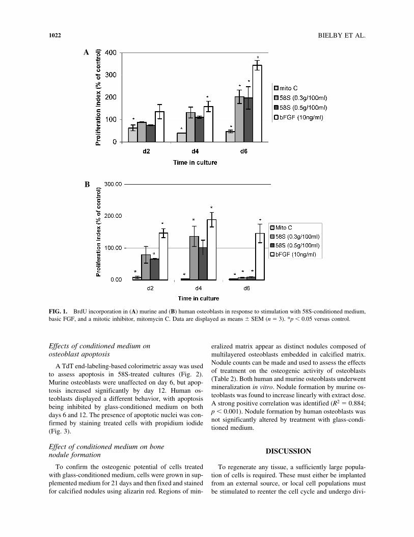
0.75 times that in control medium and the phosphorusconcentration was 0.34 or 0.25 times that in controlmedium. The sodium concentration was virtually un-changed from that in control medium (1.03 and 1.02 timescontrol). These figures are what would be expected givenwhat is known about the dissolution of sol–gel bioactiveglasses.15 As HCA formation proceeds much faster thanwith melt-derived 45S5, the silicon concentration will in-crease faster during 24 h of incubation of the sol–gelglasses.
Effects of conditioned medium on osteoblast proliferation
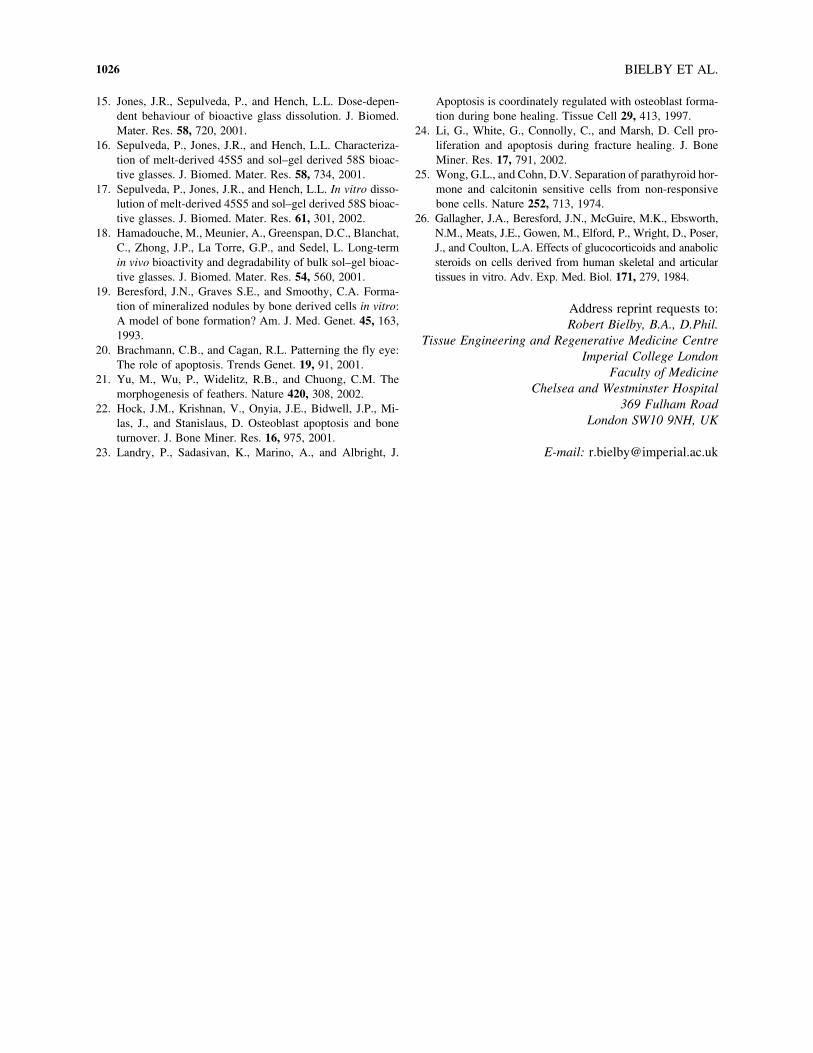
The proliferation of osteoblasts growing in culturemedium supplemented with extracts of 58S gel-glass wasassessed at 2, 4, and 6 days of culture (Fig. 1). At all timepoints, mitomycin C inhibited BrdU incorporation and,with the exception of day 2 murine osteoblasts (p 50.089), bFGF treatment (10 ng/mL) stimulated BrdU in-corporation, demonstrating that the cells were mitoticallyactive. Glass-conditioned medium did not inhibit prolif-eration of murine osteoblasts at any time point, and stim-ulated a 100% increase in proliferation on day 6 (p ,0.05). The effects of conditioned medium on human os-teoblasts were more varied. Proliferation was inhibitedon day 2, stimulated on day 4, and inhibited more stronglyon day 6. It was apparent, however, for both murine andhuman osteoblasts that the maximal increase in prolifer-ation induced by conditioned medium coincided with themaximal increase induced by bFGF treatment. Similar re-sults were obtained from an alternative assay utilizingformazan dye production (not shown), although it wasfound that this method was less sensitive than the BrdUincorporation technique. Both doses of glass condition-ing (0.3 and 0.5 g/100 mL) were effective at inducingcell proliferation and no statistically significant differ-ences were found between them. Doses higher than 0.5g/100 mL were increasingly inhibitory to cell prolifera-tion in both species and at all time points studied (datanot shown).
TABLE 1. CONCENTRATIONSa OF SILICON, CALCIUM, PHOSPHORUS, AND SODIUM IN CONDITIONED
AND CONTROL MEDIA AS MEASURED BY INDUCTIVELY COUPLED PLASMA ANALYSIS
Si Ca P Na
Unconditioned medium 0.33 6 0.12 63.42 6 0.47 29.83 6 0.40 2665.02 6 22.2958S-conditioned medium 163.98 6 1.77 51.32 6 0.32 10.38 6 0.23 2740.34 6 10.58
(0.3 g/100 mL)58S-conditioned medium 203.11 6 2.75 47.06 6 0.73 6.88 6 0.14 2711.87 6 18.53
(0.5 g/100 mL)45S5-conditioned mediumb 16.58 6 1.78 88.35 6 2.32 30.45 6 0.64 2938 6 24.62
aIn parts per million.bData from Xynos et al.9
BIELBY ET AL.1022
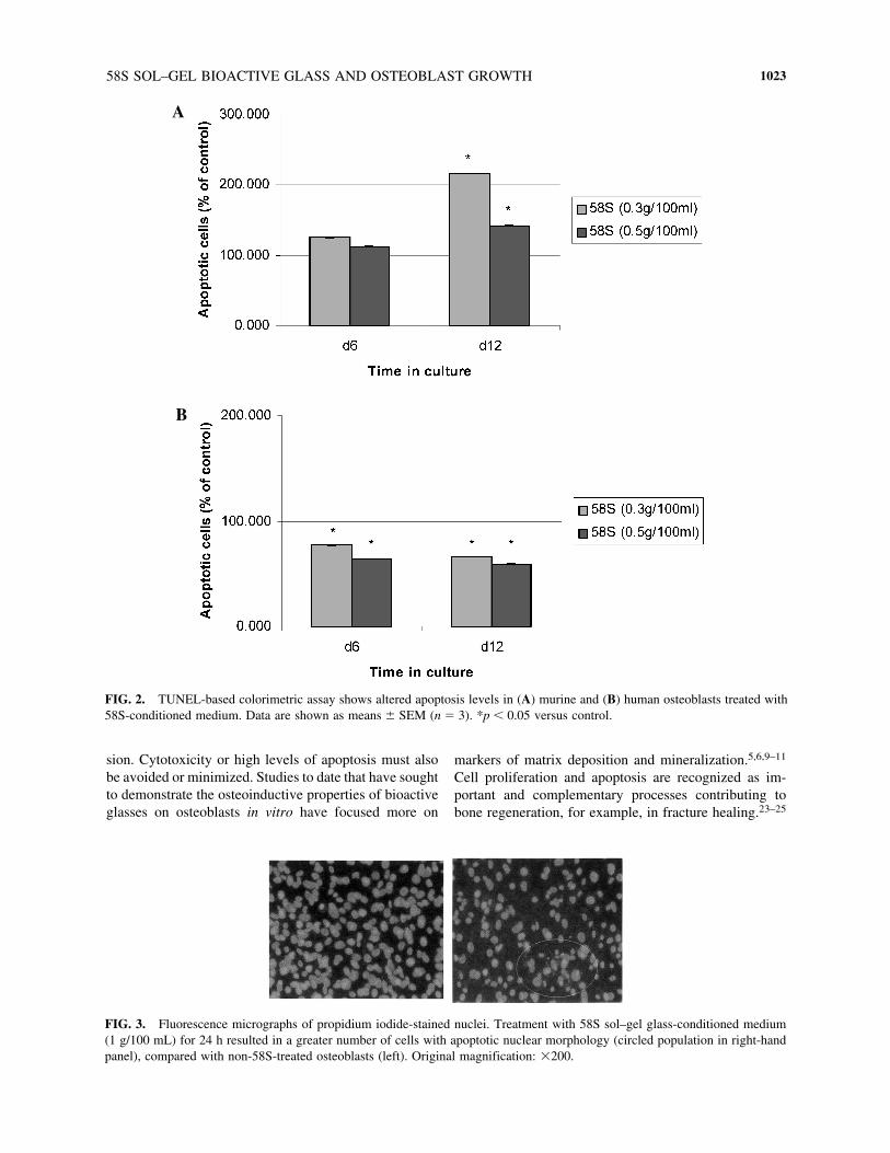
Effects of conditioned medium on osteoblast apoptosis
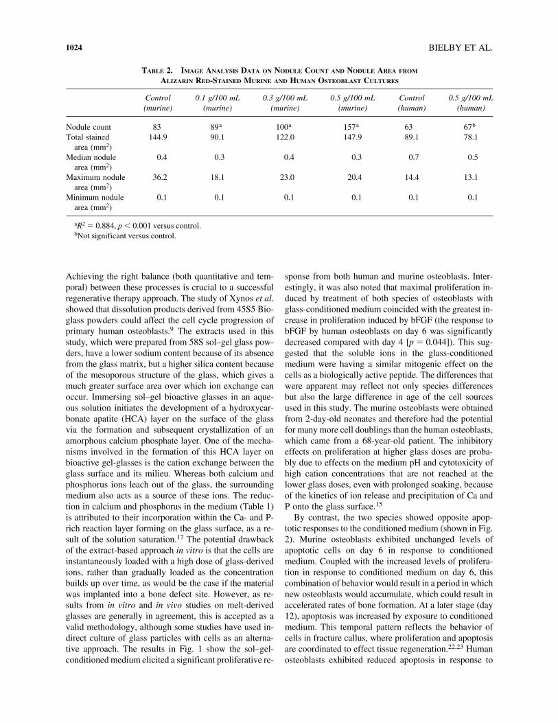
A TdT end-labeling-based colorimetric assay was usedto assess apoptosis in 58S-treated cultures (Fig. 2).Murine osteoblasts were unaffected on day 6, but apop-tosis increased significantly by day 12. Human os-teoblasts displayed a different behavior, with apoptosisbeing inhibited by glass-conditioned medium on bothdays 6 and 12. The presence of apoptotic nuclei was con-firmed by staining treated cells with propidium iodide(Fig. 3).
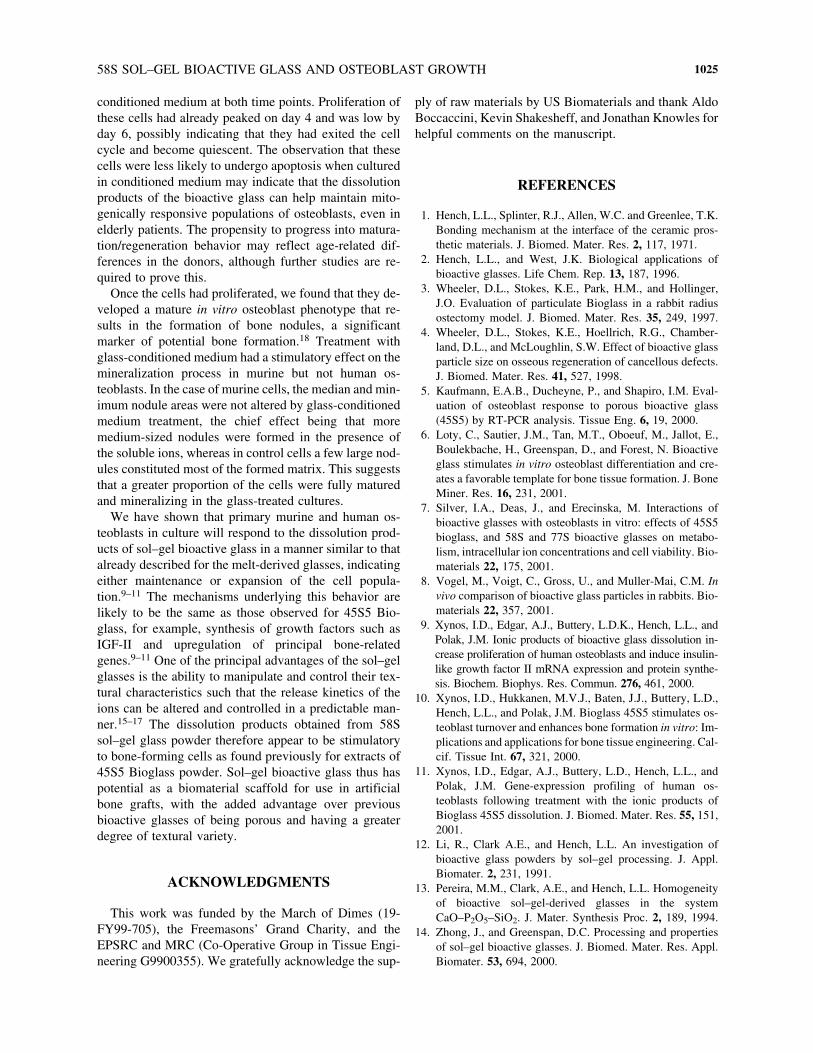
Effect of conditioned medium on bone nodule formation
To confirm the osteogenic potential of cells treatedwith glass-conditioned medium, cells were grown in sup-plemented medium for 21 days and then fixed and stainedfor calcified nodules using alizarin red. Regions of min-
eralized matrix appear as distinct nodules composed ofmultilayered osteoblasts embedded in calcified matrix.Nodule counts can be made and used to assess the effectsof treatment on the osteogenic activity of osteoblasts(Table 2). Both human and murine osteoblasts underwentmineralization in vitro. Nodule formation by murine os-teoblasts was found to increase linearly with extract dose.A strong positive correlation was identified (R2 5 0.884;p , 0.001). Nodule formation by human osteoblasts wasnot significantly altered by treatment with glass-condi-tioned medium.
DISCUSSION
To regenerate any tissue, a sufficiently large popula-tion of cells is required. These must either be implantedfrom an external source, or local cell populations mustbe stimulated to reenter the cell cycle and undergo divi-
A
B
FIG. 1. BrdU incorporation in (A) murine and (B) human osteoblasts in response to stimulation with 58S-conditioned medium,basic FGF, and a mitotic inhibitor, mitomycin C. Data are displayed as means 6 SEM (n 5 3). *p , 0.05 versus control.
58S SOL–GEL BIOACTIVE GLASS AND OSTEOBLAST GROWTH 1023
sion. Cytotoxicity or high levels of apoptosis must alsobe avoided or minimized. Studies to date that have soughtto demonstrate the osteoinductive properties of bioactiveglasses on osteoblasts in vitro have focused more on
markers of matrix deposition and mineralization.5,6,9–11
Cell proliferation and apoptosis are recognized as im-portant and complementary processes contributing tobone regeneration, for example, in fracture healing.23–25
A
B
FIG. 2. TUNEL-based colorimetric assay shows altered apoptosis levels in (A) murine and (B) human osteoblasts treated with58S-conditioned medium. Data are shown as means 6 SEM (n 5 3). *p , 0.05 versus control.
FIG. 3. Fluorescence micrographs of propidium iodide-stained nuclei. Treatment with 58S sol–gel glass-conditioned medium(1 g/100 mL) for 24 h resulted in a greater number of cells with apoptotic nuclear morphology (circled population in right-handpanel), compared with non-58S-treated osteoblasts (left). Original magnification: 3200.
BIELBY ET AL.1024
Achieving the right balance (both quantitative and tem-poral) between these processes is crucial to a successfulregenerative therapy approach. The study of Xynos et al.showed that dissolution products derived from 45S5 Bio-glass powders could affect the cell cycle progression ofprimary human osteoblasts.9 The extracts used in thisstudy, which were prepared from 58S sol–gel glass pow-ders, have a lower sodium content because of its absencefrom the glass matrix, but a higher silica content becauseof the mesoporous structure of the glass, which gives amuch greater surface area over which ion exchange canoccur. Immersing sol–gel bioactive glasses in an aque-ous solution initiates the development of a hydroxycar-bonate apatite (HCA) layer on the surface of the glassvia the formation and subsequent crystallization of anamorphous calcium phosphate layer. One of the mecha-nisms involved in the formation of this HCA layer onbioactive gel-glasses is the cation exchange between theglass surface and its milieu. Whereas both calcium andphosphorus ions leach out of the glass, the surroundingmedium also acts as a source of these ions. The reduc-tion in calcium and phosphorus in the medium (Table 1)is attributed to their incorporation within the Ca- and P-rich reaction layer forming on the glass surface, as a re-sult of the solution saturation.17 The potential drawbackof the extract-based approach in vitro is that the cells areinstantaneously loaded with a high dose of glass-derivedions, rather than gradually loaded as the concentrationbuilds up over time, as would be the case if the materialwas implanted into a bone defect site. However, as re-sults from in vitro and in vivo studies on melt-derivedglasses are generally in agreement, this is accepted as avalid methodology, although some studies have used in-direct culture of glass particles with cells as an alterna-tive approach. The results in Fig. 1 show the sol–gel-conditioned medium elicited a significant proliferative re-
sponse from both human and murine osteoblasts. Inter-estingly, it was also noted that maximal proliferation in-duced by treatment of both species of osteoblasts withglass-conditioned medium coincided with the greatest in-crease in proliferation induced by bFGF (the response tobFGF by human osteoblasts on day 6 was significantlydecreased compared with day 4 [p 5 0.044]). This sug-gested that the soluble ions in the glass-conditionedmedium were having a similar mitogenic effect on thecells as a biologically active peptide. The differences thatwere apparent may reflect not only species differencesbut also the large difference in age of the cell sourcesused in this study. The murine osteoblasts were obtainedfrom 2-day-old neonates and therefore had the potentialfor many more cell doublings than the human osteoblasts,which came from a 68-year-old patient. The inhibitoryeffects on proliferation at higher glass doses are proba-bly due to effects on the medium pH and cytotoxicity ofhigh cation concentrations that are not reached at thelower glass doses, even with prolonged soaking, becauseof the kinetics of ion release and precipitation of Ca andP onto the glass surface.15
By contrast, the two species showed opposite apop-totic responses to the conditioned medium (shown in Fig.2). Murine osteoblasts exhibited unchanged levels ofapoptotic cells on day 6 in response to conditionedmedium. Coupled with the increased levels of prolifera-tion in response to conditioned medium on day 6, thiscombination of behavior would result in a period in whichnew osteoblasts would accumulate, which could result inaccelerated rates of bone formation. At a later stage (day12), apoptosis was increased by exposure to conditionedmedium. This temporal pattern reflects the behavior ofcells in fracture callus, where proliferation and apoptosisare coordinated to effect tissue regeneration.22,23 Humanosteoblasts exhibited reduced apoptosis in response to
TABLE 2. IMAGE ANALYSIS DATA ON NODULE COUNT AND NODULE AREA FROM
ALIZARIN RED-STAINED MURINE AND HUMAN OSTEOBLAST CULTURES
Control 0.1 g/100 mL 0.3 g/100 mL 0.5 g/100 mL Control 0.5 g/100 mL(murine) (murine) (murine) (murine) (human) (human)
Nodule count 83 89.a 100.a 157.a 63 67.b
Total stained 144.9 90.1 122.0 147.9 89.1 78.1area (mm2)
Median nodule 0.4 0.3 0.4 0.3 0.7 0.5area (mm2)
Maximum nodule 36.2 18.1 23.0 20.4 14.4 13.1area (mm2)
Minimum nodule 0.1 0.1 0.1 0.1 0.1 0.1area (mm2)
aR2 5 0.884, p , 0.001 versus control.bNot significant versus control.
58S SOL–GEL BIOACTIVE GLASS AND OSTEOBLAST GROWTH 1025
conditioned medium at both time points. Proliferation ofthese cells had already peaked on day 4 and was low byday 6, possibly indicating that they had exited the cellcycle and become quiescent. The observation that thesecells were less likely to undergo apoptosis when culturedin conditioned medium may indicate that the dissolutionproducts of the bioactive glass can help maintain mito-genically responsive populations of osteoblasts, even inelderly patients. The propensity to progress into matura-tion/regeneration behavior may reflect age-related dif-ferences in the donors, although further studies are re-quired to prove this.
Once the cells had proliferated, we found that they de-veloped a mature in vitro osteoblast phenotype that re-sults in the formation of bone nodules, a significantmarker of potential bone formation.18 Treatment withglass-conditioned medium had a stimulatory effect on themineralization process in murine but not human os-teoblasts. In the case of murine cells, the median and min-imum nodule areas were not altered by glass-conditionedmedium treatment, the chief effect being that moremedium-sized nodules were formed in the presence ofthe soluble ions, whereas in control cells a few large nod-ules constituted most of the formed matrix. This suggeststhat a greater proportion of the cells were fully maturedand mineralizing in the glass-treated cultures.
We have shown that primary murine and human os-teoblasts in culture will respond to the dissolution prod-ucts of sol–gel bioactive glass in a manner similar to thatalready described for the melt-derived glasses, indicatingeither maintenance or expansion of the cell popula-tion.9–11 The mechanisms underlying this behavior arelikely to be the same as those observed for 45S5 Bio-glass, for example, synthesis of growth factors such asIGF-II and upregulation of principal bone-relatedgenes.9–11 One of the principal advantages of the sol–gelglasses is the ability to manipulate and control their tex-tural characteristics such that the release kinetics of theions can be altered and controlled in a predictable man-ner.15–17 The dissolution products obtained from 58Ssol–gel glass powder therefore appear to be stimulatoryto bone-forming cells as found previously for extracts of45S5 Bioglass powder. Sol–gel bioactive glass thus haspotential as a biomaterial scaffold for use in artificialbone grafts, with the added advantage over previousbioactive glasses of being porous and having a greaterdegree of textural variety.
ACKNOWLEDGMENTS
This work was funded by the March of Dimes (19-FY99-705), the Freemasons’ Grand Charity, and the EPSRC and MRC (Co-Operative Group in Tissue Engi-neering G9900355). We gratefully acknowledge the sup-
ply of raw materials by US Biomaterials and thank AldoBoccaccini, Kevin Shakesheff, and Jonathan Knowles forhelpful comments on the manuscript.
REFERENCES
1. Hench, L.L., Splinter, R.J., Allen, W.C. and Greenlee, T.K.Bonding mechanism at the interface of the ceramic pros-thetic materials. J. Biomed. Mater. Res. 2, 117, 1971.
2. Hench, L.L., and West, J.K. Biological applications ofbioactive glasses. Life Chem. Rep. 13, 187, 1996.
3. Wheeler, D.L., Stokes, K.E., Park, H.M., and Hollinger,J.O. Evaluation of particulate Bioglass in a rabbit radiusostectomy model. J. Biomed. Mater. Res. 35, 249, 1997.
4. Wheeler, D.L., Stokes, K.E., Hoellrich, R.G., Chamber-land, D.L., and McLoughlin, S.W. Effect of bioactive glassparticle size on osseous regeneration of cancellous defects.J. Biomed. Mater. Res. 41, 527, 1998.
5. Kaufmann, E.A.B., Ducheyne, P., and Shapiro, I.M. Eval-uation of osteoblast response to porous bioactive glass(45S5) by RT-PCR analysis. Tissue Eng. 6, 19, 2000.
6. Loty, C., Sautier, J.M., Tan, M.T., Oboeuf, M., Jallot, E.,Boulekbache, H., Greenspan, D., and Forest, N. Bioactiveglass stimulates in vitro osteoblast differentiation and cre-ates a favorable template for bone tissue formation. J. BoneMiner. Res. 16, 231, 2001.
7. Silver, I.A., Deas, J., and Erecinska, M. Interactions ofbioactive glasses with osteoblasts in vitro: effects of 45S5bioglass, and 58S and 77S bioactive glasses on metabo-lism, intracellular ion concentrations and cell viability. Bio-materials 22, 175, 2001.
8. Vogel, M., Voigt, C., Gross, U., and Muller-Mai, C.M. Invivo comparison of bioactive glass particles in rabbits. Bio-materials 22, 357, 2001.
9. Xynos, I.D., Edgar, A.J., Buttery, L.D.K., Hench, L.L., andPolak, J.M. Ionic products of bioactive glass dissolution in-crease proliferation of human osteoblasts and induce insulin-like growth factor II mRNA expression and protein synthe-sis. Biochem. Biophys. Res. Commun. 276, 461, 2000.
10. Xynos, I.D., Hukkanen, M.V.J., Baten, J.J., Buttery, L.D.,Hench, L.L., and Polak, J.M. Bioglass 45S5 stimulates os-teoblast turnover and enhances bone formation in vitro: Im-plications and applications for bone tissue engineering. Cal-cif. Tissue Int. 67, 321, 2000.
11. Xynos, I.D., Edgar, A.J., Buttery, L.D., Hench, L.L., andPolak, J.M. Gene-expression profiling of human os-teoblasts following treatment with the ionic products ofBioglass 45S5 dissolution. J. Biomed. Mater. Res. 55, 151,2001.
12. Li, R., Clark A.E., and Hench, L.L. An investigation ofbioactive glass powders by sol–gel processing. J. Appl.Biomater. 2, 231, 1991.
13. Pereira, M.M., Clark, A.E., and Hench, L.L. Homogeneityof bioactive sol–gel-derived glasses in the systemCaO–P2O5–SiO2. J. Mater. Synthesis Proc. 2, 189, 1994.
14. Zhong, J., and Greenspan, D.C. Processing and propertiesof sol–gel bioactive glasses. J. Biomed. Mater. Res. Appl.Biomater. 53, 694, 2000.
BIELBY ET AL.1026
15. Jones, J.R., Sepulveda, P., and Hench, L.L. Dose-depen-dent behaviour of bioactive glass dissolution. J. Biomed.Mater. Res. 58, 720, 2001.
16. Sepulveda, P., Jones, J.R., and Hench, L.L. Characteriza-tion of melt-derived 45S5 and sol–gel derived 58S bioac-tive glasses. J. Biomed. Mater. Res. 58, 734, 2001.
17. Sepulveda, P., Jones, J.R., and Hench, L.L. In vitro disso-lution of melt-derived 45S5 and sol–gel derived 58S bioac-tive glasses. J. Biomed. Mater. Res. 61, 301, 2002.
18. Hamadouche, M., Meunier, A., Greenspan, D.C., Blanchat,C., Zhong, J.P., La Torre, G.P., and Sedel, L. Long-termin vivo bioactivity and degradability of bulk sol–gel bioac-tive glasses. J. Biomed. Mater. Res. 54, 560, 2001.
19. Beresford, J.N., Graves S.E., and Smoothy, C.A. Forma-tion of mineralized nodules by bone derived cells in vitro:A model of bone formation? Am. J. Med. Genet. 45, 163,1993.
20. Brachmann, C.B., and Cagan, R.L. Patterning the fly eye:The role of apoptosis. Trends Genet. 19, 91, 2001.
21. Yu, M., Wu, P., Widelitz, R.B., and Chuong, C.M. Themorphogenesis of feathers. Nature 420, 308, 2002.
22. Hock, J.M., Krishnan, V., Onyia, J.E., Bidwell, J.P., Mi-las, J., and Stanislaus, D. Osteoblast apoptosis and boneturnover. J. Bone Miner. Res. 16, 975, 2001.
23. Landry, P., Sadasivan, K., Marino, A., and Albright, J.
Apoptosis is coordinately regulated with osteoblast forma-tion during bone healing. Tissue Cell 29, 413, 1997.
24. Li, G., White, G., Connolly, C., and Marsh, D. Cell pro-liferation and apoptosis during fracture healing. J. BoneMiner. Res. 17, 791, 2002.
25. Wong, G.L., and Cohn, D.V. Separation of parathyroid hor-mone and calcitonin sensitive cells from non-responsivebone cells. Nature 252, 713, 1974.
26. Gallagher, J.A., Beresford, J.N., McGuire, M.K., Ebsworth,N.M., Meats, J.E., Gowen, M., Elford, P., Wright, D., Poser,J., and Coulton, L.A. Effects of glucocorticoids and anabolicsteroids on cells derived from human skeletal and articulartissues in vitro. Adv. Exp. Med. Biol. 171, 279, 1984.
Address reprint requests to:Robert Bielby, B.A., D.Phil.
Tissue Engineering and Regenerative Medicine CentreImperial College London
Faculty of MedicineChelsea and Westminster Hospital
369 Fulham RoadLondon SW10 9NH, UK
E-mail: [email protected]