Embed Size (px)
Citation preview
FORUM REVIEW ARTICLE
Tight Junctions in Brain BarriersDuring Central Nervous System Inflammation
Caroline Coisne and Britta Engelhardt
Abstract
Homeostasis within the central nervous system (CNS) is a prerequisite to elicit proper neuronal function. TheCNS is tightly sealed from the changeable milieu of the blood stream by the blood–brain barrier (BBB) and theblood–cerebrospinal fluid (CSF) barrier (BCSFB). Whereas the BBB is established by specialized endothelial cellsof CNS microvessels, the BCSFB is formed by the epithelial cells of the choroid plexus. Both constitute physicalbarriers by a complex network of tight junctions (TJs) between adjacent cells. During many CNS inflammatorydisorders, such as multiple sclerosis, human immunodeficiency virus infection, or Alzheimer’s disease, pro-duction of pro-inflammatory cytokines, matrix metalloproteases, and reactive oxygen species are responsible foralterations of CNS barriers. Barrier dysfunction can contribute to neurological disorders in a passive way byvascular leakage of blood-borne molecules into the CNS and in an active way by guiding the migration ofinflammatory cells into the CNS. Both ways may directly be linked to alterations in molecular composition,function, and dynamics of the TJ proteins. This review summarizes current knowledge on the cellular andmolecular aspects of the functional and dysfunctional TJ complexes at the BBB and the BCSFB, with a particularemphasis on CNS inflammation and the role of reactive oxygen species. Antioxid. Redox Signal. 15, 1285–1303.
Introduction
Cerebral barriers: blood–brain barrierand blood–cerebrospinal fluid barrier—cellular composition and function
The blood–brain barrier (BBB) and the blood–cerebro-spinal fluid (CSF) barrier (BCSFB) protect the central
nervous system (CNS) from the changeable milieu of theblood stream to establish CNS homeostasis, which is a pre-requisite for proper neuronal function. Whereas the BBB islocalized at the level of highly specialized endothelial cellswithin CNS microvessels, the BCSFB is formed by the epi-thelial cells of the choroid plexus. BBB endothelial cells es-tablish a physical barrier by an elaborate network of complextight junctions (TJs) between adjacent endothelial cells com-bined with a very low pinocytotic activity and a lack of fe-nestrae thus preventing the paracellular and transcellulardiffusion of water-soluble molecules across BBB endothelialcells, respectively (71). At the same time, however, BBB en-dothelium establishes a metabolic barrier by the expression ofa number of permanently active transmembrane transportsystems and cytoplasmatic enzymes, which ensure thetransport of nutrients from the blood into the CNS and therapid exclusion of toxic metabolites out of the CNS. In addi-tion, the BBB is protected from oxidative stress by the pres-
ence of high levels of antioxidant enzymes such as peroxidedetoxifying enzymes, glutathione reductase and peroxidase,which are essential for proper brain functions (92, 115). Whilethe endothelial cells form the BBB proper, it has been wellestablished that interaction with the adjacent cells and theextracellular matrix within the neurovascular unit are a pre-requisite for complete barrier function (30). A high number ofpericytes embedded into the endothelial basement membranecontribute to proper BBB function of CNS microvascular en-dothelial cells (79) as do polarized astrocytes, whose endfeetensheath CNS microvessels. Astrocytic endfeet and the pa-renchymal basement membrane form the glia limitans (Fig. 1)(1). Thus, endothelial function and vascular morphologymake the BBB unique and distinguishable from any othermicrovessels in the body.
In analogy to the endothelial BBB TJs, the morphologicalcorrelate of the BCSFB is found at the level of unique parallelapical TJs between the choroid plexus epithelial cells (23),which inhibit the paracellular diffusion of water-solublemolecules across this epithelial barrier (Fig. 3). The choroidplexus is a villous structure consisting of an extensive networkof fenestrated capillaries that are enclosed by a single layer ofcuboidal epithelium. It extends from the ventricular surfaceinto the lumen of the ventricles. Its major known function isthe secretion of CSF. Therefore, besides their barrier function,
Theodor Kocher Institute, University of Bern, Bern, Switzerland.
ANTIOXIDANTS & REDOX SIGNALINGVolume 15, Number 5, 2011ª Mary Ann Liebert, Inc.DOI: 10.1089/ars.2011.3929
1285

choroid plexus epithelial cells are secretory active by pro-ducing the CSF. Again, similar to the BBB, the barrier andsecretory function of the choroid plexus epithelial cells aremaintained by the polarized expression of a number of spe-cific transmembrane transport systems that allow for the di-rected transport of nutrients into the CSF and the removal oftoxic agents out of the CSF. Additionally, high levels of glu-tathione-S-transferase and catalase activities have been de-
scribed in choroid plexus epithelial cells ensuring properbrain functions (44, 105).
Here we will summarize our current knowledge on thecellular and molecular basis of the functional and dysfunc-tional blood–CNS barriers with focus on CNS inflammationincluding oxidative stress components.
BBB Tight Junctions
Ultrastructural and molecular composition
The junctional complexes between CNS microvascular en-dothelial cells include adherens junctions (AJs) and TJs (Fig.2). In contrast to epithelial cells, where TJs are concentrated atthe apical side of the intercellular cleft, in endothelial cells AJsand TJs are frequently found to be intermingled along theintercellular cleft (25, 133). Nevertheless, AJs and TJs havedistinct functions at the BBB. In endothelial cells outside of theCNS, AJs are shown to initiate endothelial cell-to-cell contactsand promote their maturation and maintenance. However, inthe CNS expression of vascular endothelial (VE)-cadherindeclines with BBB maturity, suggesting that maintenance ofBBB TJs in the adult may not require high amounts of VE-cadherin (11). TJs establish a barrier function by regulating thepassage of solutes and ions through the paracellular cleft.Highly complex TJs between adjacent CNS microvascularendothelial cells are therefore primarily responsible for theunique restriction of the paracellular diffusion pathway be-tween the endothelial cells, which establishes unlike in simpleTJs, a high electrical resistance of about 1800–2000O$cm2
across the BBB (14, 21). In addition to the barrier function, TJsestablish a fence function by limiting the free movement oflipids and proteins between the apical and the basolateral cellsurface, thus establishing BBB endothelial cell polarity.
FIG. 1. Schematic view of the cellular and extracellularelements of the blood–brain barrier (BBB). BBB endothelialcells characterized by a low pinocytotic activity and complextight junctions (TJs) form the barrier proper. Continuouscross-talk with pericytes embedded in the endothelial base-ment membrane as well as with astrocytes covering theabluminal aspect of the central nervous system (CNS) mi-crovessels is required for BBB maintenance. Astrocytes laydown the parenchymal basement membrane, which togetherwith the astrocytic end-feet establishes the glia limitans, asecond barrier to the CNS parenchyma. At the postcapillarylevel the endothelial and parenchymal basement membraneborder a perivascular space, in which rare perivascular an-tigen-presenting cells can be found. The junctional complexboxed is shown in detail in Figure 2.
FIG. 3. Schematic view of the blood-cerebrospinal fluidbarrier (BCSFB). In the choroid plexus fenestrated capillarieslie within the choroid plexus stroma that is surrounded bychoroid plexus epithelial cells establishing the BCSFB. TJsbetween the choroid plexus epithelial cells are unique withexpression of claudin-1 or - 3, claudin-2, and - 11, with theexpression of claudin-11 inducing parallel non anastomosingTJ strands. In analogy to the BBB right behind the barrier rareantigen presenting cells, the epiplexus or Kolmer cells, can befound.
FIG. 2. Schematic view of the BBB junctions. Homophilicbinding to the transmembrane protein vascular endothelial(VE)-cadherin establishes cell-to-cell adhesion at the level ofthe adherens junctions (AJs). p120, b-catenin and plakoglobinbind to the cytoplasmic tail of VE-cadherin and by interact-ing with a large number of actin-binding proteins link the AJto the cytoskeleton. Occludin and claudins mediate in ahomophilic and heterophilic manner the adhesion of adja-cent outer membrane leaflets in TJs. PDZ domain containingproteins such as the ZO-proteins provide a scaffold for thesetransmembrane TJ proteins and link them to the cytoskeletonand to a large number of signaling molecules.
1286 COISNE AND ENGELHARDT
At the ultrastructural level, BBB TJ morphology closelyresembles that of epithelial cells rather than that of endothelialcells of other vascular beds. In ultrathin section electronmicrographs, BBB TJs appear as a chain of fusion points—kisses—of the outer plasma membrane leaflet of the adjacentCNS endothelial cells (32). Additionally, TJs between the en-dothelial cells of the brain microvessels have been analyzed inelaborate qualitative freeze-fracture electron microscopystudies. These studies have demonstrated that in mammalianBBB endothelial cells TJs are more complex than TJs in otherendothelial cells in the body by establishing a continuousnetwork of parallel and highly interconnected strands thatcircumscribe the apex of lateral membranes of adjacent en-dothelial cells (71, 103). In addition to the complexity of TJstrands it was suggested that the association of the TJ particleswith the protoplasmic leaflet (P-face) or the exocytoplasmicleaflet of the cell membrane (E-face) is another criterion tocorrelate morphology and physiology of TJs (163). Furtherfreeze-fracture replica electron microscopy studies havedemonstrated that the TJ particles from BBB endothelial cellsare preferentially associated with the P-face rather than the E-face (163), thus resembling TJs of epithelial cells. The P-faceassociation was shown to correlate with the sealed barrierfunction of the BBB endothelium in mammals in vivo andin vitro (163). The concept of a correlation of P-face-associatedTJ particles with BBB function is also consistent with the ob-servation that in TJs of peripheral, nonbarrier forming endo-thelial cells, P-face-associated TJ particles are rarely found andE-face-associated TJ particles clearly predominate (100, 134). Ithas been hypothesized that there is an important functionalrole for the P-face association, and thus presumably the cy-toplasmic anchoring of the TJ particles, particularly for BBBfunction (163). This is further supported by the finding thatthe transmembrane proteins of the TJs are localized to theseparticles (40) and that a molecular component, which wasidentified to associate with the P-face, claudin-3, is predomi-nantly incorporated into BBB endothelial cell TJs (see below).
In both AJs and TJs, the adhesion between adjacent endo-thelial cells is mediated by transmembrane proteins thatpromote homophilic and, at least in TJs, probably also het-erophilic interactions that establish a pericellular ziplock-likeadhesive structure sealing the intercellular cleft. In endothelialcells including those of the BBB, AJs are largely composed ofVE-cadherin, an endothelium-specific member of the cad-herin family of adhesion proteins (20). With its cytoplasmicdomain VE-cadherin binds to several protein partners, in-cluding b-catenin and plakoglobin, members of the family ofarmadillo proteins (156) as well as to p120. Junctional asso-ciation of these proteins is crucial for the functional state ofAJs (26). The association of a large set of actin-binding pro-teins such as a-catenin, vinculin, and a-actinin to AJs pro-motes the anchoring of AJs to actin filaments. Anchoring ofAJs to the cytoskeleton is thought to be a prerequisite forjunctional stabilization, but probably also for dynamic open-ing and closing of the AJs. Additional signaling componentsand enzymes associated with AJs and reviewed elsewhere ingreat detail (26) further contribute to the dynamic regulationof endothelial AJs. In addition to VE-cadherin, expression ofthe classical type II cadherin–cadherin 10 has been describedto be specifically expressed in BBB endothelium in human andmouse brain but not in the fenestrated endothelial cells of thechoroid plexus (161).
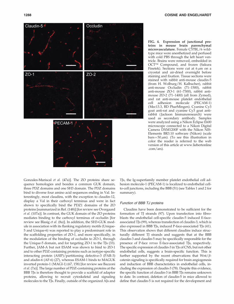
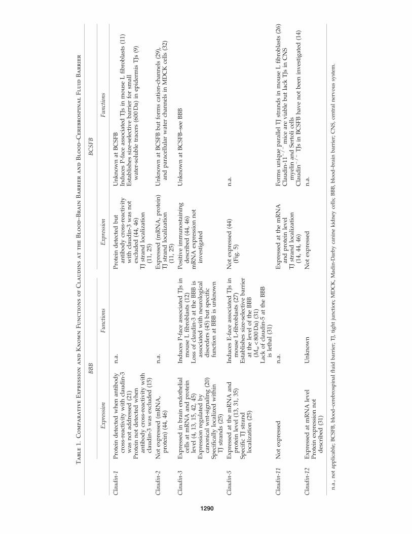
TJs of all endothelial cells, including those of the BBB, arecomposed of claudin-5, an endothelial-specific member of theclaudin family of transmembrane TJ proteins (97, 107). Theclaudins represent a gene family of integral membrane TJproteins with 24 members (42) [for review see Overgaard et al.(107a)]. Mammalian claudins range from 22 to 27 kDa andhave four transmembrane helices, a short internal amino-terminal sequence (two to six amino acids), two extracellularloops with the first loop being larger than the second and along variable cytoplasmic tail (Fig. 2). In addition to claudin-5,expression of claudin-3 and claudin-12 mRNA, but not ofclaudin-1 mRNA, is detectable in BBB endothelium (Lycket al., unpublished observations from our laboratory). At theprotein level, however, only the localization of claudin-3 andclaudin-5 to BBB TJs could be successfully demonstrated todate (53, 107, 157, 166) (Figs. 4 and 5). Previous studies havedescribed expression of claudin-1 at the protein level in BBBendothelium (77). Those studies that specifically excludedcross-reactivity of the anti-claudin-1 antibody with claudin-3failed to detect any expression of claudin-1 in CNS paren-chymal microvessels or in primary mouse or human brainendothelial cells (19, 53, 160, 166). Taken together, BBB TJsseem to be specifically assembled by the endothelial cell-specific claudin-5 together with claudin-3 and claudin-12.
Occludin, the first transmembrane component of TJs to beidentified (39), localizes to BBB TJ strands [for review seeBlasig et al. (8a)]. In fact expression levels of occludin havebeen reported to increase during brain angiogenesis and aresignificantly higher in TJs of the mature BBB than in TJs ofendothelial cells outside the CNS (56, 157). Occludin is anapproximately 60-kDa tetraspan membrane protein with twoextracellular loops, a short intracellular turn, an N-terminal,and a very long C-terminal cytoplasmic domain (146) and,therefore, at a first glance resembles claudins. Occludin does,however, not share any sequence homologies with the clau-dins. In immunoreplica electon microscopy anti-occludinantibodies exclusively label TJ strands, indicating that occlu-din directly incorporates into the TJ strands (128).
In addition, the immunoglobulin (Ig)-supergene familymembers, junctional adhesion molecule ( JAM)-A (91), andendothelial cell-selective adhesion molecule (ESAM) (106) arelocalized to TJs including those of the BBB (157). JAM-A is a32-kDa type I transmembrane protein, with two extracellularV-type Ig-domains, a single membrane-spanning domain anda cytoplasmic tail (28) [for review see Bazzoni et al. (7a)].ESAM is a 55-kDa type I transmembrane protein containing aV-type and a C2-type extracellular Ig domain, a singlemembrane-spanning domain, reminiscent of JAM-A, butdisplays a much longer 120-amino acid cytoplasmic domain(106). JAM-A has been demonstrated to physically interactwith zonula occludens-1 (ZO-1) and cingulin, indicatingthat JAM-A is a component of the multiprotein complex ofTJs (7) [for review see Bazzoni et al. (7a)]. As both proteins,JAM-A and ESAM, mediate homophilic interactions via theirextracellular domains, they may contribute to the ziplock-likeadhesive contacts at the BBB TJs (7, 72, 86) (see Tables 1 and 2for summary).
The integral membrane proteins of the BBB TJs are linkedto the endothelial cytoskeleton by TJ-associated cytoplas-mic peripheral membrane proteins of the membrane associ-ated with a guanylyl kinase-like domain (MAGUK) family,such as ZO-1, ZO-2, and ZO-3 (147, 162) [for review see
TJS IN BRAIN BARRIERS DURING CNS INFLAMMATION 1287
Gonzales-Mariscal et al. (47a)]. The ZO proteins share se-quence homologies and besides a common GUK domain,three PDZ domains and one SH3 domain. The PDZ domainsbind to diverse four amino acid sequences ending in Val. In-terestingly, most claudins, with the exception to claudin-12,display a Val in their carboxyl terminus and were in factshown to specifically bind the PDZ1 domains of the ZOproteins [summarized in Ref. (148)] [for review see Overgaardet al. (107a)]. In contrast, the GUK domain of the ZO proteinsmediates binding to the carboxyl terminus of occludin [forreview see Blasig et al. (8a)]. In addition, the SH3-GUK mod-ule in association with its flanking regulatory motifs (Unique-5 and Unique-6) was reported to play a predominant role inthe scaffolding properties of ZO-1, and more specifically, inthe modulation of the binding of occludin to ZO-1, throughthe Unique-5 domain, and for targeting ZO-1 to the TJs (33).Further, JAM-A but not ESAM was shown to bind to ZO-1and to other PDZ-containing proteins such as isotype-specificinteracting protein (ASIP)/partitioning defective-3 (PAR-3)and afadin-6 (AF-6) (27), whereas ESAM-1 binds to MAGUKinverted protein-1 (MAGI-1) (67, 158) [for review see Bazzoniet al. (7a)]. The large number of PDZ-containing proteins at theBBB TJs is therefore thought to provide a scaffold of adaptorproteins, allowing to recruit cytoskeletal and signalingmolecules to the TJs. Finally, outside of the organized AJs and
TJs, the Ig-superfamily member platelet endothelial cell ad-hesion molecule-1 (PECAM-1) is localized to endothelial cell-to-cell junctions, including the BBB (51) (see Tables 1 and 2 forsummary).
Function of BBB TJ proteins
Claudins have been demonstrated to be sufficient for theformation of TJ strands (97). Upon transfection into fibro-blasts the endothelial cell-specific claudin-5 induced E-face-associated TJs (99), whereas transfection of claudin-3, which isalso expressed in BBB TJs, induced P-face-associated TJs (41).This observation shows that different claudins induce struc-turally different TJ strands and suggests that at the BBBclaudin-3 and claudin-5 may be specifically responsible for thepresence of P-face versus E-face-associated TJs, respectively.The specific expression of claudin-3 in TJs of CNS, but not otherendothelial cells, suggests a brain-specific function. This isfurther supported by the recent observations that Wnt/b-catenin signaling is specifically required for brain angiogenesisand induction of BBB characteristics in endothelial cells, in-cluding the expression of claudin-3 (78). Despite this evidence,the specific function of claudin-3 in BBB TJs remains unknownto date. In contrast, deletion of claudin-5 in mice allowed todefine that claudin-5 is not required for the development and
FIG. 4. Expression of junctional pro-teins in mouse brain parenchymalmicrovasculature. Female C57BL/6 wild-type mice were anesthetized and perfusedwith cold PBS through the left heart ven-tricle. Brains were removed, embedded inOCT� Compound, and frozen (SakuraFinetek). Sections were cut at 6 lm on acryostat and air-dried overnight beforestaining and fixation. Tissue sections werestained with rabbit anti-mouse claudin-5(from H. Wolburg/H. Kalbacher), rabbitanti-mouse Occludin (71–1500), rabbitanti-mouse ZO-1 (61–7300), rabbit anti-mouse ZO-2 (71–1400) (all from Zymed),and rat anti-mouse platelet endothelialcell adhesion molecule (PECAM-1)(Mec13.3, BD PharMingen). Cyanine Cy3goat anti-rat and cyanine Cy3 goat anti-rabbit ( Jackson Immunoresearch) wereused as secondary antibody. Sampleswere analyzed using a Nikon Eclipse E600microscope connected to a Nikon DigitalCamera DXM1200F with the Nikon NIS-Elements BR3.10 software (Nikon) (scalebars = 50 lm). (To see this illustration incolor the reader is referred to the webversion of this article at www.liebertonline.com/ars).
1288 COISNE AND ENGELHARDT

morphology of blood vessels, but rather establishes a sizeselective barrier of BBB TJs for molecules smaller than 800 Da(107) [for review see Overgaard et al. (107a)]. On the molecularlevel little is known on how claudin–claudin interactions mayestablish such a size-selective barrier function at BBB TJs. Bothextracellular loops and possibly the transmembrane domainsof claudins have been suggested to be involved in adhesiveclaudin–claudin interactions and assembly of TJ strands (73).Recently, it could be demonstrated that a highly conservedaromatic binding core within the second extracellular loop ofclaudins mediates homophilic or heterophilic adhesive inter-actions of claudins in trans (114). Thus, the combined homo-philic and heterophilic interactions between claudin-3 andclaudin-5, and probably also claudin-12, at the BBB seem to beresponsible for the unique barrier function of the BBB TJs (73).
Mice carrying a null mutation in the occludin gene are vi-able and develop morphologically normal TJs in differenttissues, including the BBB (129). This demonstrates that oc-cludin is not essential for TJ formation [for review see Blasiget al. (8a)]. However, introduction of either C-terminally or N-terminally truncated occludin mutants into epithelial cellsresulted in disturbance of TJ integrity, proving that occludin isinvolved in TJ functions (6). Expression of occludin splicevariants has been reported and these may regulate TJ functionin distinct manners [summarized in Ref. (35)]. To what degreethis applies to BBB TJs function in vivo will need to be deter-mined. In any case, the specifically high expression levels ofoccludin in BBB TJs suggest a prominent regulatory functionof occludin in these TJs. Interestingly, recruitment of occludinto cell junctions can be achieved by JAM-A probably via in-teraction with ZO-1 (7) [for review see Gonzales-Mariscal et al.(47a)].
In epithelium, JAM-A interacting with PAR-3 could di-rectly adjust its subcellular localization, suggesting a role forJAM-A in regulating the formation of TJs and cell polarity inepithelial cells (61, 125, 142). At the blood–retinal barrier,which shares some common barrier features with the BBB, the
monoclonal antibody blockade of JAM-A resulted in an in-creased permeability of the rabbit corneal endotheliumin vitro. This suggests a direct implication of JAM-A in themaintenance of TJ integrity in this barrier endothelium (85)[for review see Frey and Antonetti (37a) and Bazzoni (7a)].The precise roles of JAM-A and ESAM in BBB TJs remain,however, to be determined as neither JAM-A nor ESAM-deficient mice display any overt BBB phenotype (16, 159) (seeTables 1 and 2 for summary).
Dynamics of TJs
Beginning to understand the individual functions of thespecific TJ proteins allows to better understand that BBB TJsare dynamic structures, which are sensitive to ambient factors.Already under physiological conditions, the diapedesis ofimmune cells during immunosurveillance or apoptosis ofbarrier cells during barrier renewal require dynamic re-modeling of TJs. In this context it has been shown that thefunction of occludin depends on its phosphorylation stage,which can be influenced by small GTPases, Rho kinases,oxidative stress, and angiogenic factors (35, 141, 169) [forreview see Lehner et al. (76a)]. Oxidative stress responses aremediated by various reactive oxygen species (ROS) such ashydrogen peroxide (H2O2), nitric oxide (NO), and peroxyni-trite. H2O2-induced in vitro BBB permeability was shown tocorrelate with higher expression levels of occludin but notZO-1 and their rearrangement at the TJs (75). Altered occludinphosphorylation has further been shown following low-doseexposure of in vitro BBB models to H2O2 (80). Phosphorylationand ubiquitination of occludin upon vascular endothelialgrowth factor stimulation has recently been described topromote internalization of TJ proteins at the blood–retinalbarrier endothelium, leading to increased permeability(101). In addition to alterations of the functional state of in-dividual TJ proteins, their expression levels in brain endo-thelium can be influenced by growth factors [for review see
FIG. 5. Expression and localization ofTJs and TJ-associated proteins in pri-mary mouse brain microvascular endo-thelial cells (pMBMECs). ConfluentpMBMEC monolayers were preparedfrom 6-week-old C57BL/6 wild-type miceas described by Coisne et al. (19). Cellswere fixed with ice-cold methanol andstained for rabbit anti-mouse claudin-3(34–1700), rabbit anti-mouse claudin-5(34–1600), rabbit anti-mouse Occludin(71–1500), and rabbit anti-mouse ZO-1(61–7300) (all from Invitrogen AG, formerZymed). Alexa Fluor 488 goat anti-rabbit(Molecular Probes) was used as second-ary antibody. Samples were analyzed us-ing a Nikon Eclipse E600 microscopeconnected to a Nikon Digital CameraDXM1200F with the Nikon NIS-ElementsBR3.10 software (Nikon) (scale bars = 50lm for claudin-3, claudin-5 and occludin,scale bars = 20 lm for ZO-1). (To see thisillustration in color the reader is referredto the web version of this article atwww.liebertonline.com/ars).
TJS IN BRAIN BARRIERS DURING CNS INFLAMMATION 1289
Ta
bl
e1.
Co
mp
ar
at
iv
eE
xp
re
ssio
na
nd
Kn
ow
nF
un
ct
io
ns
of
Cl
au
din
sa
tt
he
Bl
oo
d–
Br
ain
Ba
rr
ie
ra
nd
Bl
oo
d–
Ce
re
br
osp
in
al
Fl
uid
Ba
rr
ie
r
BB
BB
CS
FB
Ex
pre
ssio
nF
un
ctio
ns
Ex
pre
ssio
nF
un
ctio
ns
Cla
ud
in-1
Pro
tein
det
ecte
dw
hen
anti
bo
dy
cro
ss-r
eact
ivit
yw
ith
clau
din
-3w
asn
ot
add
ress
ed(2
1)P
rote
inn
ot
det
ecte
dw
hen
anti
bo
dy
cro
ss-r
eact
ivit
yw
ith
clau
din
-3w
asex
clu
ded
(15)
n.a
.P
rote
ind
etec
ted
bu
tan
tib
od
ycr
oss
-rea
ctiv
ity
wit
hcl
aud
in-3
was
no
tex
clu
ded
(44,
46)
TJ
stra
nd
loca
liza
tio
n(1
1,25
)
Un
kn
ow
nat
BC
SF
BIn
du
ces
P-f
ace
asso
ciat
edT
Jsin
mo
use
Lfi
bro
bla
sts
(11)
Est
abli
shes
size
-sel
ecti
ve
bar
rier
for
smal
lw
ater
-so
lub
letr
acer
s(6
00D
a)in
epid
erm
isT
Js(9
)
Cla
ud
in-2
No
tex
pre
ssed
(mR
NA
,p
rote
in)
(44,
46)
n.a
.E
xp
ress
ed(m
RN
A,
pro
tein
)T
Jst
ran
dlo
cali
zati
on
(11,
25)
Un
kn
ow
nat
BC
SF
Bb
ut
form
sca
tio
n-c
han
nel
s(2
9),
and
par
acel
lula
rw
ater
chan
nel
sin
MD
CK
cell
s(3
2)
Cla
ud
in-3
Ex
pre
ssed
inb
rain
end
oth
elia
lce
lls
atm
RN
Aan
dp
rote
inle
vel
(4,
13,
15,
42,
45)
Ex
pre
ssio
nre
gu
late
db
yca
no
nic
alw
nt-
sig
nal
ing
(20)
Sp
ecifi
call
ylo
cali
zed
wit
hin
TJ
stra
nd
s(2
5)
Ind
uce
sP
-fac
eas
soci
ated
TJs
inm
ou
seL
fib
rob
last
s(1
2)L
oss
of
clau
din
-3at
the
BB
Bis
asso
ciat
edw
ith
neu
rolo
gic
ald
iso
rder
s(4
5)b
ut
spec
ific
fun
ctio
nat
BB
Bis
un
kn
ow
n
Po
siti
ve
imm
un
ost
ain
ing
des
crib
ed(4
4,46
)m
RN
Aex
pre
ssio
nn
ot
inv
esti
gat
ed
Un
kn
ow
nat
BC
SF
B–s
eeB
BB
Cla
ud
in-5
Ex
pre
ssed
atth
em
RN
Aan
dp
rote
inle
vel
(13,
31,
35)
Sp
ecifi
cT
Jst
ran
dlo
cali
zati
on
(25)
Ind
uce
sE
-fac
eas
soci
ated
TJs
inm
ou
seL
-fib
rob
last
s(2
7)E
stab
lish
essi
ze-s
elec
tiv
eb
arri
erat
the
lev
elo
fth
eB
BB
(Mw
<80
0D
a)(3
1)L
ack
of
clau
din
-5at
the
BB
Bis
leth
al(3
1)
No
tex
pre
ssed
(44)
(Fig
.5)
n.a
.
Cla
ud
in-1
1N
ot
exp
ress
edn
.a.
Ex
pre
ssed
atth
em
RN
Aan
dp
rote
inle
vel
TJ
stra
nd
loca
liza
tio
n(1
4,44
,46
)
Fo
rms
un
iqu
ep
aral
lel
TJ
stra
nd
sin
mo
use
Lfi
bro
bla
sts
(26)
Cla
ud
in-1
1-
/-
mic
ear
ev
iab
leb
ut
lack
TJs
inC
NS
my
elin
and
Ser
toli
cell
sC
lau
din
-/
-T
Jsin
BC
SF
Bh
ave
no
tb
een
inv
esti
gat
ed(1
4)
Cla
ud
in-1
2E
xp
ress
edat
mR
NA
lev
elP
rote
inex
pre
ssio
nn
ot
des
crib
ed(3
1)
Un
kn
ow
nN
ot
exp
ress
edn
.a.
n.a
.,n
ot
app
lica
ble
;B
CS
FB
,b
loo
d–c
ereb
rosp
inal
flu
idb
arri
er;
TJ,
tig
ht
jun
ctio
n;
MD
CK
,M
adin
-Dar
by
can
ine
kid
ney
cell
s;B
BB
,b
loo
d–b
rain
bar
rier
;C
NS
,ce
ntr
aln
erv
ou
ssy
stem
.
1290
Frey and Antonetti (37a), in this review at the blood–retinalbarrier]. Pericyte derived angiopoietin-1 was shown by in-ducing the phosphorylation of its Tie-2 receptor to trigger adownstream signaling cascade, finally leading to increasedexpression of occludin in immortalized rat brain endothelialcells (59). Taken together, BBB TJs are highly dynamic mo-lecular complexes that will change in responseto neuroinflammatory insults generating pro-inflammatory
cytokines, chemokines, matrix metalloproteases (MMPs), andROS.
BCSFB Tight Junctions
Ultrastructural and molecular composition
The TJs between the choroid plexus epithelial cells formthe morphological correlate of the BCSFB. Freeze-fracture
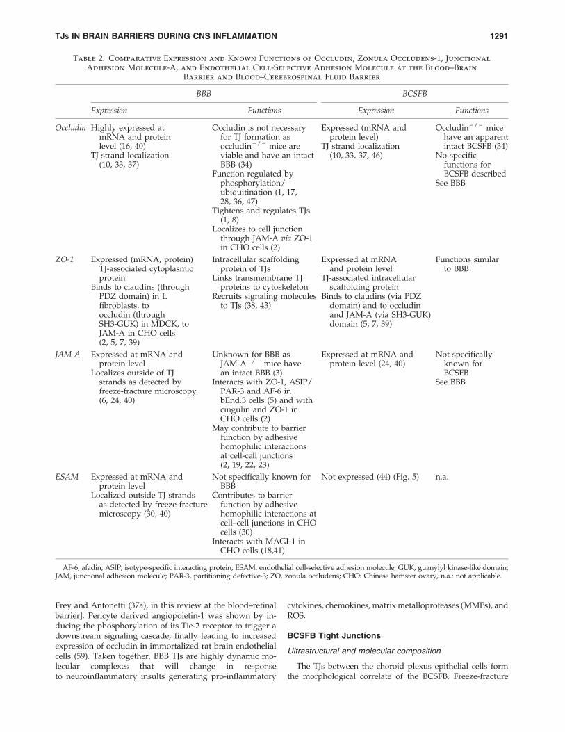
Table 2. Comparative Expression and Known Functions of Occludin, Zonula Occludens-1, Junctional
Adhesion Molecule-A, and Endothelial Cell-Selective Adhesion Molecule at the Blood–Brain
Barrier and Blood–Cerebrospinal Fluid Barrier
BBB BCSFB
Expression Functions Expression Functions
Occludin Highly expressed atmRNA and proteinlevel (16, 40)
TJ strand localization(10, 33, 37)
Occludin is not necessaryfor TJ formation asoccludin - / - mice areviable and have an intactBBB (34)
Function regulated byphosphorylation/ubiquitination (1, 17,28, 36, 47)
Tightens and regulates TJs(1, 8)
Localizes to cell junctionthrough JAM-A via ZO-1in CHO cells (2)
Expressed (mRNA andprotein level)
TJ strand localization(10, 33, 37, 46)
Occludin - / - micehave an apparentintact BCSFB (34)
No specificfunctions forBCSFB described
See BBB
ZO-1 Expressed (mRNA, protein)TJ-associated cytoplasmicprotein
Binds to claudins (throughPDZ domain) in Lfibroblasts, tooccludin (throughSH3-GUK) in MDCK, toJAM-A in CHO cells(2, 5, 7, 39)
Intracellular scaffoldingprotein of TJs
Links transmembrane TJproteins to cytoskeleton
Recruits signaling moleculesto TJs (38, 43)
Expressed at mRNAand protein level
TJ-associated intracellularscaffolding protein
Binds to claudins (via PDZdomain) and to occludinand JAM-A (via SH3-GUK)domain (5, 7, 39)
Functions similarto BBB
JAM-A Expressed at mRNA andprotein level
Localizes outside of TJstrands as detected byfreeze-fracture microscopy(6, 24, 40)
Unknown for BBB asJAM-A - / - mice havean intact BBB (3)
Interacts with ZO-1, ASIP/PAR-3 and AF-6 inbEnd.3 cells (5) and withcingulin and ZO-1 inCHO cells (2)
May contribute to barrierfunction by adhesivehomophilic interactionsat cell-cell junctions(2, 19, 22, 23)
Expressed at mRNA andprotein level (24, 40)
Not specificallyknown forBCSFB
See BBB
ESAM Expressed at mRNA andprotein level
Localized outside TJ strandsas detected by freeze-fracturemicroscopy (30, 40)
Not specifically known forBBB
Contributes to barrierfunction by adhesivehomophilic interactions atcell–cell junctions in CHOcells (30)
Interacts with MAGI-1 inCHO cells (18,41)
Not expressed (44) (Fig. 5) n.a.
AF-6, afadin; ASIP, isotype-specific interacting protein; ESAM, endothelial cell-selective adhesion molecule; GUK, guanylyl kinase-like domain;JAM, junctional adhesion molecule; PAR-3, partitioning defective-3; ZO, zonula occludens; CHO: Chinese hamster ovary, n.a.: not applicable.
TJS IN BRAIN BARRIERS DURING CNS INFLAMMATION 1291
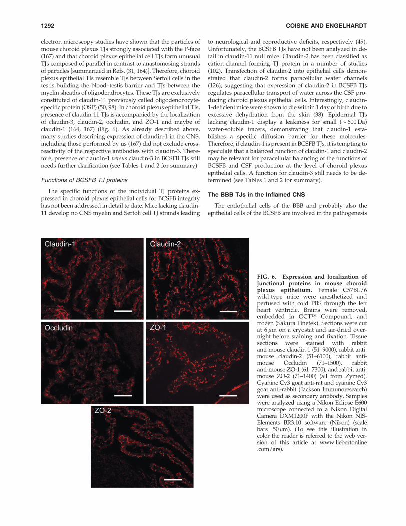
electron microscopy studies have shown that the particles ofmouse choroid plexus TJs strongly associated with the P-face(167) and that choroid plexus epithelial cell TJs form unusualTJs composed of parallel in contrast to anastomosing strandsof particles [summarized in Refs. (31, 164)]. Therefore, choroidplexus epithelial TJs resemble TJs between Sertoli cells in thetestis building the blood–testis barrier and TJs between themyelin sheaths of oligodendrocytes. These TJs are exclusivelyconstituted of claudin-11 previously called oligodendrocyte-specific protein (OSP) (50, 98). In choroid plexus epithelial TJs,presence of claudin-11 TJs is accompanied by the localizationof claudin-3, claudin-2, occludin, and ZO-1 and maybe ofclaudin-1 (164, 167) (Fig. 6). As already described above,many studies describing expression of claudin-1 in the CNS,including those performed by us (167) did not exclude cross-reactivity of the respective antibodies with claudin-3. There-fore, presence of claudin-1 versus claudin-3 in BCSFB TJs stillneeds further clarification (see Tables 1 and 2 for summary).
Functions of BCSFB TJ proteins
The specific functions of the individual TJ proteins ex-pressed in choroid plexus epithelial cells for BCSFB integrityhas not been addressed in detail to date. Mice lacking claudin-11 develop no CNS myelin and Sertoli cell TJ strands leading
to neurological and reproductive deficits, respectively (49).Unfortunately, the BCSFB TJs have not been analyzed in de-tail in claudin-11 null mice. Claudin-2 has been classified ascation-channel forming TJ protein in a number of studies(102). Transfection of claudin-2 into epithelial cells demon-strated that claudin-2 forms paracellular water channels(126), suggesting that expression of claudin-2 in BCSFB TJsregulates paracellular transport of water across the CSF pro-ducing choroid plexus epithelial cells. Interestingly, claudin-1-deficient mice were shown to die within 1 day of birth due toexcessive dehydration from the skin (38). Epidermal TJslacking claudin-1 display a leakiness for small (*600 Da)water-soluble tracers, demonstrating that claudin-1 esta-blishes a specific diffusion barrier for these molecules.Therefore, if claudin-1 is present in BCSFB TJs, it is tempting tospeculate that a balanced function of claudin-1 and claudin-2may be relevant for paracellular balancing of the functions ofBCSFB and CSF production at the level of choroid plexusepithelial cells. A function for claudin-3 still needs to be de-termined (see Tables 1 and 2 for summary).
The BBB TJs in the Inflamed CNS
The endothelial cells of the BBB and probably also theepithelial cells of the BCSFB are involved in the pathogenesis
FIG. 6. Expression and localization ofjunctional proteins in mouse choroidplexus epithelium. Female C57BL/6wild-type mice were anesthetized andperfused with cold PBS through the leftheart ventricle. Brains were removed,embedded in OCT� Compound, andfrozen (Sakura Finetek). Sections were cutat 6 lm on a cryostat and air-dried over-night before staining and fixation. Tissuesections were stained with rabbitanti-mouse claudin-1 (51–9000), rabbit anti-mouse claudin-2 (51–6100), rabbit anti-mouse Occludin (71–1500), rabbitanti-mouse ZO-1 (61–7300), and rabbit anti-mouse ZO-2 (71–1400) (all from Zymed).Cyanine Cy3 goat anti-rat and cyanine Cy3goat anti-rabbit ( Jackson Immunoresearch)were used as secondary antibody. Sampleswere analyzed using a Nikon Eclipse E600microscope connected to a Nikon DigitalCamera DXM1200F with the Nikon NIS-Elements BR3.10 software (Nikon) (scalebars = 50lm). (To see this illustration incolor the reader is referred to the web ver-sion of this article at www.liebertonline.com/ars).
1292 COISNE AND ENGELHARDT

of a wide range of inflammatory CNS disorders, includingmultiple sclerosis (MS) or its animal model experimental au-toimmune encephalomyelitis (EAE), infectious diseases (hu-man immunodeficiency virus [HIV]), as well asneurodegenerative diseases (Alzheimer’s disease [AD]). Inthis context, the contribution of both CNS barriers can beconsidered twofold, a passive role characterized by vascularleakage of blood-borne molecules into the CNS and an activerole by guiding inflammatory cell migration into the CNS.Both roles may directly be linked to alterations in the molec-ular composition of the TJ complexes or functional states of TJproteins based on their phosphorylation state. Many studiesfocused on investigating junctional alterations of the BBB inthe context of neuroinflammatory diseases. In general, moststudies rather focused on the expression of occludin and ZO-1, although only selected studies have investigated the ex-pression pattern of the complete array of TJ proteins known tobe expressed at the BBB to date. Here, we summarize ourpresent knowledge on BBB TJ alterations as described in MS,HIV, and AD as examples for inflammatory demyelinatingdiseases of the CNS, infectious diseases, and neurodegener-ative diseases, respectively. These diseases have been associ-ated with increased free radical production and chronicoxidative stress (108, 118, 119), suggesting a crucial role ofROS mediators in triggering endothelial dysfunctions thataccompany neuroinflammatory disorders. Vascular leakageof blood-borne molecules and inflammatory cell migrationacross the BBB represent two major phenomena followingBBB disruption. Both may directly be linked to alterations inthe molecular composition or dynamics of the TJ proteins.However, while TJ alteration can easily be associated withincreased paracellular flux of solutes across the BBB (inde-pendently of increased transcellular fluid-phase processes),the induction of immune cell infiltration into the CNS repre-sents a complex phenomenon that implies several features. Itnecessitates the contribution of various inflammatory medi-ators present in the CNS environment that directly affect themotility of immune cells toward the site of inflammation (bychemoattractants) and then within the CNS (by chemoat-tractants and MMPs) as well as the endothelial integrity per se(by pro-inflammatory cytokines and oxidative stress) [for re-view see Lehner et al. (76a)]. In addition, altered TJ integritymay facilitate immune cell migration across the BBB, but notnecessarily, as immune cells may rather use the paracellularroute to cross the BBB (15, 165).
Multiple sclerosis
Ultrastructural and molecular alterations. MS is an in-flammatory demyelinating disease of the CNS with an as-sumed autoimmune cause. EAE is an animal model for theinflammatory phase of MS and reflects various aspects of MS,including reversibility of the neurological disabilities, in-flammatory CNS lesion, and BBB disruption (66, 90). In bothMS and EAE, inflammatory cells composed mostly of T cellsand macrophages accumulate within the CNS parenchymabut also within perivascular spaces. CNS inflammation as-sociated with MS and EAE has been described to involve awide range of cytokines, chemokines, MMPs, and ROS pro-duced by infiltrating immune cells and also CNS resident cells(46, 93). Besides the infiltrating immune cells, pro-inflamma-tory and oxidative stress mediators are considered to have
direct impacts on BBB function in neuroinflammation. RNAprofiling in MS brain tissues, using cDNA microarrays, re-vealed differential gene expression in normal appearing whitematter (NAWM) of MS versus healthy tissues, indicating thatbeside inflammatory CNS lesion, inflammation, and degene-ration take place throughout the MS brains (52, 68). Severalalterations observed in MS tissues were characteristic ofneuroprotective mechanisms against oxidative stress, identi-cal to the ones induced following hypoxic damages, includingupregulation of hypoxia-inducible factor-1a and its down-stream partners (52, 68). In addition, higher levels of antioxi-dants such as peroxiredoxin V were reported in NAWM inacute and chronic lesions from MS patients and higher levelsof superoxide dismutase (SOD) 1/2, and heme oxygenase 1were found in active demyelinating MS lesions (58, 153).Peroxynitrites were found to be produced in astrocytes inacute and chronic MS lesions as well as in early stage of EAE,where it was associated with disease activity (22, 81, 152).Immunostainings of brain tissue biopsies from patients withvarious forms of MS showed in 20%–40% of CNS microvesselabnormalities in immunostaining for ZO-1 and occludin,characterized as discontinuous staining patterns in contrast tothe continuous staining observed in normal brain tissue (76,116). Interestingly, discontinuous immunostaining was ob-served in both active and inactive MS plaques, but also,however, to a lower extent in the NAWM, indicating thatlesion activity may not be a prerequisite for TJ alterations inMS (76, 116). Furthermore, changes observed for ZO-1 andoccludin immunostaining in MS brains were found to be in-dependent of the vascular segment, with TJ abnormalitiesobserved at the capillary level as well as in larger diametermicrovessels to the same extent (69). Similarly, disruptedimmunostaining for JAM-A has been described in CNS mi-crovessels in brain biopsy tissues from MS patients (109). InEAE, molecular alterations of BBB TJs observed seem to be inapparent contrast to the observations made in MS tissues.Here, immunofluorescence stainings demonstrated a selectiveloss of claudin-3 immunostaining specifically at those post-capillary venules, which were surrounded by inflammatorycuffs, but not in inflamed CNS microvessels, where noperivascular inflammatory cells were present (166). The im-munofluorescence staining for occludin, ZO-1, and claudin-5in inflamed CNS microvessels during EAE was found tobe unchanged, irrespective of the presence or absence ofsurrounding inflammatory cuffs (166). This apparentdiscrepancy to the observations made in MS tissues may beexplained by different interpretations of the respective find-ings. Ultrastructural studies performed on brain tissues fromEAE mice demonstrated dramatic alterations in BBB TJ mor-phology with an extended upfolding of membranes at thelevel of the cell–cell contacts (165). These upfolded junctionalmembranes might lead to the appearance of an interruptedimmunostaining for junctional proteins at the light micros-copy level. Also, the presence of inflammatory cells localizedwithin the vascular wall during their diapedesis across theBBB may lead to apparently interrupted junctional stainingpatterns. Functional changes of BBB TJs might additionallybe a consequence of the altered functional state of individualTJ proteins, induced by phosphorylation or dephosphoryla-tion. This has been specifically demonstrated for occludin,which was found to be dephosphorylated in inflamed spinalcord microvessels during EAE in a rat model (95). In addition,
TJS IN BRAIN BARRIERS DURING CNS INFLAMMATION 1293
infiltrating immune cells and their cytokine production couldalter BBB tightness. TH17 cells were reported to impair BBBintegrity through the combined production of interleukin (IL)-17A and IL-22 that downregulated occludin and to a lesserextend ZO-1 (65). Recently, the mechanism underlying BBBalteration through IL-17A was associated with the productionof ROS by NADPH oxidase and xanthine-oxidase in bEnd.3cells (60). Taken together, molecular alterations of BBB TJsoccur in MS/EAE in response to a wide range of inflamma-tory and oxidative stress mediators; however, their precisecorrelation with leukocyte infiltration and BBB leakinessneeds to be further investigated.
Role of TJs in BBB leakiness and leukocyte traffickingacross the BBB. BBB leakiness was demonstrated by vas-cular leakage of antemortem fibrinogen in association with TJalterations in active and inactive lesions in autopsy brain tis-sues from patients with primary and secondary progressiveMS. This supports the notion that BBB leakiness contributes tothe pathogenesis of the disease (76, 116).
In the early phase of MS lesion formation, ROS are crucialin mediating the infiltration of monocytes into the CNS and inimpairing BBB integrity (151) but also contribute to the per-sistence of MS lesion in favoring the phagocytosis and deg-radation of the myelin by macrophages as well as by inducingaxonal damage (54, 150). In vitro, the adhesion of monocytesto the BBB endothelium was observed to induce the produc-tion of ROS, which increased BBB permeability. This effectwas attenuated by pretreatment of the endothelium with theROS scavenger, a-lipoic acid (131). This antioxidant has al-ready demonstrated its therapeutic efficacy in vivo in pre-venting the development of clinical signs of EAE as well asdecreasing the recruitment of monocytes and T cells into theCNS (89, 96, 131). Another antioxidant, S-nitrosoglutathione,was also effective in reducing cellular infiltration into the CNSand in improving EAE symptoms in the Lewis rat model(117). S-nitrosoglutathione downregulated adhesion moleculeexpression on the BBB in vitro, through the S-nitrosylation ofp65, which subsequently inhibited nuclear factor-jB (NF-jB)activation in endothelial cells (117).
In a Theiler’s virus-induced model of MS, CD8 T cells mi-grating into the CNS across the BBB were also shown to di-rectly cause increased vascular permeability through anonapoptotic perforin-dependent mechanism (140), under-lining the notion that diapedesis of immune cells across theBBB may cause molecular changes in BBB TJs. However, ul-trastructural studies of brain tissues from mice with EAEdemonstrated that leakiness of the BBB, as identified bythe perivascular deposition of fibrin, is observed in the pres-ence and in the absence of perivascular inflammatory cells,demonstrating that leakiness of the BBB was not strictly de-pendent on prior immune cell diapedesis and the presence ofperivascular inflammatory cells. Therefore, soluble mediatorslike cytokines, chemokines, ROS, and growth factors mayrather be responsible for BBB dysfunction (165). As serialsection conventional electron microscopy in the same EAEstudy demonstrated that mononuclear cells traverse cerebralmicrovessels by a transcellular pathway leaving the BBB TJsmorphologically intact, it is tempting to speculate that a strictcorrelation of altered TJ integrity and the presence of cellularinfiltrates is only observed with specific immune cell popu-lations traversing the BBB via a paracellular route through the
endothelial TJs. Whether different immune cells traverse theBBB via a transcellular or paracellular pathway and how thismay affect BBB TJ integrity and further immune cell recruit-ment into the CNS is still a matter of debate and may dependon the CNS inflammatory context. Studying EAE in PECAM-1-deficient mice has shown that impaired integrity of BBB cell-to-cell contacts in the absence of PECAM-1 leads to increasedinflammatory cell recruitment into the CNS and increasedseverity of EAE (51), suggesting that altered vascular per-meability at the level of endothelial junctions favors para-cellular immune cell diapedesis.
Human immunodeficiency virus
Ultrastructural and molecular alterations. AIDS patientscan develop neurological disorders, such as HIV-1-associatedencephalitis (HIVE) or dementia, when HIV enters the CNS(123). Immunohistochemical studies on brain tissue of HIVEalso demonstrated a fragmentation or even absence of im-munoreactivity for occludin and ZO-1 within vessels fromsubcortical white matter, basal ganglia, and, to a lesser extent,cortical gray matter (24) [for review see Gonzales-Mariscalet al. (47a)]. These alterations were found to be tightly asso-ciated with the accumulation of activated, HIV-1-infectedbrain macrophages and fibrinogen leakage, correlating TJ al-terations with the presence of inflammatory cells and BBBleakiness. Similar observations were made in the brains ofpatients with (HIV)-1-associated dementia, where disruptedimmunoreactivity for occludin and ZO-1 was observed inCNS microvessels surrounded with CD68-positive macro-phages (10), further supporting the notion that HIV-infectedmonocytes may directly affect the expression and function ofTJ proteins by producing inflammatory mediators, includingcytokines, ROS, and metalloproteases (111). Indeed, HIV-1infection has been shown to be accompanied by endothelialdysfunction resulting from an overproduction of free radicalsthat cause chronic oxidative stress (70, 108). HIV-1-positivepatients have been reported to exhibit higher plasma levels ofhydroperoxides compared with noninfected individuals, asan indicator of enhanced free radical production and lipidperoxidation (34), and lower levels of circulating antioxidantssuch as vitamin C, cysteine, and glutathione (13, 34, 113, 137).Infected monocytes or microglial cells can release viral pro-teins such as gp120, which was shown to directly alter thepermeability of the BBB in vivo (17) and the integrity of the TJsas shown in in vitro models of the BBB by triggering protea-some-dependent degradation of ZO-1 and ZO-2 and loss ofoccludin (63, 104). Interestingly, whereas expression of clau-dins remains unaffected by gp120, exposure of brain micro-vascular endothelial cells in vitro to Tat, the principaltransactivator for HIV-1 replication actively secreted by in-fected cells (130) leads to decreased protein expression ofclaudin-1, claudin-5, and ZO-2, whereas levels of ZO-1 andoccludin remained unchanged (3). Tat-induced alterations inthe expression of TJ proteins in CNS microvessels were con-firmed in vivo, where Tat injection into the hippocampusof mice resulted in decreased expression of claudin-5 (3).This observation is, however, in apparent contrast to thestudies by Pu and colleagues observing the decreased ex-pression of occludin and ZO-1 and upregulated expression ofCOX-2 following intravenous exposure of Tat in mice (120).ZO-1 alteration was linked to the modification of the redox
1294 COISNE AND ENGELHARDT

signaling pathway of extracellular signal-regulated kinase(ERK1/2) upon intra-hippocampal injection of Tat in mice, asthe pretreatment with the antioxidant N-acetylcysteine, aprecursor of glutathione, significantly reduced the alterationof ZO-1 and the activation of ERK1/2 (121). The Tat-induceddecrease in claudin-5 expression was correlated with theactivation of multiple redox-regulated signaling pathways,including AKT/PI3K/NF-jB as well as Ras/ERK1/2 (4).Additionally, brain microvascular endothelial cells exposed toincreasing doses of Tat in vitro resulted in an increment of thecellular oxidative stress and the activation of redox-responsivetranscription factors such as the NF-jB and the activatorprotein-1 (AP-1) and a decrease in GSH levels (145). In addi-tion, HIV gp120-induced leakiness of the BBB has been asso-ciated with oxidative stress in rats in response to an increasedproduction of MMP-2 and MMP-9, following injection ofgp120 into the caudate-putamen (82). Increment of lipid per-oxidation in brain vascular endothelium and in neurons hasbeen reported, whereas prior administration of antioxidants,such as glutathione peroxidase or Cu/Zn SOD, was protec-tive against gp120-induced BBB leakiness (82, 83).
Role of TJs in BBB leakiness and leukocyte traffickingacross the BBB. Studies investigating the diapedesis ofHIV-1-infected monocytes across a human in vitro BBB modelhave shown increased Rho kinase mediated-phosphorylationof occludin and claudin-5, linking immune cell diapedesis toTJ alterations (112). However, thus, soluble mediators maydirectly influence the molecular composition of BBB TJs andsubsequently facilitate paracellular inflammatory cell diape-desis and contribute to chronic pathology. This is supportedby the observation that injection of Tat-protein into the hip-pocampus of mice induced expression of inflammatory me-diators (monocyte chemoattractant protein-1 [MCP-1], tumornecrosis factor-a [TNF-a]) and of adhesion molecules (vascu-lar cell adhesion molecule-1 [VCAM-1] and intercellular ad-hesion molecule-1 [ICAM-1]) (122). These alterations wereassociated with a marked infiltration of monocytes into braintissue of Tat-treated mice. In addition, cellular exposure to Tatwas demonstrated to activate BMECs and induce expressionof E-selectin, which can facilitate leukocyte interaction withthe BBB (57). Administration of Tat into the hippocampus ofmice was found to induce endothelial expression of MCP-1, achemokine expression of which is dependent on the redox-responsive transcription factors NF-jB and AP-1 (145). Simi-larly, intra-hippocampal injection of Tat in mice was reportedto result in the redox-mediated alteration of expression of ZO-1, associated with the accumulation of inflammatory cells inthe brain (121). These data indicate that Tat can induce redox-related inflammatory responses in the brain that may directlylead to disruption of the BBB in HIV-infected patients.
Alzheimer’s disease
Ultrastructural and molecular alterations. Dysfunction ofthe BBB has also been suggested to be critically involved inAD and cerebrovascular dysfunction was suggested to pre-cede cognitive decline and onset of neurodegenerative chan-ges in AD and AD models (8). In AD, b-amyloid (Ab)deposition within the CNS parenchyma activates microglialcells and astrocytes to produce proinflammatory mediatorssuch as IL-1 b, TNF-a, and ROS. Both proinflammatory and
oxidative stress mediators may cause alterations in BBBfunction. In addition, circulating Ab aggregates may alter BBBintegrity and contribute to the neuropathological sequelae ofAD. Ultrastructural analysis of CNS microvessels during ADdemonstrated decreased mitochondrial content and increasedpinocytotic vesicles within the brain endothelial cells (18). Anaccumulation of collagen in the vascular basement membranewas a further indication of BBB dysfunction. However, majoralterations in BBB TJs were not observed in this study. Re-duced immunostaining of the endothelial marker CD34 andthe junctional molecule PECAM-1 was, however, observed inAD brains, further supporting that there is degeneration of theendothelium during the disease progression (62). To whatdegree molecular alterations of BBB TJs develop in AD in vivoremains to be investigated. In vitro, slightly enhanced (possi-bly pathological) concentrations of Ab peptides were reportedto increase paracellular permeability of bovine brain capillaryendothelial cell monolayers (139). Ab-initiated endothelialsignaling events mediated through JNK and p38MAPK,which are downstream partners of the signaling transductionpathway of ROS, have been demonstrated to trigger increasedpermeability associated with a decrease in occludin mRNAand protein expression in the human brain endothelial cellline hCMEC/D3 (144). Interestingly, expression of claudin-5and ZO-1 remained unaffected pointing out that changes inexpression levels of individual TJ proteins at the BBB are ac-companied with alterations in barrier function. Oxidativestress has been directly implicated in the pathogenesis of AD(88, 119), as demonstrated by increased levels of lipid perox-idation, protein oxidation, and advanced glycosylation endproducts found in some areas of postmortem brain biopsies(55, 84, 87), accompanied by changes in antioxidant activitiesof catalase, SOD, gluthatione peroxidase and gluthatione re-ductase (87, 170). Ab protein has been reported to exhibit itsneurotoxic effects through the release of free radicals in vitroand in vivo in a transgenic mouse model of AD (110). There-fore, the oxidative stress response may directly contribute toBBB alteration observed in AD.
Taken together, these findings suggest that diverse neuro-logical disorders are associated with alterations in the mo-lecular architecture of TJ complexes, as the result of the actionof both pro-inflammatory and oxidative stress compounds,and can contribute to disease pathogenesis.
Role of TJs in BBB leakiness and leukocyte traffickingacross the BBB. 1–40 amino acid Ab peptide (Ab1–40)represents the major form of circulating Ab that may interactwith the BBB endothelium (127, 171) and be responsible forthe cerebrovascular alteration, possibly through the produc-tion of ROS in BBB endothelial cells (110), in parallel to Abdepositions observed in the CNS parenchyma during AD (43,149). Therefore, various in vitro studies have focused onevaluating the effect of such Ab peptide on the BBB integrity,rather than the effect of proinflammatory mediators and ROSproduced by activated microglial cells and astrocytes fol-lowing Ab deposition within the CNS parenchyma that rep-resent another cause for the alterations in BBB function. Theexposure of human brain microvascular endothelial cells toAb1–40 aggregates was shown to decrease transendothelialelectrical resistance across the endothelial monolayer andto enhance adhesion and subsequent transmigration ofmonocytes across the Ab1–40 treated BBB endothelium (48).
TJS IN BRAIN BARRIERS DURING CNS INFLAMMATION 1295

Diapedesis of monocytes across the in vitro BBB model wasfound to be further enhanced when Ab1–40 was added to thebasolateral side, suggesting that Ab1–40 aggregates initiateendothelial signaling processes enhancing monocyte diapede-sis across the BBB. A role for the putative Ab receptor, receptorfor advanced glycation end products, and for PECAM-1 hasbeen proposed in this process (45). The direct involvement ofoxidative stress in the recruitment of infiltrating immune cellswas reported in AD brains, where larger areas of COX-2- andinducible NO synthase-positive macrophage infiltration wereobserved compared to healthy control brains (36).
In these neuroinflammatory disorders, the disruption of TJproteins is quite exclusively associated with increased per-meability to blood-borne molecules. However, whether thisphenomenon is or is not associated with immune cell migra-tion across the BBB is more complex, as in the context of MS/EAE morphologically intact TJs were seen in close vicinityof immune cells penetrating the BBB via a transcellularroute (165). Recruitment of specific immune cell populationsacross the BBB greatly depends on the specific inflammatorystimulus or ROS, which regulate the expression of adhesionmolecules on the BBB that govern immune cell trafficking intothe CNS. Additionally, the maturation stage of the BBB mayhave an important impact on the role of TJ integrity in BBBfunction and neuroinflammation. Injection of IL-1 into thestriatum of juvenile rats was shown to lead to neutrophil re-cruitment into the CNS and BBB dysfunction characterized bythe loss of ZO-1 and occludin from those cerebral micro-vessels associated with neutrophil infiltrates (9). In contrast,injection of IL-1 into the striatum of adult rats did not causeincreased BBB leakiness or neutrophil recruitment (5). Thus,the maturation stage of the BBB has an influence on itsresponse to cytokines and the immature BBB seems to be moresusceptible to cytokine-induced alterations of TJ architectureand immune cell diapedesis, whether this holds true for theeffect of oxidative stress on the integrity of the BBB duringmaturation still needs to be elucidated.
The BCSFB TJs in the Inflamed CNS
Expression and function of BCSFB TJs in the inflamed CNShas been poorly investigated so far and if mostly in the contextof MS and EAE, although the involvement of the choroidplexus in CNS inflammation has been suggested by the ob-servations that immune cell counts increase in the CSF inmany inflammatory disorders of the CNS. In addition, im-paired mitochondrial functions and increased levels of NOand ROS were reported in choroid plexus from AD patients aswell as in transgenic animal models of AD, suggesting amodification of the choroid plexus epithelium integrity uponoxidative stress (154). However, no study has specificallyaddressed ROS-induced TJ modifications at the level of theBCSFB. The same holds true for MS pathology, althoughdetrimental effects of oxidative stress have been extensivelyreported in MS brain tissues (22, 81, 152). In MS, claudin-11has been identified as a putative target autoantigen, becauseincreased levels of anti-claudin-11 antibodies are found in theCSF of MS patients (12) and EAE can be induced by immu-nization with claudin-11 in susceptible mice (64, 138). OSP/claudin-11 was originally considered as a specific myelinautoantigen and its presence in the CSF was taken as a mea-sure for myelin degradation. However, presence of claudin-11
in the CSF may well reflect a modulation of the BCSFB lo-calized at the choroid plexus epithelium (167).
Ultrastructural and molecular alterations
During EAE, massive ultrastructural changes of the cho-roid plexus can be observed and seem to increase with diseaseseverity. The most prominent changes seem to affect the epi-thelium rather than the fenestrated endothelium, with elec-tron-dense or dark epithelial cells and electron-light epithelialcells lacking normal microvilli appearing adjacent to epithe-lial cells with normal morphology [summarized in Ref. (31)].Interestingly, at the morphological level BCSFB TJs seemed toremain unchanged during EAE (31). However, molecular al-terations of BCSFB TJs during EAE cannot be excluded as thecomparison of the immunoreactivities for occludin, ZO-1,claudin-1, claudin-2, and claudin-11 in the choroid plexus ofhealthy mice and EAE- mice demonstrated subtle differ-ences in immunoreactivity for the respective TJ proteinswith interrupted immunostaining reported for claudin-1 andclaudin-2 and weaker immunoreactivity for claudin-11 (167).To enter the CNS from the periphery via the choroid plexus,immune cells need appropriate trafficking cues. In this contextit has been found that expression of ICAM-1 and VCAM-1 isupregulated and expression of mucosal addressin cell adhe-sion molecule (MAdCAM)-1 is induced by choroid plexusepithelial cells during EAE (135). However, these adhesionmolecules are targeted to the apical membrane of choroidplexus epithelial cells and therefore not available for baso-lateral to apical immune cell diapedesis across the BCSFB(168).
Role of TJs in BCSFB leakiness and leukocytetrafficking across the BCSFB
The CSF of healthy individuals contains very few cells, themajority of which are central-memory CD4 + T cells, indi-cating that these cells routinely penetrate the choroid plexusepithelium [summarized in Ref. (29)]. By demonstrating thatchoroid plexus epithelial cells express CCL20, which mediatesthe migration of CCR6 + Th17 cells across the BCSFB into theCNS during initiation of EAE, the first molecular mechanismfor T cell migration across the choroid plexus could be definedand underlined the particular role of the BCSFB in mediatingimmunosurveillance of the CNS (124). Many neurologicaldisorders are characterized by the presence of increasednumbers of immune cells in the CSF, suggesting that duringinflammation trafficking mechanism across the BCSFB areupregulated. In MS brains, expression of VCAM-1 was foundto be induced in the fenestrated choroid plexus microvascu-lature and T cells were found within the choroid plexusstroma, suggesting that these T cells enter the choroid plexusparenchyma via an a4-integrin/VCAM-1-dependent mecha-nism (155). Which molecules are involved in T cell diapedesisacross the inflamed BCSFB remains to be investigated as T cellentry into the CNS during EAE is independent of CCL20.After traumatic brain injury neutrophils have been shown toenter the CNS via the choroid plexus (143). In this study en-hanced release of neutrophil chemoattractants to the baso-lateral and apical side of choroid plexus epithelium wassuggested to be involved in neutrophil diapedesis across theBCSFB, which was described to occur paracellularly through
1296 COISNE AND ENGELHARDT

the BCSFB TJs rather than transcellularly through the epi-thelial cells proper.
Upregulated expression of ICAM-1, VCAM-1, and MAd-CAM-1 on the apical surface of choroid plexus epithelial cellsmight therefore be rather important for the adhesion andmigration of Kolmer (epiplexus) cells on the choroid plexussurface and the execution of immune functions such as anti-gen presentation at the BCSFB than for the diapedesis of im-mune cells into the CSF space (31).
Conclusion: Outlook
Although our knowledge on the molecular composition ofBBB and BCSFB TJs has significantly increased, we still lackknowledge about the precise function of most TJ proteins inthese barrier locations and therefore do not yet understandthe necessity of the combined expression of these molecules atthe BBB and BCSFB TJs to regulate barrier integrity at bothsides. As most in vitro BBB models do not truthfully reproducein vivo expression of all junctional proteins and in vitromodels of the BCSFB are extremely rare, analysis of mice withtissue-specific deletion of the respective junctional moleculescombined with powerful techniques such as analysis of thetranscriptome and proteome of BBB endothelium under dif-ferent conditions may be useful to delineate the individualfunctions of the TJ proteins at these barrier tissues in vivo.
Production of free radicals that contribute to the develop-ment and the exacerbation of many inflammatory CNS dis-orders was shown to be detrimental on TJ integrity. However,in parallel antioxidative mechanisms are set up to counteractsuch excessive oxidative stress to protect CNS functions andhomeostasis, including the maintenance of BBB TJ functions.The production of antioxidant enzymes such as SOD, catalase,gluthatione peroxidase, on one hand, and of the redox-sensitive nuclear factor E2-related factor 2 and the antioxidantresponse element, on the other hand, are currently consideredfor their therapeutic potentials as an alternative or in combi-nation to other anti-inflammatory treatments. Many attemptsusing antioxidative treatments based on the modulation ofROS production, of endogenous antioxidant production,as well as supplementation with exogenous antioxidants,represent new approaches in treating inflammatory-mediatedCNS disorders. Preclinical trials in animal models havedemonstrated encouraging benefits and some have beentranslated to clinical trials. These therapeutical attempts havebeen reviewed in detail for EAE/MS (47, 94, 132), for HIV-1(37, 136) and for AD (2, 74).
Furthermore, most studies investigating the regulation ofTJ protein expression have focused on individual TJ protein;therefore, we do not yet have a general understanding on howneuroinflammatory mediators may influence the dynamics ofthe molecular architecture of the entire TJ complex. Con-sidering that each TJ protein may have a unique and specificfunction at the brain barriers in vivo, studies may be relevantto develop concepts on how altered TJ architecture may im-pact on CNS neuroinflammation. Altered TJ architecture maynot only lead to the failure to maintain a diffusion barrier towater-soluble mediators but may have an additional influ-ence on the pathways available for immune cell entry acrossthe brain barriers and therefore ultimately on the compositionof immune cell infiltrates in the CNS during neuroin-flammation. Thus, further investigations to understand the
molecular signaling mechanisms involved in the expression ofindividual TJ components in brain endothelium and choroidplexus epithelium are necessary to capitalize unrealized op-portunities on the therapeutic targeting of brain barrier TJswith the aim to specifically stabilize brain barrier functionand/or to reduce inflammatory infiltration into the CNSparenchyma.
Acknowledgments
This work has been supported by the EU FP7-funded col-laborative project JUSTBRAIN (grant no. 241 86). The authorsthank Dr. Friederike Pfeiffer and Therese Perinat for assis-tance in immunofluorescence stainings of mouse brain tissue.
References
1. Abbott NJ, Ronnback L, and Hansson E. Astrocyte-endo-thelial interactions at the blood-brain barrier. Nat RevNeurosci 7: 41–53, 2006.
2. Aliev G, Obrenovich ME, Reddy VP, Shenk JC, Moreira PI,Nunomura A, Zhu X, Smith MA, and Perry G. Antioxidanttherapy in Alzheimer’s disease: theory and practice. MiniRev Med Chem 8: 1395–1406, 2008.
3. Andras IE, Pu H, Deli MA, Nath A, Hennig B, and ToborekM. HIV-1 Tat protein alters tight junction protein expres-sion and distribution in cultured brain endothelial cells. JNeurosci Res 74: 255–265, 2003.
4. Andras IE, Pu H, Tian J, Deli MA, Nath A, Hennig B, andToborek M. Signaling mechanisms of HIV-1 Tat-inducedalterations of claudin-5 expression in brain endothelialcells. J Cereb Blood Flow Metab 25: 1159–1170, 2005.
5. Anthony DC, Bolton SJ, Fearn S, and Perry VH. Age-relatedeffects of interleukin-1 beta on polymorphonuclear neu-trophil-dependent increases in blood-brain barrier perme-ability in rats. Brain 120: 435–444, 1997.
6. Bamforth SD, Kniesel U, Wolburg H, Engelhardt B, andRisau W. A dominant mutant of occludin disrupts tightjunction structure and function. J Cell Sci 112 (Pt 12): 1879–1888, 1999.
7. Bazzoni G, Martinez-Estrada OM, Orsenigo F, CordenonsiM, Citi S, and Dejana E. Interaction of junctional adhesionmolecule with the tight junction components ZO-1, cingu-lin, and occludin. J Biol Chem 275: 20520–20526, 2000.
7a. Bazzoni G. Pathobiology of junctional adhesion molecules.Antioxid Redox Signal 15: 1221–1234, 2011.
8. Bell RD and Zlokovic BV. Neurovascular mechanisms andblood-brain barrier disorder in Alzheimer’s disease. ActaNeuropathol 118: 103–113, 2009.
8a. Blasig IE, Bellmann C, Cording J, del Vecchio G, ZwanzigerD, Huber O, and Haseloff RF. Occludin protein family:oxidative stress and reducing conditions. Antioxid RedoxSignal 15: 1195–1219, 2011.
9. Bolton SJ, Anthony DC, and Perry VH. Loss of the tightjunction proteins occludin and zonula occludens-1 fromcerebral vascular endothelium during neutrophil-inducedblood-brain barrier breakdown in vivo. Neuroscience 86:1245–1257, 1998.
10. Boven LA, Middel J, Verhoef J, De Groot CJ, and Nottet HS.Monocyte infiltration is highly associated with loss of thetight junction protein zonula occludens in HIV-1-associateddementia. Neuropathol Appl Neurobiol 26: 356–360, 2000.
11. Breier G, Breviario F, Caveda L, Berthier R, Schnurch H, GotschU, Vestweber D, Risau W, and Dejana E. Molecular cloningand expression of murine vascular endothelial-cadherin in
TJS IN BRAIN BARRIERS DURING CNS INFLAMMATION 1297
early stage development of cardiovascular system. Blood 87:630–641, 1996.
12. Bronstein JM, Lallone RL, Seitz RS, Ellison GW, and MyersLW. A humoral response to oligodendrocyte-specific pro-tein in MS: a potential molecular mimic. Neurology 53: 154–161, 1999.
13. Buhl R, Jaffe HA, Holroyd KJ, Wells FB, Mastrangeli A,Saltini C, Cantin AM, and Crystal RG. Systemic glutathionedeficiency in symptom-free HIV-seropositive individuals.Lancet 2: 1294–1298, 1989.
14. Butt AM, Jones HC, and Abbott NJ. Electrical-resistanceacross the blood-brain-barrier in anesthetized rats—adevelopmental-study. J Physiol 429: 47–62, 1990.
15. Carman CV and Springer TA. A transmigratory cup inleukocyte diapedesis both through individual vascularendothelial cells and between them. J Cell Biol 167: 377–388,2004.
16. Cera MR, Del Prete A, Vecchi A, Corada M, Martin-PaduraI, Motoike T, Tonetti P, Bazzoni G, Vermi W, Gentili F,Bernasconi S, Sato TN, Mantovani A, and Dejana E. In-creased DC trafficking to lymph nodes and contact hy-persensitivity in junctional adhesion molecule-A-deficientmice. J Clin Invest 114: 729–738, 2004.
17. Cioni C and Annunziata P. Circulating gp120 alters theblood-brain barrier permeability in HIV-1 gp120 transgenicmice. Neurosci Lett 330: 299–301, 2002.
18. Claudio L. Ultrastructural features of the blood-brain bar-rier in biopsy tissue from Alzheimer’s disease patients. ActaNeuropathol 91: 6–14, 1996.
19. Coisne C, Dehouck L, Faveeuw C, Delplace Y, Miller F,Landry C, Morissette C, Fenart L, Cecchelli R, TremblayP, and Dehouck B. Mouse syngenic in vitro blood-brainbarrier model: a new tool to examine inflammatoryevents in cerebral endothelium. Lab Invest 85: 734–746,2005.
20. Corada M, Mariotti M, Thurston G, Smith K, Kunkel R,Brockhaus M, Lampugnani MG, Martin-Padura I, Stop-pacciaro A, Ruco L, McDonald DM, Ward PA, and DejanaE. Vascular endothelial-cadherin is an important determi-nant of microvascular integrity in vivo. Proc Natl Acad Sci US A 96: 9815–9820, 1999.
21. Crone C and Olesen S-P. Electrical resistance of brain mi-crovascular endothelium. Brain Res 241: 49–55, 1982.
22. Cross AH, Manning PT, Keeling RM, Schmidt RE, andMisko TP. Peroxynitrite formation within the central ner-vous system in active multiple sclerosis. J Neuroimmunol 88:45–56, 1998.
23. Cserr HF and Bundgaard M. Blood-brain interfaces invertebrates: comparative approach. Am J Physiol 246: 277–288, 1984.
24. Dallasta LM, Pisarov LA, Esplen JE, Werley JV, Moses AV,Nelson JA, and Achim CL. Blood-brain barrier tight junc-tion disruption in human immunodeficiency virus-1 en-cephalitis. Am J Pathol 155: 1915–1927, 1999.
25. Dejana E. Endothelial cell-cell junctions: happy together.Nat Rev Mol Cell Biol 5: 261–270, 2004.
26. Dejana E, Tournier-Lasserve E, and Weinstein BM. Thecontrol of vascular integrity by endothelial cell junctions:molecular basis and pathological implications. Dev Cell 16:209–221, 2009.
27. Ebnet K, Schulz CU, Meyer Zu Brickwedde MK, Pendl GG,and Vestweber D. Junctional adhesion molecule interactswith the PDZ domain-containing proteins AF-6 and ZO-1. JBiol Chem 275: 27979–27988, 2000.
28. Ebnet K, Suzuki A, Ohno S, and Vestweber D. Junctionaladhesion molecules ( JAMs): more molecules with dualfunctions? J Cell Sci 117: 19–29, 2004.
29. Engelhardt B and Ransohoff RM. The ins and outs of T-lymphocyte trafficking to the CNS: anatomical sites andmolecular mechanisms. Trends Immunol 26: 485–495, 2005.
30. Engelhardt B and Sorokin L. The blood-brain and theblood-cerebrospinal fluid barriers: function and dysfunc-tion. Semin Immunopathol 31: 497–511, 2009.
31. Engelhardt B, Wolburg-Buchholz K, and Wolburg H. In-volvement of the choroid plexus in central nervous systeminflammation. Microsc Res Tech 52: 112–129, 2001.
32. Engelhardt B and Wolburg H. The blood-brain barrier inEAE. In: Experimental Models of Multiple Sclerosis, edited byConstantinescu ELaCS. New York: Springer Science +Business Media, Inc., 2005. pp. 415–449.
33. Fanning AS, Little BP, Rahner C, Utepbergenov D, WaltherZ, and Anderson JM. The unique-5 and - 6 motifs of ZO-1regulate tight junction strand localization and scaffoldingproperties. Mol Biol Cell 18: 721–731, 2007.
34. Favier A, Sappey C, Leclerc P, Faure P, and Micoud M.Antioxidant status and lipid peroxidation in patients in-fected with HIV. Chem Biol Interact 91: 165–180, 1994.
35. Feldman GJ, Mullin JM, and Ryan MP. Occludin: structure,function and regulation. Adv Drug Deliv Rev 57: 883–917, 2005.
36. Fiala M, Liu QN, Sayre J, Pop V, Brahmandam V, GravesMC, and Vinters HV. Cyclooxygenase-2-positive macro-phages infiltrate the Alzheimer’s disease brain and damagethe blood-brain barrier. Eur J Clin Invest 32: 360–371, 2002.
37. Fraternale A, Paoletti MF, Casabianca A, Nencioni L, Gar-aci E, Palamara AT, and Magnani M. GSH and analogs inantiviral therapy. Mol Aspects Med 30: 99–110, 2009.
37a. Frey T and Antonetti DA. Alterations to the blood–retinalbarrier in diabetes: cytokines and reactive oxygen species.Antioxid Redox Signal 15: 1271–1284, 2011.
38. Furuse M, Hata M, Furuse K, Yoshida Y, Haratake A, Su-gitani Y, Noda T, Kubo A, and Tsukita S. Claudin-basedtight junctions are crucial for the mammalian epidermalbarrier: a lesson from claudin-1-deficient mice. J Cell Biol156: 1099–1111, 2002.
39. Furuse M, Hirase T, Itoh M, Nagafuchi A, Yonemura S,Tsukita S, and Tsukita S. Occludin: a novel integral mem-brane protein localizing at tight junctions. J Cell Biol 123:1777–1788, 1993.
40. Furuse M, Sasaki H, Fujimoto K, and Tsukita S. A singlegene product, claudin-1 or - 2, reconstitutes tight junctionstrands and recruits occludin in fibroblasts. J Cell Biol 143:391–401, 1998.
41. Furuse M, Sasaki H, and Tsukita S. Manner of interaction ofheterogeneous claudin species within and between tightjunction strands. J Cell Biol 147: 891–903, 1999.
42. Furuse M and Tsukita S. Claudins in occluding junctions ofhumans and flies. Trends Cell Biol 16: 181–188, 2006.
43. Ghersi-Egea JF, Gorevic PD, Ghiso J, Frangione B, PatlakCS, and Fenstermacher JD. Fate of cerebrospinal fluid-borne amyloid beta-peptide: rapid clearance into blood andappreciable accumulation by cerebral arteries. J Neurochem67: 880–883, 1996.
44. Ghersi-Egea JF, Strazielle N, Murat A, Jouvet A, Buenerd A,and Belin MF. Brain protection at the blood-cerebrospinal fluidinterface involves a glutathione-dependent metabolic barriermechanism. J Cereb Blood Flow Metab 26: 1165–1175, 2006.
45. Giri R, Selvaraj S, Miller CA, Hofman F, Yan SD, Stern D,Zlokovic BV, and Kalra VK. Effect of endothelial cell po-
1298 COISNE AND ENGELHARDT

larity on beta-amyloid-induced migration of monocytesacross normal and AD endothelium. Am J Physiol CellPhysiol 283: C895–C904, 2002.
46. Gironi M, Bergami A, Brambilla E, Ruffini F, Furlan R,Comi G, and Martino G. Immunological markers in mul-tiple sclerosis. Neurol Sci 21: S871–S875, 2000.
47. Gonsette RE. Oxidative stress and excitotoxicity: a thera-peutic issue in multiple sclerosis? Mult Scler 14: 22–34, 2008.
47a. Gonzalez-Mariscal L, Quiros M, and Dıaz-Coranguez M.ZO proteins and redox-dependent processes. Antioxid Re-dox Signal 15: 1235–1253, 2011.
48. Gonzalez-Velasquez FJ and Moss MA. Soluble aggregatesof the amyloid-beta protein activate endothelial monolay-ers for adhesion and subsequent transmigration of mono-cyte cells. J Neurochem 104: 500–513, 2008.
49. Gow A, Southwood CM, Li JS, Pariali M, Riordan GP, Da-nias J, Bronstein JM, Brodie SE, Kachar B, and Lazzarini RA.CNS myelin and Sertoli cell tight junction stands are absentin Osp/claudin 11-null mice. J Neurochem 74: S35, 2000.
50. Gow AJ, Branco F, Christofidou-Solomidou M, Black-Schultz L, Albelda SM, and Muzykantov VR. Im-munotargeting of glucose oxidase: intracellular productionof H(2)O(2) and endothelial oxidative stress. Am J Physiol277: L271–L281, 1999.
51. Graesser D, Solowiej A, Bruckner M, Osterweil E, Juedes A,Davis S, Ruddle NH, Engelhardt B, and Madri JA. Alteredvascular permeability and early onset of experimental au-toimmune encephalomyelitis in PECAM-1-deficient mice. JClin Invest 109: 383–392, 2002.
52. Graumann U, Reynolds R, Steck AJ, and Schaeren-WiemersN. Molecular changes in normal appearing white matter inmultiple sclerosis are characteristic of neuroprotectivemechanisms against hypoxic insult. Brain Pathol 13: 554–573, 2003.
53. Hamm S, Dehouck B, Kraus J, Wolburg-Buchholz K, Wol-burg H, Risau W, Cecchelli R, Engelhardt B, and DehouckMP. Astrocyte mediated modulation of blood-brain barrierpermeability does not correlate with a loss of tight junctionproteins from the cellular contacts. Cell Tissue Res 315: 157–166, 2004.
54. Hendriks JJ, Teunissen CE, de Vries HE, and Dijkstra CD.Macrophages and neurodegeneration. Brain Res Brain ResRev 48: 185–195, 2005.
55. Hensley K, Hall N, Subramaniam R, Cole P, Harris M,Aksenov M, Aksenova M, Gabbita SP, Wu JF, Carney JM,et al. Brain regional correspondence between Alzheimer’sdisease histopathology and biomarkers of protein oxida-tion. J Neurochem 65: 2146–2156, 1995.
56. Hirase T, Staddon JM, Saitou M, Ando-Akatsuka Y, Itoh M,Furuse M, Fujimoto K, Tsukita S, and Rubin LL. Occludinas a possible determinant of tight junction permeability inendothelial cells. J Cell Sci 110: 1603–1613, 1997.
57. Hofman FM, Dohadwala MM, Wright AD, Hinton DR, andWalker SM. Exogenous tat protein activates central nervoussystem-derived endothelial cells. J Neuroimmunol 54: 19–28,1994.
58. Holley JE, Newcombe J, Winyard PG, and Gutowski NJ.Peroxiredoxin V in multiple sclerosis lesions: predominantexpression by astrocytes. Mult Scler 13: 955–961, 2007.
59. Hori S, Ohtsuki S, Hosoya K, Nakashima E, and Terasaki T.A pericyte-derived angiopoietin-1 multimeric complex in-duces occludin gene expression in brain capillary endo-thelial cells through Tie-2 activation in vitro. J Neurochem 89:503–513, 2004.
60. Huppert J, Closhen D, Croxford A, White R, Kulig P, Pie-trowski E, Bechmann I, Becher B, Luhmann HJ, WaismanA, and Kuhlmann CR. Cellular mechanisms of IL-17-induced blood-brain barrier disruption. FASEB J 24: 1023–1034, 2010.
61. Itoh M, Sasaki H, Furuse M, Ozaki H, Kita T, and Tsukita S.Junctional adhesion molecule ( JAM) binds to PAR-3: apossible mechanism for the recruitment of PAR-3 to tightjunctions. J Cell Biol 154: 491–497, 2001.
62. Kalaria RN and Hedera P. Differential degeneration of thecerebral microvasculature in Alzheimer’s disease. Neurore-port 6: 477–480, 1995.
63. Kanmogne GD, Primeaux C, and Grammas P. HIV-1 gp120proteins alter tight junction protein expression and brainendothelial cell permeability: implications for the patho-genesis of HIV-associated dementia. J Neuropathol ExpNeurol 64: 498–505, 2005.
64. Kaushansky N, Hemo R, Eisenstein M, and Ben-Nun A.OSP/claudin-11-induced EAE in mice is mediated bypathogenic T cells primarily governed by OSP192Y residueof major encephalitogenic region OSP179-207. Eur J Im-munol 37: 2018–2031, 2007.
65. Kebir H, Kreymborg K, Ifergan I, Dodelet-Devillers A,Cayrol R, Bernard M, Giuliani F, Arbour N, Becher B, andPrat A. Human TH17 lymphocytes promote blood-brainbarrier disruption and central nervous system inflamma-tion. Nat Med 13: 1173–1175, 2007.
66. Kermode AG, Thompson AJ, Tofts P, MacManus DG,Kendall BE, Kingsley DP, Moseley IF, Rudge P, andMcDonald WI. Breakdown of the blood-brain barrier pre-cedes symptoms and other MRI signs of new lesions inmultiple sclerosis. Pathogenetic and clinical implications.Brain 113 (Pt 5): 1477–1489, 1990.
67. Kimura R, Ishida T, Kuriyama M, Hirata K, and Hayashi Y.Interaction of endothelial cell-selective adhesion moleculeand MAGI-1 promotes mature cell-cell adhesion via acti-vation of RhoA. Genes Cells 15: 385–396, 2010.
68. Kinter J, Zeis T, and Schaeren-Wiemers N. RNA profiling ofMS brain tissues. Int MS J 15: 51–58, 2008.
69. Kirk J, Plumb J, Mirakhur M, and McQuaid S. Tight junc-tional abnormality in multiple sclerosis white matter affectsall calibres of vessel and is associated with blood-brainbarrier leakage and active demyelination. J Pathol 201: 319–327, 2003.
70. Kline ER and Sutliff RL. The roles of HIV-1 proteins andantiretroviral drug therapy in HIV-1-associated endothelialdysfunction. J Investig Med 56: 752–769, 2008.
71. Kniesel U and Wolburg H. Tight junctions of the blood-brain barrier. Cell Mol Neurobiol 20: 57–76, 2000.
72. Kostrewa D, Brockhaus M, D’Arcy A, Dale GE, Nelboeck P,Schmid G, Mueller F, Bazzoni G, Dejana E, Bartfai T,Winkler FK, and Hennig M. X-ray structure of junctionaladhesion molecule: structural basis for homophilic adhe-sion via a novel dimerization motif. EMBO J 20: 4391–4398,2001.
73. Krause G, Winkler L, Mueller SL, Haseloff RF, Piontek J,and Blasig IE. Structure and function of claudins. BiochimBiophys Acta 1778: 631–645, 2008.
74. Lee HP, Zhu X, Casadesus G, Castellani RJ, Nunomura A,Smith MA, Lee HG, and Perry G. Antioxidant approachesfor the treatment of Alzheimer’s disease. Expert Rev Neu-rother 10: 1201–1208, 2010.
75. Lee HS, Namkoong K, Kim DH, Kim KJ, Cheong YH,Kim SS, Lee WB, and Kim KY. Hydrogen peroxide-induced
TJS IN BRAIN BARRIERS DURING CNS INFLAMMATION 1299

alterations of tight junction proteins in bovine brain micro-vascular endothelial cells. Microvasc Res 68: 231–238, 2004.
76. Leech S, Kirk J, Plumb J, and McQuaid S. Persistent endo-thelial abnormalities and blood-brain barrier leak in pri-mary and secondary progressive multiple sclerosis.Neuropathol Appl Neurobiol 33: 86–98, 2007.
76a. Lehner C, Gehwolf R, Tempfer H, Krizbai I, Hennig B,Bauer HC, and Bauer H. Oxidative stress and blood–brainbarrier dysfunction under particular consideration of ma-trix metalloproteinases. Antioxid Redox Signal 15: 1305–1323, 2011.
77. Liebner S, Fischmann A, Rascher G, Duffner F, Grote E-H,Kalbacher H, and Wolburg H. Claudin-1 and claudin-5expression and tight junction morphology are altered inblood vessels of human glioblastoma multiforme. ActaNeuropathol 100: 323–331, 2000.
78. Liebner S and Plate KH. Differentiation of the brain vas-culature: the answer came blowing by the Wnt. J Angio-genes Res 2: 1, 2010.
79. Lindahl P, Johansson BR, Leveen P, and Betsholtz C. Peri-cyte loss and microaneurysm formation in PDGF-B-deficient mice. Science 277: 242–245, 1997.
80. Lischper M, Beuck S, Thanabalasundaram G, Pieper C, andGalla HJ. Metalloproteinase mediated occludin cleavage inthe cerebral microcapillary endothelium under pathologicalconditions. Brain Res 1326: 114–127, 2010.
81. Liu JS, Zhao ML, Brosnan CF, and Lee SC. Expression ofinducible nitric oxide synthase and nitrotyrosine in multi-ple sclerosis lesions. Am J Pathol 158: 2057–2066, 2001.
82. Louboutin JP, Agrawal L, Reyes BA, Van Bockstaele EJ,and Strayer DS. HIV-1 gp120-induced injury to the blood-brain barrier: role of metalloproteinases 2 and 9 and rela-tionship to oxidative stress. J Neuropathol Exp Neurol 69:801–816, 2010.
83. Louboutin JP, Reyes BA, Agrawal L, Maxwell CR, VanBockstaele EJ, and Strayer DS. Blood-brain barrier abnor-malities caused by exposure to HIV-1 gp120—protection bygene delivery of antioxidant enzymes. Neurobiol Dis 38:313–325, 2010.
84. Lovell MA, Ehmann WD, Butler SM, and Markesbery WR.Elevated thiobarbituric acid-reactive substances and anti-oxidant enzyme activity in the brain in Alzheimer’s disease.Neurology 45: 1594–1601, 1995.
85. Mandell KJ, Holley GP, Parkos CA, and Edelhauser HF.Antibody blockade of junctional adhesion molecule-A inrabbit corneal endothelial tight junctions produces cornealswelling. Invest Ophthalmol Vis Sci 47: 2408–2416, 2006.
86. Mandell KJ, McCall IC, and Parkos CA. Involvement of thejunctional adhesion molecule-1 ( JAM1) homodimer inter-face in regulation of epithelial barrier function. J Biol Chem279: 16254–16262, 2004.
87. Marcus DL, Thomas C, Rodriguez C, Simberkoff K, Tsai JS,Strafaci JA, and Freedman ML. Increased peroxidation andreduced antioxidant enzyme activity in Alzheimer’s dis-ease. Exp Neurol 150: 40–44, 1998.
88. Markesbery WR. Oxidative stress hypothesis in Alzhei-mer’s disease. Free Radic Biol Med 23: 134–147, 1997.
89. Marracci GH, Jones RE, McKeon GP, and Bourdette DN.Alpha lipoic acid inhibits T cell migration into the spinalcord and suppresses and treats experimental autoimmuneencephalomyelitis. J Neuroimmunol 131: 104–114, 2002.
90. Martin R and McFarland HF. Immunological aspects ofexperimental allergic encephalomyelitis and multiple scle-rosis. Crit Rev Clin Lab Sci 32: 121–182, 1995.
91. Martin-Padura I, Lostaglio S, Schneemann M, Williams L,Romano M, Fruscella P, Panzeri C, Stoppacciaro A, Ruco L,Villa A, Simmons D, and Dejana E. Junctional adhesionmolecule, a novel member of the immunoglobulin super-family that distributes at intercellular junctions and modu-lates monocyte transmigration. J Cell Bio 142: 117–127, 1998.
92. Methy D, Bertrand N, Prigent-Tessier A, Stanimirovic D,Beley A, and Marie C. Differential MnSOD and HO-1 ex-pression in cerebral endothelial cells in response to suble-thal oxidative stress. Brain Res 1003: 151–158, 2004.
93. Minagar A and Alexander JS. Blood-brain barrier disrup-tion in multiple sclerosis. Mult Scler 9: 540–549, 2003.
94. Mirshafiey A and Mohsenzadegan M. Antioxidant therapyin multiple sclerosis. Immunopharmacol Immunotoxicol 31:13–29, 2009.
95. Morgan L, Shah B, Rivers LE, Barden L, Groom AJ, ChungR, Higazi D, Desmond H, Smith T, and Staddon JM. In-flammation and dephosphorylation of the tight junctionprotein occludin in an experimental model of multiplesclerosis. Neuroscience 147: 664–673, 2007.
96. Morini M, Roccatagliata L, Dell’Eva R, Pedemonte E, Furlan R,Minghelli S, Giunti D, Pfeffer U, Marchese M, Noonan D,Mancardi G, Albini A, and Uccelli A. Alpha-lipoic acid is ef-fective in prevention and treatment of experimental autoim-mune encephalomyelitis. J Neuroimmunol 148: 146–153, 2004.
97. Morita K, Furuse M, Fujimoto K, and Tsukita S. Claudinmultigene family encoding four-transmembrane domainprotein components of tight junction strands. Proc NatlAcad Sci U S A 96: 511–516, 1999.
98. Morita K, Sasaki H, Fujimoto K, Furuse M, and Tsukita S.Claudin-11/OSP-based tight junctions of myelin sheaths inbrain and Sertoli cells in testis. J Cell Biol 145: 579–588, 1999.
99. Morita K, Sasaki H, Furuse M, and Tsukita S. Endothelialclaudin: claudin-5/TMVCF constitutes tight junctionstrands in endothelial cells. J Cell Biol 147: 185–194, 1999.
100. Muhleisen H, Wolburg H, and Betz E. Freeze-fractureanalysis of endothelial cell membranes in rabbit carotidarteries subjected to short-term atherogenic stimuli. VirchArch B Cell Pathol 56: 413–417, 1989.
101. Murakami T, Felinski EA, and Antonetti DA. Occludinphosphorylation and ubiquitination regulate tight junctiontrafficking and vascular endothelial growth factor-inducedpermeability. J Biol Chem 284: 21036–21046, 2009.
102. Muto S, Hata M, Taniguchi J, Tsuruoka S, Moriwaki K,Saitou M, Furuse K, Sasaki H, Fujimura A, Imai M, KusanoE, Tsukita S, and Furuse M. Claudin-2-deficient mice aredefective in the leaky and cation-selective paracellularpermeability properties of renal proximal tubules. Proc NatlAcad Sci U S A 107: 8011–8016, 2010.
103. Nagy Z, Peters H, and Huttner I. Fracture faces of celljunctions in cerebral endothelium during normal and hy-perosmotic conditions. Lab Invest 50: 313–322, 1984.
104. Nakamuta S, Endo H, Higashi Y, Kousaka A, Yamada H,Yano M, and Kido H. Human immunodeficiency virus type1 gp120-mediated disruption of tight junction proteins byinduction of proteasome-mediated degradation of zonulaoccludens-1 and - 2 in human brain microvascular endo-thelial cells. J Neurovirol 14: 186–195, 2008.
105. Nardacci R, Falciatori I, Moreno S, and Stefanini S. Im-munohistochemical localization of peroxisomal enzymesduring rat embryonic development. J Histochem Cytochem52: 423–436, 2004.
106. Nasdala I, Wolburg-Buchholz K, Wolburg H, Kuhn A,Ebnet K, Brachtendorf G, Samulowitz U, Kuster B,
1300 COISNE AND ENGELHARDT
Engelhardt B, Vestweber D, and Butz S. A transmembranetight junction protein selectively expressed on endothelialcells and platelets. J Biol Chem 277: 16294–16303, 2002.
107. Nitta T, Hata M, Gotoh S, Seo Y, Sasaki H, Hashimoto N,Furuse M, and Tsukita S. Size-selective loosening of theblood-brain barrier in claudin-5-deficient mice. J Cell Biol161: 653–660, 2003.
107a. Overgaard CE, Daugherty BL, Mitchell LA, and Koval M.Claudins: control of barrier function and regulation in re-sponse to oxidant stress. Antioxid Redox Signal 15: 1179–1193, 2011.
108. Pace GW and Leaf CD. The role of oxidative stress in HIVdisease. Free Radic Biol Med 19: 523–528, 1995.
109. Padden M, Leech S, Craig B, Kirk J, Brankin B, andMcQuaid S. Differences in expression of junctional adhe-sion molecule-A and beta-catenin in multiple sclerosis braintissue: increasing evidence for the role of tight junctionpathology. Acta Neuropathol 113: 177–186, 2007.
110. Pappolla MA, Chyan YJ, Omar RA, Hsiao K, Perry G,Smith MA, and Bozner P. Evidence of oxidative stress andin vivo neurotoxicity of beta-amyloid in a transgenic mousemodel of Alzheimer’s disease: a chronic oxidative para-digm for testing antioxidant therapies in vivo. Am J Pathol152: 871–877, 1998.
111. Persidsky Y and Gendelman HE. Mononuclear phagocyteimmunity and the neuropathogenesis of HIV-1 infection. JLeukoc Biol 74: 691–701, 2003.
112. Persidsky Y, Heilman D, Haorah J, Zelivyanskaya M, Per-sidsky R, Weber GA, Shimokawa H, Kaibuchi K, and IkezuT. Rho-mediated regulation of tight junctions duringmonocyte migration across the blood-brain barrier in HIV-1encephalitis (HIVE). Blood 107: 4770–4780, 2006.
113. Peterhans E. Reactive oxygen species and nitric oxide inviral diseases. Biol Trace Elem Res 56: 107–116, 1997.
114. Piontek J, Winkler L, Wolburg H, Muller SL, Zuleger N,Piehl C, Wiesner B, Krause G, and Blasig IE. Formation oftight junction: determinants of homophilic interaction be-tween classic claudins. FASEB J 22: 146–158, 2008.
115. Plateel M, Dehouck MP, Torpier G, Cecchelli R, and Teis-sier E. Hypoxia increases the susceptibility to oxidant stressand the permeability of the blood-brain barrier endothelialcell monolayer. J Neurochem 65: 2138–2145, 1995.
116. Plumb J, McQuaid S, Mirakhur M, and Kirk J. Abnormalendothelial tight junctions in active lesions and normal-appearing white matter in multiple sclerosis. Brain Pathol12: 154–169, 2002.
117. Prasad R, Giri S, Nath N, Singh I, and Singh AK. GSNOattenuates EAE disease by S-nitrosylation-mediated mod-ulation of endothelial-monocyte interactions. Glia 55: 65–77,2007.
118. Pratico D. Alzheimer’s disease and oxygen radicals: newinsights. Biochem Pharmacol 63: 563–567, 2002.
119. Pratico D. Oxidative stress hypothesis in Alzheimer’s dis-ease: a reappraisal. Trends Pharmacol Sci 29: 609–615, 2008.
120. Pu H, Hayashi K, Andras IE, Eum SY, Hennig B, and To-borek M. Limited role of COX-2 in HIV Tat-induced alter-ations of tight junction protein expression and disruption ofthe blood-brain barrier. Brain Res 1184: 333–344, 2007.
121. Pu H, Tian J, Andras IE, Hayashi K, Flora G, Hennig B, andToborek M. HIV-1 Tat protein-induced alterations of ZO-1expression are mediated by redox-regulated ERK 1/2 ac-tivation. J Cereb Blood Flow Metab 25: 1325–1335, 2005.
122. Pu H, Tian J, Flora G, Lee YW, Nath A, Hennig B, andToborek M. HIV-1 Tat protein upregulates inflammatory
mediators and induces monocyte invasion into the brain.Mol Cell Neurosci 24: 224–237, 2003.
123. Rappaport J, Joseph J, Croul S, Alexander G, Del Valle L,Amini S, and Khalili K. Molecular pathway involved inHIV-1-induced CNS pathology: role of viral regulatoryprotein, Tat. J Leukoc Biol 65: 458–465, 1999.
124. Reboldi A, Coisne C, Baumjohann D, Benvenuto F, Botti-nelli D, Lira S, Uccelli A, Lanzavecchia A, Engelhardt B,and Sallusto F. C-C chemokine receptor 6-regulated entryof TH-17 cells into the CNS through the choroid plexus isrequired for the initiation of EAE. Nat Immunol 10: 514–523,2009.
125. Rehder D, Iden S, Nasdala I, Wegener J, Brickwedde MK,Vestweber D, and Ebnet K. Junctional adhesion molecule-aparticipates in the formation of apico-basal polaritythrough different domains. Exp Cell Res 312: 3389–3403,2006.
126. Rosenthal R, Milatz S, Krug SM, Oelrich B, Schulzke JD,Amasheh S, Gunzel D, and Fromm M. Claudin-2, a com-ponent of the tight junction, forms a paracellular waterchannel. J Cell Sci 123: 1913–1921, 2010.
127. Saito Y, Buciak J, Yang J, and Pardridge WM. Vector-mediated delivery of 125I-labeled beta-amyloid peptide Abeta 1–40 through the blood-brain barrier and binding toAlzheimer disease amyloid of the A beta 1–40/vectorcomplex. Proc Natl Acad Sci U S A 92: 10227–10231, 1995.
128. Saitou M, Ando-Akatsuka Y, Itoh M, Furuse M, Inazawa J,Fujimoto K, and Tsukita S. Mammalian occludin in epi-thelial cells: its expression and subcellular distribution. EurJ Cell Biol 73: 222–231, 1997.
129. Saitou M, Furuse M, Sasaki H, Schulzke J-D, Fromm M,Takano H, Noda T, and Tsukita S. Complex phenotype ofmice lackign occludin, a component of tight junctionstrands. Mol Biol Cell 22: 4131–4142, 2000.
130. Sawaya BE, Khalili K, Gordon J, Taube R, and Amini S.Cooperative interaction between HIV-1 regulatory proteinsTat and Vpr modulates transcription of the viral genome. JBiol Chem 275: 35209–35214, 2000.
131. Schreibelt G, Musters RJ, Reijerkerk A, de Groot LR, van derPol SM, Hendrikx EM, Dopp ED, Dijkstra CD, Drukarch B,and de Vries HE. Lipoic acid affects cellular migration intothe central nervous system and stabilizes blood-brain barrierintegrity. J Immunol 177: 2630–2637, 2006.
132. Schreibelt G, van Horssen J, van Rossum S, Dijkstra CD,Drukarch B, and de Vries HE. Therapeutic potential andbiological role of endogenous antioxidant enzymes in mul-tiple sclerosis pathology. Brain Res Rev 56: 322–330, 2007.
133. Schulze C and Firth JA. Immunohistochemical localizationof adherens junction components in blood-brain-barriermicrovessels of the rat. J Cell Sci 104: 773–782, 1993.
134. Simionescu M, Ghinea N, Fixman A, Lasser M, Kukes L,Simionescu N, and Palade GE. The cerebral microvascula-ture of the rat: structure and luminal surface propertiesduring early development. J Submicrosc Cytol 20: 243–261,1988.
135. Steffen BJ, Breier G, Butcher EC, Schulz M, and EngelhardtB. ICAM-1, VCAM-1, and MAdCAM-1 are expressed onchoroid plexus epithelium but not endothelium and me-diate binding of lymphocytes in vitro. Am J Pathol 148:1819–1838, 1996.
136. Steiner J, Haughey N, Li W, Venkatesan A, Anderson C,Reid R, Malpica T, Pocernich C, Butterfield DA, and NathA. Oxidative stress and therapeutic approaches in HIVdementia. Antioxid Redox Signal 8: 2089–2100, 2006.
TJS IN BRAIN BARRIERS DURING CNS INFLAMMATION 1301

137. Stephensen CB, Marquis GS, Jacob RA, Kruzich LA, Dou-glas SD, and Wilson CM. Vitamins C and E in adolescentsand young adults with HIV infection. Am J Clin Nutr 83:870–879, 2006.
138. Stevens DB, Chen K, Seitz RS, Sercarz EE, and BronsteinJM. Oligodendrocyte-specific protein peptides induce ex-perimental autoimmune encephalomyelitis in SJL/J mice. JImmunol 15: 7501–7509, 1999.
139. Strazielle N, Ghersi-Egea JF, Ghiso J, Dehouck MP, Fran-gione B, Patlak C, Fenstermacher J, and Gorevic P. In vitroevidence that beta-amyloid peptide 1–40 diffuses across theblood-brain barrier and affects its permeability. J Neuro-pathol Exp Neurol 59: 29–38, 2000.
140. Suidan GL, McDole JR, Chen Y, Pirko I, and Johnson AJ.Induction of blood brain barrier tight junction protein al-terations by CD8 T cells. PLoS One 3: e3037, 2008.
141. Sundstrom JM, Tash BR, Murakami T, Flanagan JM, Bew-ley MC, Stanley BA, Gonsar KB, and Antonetti DA. Iden-tification and analysis of occludin phosphosites: acombined mass spectrometry and bioinformatics approach.J Proteome Res 8: 808–817, 2009.
142. Suzuki A and Ohno S. The PAR-aPKC system: lessons inpolarity. J Cell Sci 119: 979–987, 2006.
143. Szmydynger-Chodobska J, Strazielle N, Zink BJ, Ghersi-Egea JF, and Chodobski A. The role of the choroid plexus inneutrophil invasion after traumatic brain injury. J CerebBlood Flow Metab 29: 1503–1516, 2009.
144. Tai LM, Holloway KA, Male DK, Loughlin AJ, and RomeroIA. Amyloid-beta-induced occludin down-regulation andincreased permeability in human brain endothelial cells ismediated by MAPK activation. J Cell Mol Med 14: 1101–1112, 2010.
145. Toborek M, Lee YW, Pu H, Malecki A, Flora G, Garrido R,Hennig B, Bauer HC, and Nath A. HIV-Tat protein inducesoxidative and inflammatory pathways in brain endothe-lium. J Neurochem 84: 169–179, 2003.
146. Tsukita S and Furuse M. Occludin and claudins in tight-junction strands: leading or supporting players? Trends CellBiol 9: 268–273, 1999.
147. Tsukita S, Furuse M, and Itoh M. Structural and signallingmolecules come together at tight junctions. Curr Opin CellBiol 11: 628–633, 1999.
148. Tsukita S, Furuse M, and Itoh M. Multifunctional strands intight junctions. Nat Rev Mol Cell Biol 2: 285–293, 2001.
149. Ujiie M, Dickstein DL, Carlow DA, and Jefferies WA.Blood-brain barrier permeability precedes senile plaqueformation in an Alzheimer disease model. Microcirculation10: 463–470, 2003.
150. van der Goes A, Brouwer J, Hoekstra K, Roos D, van denBerg TK, and Dijkstra CD. Reactive oxygen species are re-quired for the phagocytosis of myelin by macrophages. JNeuroimmunol 92: 67–75, 1998.
151. Van der Goes A, Wouters D, Van Der Pol SM, Huizinga R,Ronken E, Adamson P, Greenwood J, Dijkstra CD, and DeVries HE. Reactive oxygen species enhance the migration ofmonocytes across the blood-brain barrier in vitro. FASEB J15: 1852–1854, 2001.
152. van der Veen RC, Hinton DR, Incardonna F, and HofmanFM. Extensive peroxynitrite activity during progressivestages of central nervous system inflammation. J Neu-roimmunol 77: 1–7, 1997.
153. van Horssen J, Schreibelt G, Drexhage J, Hazes T, DijkstraCD, van der Valk P, and de Vries HE. Severe oxidativedamage in multiple sclerosis lesions coincides with en-
hanced antioxidant enzyme expression. Free Radic Biol Med45: 1729–1737, 2008.
154. Vargas T, Ugalde C, Spuch C, Antequera D, Moran MJ,Martin MA, Ferrer I, Bermejo-Pareja F, and Carro E.Abeta accumulation in choroid plexus is associated withmitochondrial-induced apoptosis. Neurobiol Aging 31: 1569–1581, 2010.
155. Vercellino M, Votta B, Condello C, Piacentino C, Ro-magnolo A, Merola A, Capello E, Mancardi GL, Mutani R,Giordana MT, and Cavalla P. Involvement of the choroidplexus in multiple sclerosis autoimmune inflammation:a neuropathological study. J Neuroimmunol 199: 133–141,2008.
156. Vleminckx K and Kemler R. Cadherins and tissue forma-tion: integrating adhesion adn signaling. Bioessays 21: 211–220, 1999.
157. Vorbrodt AW and Dobrogowska DH. Molecular anatomyof intercellular junctions in brain endothelial and epithelialbarriers: electron microscopist’s view. Brain Res Brain ResRev 42: 221–242, 2003.
158. Wegmann F, Ebnet K, Du Pasquier L, Vestweber D, andButz S. Endothelial adhesion molecule ESAM binds directlyto the multidomain adaptor MAGI-1 and recruits it to cellcontacts. Exp Cell Res 300: 121–33, 2004.
159. Wegmann F, Petri B, Khandoga AG, Moser C, KhandogaA, Volkery S, Li H, Nasdala I, Brandau O, Fassler R, Butz S,Krombach F, and Vestweber D. ESAM supports neutrophilextravasation, activation of Rho, and VEGF-induced vas-cular permeability. J Exp Med 203: 1671–1677, 2006.
160. Weksler BB, Subileau EA, Perriere N, Charneau P, Hollo-way K, Leveque M, Tricoire-Leignel H, Nicotra A, Bour-doulous S, Turowski P, Male DK, Roux F, Greenwood J,Romero IA, and Couraud PO. Blood-brain barrier-specificproperties of a human adult brain endothelial cell line.FASEB J 19: 1872–1874, 2005.
161. Williams MJ, Lowrie MB, Bennett JP, Firth JA, and Clark P.Cadherin-10 is a novel blood-brain barrier adhesion mole-cule in human and mouse. Brain Res 1058: 62–72, 2005.
162. Wolburg H and Lippoldt A. Tight Junctions of the blood-brain barrier. development, composition and regulation.Vasc Pharmacol 28: 323–337, 2002.
163. Wolburg H, Neuhaus J, Kniesel U, Krauss B, Schmid EM,Ocalan M, Farrell C, and Risau W. Modulation of tightjunction structure in blood-brain barrier endothelial cells.Effects of tissue culture, second messengers and coculturedastrocytes. J Cell Sci 107 (Pt 5): 1347–1357, 1994.
164. Wolburg H and Paulus W. Choroid plexus: biology andpathology. Acta Neuropathol 119: 75–88, 2010.
165. Wolburg H, Wolburg-Buchholz K, and Engelhardt B.Diapedesis of mononuclear cells across cerebral venulesduring experimental autoimmune encephalomyelitisleaves tight junctions intact. Acta Neuropathol (Berl) 109:181–190, 2005.
166. Wolburg H, Wolburg-Buchholz K, Kraus J, Rascher-Eggstein G, Liebner S, Hamm S, Duffner F, Grote EH,Risau W, and Engelhardt B. Localization of claudin-3 intight junctions of the blood-brain barrier is selectively lostduring experimental autoimmune encephalomyelitis andhuman glioblastoma multiforme. Acta Neuropathol (Berl)105: 586–592, 2003.
167. Wolburg H, Wolburg-Buchholz K, Liebner S, and En-gelhardt B. Claudin-1, claudin-2 and claudin-11 are presentin tight junctions of choroid plexus epithelium of themouse. Neurosci Lett 307: 77–80, 2001.
1302 COISNE AND ENGELHARDT

168. Wolburg K, Gerhardt H, Schulz M, Wolburg H, and En-gelhardt B. Ultrastructural localization of adhesion mole-cules in the healthy and inflamed choroid plexus of themouse. Cell Tissue Res 296: 259–269, 1999.
169. Yamamoto M, Ramirez SH, Sato S, Kiyota T, Cerny RL,Kaibuchi K, Persidsky Y, and Ikezu T. Phosphorylation ofclaudin-5 and occludin by rho kinase in brain endothelialcells. Am J Pathol 172: 521–533, 2008.
170. Zemlan FP, Thienhaus OJ, and Bosmann HB. Superoxidedismutase activity in Alzheimer’s disease: possible mech-anism for paired helical filament formation. Brain Res 476:160–162, 1989.
171. Zlokovic BV, Ghiso J, Mackic JB, McComb JG, Weiss MH,and Frangione B. Blood-brain barrier transport of circulat-ing Alzheimer’s amyloid beta. Biochem Biophys Res Commun197: 1034–1040, 1993.
Address correspondence to:Dr. Caroline Coisne
Theodor Kocher InstituteUniversity of Bern
Freiestrasse 1CH-3012 Bern
Switzerland
E-mail: [email protected]
Prof. Britta EngelhardtTheodor Kocher Institute
University of BernFreiestrasse 1
CH-3012 BernSwitzerland
E-mail: [email protected]
Date of first submission to ARS Central, February 10, 2011;date of acceptance, February 19, 2011.
Abbreviations Used
Ab¼ b-AmyloidAD¼Alzheimer’s disease
AF-6¼ afadinAJ¼ adherens junction
AP-1¼ activator protein-1ASIP¼ isotype-specific interacting proteinBBB¼ blood–brain barrier
BCSFB¼ blood–cerebrospinal fluid barrier
CCL¼CC chemokine ligandCCR¼CC chemokine receptorCHO¼Chinese hamster ovaryCNS¼ central nervous systemCSF¼ cerebrospinal fluidEAE¼ experimental autoimmune
encephalomyelitisERK1/2¼ extracellular signal-regulated kinase
ESAM¼ endothelial cell-selective adhesionmolecule
GUK¼ guanylyl kinase-like domainH2O2¼hydrogen peroxide
hCMEC/D3¼human cerebral microvascularendothelial cells/D3
HIV¼human immunodeficiency virusHIVE¼human immunodeficiency
virus-1-associated encephalitisICAM¼ intercellular adhesion molecule
Ig¼ immunoglobulinIL¼ interleukin
JAM¼ junctional adhesion moleculeMAdCAM¼mucosal addressin cell adhesion
moleculeMAGI¼MAGUK inverted protein
MAGUK¼membrane-associated with a guanylylkinase-like domain
MCP-1¼monocyte chemoattractant protein-1MDCK cells¼Madin-Darby canine kidney cells
MMP¼matrix metalloproteasesMS¼multiple sclerosis
NAWM¼normal appearing white matterNF-jB¼nuclear factor-jB
NO¼nitric oxideOSP¼ oligodendrocyte-specific protein
PAR-3¼partitioning defective-3PDZ¼postsynaptic density protein (PSD95),
Drosophila disc large tumor suppressor(DlgA), Zonula occludens-1
PECAM-1¼platelet endothelial cell adhesionmolecule
PI3K¼phosphoinositide 3-kinasepMBMECs¼primary mouse brain microvascular
endothelial cellsRas¼ rat sarcoma
ROS¼ reactive oxygen speciesSOD¼ superoxide dismutase
TJ¼ tight junctionTNF-a¼ tumor necrosis factor-a
VCAM-1¼vascular cell adhesion moleculeVE-cadherin¼vascular endothelial cadherin
ZO¼ zonula occludens
TJS IN BRAIN BARRIERS DURING CNS INFLAMMATION 1303