Embed Size (px)
Citation preview

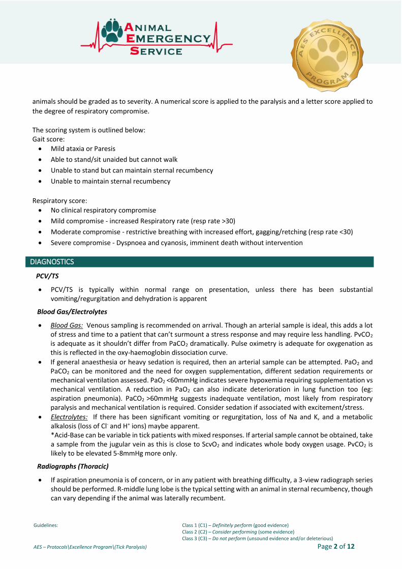
Guidelines: Class 1 (C1) – Definitely perform (good evidence) Class 2 (C2) – Consider performing (some evidence) Class 3 (C3) – Do not perform (unsound evidence and/or deleterious)
AES – Protocols\Excellence Program\(Tick Paralysis) Page 1 of 12
Tick paralysis
HISTORY
If an owner calls for advice after finding or removing any tick from their pet, consultation with a veterinarian should be recommended. Early treatment is thought to be a key element of successful recovery from tick paralysis and marked deterioration can occur in a short space of time. Animals exhibiting any of the clinical signs of tick toxicity (ascending paralysis, respiratory distress/tachypnoea, changes in phonation and regurgitation) require immediate treatment, but the early signs are often subtle and may occur singly (vomiting, mild ataxia, unilateral lameness) and may go unnoticed without a professional assessment. The second reason for veterinary assessment is that a focused examination may enable the veterinarian and or veterinary nurse to discover additional ticks on the patient. Tick preventatives can also be administered. In some cases, a tick may be found by the owner. On review of over 2000 cat cases of tick paralysis found 90% were found forward from the shoulders.
PATHOPHYSIOLOGY
The paralysis tick (Ixodes holocyclus) is a 3-host tick common to coastal southern and central Queensland, as well
as coastal NSW. All female stages are toxic, however, it is usually the adult female that causes most
envenomations. A large infestation with nymphs may also cause clinical toxicity in small animals (anectodal).
The tick injects saliva, which contains various toxins (holocyclotoxins) during feeding. Holocyclotoxins act at the
level of the neuromuscular junction and inhibit acetylcholine release from vesicles on the pre-synaptic membrane
through inhibition of voltage-gate calcium channels and the intracellular movement of extracellular calcium. They
can also cause a reduction in myocardial contractility (systolic dysfunction, related to the toxin’s blocking effect
on potassium channels and corresponding overactivity of calcium channels), leading to pulmonary oedema,
although clinically this situation is not commonly identified. Laryngeal dysfunction and megaoesophagus (leading
to regurgitation) leads to aspiration of pooled saliva and oesophageal secretions (high bacterial load) and
pneumonitis/pneumonia.
Death is through a combination of respiratory failure (primary respiratory muscle fatigue/paralysis, laryngeal
paralysis or secondary pneumonia with sepsis), and occasionally electrical failure of the heart.
At triage, respiratory function should be assessed immediately – those animals with severe respiratory distress
or cyanosis should be taken straight to the treatment room and treated with flyby oxygen. These animals will
generally have severe anxiety and will benefit from sedation as guided by the duty veterinarian. On arrival the
CLINICAL SIGNS
• Weakness (hindlimbs progressing to frontlimbs) *Unilateral lameness can be seen
• Ataxia
• Respiratory difficulties and/or tachypnea
• Changes to phonation
• Vomiting/Regurgitation
DIAGNOSTICS
• PCV/TS • Blood Gas analysis (arterial preferred)
• Radiographs (Thoracic) • Clipping the coat (2nd ticks)
Guidelines: Class 1 (C1) – Definitely perform (good evidence) Class 2 (C2) – Consider performing (some evidence) Class 3 (C3) – Do not perform (unsound evidence and/or deleterious)
AES – Protocols\Excellence Program\(Tick Paralysis) Page 2 of 12
animals should be graded as to severity. A numerical score is applied to the paralysis and a letter score applied to
the degree of respiratory compromise.
The scoring system is outlined below: Gait score:
• Mild ataxia or Paresis
• Able to stand/sit unaided but cannot walk
• Unable to stand but can maintain sternal recumbency
• Unable to maintain sternal recumbency
Respiratory score:
• No clinical respiratory compromise
• Mild compromise - increased Respiratory rate (resp rate >30)
• Moderate compromise - restrictive breathing with increased effort, gagging/retching (resp rate <30)
• Severe compromise - Dyspnoea and cyanosis, imminent death without intervention
DIAGNOSTICS
PCV/TS
• PCV/TS is typically within normal range on presentation, unless there has been substantial vomiting/regurgitation and dehydration is apparent
Blood Gas/Electrolytes
• Blood Gas: Venous sampling is recommended on arrival. Though an arterial sample is ideal, this adds a lot of stress and time to a patient that can’t surmount a stress response and may require less handling. PvCO2 is adequate as it shouldn’t differ from PaCO2 dramatically. Pulse oximetry is adequate for oxygenation as this is reflected in the oxy-haemoglobin dissociation curve.
• If general anaesthesia or heavy sedation is required, then an arterial sample can be attempted. PaO2 and PaCO2 can be monitored and the need for oxygen supplementation, different sedation requirements or mechanical ventilation assessed. PaO2 <60mmHg indicates severe hypoxemia requiring supplementation vs mechanical ventilation. A reduction in PaO2 can also indicate deterioration in lung function too (eg: aspiration pneumonia). PaCO2 >60mmHg suggests inadequate ventilation, most likely from respiratory paralysis and mechanical ventilation is required. Consider sedation if associated with excitement/stress.
• Electrolytes: If there has been significant vomiting or regurgitation, loss of Na and K, and a metabolic alkalosis (loss of Cl- and H+ ions) maybe apparent. *Acid-Base can be variable in tick patients with mixed responses. If arterial sample cannot be obtained, take a sample from the jugular vein as this is close to ScvO2 and indicates whole body oxygen usage. PvCO2 is likely to be elevated 5-8mmHg more only.
Radiographs (Thoracic)
• If aspiration pneumonia is of concern, or in any patient with breathing difficulty, a 3-view radiograph series should be performed. R-middle lung lobe is the typical setting with an animal in sternal recumbency, though can vary depending if the animal was laterally recumbent.
Guidelines: Class 1 (C1) – Definitely perform (good evidence) Class 2 (C2) – Consider performing (some evidence) Class 3 (C3) – Do not perform (unsound evidence and/or deleterious)
AES – Protocols\Excellence Program\(Tick Paralysis) Page 3 of 12
• Look for megaoesophagus on lateral series
TREATMENT
UNSTABLE Patient (Dyspnoeic with respiratory failure, RR <30, cyanotic, PvO2<60mmHg/PvCO2>60mmHg)
• Place IV Catheter if patient permits with minimal stress
• Provide Respiratory Support - If inadequate oxygenation (PvO2<60mmHg and/or SpO2 <93%), supplement patient with flow by
oxygen. If inadequate for patient needs, intubate patient - Intubation: Give 2-4mg/kg alfaxalone IV and intubate patient. Manually ventilate patient with
ambu-bag whilst owner is consulted. Mechanical ventilation is required past this stage.
STABLE Patient (SpO2 >93%, PvO2>60mmHg/PvCO2<60mmHg)
The treatment takes place on several fronts: 1. The toxin itself is treated with an antivenin from the serum of hyperimmune dogs.
2. Respiratory function must be supported through the use of oxygen therapy. Control of anxiety with sedatives or narcotics will aid in ventilation in any anxious animals. Those that are in severe respiratory distress may require tracheal intubation and ventilation.
3. Aspiration pneumonia secondary to megaoesophagus and laryngeal dysfunction must be treated aggressively.
4. Cardiovascular monitoring and support may be required in all animals that are unstable. Load reduction with diuretics may be required if pulmonary oedema or electrical cardiac dysfunction can be documented but this is rarely seen.
5. Monitoring and assistance with urinary function by regular bladder expression or catheterization.
6. Intensive nursing care to stop pressure necrosis, urine scalding, corneal desiccation and to ensure the entire tick burden has been removed.
7. Providing entire body clip to identify a second paralysis tick
Tick Anti-Serum (TAS)
The antibodies in the hyperimmune serum are used to inactivate unbound holocyclotoxin and allow removal by
the immune system. The treatment guidelines (recommended doses) remain unclear, though currently AES
recommends 10mls max of antitoxin up to 10kg, or 1 ml/kg when over 10kg, whichever is higher for canine
patients. It is not unreasonable to administer a maximum of 20ml to patients over 10kg in body weight. The
recommendation for cats is 10mls of anti- serum. These doses were adopted after a research project performed
at the original AEC practice at Bowen Hills. Published retrospective reviews of tick patients have not shown an
association between dose of serum administered and mortality or duration of hospitalisation, so this approach is
open to question. As more research is performed to answer the question surrounding dosage of anti-serum these
recommendations may change.
No patient receiving tick anti-serum from another practice is to receive further doses at AES until approved
with a senior vet. Extra anti-serum may be considered if a patient has significantly deteriorated, an additional tick

Guidelines: Class 1 (C1) – Definitely perform (good evidence) Class 2 (C2) – Consider performing (some evidence) Class 3 (C3) – Do not perform (unsound evidence and/or deleterious)
AES – Protocols\Excellence Program\(Tick Paralysis) Page 4 of 12
is found, and more than 48 hours has passed since administration of the original dose. Follow up tick serum must
be discussed with a senior veterinarian.
TAS Administration:
The anti-serum is always given intravenously as this offers the most rapid and reliable distribution throughout the
body. This can lead to potential reactions in both dogs and cats.
In canine patients, the antitoxin is virtually a preserved form of plasma, and most patients tolerate it extremely
well. The research done by the University of Queensland showed that rapid administration of the pure antiserum,
no matter what premedication was used resulted in hypotension in most of the healthy trial animals. By diluting
the serum with saline, and infusing it over a significant period of time, the incidence of reactions was markedly
reduced and was comparable between premedicated and non-premedicated patients. Routine use of
premedication in dogs is not practised.
There is a higher incidence of anaphylaxis in cats; the pulmonary effects can be devastating and difficult to
treat. It is now not recommended to pre-medicate cats prior to TAS administration because no pre-medication
has been shonw to reduce the incidence or lessen the severity of feline anaphylaxis. Corticosteroids can take a
number of hours to have an effect and can be deleterious in the event of a hypotensive anaphylactic event.
Adrenaline should be drawn up and ready to be given IV in the event of an anaphylactic event though.
• Adrenaline 0.01mg/kg IV. Never administer adrenaline subcutaneously
• 0.5ml of a 1:10000 adrenaline solution given IV
**NB: Prednisolone sodium succinate is no longer available within Australia. Due to the long period of
time for Dexamethasone to reach an active state, it is not recommended.
Very close monitoring, in combination with slow IV administration, is required in all cats, particularly those
previously treated for tick paralysis. Where a 2nd TAS administration is required in cats, a discussion with the senior
vet must be had prior to administration as it may not be given. If required, cats receiving a 2nd transfusion must
be monitored throughout the entire transfusion. Some clinics have advocated the use of intraperitoneal
administration in cats to avoid these reactions, however, due to the uncertain nature of antitoxin delivery with
this route, and the inability to remove the serum if a reaction dose occur, AES avoids this approach.
Monitor for adverse reactions:
HYPERSENSITIVITY REACTIONS
The most common reaction observed in dogs consists of bradycardia, pale mucous membranes, +/- hypotension.
This is thought to be the Bezold-Jarisch (BJ) reflex. The BJ reflex is due to severe reductions in cardiac filling. The
bradycardia is a compensatory mechanism to improve cardiac filling.
Treatment of this condition includes:
• IV fluids (isotonic crystalloids) is considered the most appropriate treatment to improve cardiac filling,
administered at 10-15ml/kg to a dog, and 5-10ml/kg to a cat.
• Flow by oxygen therapy
• Atropine 0.02-0.05mg/kg IM or IV

Guidelines: Class 1 (C1) – Definitely perform (good evidence) Class 2 (C2) – Consider performing (some evidence) Class 3 (C3) – Do not perform (unsound evidence and/or deleterious)
AES – Protocols\Excellence Program\(Tick Paralysis) Page 5 of 12
Dermatological manifestations of hypersensitivity (hives, angioedema) should increase the suspicion for
anaphylaxis. This can be treated by stopping the infusion, and giving chlorpheniramine 0.5mg/kg IM. The TAS likely
needs to be diluted further and given over a longer time, but the patient should be monitored closely for any signs
of developing systemic anaphylaxis.
SYSTEMIC ANAPHYLACTIC REACTIONS
Systemic anaphylactic reactions may be seen in either species but are more common in cats than dogs, clinical
signs can include:
CAT: Tachycardia or Bradycardia, dyspnoea, vomiting, and hypotension
DOG: Dogs (rare): Tachycardia, pale mucous membranes, poor pulse quality, vomiting/diarrhoea, and
sometimes hypotension
Treatment of systemic anaphylaxis includes:
• Immediate suspension of TAS administration
• Flow by oxygen therapy
• Adrenaline 0.01mg/kg IV (1ml/10kg IV of a 1:10000 solution)
• Crystalloid boluses given rapidly IV at 20ml/kg for dogs and 10ml/kg for cats, this can be titrated to effect
ie. reduction in heart rate and improvement in blood pressure
• Monitoring of blood pressure, heart rate, capillary refill time, pulse oximetry
• CRI of adrenaline (0.05ug/kg/min and titrated to effect) is recommended with ongoing fluid support as
necessary to maintain perfusion
Respiratory Support +/- Mechanical Ventilation
Respiratory failure in tick paralysis is a complex problem – it is related to:
• Increased oxygen consumption associated with increased respiratory effort and stress
• Fatigue/paralysis of thoracic respiratory muscles leading to reduced ventilation
• Laryngeal dysfunction leading to upper airway obstruction
• Pulmonary compromise resulting from aspiration and/or cardiac dysfunction
Multiple therapies need to be considered to effectively manage this, especially in a severe case. Sedation or
anaesthesia is a mainstay of treatment, second only to the antitoxin and almost always indicated before antitoxin
administration. Anxiety or stress results in increased oxygen consumption (by 50-100%), predisposes to
respiratory muscle fatigue and also aspiration. Aggressive and early use of sedation can be lifesaving.
SEDATION
Choices of which sedative to use include (bolded items are drugs most commonly used at AES):
• Acepromazine 0.01 – 0.05 mg/kg IV or SC (maximum of 2 mg total dose, take into account patient’s age
and health status, administer slowly IV if possible)
• Butorphanol 0.2 – 0.6 mg/kg IV or SC, CRI of 0.1 mg/kg/hr (put 10-20 mg in a 1 L fluid bag and run at
maintenance (0.1-0.3mg/kg/hr)
• Methadone 0.1 – 0.3 mg/kg IV or SQ
• Buprenorphine 10-30ug/kg IV or SQ, mainly for cats, use with ACP or diazepam, takes about 30 minutes to
take effect.

Guidelines: Class 1 (C1) – Definitely perform (good evidence) Class 2 (C2) – Consider performing (some evidence) Class 3 (C3) – Do not perform (unsound evidence and/or deleterious)
AES – Protocols\Excellence Program\(Tick Paralysis) Page 6 of 12
• Diazepam 0.1-0.2 mg/kg IV or IM, use with an opioid
• Midazolam 0.1-0.3 mg/kg/hr, use with a butorphanol CRI, can result in profound sedation/stage 1
anaesthesia.
ANAESTHESIA
Anaesthetic options for patients requiring mechanical ventilation include:
• Propofol 0.05 – 0.6 mg/kg/min with midazolam +/- butorphanol. Start at about 0.1 mg/kg, often when
used in combination with an opioid and benzodiazepine only small amounts are needed making it
moderately cost effective. Watch for the cumulative effect in cats, and high doses will cause significant
lipaemia. For this reason, alfaxalone is the preferred drug in cats.
• Thiopentone – loading dose of 4-10mg/kg then 1-4 mg/kg/hr. It is cumulative so close monitoring and
dose reduction is required
• Pentobarbitone – 2-6 mg/kg/hr, cumulative so close monitoring and dose reduction required. If
administered for longer then 3 days may seizure on wakeup to minimize this transfer to propofol 24-36
hours before wakeup.
• Alfaxan 1-2mg/kg/hr (undocumented dose). Very expensive and appears to cause respiratory depression
comparable to propofol, though spares cardiovascular function.
• Fentanyl 4-10ug/kg/hr when used in combination with midazolam. Cardiovascularly stable and quick to
wind down/up but expensive.
Should the initial dose of sedation not resolve the respiratory signs then oxygen therapy is indicated. Delivery via
the intranasal route is tolerated by most patients however small dogs and cats may respond better to an oxygen
cage. Trans-tracheal oxygenation used is also an option, although it is not commonly used. When hypoxaemia or
excessive work of breathing does not resolve through oxygen therapy alone, anaesthesia, intubation, and tracheal
oxygen therapy is administered. This step reduces oxygen consumption, protects the airway and provides efficient
oxygen supplementation (50 ml/kg/min titrated to effect).
Mechanical Ventilation:
Animals that are unable to maintain a respiratory rate greater than 10-12 breaths per minute, or any that cannot
maintain oxygen saturation at an adequate level with sedation and oxygen therapy will require anaesthetisation
with intubation +/- ventilation. The ability to use blood gas, pulse oximetry and capnography allow close
monitoring of respiratory function. Arterial oxygen partial pressure (PaO2) of 60 mmHg is the minimum acceptable
in animals on oxygen supplementation before intubation +/-ventilation is required (this equates to an SPO2 of
90%).
Hypercapnia is better tolerated than hypoxia; ideally ventilation should be commenced when the PaCO2 is
over 60 mmHg (this is similar to the end tidal CO2 level on the capnograph) however if this is not a financial option,
remember some patients may survive hypercapnia with a PaCO2 up to 100-110mmHg. Owners should be offered
ventilation when appropriate and a treatment waiver signed if this is not elected. Mechanical ventilation is usually
successful in animals with poor respiratory function, but minimal pulmonary pathology, as is seen in the earlier
phases of respiratory failure in patients with tick paralysis. Animals that show a marked deterioration in
respiratory status, which is not explained by respiratory muscle fatigue or paralysis, or have detectable increased
bronchovesicular sounds and crackles on auscultation require chest radiographs to document the pathological

Guidelines: Class 1 (C1) – Definitely perform (good evidence) Class 2 (C2) – Consider performing (some evidence) Class 3 (C3) – Do not perform (unsound evidence and/or deleterious)
AES – Protocols\Excellence Program\(Tick Paralysis) Page 7 of 12
process and treat appropriately. A patient with a perihilar interstitial to alveolar pattern may benefit from load
reduction using a diuretic (furosemide 1-2mg/kg IV), as well as anti-arrhythmic or positive inotropes. Constant
ECG monitoring should be considered in these cases as sudden catastrophic arrhythmia’s are a possibility. A lobar,
alveolar pattern on the dependent side of the lungs would be more consistent with aspiration pneumonia, and
therefore antibiotic administration is indicated. Broad-spectrum antibiotic combinations eg: first-generation
cephalosporin/penicillin or ampicillin in combination with a fluroquinolone are indicated. As well as antibiotics,
intravenous fluids are required to help moisten airway secretions to allow removal from the lungs. This can be
assisted with nebulisation and coupage, as long as it does not stress the patient. It is likely that recommendations
for antibiotic administration and collection of fluid samples for culture will change as we develop these protocols.
Supportive Care and Management
• Crystalloid therapy
o Hartmanns or Plasmalyte 148 administered at maintenance rates, though each individual animal will
need to be assessed (eg: dehydration) (C1)
o Supplements added to fluids - KCl maybe added due to GIT losses from vomiting/regurgitation
The use of intravenous fluids in dogs with tick paralysis has been associated with reduced duration of hospitalisation. There has been a historical reluctance to use intravenous fluid therapy because of a perceived risk of pulmonary oedema due to the cardiac aspects of tick toxicity. Intravenous fluids should be utilised in the setting of an objective fluid plan individually tailored to each patient. All patients will be nil per os for at least 24 hours, so fluid therapy is indicated from admission to maintain hydration and account for ongoing losses. All routine monitoring and precautions for intravenous fluid use should be observed. Particular care should be taken to avoid over-hydration in cats. This species seems most susceptible to pulmonary complications associated with fluid overload.
• GIT protectants
o Esomeprazole 0.5-1mg/kg/hr IV q12hrs (C2)
The use of gastric protectants to reduce gastric acidity is controversial in human and veterinary medicine due to
an association with nosocomial infections particularly pneumonia. On the other hand oesophageal strictures have
been reported anecdotally (and seen at AES/VSS) after tick paralysis presumably due to reflux. These medications
may be administered at the clinician’s discretion and a reason for use recorded in the clinical notes.
Esomeprazole is a proton pump inhibitor and is more effective than ranitidine for increasing gastric pH. Full
action may be delayed, and there is no pro-kinetic effect, so the general recommendation is that if anti-ulcer
medications are to be used they should both be given for the first 48 hours then esomeprazole continued as a
sole medication if needed.
These medications do little to prevent pooling of oropharyngeal secretions in the oesophagus and their
periodic emptying, seen most commonly as regurgitation – instead they reduce the chances of acidic stomach
contents entering the oesophagus, which are potentially more damaging to both the oesophagus and the
pulmonary tissue. Oesophageal/oral suctioning may help remove these secretions, however this can be painful
and requires heavy sedation if it is not to be detrimental to the patient. Patient positioning can also help prevent
aspiration of regurgitated material by keeping them in sternal recumbency with the trachea angled ventrally.
Some patients may also tolerate placement of a naso-oesophageal feeding tube for regular decompression. NG

Guidelines: Class 1 (C1) – Definitely perform (good evidence) Class 2 (C2) – Consider performing (some evidence) Class 3 (C3) – Do not perform (unsound evidence and/or deleterious)
AES – Protocols\Excellence Program\(Tick Paralysis) Page 8 of 12
tube placement is ideally avoided, to prevent reflux of stomach acid into the oesophagus. Use is limited by patient
size as the tube needs to be greater than 6Fr diameter to allow effective removal of thick secretions.
• Anti-emetics
o Metoclopramide 0.5mg/kg IV, then CRI at 2mg/kg/day (C2)
o Maropitant 1mg/kg SC q24hrs (C2)
o Ondansetron 0.1-0.4mg/kg IV q8hrs (C2)
The rationale for anti-emetic use is to reduce the incidence of vomiting in order to (hopefully) reduce the incidence of aspiration pneumonia. Patient’s presenting with a history of vomiting or regurgitation should be started on an antiemetic. Metoclopramide may be a useful antiemetic because it increases gastric emptying and increases lower oesophageal sphincter tone, preventing reflux as well as acting as a central emetic.
• Antibiotics (given in response to aspiration pneumonia)
o Ampicillin 22mg/kg IV q8hrs (C1) or Cephazolin 22mg/kg IV q8hrs (C1)
o Enrofloxacin 5mg/kg/hr SC q12hrs (C1)
Table 1: A comprehensive list of medications involved is included below.
DRUG DOSE ROUTE/RATE
Sedatives
Acepromazine 0.01-0.05 mg/kg (dogs - max of 2 mg per dose)
Up to 0.1 mg/kg (cats)
IV/SC (lower end of spectrum for IV dosing)
Methadone 0.1-0.3 mg/kg IV/SC
Butorphanol 0.1-0.6 mg/kg
0.1 mg/kg/hr
IV
IV CRI
Buprenorphine 10-30 ug/kg (cats) IV/SC
Diazepam 0.1-0.2 mg/kg IV/IM (with opioid)
Midazolam 0.1-0.3 mg/kg/hr IV CRI
Anaphylactic reactions
Adrenalin 0.01 - 0.1 mg/kg IM/IV
Tick Serum (dilute 50:50 with saline)
Dogs 10ml min or 1ml/kg – whichever is greater
Slow IV (1hr)
Cats 10ml max Slow IV (1hr)
Anaesthetics (used in combination with sedatives such as midazolam +/- butorphanol)

Guidelines: Class 1 (C1) – Definitely perform (good evidence) Class 2 (C2) – Consider performing (some evidence) Class 3 (C3) – Do not perform (unsound evidence and/or deleterious)
AES – Protocols\Excellence Program\(Tick Paralysis) Page 9 of 12
Propofol 0.05-0.6 mg/kg/min IV CRI
Alfaxalone 1-2mg/kg/hr IV CRI
Pentobarbitone 2-6 mg/kg/hr IV CRI
Fentanyl 4-10 ug/kg/hr IV CRI
Antiemetics/Protectants
Metoclopramide 0.5 mg/kg
1-2 mg/kg/day
IV/SC q 6 hrs
IV CRI (approx 10mg in 1L IV fluids @ maintenance)
Esomeprazole 0.7-1.0mg/kg IV q24hrs
Entire body clipping
This is recommended highly in all cases of tick paralysis. In a recent study, 9% of cats with tick paralysis had a second tick identified. To prevent the continuing exposure to tick venom and ensuing paralysis requires removal of all ticks. This can be performed under sedation (mainly for dogs), and general anaesthesia (for all cats) to reduce stress and control respiration.
Nursing Care
The majority of tick paralysis patients are unable to self-position, urinate or even blink, so intensive nursing care
is critical to reduce complications such as bladder atony or corneal ulceration, as well as improving patient comfort
and recovery in hospital. Soft, dry bedding and regular turning is important to prevent pressure sores. Regular
bladder checks are indicated, with expression, or placement of an indwelling urinary catheter, to prevent detrusor
muscle atony, overfill incontinence or urinary tract infections. Regular artificial tear administration is required,
especially if the tick was attached around the eye or CN5. The artificial tear product should be preservative-free
as preservatives in these preparations have been shown to induce corneal ulceration in humans (e.g Celluvisc).
Corneal ulceration secondary to desiccation needs to be identified quickly and can be treated with antibiotics and
a temporary tarsorrhaphy (either non-surgical or surgical). All patients admitted with tick paralysis should receive
a tick clip and tick bath as well as repeated tick searches at every check as it is not uncommon to find more than
one tick.
AES nurses perform full TPR’s on tick patients every 2-4 hours. This includes SpO2 monitoring, bladder
palpation and expression, application of artificial tears, oral moistening and patient turning. If the patient is
receiving oxygen therapy or there are concerns about ventilation then oxygen saturations are measured every 2
hours and blood gas parameters are measured at the vet’s discretion. If the patient has palpebral paralysis then
artificial tears are applied every 2 hours and a temporary tarsorrhaphy is considered. AES now has access to
contact lenses and has had success with preventing corneal ulceration for anaesthetised patients. AES uses
absorbent disposable bedding to reduce the amount of washing of sheets/towels, but once a patient urinates or
defecates on its bedding it is changed immediately to reduce urine/faecal scalding. The nursing and care of a
patient being ventilated is beyond the scope of this protocol. Patients that are regurgitating and retching also
require more intensive nursing care as aspiration is a high risk especially in patients that are severely affected.

Guidelines: Class 1 (C1) – Definitely perform (good evidence) Class 2 (C2) – Consider performing (some evidence) Class 3 (C3) – Do not perform (unsound evidence and/or deleterious)
AES – Protocols\Excellence Program\(Tick Paralysis) Page 10 of 12
Immediate suctioning of the oral and pharyngeal areas after a regurgitation event is of extreme importance to
reduce the risk aspiration.
Animals can be discharged when they are no longer ataxic and when eating and drinking can be accomplished
without gagging, and voluntary urination documented. Water and food trials are usually implemented when the
patient is walking well, has stopped regurgitating and has a palpable swallow or gag reflex. Monitoring during the
trial is imperative and if there is any evidence of laryngeal dysfunction (as evidenced by a cough or retch) then a
repeat trial is performed in 6-12 hours time.
FURTHER TREATMENT & MONITORING
Post discharge instructions need to include restricting activity, keeping the patient cool and supervision of eating
and drinking until complete normality returns and monitoring of urination patterns.
COSTS AND HOSPITALISATION
• Hospitalisation time to expect: 2-7 days
• Costs whilst hospitalized: $1500-$10000
PROGNOSIS AND RISK FACTORS
• 2% mortality in cats receiving TAS and followed the veterinarian’s instructions
• 8% mortality in cats that do not receive TAS
• 9% of cats receiving TAS have a reaction, of which 6% of these will die. Compare to 1% mortality with cats without a TAS reaction
• 88% mortality of cases where ventilation is recommended, but not instituted. Compared to 18% mortality when recommended and instituted
• 4% survival for cats requiring CPR for respiratory arrest
• Overall mortality for cats is 3%
• Increased risk of death for cats - high gait score (4) - high respiratory score (D) - TAS reactions - Mechanical ventilation - Hypothermia (<35oC)
• Reduced risk of death for cats - Clipping entire body for paralysis ticks
- Receiving TAS
- Implementing ventilation when advised

Guidelines: Class 1 (C1) – Definitely perform (good evidence) Class 2 (C2) – Consider performing (some evidence) Class 3 (C3) – Do not perform (unsound evidence and/or deleterious)
AES – Protocols\Excellence Program\(Tick Paralysis) Page 11 of 12
REFERENCES
1. Atwell RB, Campbell FE, Evans EA. (2001) Prospective survey of tick paralysis in dogs. AVJ. 79: 412-418.
2. Westwood MN, Emery DL, Dhand NK. Clinical presentation and treatment of tick paralysis in dogs and
cats in Sydney (2001-2010). Australian veterinary journal 2013;91:491-498.
3. Schull D. Acute side effects attributed to the use of tick antitoxin serum: A review of availabl
descriptions. AUSTRALIAN VETERINARY PRACTITIONER 2007;37:98-+.
4. Shmuel DL, Cortes Y. Anaphylaxis in dogs and cats. Journal of veterinary emergency and critical care
(San Antonio, Tex : 2000) 2013;23:377-394.
5. Kinsella SM, Tuckey JP. (2001) Perioperative bradycardia and asystole: relationship to vasovagal
syncope and the Bezold-Jarisch reflex. Bri J Ana. 86: 859-868.
6. Keir I. (2014) Concerns over treatment of anaphylaxis. Journal of veterinary emergency and critical
care (San Antonio, Tex : 2000) 24: 134-discussion 134.
7. Webster R, Haskins S, Mackay B. (2013) Management of respiratory failure from tick paralysis. AVJ. 91:
499-504.
8. Ruple-Czerniak A, Aceto HW, Bender JB, Paradis MR, Shaw SP. (2013) Using syndromic surveillance to
estimate baseline rates for healthcare-associated infections in critical care units of small animal
referral hospitals. JVIM. 27: 1392-1399.
9. Chand KK, Lee K-M, Lavidis NA, Rodriguez M, Ijaz H, Koehbach J, Clark RJ, Lew-Tabor A, Noakes PG.
(2016) Tick holocyclotoxins trigger host paralysis by presynaptic inhibition. Nature, Scientific Reports.
6: 29446; doi: 10.1038/srep29446
10. Leister E, Morton, J, Atwell R, Webster R. (2018) Clinical presentations, treatments, and risk factors for
mortality in cats with tick paralysis caused by Ixodes holocyclus: 2077 cases (2008-2016). JFMS 20(6)
465-478.
*Use of any of this material is not for retail or public disclosure. It is intended for personal use and knowledge only

Guidelines: Class 1 (C1) – Definitely perform (good evidence) Class 2 (C2) – Consider performing (some evidence) Class 3 (C3) – Do not perform (unsound evidence and/or deleterious)
AES – Protocols\Excellence Program\(Tick Paralysis) Page 12 of 12
Author(s): Dr Caitlin Logan & Dr Courtney Reddrop Reviewed: Dr Gerardo Poli & Dr Rob Webster Date: 9th October 2017