Embed Size (px)
Citation preview
2975Research Article
IntroductionThyroid hormone (TH or T3) regulates many processes in thedevelopment of mammals before the onset of function in avariety of organ systems. This pleiotropic nature of TH ismediated through interactions with ligand-modulated nuclearreceptors encoded by two genes TR� and TR� (Wu andKoenig, 2000) that can either positively or negatively regulatetarget genes in response to T3 (Lazar, 2003). To date it isunknown whether T3 target genes can differ in their sensitivityto distinct thyroid hormone receptor (TR) isoforms or to TRshaving the ligand bound (holoreceptors) or not bound(aporeceptors). This is a crucial question considering thepathologies associated with hypothyroidism or TR mutations(O’Shea and Williams, 2002). The presumptive redundanteffects of coexpressed TRs could definitively mask theindividual TR responsiveness of genes and dramatically definethe pathological phenotype. To unravel the question of TRspecificity at the single-cell level, we focused on two cochlearouter hair cell (OHC) genes that are concomitantly expressedduring a crucial time period during which hypothyroidismleads to irreversible hearing deficits in adults (Deol, 1973;Uziel et al., 1985). Neurosensory deafness in humans androdents is presumed to be related to mutations in the TR�,leading to resistance to thyroid hormone (RTH), caused by a
transdominant negative transcriptional effect (Refetoff et al.,1993; Weiss and Refetoff, 2000). A role of TR�1 for auditoryfunction was suggested in a study of TR�2-deficient miceexhibiting increased expression of TR�1, which was proposedto compensate for missing TR� activity (Ng et al., 2001).However, no T3 target genes have been identified in the cochleaat the level of TR-isoform-specific transcriptional regulation.
During a developmental period before the onset of hearing,the gene that encodes the outer hair cell motor protein prestin,Slc26a5 (Zheng et al., 2000), is expressed from approximatelypostnatal day (P) 3 in euthyroid rats, whereas its expression isdelayed under conditions of hypothyroidism (Weber et al.,2002). The voltage-dependent K+ channel KCNQ4, which isresponsible for the predominant K+ conductance, IK,n, ofmature OHCs (Marcotti and Kros, 1999) is expressed inrodents from approximately P6 onwards (present study)(Kharkovets et al., 2000). In order to study the specific effectof TH on the expression of these genes, a variety of animalmodels of hypothyroidism and several TR mutant mice wereused to examine their TH dependency. These included (1)animals with goitrogen-induced hypothyroidism; (2) animalsthat acquired hypothyroidism as a consequence of a naturallyoccurring point mutation in the thyrotropin receptor (Tshrhyt
mutants) (for a review, see Walsh and McGee, 2001); (3)
Thyroid hormone (TH or T3) and TH-receptor �� (TR��)have been reported to be relevant for cochlear developmentand hearing function. Mutations in the TR�� gene result indeafness associated with resistance to TH syndrome. Theeffect of TR��1 on neither hearing function nor cochlear T3target genes has been described to date. It is also uncertainwhether TR��1 and TR�� can act simultaneously ondifferent target genes within a single cell. We focused ontwo concomitantly expressed outer hair cell genes, thepotassium channel Kcnq4 and the motor protein prestinSlc26a5. In outer hair cells, TH enhanced the expression ofthe prestin gene through TR��. Simultaneously Kcnq4expression was activated in the same cells by derepression
of TR��1 aporeceptors mediated by an identified TH-response element, which modulates KCNQ4 promoteractivity. We show that T3 target genes can differ in theirsensitivity to TH receptors having the ligand either bound(holoreceptors) or not bound (aporeceptors) within singlecells, and suggest a role for TR��1 in final celldifferentiation.
Supplementary material available online athttp://jcs.biologists.org/cgi/content/full/119/14/2975/DC1
Key words: KCNQ4, Prestin, Final differentiation, TR aporeceptors
Summary
Thyroid hormone receptors TR��1 and TR��differentially regulate gene expression of Kcnq4 andprestin during final differentiation of outer hair cellsHarald Winter1, Claudia Braig1, Ulrike Zimmermann1, Hyun-Soon Geisler1, Jürgen-Theodor Fränzer1,Thomas Weber1, Matthias Ley1, Jutta Engel2, Martina Knirsch2, Karl Bauer3, Stephanie Christ3,Edward J. Walsh4, JoAnn McGee4, Iris Köpschall1, Karin Rohbock1 and Marlies Knipper1,*1University of Tübingen, Department of Otolaryngology, Tübingen Hearing Research Centre (THRC), Laboratory of Molecular Neurobiology,Elfriede-Aulhorn-Str. 5, 72076 Tübingen, Germany2University of Tübingen, Institute of Physiology II and Department of Otolaryngology, THRC, Gmelinstr. 5, 72076 Tübingen, Germany3Max-Planck-Institute for Experimental Endocrinology, Feodor-Lynen-Str. 7, 30625 Hannover, Germany4Developmental Auditory Physiology Laboratory, Boys Town National Research Hospital, 555 North 30th Street, Omaha, NE 68131, USA*Author for correspondence (e-mail: [email protected])
Accepted 6 April 2006Journal of Cell Science 119, 2975-2984 Published by The Company of Biologists 2006doi:10.1242/jcs.03013
Jour
nal o
f Cel
l Sci
ence

2976
athyroid, Pax8–/– mutants (Christ et al., 2004; Macchia, 2000;Mansouri et al., 1998; Tell et al., 1999); (4) TR�1–/– (Rusch etal., 1998; Wikstrom et al., 1998), TR�–/– (Forrest et al., 1996a;Forrest et al., 1996b; Gauthier et al., 1999), TR�1–/–/�–/–
(Gothe et al., 1999; Rusch et al., 2001) mutants; and (5)euthyroid TR�1m/+ mice carrying a dominant-negative TR�1R384C point mutation (Tinnikov et al., 2002). The differentmutant strains used in this study are listed and summarized inTable 1.
Both the prestin gene (Weber et al., 2002) and Kcnq4(present study) were identified as simultaneously expressedT3-dependent genes that are, however, differentially regulatedby either TR� or TR�1. A role for TR�1 in inner-eardevelopment and specifically final differentiation of OHCs wasdemonstrated. Furthermore, the data show for the first time thatapo-TR�1 can repress a T3 target gene independently but inparallel to TR� acting on a different T3 target genewithin the same cell.
ResultsKCNQ4 and prestin protein expression duringearly stages of auditory functionBefore the onset of hearing, the subcellularlocalization of the two outer hair cell proteinsKCNQ4 and prestin overlapped between P4 and P7,occupying the entire basolateral membrane area ofthe OHCs (Fig. 1A, P8). Between P8 and P12,however, the distribution of KCNQ4 gradually shiftedto the basal pole of the OHC membrane, whereasprestin shifted to the lateral membrane (Fig. 1A, P12),resulting in a completely separated but neighboringdistribution from P12 onwards (Fig. 1B).
KCNQ4 and prestin protein expression aredifferentially affected by hypothyroidismIn the absence of thyroid hormone, the expression ofprestin was reduced and the distribution pattern
Journal of Cell Science 119 (14)
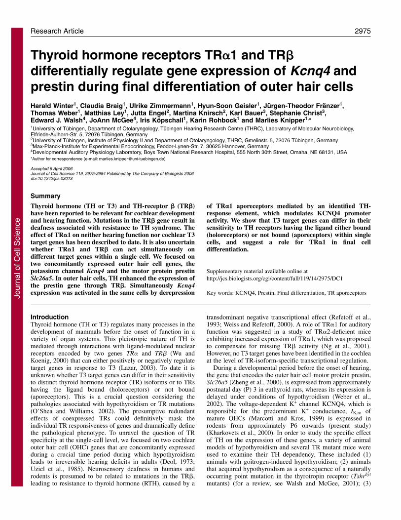
remained immature (Fig. 2A, hypo, red) (Weber et al., 2002).By contrast, KCNQ4 expression was completely lacking in theOHCs of hypothyroid rats (Fig. 2A, hypo, green). Usingnorthern blot analysis both the ~3.9 kb and the ~3.8 kb Kcnq4transcripts were detected (Fig. 2B, con as) (Kubisch et al.,1999), which were significantly reduced in the cochleae ofhypothyroid animals (Fig. 2B, hypo, as). The correspondingsense probes showed no signal (Fig. 2B, con, sense). ReducedKcnq4 mRNA levels were also noted in the cochleae of P12hypothyroid rats (Fig. 2C, hypo) compared with controlanimals (Fig. 2C, con) using semi-quantitative RT-PCR. Thehousekeeping gene cyclophilin was used as an internal control.In addition, Kcnq4 mRNA was detected by in situhybridization in both outer and inner hair cells of controlanimals (Fig. 2D, control) (see also Oliver et al., 2003) and wasabsent in age-matched hypothyroid rats (Fig. 2D, hypo).
Table 1. Mouse mutants usedStrain Molecular characterization Pathophysiology Reference
Pax8–/– Targeted inactivation of the Pax8 gene Athyroidism Christ et al., 2004Absence of thyroid follicular cells Mansouri et al., 1998Deafness, degeneration of outer hair cells
Tshrhyt/hyt Naturally inherited, autosomal recessive P556L point Primary, congenital hypothyroidism Beamer et al., 1981mutation in the TshR Hypoplasia, retarded growth, infertility Sprenkle et al., 2001a
Deafness Sprenkle et al., 2001bSprenkle et al., 2001cStein et al., 1994
TR�1–/– Replacement of TR�1-specific coding region with Abnormal heart rate Wikstrom et al., 1998that of TR�2 Reduced body temperature
TR�–/– Targeted inactivation of the TR� gene Recessive resistance to thyroid hormone Forrest et al., 1996bHyperthyroidismDeafness
TR�1–/–�–/– Compound mutant mice, generated by crossing TR�1 Hyperthyroidism Gothe et al., 1999and TR� mice Retarded growth Rusch et al., 1998
Retarded bone maturationFemale infertilityDeafness
TR�1m/+ Dominant-negative R384C mutation introduced in Tenfold reduced ligand binding Tinnikov et al., 2002TR�1 Retarded postnatal development and growth
Cardiac function abnormalities
Fig. 1. Coincident redistribution of KCNQ4 and prestin in rat OHCs.(A) Immunohistochemistry shows KCNQ4 (green) and prestin (red) in OHCsbefore (P8) and at onset of hearing (P12). (B) Double immunohistochemistryof prestin (red) and KCNQ4 (green) in a mature (P21) OHC. Arrows, KCNQ4and prestin protein; arrowheads, basal pole of OHCs. Bars, 20 �m.
Jour
nal o
f Cel
l Sci
ence
2977TH-dependent Kcnq4 and prestin expression in OHCs
In two commonly used mouse models of hypothyroidism,i.e. Tshrhyt mutants (Hyt/Hyt) which have a point mutation inthe thyrotropin receptor, and Pax8–/– mutants which lackthyroid follicular cells, similar KCNQ4 and prestin expressionpatterns were observed (data not shown). This verifies theuniformity of TH influence, independent of the mode ofinduction of hypothyroidism.
KCNQ4 but not prestin protein expression differsdepending on the absence of thyroid hormone or itsreceptorsKCNQ4 and prestin protein expression profiles in the absenceof TH were compared with those in TR�1–/–/�–/– mutantmice, which carry deletions of both TR� and TR�.Immunohistochemical analyses were carried out withantibodies against KCNQ4 or prestin, along with the efferent-
specific marker synaptophysin (Gil-Loyzaga and Pujol, 1988;Knipper et al., 1995), shown for P10 animals in Fig. 3.
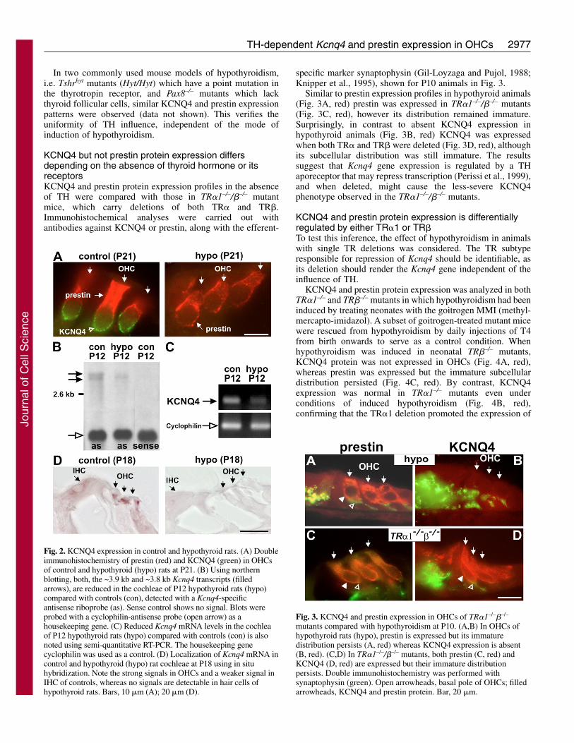
Similar to prestin expression profiles in hypothyroid animals(Fig. 3A, red) prestin was expressed in TR�1–/–/�–/– mutants(Fig. 3C, red), however its distribution remained immature.Surprisingly, in contrast to absent KCNQ4 expression inhypothyroid animals (Fig. 3B, red) KCNQ4 was expressedwhen both TR� and TR� were deleted (Fig. 3D, red), althoughits subcellular distribution was still immature. The resultssuggest that Kcnq4 gene expression is regulated by a THaporeceptor that may repress transcription (Perissi et al., 1999),and when deleted, might cause the less-severe KCNQ4phenotype observed in the TR�1–/–/�–/– mutants.
KCNQ4 and prestin protein expression is differentiallyregulated by either TR�1 or TR�To test this inference, the effect of hypothyroidism in animalswith single TR deletions was considered. The TR subtyperesponsible for repression of Kcnq4 should be identifiable, asits deletion should render the Kcnq4 gene independent of theinfluence of TH.
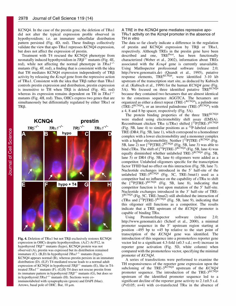
KCNQ4 and prestin protein expression was analyzed in bothTR�1–/– and TR�–/– mutants in which hypothyroidism had beeninduced by treating neonates with the goitrogen MMI (methyl-mercapto-imidazol). A subset of goitrogen-treated mutant micewere rescued from hypothyroidism by daily injections of T4from birth onwards to serve as a control condition. Whenhypothyroidism was induced in neonatal TR�–/– mutants,KCNQ4 protein was not expressed in OHCs (Fig. 4A, red),whereas prestin was expressed but the immature subcellulardistribution persisted (Fig. 4C, red). By contrast, KCNQ4expression was normal in TR�1–/– mutants even underconditions of induced hypothyroidism (Fig. 4B, red),confirming that the TR�1 deletion promoted the expression of
Fig. 2. KCNQ4 expression in control and hypothyroid rats. (A) Doubleimmunohistochemistry of prestin (red) and KCNQ4 (green) in OHCsof control and hypothyroid (hypo) rats at P21. (B) Using northernblotting, both, the ~3.9 kb and ~3.8 kb Kcnq4 transcripts (filledarrows), are reduced in the cochleae of P12 hypothyroid rats (hypo)compared with controls (con), detected with a Kcnq4-specificantisense riboprobe (as). Sense control shows no signal. Blots wereprobed with a cyclophilin-antisense probe (open arrow) as ahousekeeping gene. (C) Reduced Kcnq4 mRNA levels in the cochleaof P12 hypothyroid rats (hypo) compared with controls (con) is alsonoted using semi-quantitative RT-PCR. The housekeeping genecyclophilin was used as a control. (D) Localization of Kcnq4 mRNA incontrol and hypothyroid (hypo) rat cochleae at P18 using in situhybridization. Note the strong signals in OHCs and a weaker signal inIHC of controls, whereas no signals are detectable in hair cells ofhypothyroid rats. Bars, 10 �m (A); 20 �m (D).
Fig. 3. KCNQ4 and prestin expression in OHCs of TR�1–/–�–/–
mutants compared with hypothyroidism at P10. (A,B) In OHCs ofhypothyroid rats (hypo), prestin is expressed but its immaturedistribution persists (A, red) whereas KCNQ4 expression is absent(B, red). (C,D) In TR�1–/–/�–/– mutants, both prestin (C, red) andKCNQ4 (D, red) are expressed but their immature distributionpersists. Double immunohistochemistry was performed withsynaptophysin (green). Open arrowheads, basal pole of OHCs; filledarrowheads, KCNQ4 and prestin protein. Bar, 20 �m.
Jour
nal o
f Cel
l Sci
ence
2978
KCNQ4. In the case of the prestin gene, the deletion of TR�1did not alter the typical expression profile observed inhypothyroidism; i.e. an immature subcellular distributionpattern persisted (Fig. 4D, red). These findings collectivelyvalidate the view that apo-TR�1 represses KCNQ4 expression,but does not affect the expression of prestin.
Treatment with T4 rescued the KCNQ4 phenotype fromneonatally induced hypothyroidism in TR�–/– mutants (Fig. 4E,red), while not affecting the normal phenotype in TR�1–/–
mutants (Fig. 4F, red), a finding that is consistent with the ideathat TH mediates KCNQ4 expression independently of TR�activity by releasing the Kcnq4 gene from the repressive actionof TR�1. Consistent with the idea that TR� rather than TR�1controls prestin expression and distribution, prestin expressionis insensitive to TH when TR� is deleted (Fig. 4G, red)whereas its expression remains dependent on TH in TR�1–/–
mutants (Fig. 4H, red). Thus, OHCs express two genes that aresimultaneously but differentially regulated by either TR�1 orTR�.
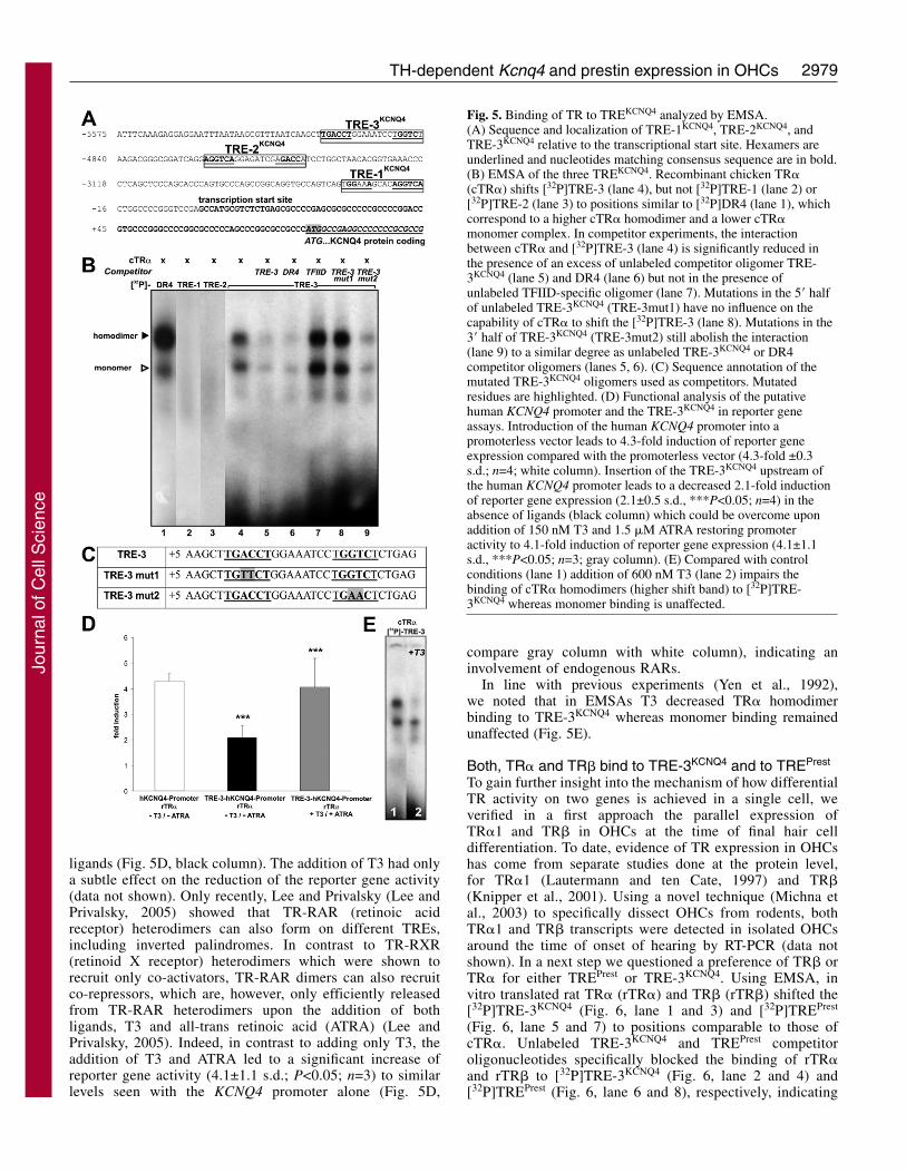
A TRE in the KCNQ4 gene mediates repressive apo-TR�1 activity on the Kcnq4 promoter in the absence ofTH in vitroThe data so far clearly indicate a difference in the regulationof prestin and KCNQ4 expression by TR� or TR�1,respectively. Although TREs in the prestin gene have beendescribed and one, TREPrest, has been functionallycharacterized (Weber et al., 2002), information about TREsassociated with the Kcnq4 gene is currently unavailable.Using MatInspector professional software (release 2.0;http://www.genomatix.de) (Quandt et al., 1995), putativeresponse elements, TREKCNQ4, were identified 3-10 kbupstream of the transcription start site, as deduced by Kubischet al. (Kubisch et al., 1999) for the human KCNQ4 gene (Fig.5A). We focused on three identified putative TREKCNQ4
because they contained two hexamers that are almost identicalto the consensus sequence AGGTCA. The hexamers areorganized as either a direct repeat (TRE-1KCNQ4), a palindrome(TRE-2KCNQ4), or an inverted palindrome (TRE-3KCNQ4) witha 4, 8 and 8 bp spacer, respectively (Fig. 5A).
The protein binding properties of the three TREKCNQ4
were studied using electromobility shift assay (EMSA).Recombinant chicken TR� (cTR�) shifted [32P]TRE-3KCNQ4
(Fig. 5B, lane 4) to similar positions as a 32P-labeled controlTRE (DR4; Fig. 5B, lane 1), which correspond to a homodimercomplex with a lower electromobility and a monomer complexwith a higher electromobility. Neither [32P]TRE-1KCNQ4 (Fig.5B, lane 2) nor [32P]TRE-2KCNQ4 (Fig. 5B, lane 3) was able tobind cTR�. The shift of [32P]TRE-3KCNQ4 (Fig. 5B, lane 4) wasequally diminished whether unlabeled TRE-3KCNQ4 (Fig. 5B,lane 5) or DR4 (Fig. 5B, lane 6) oligomers were added as acompetitor. Unlabeled oligomers specific for the transcriptionfactor TFIID had no effect on this interaction (Fig. 5B, lane 7).Nucleotide exchanges introduced in the 5� half-site of theunlabeled TRE-3KCNQ4 (Fig. 5C, TRE-3mut1) used as acompetitor had no influence on the capability of cTR� to shiftthe [32P]TRE-3KCNQ4 (Fig. 5B, lane 8), indicating thatcompetitor function is lost upon mutation of the 5� half-site.Nucleotide exchanges introduced in the 3� half-site of TRE-3KCNQ4 (Fig. 5C, TRE-3mut2) still abolished the interaction ofcTR� and [32P]TRE-3KCNQ4 (Fig. 5B, lane 9), indicating thatthis oligomer still functions as a competitor. The resultsindicate that a TRE upstream of the KCNQ4 promoter iscapable of binding TR�.
Using PromoterInspector software (release 2.0;http://www.genomatix.de) (Scherf et al., 2000), a minimalpromoter sequence in the 5� upstream region starting atposition –495 bp to +45 bp relative to the start point oftranscription of the KCNQ4 gene was identified. Theintroduction of this sequence into a promoterless reporter genevector led to a significant 4.3-fold (±0.3 s.d.; n=4) increase inreporter gene activation (Fig. 5D, white column) whencompared with the promoterless vector, specifying the minimalpromoter of KCNQ4.
A series of transfections were performed to examine theTH responsiveness of the reporter gene expression upon thesubcloning of the TRE-3KCNQ4 upstream of the KCNQ4promoter sequence. The introduction of the TRE-3KCNQ4
upstream of the identified promoter sequence led to asignificant decline of the reporter gene activity to 2.1±0.5 s.d.(P<0.05; n=4) with co-transfected TR� in the absence of
Journal of Cell Science 119 (14)
Fig. 4. Deletion of TR�1 but not TR� exclusively restores KCNQ4expression in OHCs despite hypothyroidism. (A,C) At P12, inhypothyroid TR�–/– mutants (hypo), KCNQ4 protein was notobserved (A), prestin was expressed but its distribution remainedimmature (C). (B,D) In hypothyroid TR�1–/– mutants (hypo),KCNQ4 appears normal (B), whereas prestin persists in an immaturedistribution (D). (E,F) T4-mediated rescue leads to a normal adultexpression of KCNQ4 in hypothyroid TR�–/– mutants (E), like in T4-treated TR�1–/– mutants (F). (G,H) T4 does not rescue prestin fromits immature pattern in hypothyroid TR�–/– mutants (G), but does soin hypothyroid TR�1–/– mutants (H). Sections were co-immunolabeled with synaptophysin (green) and DAPI (blue).Arrows, basal pole of OHC. Bar, 10 �m.
Jour
nal o
f Cel
l Sci
ence
2979TH-dependent Kcnq4 and prestin expression in OHCs
ligands (Fig. 5D, black column). The addition of T3 had onlya subtle effect on the reduction of the reporter gene activity(data not shown). Only recently, Lee and Privalsky (Lee andPrivalsky, 2005) showed that TR-RAR (retinoic acidreceptor) heterodimers can also form on different TREs,including inverted palindromes. In contrast to TR-RXR(retinoid X receptor) heterodimers which were shown torecruit only co-activators, TR-RAR dimers can also recruitco-repressors, which are, however, only efficiently releasedfrom TR-RAR heterodimers upon the addition of bothligands, T3 and all-trans retinoic acid (ATRA) (Lee andPrivalsky, 2005). Indeed, in contrast to adding only T3, theaddition of T3 and ATRA led to a significant increase ofreporter gene activity (4.1±1.1 s.d.; P<0.05; n=3) to similarlevels seen with the KCNQ4 promoter alone (Fig. 5D,
compare gray column with white column), indicating aninvolvement of endogenous RARs.
In line with previous experiments (Yen et al., 1992),we noted that in EMSAs T3 decreased TR� homodimerbinding to TRE-3KCNQ4 whereas monomer binding remainedunaffected (Fig. 5E).
Both, TR� and TR� bind to TRE-3KCNQ4 and to TREPrest
To gain further insight into the mechanism of how differentialTR activity on two genes is achieved in a single cell, weverified in a first approach the parallel expression ofTR�1 and TR� in OHCs at the time of final hair celldifferentiation. To date, evidence of TR expression in OHCshas come from separate studies done at the protein level,for TR�1 (Lautermann and ten Cate, 1997) and TR�(Knipper et al., 2001). Using a novel technique (Michna etal., 2003) to specifically dissect OHCs from rodents, bothTR�1 and TR� transcripts were detected in isolated OHCsaround the time of onset of hearing by RT-PCR (data notshown). In a next step we questioned a preference of TR� orTR� for either TREPrest or TRE-3KCNQ4. Using EMSA, invitro translated rat TR� (rTR�) and TR� (rTR�) shifted the[32P]TRE-3KCNQ4 (Fig. 6, lane 1 and 3) and [32P]TREPrest
(Fig. 6, lane 5 and 7) to positions comparable to those ofcTR�. Unlabeled TRE-3KCNQ4 and TREPrest competitoroligonucleotides specifically blocked the binding of rTR�and rTR� to [32P]TRE-3KCNQ4 (Fig. 6, lane 2 and 4) and[32P]TREPrest (Fig. 6, lane 6 and 8), respectively, indicating
Fig. 5. Binding of TR to TREKCNQ4 analyzed by EMSA.(A) Sequence and localization of TRE-1KCNQ4, TRE-2KCNQ4, andTRE-3KCNQ4 relative to the transcriptional start site. Hexamers areunderlined and nucleotides matching consensus sequence are in bold.(B) EMSA of the three TREKCNQ4. Recombinant chicken TR�(cTR�) shifts [32P]TRE-3 (lane 4), but not [32P]TRE-1 (lane 2) or[32P]TRE-2 (lane 3) to positions similar to [32P]DR4 (lane 1), whichcorrespond to a higher cTR� homodimer and a lower cTR�monomer complex. In competitor experiments, the interactionbetween cTR� and [32P]TRE-3 (lane 4) is significantly reduced inthe presence of an excess of unlabeled competitor oligomer TRE-3KCNQ4 (lane 5) and DR4 (lane 6) but not in the presence ofunlabeled TFIID-specific oligomer (lane 7). Mutations in the 5� halfof unlabeled TRE-3KCNQ4 (TRE-3mut1) have no influence on thecapability of cTR� to shift the [32P]TRE-3 (lane 8). Mutations in the3� half of TRE-3KCNQ4 (TRE-3mut2) still abolish the interaction(lane 9) to a similar degree as unlabeled TRE-3KCNQ4 or DR4competitor oligomers (lanes 5, 6). (C) Sequence annotation of themutated TRE-3KCNQ4 oligomers used as competitors. Mutatedresidues are highlighted. (D) Functional analysis of the putativehuman KCNQ4 promoter and the TRE-3KCNQ4 in reporter geneassays. Introduction of the human KCNQ4 promoter into apromoterless vector leads to 4.3-fold induction of reporter geneexpression compared with the promoterless vector (4.3-fold ±0.3s.d.; n=4; white column). Insertion of the TRE-3KCNQ4 upstream ofthe human KCNQ4 promoter leads to a decreased 2.1-fold inductionof reporter gene expression (2.1±0.5 s.d., ***P<0.05; n=4) in theabsence of ligands (black column) which could be overcome uponaddition of 150 nM T3 and 1.5 �M ATRA restoring promoteractivity to 4.1-fold induction of reporter gene expression (4.1±1.1s.d., ***P<0.05; n=3; gray column). (E) Compared with controlconditions (lane 1) addition of 600 nM T3 (lane 2) impairs thebinding of cTR� homodimers (higher shift band) to [32P]TRE-3KCNQ4 whereas monomer binding is unaffected.
Jour
nal o
f Cel
l Sci
ence
2980
that both receptors, TR� and TR�, bind to both TRE-3KCNQ4
and TREPrest.
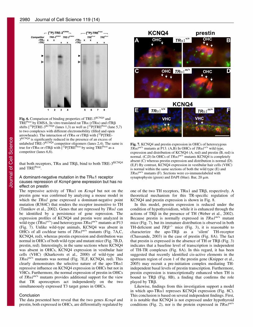
A dominant-negative mutation in the TR�1 receptorcauses repression of Kcnq4 gene expression but has noeffect on prestinThe repressive activity of TR�1 on Kcnq4 but not on theprestin gene was confirmed by analyzing a mouse model inwhich the TR�1 gene expressed a dominant-negative pointmutation (R384C) that renders the receptor insensitive to TH(Tinnikov et al., 2002). Genes that are repressed by TR�1 canbe identified by a persistence of gene repression. Theexpression profiles of KCNQ4 and prestin were analyzed inwild-type (TR�1+/+) and heterozygous TR�1m/+ mutants at P13(Fig. 7). Unlike wild-type animals, KCNQ4 was absent inOHCs of all cochlear turns of TR�1m/+ mutants (Fig. 7A,C,KCNQ4, red), whereas prestin expression and distribution wasnormal in OHCs of both wild-type and mutant mice (Fig. 7B,D,prestin, red). Interestingly, in the same sections where KCNQ4was absent in OHCs, KCNQ4 expression in vestibular haircells (VHC) (Kharkovets et al., 2000) of wild-type andTR�1m/+ mutants was normal (Fig. 7E,F, KCNQ4, red). Thisclearly demonstrates the selective nature of the apo-TR�1repressive influence on KCNQ4 expression in OHCs but not inVHCs. Furthermore, the normal expression of prestin in OHCsof TR�1m/+ mutants provides additional support for the viewthat TR aporeceptors act independently on the twosimultaneously expressed T3 target genes in OHCs.
ConclusionThe data presented here reveal that the two genes Kcnq4 andprestin, both expressed in OHCs, are differentially regulated by
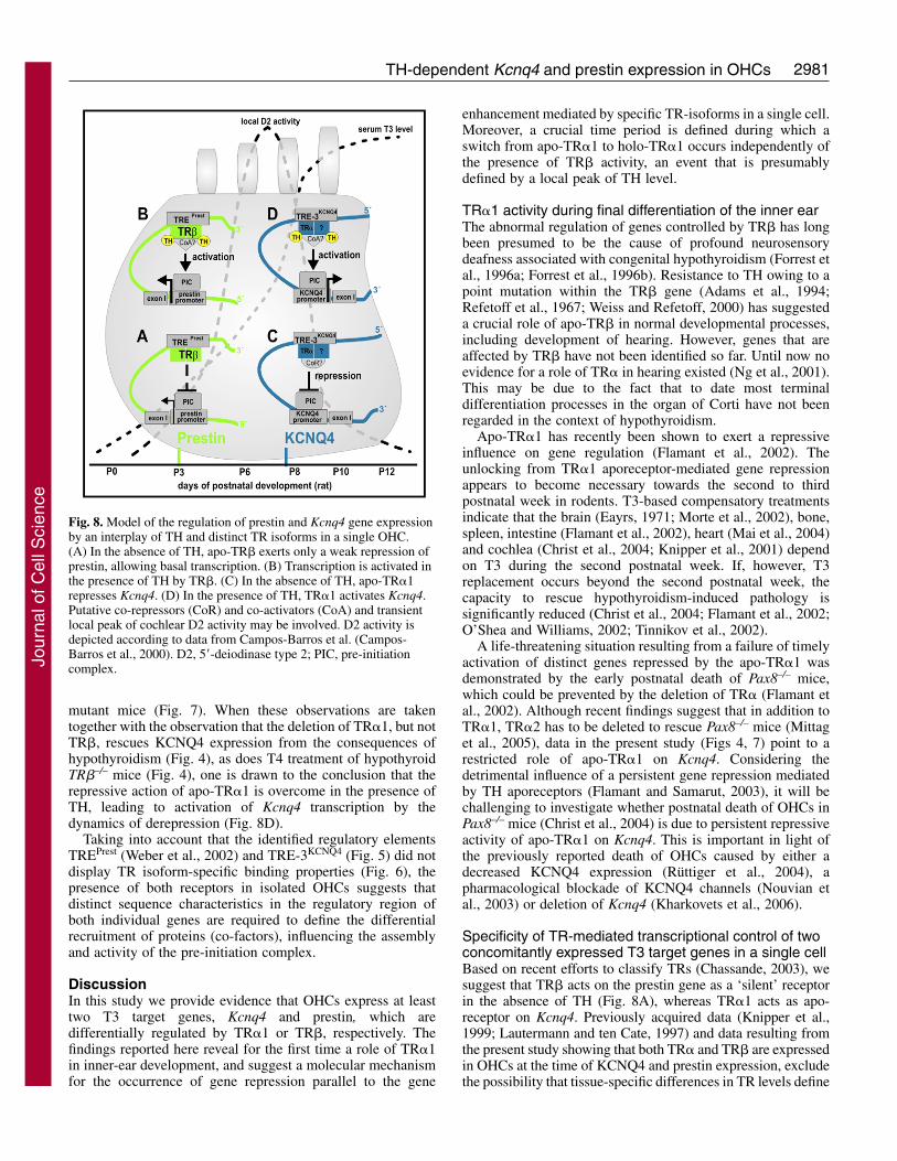
one of the two TH receptors, TR�1 and TR�, respectively. Atheoretical mechanism for this TR-specific regulation ofKCNQ4 and prestin expression is shown in Fig. 8.
In this model, prestin expression is reduced under thecondition of hypothyroidism, while it is enhanced through theactions of TR� in the presence of TH (Weber et al., 2002).Because prestin is normally expressed in TR�1m/+ mutantmice (Fig. 7), but its immature distribution is observed in bothTH-deficient and TR�–/– mice (Fig. 3), it is reasonable tocharacterize the apo-TR� as a ‘silent’ TH-receptor(Chassande, 2003) in the case of prestin (Fig. 8A). The factthat prestin is expressed in the absence of TH or TR� (Fig. 3)indicates that a baseline level of transcription is independentof TH-TR complexes (Fig. 8A). In this regard, it has beensuggested that recently identified cis-active elements in theupstream region of exon 1 of the prestin gene (Knipper et al.,2005) may recruit a pre-initiation complex mediating TH-independent basal levels of prestin transcription. Furthermore,prestin expression is transcriptionally enhanced when TH isbound to TR� (Fig. 8B), a finding that confirms the roleplayed by TR�.
Likewise, findings from this investigation support a modelin which apo-TR�1 represses KCNQ4 expression (Fig. 8C).This conclusion is based on several independent findings. First,it is notable that KCNQ4 is not expressed under hypothyroidconditions (Fig. 2), nor is the protein expressed in TR�1m/+
Journal of Cell Science 119 (14)
Fig. 6. Comparison of binding properties of TRE-3KCNQ4 andTREPrest by EMSA. In vitro translated rat TR� (rTR�) and rTR�shifts [32P]TRE-3KCNQ4 (lanes 1,3) as well as [32P]TREPrest (lane 5,7)to two complexes with different electromobility (filled and openarrowheads). The interaction of rTR� or rTR� with [32P]TRE-3KCNQ4 is significantly reduced in the presence of an excess ofunlabeled TRE-3KCNQ4 competitor oligomers (lanes 2,4). The same istrue for rTR� or rTR� with [32P]TREPrest by using TREPrest as acompetitor (lanes 6,8).
Fig. 7. KCNQ4 and prestin expression in OHCs of heterozygousTR�1m/+ mutants at P13. (A,B) In OHCs of TR�1+/+ wild type,expression and distribution of KCNQ4 (A, red) and prestin (B, red) isnormal. (C,D) In OHCs of TR�1m/+ mutants KCNQ4 is completelyabsent (C) whereas prestin expression and distribution is normal (D).(E,F) By contrast, KCNQ4 expression in vestibular hair cells (VHC)is normal within the same sections of both the wild type (E) andTR�1m/+ mutants (F). Sections were co-immunolabeled withsynaptophysin (green) and DAPI (blue). Bar, 20 �m.
Jour
nal o
f Cel
l Sci
ence
2981TH-dependent Kcnq4 and prestin expression in OHCs
mutant mice (Fig. 7). When these observations are takentogether with the observation that the deletion of TR�1, but notTR�, rescues KCNQ4 expression from the consequences ofhypothyroidism (Fig. 4), as does T4 treatment of hypothyroidTR�–/– mice (Fig. 4), one is drawn to the conclusion that therepressive action of apo-TR�1 is overcome in the presence ofTH, leading to activation of Kcnq4 transcription by thedynamics of derepression (Fig. 8D).
Taking into account that the identified regulatory elementsTREPrest (Weber et al., 2002) and TRE-3KCNQ4 (Fig. 5) did notdisplay TR isoform-specific binding properties (Fig. 6), thepresence of both receptors in isolated OHCs suggests thatdistinct sequence characteristics in the regulatory region ofboth individual genes are required to define the differentialrecruitment of proteins (co-factors), influencing the assemblyand activity of the pre-initiation complex.
DiscussionIn this study we provide evidence that OHCs express at leasttwo T3 target genes, Kcnq4 and prestin, which aredifferentially regulated by TR�1 or TR�, respectively. Thefindings reported here reveal for the first time a role of TR�1in inner-ear development, and suggest a molecular mechanismfor the occurrence of gene repression parallel to the gene
enhancement mediated by specific TR-isoforms in a single cell.Moreover, a crucial time period is defined during which aswitch from apo-TR�1 to holo-TR�1 occurs independently ofthe presence of TR� activity, an event that is presumablydefined by a local peak of TH level.
TR�1 activity during final differentiation of the inner earThe abnormal regulation of genes controlled by TR� has longbeen presumed to be the cause of profound neurosensorydeafness associated with congenital hypothyroidism (Forrest etal., 1996a; Forrest et al., 1996b). Resistance to TH owing to apoint mutation within the TR� gene (Adams et al., 1994;Refetoff et al., 1967; Weiss and Refetoff, 2000) has suggesteda crucial role of apo-TR� in normal developmental processes,including development of hearing. However, genes that areaffected by TR� have not been identified so far. Until now noevidence for a role of TR� in hearing existed (Ng et al., 2001).This may be due to the fact that to date most terminaldifferentiation processes in the organ of Corti have not beenregarded in the context of hypothyroidism.
Apo-TR�1 has recently been shown to exert a repressiveinfluence on gene regulation (Flamant et al., 2002). Theunlocking from TR�1 aporeceptor-mediated gene repressionappears to become necessary towards the second to thirdpostnatal week in rodents. T3-based compensatory treatmentsindicate that the brain (Eayrs, 1971; Morte et al., 2002), bone,spleen, intestine (Flamant et al., 2002), heart (Mai et al., 2004)and cochlea (Christ et al., 2004; Knipper et al., 2001) dependon T3 during the second postnatal week. If, however, T3replacement occurs beyond the second postnatal week, thecapacity to rescue hypothyroidism-induced pathology issignificantly reduced (Christ et al., 2004; Flamant et al., 2002;O’Shea and Williams, 2002; Tinnikov et al., 2002).
A life-threatening situation resulting from a failure of timelyactivation of distinct genes repressed by the apo-TR�1 wasdemonstrated by the early postnatal death of Pax8–/– mice,which could be prevented by the deletion of TR� (Flamant etal., 2002). Although recent findings suggest that in addition toTR�1, TR�2 has to be deleted to rescue Pax8–/– mice (Mittaget al., 2005), data in the present study (Figs 4, 7) point to arestricted role of apo-TR�1 on Kcnq4. Considering thedetrimental influence of a persistent gene repression mediatedby TH aporeceptors (Flamant and Samarut, 2003), it will bechallenging to investigate whether postnatal death of OHCs inPax8–/– mice (Christ et al., 2004) is due to persistent repressiveactivity of apo-TR�1 on Kcnq4. This is important in light ofthe previously reported death of OHCs caused by either adecreased KCNQ4 expression (Rüttiger et al., 2004), apharmacological blockade of KCNQ4 channels (Nouvian etal., 2003) or deletion of Kcnq4 (Kharkovets et al., 2006).
Specificity of TR-mediated transcriptional control of twoconcomitantly expressed T3 target genes in a single cellBased on recent efforts to classify TRs (Chassande, 2003), wesuggest that TR� acts on the prestin gene as a ‘silent’ receptorin the absence of TH (Fig. 8A), whereas TR�1 acts as apo-receptor on Kcnq4. Previously acquired data (Knipper et al.,1999; Lautermann and ten Cate, 1997) and data resulting fromthe present study showing that both TR� and TR� are expressedin OHCs at the time of KCNQ4 and prestin expression, excludethe possibility that tissue-specific differences in TR levels define
Fig. 8. Model of the regulation of prestin and Kcnq4 gene expressionby an interplay of TH and distinct TR isoforms in a single OHC.(A) In the absence of TH, apo-TR� exerts only a weak repression ofprestin, allowing basal transcription. (B) Transcription is activated inthe presence of TH by TR�. (C) In the absence of TH, apo-TR�1represses Kcnq4. (D) In the presence of TH, TR�1 activates Kcnq4.Putative co-repressors (CoR) and co-activators (CoA) and transientlocal peak of cochlear D2 activity may be involved. D2 activity isdepicted according to data from Campos-Barros et al. (Campos-Barros et al., 2000). D2, 5�-deiodinase type 2; PIC, pre-initiationcomplex.Jo
urna
l of C
ell S
cien
ce
2982
TR specificity, as previously suggested (Dillmann, 2002). Wecannot, however, rule out the fact that differences in the levelof either TR� or TR� in OHCs may play a role in TRspecificity. As both TRs were able to bind to both TREPrest andTREKCNQ4 (Fig. 6), we can be certain that the TR specificityseen in the case of Kcnq4 and prestin is not determined bydifferences in sequence-specific binding properties of the TREs,as previously described (Olson et al., 1998).
Local changes in T3 level should instead be considered toplay a crucial role in influencing TR specificity. Indeed, inhumans and rodents the critical developmental time period ofthe inner ear occurs in parallel to the natural rise of TH bloodplasma levels (Deol, 1973; Knipper et al., 2000; Uziel et al.,1985).
The importance of T3 availability as a factor controlling TR-regulated transcription was clearly demonstrated in a recentstudy of TR�1m/m mutant mice. In this strain, the affinity ofTR�1 for TH is reduced as a consequence of a point mutation(Tinnikov et al., 2002) leading to mortality within the first3 postnatal weeks, as in Pax8–/– mice. The pathologicalconsequences of the condition were reversible uponcompensatory T3 levels (Tinnikov et al., 2002). Theimportance of circulating T3 for the conversion of apo-TR�1to the holoreceptor configuration was also emphasized instudies analyzing TR�1 function in heart tissue, intestine, boneand brain (Flamant et al., 2002; Mai et al., 2004).
The role of local T3 levels, which are determined bydeiodinase activity, has been shown in target tissues duringamphibian metamorphosis (Becker et al., 1997; Huang et al.,1999) and rat brain development (Kaplan and Yaskoski, 1981;Obregon et al., 1991). Hearing loss in 5�-deiodinase type 2(D2)-deficient mice (Ng et al., 2004) was only recentlydiscussed in the context of a striking peak of D2 activity in thecochlea between P5 and P10 (Campos-Barros et al., 2000).Although the role of deiodinase in the switch from apo-TR toholo-TR has been hypothesized (Chassande, 2003), supportingdata have been unavailable until now.
The data in the present study show that the time period overwhich the two T3 target genes in OHCs are activated and/ormodulated is brief and occurs between P6 and P10, when thereis a peak in 5�-deiodinase activity (Campos-Barros et al., 2000)and a steep rise in the TH blood plasma level (Knipper et al.,2000). Local T3 levels thus may split the genes into TR�1-affected or -unaffected categories. T3 may induce aconformational change in the receptor that destabilizes its bondwith co-repressors and facilitates the binding of co-activators(Lazar, 2003).
Retinoic acid, shown to be required as an additional ligandto overcome the repressive effect on Kcnq4 promoter activityin vitro (Fig. 6) should be considered as a further ligand neededfor derepression. Lee and Privalsky (Lee and Privalsky, 2005)reported that TR�1 forms heterodimers with RAR, which incontrast to TR-RXR heterodimer recruit both co-activators andco-repressors. TR�1-RAR heterodimers bound to the invertedpalindrome TRE-3KCNQ4 in OHCs may thus recruit co-repressors until TH levels rise during early postnataldevelopment. Although several studies underscore theimportance of RAR for inner-ear development (Raz and Kelley,1999; Romand et al., 2002), changes in retinoic acid levelsduring postnatal development have not been reported(Romand, 2003).
TR� activity in the maturation of OHCsAs higher T3 levels seem to be required to release apo-TR�1from TRE-3KCNQ4, lower T3 levels may be sufficient for therecruitment of the TR� complex to TREPrest (Fig. 8). Prestinexpression and distribution are known to be diminished andimmature in hypothyroidism (Weber et al., 2002), as well as inTR�–/– and TR�1–/–/�–/– mutants as shown in the present study.Although we cannot rule out the possibility that the influenceof TH on the redistribution of prestin is indirect, this conditionmay be causally linked to the reduced nonlinear capacitance ofOHCs observed in TR�–/– and TR�1–/–/�–/– mutants (Rusch etal., 2001).
In conclusion, the characterization of differentially regulatedT3 target genes in the same cell may provide the basis for novelinsight into the mechanism of subcellular TR specificity. Giventhe fact that TR�1 and TR� show no preference in binding toTREPrest or TRE-3KCNQ4, the specificity of TR binding inpostnatal OHCs must be achieved by additional gene-specificdifferences in the upstream regions of the prestin and Kcnq4genes. These differences influence the level of TR-associatedDNA-bound transcription factors, as previously proposed(Glass and Rosenfeld, 2000). The composition of regulatoryelements may then act in concert with specific local T3 levelsto alter dissociation or recruitment rates of co-repressor or co-activator complexes (Fig. 8) (Hermanson et al., 2002; Kameiet al., 1996; Onate et al., 1995).
Until now human patients with mutant TR�1 have not beenidentified; as either the effects on a human phenotype are toomild, spontaneous miscarriages occur at embryonic or fetalstages of development, and/or the symptoms have not beenattributed to defects in TR�1 (Tinnikov et al., 2002). Based onthe findings reported here, the latter explanation may applymost appropriately in the case of the auditory system.
Materials and MethodsAnimals and drug administrationWistar rats, Pax8–/– (Mansouri et al., 1998), Tshrhyt (hyt/hyt) (Walsh and McGee,2001), TR�1–/– (Wikstrom et al., 1998), TR�–/– (Forrest et al., 1996a), TR�1–/–/�–/–
(Gothe et al., 1999) and TR�1m/+ (Tinnikov et al., 2002) mice were used (Table 1).MMI treatment of mice, thyroxin (T4) administration, and T4 and T3 plasma titerdeterminations were performed as described (Knipper et al., 2000; Weber et al.,2002). Animal experiments were approved and complied with all protocolrequirements at the University of Tübingen.
Tissue preparationCochleae of untreated (control), MMI-treated (hypothyroid) and T4-treated(hypothyroid+T4) animals were prepared and cryosectioned as described (Knipperet al., 1998).
Riboprobe synthesis and in situ hybridizationUsing the KCNQ4-specific oligonucleotide primers rKCNQ4-USP3 and rKCNQ4-DSP9 (supplementary material, Table S1) a PCR fragment was amplified from ratcochlear cDNA, cloned into pCR®II TOPO Vector (Invitrogen) and sequenced.Kcnq4-specific riboprobes were synthesized and in situ hybridization wasperformed as described (Knipper et al., 1999; Knipper et al., 2000; Weber et al.,2002).
Northern blotNorthern blots were performed as described (Knipper et al., 1998; Knipper et al.,1999). The effect of TH on mRNA levels was semi-quantitatively evaluated usingmRNAs isolated from a similar number of cochleae as described (Knipper et al.,1998). To ensure the loading of similar amounts of mRNA per lane blots wereprobed with the housekeeping gene cyclophilin (Thellin et al., 1999).
Semi-quantitative RT-PCRSemi-quantitative RT-PCR was performed as described (Rüttiger et al., 2006). Inbrief, cochlear mRNA from P12 hypothyroid and control rats was isolated using theDynabeads mRNA Direct Kit (Dynal) and reverse-transcribed with Superscript II
Journal of Cell Science 119 (14)
Jour
nal o
f Cel
l Sci
ence
2983TH-dependent Kcnq4 and prestin expression in OHCs
(Invitrogen). Using the oligonucleotides rKCNQ4-USP3 and rKCNQ4-DSP3 andcyclophilin-f and cyclophilin-r (supplementary material Table S1) as internalcontrols semi-quantitative RT-PCR was performed on equivalent amounts of mRNAisolated from four cochleae of control and hypothyroid rats. PCR was repeated threetimes using different animals.
Generation of KCNQ4 antibodyAn antibody against a conserved part of the C-terminus of KCNQ4(CQTLSISRSVSTNMD-COOH) was raised in rabbits, purified by affinitychromatography and tested by immunoblotting and immunohistochemistry bypreincubation with the synthetic peptide (data not shown).
Fluorescence immunohistochemistryCochlear sections of rats and mice were stained and imaged as described (Knipperet al., 1998; Knipper et al., 2000). The rabbit and goat (Santa Cruz) polyclonalantibody against KCNQ4, rabbit polyclonal antibody against prestin (Weber et al.,2002), sheep polyclonal antibody against synaptophysin (The Binding Site) werevisualized with Cy3- (Jackson ImmunoResearch Laboratories) or Alexa Fluor 488-conjugated secondary antibodies (Molecular Probes) and counter-stained with DAPI(Vector Laboratories).
Oligonucleotides and labelingIn EMSA, 3.85 pmol of the double-stranded synthetic oligonucleotides TRE-1KCNQ4, TRE-2KCNQ4, TRE-3KCNQ4, TRE-3mut1, TRE-3mut2, DR4, TREPrest andTFIID (supplementary material, Table S1) were labeled with [�-32P]ATP (5000Ci/mmol; Amersham Biosciences) by phosphorylation with T4 polynucleotidekinase (Promega) and purified with the QIAquick Nucleotide Removal Kit (Qiagen).
Cloning of the human KCNQ4 promoterGenomic DNA was isolated from human blood with the QIAmp Blood Kit (Qiagen).PCR was performed with the oligonucleotide primers PromK4USP andPromK4DSP and nested primers sPromK4USP(BglII) and sPromK4DSP(HindIII)(supplementary material Table S1). A 739 bp PCR fragment was amplified, clonedinto the pGL3basic vector (Promega) and sequenced.
Cloning of the reporter gene constructsThe synthetic double-stranded oligonucleotide TRE-3KCNQ4 (supplementarymaterial Table S1) was cloned into the SacI and XhoI sites of the pGL3 vectorcontaining the putative human KCNQ4 promoter. The previously described rat TR�expression vector was used (Weber et al., 2002).
Transfection and reporter gene assaysHEK293 cells were cultured and transfected with 500 ng of each reporter construct,500 ng of the control plasmid RSV-LacZ, and 500 ng of the TR� expression vectorfor 12 hours as described (Weber et al., 2002). Transfected cells were incubated for48 hours in fresh medium containing serum either depleted of T3 and retinoic acid(Weber et al., 2002) or containing 150 nM T3 (Sigma) or 1.5 �M all-trans-retinoicacid (ATRA; Sigma). The luciferase assay system (Promega) and the �-galactosidase enzyme system (Promega) were used to determine luciferase activityand transfection efficiency as described (Weber et al., 2002). Four independentexperiments were performed, each containing three to six independently preparedtransfection mixtures.
In vitro translationRat TR� and TR� were translated in vitro from plasmids (Weber et al., 2002) usingthe TNT® T7 coupled reticulocyte lysate system (Promega).
Electromobility-shift assay (EMSA)EMSAs were performed as described (Weber et al., 2002). Unlabeled competitoroligonucleotides (200-fold excess) or T3 (150 nM) were preincubated withrecombinant chicken TR� (Santa Cruz) for 30 minutes before adding theradiolabeled oligonucleotides.
We thank B. Vennström for the TR�1m/+ mice, R. Panford-Walshfor reading and correcting this manuscript and D. Schmollinger fortechnical assistance. This work was supported by the FederalMinistry of Education and Research (01KS9602), theInterdisciplinary Center of Clinical Research Tübingen, the DeutscheForschungsgemeinschaft DFG KN316/4-1 and the NIH-NIDCD-5R01DC04566.
ReferencesAdams, M., Matthews, C., Collingwood, T. N., Tone, Y., Beck-Peccoz, P. and
Chatterjee, K. K. (1994). Genetic analysis of 29 kindreds with generalized andpituitary resistance to thyroid hormone. Identification of thirteen novel mutations inthe thyroid hormone receptor beta gene. J. Clin. Invest. 94, 506-515.
Beamer, W. J., Eicher, E. M., Maltais, L. J. and Southard, J. L. (1981). Inheritedprimary hypothyroidism in mice. Science 212, 61-63.
Becker, K. B., Stephens, K. C., Davey, J. C., Schneider, M. J. and Galton, V. A. (1997).The type 2 and type 3 iodothyronine deiodinases play important roles in coordinatingdevelopment in Rana catesbeiana tadpoles. Endocrinology 138, 2989-2997.
Campos-Barros, A., Amma, L. L., Faris, J. S., Shailam, R., Kelley, M. W. and Forrest,D. (2000). Type 2 iodothyronine deiodinase expression in the cochlea before the onsetof hearing. Proc. Natl. Acad. Sci. USA 97, 1287-1292.
Chassande, O. (2003). Do unliganded thyroid hormone receptors have physiologicalfunctions? J. Mol. Endocrinol. 31, 9-20.
Christ, S., Biebel, U. W., Hoidis, S., Friedrichsen, S., Bauer, K. and Smolders, J. W.(2004). Hearing loss in athyroid pax8 knockout mice and effects of thyroxinesubstitution. Audiol. Neurootol. 9, 88-106.
Deol, M. S. (1973). An experimental approach to the understanding and treatment ofhereditary syndromes with congenital deafness and hypothyroidism. J. Med. Genet. 10,235-242.
Dillmann, W. H. (2002). Cellular action of thyroid hormone on the heart. Thyroid 12,447-452.
Eayrs, J. T. (1971). Thyroid and developing brain: anatomical and behavioral effects. InHormones in Development (ed. M. Hamburgh and E. J. W. Barrington), pp. 345-355.New York: Appleton Centruy Crofts.
Flamant, F. and Samarut, J. (2003). Thyroid hormone receptors: lessons from knockoutand knock-in mutant mice. Trends Endocrinol. Metab. 14, 85-90.
Flamant, F., Poguet, A. L., Plateroti, M., Chassande, O., Gauthier, K.,Streichenberger, N., Mansouri, A. and Samarut, J. (2002). Congenital hypothyroidPax8(–/–) mutant mice can be rescued by inactivating the TRalpha gene. Mol.Endocrinol. 16, 24-32.
Forrest, D., Erway, L. C., Ng, L., Altschuler, R. and Curran, T. (1996a). Thyroidhormone receptor beta is essential for development of auditory function. Nat. Genet.13, 354-357.
Forrest, D., Hanebuth, E., Smeyne, R. J., Everds, N., Stewart, C. L., Wehner, J. M.and Curran, T. (1996b). Recessive resistance to thyroid hormone in mice lackingthyroid hormone receptor beta: evidence for tissue-specific modulation of receptorfunction. EMBO J. 15, 3006-3015.
Gauthier, K., Chassande, O., Plateroti, M., Roux, J. P., Legrand, C., Pain, B.,Rousset, B., Weiss, R., Trouillas, J. and Samarut, J. (1999). Different functions forthe thyroid hormone receptors TRalpha and TRbeta in the control of thyroid hormoneproduction and post-natal development. EMBO J. 18, 623-631.
Gil-Loyzaga, P. and Pujol, R. (1988). Synaptophysin in the developing cochlea. Int. J.Dev. Neurosci. 6, 155-160.
Glass, C. K. and Rosenfeld, M. G. (2000). The coregulator exchange in transcriptionalfunctions of nuclear receptors. Genes Dev. 14, 121-141.
Gothe, S., Wang, Z., Ng, L., Kindblom, J. M., Barros, A. C., Ohlsson, C., Vennstrom,B. and Forrest, D. (1999). Mice devoid of all known thyroid hormone receptors areviable but exhibit disorders of the pituitary-thyroid axis, growth, and bone maturation.Genes Dev. 13, 1329-1341.
Hermanson, O., Glass, C. K. and Rosenfeld, M. G. (2002). Nuclear receptorcoregulators: multiple modes of modification. Trends Endocrinol. Metab. 13, 55-60.
Huang, H., Marsh-Armstrong, N. and Brown, D. D. (1999). Metamorphosis is inhibitedin transgenic Xenopus laevis tadpoles that overexpress type III deiodinase. Proc. Natl.Acad. Sci. USA 96, 962-967.
Kamei, Y., Xu, L., Heinzel, T., Torchia, J., Kurokawa, R., Gloss, B., Lin, S. C.,Heyman, R. A., Rose, D. W., Glass, C. K. et al. (1996). A CBP integrator complexmediates transcriptional activation and AP-1 inhibition by nuclear receptors. Cell 85,403-414.
Kaplan, M. M. and Yaskoski, K. A. (1981). Maturational patterns of iodothyroninephenolic and tyrosyl ring deiodinase activities in rat cerebrum, cerebellum, andhypothalamus. J. Clin. Invest. 67, 1208-1214.
Kharkovets, T., Hardelin, J. P., Safieddine, S., Schweizer, M., El-Amraoui, A., Petit,C. and Jentsch, T. J. (2000). KCNQ4, a K+ channel mutated in a form of dominantdeafness, is expressed in the inner ear and the central auditory pathway. Proc. Natl.Acad. Sci. USA 97, 4333-4338.
Kharkovets, T., Dedek, K., Maier, H., Schweizer, M., Khimich, D., Nouvian, R.,Vardanyan, V., Leuwer, R., Moser, T. and Jentsch, T. J. (2006). Mice with alteredKCNQ4 K(+) channels implicate sensory outer hair cells in human progressivedeafness. EMBO J. 25, 642-652.
Knipper, M., Zimmermann, U., Rohbock, K., Kopschall, I. and Zenner, H. P. (1995).Synaptophysin and GAP-43 proteins in efferent fibers of the inner ear during postnataldevelopment. Brain Res. Dev. Brain Res. 89, 73-86.
Knipper, M., Bandtlow, C., Gestwa, L., Kopschall, I., Rohbock, K., Wiechers, B.,Zenner, H. P. and Zimmermann, U. (1998). Thyroid hormone affects Schwann celland oligodendrocyte gene expression at the glial transition zone of the VIIIth nerveprior to cochlea function. Development 125, 3709-3718.
Knipper, M., Gestwa, L., Ten Cate, W. J., Lautermann, J., Brugger, H., Maier, H.,Zimmermann, U., Rohbock, K., Kopschall, I., Wiechers, B. et al. (1999). Distinctthyroid hormone-dependent expression of TrKB and p75NGFR in nonneuronal cellsduring the critical TH-dependent period of the cochlea. J. Neurobiol. 38, 338-356.
Knipper, M., Zinn, C., Maier, H., Praetorius, M., Rohbock, K., Kopschall, I. andZimmermann, U. (2000). Thyroid hormone deficiency before the onset of hearingcauses irreversible damage to peripheral and central auditory systems. J. Neurophysiol.83, 3101-3112.
Knipper, M., Richardson, G., Mack, A., Muller, M., Goodyear, R., Limberger, A.,Rohbock, K., Kopschall, I., Zenner, H. P. and Zimmermann, U. (2001). Thyroid
Jour
nal o
f Cel
l Sci
ence
2984
hormone-deficient period prior to the onset of hearing is associated with reduced levelsof beta-tectorin protein in the tectorial membrane: implication for hearing loss. J. Biol.Chem. 276, 39046-39052.
Knipper, M., Weber, T., Winter, H., Braig, C., Cimerman, J., Fraenzer, J.-T. andZimmermann, U. (2005). Individual characteristics of members of the SLC26 familyin vertebrate and their homologues in insects. In Epithelial Anion Transport in Healthand Disease: The Role of the SLC26 Transporters Family (Novartis FoundationSymposium 273) (ed. D. J. Chadwick). Chichester: Novartis Foundation, John Wiley& Sons. In press.
Kubisch, C., Schroeder, B. C., Friedrich, T., Lutjohann, B., El-Amraoui, A., Marlin,S., Petit, C. and Jentsch, T. J. (1999). KCNQ4, a novel potassium channel expressedin sensory outer hair cells, is mutated in dominant deafness. Cell 96, 437-446.
Lautermann, J. and ten Cate, W. J. (1997). Postnatal expression of the alpha-thyroidhormone receptor in the rat cochlea. Hear. Res. 107, 23-28.
Lazar, M. A. (2003). Thyroid hormone action: a binding contract. J. Clin. Invest. 112,497-499.
Lee, S. and Privalsky, M. L. (2005). Heterodimers of retinoic acid receptors and thyroidhormone receptors display unique combinatorial regulatory properties. Mol.Endocrinol. 19, 863-878.
Macchia, P. E. (2000). Recent advances in understanding the molecular basis of primarycongenital hypothyroidism. Mol. Med. Today 6, 36-42.
Mai, W., Janier, M. F., Allioli, N., Quignodon, L., Chuzel, T., Flamant, F. andSamarut, J. (2004). Thyroid hormone receptor alpha is a molecular switch of cardiacfunction between fetal and postnatal life. Proc. Natl. Acad. Sci. USA 101, 10332-10337.
Mansouri, A., Chowdhury, K. and Gruss, P. (1998). Follicular cells of the thyroid glandrequire Pax8 gene function. Nat. Genet. 19, 87-90.
Marcotti, W. and Kros, C. J. (1999). Developmental expression of the potassium currentIK,n contributes to maturation of mouse outer hair cells. J. Physiol. 520, 3, 653-660.
Michna, M., Knirsch, M., Hoda, J. C., Muenkner, S., Langer, P., Platzer, J.,Striessnig, J. and Engel, J. (2003). Cav1.3 (alpha1D) Ca2+ currents in neonatal outerhair cells of mice. J. Physiol. 553, 747-758.
Mittag, J., Friedrichsen, S., Heuer, H., Polsfuss, S., Visser, T. J. and Bauer, K. (2005).Athyroid Pax8–/– mice cannot be rescued by the inactivation of thyroid hormonereceptor alpha1. Endocrinology 146, 3179-3184.
Morte, B., Manzano, J., Scanlan, T., Vennstrom, B. and Bernal, J. (2002). Deletionof the thyroid hormone receptor alpha 1 prevents the structural alterations of thecerebellum induced by hypothyroidism. Proc. Natl. Acad. Sci. USA 99, 3985-3989.
Ng, L., Rusch, A., Amma, L. L., Nordstrom, K., Erway, L. C., Vennstrom, B. andForrest, D. (2001). Suppression of the deafness and thyroid dysfunction in Thrb-nullmice by an independent mutation in the Thra thyroid hormone receptor alpha gene.Hum. Mol. Genet. 10, 2701-2708.
Ng, L., Goodyear, R. J., Woods, C. A., Schneider, M. J., Diamond, E., Richardson,G. P., Kelley, M. W., Germain, D. L., Galton, V. A. and Forrest, D. (2004). Hearingloss and retarded cochlear development in mice lacking type 2 iodothyroninedeiodinase. Proc. Natl. Acad. Sci. USA 101, 3474-3479.
Nouvian, R., Ruel, J., Wang, J., Guitton, M. J., Pujol, R. and Puel, J. L. (2003).Degeneration of sensory outer hair cells following pharmacological blockade ofcochlear KCNQ channels in the adult guinea pig. Eur. J. Neurosci. 17, 2553-2562.
Obregon, M. J., Ruiz de Ona, C., Calvo, R., Escobar del Rey, F. and Morreale deEscobar, G. (1991). Outer ring iodothyronine deiodinases and thyroid hormoneeconomy: responses to iodine deficiency in the rat fetus and neonate. Endocrinology129, 2663-2673.
Oliver, D., Knipper, M., Derst, C. and Fakler, B. (2003). Resting potential andsubmembrane calcium concentration of inner hair cells in the isolated mouse cochleaare set by KCNQ-type potassium channels. J. Neurosci. 23, 2141-2149.
Olson, D. P., Sun, B. and Koenig, R. J. (1998). Thyroid hormone response elementarchitecture affects corepressor release from thyroid hormone receptor dimers. J. Biol.Chem. 273, 3375-3380.
Onate, S. A., Tsai, S. Y., Tsai, M. J. and O’Malley, B. W. (1995). Sequence andcharacterization of a coactivator for the steroid hormone receptor superfamily. Science270, 1354-1357.
O’Shea, P. J. and Williams, G. R. (2002). Insight into the physiological actions ofthyroid hormone receptors from genetically modified mice. J. Endocrinol. 175, 553-570.
Perissi, V., Staszewski, L. M., McInerney, E. M., Kurokawa, R., Krones, A., Rose, D.W., Lambert, M. H., Milburn, M. V., Glass, C. K. and Rosenfeld, M. G. (1999).Molecular determinants of nuclear receptor-corepressor interaction. Genes Dev. 13,3198-3208.
Quandt, K., Frech, K., Karas, H., Wingender, E. and Werner, T. (1995). MatInd andMatInspector: new fast and versatile tools for detection of consensus matches innucleotide sequence data. Nucleic Acids Res. 23, 4878-4884.
Raz, Y. and Kelley, M. W. (1999). Retinoic acid signaling is necessary for thedevelopment of the organ of Corti. Dev. Biol. 213, 180-193.
Refetoff, S., DeWind, L. T. and DeGroot, L. J. (1967). Familial syndrome combiningdeaf-mutism, stuppled epiphyses, goiter and abnormally high PBI: possible target organrefractoriness to thyroid hormone. J. Clin. Endocrinol. Metab. 27, 279-294.
Refetoff, S., Weiss, R. E. and Usala, S. J. (1993). The syndromes of resistance to thyroidhormone. Endocr. Rev. 14, 348-399.
Romand, R. (2003). The roles of retinoic acid during inner ear development. Curr. Top.Dev. Biol. 57, 261-291.
Romand, R., Hashino, E., Dolle, P., Vonesch, J. L., Chambon, P. and Ghyselinck, N.B. (2002). The retinoic acid receptors RARalpha and RARgamma are required for innerear development. Mech. Dev. 119, 213-223.
Rusch, A., Erway, L. C., Oliver, D., Vennstrom, B. and Forrest, D. (1998). Thyroidhormone receptor beta-dependent expression of a potassium conductance in inner haircells at the onset of hearing. Proc. Natl. Acad. Sci. USA 95, 15758-15762.
Rusch, A., Ng, L., Goodyear, R., Oliver, D., Lisoukov, I., Vennstrom, B., Richardson,G., Kelley, M. W. and Forrest, D. (2001). Retardation of cochlear maturation andimpaired hair cell function caused by deletion of all known thyroid hormone receptors.J. Neurosci. 21, 9792-9800.
Rüttiger, L., Sausbier, M., Zimmermann, U., Winter, H., Braig, C., Engel, J., Knirsch,M., Arntz, C., Langer, P., Hirt, B. et al. (2004). Deletion of the Ca2+-activatedpotassium (BK) {alpha}-subunit but not the BK{beta}1-subunit leads to progressivehearing loss. Proc. Natl. Acad. Sci. USA 101, 12922-12927.
Rüttiger, L., Panford-Walsh, R., Schimmang, T., Tan, J., Zimmermann, U., Rohbock,K., Kopschall, I., Limberger, A., Muller, M., Franzer, J.-T. et al. (2006). BDNFmRNA expression and protein localization are changed in age-related hearing loss.Neurobiol. Aging doi:10.1016/j.neurobiolaging.2006.02.008.
Scherf, M., Klingenhoff, A. and Werner, T. (2000). Highly specific localization ofpromoter regions in large genomic sequences by PromoterInspector: a novel contextanalysis approach. J. Mol. Biol. 297, 599-606.
Sprenkle, P. M., McGee, J., Bertoni, J. M. and Walsh, E. J. (2001a). Consequences ofhypothyroidism on auditory system function in Tshr mutant (hyt) mice. J. Assoc. Res.Otolaryngol. 2, 312-329.
Sprenkle, P. M., McGee, J., Bertoni, J. M. and Walsh, E. J. (2001b). Development ofauditory brainstem responses (ABRs) in Tshr mutant mice derived from euthyroid andhypothyroid dams. J. Assoc. Res. Otolaryngol. 2, 330-347.
Sprenkle, P. M., McGee, J., Bertoni, J. M. and Walsh, E. J. (2001c). Prevention ofauditory dysfunction in hypothyroid Tshr mutant mice by thyroxin treatment duringdevelopment. J. Assoc. Res. Otolaryngol. 2, 348-361.
Stein, S. A., Oates, E. L., Hall, C. R., Grumbles, R. M., Fernandez, L. M., Taylor, N.A., Puett, D. and Jin, S. (1994). Identification of a point mutation in the thyrotropinreceptor of the hyt/hyt hypothyroid mouse. Mol. Endocrinol. 8, 129-138.
Tell, G., Pellizzari, L., Esposito, G., Pucillo, C., Macchia, P. E., Di Lauro, R. andDamante, G. (1999). Structural defects of a Pax8 mutant that give rise to congenitalhypothyroidism. Biochem. J. 341, 89-93.
Thellin, O., Zorzi, W., Lakaye, B., De Borman, B., Coumans, B., Hennen, G., Grisar,T., Igout, A. and Heinen, E. (1999). Housekeeping genes as internal standards: useand limits. J. Biotechnol. 75, 291-295.
Tinnikov, A., Nordstrom, K., Thoren, P., Kindblom, J. M., Malin, S., Rozell, B.,Adams, M., Rajanayagam, O., Pettersson, S., Ohlsson, C. et al. (2002). Retardationof post-natal development caused by a negatively acting thyroid hormone receptoralpha1. EMBO J. 21, 5079-5087.
Uziel, A., Legrand, C. and Rabie, A. (1985). Corrective effects of thyroxine on cochlearabnormalities induced by congenital hypothyroidism in the rat. I. Morphological study.Brain Res. 351, 111-122.
Walsh, E. J. and McGee, J. (2001). Hypothyroidism in the Tshr mutant mouse. InHandbook of Mouse Auditory Research: From Behavior to Molecular Biology (ed. J.F. Willot), pp. 537-555. Boca Raton, FL: CRC Press.
Weber, T., Zimmermann, U., Winter, H., Mack, A., Kopschall, I., Rohbock, K.,Zenner, H. P. and Knipper, M. (2002). Thyroid hormone is a critical determinant forthe regulation of the cochlear motor protein prestin. Proc. Natl. Acad. Sci. USA 99,2901-2906.
Weiss, R. E. and Refetoff, S. (2000). Resistance to thyroid hormone. Rev. Endocr. Metab.Disord. 1, 97-108.
Wikstrom, L., Johansson, C., Salto, C., Barlow, C., Campos Barros, A., Baas, F.,Forrest, D., Thoren, P. and Vennstrom, B. (1998). Abnormal heart rate and bodytemperature in mice lacking thyroid hormone receptor alpha 1. EMBO J. 17, 455-461.
Wu, Y. and Koenig, R. J. (2000). Gene regulation by thyroid hormone. TrendsEndocrinol. Metab. 11, 207-211.
Yen, P. M., Darling, D. S., Carter, R. L., Forgione, M., Umeda, P. K. and Chin, W.W. (1992). Triiodothyronine (T3) decreases binding to DNA by T3-receptorhomodimers but not receptor-auxiliary protein heterodimers. J. Biol. Chem. 267, 3565-3568.
Zheng, J., Shen, W., He, D. Z., Long, K. B., Madison, L. D. and Dallos, P. (2000).Prestin is the motor protein of cochlear outer hair cells. Nature 405, 149-155.
Journal of Cell Science 119 (14)
Jour
nal o
f Cel
l Sci
ence


![trtr trn tru tr[] ujosephschwartzdermatology.com/wp-content/uploads/... · trtr UEI trtr E] utr E] Y-ES tr tr B D tr tr tr tr tr NO tr EI tr u u u EI E tr OlherSystemic: Diobetes](https://img.pdfslide.us/doc/110x75/5f655dabeca5702d4204d061/trtr-trn-tru-tr-ujosep-trtr-uei-trtr-e-utr-e-y-es-tr-tr-b-d-tr-tr-tr-tr-tr-no.jpg)


![TURKEY——— [TR] STAR TV HD [TR] STAR TV HD [L] [TR] STAR TV ... · [tr] hilal tv [tr] sinevizyonlaŔ da ne var [tr] sinevizyon 1 hd [tr] sinevizyon 2 hd [tr] sinevizyon 3 hd](https://img.pdfslide.us/doc/110x75/5e1690ad410818078675a933/turkeyaaa-tr-star-tv-hd-tr-star-tv-hd-l-tr-star-tv-tr-hilal.jpg)
![Synthesis and Study of Many New Chalcone Derivatives · to give1-(1H-benzo[d]imidazol-2-yl) ethanone [2]. Reaction of this compound with different substituted benzaldehydes or 1-(1H-benzo[d]imidazol-2-yl)ethanone,](https://img.pdfslide.us/doc/110x75/5faa6f861699493f7c4fefcc/synthesis-and-study-of-many-new-chalcone-derivatives-to-give1-1h-benzodimidazol-2-yl.jpg)


![REACTIONS OF IMIDAZOL IN)-2-YLIDENES WITH ELECTRON ...ajarduengo.net/FormattedFinalPSSi2015.pdf · [9,10] We recently reported chemistry of imidazol-2-ylidenes with cyano-olefins](https://img.pdfslide.us/doc/110x75/60ab20f98248c34e06044e45/reactions-of-imidazol-in-2-ylidenes-with-electron-910-we-recently-reported.jpg)