Embed Size (px)
Citation preview

THYROID CYTOLOGY
STRUCTURED REPORTING
PROTOCOL
(2nd Edition, 2019)

ii
ISBN: 978-1-76081-109-9
Publications number (SHPN): (CI) 190190
Online copyright
© RCPA 2019
This work (Protocol) is copyright. You may download, display, print and
reproduce the Protocol for your personal, non-commercial use or use within your
organisation subject to the following terms and conditions:
1. The Protocol may not be copied, reproduced, communicated or displayed, in
whole or in part, for profit or commercial gain.
2. Any copy, reproduction or communication must include this RCPA copyright
notice in full.
3. With the exception of Chapter 6 - the checklist, no changes may be made to
the wording of the Protocol including any Standards, Guidelines,
commentary, tables or diagrams. Excerpts from the Protocol may be used
in support of the checklist. References and acknowledgments must be
maintained in any reproduction or copy in full or part of the Protocol.
4. Regarding Chapter 6 of the Protocol - the checklist:
o The wording of the Standards may not be altered in any way and must be
included as part of the checklist.
o Guidelines are optional and those which are deemed not applicable may be
removed.
o Numbering of Standards and Guidelines must be retained in the checklist,
but can be reduced in size, moved to the end of the checklist item or
greyed out or other means to minimise the visual impact.
o Additional items for local use may be added but must not be numbered as
a Standard or Guideline, in order to avoid confusion with the RCPA
checklist items.
o Formatting changes regarding font, spacing, tabulation and sequencing
may be made.
o Commentary from the Protocol may be added or hyperlinked to the
relevant checklist item.
Apart from any use as permitted under the Copyright Act 1968 or as set out
above, all other rights are reserved. Requests and inquiries concerning
reproduction and rights should be addressed to RCPA, 207 Albion St, Surry Hills,
NSW 2010, Australia.
First published: June 2019 2nd Edition (version 2.0)

iii
Disclaimer
The Royal College of Pathologists of Australasia ("College") has developed these
protocols as an educational tool to assist pathologists in reporting of relevant
information for specific cancers. The use of these standards and guidelines is
subject to the clinician’s judgement in each individual case.
The College makes all reasonable efforts to ensure the quality and accuracy of the
protocols and to update the protocols regularly. However subject to any
warranties, terms or conditions which may be implied by law and which cannot be
excluded, the protocols are provided on an "as is" basis. The College does not
warrant or represent that the protocols are complete, accurate, error-free, or up
to date. The protocols do not constitute medical or professional advice. Users
should obtain appropriate medical or professional advice, or where appropriately
qualified, exercise their own professional judgement relevant to their own
particular circumstances. Users are responsible for evaluating the suitability,
accuracy, currency, completeness and fitness for purpose of the protocols.
Except as set out in this paragraph, the College excludes: (i) all warranties, terms
and conditions relating in any way to; and (ii) all liability (including for
negligence) in respect of any loss or damage (including direct, special, indirect or
consequential loss or damage, loss of revenue, loss of expectation, unavailability
of systems, loss of data, personal injury or property damage) arising in any way
from or in connection with; the protocols or any use thereof. Where any statute
implies any term, condition or warranty in connection with the provision or use of
the protocols, and that statute prohibits the exclusion of that term, condition or
warranty, then such term, condition or warranty is not excluded. To the extent
permitted by law, the College's liability under or for breach of any such term,
condition or warranty is limited to the resupply or replacement of services or
goods.

iv
Contents
Scope ............................................................................................. v
Abbreviations ................................................................................ vi
Definitions .................................................................................... vii
Introduction ................................................................................... 1
Authority and development ............................................................ 5
1 Pre-analytical ....................................................................... 8
2 Specimen collection and handling ...................................... 10
3 Terminology, microscopic findings, interpretation &
recommendations .............................................................. 15
4 Ancillary studies ................................................................ 28
5 Synthesis and overview...................................................... 32
6 Structured checklist ........................................................... 33
7 Formatting of pathology reports ........................................ 42
Appendix 1 Pathology request information .............................. 43
Appendix 2 Guidelines for formatting of a pathology report ..... 47
Appendix 3 Examples of cytopathology reports ........................ 48
References ............................................................................... 50

v
Scope
This protocol contains standards and guidelines for the preparation, interpretation
and reporting of thyroid cytology material obtained by fine needle aspiration
biopsy (FNA). The aspirates may be direct aspirations or radiologically guided
aspirations.
The aim is to improve quality of the final cytopathology report that is issued to
clinicians, and in particular to improve the decision for management of thyroid
lesions. However this reporting format allows flexibility in the report to reflect any
specific issues as appropriate and to include additional information as free text.

vi
Abbreviations
ASC Australian Society of Cytology
ATA American Thyroid Association
ATC Anaplastic thyroid carcinoma
BRAF B-Raf proto-oncogene
BSCC
British Society of Clinical Cytology
EVPTC Encapsulated variant of papillary thyroid carcinoma
FISH Fluorescence in situ hybridisation
FNA Fine Needle Aspiration
FVPTC
Follicular variant of papillary thyroid carcinoma
IAP International Academy of Pathology
IHC Immunohistochemistry
IHI Individual Healthcare Identifier
LBC
Liquid based cytology
MTC Medullary thyroid carcinoma
NCI
National Cancer Institute
NIFTP Non-invasive follicular thyroid neoplasm with papillary-like
nuclear features
NZSP New Zealand Society of Pathologists
Royal Australasian and New Zealand College of Radiologists
(RANZCR)
PCR Polymerase chain reaction
PPV Positive predictive value
PTC Papillary thyroid carcinoma
QA Quality Assurance
QI Quality improvement
RACS Royal Australasian College of Surgeons
RANZCR Royal Australasian and New Zealand College of Radiologists
RAS Rat sarcoma virus
RCPA Royal College of Pathologists of Australasia
ROM Reported risk of malignancy
TBSRTC
The Bethesda System for Reporting Thyroid Cytopathology
TCGA The Cancer Genome Atlas
US Ultrasound
WHO World Health Organization

vii
Definitions
The table below provides definitions for general or technical terms used in this
protocol. Readers should take particular note of the definitions for ‘standard’,
‘guideline’ and ‘commentary’, because these form the basis of the protocol.
Ancillary study An ancillary study is any pathology investigation that may form
part of a cancer pathology report but is not part of routine
cytological assessment.
Clinical
information
Patient information required to inform pathological assessment,
usually provided with the specimen request form, also referred
to as “pre-test information”.
Commentary Commentary is text, diagrams or photographs that clarify the
standards (see below) and guidelines (see below), provide
examples and help with interpretation, where necessary (not
every standard or guideline has commentary).
Commentary is used to:
• define the way an item should be reported, to foster
reproducibility
• explain why an item is included (eg how does the item
assist with clinical management).
• cite published evidence in support of the standard or
guideline
• state any exceptions to a standard or guideline.
In this document, commentary is prefixed with ‘CS’ (for
commentary on a standard) or ‘CG’ (for commentary on a
guideline), numbered to be consistent with the relevant
standard or guideline, and with sequential alphabetic lettering
within each set of commentaries (eg CS1.01a, CG2.05b).
General
commentary
General commentary is text that is not associated with a specific
standard or guideline. It is used:
• to provide a brief introduction to a chapter, if necessary
• for items that are not standards or guidelines but are
included in the protocol as items of potential importance,
for which there is currently insufficient evidence to
recommend their inclusion. (Note: in future reviews of
protocols, such items may be reclassified as either
standards or guidelines, in line with diagnostic and
prognostic advances, following evidentiary review).
Guideline Guidelines are recommendations; they are not mandatory, as
indicated by the use of the word ‘should’. Guidelines cover items
that are not essential for clinical management, but are
recommendations.
Guidelines include key observational and interpretative findings
that are fundamental to the interpretation, diagnosis and
conclusion. Such findings are essential from a clinical

viii
governance perspective, because they provide a clear,
evidentiary decision-making trail.
Guidelines are not used for research items.
In this document, guidelines are prefixed with ‘G’ and numbered
consecutively within each chapter (eg G1.10).
Microscopic
findings
In this document, the term ‘microscopic findings’ refers to
cytological/morphological assessment.
Prognostic
factor
A prognostic factor is a measurement that is associated with
clinical outcome in the absence of therapy or with the
application of a standard therapy. It can be thought of as a
measure of the natural history of the disease.
Standard Standards are mandatory, as indicated by the use of the term
‘must’. Their use is reserved for core items essential for the
clinical management, and key information (including
observations and interpretation) which is fundamental to the
diagnosis and conclusion. These elements must be recorded and
at the discretion of the pathologist included in the pathology
report according to the needs of the recipient of the report.
The summation of all standards represents the minimum
dataset for the cancer.
In this document, standards are prefixed with ‘S’ and numbered
consecutively within each chapter (eg S1.02).
Structured
report
A report format which utilises standard headings, definitions and
nomenclature with required information.
Synoptic report A structured report in condensed form (as a synopsis or precis).
Synthesis Synthesis is the process in which two or more pre-existing
elements are combined, resulting in the formation of something
new.
The Oxford dictionary defines synthesis as “the combination of
components or elements to form a connected whole”.
In the context of structured pathology reporting, synthesis
represents the integration and interpretation of information
from two or more modalities to derive new information.

1
Introduction
On clinical examination, about 5% of the population are found to have thyroid
nodules and with ultrasound examination, 25% have nodules. Fifty percent of
people with clinically detected solitary nodules have additional nodules when
examined further by ultrasonography. About 5% of thyroid nodules are likely to
be malignant.1,2 The incidence of thyroid cancer is increasing, particularly in
women, where it is among the top ten cancers diagnosed in Australia. In 2007
1787 new cases of thyroid cancer were reported compared to 859 new cases in
1997 – a twofold increase in 10 years.3
Fine-needle aspiration is currently considered to be the best test for triaging
thyroid nodules. The goal of cytological assessment is to provide guidance for
patient management. The decision to assess the nodule by FNA is largely guided
by clinical and radiological findings.
In Australia, a survey by Royal College of Pathologists of Australasia (RCPA)
Quality Assurance (QA) programme demonstrated that radiologists perform
approximately - 65% of aspirations while pathologists perform only 11%. The
quality and the quantity of smears and other material provided to the pathologist
are therefore largely dependent on the skills of radiologists performing the
procedure, and others who are involved in sample preparation before the
specimen arrives in the laboratory.
If well-established and stringent diagnostic criteria are used, a majority of thyroid
nodules can be safely and accurately categorised by cytological assessment.
Often a specific diagnosis, such as a colloid goitre, thyroiditis, or a specific thyroid
malignancy (eg papillary, medullary, anaplastic carcinoma) can be made. In some
cases the use of ancillary techniques may enhance diagnostic accuracy.
Some aspirates are non-diagnostic due to technical issues such as insufficient
cellularity, poor quality of sample preparation or technical artefacts due to blood
staining, ultrasound gel etc. In others, the distinction between benign and
malignant nodules may not be possible due to interpretative difficulties. To
ensure the best possible assessment of thyroid nodules some guidance on
sampling, preparation of material, interpretation and reporting is warranted.
The RCPA QA survey showed that thyroid FNAs constitute between 0-62% of the
total workload of cytology laboratories. Of 143 labs surveyed, 60 used no defined
classification system for reporting thyroid FNAs, 53 used the Bethesda system,
and 14 used other systems for reporting thyroid cytology. Results of the survey
revealed an overwhelming consensus for a uniform reporting system suitable for
Australasia.
In an attempt to develop a more standardised reporting system for thyroid FNAs,
the RCPA and the Australian Society of Cytology (ASC) formed a working party of
cytopathologists, cytologists and clinicians with an interest and expertise in this
field. The aim of the group was to prepare guidelines in an effort to standardise
FNA techniques and smear preparation. It also sought to create a uniform
approach to terminology, reporting, and management recommendations. In
addition, information regarding the value of using ancillary techniques to improve
diagnostic accuracy is provided. It is hoped that the document, by providing a
more standardised reporting system, will enhance laboratory quality assurance
programs and allow for the future development of appropriate performance

2
measures as well as being of educational and research value to all those involved
in the assessment and management of thyroid nodules.
Following endorsement by the RCPA and Australian Society of Cytology (ASC), the
Australasian classification system has been renamed the RCPA/ASC system.
A second edition was deemed appropriate based on the updates of the revised
World Health Organization Classification of Thyroid Tumours 2017 and molecular
pathology of thyroid tumours.
Importance of cytopathology reporting
The role of the cytopathologist and cytologist in the management of thyroid
nodules is to provide guidance to clinicians in planning further management.
While accurate assessment of cytology material is vital, translation of the
pathological assessment and interpretation into a report is an equally important
exercise. The reporting categories, system and terminology should be
reproducible, predictive of outcome, and translate to clear management
guidelines. The “inconclusive/grey” area should thus be within an acceptable
range.
Benefits of standard/uniform reporting
It is not uncommon to find inconsistencies in approaches to handling and
reporting of thyroid cytology within a single institution, across organisations,
states, and countries. An effective way to overcome this situation is to create a
standardised approach that will ensure that the aspirated material is optimally
prepared, and key features observed, interpreted, and translated into the report
to guide clinicians in further management of the lesion. It is also desirable to
document important features, for the purposes of audits, QA and quality
improvement (QI) purposes.
The recently introduced Bethesda system for Reporting Thyroid Cytopathology
(TBSRTC)4, Papanicolaou Society Classification5 and British Thyroid Association
Terminology6 provide standard reporting and terminology guidelines. There are
several systems in use in Australasia7-9 either in their original form or modified to
suit local needs. Our aim is to generate a standard approach not limited to
reporting terminology, but including sampling, preparation and recommendations
incorporating relevant available evidence-based information. The most widely
utilised system, the Bethesda system, was used as the framework to prepare
Australasian reporting guidelines. It is important to note that these guidelines are
not restricted to reporting terminology alone, but incorporate other essential pre-
analytical and post-analytical aspects of thyroid cytology. The guidelines are
aimed at modifying aspects of Bethesda terminology to suit the Australasian
setting, and reconciling any inconsistencies between terminologies that are
currently in use within the Bethesda framework. These guidelines will evolve
further when evidence and data on their merits and shortcomings become
available. It is hoped that a standard approach to all aspects of thyroid cytology
will allow optimal management of thyroid lesions by clinicians.

3
Design of this protocol
This protocol defines the relevant information to be assessed and recorded in
cytology reporting of thyroid lesions. Mandatory elements (standards) are
differentiated from those that are not mandatory but represent best practice
(guidelines). The structure provided in the following chapters, headings and
subheadings describe the elements of information and their groupings, but does
not necessarily represent the format of either a pathology report (Chapter 7) or a
checklist (Chapter 6). These, and the structured pathology request form
(Appendix 1) are templates that represent information from this protocol,
organised and formatted differently to suit different purposes.
Key documentation
• Guidelines for Authors of Structured Cancer Pathology Reporting
Protocols10
• The Pathology Request-Test-Report Cycle — Guidelines for Requesters and
Pathology Provider11
• Guidelines of the Papanicolaou Society of Cytopathology for fine needle
aspiration procedure and reporting5
• Techniques for Thyroid FNA: A Synopsis of the National Cancer Institute
Thyroid Fine-Needle Aspiration State of the Science Conference12
Changes since the last edition
Following endorsement by the RCPA and Australian Society of Cytology (ASC), the
Australasian classification system has been renamed the RCPA/ASC system.
Updates after the revised World Health Organization Classification of Thyroid
Tumours 2017 and new molecular updates:
Introduction Following endorsement by the RCPA and
Australian Society of Cytology (ASC), the
Australasian classification system has
been renamed the RCPA/ASC system.
Chapter 2 Collection of Thyroid FNA samples.
Liquid based cytology (LBC)
Minor edits to commentary.
Preparation of aspirate material obtained Further detail added on
testing of cystic fluid.
Chapter 3 Section CG3.01a – added. A review of changes to the
World Health Organization
Classification of thyroid
Tumours 2017

4
Table 2 Category Guidelines
Indeterminate OR Follicular lesion of
undetermined significance (Category 3)
Further detail added to the
introductory notes.
Table 2 Category Guidelines Suggestive
of a follicular neoplasm (Category 4)
Further detail added to the
introductory notes.
Table 2 Category Guidelines Suspicious
of malignancy (Category 5)
Further detail added to the
introductory notes.
Chapter 4 Further information added to the
introductory commentary.
CG4.01b Characterisation of cells in the
aspirate, Primary thyroid origin
Additional information added.
Appendix 3 Example reports. Suggestive of a
follicular neoplasm (Category 4)
Example report 3 added
Example reports. Suspicious of
malignancy (Category 5)
Example report 4 added

5
Authority and development
This section provides information about the process undertaken to develop this
protocol.
Protocol developers
The 1st edition of this protocol, published in July 2014, was developed jointly for
the RCPA and ASC by the following expert committee:
Expert committee
Clinical Professor Priyanthi Kumarasinghe (Chair and lead author), Pathologist
Dr David Papadimos, (Co-chair), Pathologist
Ms Anne Beaty, Senior Scientist
Dr Peter Bethwaite, Pathologist
Dr Stephen Braye, Pathologist
Associate Professor Chris Carter, Pathologist
Professor Guan Chong, Surgeon
Associate Professor Margaret Cummings, Pathologist
Dr Peter Downey, Radiologist
Dr Felicity Frost, Pathologist
Dr Christine Loo, Pathologist
Dr Min En Nga, Pathologist
Dr Hieu Nguyen, Surgeon
Dr Vijay Panicker, Endocrinologist
Dr Andrew Parker, Pathologist
Ms Gillian Phillips, Senior scientist
Associate Professor Wendy Raymond, Pathologist
Associate Professor Elizabeth Salisbury, Pathologist
Associate Professor Paul Shield, Cytologist
Dr Jane Twin, Pathologist
This second edition was developed by the same expert committee to include
changes per the World Health Organization Classification of Thyroid Tumours
2017 and other molecular updates.

6
Stakeholders
ACT Health
ACT Cancer Registry
Australian Pathology
Australian Cancer Network
Australian Commission on Safety and Quality in Health Care
Australian Digital Health Agency
Australian Institute of Health and Welfare
Australian Society of Cytology
Cancer Australia
Cancer Council ACT
Cancer Council Queensland
Cancer Council Victoria
Cancer Council Western Australia
Cancer Institute NSW
Cancer Services Advisory Committee (CanSAC)
Cancer Voices NSW
Clinical Oncology Society of Australia (COSA)
Department of Health, Australia
Health Informatics Society of Australia (HISA)
Independent Review Group of Pathologists
Medical Software Industry Association (MSIA)
Ministry of Health, New Zealand
National Pathology Accreditation Advisory Council (NPAAC)
New Zealand Cancer Registry
Northern Territory Cancer Registry
Public Pathology Australia
Queensland Cooperative Oncology Group (QCOG)
RCPA Anatomical Pathology Advisory Committee (APAC)
Representatives from laboratories specialising in anatomical pathology across
Australia
Royal Australasian College of Physicians (RACP)

7
South Australia Cancer Registry
Standards Australia
Tasmanian Cancer Registry
The Medical Oncology Group of Australia
The Royal Australasian College of Surgeons (RACS)
The Royal Australian and New Zealand College of Radiologists (RANZCR)
The Royal Australian College of General Practitioners (RACGP)
The Royal College of Pathologists of Australasia (RCPA)
Western Australia Clinical Oncology Group (WACOG)
Secretariat
Meagan Judge, Royal College of Pathologists of Australasia
Development process This protocol has been developed following the nine-step process set out in
Guidelines for Authors of Structured Cancer Pathology Reporting Protocols.10
Where no reference is provided, the authority is the consensus of the expert
group.

8
1 Pre-analytical
This chapter relates to information that should be recorded on receipt of the
specimen in the laboratory.
Clinical information is a prerequisite for accurate diagnosis. Some of this
information may be received on generic pathology request forms; however, the
additional information required by the pathologist specifically for the reporting of
Thyroid FNA is outlined in Appendix 1. Appendix 1 also includes a standardised
request information sheet that may be useful in obtaining all relevant information
from the requestor.
Collection procedures affect the quality of the specimen and recommendations for
appropriate collection are included in Chapter 2.
S1.01 All demographic information provided on the request form and
with the specimen(s) must be recorded.
CS1.01a The Royal College of Pathologists of Australasia (RCPA) The
Pathology Request-Test-Report Cycle — Guidelines for
Requesters and Pathology Providers must be adhered to.13
This document specifies the minimum information to be
provided by the requesting clinician for any pathology test.
CS1.01b The patient’s ethnicity must be recorded, if known. In
particular whether or not the patient identifies as Aboriginal
and/ or Torres Strait Islander in Australia, or Maori in New
Zealand. This is in support of government initiatives to
monitor the health of those who identify as indigenous,
particularly in relation to cancer.
CS1.01c The patient’s health identifiers may include the patient’s
Medical Record Number as well as a national health number
such as a patient’s Individual Healthcare Identifier (IHI)
(Australia) or the National Healthcare Identifier (New
Zealand).
S1.02 All information as documented on the request form must be
recorded verbatim in the final pathology report.
CS1.02a The request information may be recorded as a single text
(narrative) field or it may be recorded in a structured format.
CS1.02b The copy doctors requested on the request form must be
recorded.
S1.03 The pathology accession number of the specimen must be
recorded.
S1.04 The principal clinician involved in the patient’s care and
responsible for investigating the patient must be recorded.
CS1.04a Knowledge of the clinical presentation is an essential part of
the WHO classification, yet it may not be available for a
number of reasons:
• The clinical assessment and staging may be
incomplete at the time of biopsy.

9
• The pathology request is often authored by the
clinician performing the FNA (eg radiologist) rather
than the clinician who is investigating and managing
the patient.
• The identity of this clinician is often not indicated on
the pathology request form
In practice therefore, it is important in such cases that the
reporting pathologist should be able to communicate with the
managing clinician for clarification.
G1.01 Any clinical information received in other communications from the
requestor or other clinician should be recorded.

10
2 FNA collection and preparation of material
This chapter relates to the collection and preparation of FNA material.
Prior to collection
➢ Local guidelines for obtaining consent should be followed and the usual
risks of the procedure (bleeding, tenderness, infection) explained to the
patient. Special considerations may be required for paediatric patients.
Collection of Thyroid FNA samples
➢ A detailed discussion of the general principles of performance of fine
needle aspiration biopsy and all technical requirements is outside the
scope of this document. Readers are referred to publications by the
National Cancer Institute (NCI), Papanicolaou Society and British Society
of Clinical Cytology (BSCC)5,12,14 for detailed reviews of the published
literature.
The procedure may be a palpation-guided aspiration by a pathologist or a
clinician or an ultrasound guided aspiration performed by a radiologist.
The following are generally accepted guidelines relating to specimen
procurement, either with or without ultrasound guidance:
1. The patient may be in a sitting or lying position, but with the neck
extended to facilitate access to the thyroid.
Most aspirators recommend 25 or 27 gauge needles with a long or
regular bevel. When draining a cystic lesion, a 23 gauge needle
may be beneficial.
2. Sampling may be performed with aspiration utilising a syringe,
with or without a pistol-grip device, or without aspiration by
needle alone. No statistically significant improvement in adequacy
rates has been demonstrated with aspiration over non-aspiration
sampling, however some authors prefer the increased tactile
sensation non-aspiration provides and claim it causes less
bleeding.5,15 Alternatively, aspiration by an assistant via a
connecting tube allows maintenance of tactile sensation.
Aspiration may be particularly helpful to drain cysts or following
unsuccessful needle-only sampling.16 Larger gauge needles with
aspiration may be required to drain viscous colloid cysts.
3. Ultrasound gel used in ultrasound – guided aspirations should be
thoroughly removed prior to aspiration to prevent gel artefact
which may seriously impede interpretation.
4. Cellular material is obtained by the cutting action of the trailing
edge of the needle (heel of the bevel) and is retained in the
needle lumen by forward motion and capillary tension.
5. The needle should be oscillated back and forth approximately
three times per second and withdrawn after 3-5 seconds, following
release of any aspiration pressure applied. Longer dwell times
result in capillary blood contamination and clotting within the

11
needle. Appropriate haemostasis should be applied by local
pressure between each pass.
There is no clear scientific evidence to indicate the optimum
number of passes. A pragmatic recommendation, based in part on
the Papanicolaou Society Task Force review of the literature, is as
follows:
a) FNA with rapid interpretation by on site evaluation
available: one or two passes from different areas of the
lesion, smeared onto an appropriate number of slides
with a representative slide stained for adequacy.
No more tissue is needed if (1) a cyst is completely
drained and no residual mass is identified, (2) a specific
malignancy is identified (and no ancillary tests are
deemed necessary), or (3) the aspirate appears
adequate.
Additional FNA is recommended if (1) there is a residual
mass after draining a cyst, (2) cellularity is inadequate
or, (3) to enrich a sample for cell block preparation,
flow cytometry, molecular techniques or electron
microscopy.
Attendance at the FNA procedure by a cytopathologist,
scientist or cytotechnologist is valuable in determining
specimen adequacy and achieving optimal preparation
and handling of the specimen. There is a high level of
concordance between the on-site evaluation provided
by cytology staff and the final diagnosis.17-19 On-site
evaluation generally requires fewer passes than
predetermined clinical protocols and achieves higher
adequacy rates.20
Following the preparation of direct smears from each
pass, the needle may be rinsed in saline or cell
transport medium to harvest any residual material.
Although cell yield is often low, needle washout samples
may be useful for adjunctive testing such as flow
cytometry for suspected lymphoproliferative disorders.
However, if the requirement for ancillary tests is
anticipated, a dedicated pass for cell block (for
immunohistochemical stains) or flow cytometry is
recommended. Further cytology preparations may also
be made but are rarely contributory.
b) FNA without rapid interpretation by on site evaluation
available: two to five passes from different sites within
the lesion with representative material from each pass
smeared onto an appropriate number of slides and the
remaining tissue rinsed into a collection tube with cell
transport medium (eg Hanks, RPMI) without fixative
(unless delay in immediate processing is expected).
c) Liquid based cytology (LBC): LBC preparations are most
appropriate for assessing needle wash material and
may be preferred by some laboratories when specimens
are collected by staff with no expertise in smear
techniques. LBC may also be used when a large amount
of cyst fluid is aspirated. LBC should not be used as a

12
replacement for direct smears but may have utility in
cell block preparation and for molecular studies. There
is no consistent evidence supporting use of liquid based
preparation methods for thyroid cytology samples,
either as an adjunct or a replacement for direct smears,
in terms of adequacy rates and the data is variable in
assessing accuracy of diagnosis.21,22
Direct smears are essential if on-site adequacy
assessment is required and air-dried preparations are
not possible with LBC methods. Direct smears provide
important diagnostic information which can be lost in
LBC material. There are subtle differences in the
appearance of the cytological material following LBC
preparation which require experience for reliable
interpretation.
Liquid based cytology is not used in routine practice in
many centres in Australia.
6. Smears should be clearly labelled in lead pencil (on the side of the
smeared material) with at least two identifiers (patient’s
name/date of birth/unit record number) and the site of aspirate
and labelled as to whether air-dried or alcohol fixed.
7. Air dried smears should be transported in a container separate
from the alcohol – fixed preparations and also separate from any
formalin fixed specimens as the vapours may create artefacts.
Preparation of aspirate material obtained
➢ Laboratories should have available written instructions detailing their
recommendations for specimen handling.
• The FNA material, including visible tissue fragments, should be
smeared over one or two slides per pass to allow preparation of
both air dried and alcohol fixed smears. Excessive blood
contamination can make smearing of material obtained from the
thyroid difficult, and more slides may be required to adequately
smear the material. There are a variety of techniques available to
produce a near-monolayer of cells and gently tease apart tissue
fragments while preserving architectural and cellular detail. A
number of these are detailed and illustrated at
http://www.papsociety.org/fna.html and in texts on the subject,
such as Orell, Sterrett and Whitaker.23 Thin, well spread smears
facilitate rapid alcohol fixation or rapid air-drying, essential for
optimal staining.
• The use of both alcohol-fixed and air-dried smears is optimal as
the two techniques are complementary. However exclusive use of
one type is acceptable. Slides can be alcohol fixed by either
immersion in 95% ethanol or by spraying with a commercial
fixative, and are then stained with either the Papanicolaou stain or
haematoxylin and eosin. Air-dried slides are stained with a
Romanowsky stain and the rapid modified Wright Giemsa (or ‘Diff
Quik’) stain or similar is generally favoured.

13
• Cyst fluid may be assessed by making direct smears or prepared
in the laboratory by centrifugation (eg for cell blocks) or LBC
methods to concentrate the cellular material. The latter is useful
to effectively sample large volume (>5 mL) specimens.
The reported risk of malignancy (ROM) is low in simple cysts of
less than 3 cm in size.24 A recent Japanese study has shown a
ROM in cyst fluid only samples equal to the benign category (2%
vs 0-3%) and lower than that of non-cystic inadequate samples
(2.0% vs 5.6%, p<0.01).25 These findings support a conservative
approach to cyst fluid samples. Additional clinical and radiological
information, such as disappearance of the cyst following
aspiration, is helpful.
Record of Procedure
If the procedure is undertaken by a cytopathologist then the following information
should be recorded:
S2.01 The date of the FNA must be recorded.
S2.02 The identity of the FNA operator (pathologist, radiologist/other
must be recorded.
CS2.02a It is not essential that this information be included in the final
report, however the information must be readily available in
the event that it is required.
S2.03 The location of each sampled nodule must be recorded.
G2.01 A description of the nodule(s) should be recorded.
CG2.01a The description should include the size of each nodule,
whether calcification is present, the number of passes
undertaken and whether the nodule was aspirated to dryness.
If aspirated to dryness, any residual mass should be noted.
G2.02 If lymph nodes are sampled this should be recorded and include the
location, size and cervical lymph node level.
CG2.02a Neat samples of cystic fluid or the supernatant or a needle
rinse/wash of suspected metastases of papillary thyroid
carcinoma (PTC) may be tested for thyroglobulin, or for
parathyroid hormone when parathyroid tumours are
suspected, if appropriate. Please refer to ancillary testing
(chapter 4).
G2.03 The general appearance of the aspirate should be described.
G2.04 Any difficulties experienced with the aspiration may be recorded.
G2.05 If used, the level and type (including dose) of sedation should be
recorded.
G2.06 Record any additional relevant information on the procedure.
Specimen information
The following standards and guidelines should be recorded about the specimen:

14
S2.04 The number of air dried and alcohol fixed slides and other
specimens collected such as needle rinses must be reported.
S2.05 The distribution of biopsy material for investigational purposes
must be reported.
CS2.05a This provides a checklist to indicate how many and what sort
of ancillary tests may have been performed on a specimen in
which results are still pending.

15
3 Terminology, microscopic findings, interpretation & recommendations
This section relates to purely cytological (morphological) assessment. Information
derived from multiple investigational modalities, or from two or more chapters, is
described in Chapter 5. The most widely known Bethesda system is used as the
framework to prepare Australasian guidelines for reporting.
S3.01 The general classification (category descriptor) of the aspirate
must be recorded.
CS3.01a Following general categorisation, a specific diagnosis
must be stated, favoured or suggested, where applicable.
G3.01 The Bethesda System for Reporting Thyroid Cytopathology4
classification is included below in Table 1 with comments and
recommendations for use in the Australasian setting. These are
further discussed in detail in Table 2 under each general category.
CG3.01a The WHO classification of thyroid tumours in 2017
introduced a category of encapsulated follicular patterned
thyroid tumours.26 This resulted in reclassification of
previous encapsulated and circumscribed follicular
variants of PTC as “non-invasive follicular thyroid
neoplasm with papillary-like nuclear features (NIFTP)”.
The rising incidence of papillary thyroid carcinoma in the
world has been linked in part to the inclusion of a non-
invasive follicular variant of papillary thyroid carcinoma.27
The RCPA/ASC expert committee is of the opinion that in
Australasia, the diagnosis of encapsulated variant of
papillary thyroid carcinoma (EVPTC) for the lesions that
are now referred to as NIFTP is not as high as is reported
in the United States.28 Many such lesions would be
considered in the spectrum of follicular adenomas in the
Australasian setting and this practice will continue with
greater confidence based on the Rat sarcoma virus (RAS)
driven pathway associated with “NIFTPs” (the Cancer
Genome Atlas [TCGA] data).29-31 Similar to follicular
adenomas (FAs), most “NIFTPs” are expected to fall into
the category of “Suggestive of a follicular neoplasm”
(RCPA/ASC category 4) in our cytology practice. It is
unlikely that these lesions would be assigned “Suspicious
of malignancy” category given the recommendation to
maintain a high positive predictive value (PPV),
approaching 90%, with adherence to strict criteria for the
cytological diagnosis of papillary carcinoma. This high
PPV, ranging from 80-90%, was proven in at least 2
centres in Australia recently 32,33 and most likely reflects
the higher diagnostic threshold in the Australasian
setting, often requiring papillary fragments in addition to
papillary carcinoma-type nuclear features for a
cytological diagnosis. Some NIFTPs undoubtedly will be
placed in category 3 (RCPA/ASC “Indeterminate”),
particularly if of low cellularity, although the repetitive
follicular pattern which is a hallmark of these lesions, will
correctly assign them to the category 4. Currently

16
institutional audits are being performed to confirm these
opinions. Absence of B-Raf proto-oncogene (BRAF) V600E
point mutation and the presence of mutually exclusive
RAS mutation may be of value, again based on recent
molecular data, but this approach is currently not routine.
Another development has been the ability to perform
molecular testing on cell scrapes.34 This technique has
been tested successfully to demonstrate the specific
BRAF V600E mutation which may be considered as an
additional criterion to confirm papillary thyroid carcinoma
in the correct cytology, setting.33
CG3.01b Further information on interpretation, problems and
issues and recommendations on each category as it
pertains to the Australasian environment is explained in
detail in Table 2.
CG3.01c A category descriptor should always be stated in a report
with the category number when using the classification
system in Table 1. The number should NOT be used
without the descriptor to avoid any potential
miscommunication with other numbered categories used
for other sites eg breast.
S3.02 The report must include a summary of the cytological findings.
CS3.02a Include in the conclusion or comment, any
recommendations as appropriate. As the pathologist may
not have the full clinical information (such as a previous
indeterminate result), appropriate wording regarding the
recommendation such as “consider in appropriate clinical
context” may be used.
Any management recommendations should ideally be
evidence–based.

17
Table 1: General categories with comparison to the Bethesda system and
comments/recommendations
RCPA/ASC
Classification/terminology
Bethesda Category
number
Comments and
recommendations *
Non-diagnostic Non-
diagnostic/Unsatisfactory
1
Benign Benign 2
Indeterminate OR
Follicular lesion of
undetermined significance
Atypia of undetermined
significance/ Follicular
lesion of undetermined
significance
3 1. The use of the term atypia in the general diagnostic category is discouraged.
2. The word atypia is used to describe a cellular or architectural feature.
3. Includes follicular and non follicular lesions.
4. Most cases will have a benign follow up.
5. The choice of recommended term is dependent on the circumstances.
Suggestive of a follicular
neoplasm
Follicular neoplasm or
Suspicious for FN
4 The use of the term
suspicious is not
recommended
Suspicious of malignancy Suspicious of malignancy 5
Malignant Malignant 6
* See detailed discussion below under each general category.

18
Table 2: Category guidelines
NON-DIAGNOSTIC (CATEGORY 1)4,6-8,35-37
1. Introduction
If the general category of Non-diagnostic (Category 1) is reported then a reason
for this choice of category should also be included in the report.
For a thyroid aspirate to be adequately assessed and be reliably interpreted
there must be sufficient well preserved cellular material, in order to prevent a
false negative result. In the literature there is no definition of adequacy with
level 1 evidence, but there are numerous definitions based on expert opinion
(Level V).4,7,35,37,38 The most recent definition, and one which has been most
accepted, is that of the Bethesda System for Reporting Thyroid Cytopathology:4
“greater than 6 groups of well-visualised follicular cells, with at least 10 cells per
group (preferably on a single slide)”. The British Thyroid Association definition is
very similar. This definition states that to be adequate “smears usually contain 6
or more groups of 10 or more thyroid follicular cells but the balance between
cellularity and colloid may be more important”.
Percentage of FNA cases falling in this category: 10-15%4,7,8
2. Interpretation
For the purpose of this document, the definition of adequacy is: “Greater than 6
groups of well-visualised follicular cells, with at least 10 cells per group
(preferably on a single slide)”, as is defined in the Bethesda System for
Reporting Thyroid Cytopathology.4
3. Cytological findings
The rationale behind this definition is that it is thought that a sheet of 10 cells is
sufficient to exclude a microfollicular architecture, and therefore is categorised
as Indeterminate or Follicular lesion of undetermined significance.
Reasons for an inadequate smear
a) There are less than 6 well preserved groups of 10 follicular epithelial
cells.
b) Cyst fluid containing only cyst macrophages without epithelial cells,
where there is no history of complete aspiration of the cyst, may be
deemed inadequate as a cystic neoplasm cannot be excluded.
c) If there is abundant material but the preservation is poor due to
inadequate fixation or too much blood.
The reason for the inadequate smear should be clearly stated in the report.
4. Practical issues, problems
Exceptions
There are however several exceptions to the definition above, when the smears
may be considered adequate:

19
a) A cyst aspirated to dryness without residual mass or atypical features by
imaging should be categorised as benign even in the absence of
epithelium (please refer to the benign section).
b) Smears with abundant thick colloid may be accepted as indicating a
benign colloid nodule even if the aspirate is paucicellular or acellular.
c) There is inflammation with specific features indicative of thyroiditis, such
as Hashimoto’s thyroiditis or granulomatous thyroiditis. In this situation
there may be no follicular epithelial cells but a background of
inflammatory cells.
d) The smears are paucicellular but the cells present cannot be deemed
benign with certainty. In this situation, these cases should be placed in
the appropriate category with an appropriate recommendation (repeat
aspiration or specialist surgical opinion should be sought).
5. Recommendation
A categorisation of non-diagnostic should trigger a repeat aspirate under
ultrasound guidance. There is no level 1 evidence regarding the optimum
interval for a repeat aspiration. A 3 month interval has been suggested to
prevent false-positive misinterpretations due to reactive/reparative changes.
However the repeat interval may be decided by the clinician depending on the
clinical circumstances.39,40
BENIGN (CATEGORY 2)
1. Introduction
Most thyroid nodules are benign, and by accurately and reliably identifying these
lesions as such, patients can avoid further unnecessary investigation and
surgery (the introduction of FNA has reduced the number of thyroid operations
by 35-75%).41
Percentage of FNA cases falling in this category: 40-60%4,7,8
2. Interpretation
Benign; where possible a specific diagnosis should be given:
• Colloid nodule/follicular nodule/multinodular goitre
• Cyst
• Diffuse hyperplasia (Graves disease)
• Lymphocytic thyroiditis (Hashimoto)
• Acute thyroiditis
• Granulomatous thyroiditis (palpation, de Quervain)
3. Cytological findings
Minimum diagnostic criteria required with no significant atypia.
a) Colloid nodule/follicular nodule/multinodular goitre

20
i. Abundant colloid generally with low cell to colloid ratio. Colloid has
varied appearance ranging from watery thin (almost invisible and
detected by even spread of material across slide) to typical
‘cracked pavement’ to thick (cracked glass or globular
appearance).
ii. Cytologically benign follicular epithelial cells including flat sheets
and intact follicles. Hyperplasia and/or metaplasia may be present.
iii. Hypercellularity permissible and often present.
iv. Microfollicular regions may be present but do not predominate.
v. Often have cystic changes with foamy macrophages.
vi. Siderophages (previous bleeding) often present.
vii. Goitre is characterised by the combination of the above features.
Fibrosis and (non-psammomatous) calcification may be present
but not commonly observed in cytology preparations.
b) Cyst
i. Cystic fluid with benign epithelium is classified as such.
ii. A cyst aspirated to dryness without residual mass or atypical
features by imaging can be categorised as benign in the absence
of epithelium.
iii. Cyst fluid without epithelium, not aspirated to dryness and without
adequate clinical or radiological information should be assigned to
Non-diagnostic (Category 1) (that is, if reporting pathologist has
significant doubt about benignity then considered non-diagnostic).
c) Diffuse hyperplasia (Graves disease)
i. Hyperplasia is not synonymous with hypercellular (though both
often co-exist).
ii. Cytological features of hyperplasia include follicular cells with pale,
vacuolated cytoplasm and ‘fire-flares’. May be associated with
some anisonucleosis and mild atypia including grooves.
iii. When considering a diagnosis of Graves disease an attempt should
be made to correlate with thyroid function tests and
autoantibodies.
d) Lymphocytic thyroiditis (Hashimoto)
i. Often hypercellular with reduced colloid.
ii. Lymphocytes present (by definition) but variable numbers from
background cells to prominent with germinal centres (may
overwhelm the epithelial component). Lymphocytes may be seen
within epithelial cell groups. Plasma cells may or may not be
present.
iii. Epithelial cells usually show oncocytic metaplasia (but not
always); oncocytic cells usually have anisonucleosis.
iv. When considering a diagnosis of Hashimoto thyroiditis an attempt
should be made to correlate with thyroid function tests and
autoantibodies.
e) Acute thyroiditis

21
i. Characterised by acute inflammation: neutrophils, macrophages
and cellular debris.
ii. Correlates with typical clinical features.
f) Granulomatous thyroiditis (palpation, de Quervain)
i. Granulomatous inflammation
ii. Consider differential diagnoses associated with granulomata
(infection, etc.)
4. Practical issues, problems and suggestions for ancillary testing
a) A confident diagnosis of a cyst without epithelium as benign requires
clinicopathological/radiological correlation – the use of standardised
request forms will facilitate provision of this information.
b) A definitive diagnosis of autoimmune thyroiditis may be made with the
knowledge of autoantibody results and thyroid function tests. These tests
may not have been performed prior to FNA and a recommendation to
perform these tests should be made.
c) Colloid nodule/follicular nodule/multinodular goitre: These lesions may
have variable features including some hypercellularity, minor nuclear
"atypia" (often seen in areas of hyperplasia) and even a minor
microfollicular component. The presence of these features is acceptable
for benign categorisation.
d) Dual benign pathology may be present eg colloid nodule and lymphocytic
thyroiditis.
e) False negative diagnoses: Assessment of all cytology specimens requires
appropriate interpretation of “atypia”; for thyroid cytology this may be
cytomorphological or architectural pattern or both. Well differentiated
thyroid carcinomas often have minimal morphological atypia with the
diagnosis primarily based on pattern. Nevertheless, the presence of
atypia should not automatically result in non-benign categorisation.
There should be awareness of the suboptimal reproducibility of the
histological distinction between follicular nodule, adenoma and well
differentiated follicular carcinoma (despite histology being considered the
“gold standard’).42-44 It is noted that there is no reliable method of
auditing the rate of false negative diagnoses.41
5. Recommendation
No specific recommendation based on cytology alone.
INDETERMINATE OR FOLLICULAR LESION OF UNDETERMINED
SIGNIFICANCE (CATEGORY 3)
1. Introduction
This category reflects some degree of uncertainty in the cytological findings.
Some features of a colloid nodule or of a hyperplastic or follicular nodule are
present, but other features raise the less likely possibility of a follicular neoplasm
(adenoma or well differentiated carcinoma).

22
Some experts are of the opinion that judicious use of criteria to differentiate
categories 2 and 4 will reduce the rate of cytology category 3 in both the
RCPA/ASC and Bethesda systems. A lesion with “Indeterminate” cytology
category may be followed up, as for benign nodules, if there are no clinical or
radiologically concerning features. Data supporting a conservative approach for
these lesions is suggested by Asian experts.45 If these lesions turn out to be
neoplasms they are more likely to be RAS driven follicular neoplasm than BRAF
driven carcinomas. Widely invasive follicular carcinoma or other aggressive
carcinomas are unlikely to be placed in this category, provided cellular material
is well preserved and adequate. However, BRAF negative PTC may occur in this
group. Correlation with radiological and clinical findings is essential. Recent
American Thyroid Association (ATA) guidelines recommend radiological risk
categorisation that may add important clinical information to the pathologists.46
The proportion of cases in this category should be in the range of approximately
10% of the total number of cases.
Percentage of FNA cases falling in this category: 3-20%4,7,8
The use of the term “atypia” as a general diagnostic category is strongly
discouraged. The term “Atypical” conveys a greater degree of concern for
neoplasia, and therefore could trigger surgery which, in most cases, is likely to
be inappropriate for this category. Some studies have shown high rates of BRAF
mutated papillary carcinomas in this category.47 Among other reasons, lesions
with cytological “atypia” and true architecturally “atypical” sheets representative
of true PTCs may inadvertently be placed in this category.
The term atypia may be used to describe a cellular or architectural feature as
appropriate but not in the general category. In this category, the descriptors
"indeterminate” and “follicular lesion of undetermined significance" are equally
acceptable and can be used interchangeably and appropriately.
2. Interpretation
The cytological findings are indeterminate such that distinction between a non-
neoplastic nodule (including colloid nodule, hyperplastic or follicular nodule, or
occasionally thyroiditis) and a follicular neoplasm cannot be made with certainty.
This category may include cases in which cells showing a degree of cytological
atypia are present in greater numbers than regarded as non diagnostic or
suboptimal. Most cases in this category will have a benign outcome.
Estimated likelihood of malignancy: VERY LOW4,8
3. Cytological findings
1. A mixed pattern, with some flat sheets, some microfollicles and scant to
moderate colloid. The findings raise the possibility of a neoplasm rather
than strongly suggesting its likelihood.
2. Findings that raise the possibility of thyroiditis (eg the presence of
moderate numbers of lymphocytes or lymphoid tangles but with an
accompanying predominant microfollicular pattern), especially when
there is clinical or imaging concern about neoplasm.
3. A history of Hashimoto thyroiditis but with a prominent Hürthle
(oncocytic) cell population (benign Hürthle cell nodule versus neoplasm),
especially when there is clinical concern about neoplasm.

23
4. A predominant pattern of colloid nodule but with a subpopulation of cells
that exhibit nuclear atypia (for example nuclear enlargement or pale
chromatin or nuclear grooves, raising the possibility of a very low
likelihood of a papillary lesion) in which a repeat FNA may be useful in
excluding malignancy but not surgery.
5. A predominant lymphoid population with few follicular cells in a worrying
clinical setting (eg elderly patient, longstanding lymphocytic thyroiditis,
enlarging goitre), some degree of monotony or concerning nuclear
changes that may warrant repeat aspiration, with or without ancillary
testing, but not surgery.
4. Practical issues, problems and suggestions for ancillary testing
This category includes a range of pathological entities. The suggested
management guideline is to repeat the FNA after an appropriate interval.
Surgery is generally not indicated.
Studies have shown that repeat FNA results in a more definitive cytological
diagnosis in 62.2% to 80% of cases.8,48,49
The incidence of malignancy on follow-up in this category is very low.4,8
Ancillary testing
In selected cases appropriate immunohistochemical tests or molecular tests may
be applied.50-53
Cases in which the possibility of a low grade lymphoma is raised might benefit
from a repeat FNA and sending cytological material for flow cytometry, in order
to characterise the lymphoid population and assess for the presence of clonality.
It should be noted, however, that false positive results of clonality may
sometimes be obtained,54,55 and clinicopathological correlation is required.
5. Recommendation
Repeat FNA after 3 months or at a shorter interval depending on clinical
circumstances. In cases where there are subsequent ‘indeterminate/FLUS’
results the patient should be referred for specialist opinion. There is no level 1
evidence regarding the optimum interval for a repeat aspiration. A 3 month
interval has been suggested to prevent false-positive misinterpretations due to
reactive/reparative changes. However the repeat interval may be decided by the
clinician depending on the clinical circumstances.39,40
In cases with a concerning lymphoid population, repeat the FNA with material for
flow cytometry. If there is a genuine suspicion of a lymphoma Suspicious of
malignancy (Category 5) is more appropriate.
SUGGESTIVE OF A FOLLICULAR NEOPLASM (CATEGORY 4)
1. Introduction
This category includes nodules in which the cytological features strongly suggest
neoplasm with a follicular architecture, but the suspicion of malignancy is not
high. Therefore, the term ‘suggestive’ is recommended over ‘suspicious’.
There was a suggestion by some members of the expert committee to use the
all-encompassing term “suggestive of a neoplasm” for this category. This

24
suggestion was based on the observation that although the majority of the
lesions in this category are of thyroid follicular epithelial cell origin some cases,
such as unsuspected medullary carcinoma, metastatic tumours and lymphomas,
derived from non-follicular cells, may be placed in this category. Some members
believed such cases could be placed in this category if the category could be
subdivided into follicular and non-follicular neoplasms. However, there was no
consensus for this approach among the expert group and the decision was to
retain the term “Suggestive of a follicular neoplasm”.
Percentage of FNA cases falling in this category: ~10%4,7,8
2. Interpretation
“Findings that strongly suggest a follicular neoplasm, in which malignancy
cannot be excluded.”
The spectrum of entities range from benign to malignant and include cellular
hyperplastic nodule, follicular adenoma, follicular carcinoma, Hürthle cell
neoplasm and follicular variant of papillary thyroid carcinoma (FVPTC). Lesions
that are highly suggestive of a neoplasm with minimal, subtle nuclear features
which are insufficient to classify as Suspicious of malignancy (Category 5) are
often included in this category. A proportion of these cases may be classical
papillary thyroid carcinoma (PTC) on histology. Some of these cases may be
controversial even at histology.8 However the presence of nuclear features that
strongly suggest PTC should warrant classification in Suspicious of malignancy
(Category 5).
There should be enough justification for these lesions to be surgically managed.
Estimated likelihood of malignancy: LOW TO MODERATE4,8
3. Cytological findings
1. Suggestive of follicular neoplasm - Scant or absent colloid and abundant
blood may be present. Cellularity may be variable with follicular elements
arranged in one or more of three patterns: predominant, repetitive
microfollicles, syncytial sheets or trabeculaea.
2. Suggestive of Hürthle cell neoplasm – A predominant oncocytic cell
population with no significant lymphoid background. Other clues to
neoplastic Hürthle cell lesions include cell dissociation, monotony,
presence of cherry red macronucleoli and traversing vessels in larger
tissue fragments. Colloid is variable, ranging from scanty to
moderate.37,57
a Definitions:56 Clark D and Faquin W (2005). Thyroid Cytopathology 2nd edition. Essentials in
Cytopathology. Series editor: Rosenthal DL. Springer, New York
Microfollicle - small follicular groups of approximately 6-12 follicular cells in a ring-like or wreath like arrangement, which may or may not have a small central colloid droplet evident.
Syncytial group - three dimension groups of overlapping follicular cells appearing as crowded irregularly shaped groups distinguishable from macrofollicles by their lack of associated colloid and absence of an orderly / honeycomb cell arrangement.
Trabecular architecture - rounded and overlapping follicular cells forming ribbons or trabeculae.

25
3. In some cases in this category there are epithelial cells with subtle
nuclear atypia (enlargement, pleomorphism, irregularity or focal
grooves). Papillary thyroid carcinoma may be a consideration but
there are insufficient nuclear features to categorise as suspicious of
PTC.
4. Rare variants of medullary carcinoma may present with a follicular
pattern.
4. Practical issues, problems and suggestions for ancillary testing
Although a fairly narrow cytological spectrum forms this category, the
histologic outcomes include several different neoplasms which in reality, are
quite different in terms of clinical behaviour and diagnostic criteria (eg
follicular adenoma, follicular carcinoma, PTC). Cytological findings and
recommendations for management must be clearly stated.
Ancillary testing
Ancillary testing might be useful in this category, depending on the cytological
findings and availability of resources and expertise.50-53 Please refer to chapter 4.
5. Recommendation
Refer to specialist surgeon.
SUSPICIOUS OF MALIGNANCY (CATEGORY 5)
1. Introduction
This category includes cases diagnosed as suspicious of malignancy and typically
fall into two groups:
a. Qualitative: cellular aspirates in which the cytological features are
insufficient to make a definite diagnosis of malignancy.
b. Quantitative: highly atypical cells, but insufficient in number for a definite
diagnosis of malignancy.
This category should demonstrate a high positive predictive value of malignancy.
A PPV of up to 90% was reported in 2 Australian studies.32,33 One of the studies
also evaluated the use of BRAF mutation status by molecular testing in cell
blocks, neat fluids and cell scrapes to strengthen a diagnosis of PTC, with
encouraging results (International Academy of Pathology abstract).33
Percentage of FNA cases falling in this category: 2-3%4,7,8
2. Interpretation
This category specifically excludes follicular neoplasms.
When using this category the cytopathologist may give an indication of which
type of malignancy the features would favour, for example, “suspicious of
malignancy, favour medullary carcinoma” or “suspicious of medullary
carcinoma”.

26
Estimated likelihood of malignancy: HIGH
3. Cytological findings
a. The most common diagnosis falling into this category is “suspicious of
papillary carcinoma”. Paucicellular, cystic lesions with some nuclear or
background features suspicious of papillary carcinoma (such as
multinucleated giant cells, psammomatous calcifications or squamoid
cells) may also be reported in this category.
b. A similar approach is applied to other primary thyroid carcinomas
(medullary, poorly differentiated and anaplastic) and metastases. These
cases need to be carefully correlated with radiological and clinical findings
with a clear recommendation for management.
c. Lesions that show spindle cells or a predominant single cell population
may raise the possibility of malignancy. These lesions are rare and
include primary benign and malignant neoplasms, such as medullary
carcinoma, metastatic malignancies or non-neoplastic lesions such as
nodular fasciitis.
4. Practical issues, problems and suggestions for ancillary testing
a. A repeat FNA may be appropriate to provide more conclusive evidence of
malignancy and material for a cell block for ancillary studies.
b. In selected cases other investigations (ie serum calcitonin assay for
suspected medullary carcinoma) may be recommended.
c. In cases where there is a suspicion of lymphoma, a repeat FNA with a
recommendation for flow cytometry may establish a definitive diagnosis.
d. When ancillary tests are performed a supplementary report should be
issued with relevant findings and additional information.
e. A more definitive classification may be obtained with consultation or
further clinical information.
5. Recommendation
The recommendation for this category would be specialist referral.
In selected cases, other investigations and repeat aspiration for ancillary testing
(as described above) may be recommended.
MALIGNANT (CATEGORY 6)
1. Introduction
This category should be used when there is sufficient cytological evidence to
support an unequivocal diagnosis of malignancy.
Patients with a diagnosis of malignancy on FNA will often proceed to definitive
cancer therapy and care must be exercised to ensure that a cytological diagnosis
of malignancy can be made confidently.
This category does not include a diagnosis of follicular carcinoma as the
diagnostic features of malignancy in follicular carcinoma, such as vascular and

27
capsular invasion, can only be assessed histologically. These lesions are included
under Suggestive of a follicular neoplasm (Category 4).
Percentage of FNA cases falling in this category: 3-4%4,7,8
2. Interpretation
Cytological features of malignancy are present. Where possible a specific
malignancy should be stated, favoured or suggested.
Estimated likelihood of malignancy: VERY HIGH (99-100%)
3. Cytological findings
The cytological features of the various thyroid malignancies are well described in
standard texts and the literature and will not be repeated here.4,7,8
Lesions which can be diagnosed as malignant on FNA include:
1. Primary malignant tumours of the thyroid
a. Papillary thyroid carcinoma
b. Medullary carcinoma
c. Poorly differentiated carcinoma
d. Anaplastic carcinoma
e. Lymphoma
2. Metastatic tumours to the thyroid.
For specific diagnoses and subtyping ancillary testing may be indicated, in
particular, for a specific diagnosis of a lymphoma or metastases. Please refer to
the chapter on ancillary testing.
4. Practical issues, problems
a. If there are insufficient cytological features for a definitive diagnosis of
PTC classify as Suspicious of malignancy (Category 5).
b. If the FNA has been examined at the time of aspiration by a scientist or
pathologist and there is suspicion of medullary carcinoma then collection
of material for immunohistochemical staining for Calcitonin (on cell block
or unstained smears) is recommended.
c. A definitive diagnosis of lymphoma typically requires ancillary studies in
combination with flow cytometry.
5. Recommendation
Specialist referral as appropriate.
28
4 Ancillary studies
Ancillary testing is not required for the majority of thyroid aspirates and is not
recommended as part of routine practice. When appropriate, an adequate
specimen should be available before proceeding to ancillary studies. Cell blocks
are the most suitable for histochemistry and immunohistochemistry (IHC).
Molecular testing can be performed on aspiration fluid, cyst fluid, cell blocks and
liquid based preparations (LBP).8,37,50,58
Justification for the new histological category of NIFTPs is based on recent
advances in understanding the molecular basis of thyroid tumours. With emerging
convincing molecular data, including those of TCGA project, RAS and BRAF
mutations are accepted as two mutually exclusive powerful drivers for the
development and progression of most differentiated carcinomas.28-31 Poorly
differentiated and undifferentiated thyroid carcinomas are believed to accumulate
additional mutations. There is strong evidence that NIFTPs are RAS driven and
different to BRAF driven true papillary neoplasms, and these lesions constitute the
major proportion of neoplasms in routine practice. TCGA data has also shown that
there are a few other molecular alterations which are related to less common
tumours that generally do not fall into categories 3 and 4. Several updates on the
molecular basis of thyroid tumours and the potential use of molecular techniques
in thyroid cytology had been published worldwide and in Australasia.59-62 The
impact of molecular testing on risk categorisation will need to be re-visited as
further data become available and molecular testing becomes more widespread.
This will potentially be valuable to improve the positive predictive value in
category 3-5 lesions in the Australasian setting to raise the threshold for surgery,
a practice which has been endorsed by the Asian group.45
The practice of using ancillary tests is essentially based on availability of
resources and expertise as well as the clinical demand.
G4.01 The results of any ancillary tests performed should be incorporated
into the pathology report.
CG4.01a Ancillary studies may be done in several situations:
1. To characterise cells in the aspirate
2. To confirm and classify a specific malignancy
3. To refine categorisation of a cytology sample
4. To detect genetic or molecular characteristics and
provide prognostic information.
CG4.01b 1. Characterisation of cells in the aspirate
Cysts
A panel of cytokeratin stains may be useful to confirm
the presence of epithelial cells. This exercise may be
valuable when assessing specimens that may be deemed
non-diagnostic due to paucity of cells.63,64
Primary thyroid origin
• The value of appropriate immunohistochemical stains
to confirm primary thyroid origin in unusual thyroid

29
lesions, and to confirm specific subtypes such as PTC
and MTC is emphasised. An Australian study has
shown the value of demonstration of BRAF mutation
by BRAF V600E specific immunohistochemical stains
and molecular techniques.59 A recent US study has
re-confirmed this experience.65 Another study from
the same Australian institution has demonstrated
that, when papillary thyroid carcinoma is suspected,
the suspicious category can be reduced by testing for
BRAF V600E mutation.33 Molecular testing may be
performed on cell scrapes.34 This technique has been
tested successfully and demonstrates BRAF V600E
mutation, adding an additional criterion to confirm
papillary thyroid carcinoma in the appropriate
cytological setting.33 With respect to BRAF testing,
smears are best scraped and sent directly for
molecular testing. Cytoplasmic positivity for BRAF
V600E mutant protein by immunohistochemical
stains tend to be weak. Interpretation is
exceptionally difficult in cell blocks made out of cell
scrapes in our experience.
• TTF-1 and thyroglobulin are useful to establish
thyroid origin. TTF1 is considered a sensitive marker
for thyroid and lung carcinomas. However TTF1 does
not have perfect specificity and rare neoplasms from
other primary sites, such as breast, colon and
prostate have been reported to express TTF-1.66-
68Thyroglobulin is more specific but interpretation of
cytoplasmic positivity may be difficult due to
background staining.
• PAX8 is another marker that shows consistent
positivity in thyroid epithelial cells but is also
expressed in renal, ovarian and pancreatic
epithelium.69
• CDX2 is known to be positive in columnar cell
variants of PTC.70
• Mucin production is not a feature of primary thyroid
neoplasms with the exception of rare tumours such
as mucoepidermoid carcinomas.
2. Confirmation and classification of a specific
malignancy
Papillary, medullary and anaplastic carcinomas may be
diagnosed with a very high degree of accuracy on
cytomorphology alone. There are times when the cells
are cytologically malignant but the subtyping is
uncertain. In other situations the full complement of
cytological criteria may be lacking leading to a suspicious
rather than a definitive report. Judicious use of IHC may
be valuable to arrive at a conclusive diagnosis.
Papillary thyroid carcinoma (PTC)

30
• CK19, HBME1 and Galactin combinations have
been shown to be reliable in the diagnosis of
classical PTC.71-74
• The V600E mutation of the BRAF kinase gene is a
common event in PTC and the majority of classic
(45-77%) and tall cell variants (80%).60,61
Detection of this mutation in aspirates should
virtually confirm the diagnosis of PTC therefore
can be used to confirm a diagnosis in suspicious
cases.62,75,50,76
• RET ⁄ PTC and PAX8 ⁄ PPARc gene abnormalities
have been described in several thyroid
neoplasms. However their use in thyroid cytology
is not currently established.
Medullary thyroid carcinoma (MTC)
• IHC for calcitonin, CEA and neuroendocrine
markers in cell block preparations may confirm
medullary thyroid carcinoma.
Anaplastic thyroid carcinoma (ATC)
• Generally ATC does not show any specific IHC
pattern. However metastatic malignancies would
be the primary differential diagnosis and a
negative reaction to site specific antibodies may
be of help.
3. To refine categorisation of a cytology sample
• An aspirate categorised as suspicious of malignancy
may be confirmed as PTC with a panel of IHC (HBME-
1, CK19, Galectin and BRAF)77,78 and/or mutation
testing for BRAF. However there is no universal
acceptance of this in clinical practice.
• Commercially available molecular diagnostic kits for
“inconclusive” categories are available including
Afirma® Thyroid FNA Analysis (Veracyte Inc, San
Francisco, CA) and miRInform® Thyroid (Asuragen®,
Austin, TX). However the accuracy, advantages, cost
effectiveness and use of these tests in routine
practice needs further evaluation.58
4. To detect genetic or molecular characteristics
and provide prognostic information
A panel of mutations including those in the BRAF V600E
and RAS genes and rearrangements involving RET ⁄ PTC
and PAX8 ⁄ PPARc have been recommended in the
management of thyroid nodules. Of these the BRAF
mutation is the most evaluated marker for prognosis in
relation to PTC as it appears to play a key role in the
development and progression of this disease. PTCs with
the BRAF V600E mutation are believed to be associated

31
with advanced stage and aggressive biological behaviour
although this notion is being questioned lately.79-81
The recent introduction of the selective BRAF V600E
inhibitor PLX4032 in the management of melanomas
harbouring the T1799A point mutation has renewed
interest in the identification of BRAF mutated thyroid
carcinomas as possible targets for alternative therapy for
otherwise treatment-resistant BRAF T1799A-mutated
PTC.
CG4.01c Flow cytometry
Cell surface marker analysis by flow cytometry is
indicated in suspected lymphoproliferative disorders.
The decision to submit material for flow cytometry may
have to be made at the time of aspiration with on site
evaluation. This decision may be guided by the clinical
circumstances in addition to the presence of suspicious
or concerning cytological features. Flow cytometry
enables characterisation of the lymphoid population with
confirmation of clonality. It should be noted, however,
monoclonality may occur in Hashimoto thyroiditis.54,55
The results should be interpreted in conjunction with the
clinical and cytological findings.
CG4.01d Thyroglobulin assay in lymph nodes
Thyroglobulin assay in aspirates of suspected metastatic
papillary thyroid carcinoma can be helpful in establishing
a diagnosis. Aspiration samples, the supernatant of cyst
fluid or a needle rinse may be tested for thyroglobulin
levels.82-84
CG4.01e Parathyroid hormone assay
In lesions suspected of being parathyroid tumours or
cysts, material can be sent for parathyroid hormone
testing (needle washed in 0.1- 0.5ml of normal saline).85

32
5 Synthesis and overview
Information that is synthesised from multiple modalities and therefore cannot
reside solely in any one of the preceding chapters is described here.
By definition, synthetic elements are inferential rather than observational, often
representing high-level information that is likely to form part of the report
‘Summary’ or ‘Diagnosis’ section in the final formatted report.
Overarching case comment is synthesis in narrative format. Although it may not
necessarily be required in any given report, the provision of the facility for
overarching commentary in a cancer report is essential.
G5.01 A field for free text or narrative in which the reporting pathologist can
give overarching case comment should be provided.
CG5.01a This field may be used, for example, to:
• list any relevant ancillary tests
• document any noteworthy adverse gross histological
features in the cell block
• express any diagnostic subtlety or nuance that is
beyond synoptic capture
• document further consultation or results still
pending.
CG5.01b Use of this field is at the discretion of the reporting
pathologist.

33
6 Structured checklist
The following checklist includes the standards and guidelines for this protocol
which must be considered when reporting, in the simplest possible form. The
summation of all “Standards” is equivalent to the “Minimum Data Set” for
reporting thyroid FNA cases. For emphasis, standards (mandatory elements) are
formatted in bold font.
S6.01 The structured checklist provided below may be modified as
required but with the following restrictions:
a. All standards and their respective naming conventions,
definitions and value lists must be adhered to.
b. Guidelines are not mandatory but are recommendations and
where used, must follow the naming conventions, definitions
and value lists given in the protocol.
G6.01 The order of information and design of the checklist may be varied
according to the laboratory information system (LIS) capabilities and as
described in Functional Requirements for Structured Pathology
Reporting of Cancer Protocols.86
CG6.01a Where the LIS allows dissociation between data entry and
report format, the structured checklist is usually best
formatted to follow pathologist workflow. In this situation,
the elements of synthesis or conclusions are necessarily at
the end. The report format is then optimised independently
by the LIS.
CG6.01b Where the LIS does not allow dissociation between data
entry and report format, for example where only a single
text field is provided for the report, pathologists may elect to
create a checklist in the format of the final report. In this
situation, communication with the clinician takes precedence
and the checklist design is according to principles given in
Chapter 7.
G6.02 Where the checklist is used as a report template (see G6.01), the
principles in Chapter 7 and Appendix 2 apply.
CG6.02a All extraneous information, tick boxes and unused values
should be deleted.
G6.03 Additional comment may be added to an individual response where
necessary to describe any uncertainty or nuance in the selection of a
prescribed response in the checklist. Additional comment is not required
where the prescribed response is adequate.

34
Values in italics are conditional on previous responses.
Values in all caps are headings with sub values.
S/G Item description Response type Conditional
Request information
S1.01 Demographic information
provided
S1.02 Information provided on
request form
Text
OR
Structured entry as below:
CLINICAL INFORMATION
History of prior
surgery/radiation
Text
Thyroid function test results (T3,
T4, TSH, Thyroid antibodies)
Text
NODULES Note: Repeat for each nodule identified
Location and size Text
Radiologic/sonographic
appearance
Multi select value list (select all that apply):
• Cystic
• Solid
• Mixed

35
S/G Item description Response type Conditional
• Other (describe)
Calcification Single selection value list:
• Absent
• Present
PROCEDURAL INFORMATION (if
applicable)
Date of FNA Date
FNA operator (name, contact
details, role ie
radiologist/surgeon etc)
Text
Method of aspiration Single selection value list:
• Direct by palpation
• Ultrasound guided
Aspirated nodule location Text
Note: repeat for each aspirated nodule.
Nodule aspirated to dryness Single selection value list:
• No
• Yes
Note: repeat for each aspirated nodule.
If yes, record if there is
residual mass
Residual mass Single selection value list:
• No

36
S/G Item description Response type Conditional
• Yes
Number of passes Numeric: ____
Note: repeat for each aspirated nodule.
Aspirate appearance Text
LYMPH NODES Single selection value list:
• Not sampled
• Sampled
If sampled, record the
location, size and level
Lymph nodes sampled (location,
size and level)
Text
Any difficulties with the
aspiration
Text
Sedation (level and type) Text
S1.03 Pathology accession number Alpha-numeric
S1.04 Principal clinician caring for
the patient
Text
G1.01 Any other clinical information
received
Text
Record of procedure (if FNA performed by Cytopathologist)
S2.01 Date of FNA Date
S2.02 FNA operator (name, contact
details)
Text

37
S/G Item description Response type Conditional
S2.03 Location of sampled
nodule(s)
Text
Note: repeat for each aspirated nodule.
G2.01 Description of nodule(s) Note: repeat for each aspirated nodule.
Size Numeric: ___mm
Calcification Single selection value list:
• Absent
• Present
Number of passes Numeric: ______
Aspirated to dryness Single selection value list:
• No
• Yes
If yes, record if there is
residual mass
Residual mass Single selection value list:
• No
• Yes
G2.02 LYMPH NODES Single selection value list:
• Not sampled
• Sampled
If sampled, record the
location , size and level
Lymph nodes sampled (location,
size and level)
Text
G2.03 Aspirate appearance Text

38
S/G Item description Response type Conditional
G2.04 Any difficulties with the
aspiration
Text
G2.05 Sedation (level and type) Text
G2.06 Other relevant information Text
Specimen information
S2.04 Specimen types/slides Text
S2.05 Distribution of biopsy
material
Multi select value list (select all that apply):
• Flow cytometry
• Immunohistochemistry
• Cytogenetics
• Fluorescence in situ hybridisation (FISH)
• Molecular testing
• Microbiology
• Tissue bank
• Other (specify)
Microscopic findings
S3.01 Classification Text
G3.01 Further specific diagnosis (if
applicable)
Text
S3.02 Summary of cytological
findings
Text

39
S/G Item description Response type Conditional
Recommendation (if
applicable)
Text
Ancillary findings
G4.01 IMMUNOHISTOCHEMISTRY Conditional on
immunohistochemistry
being requested in
S2.05.
Antibodies List (as applicable):
• Positive antibodies
• Negative antibodies
• Equivocal antibodies
Interpretive comment Text
FLOW CYTOMETRY Conditional on flow
cytometry being
requested in S2.05.
Antibodies List (as applicable):
• Positive antibodies
• Negative antibodies
• Equivocal antibodies
Interpretive comment Text
CYTOGENETICS Conditional on
cytogenetics being
requested in S2.05.

40
S/G Item description Response type Conditional
Result Text
Interpretive comment Text
MOLECULAR GENETICS Conditional on
molecular testing
being requested in
S2.05.
Result Text
Interpretive comment Text
FISH Conditional on FISH
being requested in
S2.05.
Result Text
Interpretive comment Text
MICROBIOLOGY (including
staining cultures and PCR)
Conditional on
microbiology being
requested in S2.05.
Result Text
Interpretive comment Text
Test result type Text
Note: Test result type, result and interpretive comment will
need to repeat for each other test performed.
Conditional on ‘other’
being requested in
S2.05.
Result Text
Note: Test result type, result and interpretive comment will
need to repeat for each other test performed.

41
S/G Item description Response type Conditional
Interpretive comment Text
Note: Test result type, result and interpretive comment will
need to repeat for each other test performed.
Synthesis and overview
G5.01 Overarching comment Text
42
7 Formatting of pathology reports
Good formatting of the pathology report is essential for optimising communication
with the clinician, and will be an important contributor to the success of cancer
reporting protocols. The report should be formatted to provide information clearly
and unambiguously to the treating doctors, and should be organised with their
use of the report in mind. In this sense, the report differs from the structured
checklist, which is organised with the pathologists’ workflow as a priority.
Uniformity in the format as well as in the data items of cancer reports between
laboratories makes it easier for treating doctors to understand the reports; it is
therefore seen as an important element of the systematic reporting of cancer. For
guidance on formatting pathology reports, please refer to Appendix 2.

43
Appendix 1 Pathology request information
This appendix describes the information that should be collected before the
pathology test. Some of this information can be provided on generic pathology
request forms; any additional information required specifically for the reporting of
a thyroid FNA may be provided by the clinician on a separate request information
sheet. An example request information sheet is included below. Elements which
are in bold text are those which cytopathologists consider to be required
information. Those in non-bold text are recommended.
Also included in this appendix are the procedures that are recommended before
handover of specimens to the laboratory.
Patient information
➢ Adequate demographic and request information should be provided with
the specimen.
• Items relevant to cancer reporting protocols include:
• patient name
• date of birth
• sex
• identification and contact details of requesting doctor, who
referred the patient to the radiologist or cytopathologist
• date of request
• The patient’s ethnicity should be recorded, if known. In particular
whether or not the patient identifies as Aboriginal and/ or Torres
Strait Islander in Australia, or Maori in New Zealand, should be
documented. This is in support of government initiatives to
monitor the health of those who identify as indigenous,
particularly in relation to cancer.
➢ The patient’s health identifiers should be provided.
• The patient’s health identifiers may include the patient’s Medical
Record Number as well as a national health number such as a
patient’s Medicare number (Australia), Individual Healthcare
Identifier (IHI) (Australia) or the National Healthcare Identifier
(New Zealand).
Clinical Information
➢ Any history of prior surgery, radiation to head and neck area and prior
FNA will be important information to include in the request information.
➢ Results of any thyroid function tests eg T3, T4, TSH and Thyroid
antibodies should be included. These are most helpful in cases of
multiple nodules.

44
Procedural Information
If the request is accompanied by biopsied material the following information on
the procedure should be submitted with the request form:
➢ The date of the FNA.
➢ The name, contact details and role (radiologist/surgeon/other)
of the clinician who performed the procedure.
➢ The method of aspiration ie direct by palpation or ultrasound guided.
➢ The number of passes undertaken.
➢ The general appearance of the aspirate.
➢ Whether or not the nodule(s) are aspirated to dryness, ie completely
aspirated. If aspirated to dryness, any residual mass should be noted.
➢ Whether lymph nodes are sampled and if so, the location, size and level
should be recorded.
➢ Any difficulties experienced with the aspiration may be described.
➢ If used, the level and type (including dose) of sedation.
➢ The radiologic and ultrasonographic appearances of the nodule(s)
(those to be sampled/sampled, and other nodules not sampled) and
surrounding thyroid should be described.
• The location and size of the nodule(s) should be described.
• The description of the nodules may include:
• Cystic
• Solid
• Mixed
• Other (describe)
• The presence or absence of any calcification should be included.
➢ Any other relevant clinical information should be recorded.
• Submission of a copy of the referring doctor’s letter to the
radiologist and the radiology report with the request form are
desirable.
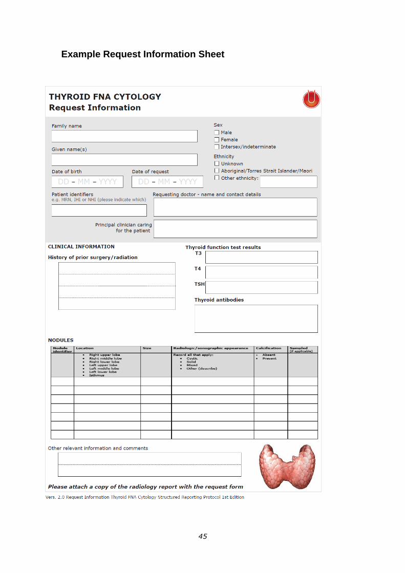
45
Example Request Information Sheet

46

47
Appendix 2 Guidelines for formatting of
a pathology report
Layout
Headings and spaces should be used to indicate subsections of the report, and
heading hierarchies should be used where the LIS allows it. Heading hierarchies
may be defined by a combination of case, font size, style and, if necessary,
indentation.
• Grouping similar data elements under headings and using ‘white space’
assists in rapid transfer of information.87
Descriptive titles and headings should be consistent across the protocol, checklist
and report.
When reporting on different tumour types, similar layout of headings and blocks
of data should be used, and this layout should be maintained over time.
• Consistent positioning speeds data transfer and, over time, may reduce
the need for field descriptions or headings, thus reducing unnecessary
information or ‘clutter’.
Within any given subsection, information density should be optimised to assist in
data assimilation and recall.
• Configuring reports in such a way that they ‘chunk’ data elements into a
single unit will help to improve recall for the clinician.87
• ‘Clutter’ should be reduced to a minimum.87 Thus, information that is not
part of the protocol (eg billing information, SNOMED codes, etc) should not
appear on the reports or should be minimized.
• Injudicious use of formatting elements (eg too much bold, underlining or
use of footnotes) constitutes clutter and may distract the reader from the
key information.
Where a structured report checklist is used as a template for the actual report,
any values provided in the checklist, but not applying to the case in question,
must be deleted from the formatted report.
Reports should be formatted with an understanding of the potential for the
information to mutate or be degraded as the report is transferred from the LIS to
other health information systems.
As a report is transferred between systems:
• text characteristics such as font type, size, bold, italics and colour are
often lost
• tables are likely to be corrupted as vertical alignment of text is lost when
fixed font widths of the LIS are rendered as proportional fonts on screen or
in print
• spaces, tabs and blank lines may be stripped from the report, disrupting
the formatting
• supplementary reports may merge into the initial report.
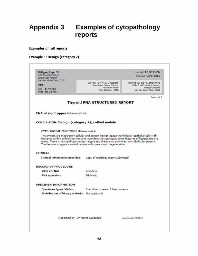
48
Appendix 3 Examples of cytopathology reports
Examples of full reports
Example 1: Benign (category 2)

49
Example 2: Suggestive of follicular neoplasm (category 4)

50
Examples of abbreviated reports
Each report requires a conclusion with the descriptor and category number. A brief
statement with further clarification is often required. Several options and examples
are given below. These example reports are only guides.
NON DIAGNOSTIC (Category 1)
Example 1
US FNA left thyroid lobe
CONCLUSION:
NON-DIAGNOSTIC (Category 1); inadequate cellularity for assessment.
CYTOLOGICAL FINDINGS (MICROSCOPIC):
(insert as appropriate)
RECOMMENDATION:
Repeat aspiration in 3 months or as clinically indicated.
Example 2
US FNA left thyroid lobe
CONCLUSION:
NON-DIAGNOSTIC (Category 1); smears are heavily blood stained with a few
scattered poorly preserved cells.
CYTOLOGICAL FINDINGS (MICROSCOPIC):
(insert as appropriate)
RECOMMENDATION:
Repeat aspiration in 3 months or as clinically indicated.
Example 3
US FNA left thyroid lobe
CONCLUSION:
NON-DIAGNOSTIC (Category 1); unsatisfactory smears with poor
preservation and smearing artifacts.
CYTOLOGICAL FINDINGS (MICROSCOPIC):
(insert as appropriate)
RECOMMENDATION:
Repeat aspiration in 3 months or as clinically indicated.

51
Example 4
US FNA left thyroid lobe
CONCLUSION:
NON-DIAGNOSTIC (Category 1); a cystic lesion with no epithelial cells
(please see comment).
CYTOLOGICAL FINDINGS (MICROSCOPIC):
The smears and cell block made of 5 ml fluid received contain foamy
macrophages and granular debris. No intact epithelial cells or malignant cells are
seen.
RECOMMENDATION:
Repeat aspiration in 3 months or as clinically indicated.
Comment: If the cyst was aspirated to dryness with no residual mass remaining
this lesion may be considered benign. Please correlate with radiological
appearance.
BENIGN (CATEGORY 2)
Example 1
FNA of right upper lobe nodule
CONCLUSION:
Benign (category 2); lymphocytic thyroiditis
CYTOLOGICAL FINDINGS (MICROSCOPIC):
The smears are moderately cellular and contains follicular epithelial
cells with a mixed population of lymphocytes in background blood;
colloid is difficult to discern. The epithelial cells are variable with
some focal oncocytic change; these oncocytic cells have some
irregularities of the nuclear membrane and very occasional grooves.
The lymphoid population appears benign. The features suggest
lymphocytic thyroiditis.
RECOMMENDATION:
Correlation with autoantibodies and thyroid function tests may be useful.
Example 2
FNA of right upper lobe nodule
CONCLUSION:
Benign (category 2); cellular colloid nodule
CYTOLOGICAL FINDINGS (MICROSCOPIC):
The smears are moderately cellular with abundant follicular epithelial
tissue fragments and dispersed intact cells in a background of thin

52
colloid and blood. The epithelial cells are uniform; some stripped
nuclei are present. There are some intact follicles and also occasional
microfollicles though the latter feature does not predominate. There is
no significant nuclear atypia. The features suggest a cellular colloid
nodule.
RECOMMENDATION:
Nil
Example 3
FNA of right upper lobe nodule
CONCLUSION:
Benign (category 2); cyst
CYTOLOGICAL FINDINGS (MICROSCOPIC):
The aspirate contains abundant thin colloid with admixed
macrophages. There is no epithelial sampling.
RECOMMENDATION:
Repeat aspiration if cyst re-accumulates.
Comment: This has been categorised as a benign cyst given the
clinical information of no residual mass/no complex features by
ultrasound.
INDETERMINATE (CATEGORY 3)
Example 1
US FNA right thyroid
CONCLUSION:
INDETERMINATE/ FOLLICULAR LESION OF UNDETERMINED
SIGNIFICANCE (CATEGORY 3); Unable to differentiate between colloid nodule
and neoplasm.
CYTOLOGICAL FINDINGS (MICROSCOPIC):
The smears are heavily bloodstained and show scattered crowded clusters of
thyroid epithelial cells. No colloid is seen. No features of papillary carcinoma are
noted.
RECOMMENDATION:
Repeat aspiration in 3 months or as clinically indicated.

53
Example 2
Left thyroid nodule FNA
CONCLUSION:
INDETERMINATE/FOLLICULAR LESION OF UNDETERMINED
SIGNIFICANCE (Category 3); features indeterminate between adenomatous
nodule and follicular neoplasm
CYTOLOGICAL FINDINGS (MICROSCOPIC):
Relatively hypocellular smears show both flat sheets as well as microfollicular
arrangements of follicular cells with no significant nuclear atypia. Minimal colloid
is present in the background.
RECOMMENDATION:
Repeat aspiration in 3 months or as clinically indicated. Suggest correlation with
clinical and radiological findings.
Example 3
Left thyroid nodule, FNA
CONCLUSION:
INDETERMINATE (Category 3); microfollicular pattern with lymphocytic
infiltrates, raising the possibility of thyroiditis
CYTOLOGICAL FINDINGS (MICROSCOPIC):
Smears show prominent microfollicular arrangements of follicular cells,
accompanied by a background lymphoid population composed predominantly of
small lymphocytes, admixed with larger, more activated forms. Minimal colloid is
identified. No significant nuclear atypia is noted.
RECOMMENDATION:
Repeat aspiration in 3 months and correlate with clinical, biochemical and
relevant serological investigations.
Comment: The findings raise the possibility of a lymphocytic thyroiditis.
Example 4
Left thyroid nodule, FNA
CONCLUSION: INDETERMINATE/FOLLICULAR LESION OF UNDETERMINED
SIGNIFICANCE (Category 3); Features indeterminate between adenomatous
nodule and follicular neoplasm.
CYTOLOGICAL FINDINGS (MICROSCOPIC):

54
Relatively hypocellular smears show both flat sheets as well as microfollicular
arrangements of follicular cells with no significant nuclear atypia. Minimal colloid
is present in the background.
RECOMMENDATION:
Repeat FNA after an appropriate interval and correlation with clinical and
radiological findings.
Comment: The likelihood of malignancy is low.
Example 5
Left thyroid nodule, FNA
CONCLUSION: INDETERMINATE (Category 3); Microfollicular pattern with lymphocytic
infiltrates, raising the possibility of thyroiditis
CYTOLOGICAL FINDINGS (MICROSCOPIC):
Smears show prominent microfollicular arrangements of follicular cells,
accompanied by a background lymphoid population composed
predominantly of small lymphocytes, admixed with larger, more activated
forms. Minimal colloid is identified. No significant nuclear atypia is noted.
RECOMMENDATION:
Repeat FNA after an appropriate interval and correlate with clinical,
biochemical and relevant serological investigations.
Comment: The likelihood of malignancy is low. The findings raise the
possibility of a lymphocytic thyroiditis.
SUGGESTIVE OF A FOLLICULAR NEOPLASM (CATEGORY 4)
Example 1
US FNA right thyroid
CONCLUSION:
Suggestive of a follicular neoplasm (Category 4); favour follicular neoplasm
CYTOLOGICAL FINDINGS (MICROSCOPIC):
The smears are heavily bloodstained and cellular, showing many equal sized
repetitive rounded microfollicles, some of which contain colloid in the central
lumen. No thin colloid is seen. No features of papillary carcinoma are noted.
RECOMMENDATION:
Suggest referral to specialist surgeon/excision.

55
Example 2
US FNA right thyroid
CONCLUSION:
Suggestive of a follicular neoplasm (Category 4)
CYTOLOGICAL FINDINGS (MICROSCOPIC):
The smears are cellular and a mixture of monolayered sheets and crowded
aggregates of heavily bloodstained and cellular showing many equal sized
repetitive rounded microfollicles, some of which contain colloid in the central
lumen. No thin colloid is seen. No features of papillary carcinoma are noted.
RECOMMENDATION:
Suggest referral to specialist surgeon/excision.
Example 3
US FNA right thyroid
CONCLUSION:
Suggestive of a follicular neoplasm (Category 4)
CYTOLOGICAL FINDINGS (MICROSCOPIC):
The smears are cellular and comprise monolayered sheets in addition to many
equal sized, repetitive, microfollicles, some of which contain colloid in the central
lumen. No thin colloid is seen. There are scattered nuclear grooves and possible
intranuclear inclusions. No papillary architecture or psammoma bodies are
noted.
RECOMMENDATION:
Suggest referral to specialist surgeon/excision. The possibility of a non-invasive
follicular thyroid neoplasm with papillary-like nuclear features (NIFTP) cannot be
excluded
SUSPICIOUS OF MALIGNANCY (Category 5)
Example 1
US FNA left thyroid lobe
CONCLUSION:
SUSPICIOUS OF MALIGNANCY (Category 5); suspicious of medullary
carcinoma.
CYTOLOGICAL FINDINGS (MICROSCOPIC):
(as appropriate)
RECOMMENDATION:

56
Referral to specialist surgeon +/- repeat FNA for ancillary testing as appropriate.
Serum calcitonin assay may be of value.
Example 2
US FNA left thyroid lobe
CONCLUSION:
SUSPICIOUS OF MALIGNANCY (Category 5); favour papillary carcinoma.
CYTOLOGICAL FINDINGS (MICROSCOPIC):
(as appropriate)
RECOMMENDATION:
Referral to specialist surgeon +/- repeat FNA for ancillary testing.
Example 3
US FNA left thyroid lobe
CONCLUSION:
SUSPICIOUS OF MALIGNANCY (Category 5); features are highly suggestive,
but not diagnostic, of papillary carcinoma.
CYTOLOGICAL FINDINGS (MICROSCOPIC):
(as appropriate)
RECOMMENDATION:
Referral to specialist surgeon
Comment: BRAF V600E mutation testing may confirm papillary thyroid
carcinoma
Example 4
US FNA left thyroid lobe
CONCLUSION:
SUSPICIOUS OF MALIGNANCY (Category 5); features are highly suggestive
but not diagnostic of papillary carcinoma.
CYTOLOGICAL FINDINGS (MICROSCOPIC):
(as appropriate)
RECOMMENDATION:
Referral to specialist surgeon.
Comment: Cell block was insufficient for further assessment and ancillary
testing.

57
Example 5
US FNA left thyroid lobe
CONCLUSION:
SUSPICIOUS OF MALIGNANCY (Category 5); some features favour a poorly
differentiated thyroid carcinoma.
CYTOLOGICAL FINDINGS (MICROSCOPIC):
(as appropriate)
RECOMMENDATION:
Referral to specialist surgeon.
Example 6
US FNA left thyroid lobe
CONCLUSION:
SUSPICIOUS OF MALIGNANCY (Category 5); features are highly suggestive
of a lymphoma.
CYTOLOGICAL FINDINGS (MICROSCOPIC):
(as appropriate)
RECOMMENDATION:
Referral to specialist management.
A repeat FNA for ancillary testing and flow cytometry would be helpful.
Example 7
US FNA left thyroid lobe
CONCLUSION:
SUSPICIOUS OF MALIGNANCY (Category 5); it is difficult to further
characterise the neoplasm.
CYTOLOGICAL FINDINGS (MICROSCOPIC):
(as appropriate)
RECOMMENDATION:
Referral to specialist management. A repeat FNA for ancillary testing may be
helpful.
MALIGNANT (CATEGORY 6)
Example 1
US FNA left thyroid lobe

58
CONCLUSION:
MALIGNANT (Category 6); papillary thyroid carcinoma.
CYTOLOGICAL FINDINGS (MICROSCOPIC):
(as appropriate)
RECOMMENDATION:
Referral to specialist surgeon.
Example 2
US FNA left thyroid lobe
CONCLUSION:
MALIGNANT (Category 6); papillary thyroid carcinoma with focal poorly
differentiated features.
CYTOLOGICAL FINDINGS (MICROSCOPIC):
(as appropriate)
RECOMMENDATION:
Referral to specialist surgeon.
Example 3
US FNA left thyroid lobe
CONCLUSION:
MALIGNANT (Category 6); medullary thyroid carcinoma.
CYTOLOGICAL FINDINGS (MICROSCOPIC):
(as appropriate)
RECOMMENDATION:
Referral to specialist surgeon.
Comment: Suggest clinicopathological correlation.
Example 4
US FNA left thyroid lobe
CONCLUSION:
MALIGNANT (Category 6); an undifferentiated malignancy. Possibilities would
include an anaplastic thyroid carcinoma of thyroid and metastatic malignancy.
CYTOLOGICAL FINDINGS (MICROSCOPIC):
(as appropriate)

59
RECOMMENDATION:
Referral to specialist surgeon. A repeat FNA for ancillary studies may be helpful
in further characterisation of malignant cells.
Comment: Suggest clinicopathological correlation.
Example 5
US FNA left thyroid lobe
CONCLUSION:
MALIGNANT (Category 6); consistent with metastatic renal cell carcinoma in
the given clinical setting.
CYTOLOGICAL FINDINGS (MICROSCOPIC):
(as appropriate)
RECOMMENDATION:
Referral to specialist management.
Comment: Cytological features and are similar to those of the primary renal cell
carcinoma (Ref XXXXXXXXX) and the immunohistochemical profile is consistent
with a renal cell carcinoma.

60
References
1 Tan H, Gharib H and Reading CC (1995). Solitary thyroid nodule. Comparison between palpation and ultrasonography. Arch Intern Med. 155:2418-2423.
2 Mackenzie EJ and Mortimer RH (2004). Thyroid nodules and thyroid cancer. Medical Journal of Australia 180(5):242-247.
3 AIHW (Australian Institute of Health and Welfare). Australian Cancer Incidence and Mortality (ACIM) books. Available at http://www.aihw.gov.au/acim-books/ Accessed: 3rd Dec 2012.
4 Ali S and Cibas ES (eds) (2010). The Bethesda System for Reporting Thyroid Cytopathology. Definitions, Criteria and Explanatory Notes. 1st Edition, Springer.
5 The Papanicolaou Society of Cytopathology Task Force on Standards for Practice (1997). Guidelines of the Papanicolaou Society of Cytopathology for fine needle aspiration procedure and reporting. Diagn Cytopathol 17:239-247.
6 British Thyroid Association Royal College of Physicians (2007). Guidelines for the management of thyroid cancer 2nd edition. Report of the Thyroid Cancer Guidelines Update Group. Perros P (eds). Royal College of Physicians, London.
7 Orell SR and Philips J (2000). Broadsheet number 57: problems in fine needle biopsy of the thyroid. Pathology 32(3):191-198.
8 Nga Min En, Kumarasinghe MP, Tie B, Sterrett GF, Wood B, Walsh J, Nguyen H, Whyte A and Frost F (2010). Experience with standardized thyroid fine-needle aspiration reporting categories. Cancer Cytopathology 118(6):423-433.
9 Tsan CJ, Serpell JW and Poh YY (2007). The impact of synoptic cytology reporting on fine-needle aspiration cytology of thyroid nodules. ANZ J Surg. 77(11):991-995.
10 RCPA (Royal College of Pathologists of Australasia (2009 ). Guidelines for Authors of Structured Cancer Pathology Reporting Protocols. RCPA, Surry Hills NSW.
11 RCPA (Royal College of Pathologists of Australasia) (2004). The Pathology Request-Test-Report Cycle — Guidelines for Requesters and Pathology Providers, RCPA, Surry Hills, NSW.

61
12 Pitman M, Abele J, Ali S, Duuick D, Elsheikh TM, Jeffrey B, Powers C, Randolph G, Renshaw A and Scout L (2008). Techniques for Thyroid FNA: A Synopsis of the National Cancer Institute Thyroid Fine-Needle Aspiration State of the Science Conference. Diagn Cytopathol 36:407-424.
13 RCPA (Royal College of Pathologists of Australasia) (2004). Chain of Information Custody for the Pathology Request-Test-Report Cycle — Guidelines for Requesters and Pathology Providers. RCPA, Surry Hills, NSW.
14 Kocjan G, Chandra A, Cross P, Denton K, Giles T, Herbert A, Smith P, Remedios D and Wilson P (2009). BSCC Code of Practice – fine needle aspiration cytology. Cytopathology. 20:283-296.
15 Kumarasinghe MP and Sheriffdeen AH (1995). Fine needle sampling without aspiration. Pathology 27:330-332.
16 Abele J (2012). Putting Aspiration Back Into Thyroid Fine-Needle Biopsy—The Re-Emerging Role of Vacuum Assistance. Cancer Cytopathology 120:366-372.
17 Olson MT, Armanda D, Tatsas AD and Ali SZ (2012). Cytotechnologist-attended on-site adequacy evaluation of thyroid fine needle aspiration. Comparison with cytopathologists and correlation with the final interpretation. Am J Clin Pathol 138:90-95.
18 Burlingame OO, Kesse KO, Silverman SG and Cibas ES (2012). On-site adequacy evaluations performed by cytotechnologists correlation with final interpretations of 5241 image-guided fine-needle aspiration biopsies. Cancer (Cancer Cytopathol) 120:177-184.
19 Jing X, Wey E and Michael CW (2011). Retrospective evaluation of instituted standard adequacy criteria for on-site adequacy assessment of thyroid fine-needle aspiration. Diagn. Cytopathol. 39:391-394.
20 Schmidt RL, Kordy MA, Howard K, Layfield LJ, Hall BJ and Adler DG (2013). Risk-benefit analysis of sampling methods for fine-needle aspiration cytology: a mathematical modeling approach. Am J Clin Pathol 139(3):336-344.
21 Saleh H, Bassily N and Hammoud MJ (2009). Utility of a liquid-based, monolayer preparation in the evaluation of thyroid lesions by fine needle aspiration biopsy: comparison with the conventional smear method. Acta Cytol 53(2):130-136.

62
22 Fischer AH, Clayton AC, Bentz JS, Wasserman PG, Henry MR, Souers RJ and Moriarty AT (2013). Performance differences between conventional smears and liquid-based preparations of thyroid fine-needle aspiration samples: analysis of 47,076 responses in the College of American Pathologists Interlaboratory Comparison Program in Non-Gynecologic Cytology. Arch Pathol Lab Med 137(1):26-31.
23 Orell SR, Sterrett GF and Whitaker D (2005). Fine Needle Aspiration Cytology. 4th Ed Elsevier Churchill Livingstone, London.
24 Choi KU, Kim JY, Park DY, Lee CH, Sol MY, Han KT and Kim YG (2005). Recommendations for the management of cystic thyroid nodules. ANZ J Surg 75(7):537-541.
25 Takada N, Hirokawa M, Suzuki A, Higuchi M, Kuma S and Miyauchi A (2017). Reappraisal of "cyst fluid only" on thyroid fine-needle aspiration cytology. Endocr J 64(8):759-765.
26 WHO (World Health Organization) (2017). WHO classification of tumours of endocrine organs (4th edition). Lloyd RV, Osamura R, Kloppel G and Rosai J. IARC Press, Lyon.
27 Seethala RR, Baloch ZW, Barletta JA, Khanafshar E, Mete O, Sadow PM, LiVolsi VA, Nikiforov YE, Tallini G and Thompson LD (2018). Noninvasive follicular thyroid neoplasm with papillary-like nuclear features: a review for pathologists. Mod Pathol 31(1):39-55.
28 Strickland KC, Howitt BE, Marqusee E, Alexander EK, Cibas ES, Krane JF and Barletta JA (2015). The Impact of Noninvasive Follicular Variant of Papillary Thyroid Carcinoma on Rates of Malignancy for Fine-Needle Aspiration Diagnostic Categories. Thyroid 25(9):987-992.
29 Giordano TJ, Kuick R, Thomas DG, Misek DE, Vinco M, Sanders D, Zhu Z, Ciampi R, Roh M, Shedden K, Gauger P, Doherty G, Thompson NW, Hanash S, Koenig RJ and Nikiforov YE (2005). Molecular classification of papillary thyroid carcinoma: distinct BRAF, RAS, and RET/PTC mutation-specific gene expression profiles discovered by DNA microarray analysis. Oncogene 24(44):6646-6656.
30 Nikiforov YE, Seethala RR, Tallini G, Baloch ZW, Basolo F, Thompson LD, Barletta JA, Wenig BM, Al Ghuzlan A, Kakudo K, Giordano TJ, Alves VA, Khanafshar E, Asa SL, El-Naggar AK, Gooding WE, Hodak SP, Lloyd RV, Maytal G, Mete O, Nikiforova MN, Nose V, Papotti M, Poller DN, Sadow PM, Tischler AS, Tuttle RM, Wall KB, LiVolsi VA, Randolph GW and Ghossein RA (2016). Nomenclature Revision for Encapsulated Follicular Variant of Papillary Thyroid Carcinoma: A Paradigm Shift to Reduce Overtreatment of Indolent Tumors. JAMA Oncol 2(8):1023-1029.

63
31 Cancer Genome Atlas Research Network (2014). Integrated genomic characterization of papillary thyroid carcinoma. Cell 159(3):676-690.
32 Sarkis LM, Norlen O, Aniss A, Watson N, Delbridge LW, Sidhu SB, Sywak MS and Gill AJ (2014). The Australian experience with the Bethesda classification system for thyroid fine needle aspiration biopsies. Pathology 46(7):592-595.
33 Gunawardena D, Frost F, Kumarasinghe MP, Sterrett GF, Nguyen H, Minchin D, Lisewski D, Ryan S, Yew MK, Wong SL, Walsh J and Boeddinghaus R (2017). Clinical significance of malignant and suspicious categories in thyroid FNA cytology: a 10-year experience of a single institution. Pathology 49 (S134).
34 Shi Q, Ibrahim A, Herbert K, Carvin M, Randolph M, Post KM, Curless K, Chen S, Cramer HM, Cheng L and Wu HH (2015). Detection of BRAF mutations on direct smears of thyroid fine-needle aspirates through cell transfer technique. Am J Clin Pathol 143(4):500-504.
35 Cibas ES (2009). Cervical and vaginal cytology. In: Cytology. Diagnostic principles and clinical correlates. 3rd edition, Saunders Elsevier, Philadelphia, Edinburgh.
36 Kocjan G, Cochand-Priollet B, de Agustin PP, Bourgain C, Chandra A, Daneshbod Y, Deery A, Duskova J, Ersoz C, Fadda G, Fassina A, Firat P, Jimenez-Ayala B, Karakitsos P, Koperek O, Matesa N, Poller D, Thienpont L, Ryska A, Schenck U, Sauer T, Schmitt F, Tani E, Toivonen T, Tötsch M, Troncone G, Vass L and Vielh P (2010). Diagnostic terminology for reporting thyroid fine needle aspiration cytology: European Federation of Cytology Societies thyroid working party symposium, Lisbon 2009. Cytopathology. 21(2):86-92.
37 Kini SR (2008). Thyroid Cytopathology: A Text and Atlas Lippincott Williams and Wilkins 38 Perros P (ed) (2007). British Thyroid Association, Royal college of Physicians: Guidelines for the Management of Thyroid Cancer. 2nd ed. Report of the Thyroid Cancer Guidelines Update Group, Royal College of Physicians, London, United Kingdom.
39 Cibas ES, Ali SZ and NCI Thyroid FNA State of the Science Conference (2009). The Bethesda System For Reporting Thyroid Cytopathology. Am J Clin Pathol 132(5):658-665.
40 Singh RS and Wang HH (2011). Timing of repeat thyroid fine-needle aspiration in the management of thyroid nodules. Acta Cytol 55(6):544-548.
41 DeMay RM (2011). Chapter 12 In: The Art and Science of Cytopathology, American Society for Clinical Pathology.

64
42 Lloyd RV et al (2004). Observer Variation in the Diagnosis of Follicular Variant of Papillary Thyroid Carcinoma. Am J Surg Pathol 28:1336-1340.
43 Hirokawa M et al (2002). Observer Variation of Encapsulated Follicular Lesions of the Thyroid Gland. Am J Surg Pathol 26(11):1508-1514.
44 Baloch ZW and LiVolsi VA (2007). Our approach to follicular-patterned lesions of the thyroid J Clin Pathol 60:244-250.
45 Kakudo K, Higuchi M, Hirokawa M, Satoh S, Jung CK and Bychkov A (2017). Thyroid FNA cytology in Asian practice-Active surveillance for indeterminate thyroid nodules reduces overtreatment of thyroid carcinomas. Cytopathology 28(6):455-466.
46 Haugen BR, Alexander EK, Bible KC, Doherty GM, Mandel SJ, Nikiforov YE, Pacini F, Randolph GW, Sawka AM, Schlumberger M, Schuff KG, Sherman SI, Sosa JA, Steward DL, Tuttle RM and Wartofsky L (2016). 2015 American Thyroid Association Management Guidelines for Adult Patients with Thyroid Nodules and Differentiated Thyroid Cancer: The American Thyroid Association Guidelines Task Force on Thyroid Nodules and Differentiated Thyroid Cancer. Thyroid 26(1):1-133.
47 Park HJ, Moon JH, Yom CK, Kim KH, Choi JY, Choi SI, Ahn SH, Jeong WJ, Lee WW and Park SY (2014). Thyroid "atypia of undetermined significance" with nuclear atypia has high rates of malignancy and BRAF mutation. Cancer Cytopathol 122(7):512-520.
48 Yassa L, Cibas ES and Benson CB et al (2007). Long term assessment of a multidisciplinary approach to thyroid nodule diagnostic evaluation. Cancer 111:508-516.
49 Nayar R and Ivanovic M (2009). The indeterminate thyroid FNA: Experience from an academic center using terminology similar to that proposed in the 2007 NCI Thyroid Fine Needle Aspiration State of the Science Conference. Cancer Cytopathology(117):195-202.
50 Nikiforova MN and Nikiforov YE (2009). Molecular diagnostics and predictors in thyroid cancer. Thyroid 19:1351-1361.
51 Mehta V, Nikiforov YE and Ferris RL (2013). Use of molecular biomarkers in FNA specimens to personalize treatment for thyroid surgery. Head Neck 35:1499-1506.
52 Cooper DS, Doherty GM, Haugen BR, Kloos RT, Lee SL, Mandel SJ, Mazzaferri EL, McIver B, Pacini F, Schlumberger M, Sherman SI, Steward DL and Tuttle RM (2009). Revised American

65
Thyroid Association Management Guidelines for Patients with Thyroid Nodules and Differentiated Thyroid Cancer. (The American Thyroid Association (ATA) Guidelines Taskforce on Thyroid Nodules and Differentiated Thyroid Cancer). Thyroid 19:1167-1214.
53 Hassell LA, Gillies EM and Dunn ST (2012). Cytologic and molecular diagnosis of thyroid cancers. Is it time for routine reflex testing? . Cancer Cytopathology 120(1):7-17.
54 Kussick SJ, Kalnoski M, Braziel RM and Wood BL (2004). Prominent Clonal B-Cell Populations Identified by Flow Cytometry in Histologically Reactive Lymphoid Proliferations. Am J Clin Pathol 121:464-472.
55 Savage EC, Vanderheyden AD and Bell AM et al (2011). Diagnostic Accuracy of Flow Cytometry Obtained From Fine-Needle Aspirates : A 10-Year Experience With 451 Cases Am J Clin Pathol 135:304-309.
56 Clark D and Faquin W (2005). Thyroid Cytopathology 2nd edition. Essentials in Cytopathology. Series editor: Rosenthal DL. Springer, New York 57 Mac De May R (2012). The Art and Science of Cytopathology. 2nd Edition. American Society for Clinial Pathology Press.
58 Alexander EK, Kennedy GC, Baloch ZW, Cibas ES, Chudova D, Diggans J, Friedman L, Kloos RT, LiVolsi VA, Mandel SJ, Raab SS, Rosai J, Steward DL, Walsh PS, Wilde JI, Zeiger MA, Lanman RB and Haugen BR (2012). Preoperative Diagnosis of Benign Thyroid Nodules with Indeterminate Cytology. New England Journal of Medicine 367(8):705-715.
59 Leslie C, Grieu-Iacopetta F, Richter A, Platten M, Murray J, Frost FA, Amanuel B and Kumarasinghe MP (2015). BRAF p.Val600Glu (V600E) mutation detection in thyroid fine needle aspiration cell block samples: a feasibility study. Pathology 47(5):432-438.
60 Kimura ET, Nikiforova MN and Zhu Z et al (2003). High prevalence of BRAF mutations in thyroid cancer: genetic evidence for constitutive activation of theRET/PTC–RAS–BRAF signalling pathway in papillary thyroid carcinoma. Cancer Res. 63:1454-1457.
61 Nikiforova MN, Kimura ET and Gandhi M et al (2003). BRAF mutations in thyroid tumors are restricted to papillary carcinomas and anaplastic or poorly differentiated carcinomas arising from papillary carcinomas. J Clin Endocrinol Metab 88:5399 -5404.
62 Ohori NP, Singhal R, Nikiforova MN, Yip L, Schoedel KE, Coyne C, McCoy KL, LeBeau SO, Hodak SP, Carty SE and Nikiforov YE (2013). BRAF mutation detection in indeterminate thyroid cytology specimens: underlying cytologic, molecular, and pathologic characteristics of papillary thyroid carcinoma. Cancer Cytopathol. 121(4):197-205.

66
63 Judkins AR, Roberts SA and LiVolsi VA (1999). Utility of immunohistochemistry in the evaluation of necrotic thyroid tumours. Hum.Pathol. 30:1373-1376.
64 Chai SM and Kumarasinghe MP (2011). Diagnosis of necrotic and degenerate thyroid lesions: value of Immunohistochemistry Histopathology 59:496-503.
65 Smith AL, Williams MD, Stewart J, Wang WL, Krishnamurthy S, Cabanillas ME and Roy-Chowdhuri S (2018). Utility of the BRAF p.V600E immunoperoxidase stain in FNA direct smears and cell block preparations from patients with thyroid carcinoma. Cancer Cytopathol 126(6):406-413.
66 Xu B, Thong N, Tan D and Khoury T (2010). Expression of thyroid transcription factor-1 in colorectal carcinoma. Appl Immunohistochem Mol Morphol. 18(3):244-249.
67 Robens J, Goldstein L, Gown AM and Schnitt SJ (2010). Thyroid transcription factor-1 expression in breast carcinomas. Am J Surg Pathol 34(12):1881-1885.
68 Leite KR, Mitteldorf CA, Srougi M, Dall'oglio MF, Antunes AA, Pontes J Jr and Camara-Lopes LH (2008). Cdx2, cytokeratin 20, thyroid transcription factor 1, and prostate-specific antigen expression in unusual subtypes of prostate cancer. Ann Diagn Pathol. 12(4):260-266.
69 Nonaka D, Tang Y, Chiriboga L, Rivera M and Ghossein R (2008). Diagnostic utility of thyroid transcription factors Pax8 and TTF-2 (FoxE1) in thyroid epithelial neoplasms. Mod Pathol. 21(2):192-200.
70 Enriquez ML, Baloch ZW, Montone KT, Zhang PJ and LiVolsi VA (2012). CDX2 expression in columnar cell variant of papillary thyroid carcinoma. Am J Clin Pathol. 137(5):722-726.
71 Schmitt AC, Cohen C and Siddiqui MT (2010). Paired Box Gene 8, HBME-1, and Cytokeratin 19 Expression in Preoperative Fine-Needle Aspiration of Papillary Thyroid Carcinoma. Cancer Cytopathology 118(4):196-202.
72 Nga ME, Lim GS, Soh CH and Kumarasinghe MP (2008). HBME-1 and CK19 are highly discriminatory in the cytological diagnosis of papillary thyroid carcinoma. Diagn Cytopathol. 36(8):550-556.
73 Cui W, Sang W, Zheng S, Ma Y, Liu X and Zhang W (2012). Usefulness of cytokeratin-19, galectin-3, and Hector Battifora mesothelial-1 in the diagnosis of benign and malignant thyroid nodules. Clin Lab 58(7-8):673-680.

67
74 Cheng S, Serra S, Mercado M, Ezzat S and Asa SL (2011). A high-throughput proteomic approach provides distinct signatures for thyroid cancer behavior. Clin Cancer Res. 17(8):2385-2394.
75 Koperek O, Kornauth C, Capper D, Berghoff AS, Asari R, Niederle B, von Deimling A, Birner P and Preusser M (2012). Immunohistochemical Detection of the BRAF V600E-mutated Protein in Papillary Thyroid Carcinoma. Am J Surg Pathol 36(6):844-850.
76 Lee JH, Lee ES and Kim YS (2007). Clinicopathologic significance of BRAF V600E mutation in papillary carcinomas of the thyroid: A meta-analysis. Cancer 110:38-46.
77 Toll AD, Rossi ED and Ali SZ (2014). Role of ancillary testing in thyroid fine needle aspiration: Review and update. J Am Soc Cytopathol 3(4):218-224.
78 Rossi ED, Martini M, Capodimonti S, Cenci T, Straccia P, Angrisani B, Ricci C, Lanza P, Lombardi CP, Pontecorvi A, Larocca LM and Fadda G (2014). Analysis of immunocytochemical and molecular BRAF expression in thyroid carcinomas: A cytohistologic institutional experience. Cancer Cytopathol 122(7):527-535.
79 Xing M, Alzahrani AS and Carson KA et al (2013). Association between BRAF V600E mutation and mortality in patients with papillary thyroid cancer. JAMA 309(14):1493-1501.
80 Sancisi V, Nicoli D, Ragazzi M, Piana S and Ciarrocchi A (2012). BRAFV600E mutation does not mean distant metastasis in thyroid papillary carcinomas. J Clin Endocrinol Metab. 97(9):E1745-1749.
81 Li C, Han PA and al LKe (2013). Does BRAF V600E Mutation Predict Aggressive Features in Papillary Thyroid Cancer? Results From Four Endocrine Surgery Centers. The Journal of Clinical Endocrinology & Metabolism 98(9):3702-3712.
82 Cunha N, Rodrigues F and Curado F at al (2007). Thyroglobulin detection in fine-needle aspirates of cervical lymph nodes: a technique for the diagnosis of metastatic differentiated thyroid cancer. Eur J Endocrin. 157:101-107.
83 Giovanella L, Ceriani L and Suriano S (2011). Lymph node thyroglobulin measurement in diagnosis of neck metastases of differentiated thyroid carcinoma. J Thyroid Res 2011: 621839:doi:10.4061/2011/621839.

68
84 Baloch ZW, Barroeta JE, Walsh J, Gupta PK, Livolsi VA and Langer JE et al (2008). Utility of Thyroglobulin measurement in fine-needle aspiration biopsy specimens of lymph nodes in the diagnosis of recurrent thyroid carcinoma. Cytojournal. 5(1).
85 Mayo Clinic (2014 ). Online Test Catalog. Available at www.mayomedicallaboratories.com/test-catalog Accessed 8th August 2014. 86 Royal College of Pathologists of Australasia (2011). Functional Requirements for Laboratory Information Systems to support Structured Pathology Reporting of Cancer Protocols https://www.rcpa.edu.au/Library/Practising-Pathology/Structured-Pathology-Reporting-of-Cancer/Implementation.
87 Valenstein PN (2008). Formatting pathology reports: applying four design principles to improve communication and patient safety. Arch Path Lab Med. 132(1):84–94.