Embed Size (px)
Citation preview
Thursday 30 March 2017
Rhinitis, 14:15 - 16:06
O01 Long-term follow-up of local allergic rhinitis patients
Ibon Eguiluz Gracia1, Carmen Rondón1, Paloma Campo1, Ana Prieto1, Lina Mayorga1, Luisa Galindo1, Ana Molina1, Miguel Blanca1, Maria Jose Torres1
1Hospital Regional Universitario de Malaga and IBIMA, Málaga, Spain
Keywords: Local Allergic Rhinitis, Allergic Rhinitis, Sensitization
Introduction
There are few data available regarding natural history of local allergic rhinitis (LAR). We previously reported the results of the first 5-years of follow-up observing a similar rate of development of systemic
allergic rhinitis (AR) in both LAR patients and healthy controls.
Objective: To explore the natural history of a population with LAR and the development of AR and
comorbidities over a 10 year period
Methods
A cohort of 194 patients with LAR of recent onset (<18 months) and 130 age- and sex-matched healthy controls were prospectively evaluated in a 10-year follow-up study (2005-2016). All participants provided
informed consent and ethic committee of the hospital approved the study. Clinical-demographic
questionnaire, spirometry, SPT and specific IgE (sIgE) to aeroallergens were evaluated yearly. Nasal
allergen provocation tests (NAPT) with D. pteronyssinus (DP), Alternaria, Olea europea, and grass pollen
were performed at baseline, and after 5 and 10 years. Results
A total of 151 patients (78%) and 90 controls (69%) completed the study. At baseline, most patients had
moderate-to-severe persistent-perennial rhinitis. Conjunctivitis (52%) and asthma (19%) were the main
comorbidities, and DP the most frequent sensitizing aeroallergen (51.1%). During the 10 years of
evaluation 21 new cases of asthma (12.5%, P=0.007) and 17 new cases of conjunctivitis (10%, P=0.067) were diagnosed. After 10 years of evolution a similar rate of development of AR was detected in patients
and healthy controls (11.3% vs 10%, P=0.761). In 5 patients, conversion to systemic atopy occurred in the
last year of evaluation (3%).
Conclusions
LAR is a well-differentiated clinical entity with a low rate of development of systemic atopy. This study
was funded by the Institute of Health “Carlos III” of the Spanish Ministry of Economy and Competitiveness through the RETICS ARADyAL (RD16/0006/0001) and the FIS PI14/00864
O02 ILC2-Activation Aggravates Th2-dependent Nasal inflammation in mice
Taiyo Morikawa1, Ayumi Fukuoka1, Kazufumi Matsushita1, Shigeharu Fujieda2, Tomohiro Yoshimoto1
1. Hyogo College of Medicine, Nishinomiya, Hyogo, Japan
2. University of Fukui, Fukui, Japan
Keywords: Nasal Allergy, Th2 Cells, Group 2 Innate Lymphoid Cells, IL-33, IL-13
Introduction
It is well known that T helper (Th) 2 cells and group 2 innate lymphoid cells (ILC2s) contribute to allergic diseases. However, their exact role and relationship in nasal allergic disorders is unclear. We sought to
investigate the cooperation of Th2 cells and ILC2s in a mouse model of nasal allergic disorder. Methods
To differentially activate Th2 cells and/or ILC2s in nasal mucosa, mice were intranasally administered
ovalbumin (OVA) antigen, papain, an ILC2-activatior, or both for 2 weeks. Epithelial thickness and
number of eosinophils in the nasal mucosa were evaluated at 24 hours after the final challenge.
Results Intranasal administration of OVA and papain preferentially activated Th2 cells and ILC2s, respectively, in
the nose. Both OVA and papain increased the nasal epithelial thickness and number of eosinophils, and
their coadministration significantly enhanced the symptoms. ILC2- and Rag2-deficient mice showed a
partial decrease in OVA-plus-papain-induced nasal epithelial thickening and eosinophilia. Interleukin
(IL)-33- and ST2-deficient mice showed decreased OVA-plus-papain-induced, but not OVA-alone-induced nasal epithelial thickening and eosinophilia. IL-5 induced eosinophilia only, but IL-13 contributed
to both nasal epithelial thickening and eosinophilia.
Conclusions
IL-33/ST2-pathway-mediated ILC2 activation exacerbated additively Th2-cell-induced nasal type 2
inflammation. Furthermore, IL-13, but not IL-5, contributes to exacerbation of nasal type 2 inflammation.
O03 Allergen endotoxins induce non-IgE-mediated Nasal hypersensitivity in mice via
monocyte/macrophage-dependent pathway
Tomohiro Yoshimoto, Naruhito Iwasaki, Kazufumi Matsushita
Hyogo College of Medicine, Nishinomiya, Japan
Keywords: Non-Allergic Rhinitis, Endotoxin, Histamine, T Cells, Monocytes/Macrophages
Introduction
Allergen-mediated cross-linking of IgE on mast cells/basophils is a well-recognized trigger for type-1
allergic diseases such as allergic rhinitis (AR). However, allergens may not be the only trigger for AR, and several allergic-like reactions are induced by non-IgE-mediated mechanisms. Here, we describe a novel
non-IgE-mediated, endotoxin-triggered nasal type-1-hypersensitivity reaction in mice.
Methods
To investigate whether endotoxin affects sneezing responses, mice were intraperitoneally immunized with
ovalbumin (OVA), and then nasally challenged with endotoxin-free or endotoxin-containing OVA. To
investigate the role of T cells and mechanisms of the endotoxin-induced response, mice were adoptively transferred with in vitro differentiated OVA-specific Th2 cells, and then nasally challenged with
endotoxin-free or endotoxin-containing OVA. Immediately after each nasal challenge, the frequency of
sneezing was counted for 10 minutes. The mice were sacrificed 24 hours after the final nasal challenge,
and noses were dissected for analyzing infiltrating inflammatory cells.
Results Endotoxin-containing, but not endotoxin-free, OVA elicited sneezing responses in mice independent from
IgE-mediated signaling. OVA-specific Th2 cell adoptive transfer to mice demonstrated that local
activation of antigen-specific Th2 cells was required for the response. The Toll-like receptor 4-MyD 88-
signaling pathway was indispensable for endotoxin-containing OVA-elicited rhinitis. In addition,
lipopolysaccharide directly triggered sneezing responses in OVA-specific Th2-transferred and nasally endotoxin-free OVA-primed mice. Although an antihistamine, diphenhydramine, suppressed sneezing
responses, mast-cell/basophil-depleted mice had normal sneezing responses to endotoxin-containing
OVA. Clodronate treatment abrogated endotoxin-containing OVA-elicited rhinitis, suggesting the
involvement of monocytes/macrophages in this response.
Conclusions Antigen-specific nasal activation of CD4+ T cells followed by endotoxin exposure induces mast
cell/basophil-independent histamine release in the nose that elicits sneezing responses. Thus,

environmental or nasal residential bacteria may exacerbate AR symptoms. In addition, this novel phenomenon might explain currently unknown mechanisms in non–IgE-mediated allergic disorders, such
as non-IgE-mediated gastrointestinal food allergy in infants.
Thursday 30 March 2017
Omics in Rhinology, 16:30 - 17:44
O04 Increased microbial abundance and decreased diversity in preschool children at risk for
asthma
Tamar Smulders1, Danielle Van Egmond1, Kees Van Drunen1, Marc Van Der Schee1
1AMC-UvA, Amsterdam, Netherlands, The
Keywords: Asthma, Respiratory Microbiome, Rhinovirus, Wheeze, IS-Pro
Introduction
Recent studies suggest that a specific respiratory microbiome is associated with an increased risk of viral infection and that in turn these infections influence the composition of the resident microbiota. A
commonly found virus that causes infection in preschool children is Rhinovirus (RV), wheeze is an
important symptom. Children with a symptomatic RV infection have an increased risk to develop
persistent wheeze and asthma.
If children with a RV infection have a specific respiratory microbiome this may hold implications for
early diagnosis of asthma and novel therapeutic approaches aimed at restoring microbial dysbiosis. Methods
We aimed to study this by comparing the nasal microbiome of children with physician confirmed wheeze,
during acute symptoms and after recovery, to symptomatic controls with non-wheezing respiratory illness
and asymptomatic healthy controls. Specificity of our outcomes for RV was determined by matched
analysis in RV negative (RV-) children. As part of the EUROPA-study children were visited within 8 hours of exhibiting respiratory symptoms and again upon recovery for assessment of symptoms and nasal
swab collection. Swabs were tested by qPCR for 14 respiratory viruses. Bacterial microbiota analysis was
done by IS-pro, a 16S-23S PCR-based bacterial profiling technique. Relative abundance and Shannon
diversity index were compared between groups
Results 160 pre-school children were included in the study. RV induced wheeze had the highest normalized total
bacterial abundance (mean±SEM; 0.95 ± 0.08) followed by symptomatic controls (0.77±0.08) and
asymptomatic controls (0.57 ± 0.09, p = 0.01). This increase was related to an upregulation of bacterial
species belonging to the phyla of Firmicutes and Bacteroidetes of which the latter persisted after recovery
from infection. None of the control subjects carried Bacteroidetes. Microbial diversity was significantly (p
= 0.04) decreased in RV+ wheezing children (Median [IQR] 1.60 [0.97] ) compared to RV- wheezing children (2.00 [0.75]).
Conclusions
We established an increased microbial abundance and decreased microbial diversity in children with
Rhinovirus induced wheeze who are at an increased risk to develop asthma. For a subset of these children
this was primarily attributed to shifts in various species belonging to the phylum of Bacteroidetes. Our findings hold potential implications for the early diagnosis of asthma and novel therapeutic approaches

aimed at restoring microbial dysbiosis.
O05 Proteomics of eosinophilic mucin
Achim Georg Beule1
1Klinik für Hals, Nasen- und Ohrenheilkunde, Universitätsklinikum Münster, Münster, Germany
Introduction
Eosinophilic mucin is a clinical relevant nasal secretion in patients with chronic rhinosinusitis, (CRS)
warning the surgeon of increased risks of recurrence of both nasal polyps and symptoms. While the pathophysiology leading to this thick, glue-like mucus is poorly understood, physicians are increasingly
confronted to employ topical and or systemic strategies to avoid postoperative deterioration. Aim of this
study was to determine a typical proteomics signature of eosinophilic mucin to improve our understanding
of the underlying pathophysiology.
Methods Nasal secretions were collected from 10 healthy volunteers and compared to intraoperatively collected
specimens of eosinophilic mucin. Pooled samples were analyzed using 2D-Gel PAGE, MALDI-MS and
IPA analysis. Gender contributions was balanced and topical medication as well as allergy as confounding
factors excluded.
Results 593 proteins were identified, of which 373 proteins were present in both pooled samples (mucin only 76,
healthy lavage only 144 proteins). 303 out of 393 proteins were regulated in their expression. If a cut-off
value of 1.5 was determined, 43 were induced and 40 were reprimed in eosinophilic mucin.
Conclusions
Based on our results, specific proteomic changes could be observed in eosinophilic mucin enabling further
insight into the specific pathophysiology leading to deterioration in this subgroup of CRS patients.
Thursday 30 March 2017
Welcome reception and poster discussions, 17:45 - 19:45
P01 Probiotics impregnated bedding covers in house dust mite allergic rhinitis patients: a pilot
randomized controlled trial
Margot Berings1, Anton Jult2, Hanne Vermeulen2, Natalie De Ruyck1, Lara Derycke1, Hakan Ucar3, Philip Ghekiere3, Robin Temmerman4, John Ellis5, Claus Bachert1, Bart Lambrecht6, Melissa Dullaers6,
Philippe Gevaert6
1. Upper Airways Research Laboratory, Ghent University, Ghent, Belgium 2. Ghent University, Ghent, Belgium
3. BekaertDeslee Innovation bvba, Waregem, Belgium
4. Chrisal NV, Lommel, Belgium
5. Devan Chemicals NV, Ronse, Belgium
6. Laboratory of Immunoregulation, VIB Inflammation Research Center, Ghent University, Ghent, Belgium
Keywords: Allergy, Rhinitis, House Dust Mite, Probiotics, Avoidance
Introduction
House dust mite exposure is a major cause of allergy worldwide. As current evidence for existing house
dust mite avoidance measures is low, new methods are being developed.
Objectives: To explore the effect of probiotics-impregnated bedding covers on symptoms and quality of life of patients with allergic rhinitis to house dust mite.
Methods
A double-blind, randomised, placebo-controlled, crossover trial was conducted at Ghent University
Hospital, Belgium. The pilot trial included 20 adult patients with allergic rhinitis to house dust mite. The
trial consisted of an 8-week period with untreated (placebo) covers and an 8-week period with probiotics-impregnated covers in random order, with a washout period in between of at least four weeks. Der p 1
concentrations were measured in dust samples collected from mattresses and pillows. Symptoms and
quality of life were assessed through self-reported questionnaires.
Product information: The probiotics-based textile treatment contains five different probiotic and natural
bacterial strains of Bacillus species (Bacillus subtilis, Bacillus amyloliquefaciens and Bacillus pumilus).
The probiotics are incorporated in microcapsules, which are diffusely inserted in the textile. The probiotics remain non-active in the closed microcapsules, until upon friction forces a small number of the
microcapsules rupture and release their probiotic bacteria.
Results
There was a comparable and significant reduction of Der p 1 levels with both the probiotics -impregnated
covers and the untreated covers. Several symptom and quality of life scores improved significantly with the probiotics-impregnated covers, whereas no significant changes were observed with the untreated
covers. The effects of the probiotics-impregnated covers on symptoms and quality of life scores however
were not significant compared to the placebo covers (except for a subscore ‘NRQLQ sleeptime’).
Conclusions
This pilot study suggests that probiotics-impregnated bedding covers may improve symptoms and quality of life of patients with allergic rhinitis to house dust mite. Although the effects of the probiotics -
impregnated covers were not significant compared to the untreated covers, these findings are promising
and warrant a future large-scale clinical trial.
Prior to patient enrolment, the trial was registered at clinical.trials.gov (NCT01997606).
P02 Reliable mite-specific IgE testing in nasal secretions by means of allergen microarray Stefania Arasi1, Margot Berings2, Serena Perna1, Natalie De Ruyck2, Yvonne Resch3, Christian Lupinek3,
Kuan-Wei Chen3, Susanne Vrtala3, Rudolf Valenta3, Paolo Maria Matricardi1, Philippe Gevaert2
1. Department of Pediatric Pneumology and Immunology, Charité Medical School, Berlin, Germany 2. Upper Airway Research Laboratory, Ghent University, Ghent, Belgium
3. Medical University of Vienna, Vienna, Austria
Keywords: Allergen Molecules, Allergic Rhinitis, House Dust Mite Allergy, Immunoglobulin E, Nasal
Secretions
Introduction
In nasal secretions (NS) IgE is present at very low concentrations and difficult to detect with standard methods.
To evaluate the performance of a customized microarray chip (Thermo Fisher Scientific, TFS, Uppsala,
Sweden) derived from the new ImmunoCAP ISAC 103TM for simultaneous detection of IgE to 13
components of Dermatophagoides pteronyssinus (D.pt.) and 2 of Dermatophagoides farinae (D.fa.) in
serum and NS. Methods
NS were collected with both Filter Disks (FD) and Sinus Packs (SP) in 30 adult patients with allergic

rhinitis to HDM and 29 adult non-allergic controls. NS samples were diluted 1:20 for FD and 1:10 for SP. Specific IgE to 13 D.pt. and 2 D.fa. components (Der p 1, rDer p 2, Der p 4, rDer p 5, rDer p 7, rDer p 10,
rDer p 11, rDer p 14, rDer p 15, rDer p 18, rDer p 21, rDer p 23, clone 16; Der f 1 and Der f 2) were
measured with the microarray both in serum (cut-off ≥0.10 ISAC standardized units [ISU]) and in NS. In
NS the cut-off for IgE positivity needs to be determined. Hence, several analytical cut-off levels were
tested (final cut-off reported below). Sensitivity and specificity of IgE tests in NS were calculated using the results of IgE tests in serum as a reference.
Results
With the microarray, the best balance of sensitivity and specificity was achieved with a cut-off of ≥0.03
ISU in the diluted NS. With this cut-off, nasal IgE to major allergen molecules (nDer p 1, nDer f 1, rDer p
2, rDer f 2, rDer p 23) identified the mite-allergic patients with 90% sensitivity and 100% specificity. Among the mite allergic subjects, prevalences of IgE positivity to 13 of the 15 examined components were
similar in serum and NS: 83-97% for Der p 2 and Der f 2; 70-87% for Der p 23; 20-33% for Der p 5 and
Der p 7; and 0-23% for all others. IgE to Der p 1 and Der f 1 were detected more frequent in serum (80 -
83%) than in NS (37-60%).
Among the non-allergic controls, no IgE to the components was detected in sera and NS, with the
exception of IgE to Der p 5 that was detected (= 0.03 ISU) in 4 controls but only in their SP samples. Conclusions
An ISAC-derived microarray technique detected nasal IgE to major allergen molecules D.pt. and D.fa
(nDer p 1, nDer f 1, rDer p 2, rDer f 2, rDer p 23) identifying the mite-allergic patients with 90%
sensitivity and 100% specificity when referred to serum. These promising findings need confirmation in
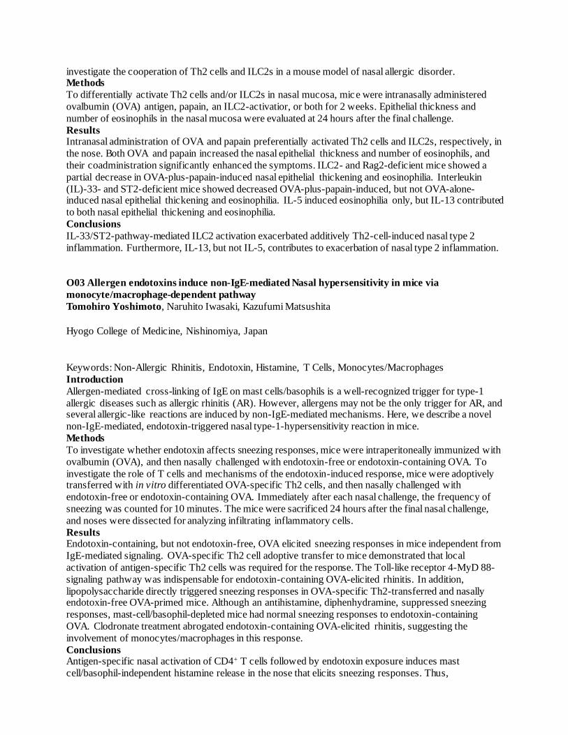
larger studies. Table 1. Prediction of house dust mite (HDM) allergy and serum IgE responses by testing IgE in nasal secretions in 30 HDM allergic patients and 29 healthy controls.
Filter Disk
Sensitivity Specificity PPV NPV Accuracy LR+ LR-
% (95%
CI)† %
(95%
CI)† %
(95%
CI)† %
(95%
CI)† %
ANY major
molecules ° 90 (73 - 98) 100
(83 -
100) 100
(82 -
100) 91
(75 -
98) 95 ∞ 0.1
nDer p 1 46 (26 -66) 100 (85 - 100)
100 (62 -100)
73 (58 - 85)
78 ∞ 0.5
nDer f 1 68 (46 - 85) 100 (85 -
100) 100
(73 -
100) 81
(66 -
91) 86 ∞ 0.3
rDer p 2 90 (73 - 98)
100 (83 - 100)
100 (81 - 100)
91 (76 - 98)
95 ∞ 0.1
rDer f 2 90 (73 - 98) 100 (83 -
100) 100
(81 -
100) 91
(76 -
98) 95 ∞ 0.1
rDer p 23 81 (61 - 93) 100 (85 - 100)
100 (77 - 100)
87 (72 - 96)
92 ∞ 0.2
OTHER
molecules
rDer p 4 0 (0 - 53) 98 (90 - 100)
0 (0 - 99)
88 (77 - 95)
86 0.0 1.0
rDer p 5 80 (44 - 97) 100 (89 - 100)
100 (52 - 100)
96 (87 - 100)
97 ∞ 0.2
rDer p 7 78 (40 - 97)
100 (90 - 100)
100 (47 - 100)
96 (87 - 100)
97 ∞ 0.2
rDer p 21 57 (18 - 90) 100 (90 -
100) 100
(28 -
100) 95
(85 -
99) 95 ∞ 0.4
ALL molecules¥ 71 (63 - 77) 100 (99 - 100)
98 (93 - 100)
93 (91 - 95)
94 167.4 0.3
Sinus Pack
Sensitivity Specificity PPV NPV Accuracy LR+ LR-
% (95%
CI)† %
(95%
CI)† %
(95%
CI)† %
(95%
CI)† %
ANY major
molecules ° 87 (69 - 96) 100
(83 -
100) 100
(81 -
100) 88
(72 -
97) 93 ∞ 0.1
nDer p 1 58 (37 - 78) 100 (85 - 100)
100 (68 - 100)
78 (63 - 89)
83 ∞ 0.4
nDer f 1 48 (28 - 69) 100 (85 -
100) 100
(64 -
100) 72
(57 -
84) 78 ∞ 0.5
rDer p 2 86 (68 - 96) 100 (83 - 100)
100 (80 - 100)
88 (73 - 97)
93 ∞ 0.1
rDer f 2 86 (68 - 96) 100 (83 -
100) 100
(80 -
100) 88
(73 -
97) 93 ∞ 0.1
rDer p 23 81 (61 - 93)
100 (85 - 100)
100 (77 -100)
87 (72 - 96)
92 ∞ 0.2
OTHER
molecules
rDer p 4 14 (0 - 58) 98 (90 - 100)
50 (1 - 99)
89 (78 - 96)
88 7.4 0.9
rDer p 5 70 (35 - 93) 90 (78 - 97)
58 (28 - 85)
94 (82 - 99)
86 6.9 0.3
rDer p 7 67 (30 - 93)
100 (90 - 100)
100 (42 - 100)
94 (84 - 99)
95 ∞ 0.3
rDer p 21 57 (18 - 90) 100 (90 - 100)
100 (28 - 100)
95 (85 - 99)
95 ∞ 0.4

ALL molecules¥ 68 (60 - 75) 99 (98 - 100)
95 (90 - 98)
93 (91 - 94)
94 80.3 0.3
† exact binomial confidence limits (95 % CI) for test sensitivity, specificity, PPV (positive
predictive value); NPV (negative predictive value), LR+ (positive likelihood ratio), LR- (negative likelihood ratio). ° outcomes referred to at least one of the major allergen molecules (nDer p 1, nDer f 1, rDer p 2, rDer f 2, rDer p 23).
¥ including outcomes of rDer p 10, rDer p 11, rDer p 14, rDer p 15, Clone 16, rDer p 18, all characterized by a low positive sample size (n<5).
P03 Correlation between rhinomanometry and spirometry parameters in 971 adults
Ivânia Gonçalves1, Tiago Jacinto2, Rita Amaral3, Ana M. Pereira3, Luís M. Araújo4, Mariana Couto1, João A. Fonseca5
1. CUF Porto - Instituto & Hospital, Porto, Portugal
2. CUF Porto - Instituto & Hospital; CINTESIS - Center for Health Technology and Services Research; ESS - IPP - School of Health; Polytechnic of Porto, Porto, Portugal
3. CUF Porto - Instituto & Hospital; CINTESIS - Center for Health Technology and Services
Research, Porto, Portugal
4. CUF Porto - Instituto & Hospital; Allergy Unit, FMUP - Faculty of Medicine, University of Porto,
Porto, Portugal 5. CUF Porto - Instituto & Hospital; CINTESIS - Center for Health Technology and Services
Research; MEDCIDES - Department of Health Information and Decision Sciences, Faculty of
Medicine, University of Porto, Porto, Portugal
Keywords: Rhinomanometry; Spirometry
Introduction
There is a lack of published studies about the association between rhinomanometry and spirometry results.
Some studies have shown a moderate correlation between spirometry parameters and other nasal objective measures such as Peak Nasal Inspiratory Flow (PNIF). We aimed to study the correlation between
rhinomanometry and spirometry parameters.
Methods
We included all adults (age ≥ 18 years) who performed rhinomanometry and spirometry consecutively on
the same day, at CUF Porto - Instituto and Hospital from November 2010 to July 2016. When more than one rhinomanometry was performed, only the first one was included in the analysis. We included gender,
age, height, rhinomanometry parameters (inspiratory and expiratory total nasal airflow, inspiratory
(RAARi) and expiratory (RAARe) mean airflow resistance at a sample pressure of 150 Pa and side ratio)
and spirometric parameters (forced vital capacity (FVC), forced expiratory volume in 1 s (FEV1), mid-
flow rate/forced expiratory flow at 25–75% of FVC (FEF25-75) and FEV1/FVC). Pearson’s correlation
was used to evaluate unadjusted correlations and partial correlations were used to adjust parameters for age, gender and height.
Results
A total of 971 adults were included, 623 (64%) females, with a mean (sd) height of 166.0 (9.0) cm and age
of 38.3 (14.1) years (min-max: 18-80). Correlations between spirometry and rhinomanometry variables
are presented in table 1. The correlations between FEV1/FVC and either inspiratory total nasal airflow or mean RAARi, were the only ones with statistical significance (r=-0.083, p=0.016 and r=0.011, p=0.039,
respectively). After adjusting for age, gender and height, no statistically significant associations were

found between the parameters. Conclusions
Rhinomanometry and spirometry parameters were not significantly correlated after adjustment to
confounders, which suggests that rhinomanometry measurements are not influenced by respiratory
capacity measured with spirometry, contrary to PNIF. This may be advantageous, especially in patients
with low respiratory functional capacity.
FVC
(%) p
FEV1
(%) p
FEV1/FVC
(%) p
MEF25-75
(%) p
Insp total nasal airflow
(ml/s) -0.027 0.575 0.017 0.615 -0.083 0.016 -0.008 0.808
Exp total nasal airflow
(ml/s) 0.032 0.359 0.052 0.131 -0.055 0.116 0.047 0.173
Mean RAARi
(kPa*s/L) 0.092 0.880 -0.027 0.424 0.011 0.039 -0.047 0.160
Mean RAARe
(kPa*s/L) -0.006 0.862 -0.044 0.186 -0.012 0.731 -0.057 0.087
Side ratio -0.032 0.341 -0.046 0.165 -0.023 0.495 -0.023 0.487
P04 Profile of patients with persistent allergic rhinitis prescribed MP-AzeFlu®* In routine clinical
practice: pooled data from austria, ireland and sweden
Par Stjarne1, Ranbir Kaulsay2, Wolfgang Pohl3
1. Karolinska Institute, Stockholm, Sweden 2. Clontarf Clinic, Dublin, Ireland
3. Karl Landsteiner Institut fur Experimentelle und Klinische Pneumologie, Vienna, Austria
Keywords: MP-AzeFlu, Dymista, Azelastine, Fluticasone Propionate
Introduction
The aims of this study were (i) to characterise patients with persistent allergic rhinitis (PER) prescribed Meda Pharma's AzeFlu (MP-AzeFlu; a novel formulation of azelastine hydrochloride, fluticasone
propionate and excipients in a single spray) in real-life in Austria, Ireland and Sweden and (ii) to quantify
the personal symptomatic burden of PER in these countries prior to MP-AzeFlu prescription.
Methods
428 patients (≥12 years old) with moderate-to-severe PER were recruited into 3, prospective, non-
interventional studies carried out in Austria (n=214), Ireland (n=53) and Sweden (n=161). MP-AzeFlu was prescribed according to label. Information was gathered on patient demographics, AR phenotype,
allergen sensitization, symptomatology, previous AR treatments in the last year (prior to MP-AzeFlu
prescription) and reason for MP-AzeFlu prescription. Data for all countries are pooled.
Results
Classified traditionally, slightly more patients had both seasonal AR (SAR) and perennial AR (PAR) (n=254; 59.3%) vs. PAR alone (n=174; 40.7%). Sensitization to house dust mite predominated (n=261;
61.0%), followed by animal dander, and at least 50.5% (n=216) were poly-sensitized. Prior to MP-AzeFlu
prescription patients reported troublesome symptoms (n=268; 62.6%), impairment of daily activities
(n=238; 55.6%), sleep disturbance (n=235; 54.9) and impairment of school/work (n=177; 41.4%).
Congestion was considered the most bothersome symptom by most patients (n=254; 59.3%). The most frequent reason for MP-AzeFlu prescription was that other therapies were not sufficient in the past
(n=299; 69.9%) or not sufficient to treat acute symptoms (n=87; 20.3%). Most of these PER patients were
previously treated with oral antihistamines (n=274; 64.0%), intranasal corticosteroids (n=236; 55.1%) or

intranasal anti-histamines (n=97; 22.7%). 59.3% (n=254) of patients reported using ≥2 AR therapies in the past year, but 9.6% (n=41) reported using no AR therapy at all.
Conclusions
Many patients in Europe live with uncontrolled persistent disease despite treatment with mono- and
multiple therapies. A more effective treatment option, like MP-AzeFlu, should improve AR control and
reduce the number of patients requiring immunotherapy.
*MP-AzeFlu, a registered trademark of Meda AB, is marketed in the U.S. as Dymista®, a registered
trademark of Meda Pharma Inc., both Mylan Companies
P05 Measurement of Nasal specific IgE in patients with local allergic rhinitis
Paloma Campo1, Ibon Eguiluz2, Carmen Rondon3, Maria Carmen Plaza4, Ana Maria Prieto5, Luisa
Galindo1, Cristobalina Mayorga2, Miguel Blanca4, Maria Jose Torres5
1. Allergy Unit, Malaga, Spain 2. IBIMA, Malaga, Spain
3. Research Laboratory, Malaga, Spain
4. Regional University Hospital of Malaga, Malaga, Spain
5. UMA, Malaga, Spain
Keywords: IgE, Local Allergic Rhinitis, Nasal
Introduction Prior methods used for measuring nasal specific IgE (NsIgE) in local allergic rhinitis (LAR) have shown a
variable sensitivity: 22% for D. Pteronyssinus (DP) using the Greiff/Grünberg method and lower with
Naclerio method. In this study a novel method of detection of NsIgE in patients with confirmed LAR to
DP was evaluated.
Methods Sixteen LAR (positive nasal allergen provocation test to DP (NAPT-DP), negative skin testing/sIgE to
DP), 10 allergic rhinitis (AR) as positive control (positive NAPT-DP and skin testing/sIgE to DP), and 12
healthy controls as negative control (negative NAPT-DP and skin testing/sIgE to DP) were recruited. DP-
ImmunoCAP® solid phase was applied directly in the lower turbinate of each nostril for 10 minutes
before and 24 hours after NAPT-DP and analyzed following the manufacturer´s instructions. ROC curves
were performed to obtain the optimal cut-off point of nasal sIgE value to calculate sensitivity (S) and specificity (SP), and outcomes were compared with NAPT-DP result (gold standard test). Study was
approved by local ethics committee.
Results
All LAR and AR subjects had a positive response to NAPT-DP, and none in the healthy control group. At
24 hours after NAPT-DP, mean NsIgE values were 0.119 kU/L in LAR, 1.600 kU/L in AR and 0.115 kU/L in healthy controls. ROC curves using NsIgE values obtained 24 hours after NAPT-DP were
performed. In LAR subjects, the area under the curve (AUC) was 0.7277, p=0.0054. The optimal cut-off
point to discriminate LAR subjects from controls was 0.135 kU/L, obtaining a S=20.31% and SP=88.09%.
In AR (positive control group) the AUC was 0.9798, p=<0.0001, and the optimal cut-off point was 0.170
kU/L with S =95% and SP=100%. Conclusions
Measurement of NsIgE by direct application of DP-ImmunoCAP® in LAR shows similar sensitivity to
other methods and good specificity, with the advantage of being non-invasive, easier to perform and
faster. Funded by Institute of Health “Carlos III” (Ministry of Economy and Competitiveness) RETICS
ARADyAL (RD16/0006/0001), FIS PI14/00864 and Consejería de Salud PI-0346-2016.

P06 Patients with persistent allergic rhinitis get a better night’s sleep on MP-AzeFlu®*: individual and pooled data from Austria, Ireland and Sweden
Ranbir Kaulsay1, Wolfgang Pohl2, Par Stjarne3
1. Clontarf Clinic, Dublin, Ireland 2. Karl Landsteiner Institut fur Experimentelle und Klinische Pneumologie, Vienna, Austria
3. Karolinska Institute, Stockholm, Sweden
Keywords: Dymista, MP-AzeFlu, Azelastine, Fluticasone, Rhinitis
Introduction
Most allergic rhinitis (AR) patients attending clinic have moderate/severe persistent disease and frequently
report reduced sleep quality. Meda Pharma’s AzeFlu (MP-AzeFlu) comprises intranasal azelastine hydrochloride, fluticasone propionate and a novel formulation, in a single device. Its real-life effectiveness
has been established in AR during 14 days. However, its impact on sleep quality is unknown. This study
assessed the impact of MP-AzeFlu on sleep quality when used in routine clinical practice by patients with
persistent AR (PER).
Methods
428 patients (≥12 years old) with moderate-to-severe PER were recruited into 3, prospective, non-interventional studies carried out in Austria (n=214), Ireland (n=53) and Sweden (n=161). MP-AzeFlu
was prescribed according to its summary of product characteristics. Patients assessed their sleep quality
(7-days reflective) on days 7, 14, 21, 28, 35 and 42 using a 5-point scale from ‘very good’ to ‘very bad’.
Results
Many patients in each country reported sleep disturbance prior to MP-AzeFlu prescription: n=112 (52.3%) in Austria; n=41 (77.4%) in Ireland; n=82 (50.9%) in Sweden. MP-AzeFlu treatment (1 spray/nostril bd;
daily doses:AZE=548μg;FP=200μg) was associated with improved sleep quality, evidenced by an increase
in the proportion of patients reporting ‘very good’ and ‘good’ quality sleep in the first 28 days of
treatment, and a corresponding reduction in the proportion of patients reporting ‘bad’ or ‘very bad’ sleep
quality. Sleep quality improved at each assessed time point (Table). Improved sleep quality occurred irrespective of phenotype (when classified traditionally) - in those with perennial AR (PAR) only and in
those with both PAR & seasonal AR.
Conclusions
MP-AzeFlu improves sleep quality in patients with moderate-to-severe PER in a real-world pan-European
setting. *MP-AzeFlu, a registered trademark of Meda AB, is marketed in the U.S. as Dymista®, a registered trademark of Meda Pharma Inc., both Mylan Companies
Character count: 2352 (limit = 2500)
Austria† (n≤214) Ireland (n≤53) Sweden†(n≤161) Pooled† (n≤ 428)
Day 0 Day 28 Day 0 Day 28 Day 0 Day 28 Day 0 Day 28
Very good 2.4% 35.3% 0.0% 24.5% 3.7% 15.7% 2.6% 26.5%
Good 25.1% 44.6% 24.5% 50.9% 28.6% 44.4% 26.4% 46.0%
Fair 36.5% 17.3% 32.1% 15.1% 34.2% 25.9% 35.1% 20.1%
Bad 28.0% 2.2% 32.1% 5.7% 27.3% 12.0% 28.3% 6.4%
Very bad 8.1% 0.7% 9.4% 0.0% 6.2% 1.9% 7.5% 1.0%
† % without missing values

P07 Facial infrared thermography in the assessment of Nasal provocation test
Magdalena Herknerova1
1Hospital Na Homolce, Prague, Czech Republic
Keywords: Infrared Thermography, Nasal Provocation, Bounding Box
Introduction Infrared thermography is a diagnostic tool in more medical disciplines. It detects temperature changes in
the infra-red spectrum range and was originally designed to test the heating qualities of
buildings. Potential benefits in allergology and ENT are not widely evaluated. We asked if this method
can give us an objective information about a patient during nasal provocation test.
Methods As it is difficult to detect always the same point in the face during repeating measurements, a bounding
box to detect the face of a patient was designed.
Forehead, nasal, cheek and chin facial areas were defined.
Results
We present one example of our software in the evaluation of facial infrared thermography. This method
detects temperature changes in different areas of the face. The nasal area was the most sensitive one in the detection of temperature change during nasal provocation test followed by the cheek area.
Conclusions
Facial infrared thermography might be- besides other important parametres like nasal symptome score and
active anterior rhinomanometry- an objective, non- invasive and feasible tool in the assessment of allergic
response during nasal provocation test.
P08 Intranasal Condyloma Acuminatum with malignant transformation
Tengchin Wang1, Chiejun Wu2
1. Department of Otolaryngology, Tainan Municipal Hospital, Tainan City, Taiwan 2. Department of Pathology, Tainan Municipal Hospital, Tainan City, Taiwan
Keywords: Condyloma Acuminatum, Immunocompromised
Introduction
Condyloma acuminatum is a venereal diasese transmitted by the human papillomavirus (HPV). Generally,
it is a benign entity but carcinomatous change has been reported in anogenital area. The malignant
transformation is associated with the immunocompromised status ,especially HIV. Condyloma acuminatum is uncommonly identified in the nasal cavity, the malignant transformation is extremely rare.
Methods
A mid-aged patient with diabetes mellitus and psoriasis had suffered from progressive right nasal
obstruction with epistaxis for six months. Physical examination revealed a cauliflower-like lesion over the
right nasal vestibule,expanding to the septum. Due to this patient had history of penile condyloma acuminatum, biopsy was done and sent hc2 high-risk-HPV DNA testing. The results was compatible with
condyloma acuminatum and negative for high-risk-HPV infection
Results
Eradication surgery was performed. Necrotic tissue with pus content was buried in the lesion. This mass
was completely excised eventually. The formal histopathology reported condyloma acuminatum with focal invasive squamous cell carcinoma. We suggested this patient receiving adjuvant radiotherapy or re-
operation because there was no planed safe margin in advance, and the examinations for HIV and syphilis
were also advised. But the patient refused and lost of follow-up thereafter. Conclusions
Human papillomavirus types 16 and 18 are found in up to 90% of patients with cervical carcinoma,
however HPV type 6 and HPV type 11 are the main factor in developing giant condyloma acuminatum,
which is reported 56% incidence of malignant transformation. Abscesses and fistulas are more common in
lesions as described in our case. Immunosuppression, coexisting HIV infection, and unhygienic conditions play a role in malignant transformation, therefore AIDS togethers with other venereal diseases should be
examined. Electrocautery or laser total excision could be applied for treatment. Non-resectable lesions,
radiotherapy could be applied either alone or along with chemotherapy. In conclusion, condyloma
acuminatum with invasive squamous cell carcinoma is rarely found in the nasal cavity, more cases should
be obtained for more comprehensive understanding.
P09 Rhinitis: classification and diagnosis algorithm proposal
Jonathan Kilimajer1
1Subiza Asthma and Allergy Center, Madrid, Spain
Introduction One of the most common problems presenting among physicians are rhinitis symptoms.Challenges in the
diagnosis results from the factor that differential diagnosis is extensive and sometimes symptoms of
allergic, nonallergic, and mixed rhinitis are often indistinguishable. Searching in the literature there is a
clear consensus on the classification but is difficult to found one that include and ordered all the different
etiologies. It is also difficult to found an specific diagnosis algorithm for these diseases. Methods
After checking over and try to reunite different consensus made in allergy an ENT for classification and
diagnosis of rhinitis we propose two tables with both of these sections.
Results
Table 1. Classification of Rhintis
Table 2. Diagnosis algorithm for Rhinitis
Conclusions
When approaching rhinitis, diagnostic challenge is to determine the etiology, specifically whether it is
allergic, nonallergic, or perhaps an overlap of both conditions. With these proposal tables it could be more easy to differentiate and lead to a correct diagnosis.

Table 1. Classification of Rhinitis
ACUTE
Infectious symptoms
Anterior rhinorrhea , stuffiness, sneezing, itchy nose
+Ocular symptoms +/- asthma
(PROBABLY ALLERGIC RHINITIS ) ACUTE RHINOSINUSITIS BACTERIAN RHINITIS
VIRAL RHINITIS
PresentAbsent
Post nasal dripMucopurulent rhinorrhea
Skin prick tests SPT ( If negative : IgE )
SPT ( - ) IgE ( + ) SPT ( +) SPT ( - ) IgE ( - )
ALLERGY
In relation with :allergens
Search forother etiology
ALLERGICRHINITIS
In relation with :work
Nasal Provocation
Nasal Provocation
In relation withallergens
In relation withwork
NON ALLERGICOCUPATIONAL
RHINITIS
LOCALRHINITIS
ALLERGICOCUPATIONAL
RHINITIS
CHRONIC
Nasal Obstruction+/- Hyposmia
+/- Post nasal drip
ENDOSCOPYNasal structural abnormalities
YES NO
POLYPOSISANATOMIC DEFECTSATROPHIC RHINITISGRANULOMATOUS
(Confirm with TAC, RM, Biopsy)
Anterior rhinorrhea , stuffiness, sneezing, itchy nose
DRUGS HORMONALEMOTIONALSENILEGUSTATORY
Nasal Provocation
( + )
Relation with externalagents
VASMOTORRHINITIS
( - ) ( + ) ( + )
NO YES
NARESMastocytosis
( - )
NO
ByopsyCulture
( - )
Post nasal dripMucopurulentrhinorrhea
CHRONICRHINOSINUSITIS
Table 2. Diagnosis algorithm for Rhinitis
RHINITIS
ALLERGIC NON ALLERGIC
Allergic Rhinitis(systemic athopy )
Local Allergic Rhinitis(without systemic athopy)
Mixed rhinitis Infectious Non infectious
INFLAMATORIESR. Eosinophilic
Polyps
R. Granulomatous
R. Primary atrophic
Mastocitosytosis
NON INFLAMATORIESR. Colinérgic
R. Drugs
R. Hormonal
R. Structural
R. Irritative
R. Emotional
R. Senile
P10 Real life effectiveness of MP-AzeFlu®* in persistent allergic rhinitis, assessed by visual analogue scale and endoscopy: pooled data from Austria, Ireland and Sweden
Wolfgang Pohl1, Ranbir Kaulsay2, Par Stjarne3
1. Karl Landsteiner Institut fur Experimentelle und Klinische Pneumologie, Vienna, Austria 2. Clontarf Clinic, Dublin, Ireland
3. Karolinska Institute, Stockholm, Sweden
Keywords: MP-AzeFlu, Dymista, Azelastine, Fluticasone
Introduction
Most allergic rhinitis (AR) patients attending clinic have moderate/severe persistent disease. Meda
Pharma's AzeFlu (MP-AzeFlu) comprises intranasal azelastine hydrochloride, fluticasone propionate and a novel formulation, in a single device. Its real-life effectiveness has been established in AR during 14 days,
but its effectiveness in those with persistent AR (PER) over the longer term is unknown. We assessed the
effectiveness of MP-AzeFlu in routine clinical practice in PER patients, using the ARIA-endorsed
language of AR control (i.e. visual analogue scale (VAS)). A VAS score cut-off of 50 mm is
recommended to assess control and guide treatment decisions.
Methods 428 patients (≥12 yrs old) with moderate/severe PER were recruited into 3, prospective, non-
interventional studies in Austria (n=214), Ireland (n=53) and Sweden (n=161). MP-AzeFlu was prescribed
according to label. Patients assessed symptom severity using a VAS from 0 mm (not at all bothersome) to
100 mm (very bothersome) in the AM prior to MP-AzeFlu use, on Days 0, 1, 3, 7, 14, 21, 28, 35 and 42.
An endoscopy was performed in the Irish study on Days 0 and 28. Symptoms of ‘oedema’, ‘discharge’ and ‘redness’ were scored on a 3-point scale for both nostrils (max score=12).
Results
Patients treated with MP-AzeFlu (1 spray/nostril bd; daily doses:AZE=548μg;FP=200μg) had a VAS
score reduction from 59.0 mm (SD 25.0) at Day 0 to 28.8 mm (SD 23.2) at Day 28 (p<0.0001 ) and 23.2
mm (SD 21.5) on Day 42 (p<0.0001), a reduction of 35.8 mm. This reduction was rapid, with statistical significance vs baseline noted from Day 1 (p<0.0001), and was consistent irrespective of phenotype,
patient age, and disease severity. On average MP-AzeFlu-treated patients achieved the 50 mm VAS score
cut-off before Day 3. Total endoscopy score reduced from 7.5 (SD 3.1) at baseline to 3.5 (SD 2.5) at Day
28. The % of patients with severe oedema reduced from 53.1% at baseline to 3.8% at Day 28. A similar
reduction in the incidence of thick/mucusy discharge (28.3% to 4.8%) and severe redness (34.9% to 0%)
was also observed.
Conclusions
MP-AzeFlu provides effective and rapid PER control in a real-world pan-European setting assessed by
VAS. Symptom improvement was noted from Day 1, sustained for 42 days and was associated with
improvement in mucosal appearance after 28 days.
*MP-AzeFlu, a registered trademark of Meda AB, is marketed in the U.S. as Dymista®, a registered
trademark of Meda Pharma Inc., both Mylan Companies
P11 Overadditive effects of MP-AzeFlu* on antiinflammatory genes and inhibition of
proinflammatory mediators compared to Fluticasone Propionate and Azelastine in Sinonasal
Fibroblasts.
Laura Pujols1, Jordi Roca-Ferrer1, Borja Callejas1, Mireya Fuentes-Prado1, Maria Perez-Gonzalez1, Isam Alobid2, Antonio Valero2, Cesar Picado2, Ruth Murray3, Joaquim Mullol2

1. IDIBAPS & CIBERES, Barcelona, Spain 2. IDIBAPS, CIBERES & University of Barcelona Hospital Clinic, Barcelona, Spain
3. Medscript, Dundalk, Ireland
Keywords: MP-AzeFlu, Azelastine, Fluticasone Propionate, GILZ
Introduction
MP-AzeFlu*, a novel intranasal formulation of azelastine hydrochloride (AZE) and fluticasone propionate
(FP), has demonstrated superior clinical effects compared to these drugs in monotherapy in patients with
allergic rhinitis and chronic rhinitis. MP-AzeFlu has previously shown greater antiinflammatory potency than FP and AZE in an in vitro model of eosinophilic inflammation.Our aim was to compare the effect of
MP-AzeFlu, FP, and AZE on antiinflammatory gene transactivation and proinflammatory mediator
inhibition in sinonasal cultured fibroblasts from nasal mucosa (NM) and chronic rhinosinusitis with nasal
polyps (CRSwNP).
Methods NM and CRSwNP fibroblast cultures (n=6) were incubated with serial dilutions of MP-AzeFlu (1:102 -
1:104) or FP or AZE (equivalent dilutions) for 2 to 24h with/without 1ng/ml IL-1b. Glucocorticoid-
induced leucine zipper (GILZ), mitogen-activated protein kinase phosphatase-1 (MKP-1), and COX-2
gene (RT-PCR) and IL-8 and GM-CSF protein (ELISA) expression was analysed. Results are expressed
as fold-increase or % inhibition over control (mean±SEM). Results
MP-AzeFlu and FP (all dilutions, P<0.05) and AZE (1:102, P<0.05) increased GILZ and MKP-1 gene
expression in NM and CRSwNP fibroblasts. MP-AzeFlu (1:102) had an overadditive effect on GILZ
expression vs FP or AZE and on MKP-1 expression vs AZE (p<0.05) (Table). MP-AzeFlu and FP
provoked a similar inhibition of IL-1b-induced COX-2, IL-8, and GM-CSF in NM and CRSwNP
fibroblasts (all dilutions, P<0.05) but greater than AZE, which had no inhibitory effect. Conclusions
The superior clinical effect of MP-AzeFlu compared to corticosteroid or antihistamine monotherapy may
be related to the overadditive induction of antiinflammatory genes and the inhibition of proinflammatory
mediators. Our findings reveal some molecular basis for the therapeutic benefit of MP-AzeFlu in rhinitis
and potentially in CRSwNP.
This study has been sponsored by a research grant form MYLAN - MEDA. (*) Dymista
NM fibroblasts CRSwNP fibroblasts
2h 6h 2h 6h
GILZ (fold increase)
MP-AzeFlu*
(1:102) 11.2±1.1 36.5±7.9 14.7±4.5 31.4±9.2
FP (1:102) 9.4±1.3† 19.8±2.4† 13.9±3.8 15.3±4.5†
AZE (1:102) 1.8±0.1† 7.9±2.8† 2.5±0.8† 2.8±0.9†
MKP-1 (fold increase)
MP-AzeFlu*
(1:102) 6.5±1.4 5.4±1.7 7.2±2.1 6.2±2.4
FP (1:102) 5.4±0.6 4.7±1.0 5.7±1.5 4.8±1.6
AZE (1:102) 2.1±0.3† 1.8±0.6† 2.9±1.0† 1.9±1.3†
Proinflammatory markers (% inhibition vs control at 24h)
COX-2 IL-8 GM-CSF COX-2 IL-8 GM-CSF

MP-AzeFlu
(1:102) 72.0±7.1 61.5±5.5 80.0±5.7 91.3±0.5 50.4±4.3 85.6±4.0
FP (1:102) 93±2.4 69.1±3.2 81.4±4.8 96.8±0.7 61.3±5.6 87.5±3.7
(†) p<0.05 vs MP-AzeFlu*
Friday 31 March 2017
Chronic Rhinosinusitis: mechanisms, 08:30 - 10:33
O06 TLR4 mediates epithelial barrier dysfunction by Staphylococcus Aureus Enterotoxin B in
CRSwNP Brecht Steelant1, Katleen Martens1, Guy Boeckxstaens1, Sven F. Seys1, Peter W. Hellings1
1KU Leuven, Leuven, Belgium
Keywords: TLR4; CRSwNP; Epithelial Integrity; Tight Junction
Introduction Epithelial barrier dysfunction plays a role in the pathophysiology of chronic rhinosinusitis with nasal
polyps (CRSwNP). Toll-like receptors (TLRs) are thought to regulate epithelial barrier integrity, though
its function has not been studied in CRSwNP thus far. The aim of this study was to investigate if TLR2
and TLR4 signaling is involved in regulating barrier function in CRSwNP.
Methods
Primary nasal epithelial cells from controls and CRSwNP patients (both n = 5) were isolated and grown at air-liquid interface on transwell inserts for 21 days. Epithelial integrity was evaluated by measuring
transepithelial electrical resistance (TER) together with the expression of occludin and zonula occludens
1. TLR2 and TLR4 expression was evaluated using qRT-PCR. Primary nasal epithelial cells were
stimulated with Staphylococcus aureus enterotoxin B (SEB) for 4 hours and TER was evaluated. In vitro,
TLR2 and TLR4 signaling was blocked to evaluate the contribution to barrier dysfunction. In vivo, wild type and TLR4-/- transgenic mice were used to study the role of TLR activation by SEB in increasing
mucosal permeability for FD4.
Results
TER of CRSwNP cultures was significantly decreased compared to controls, which was associated with
decreased expression of occludin. Stimulation with SEB significantly decreased TER in CRSwNP cultures associated with decreased occludin, and had no effect in control cultures. TLR2 and TLR4 expression was
elevated in CRSwNP, which could explain the differential SEB response. Moreover, occludin levels
correlated inversely with TLR4 expression in CRSwNP. Antagonizing TLR signaling prevented SEB-
induced decrease in TER in vitro. In vivo, SEB increased mucosal permeability for FD4 in wild type mice
by affecting the expression of occludin. No increased FD4 permeability nor decreased expression of
occludin was found in TLR4-/- transgenic mice. Conclusions
TLR4 expression is increased in CRSwNP and correlated inversely with occludin levels. TLR4 seems a
key factor in regulating barrier integrity in CRSwNP.

O07 Investigation Of The Th17 Inflammatory Response In Chronic Rhinosinusitis
Timothy C Biggs1, Stephen M Hayes1, Philip G Harries2, Sylvia Pender1, Rami J Salib1
1. University of Southampton, Southampton, United Kingdom 2. University Hospital Southampton NHS Foundation Trust, Southampton, United Kingdom
Introduction T-helper 17 (Th17) cells represent a distinct T-cell linage, involved in the host defence against
extracellular pathogens. There have been limited studies of a Th17 mediated inflammatory response in
chronic rhinosinusitis patients. We aimed to study Th17 responses in chronic rhinosinusitis with and
without nasal polyps (CRSwNP and CRSsNP respectively) including stimulation with Staphylococcus
aureus enterotoxin B (SEB). Methods
In total, 31 patients were included within the study. Nasal polyp, CRSsNP sinonasal mucosa and control
mucosa was harvested at the time of surgery. Th17 cytokine gene expression was assessed using real time
quantitative polymerase chain reaction (RT-qPCR). In addition, nasal polyp and control tissues were
cultured with SEB for 24 hours using an ex-vivo nasal explant model, prior to cytokine analysis using RT-
qPCR and Luminex. Results
IL-17A gene expression was significantly upregulated in CRSsNP sinonasal mucosa compared to nasal
polyps (p<0.05) and control mucosa (p<0.01). Upon stimulation with SEB, IL-17A gene expression and
protein production was significantly upregulated (p<0.05) within nasal polyps.
Conclusions This study demonstrates a Th17 response in CRSsNP sinonasal mucosa, and in nasal polyps when
stimulated with SEB. These results highlight the likely role of bacteria and their toxins in driving the
inflammatory response in CRS, resulting in disease resistance and long term chronicity.
O08 Regulation Of Hyperplastic Cell Growth In Chronic Rhinosinusitis With Nasal Polyposis
Jean Kim1, Hyun Sil Lee1
1Johns Hopkins University School of Medicine, Baltimore, United States
Keywords: Nasal Polyposis, Epithelial Cell Growth, VEGF, HIF
Introduction We have previously shown that VEGF is overexpressed in the epithelium of hyperplastic nasal polyps in
patients with chronic rhinosinusitis with nasal polyposis (CRSwNP). We hypothesized that the HIF family
of transcription factors function to regulate this process.
Methods
We used human primary nasal epithelial cells grown in submerged culture (PNEC) and whole human polyp tissue in air liquid interface culture to examine to examine HIF and VEGF expression by
immunohistochemistry, flow cytometry and realtime PCR analysis. Function was assessed by exposure to
known pharmacologic inhibitor digoxin and specific siRNA knockdown of HIF on cell growth and
apoptosis by DNA binding assay of PNEC and TUNEL assay of polyp tissue, respectively.
Results We now report that the HIF family of transcription factors, both HIF1-alpha (HIF1α) and HIF2-alpha
(HIF2α), which known regulators of expression of VEGF, are highly expressed in PNEC from CRSwNP
subjects, as compared to normal control subjects when analyzed by flow cytometry of PNEC and
immunohistochemistry of sinonasal surgical tissue. PNEC from CRSwNP patients express high
constitutive levels of mRNA of HIF1α and HIF2α. Inhibition of HIF1α by cardiac glycoside digoxin
results in 65% inhibition of PNEC growth rates analyzed by DNA binding assay (p<0.001, n=5). Further studies show that siRNA knockdown of HIF1α results in inhibition of VEGF mRNA expression by 75%
(p<0.01, n=3) and subsequent 55% inhibition of PNEC growth rate as measured by DNA binding assay
(p<0.01, n=3). Exposure of whole human nasal polyps in ex vivo culture to HIF1α inhibitor digoxin or
VEGF co-receptor inhibitor anti-neuropilin1 antibody results in 90% (p<0.01, n=5) and 2-fold (p<0.005,
n=5) increase in apoptosis respectively of PNEC as assessed by TUNEL assay. Conclusions
Collectively, these data indicate that HIF regulates VEGF in promoting hyperplastic nasal epithelial cell
growth and survival observed in CRSwNP.
O09 Interleukin 7 and Interleukin 15 interaction with the inflammation severity, predisposing
factors and phenotype in chronic rhinosinusitis
Livije Kalogjera1, Nada Vrkic2, Anita Topic3, Dejan Tomljenovic4, Tomislav Greguric5, Patricija
Bankovic Radovanovic6
1. ORL/HNS Dept. University Hospital Centre, Zagreb, Croatia 2. Biochemistry Lab,. University Hospital Centre, Zagreb, Croatia
3. Clinical Institute of Chemistry, Zagreb, Croatia
4. ORL/HNS Dept, University Hospital Centre, Zagreb, Croatia 5. Clinical Dept of Radiology, University Hospital Centre, Zagreb, Croatia
6. Dept of clinical chemistry, Genera Hospital Pula, Pula, Croatia
Keywords: IL-7, IL-15, Chronic Rhinosinusitis, Nasal Polyps,T Cell Subset
Introduction
Two major clinical phenotypes of chronic rhinosinusitis (CRS), with (CRSwNP) and without nasal polyps
(CRSsNP) may show different regulation of inflammation, interplay between T cell subsets, remodelling,
response to microbiome and association with predisposing comorbidities, like allergy, asthma and aspirin intolerance. IL-7 and IL-15 belong to common gamma chain cytokines, and are essentially involved in T
cell homeostasis and proliferation. Several papers indicated that they might be involved in the immune
response in asthma, however, their role in CRS was not evaluated. Our aim was to analyze interaction
between IL-7 and IL-15 related to phenotypes and comorbidities.
Methods
The study included 50 patients(both gender, age 18-60 years, 32 with CRSsNP, and 18 with CRSwNP), with CRS according to the EPOS criteria, who were submitted to endoscopic sinus surgery. Ethmoid sinus
mucosa or nasal polyps were collected at surgery and immediately tissue homogenates were prepared and
incubated with protease inhibitor. Assays (R&D System, Quantikine ELISA, UK) for interleukins IL-4,
IL-5, IL-7, IL-15 and for interferon gamma (IFN-γ) employs the quantitative sandwich enzyme
immunoassay technique. Tissue homogenates (n=70) were analysed according to the manufacurer’s instructions (29 from non-allergic CRSsNP, 17 from allergic asthma patients, 11 from non-allergic
asthma, 9 from allergic non-asthmatic CRSsNP and 4 from aspirin intolerant CRSwNP). Polyp and
ethmoid sinus mucosa samples were taken in CRSwNP, and only ethmoid sinus mucosa from patients
with CRSsNP, respectively, from the worse side according to imaging.
Results Results. IL-5 was significantly higher (mean 38,69 pg/mL vs. 9,47 respectively ), but IL-7 (0,1397 pg/mL
vs. 0,2638, respectively), and IFN-γ (22,44 pg/mL vs. 41,71, respectively) significantly lower in samples
from CRSwNP, and these differences were also significant when asthmatic were compared to non-
asthmatic, irrespective of phenotype and sensitization. Interestingly, IFN-γ was significantly higher in
patients with allergic sensitization, while IL-4 was significantly higher in patients with ASA intolerance. IL-15 significantly correlated with IL-7 (rho 0,44), IL-4 (rho 0,31) and IFN-γ significantly with IL-4 (rho
0,51).

Conclusions
IL-7 and IL-15 are significantly upregulated in CRSsNP compared to CRSwNP and in non-asthmatics
compared to asthmatics.
O10 Problems And Solutions In The Treatment Of Acute And Chronic Rhinosinusitis Claus - Bachert1, Rainer Jund1
1URL, Ghent, Botswana
Keywords: Acute Rhinosinusitis, Chronic Rhinosinusitis, Phytotherapy, BN1016 Introduction
Acute and chronic rhinosinusitis affect everyone several times per year and 11% of the EU population,
respectively.
Methods
Double-blind randomized placebo-controlled trials
Results Recently, the herbal drug BNO 1016 showed evidence for a significant and clinically relevant reduction of
symptoms and increase of quality of life. In an analysis of pooled data of two similar randomized placebo-
controlled clinical trials including 589 patients, the Major Symptom Score (MSS) improved from
10.02±1.61 to 2.47±2.55 for BNO 1016 and from 9.87±1.52 to 3.63±3.63 for placebo; the difference
between treatment groups at end of therapy (1.16 score point ± [SD]) was statistically significant in favor of BNO 1016 (p<0.0001); patient-assessed quality of life also improved significantly (p=0.0015) versus
placebo. The results were confirmed by ultrasonography. In summary, the herbal dry extract BNO 1016 is
efficacious and well tolerated in patients with acute viral rhinosinusitis.
The objective of a recent clinical trial therefore was to assess the efficacy, safety and tolerability of dry
extract BNO 1016 in patients with chronic rhinosinusitis. 927 patients suffering from chronic rhinosinusitis (> 12 weeks and 6 ≤ MSS ≤ 12) were enrolled in a randomised placebo-controlled trial with
a treatment period of 12 weeks. The primary endpoint was the mean Major Symptom Score (MSS) over
week 8 and week 12 compared to placebo. Secondary endpoint included further MSS related calculations
and responder rates over time. Finally, safety and tolerability were evaluated. Although the results of the
secondary endpoints showed a clear trend towards superior efficacy, BNO 1016 was not superior over
placebo regarding the primary endpoint. Additional post-hoc sensitivity analyses in patients with a baseline MSS ≥ 10, duration of disease > 1 year and diagnosed by specialists in otorhinolaryngology
indicated that those patients significantly benefited from BNO 1016. Therapy was superior for the primary
endpoint analysis. Patients were less impaired with respect to work and daily activities. A good safety and
tolerability of BNO 1016 was assured in all patients.
Conclusions Rhinosinusitis, a frequent cause of disease and associated sequalae, still remains a challenge for treatment.
BNO 1016 can safely be administered in patients with acute and chronic rhinosinusitis and offers a
suitable treatment option

Friday 31 March 2017
CRS & Aspirin Exacerbated Respiratory Disease (AERD), 16:10 - 17:12
O11 Transglutaminase 2 expression but not lipid mediator profiles discriminate Nasal polyps from
steroid treated Aspirin tolerant and intolerant patients Pascal Haimerl1, Adam M Chaker2, Yvonne Schober3, Sonja Schindela1, Andreas Nockher3, Carsten B
Schmidt-Weber1, Julia Esser-Von Bieren1
1. Center of Allergy and Environment (ZAUM), Technical University of Munich and Helmholtz Center Munich, Munich, Germany
2. Department of Otolaryngology, Allergy Section, Klinikum Rechts der Isar; Center of Allergy and
Environment (ZAUM), Technical University of Munich, Munich, Germany
3. Institute of Laboratory Medicine and Pathobiochemistry, Molecular Diagnostics, Philipps
University Marburg, Marburg, Germany
Keywords: AERD, Allergy, Eicosanoids, Nasal Polyps, Transglutaminase 2
Introduction Aspirin exacerbated respiratory disease (AERD) is characterized by acute aspirin-triggered respiratory
distress, chronic asthma and nasal polyps that are often refractory to steroid treatment. AERD patients
show characteristic abnormalities in lipid mediator (eicosanoid) metabolism and signaling, but the
mechanisms that discriminate nasal polyp development in AERD and aspirin tolerant chronic
rhinosinusitis with nasal polyps (AT CRSwNP) remain obscure. We thus compared eicosanoid profiles in nasal polyps from AERD and AT CRSwNP patients and analyzed the expression of factors involved in
airway remodeling.
Methods
Nasal polyp tissues (n=10 AT CRSwNP, n=4 AERD) were obtained from patients undergoing functional
sinus surgery. All patients were treated with systemic steroids preoperatively. Expression of eicosanoid pathway proteins and regulatory factors in nasal polyp tissues or healthy turbinates (n=3) was studied by
immunofluorescence and immunohistochemistry followed by automated image analysis. Supernatants
from overnight cultures of nasal polyps were processed and subjected to lipid mediator analysis by LC-
MS/MS or immunoassay.
Results
Nasal polyp tissues from steroid treated AT CRSwNP and AERD patients expressed high levels of leukotriene (LT) biosynthetic enzymes (5-lipoxygenase, Leukotriene C4 synthase) despite low levels of
infiltrating granulocytes. LT enzymes were abundant in the nasal polyp epithelium and in macrophages
and all nasal polyp tissues released significant amounts of LTs. The overall eicosanoid profile was similar
in nasal polyps from AT CRSwNP and AERD patients with a tendency for increased thromboxane
production by AERD tissues. In contrast AERD tissues expressed significantly lower levels of the enzymes transglutaminase 2 (TG2) and microsomal prostaglandin E2 synthase (mPGES-1). TG2 was
particularly abundant in tissues from house dust mite allergic patients.
Conclusions
Our findings identify TG2 as a potential mechanism that discriminates nasal polyp endotypes. We further
show that TG2 expression and LT production in nasal polyp epithelium are resistant to steroid treatment. Thus, targeting TG2 might be promising in allergic CRSwNP patients and novel strategies targeting
steroid resistant lipid mediator abnormalities in nasal polyps are urgently needed.

Friday 31 March 2017
Poster discussion session, 17:15 - 18:30
P12 Effects of Cytokine secretion from Nasal polyps on peripheral lymphocytes in a co-culture
system
Pascal Ickrath1, Norbert Kleinsasser1, Niklas Beyersdorf2, Xin Ding2, Rudolf Hagen1, Stephan
Hackenberg1
1. Department of Otorhinorhinology, University of Wuerzburg, Wuerzburg, Germany 2. Institute of Immunology and Virology, Wuerzburg, Germany
Keywords: Chronic Rhinosinusitis With Nasal Polyps, T Cell Subsets, Co-Culture System
Introduction
T cell subpopulations in nasal polyps differ from peripheral lymphocytes in patients with CRSwNP. The direct influence of the nasal polypoid tissue by secretion of cytokines on the differentiation or activation
of T cells still remains unclear. To study the effects of polypoid tissue on peripheral lymphocytes in vitro,
we developed a co-culture system with nasal polyp tissue and peripheral lymphocytes to directly measure
these interactions. Additionally, we assessed the cytokine secretion by polyp tissue, which may induce T
cell responses in this system.
Methods Tissue and blood samples were collected from 10 patients undergoing nasal sinus surgery. Polypoid tissue
was cultured under air-liquid interface conditions. Afterwards, CD3/CD28 activated PBMC of the same
patients were added using cytokine free medium. After 3 days, lymphocytes were separated and analyzed
by multicolor flow cytometry. Monoculture PBMC served as control group. Additionally, cytokine
secretion of the polyp tissue was measured using a human Th1/Th2/Th17 antibody array. Results
There was a significantly higher amount of CD4+ and CD8+ T cells in the co-cultured system than in
PBMC alone. Terminal differentiated CD8+ T cells were significantly increased, while central memory
CD8+ T cells significantly decreased in the co-culture. HLA-DR was downregulated in co-cultured CD3+
lymphocytes. Conventional memory CD4+ T cells significantly increased and resting regulatory T cells significantly decreased in the co-cultured system. Cytokine analysis showed a secretion of IL-6, GM-CSF
and MIP-3.
Conclusions
It could be demonstrated that nasal polypoid tissue has an effect on the PBMC phenotype. The secretion
of pro-inflammatory cytokines may play a regulatory role in this context. Interestingly, the clinically
known downregulation of HLA-DR in CD3+ lymphocytes was significantly reproducible in vitro. The modulating effect of the polypoid tissue on the activation of lymphocytes must be further investigated in
order to determinate its impact on this disease.
P13 A new cytological approach to improve the final diagnosis of Eosinophilic Granulomatosis with Polyangiitis (EGPA)
Daniela Cangiano1, Francesco Cinetto1, Giuseppe Brescia2, Gino Marioni2, Claudia Zanotti2, Franco
Schiavon3, Roberto Padoan3, Ilaria Caputo4, Raffaella Neri1, Carlo Agostini1
1. Department of Medicine DIMED,University of Padua, Padova, Italy 2. Department of Neurosciences DNS, Otolaryngology Section,University of Padua, Padova, Italy
3. Department of Internal Medicine, University of Padua, Padova, Italy
4. University of Padua, Padova, Italy
Introduction
Eosinophilic granulomatosis with polyangiitis (EGPA) is an uncommon systemic necrotizing vasculitis that affects small to medium sized vessels and is associated with severe asthma, allergic rhinitis, nasal
polyposis and blood and tissue eosinophilic infiltration. Antineutrophil cytoplasmic autoantibodies
(ANCA) are present in about 40% of cases. The presence of four or more of the above described findings
yields a sensitivity of 85% and a specificity of 99.7% for the final diagnosis of EGPA. The aim of the
study was to develop a new diagnostic tool to support the diagnostic and prognostic work-up of EGPA
patients with the possibility to evaluate extracellular mediators in nasal secretion. Methods
Nasal secretions were gained from 40 patients of which 20 EGPA, 10 suspected EGPA and 10 controls
after positioning the cotton pieces in the nasal middle meatus where they were left for 10 minutes.
Subsequently the cotton pieces were put in 5 ml of saline solution and left in ice until processing. The
liquid obtained from the squeezing of the cottons was centrifuged two times at 1600 RPM for 10 minutes. Supernatant was frozen after first centrifugation. Trypan Blue was added to 10 ul of sample to calculate
the number of cells and their vitality. Finally, 100 ul of sample with containing 1,5-2,00x x105cells were
centrifuged with Cytospin at 300 RPM for 15 minutes. The slides were stained with May
Grunwald/Giemsa and cell population analysed at microscope with objective oil immersion 40X of
magnification. The remaining cells were frozen or cultured. Results
The mean number of cells obtained after centrifugation was 2.00-4.08x10⁶ cells/ml for EGPA patients and
6.42-8.62x10⁵cells/ml for control group and suspected EGPA. Cytologic analysis allowed us to count an
increase in the percentage of eosinophils in EGPA patients with active ENT disease and in some of the
patients with a suspect of EGPA, where this percentage correlated with histologic evidence of disease.
Conclusions This method seems preliminarily a reliable cytologic examination allowing fresh cells isolation and
analysis of extracellular mediators. Compared to the direct slither of the mucus on the surface of the glass,
cytospin significantly reduced any dye accumulation. Further studies are ongoing to support the described
method, in order to confirm the cytologic diagnostic reliability to identify prognostic biomarkers of
EGPA.
P14 Enhanced Type I Interferon response contributes to Eosinophilic chronic rhinosinusitis
JI HEUI Kim1, Yong Ju Jang1, Ji Youn Lim1, Sung Hee Kim1
1Asan Medical Center, Seoul, South Korea
Keywords: Chronic Rhinosinusitis, Type I Interferon, Eosinophil
Introduction
Type I interferons (IFN-I) response has been implicated in the eosinophilic inflammation besides its
antiviral function. This study aimed to investigate the role of IFN-I response in the pathogenesis of
eosinophilic chronic rhinosinusitis (ECRS). Methods
The expressions of IFN-I and IFN-I receptor (IFNAR1) in sinonasal tissue from controls and patients with
CRS with nasal polyp (NP) were measured using real time-PCR, ELISA, and/or immunohistochemistry.
The levels of CCL11/eotaxin-1, IL-5, and IL-13 in sinonasal tissue of human subjects were determined by
ELISA. ECRS in the wild type (WT) and IFNAR1 knockout (Ifnar1-/-) mice was induced by intranasal challenge of aspergillus protease plus ovalbumin (OVA). Stromal cells cultured from NP tissue were
stimulated by exogenous IFN-β and their CCL11/eotaxin-1 production was measured by ELISA.
Results
IFN-β, IFNAR1, IL-5, IL-13, and CCL11 expressions were increased in NP tissues from ECRS compared with uncinated process mucosa from controls. IFN-β levels positively correlated with IL-13 and CCL11
levels. The histological severity of Aspergillus protease plus OVA induced-ECRS was less in Ifnar1-/- than
in WT mice. The levels of IL-4, IL-5, and CCL11 in the nasal lavage fluid of Ifnar1-/- mice were
significantly lower than those in WT mice. The serum total IgE levels in Ifnar1-/- mice were also lower
than those in WT mice. NP stromal cells produced significantly greater amount of CCL11/eotaxin-1 by concentration-dependent stimulation of IFN-β.
Conclusions
Our results showed that IFN-I response was up-regulated in the NP tissues from ECRS, IFN-β increased
CCL11/eotaxin production in the NP stromal cell culture model, and development of ECRS was inhibited
by deficiency in IFN-I signaling. Therefore, increased IFN-I response in the sinonasal mucosa may underlie in the pathogenesis of ECRS.
P15 In search of diverse endotypes of chronic rhinosinusitis with Nasal polyps in patients living in
different geographical regions of the Russian Federation: pilot study
Elena Savlevich1, Leonid Gaganov2, Maria Kochnova2, Victor Egorov2
1. Central State Medical Academy of Department for Presidential Affairs of the Russian Federation, Moscow, Russia
2. Moscow Regional Research and Clinical Institute (MONIKI), Moscow, Russia
Keywords: Nasal Polyp, CRSwNP, Eosinophils, Endotypes
Introduction Chronic rhinosinusitis with nasal polyps (CRSwNP) affects 4% of the total population (C.A.Akdis, 2013).
The prevalence of CRSwNP in Russian Federation (RF) is 1,5 % or 4.9 per 10,000 population (Lopatin
A, 2014). The ratio and number of neutrophils and eosinophils in the vast majority of nasal polyps (NP)
are diverse. According to the data from research neutrophilic types of adult bilateral polyps do occur,
predominantly in Asian subjects and some populations in North America ( Zhang N, 2006). Russia is a multiethnic country that separates Europe and Asia. It is of interest as a target for epidemiological studies.
The aim was to explore the histology of NP biopsies obtained from operated patients with CRSwNP living
in different regions of RF.
Methods
NP samples obtained from 152 patients during polypectomy were assaying histologically to identify
neutrophils and eosinophils count and ratio of eosinophils to neutrophils (ENR) in biopsies. There were 10-11 samples of polyp’s tissue from 15 ENT hospitals from different regions of the country. There was
equal quantity of participants of 12 nationalites belonging to Caucasian or Mongoloid race. All patients
completed a questionnaire including data about symptoms, nationality, comorbidities, atopic reactions and
drug hypersensitivity.
Results There was predominance of eosinophil’s infiltration (>90% of all cells) in all NP samples. There were no
significance between clinical symptoms and NER. The NP with predominant eosinophil’s count (ENR
median total=6,8) were isolated from Siberian and Volzhsky region habitants (Tomsk ENR=23,3, Irkutsk
= 9,14, Nizhniy Novgorod =11,3). There were no significant differences in ENR in patients of different
nationalities. Moreover the predominance of eosinophilic infiltration in Russian (Caucasoid) were revealed in 69,5% of cases versus patients with other nationalities patients (83%). In most biopses from
Russian patients ENR =6,0 in comparison to others ENR =6,9.
Conclusions
There’re some hypotheses about triggering factors of CRSwNP. The most popular one is the influence of
microbial agents (C.Bachert). Probably the features of infectious and parasitic diseases (zoonoses, insect bites, ticks, infected fish, etc.) and climate factors are cause of debut of formation of polyps . Russia is
excellent model for epidemiological studies that could reveal these factors.

P16 Eosinophilic oesophagitis with sinonasal polyposis: a single eosinophilic disorder?
Jie Shen Fok 1
1Flinders Medical Centre, Bedford Park, Australia
Keywords: Eosinophilic Oesophagitis, Sinonasal Polyposis, Eosinophilic Disorder Introduction
Is concurrent eosinophilic oesophagitis and sinonasal polyposis a pure coincidence? Or, do they share
common pathogenesis? We report a case series of four subjects with both conditions managed at a local
allergy clinic in Adelaide, South Australia, Australia, highlighting certain features common to both.
Methods N/A
Results
All four subjects ranged from age 50 to 61 at the time of presentation, seeking treatment for chronic nasal
symptoms, which were managed predominantly with intranasal corticosteroids. All four were asthmatic
and have had intranasal surgery in the past, with two of them having had four surgeries to date. Results of
polyp biopsy were not available. Peripheral eosinophilia was characteristic of all four, ranging from 0.6 to 1.56 x 10⁹. Background of atopy was further evaluated with skin prick testing with aeroallergens and food
allergens with no concordant findings noted across all four subjects. Nasal symptoms preceded
oesophageal symptoms by 2 to 3 years. All subjects had oesophageal biopsy confirming eosinophilic
oesophagitis. Three subjects were aspirin sensitive. Aspirin or sodium salicylate desensitisation was tried
without success. Two subjects responded to low salicylate diet with the remaining two not yet trialled on such. Three subjects responded symptomatically to montelukast.
Conclusions
Is there a single factor driving the pathomechanistic pathway e.g. IL-5? Nasal polyposis occurs frequently
in patients with intolerance to salicylate acid and those with allergic fungal sinusitis. It has been
demonstrated that IL-5 has a key role in the pathophysiology of eosinophil dominated polyps. Additionally, adhesion molecules VCAM-1 is recognised to play a role in the extravasation of eosinophils
into nasal polyps and cytokines such as IL-3, IL-4 and TNF-a can induce VCAM-1 expression in
microvascular endothelium from the polyps. Future planned testing includes eosinophil activation markers
on flow cytometry, along with serum levels of cytokines mentioned.
Further questions then arise for the management of future patients, especially in regards to the
eosinophilic oesophagitis component. Is it worthwhile to have six food elimination diet based on skin prick testing results, followed by a low salicylate diet? In the case where aspirin sensitivity is confirmed,
is it worthwhile to have a direct move to low salicylate diet? Is it worthwhile to attempt sodium salicylate
desensitisation on all aspirin sensitive or dietary salicylate sensitive patients?
P17 Extended draf IIb.2 Procedure in the treatment of unilateral frontal sinusitis
Tengchin Wang1
1Department of otolaryngology, Tainan Municipal Hospital, Tainan City, Taiwan
Keywords: Draf IIb Procedure Introduction
Traditionally, Draf III procedure is considered for accessing the refractory pathogens inside the frontal
sinus. In case of confined, predominantly unilateral lesions, Draf IIb procedure also provides wide access
to the frontal sinus. This approach can be extended without destruction of the contralateral frontal sinus
drainage pathway.

Methods We report on a patient suffering from left frontal pain and nasal obstruction for 6 months. He received
examination in our department, and fiberscopy revealed polyposis. Computed tomography displayed the
over-pneumatized right frontal sinus pushing intersinus septum toward to left side and causing narrowed
left frontal space . We decided to performe Draf IIb.2 procedure.
Results Once the frontal recess is identified, we started using the irrigated RAD curved burr (3.6 mm 55°) to
remove partial anterior-superior portion of middle turbinate and frontal beak. After identifying intersinus
septum, we removed its lower part and formed a drainage pathway through rightl frontal sinus. There
were no sequelas and patient felt well during follow-up.
Conclusions In case of confined, unilateral frontal lesions, less destructive, limited approaches, defined as extended
Draf IIb, can be applied without disturbing the contralateral frontal sinus drainage pathway according to
the literatures. Gotlib et al. have classified extended Draf IIb procedure into 3 subtypes; Extended Draf
IIb.2 ( or mini-Lothrop) refers to Draf IIb procedure with removal of the lower intersinus septum. This
technique is applicable for bilateral frontal lesions with one side limiting in frontal sinus or unilateral
frontal sinus disease with intersinus septum deviation towards the lesion. It can minimiz destruction of the contralateral frontal sinus drainage pathway when comparing to Draf III. The nasal septum can be kept
intact as compared to Draf IIb.1 and Draf IIb.3 procedures.
P18 Nasal epithelial transcriptome in subjects with birch pollen allergic rhinitis: evaluation of the seasonal and immunotherapy-related alterations
Tanzeela Hanif, Jutta Renkonen, Sakari Joenväärä, Matti Kankainen, Mika Mäkelä, Paula Kauppi, Anna
Pelkonen, Pirkko Mattila, Risto Renkonen, Sanna Toppila-Salmi
University of Helsinki, Helsinki, Finland
Keywords: Allergic Rhinitis, Birch Pollen, Gene Ontology, Immunotherapy, KEGG
Introduction
Birch pollen allergic rhinitis (AR) is one of the common allergic diseases in Europe. Little knowledge
exists on changes in nasal epithelial transcriptome during allergen exposure or allergen specific
immunotherapy. The aim of this study was to evaluate the seasonal and immunotherapy-related alterations
in nasal epithelial transcriptome in subjects with or without birch pollen allergic rhinitis (AR). Methods
The study subjects were healthy nonsmoking adults with or without birch pollen AR (N=11). We used the
RNA sequencing to demonstrate the alterations in transcriptome of study subjects. The whole study
spanned on two years i.e. 4 seasons, where half of the AR patients started the immunotherapy during the
second year. We used edgeR for differential expression analysis and R environment for further Bioinformatics. Gene Ontology (GO) and KEGG pathway enrichment was done using DAVID (Database
for Annotation, Visualization, and Integrated Discovery).
Results
We found a significant change in gene expression of the nasal epithelial cells in AR patients not having
the immunotherapy compared to healthy controls. We found immunity related GO terms up or down -regulated in these patients. We also observed variations in biological processes of IL-2 production,
cytokines production, regulation of lymphocyte and leukocyte differentiation. Immunotherapy induced a
down regulation of GO terms related to immune response in AR patients.
Conclusions
Birch pollen AR induces in-seasonal changes in gene expression. It can alter the regulation of immunity related functions and pathways in AR patients. Immunotherapy might normalize the barrier function by
decreasing epithelial immune response. More research with larger number of samples from different

populations is needed.
Saturday 1 April 2017
Allergen Immunotherapy: Hot Topics, 09:00 - 10:39
O12 Subcutaneous house dust mite immunotherapy is associated with a long-term suppression of
allergen-induced basophil activation: a prospective study
Margot Berings1, Natalie De Ruyck1, Lara Derycke1, Gabriele Holtappels1, Claus Bachert1, Bart N
Lambrecht2, Melissa Dullaers2, Philippe Gevaert1
1. Upper Airways Research Laboratory, Ghent University, Ghent, Belgium 2. Laboratory of Immunoregulation, VIB Inflammation Research Center, Ghent University, Ghent,
Belgium
Keywords: Allergy, Rhinitis, Immunotherapy, House Dust Mite, Basophils
Introduction The effect of allergen-specific immunotherapy (IT) on basophils remains incompletely understood.
Several studies have shown an early suppression of basophil activation, and a few studies have reported
long-term suppression in response to IT. These studies involved venom, grass, birch or cat IT. To our
knowledge, no previous studies investigated the effect on basophil activation in the context of house dust
mite (HDM) IT. Objectives: To study the effect of HDM IT on allergen-stimulated basophil activation and to evaluate its
relation with clinical response to treatment.
Methods
Patients with HDM allergic rhinitis undergoing conventional HDM subcutaneous IT (HDM SCIT, n=23)
and patients not undergoing IT (HDMA, n=10) were studied prospectively. Basophil activation
experiments were performed before start of IT (visit 0), halfway initiation (visit 1), at the end of initiation (visit 2), after 4 months of maintenance (visit 3), and after one year of treatment (visit 4). The same time
line was followed for the HDMA subjects. Whole blood samples were stimulated with various
concentrations (0.0023-22.7 ng/ml) of Dermatophagoides pteronyssinus extract. Activated basophils were
identified with flowcytometry by expression of CD63. Dose-response curve metrics (EC50, CD-sens, and
Area Under the Curve) were determined as measures for basophil allergen-sensitivity. Results
HDM IT induced a significant decrease of basophil activation over time (p<0.001; change observed from
visit 2, most marked at visit 3, maintained at visit 4). In the patients not undergoing IT no significant
changes in basophil activation were observed (p=0.523) and the difference between both groups was
significant (HDM SCIT vs. HDMA, p=0.005,). After one year of treatment with IT, three patients reported “no change”, five patients “a little improvement”, six patients “improvement”, and nine patients "large
improvement” of allergy symptoms. None of the patients reported worsening of symptoms. Within the
HDM SCIT group, the change in allergen-stimulated basophil activation was not correlated with the
change in patient-reported symptom control (VAS score, rs=0.26, p=0.24).
Conclusions
A long-term decrease of allergen-stimulated basophil-activation was observed in response to treatment with house dust mite subcutaneous immunotherapy. The degree of change in basophil activation was
however not associated with the degree of symptom improvement.
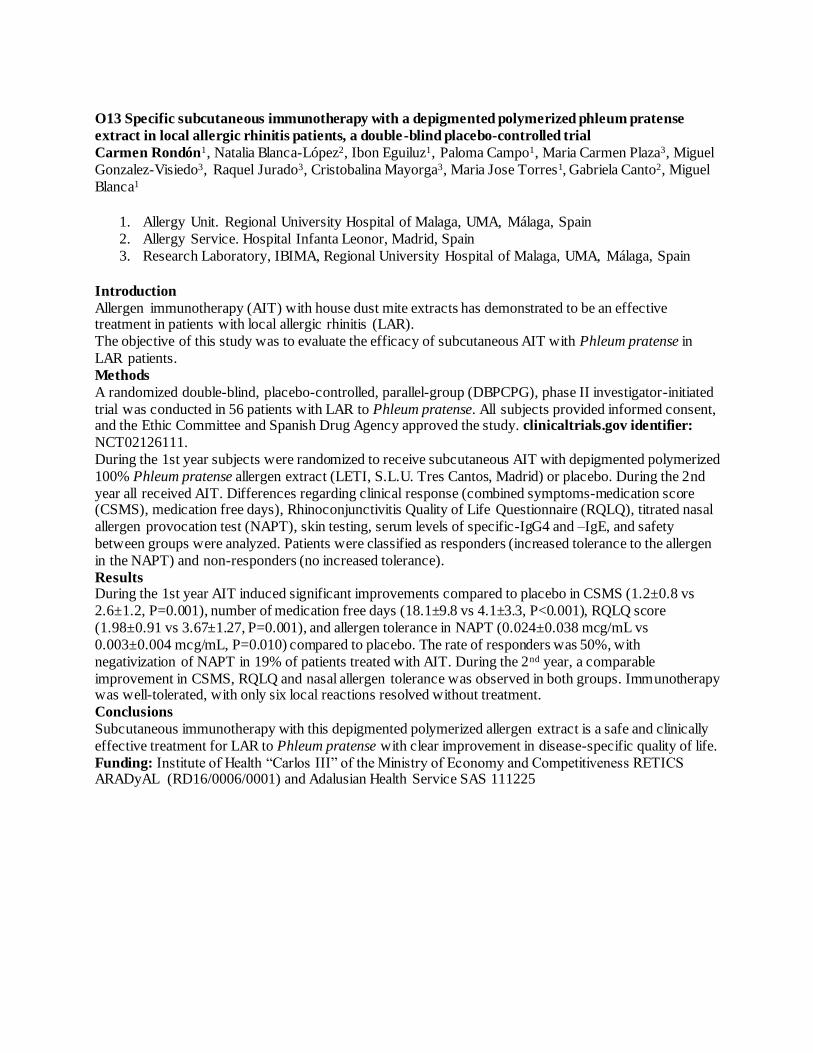
O13 Specific subcutaneous immunotherapy with a depigmented polymerized phleum pratense
extract in local allergic rhinitis patients, a double-blind placebo-controlled trial
Carmen Rondón1, Natalia Blanca-López2, Ibon Eguiluz1, Paloma Campo1, Maria Carmen Plaza3, Miguel
Gonzalez-Visiedo3, Raquel Jurado3, Cristobalina Mayorga3, Maria Jose Torres1, Gabriela Canto2, Miguel
Blanca1
1. Allergy Unit. Regional University Hospital of Malaga, UMA, Málaga, Spain
2. Allergy Service. Hospital Infanta Leonor, Madrid, Spain
3. Research Laboratory, IBIMA, Regional University Hospital of Malaga, UMA, Málaga, Spain
Introduction
Allergen immunotherapy (AIT) with house dust mite extracts has demonstrated to be an effective treatment in patients with local allergic rhinitis (LAR).
The objective of this study was to evaluate the efficacy of subcutaneous AIT with Phleum pratense in
LAR patients.
Methods
A randomized double-blind, placebo-controlled, parallel-group (DBPCPG), phase II investigator-initiated
trial was conducted in 56 patients with LAR to Phleum pratense. All subjects provided informed consent, and the Ethic Committee and Spanish Drug Agency approved the study. clinicaltrials.gov identifier:
NCT02126111.
During the 1st year subjects were randomized to receive subcutaneous AIT with depigmented polymerized
100% Phleum pratense allergen extract (LETI, S.L.U. Tres Cantos, Madrid) or placebo. During the 2nd
year all received AIT. Differences regarding clinical response (combined symptoms-medication score (CSMS), medication free days), Rhinoconjunctivitis Quality of Life Questionnaire (RQLQ), titrated nasal
allergen provocation test (NAPT), skin testing, serum levels of specific-IgG4 and –IgE, and safety
between groups were analyzed. Patients were classified as responders (increased tolerance to the allergen
in the NAPT) and non-responders (no increased tolerance).
Results During the 1st year AIT induced significant improvements compared to placebo in CSMS (1.2±0.8 vs
2.6±1.2, P=0.001), number of medication free days (18.1±9.8 vs 4.1±3.3, P<0.001), RQLQ score
(1.98±0.91 vs 3.67±1.27, P=0.001), and allergen tolerance in NAPT (0.024±0.038 mcg/mL vs
0.003±0.004 mcg/mL, P=0.010) compared to placebo. The rate of responders was 50%, with
negativization of NAPT in 19% of patients treated with AIT. During the 2nd year, a comparable
improvement in CSMS, RQLQ and nasal allergen tolerance was observed in both groups. Immunotherapy was well-tolerated, with only six local reactions resolved without treatment.
Conclusions
Subcutaneous immunotherapy with this depigmented polymerized allergen extract is a safe and clinically
effective treatment for LAR to Phleum pratense with clear improvement in disease-specific quality of life.
Funding: Institute of Health “Carlos III” of the Ministry of Economy and Competitiveness RETICS ARADyAL (RD16/0006/0001) and Adalusian Health Service SAS 111225


![Diagnosis and Management of Rhinitis: Complete Guidelines ... · different forms of rhinitis (allergic, non-allergic, occupational rhinitis, hormonal rhinitis [pregnancy and hypothyroidism],](https://img.pdfslide.us/doc/110x75/5d61f07588c993197b8b51b8/diagnosis-and-management-of-rhinitis-complete-guidelines-different-forms.jpg)