Embed Size (px)
Citation preview

ThrombophlebitisFormation of a venous clot depends on the
presence of of at least of one of Virchow’s triad factors
-venous stasis-injury to vessel wall-hypercoagulable state
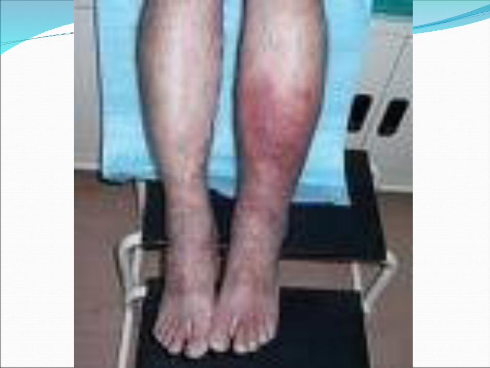
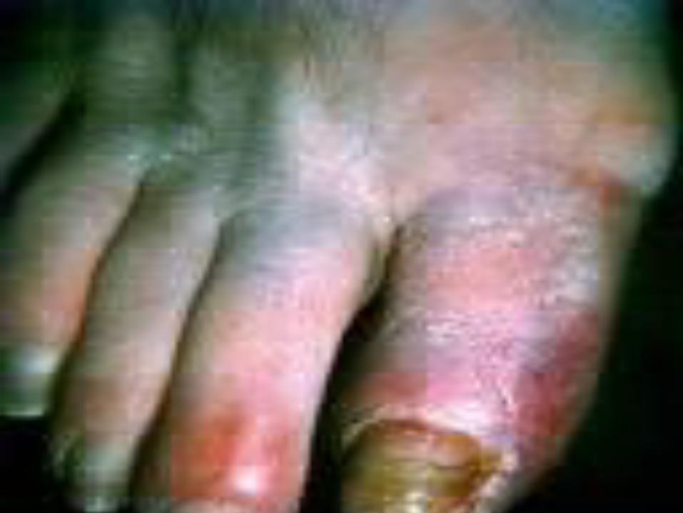
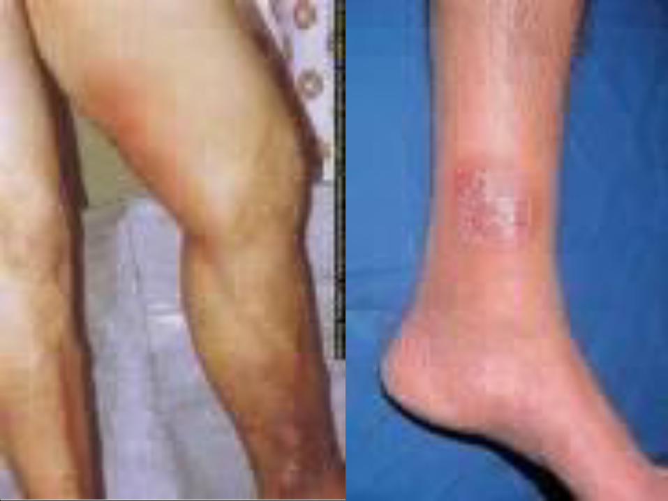
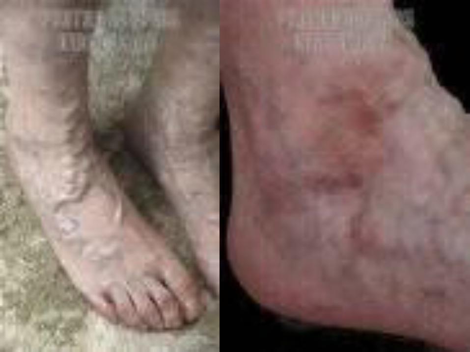
SIGNS AND SYMPTOMSpain in the part of the body affected
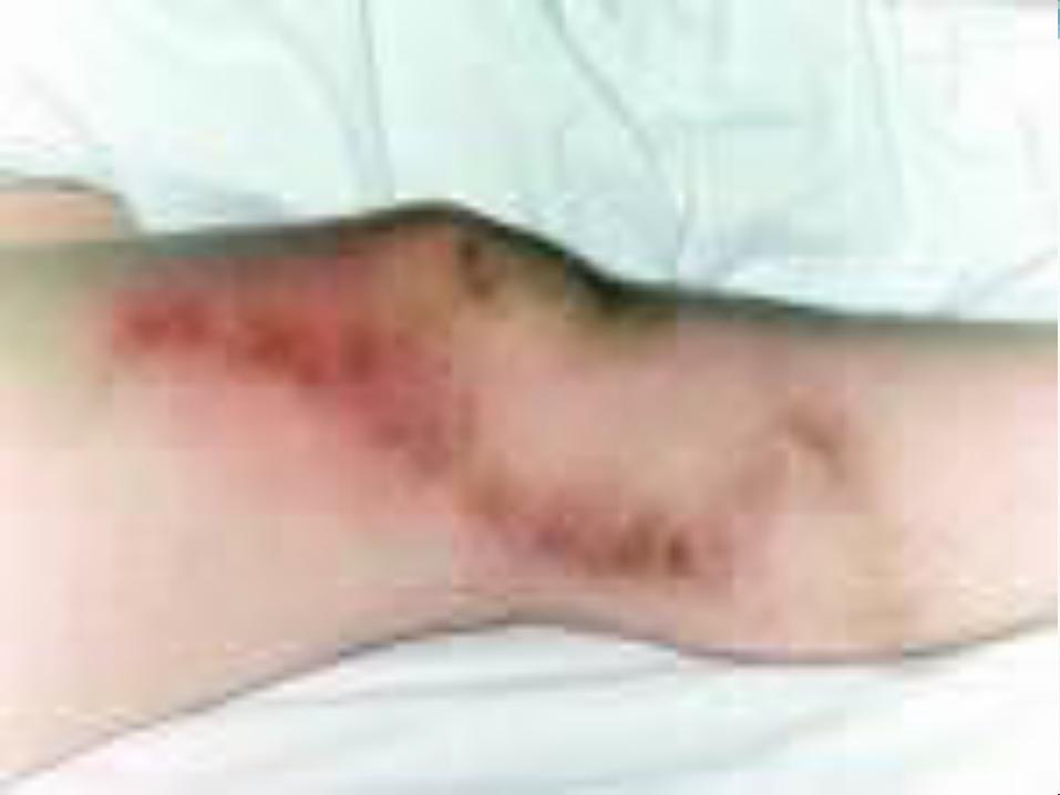
skin redness or inflammation (not always present)
swelling (edema) of the extremities (ankle and foot).
CAUSES
Thrombophlebitis is related to a thrombus in the vein. Risk factors include prolonged sitting and disorders related to blood clotting
Specific disorders associated with thrombophlebitis include superficial thrombophlebitis (affects veins near the skin surface) and deep venous thrombosis (affects deeper, larger veins)
Clinical risk factors for deep vein thrombosisTrauma, travelHypercoagulable,
hormone replacementRecreational drugs(IV
drugs)Old (age >60y)Malignancy
Obesity, obstetricsSurgery, smokingImmobilizationBirth control, blood group
ASickness
PathophysiologyMost common cause of hereditary hemophilia is factor V
Leiden
Thrombi usually form at the venous cusps of deep veins where altered or static blood flow causes clot formation
Alternatively, clots form from intimal defects
Clots are composed from fibrin, red cells and platelets and cause partial/complete obstruction of vein
Pathophysiology
Postphlebitic syndrome (PPS) may develop after the resolution of a DVT
PPS is due valvular incompetence, persistent outflow obstruction and abnormal microcirculation.

Superficial ThrombophlebitisThrombosis can occur in any superficial vein
primarily the saphenous vein and its tributariesLocal pain, redness, and tenderness are characteristic
findings.Mild cases can be treated with warm compresses,
analgesia and elastic supportsSevere cases can be debilitating and should be
managed by bed rest, elevation of extremity, support stockings, and analgesia.
Antibiotics and anticoagulants are useful in septic thrombophlebitis
Deep Vein ThrombosisClinical exam is unreliable for detection or exclusion
of a DVTPain, redness, swelling, and warmth are present in
less than half the patients with confirmed DVT.Pain in calf with dorsiflexion of ankle with the leg
straight (Homan’s sign) is unreliable
Deep Vein Thrombosis the leg is white due to arterial spasm secondary to
massive iliofemoral thrombosis, often mistaken for arterial occlusion.
PPS can be difficult to differentiate from recurrent DVT due to pain, swelling and ulceration of the skin.
Up to to one third of the patients with DVT can develop PPS.
Deep Vein Thrombosis-DiagnosisAll patients with any signs or symptoms suggestive DVT
should undergo an objective diagnostic evaluationVenography was the historical “gold standard” for
detection of DVT with 100% sensitivity and specificity but it is invasive and can cause contrast-related reactions, phlebitis and DVT .
Axillary and Subclavian Vein thrombosis2-4% of DVTs occur in axillary or subclavian veinRisks include recent central venous catheters or
pacemakers, IV drug use, malignancy, hypercoagulable states and excessive or unusual exercise, chronic compression(cervical rib, scalene or web)
Treatment includes anticoagulation alone or preceded by thrombolysis.
Pelvic Vein ThrombosisUsually it’s an extension of a clot from the femoral
vein. An isolated pelvic vein thrombosis is rare and can be
a complication in the postpartum period, after pelvic surgery or trauma.
Septic pelvic vein thrombophlebitis is a life-threatening condition after post-partum endometritis and is usually diagnosed with CT or MRI.
COMPLICATIONSThe most serious complication occurs when the blood
clot dislodges, traveling through the heart and occluding the dense capillarynetwork of the lungs; this is a pulmonary embolism which can be potentially life threatening
TreatmentBed rest, leg elevation and elastic stockings are of
unproven benefit in the management of DVT.Aggressive anticoagulation will prevent extension of
the clot.Early ambulation after adequate anticoagulation is a
safe approachPrimary objective of treating DVT is the prevention of
pulmonary embolus
TreatmentMedications analgesics (pain medications)anticoagulants e.g warfarin or heparin to prevent new
clot formationthrombolytics to dissolve an existing clot such as
intravenous streptokinase.nonsteroidal anti-inflammatory medications (NSAIDS)
such as ibuprofen to reduce pain and inflammationantibiotics (if infection is present) selection will usually
depend with the causative agent.Support stockings and wraps to reduce discomfort
TreatmentIn pregnant pt who cannot have heparin, danaproid
should be used.
Warfarin is contraindicated in pregnancy, active bleeding, recent major surgery (thoracoabdominal, nervous system, spine, eye)
The patient may be advised to do the followingElevate the affected area to reduce swelling.Keep pressure off of the area to reduce pain and
decrease the risk of further damage.Apply moist heat to reduce inflammation and pain.Surgical removal, stripping, or bypass of the vein is rarely
needed but may be recommended in some situations.

![Behcet’s Disease: Radiologic Diagnosis · with Behcet’s disease is the rupture of a large aortic or arterial aneurysm [17]. Venous occlusion: Deep and superficial veins thrombophlebitis](https://img.pdfslide.us/doc/110x75/5ace29797f8b9a875a8eac4b/behcets-disease-radiologic-behcets-disease-is-the-rupture-of-a-large-aortic.jpg)