Embed Size (px)
Citation preview
THROMBOPHILIA DVT PROPHYLAXIS
VTE MANAGEMENT
DR. ASHISH
Moderator: DR. MEERA KHARBANDAwww.anaesthesia.co.in [email protected]
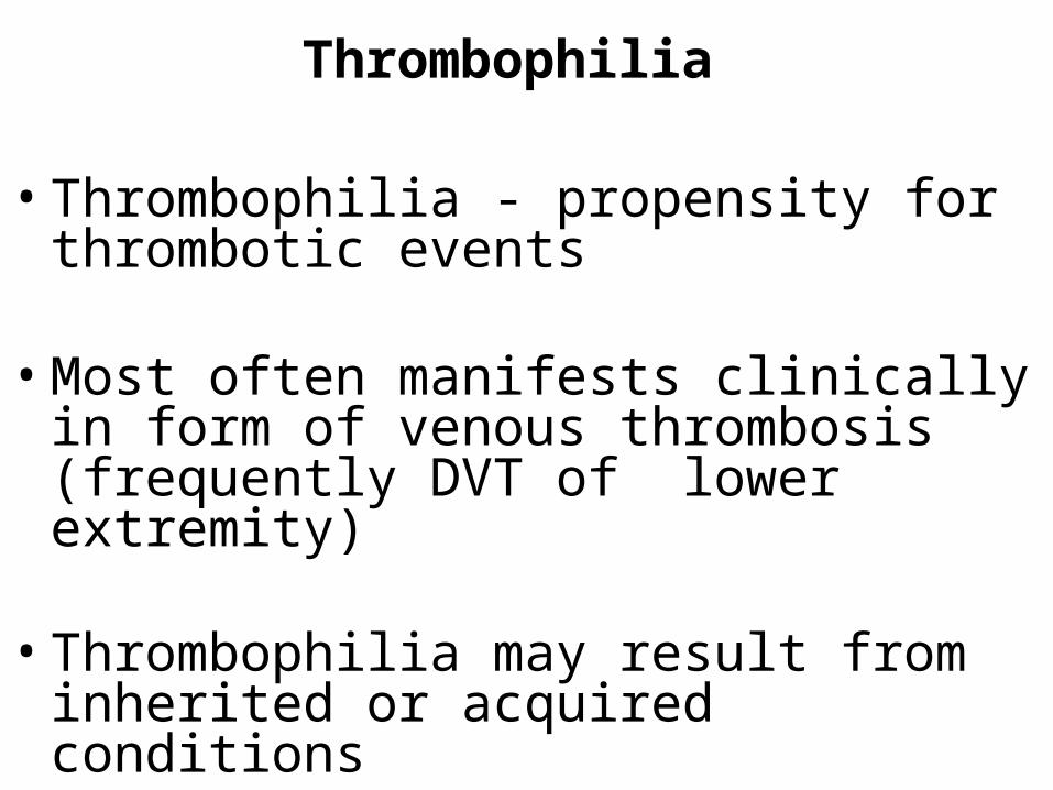
Thrombophilia
• Thrombophilia - propensity for thrombotic events
• Most often manifests clinically in form of venous thrombosis (frequently DVT of lower extremity)
• Thrombophilia may result from inherited or acquired conditions
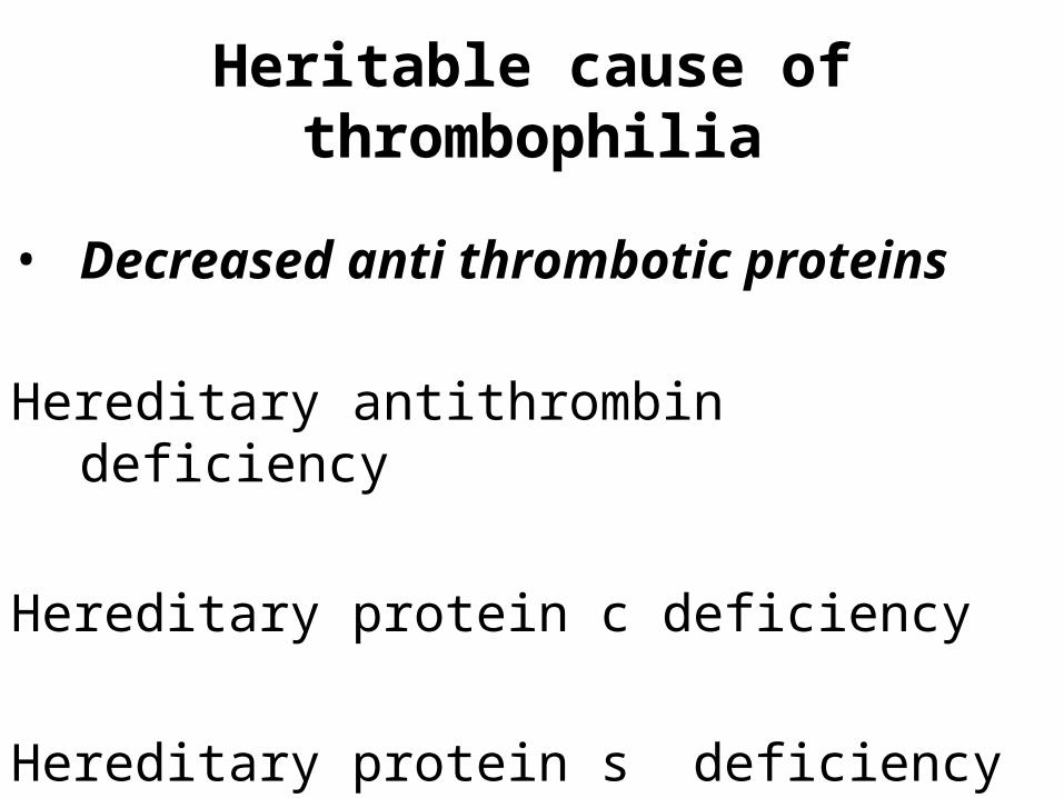
Heritable cause of thrombophilia
• Decreased anti thrombotic proteins
Hereditary antithrombin deficiency
Hereditary protein c deficiency
Hereditary protein s deficiency
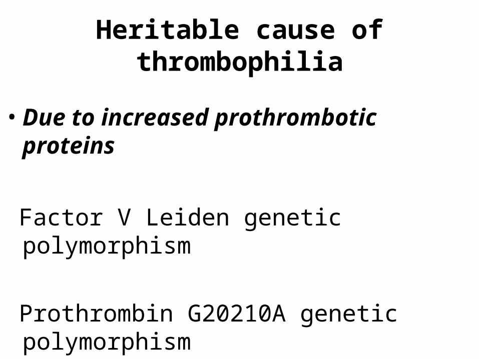
Heritable cause of thrombophilia
• Due to increased prothrombotic proteins
Factor V Leiden genetic polymorphism
Prothrombin G20210A genetic polymorphism

Acquired causes of thrombophilia
• Myeloproliferative disorders
• Malignancies
• Pregnancy & OCP use
• Nephrotic syndrome patients
• Antiphospholipid antibodies
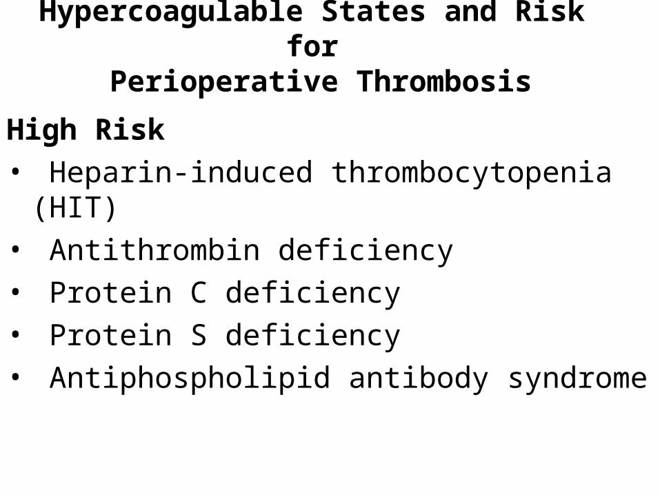
Hypercoagulable States and Risk for Perioperative Thrombosis
High Risk
• Heparin-induced thrombocytopenia (HIT)
• Antithrombin deficiency
• Protein C deficiency
• Protein S deficiency
• Antiphospholipid antibody syndrome
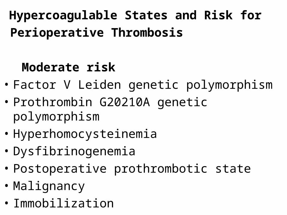
Hypercoagulable States and Risk for
Perioperative Thrombosis
Moderate risk
• Factor V Leiden genetic polymorphism
• Prothrombin G20210A genetic polymorphism
• Hyperhomocysteinemia
• Dysfibrinogenemia
• Postoperative prothrombotic state
• Malignancy
• Immobilization
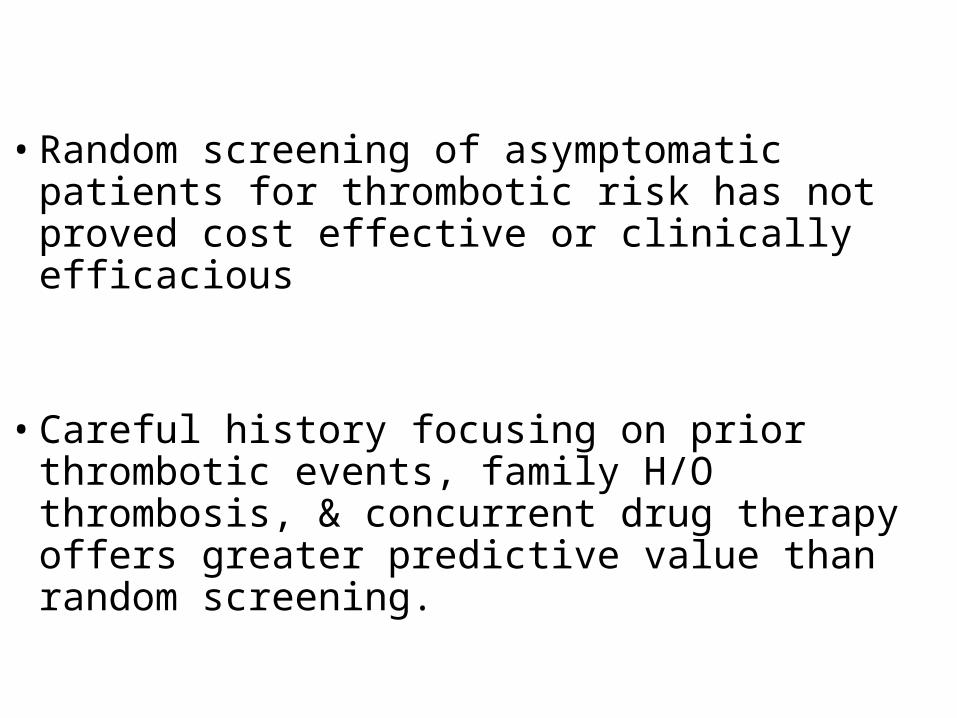
• Random screening of asymptomatic patients for thrombotic risk has not proved cost effective or clinically efficacious
• Careful history focusing on prior thrombotic
events, family H/O thrombosis, & concurrent drug therapy offers greater predictive value than random screening.
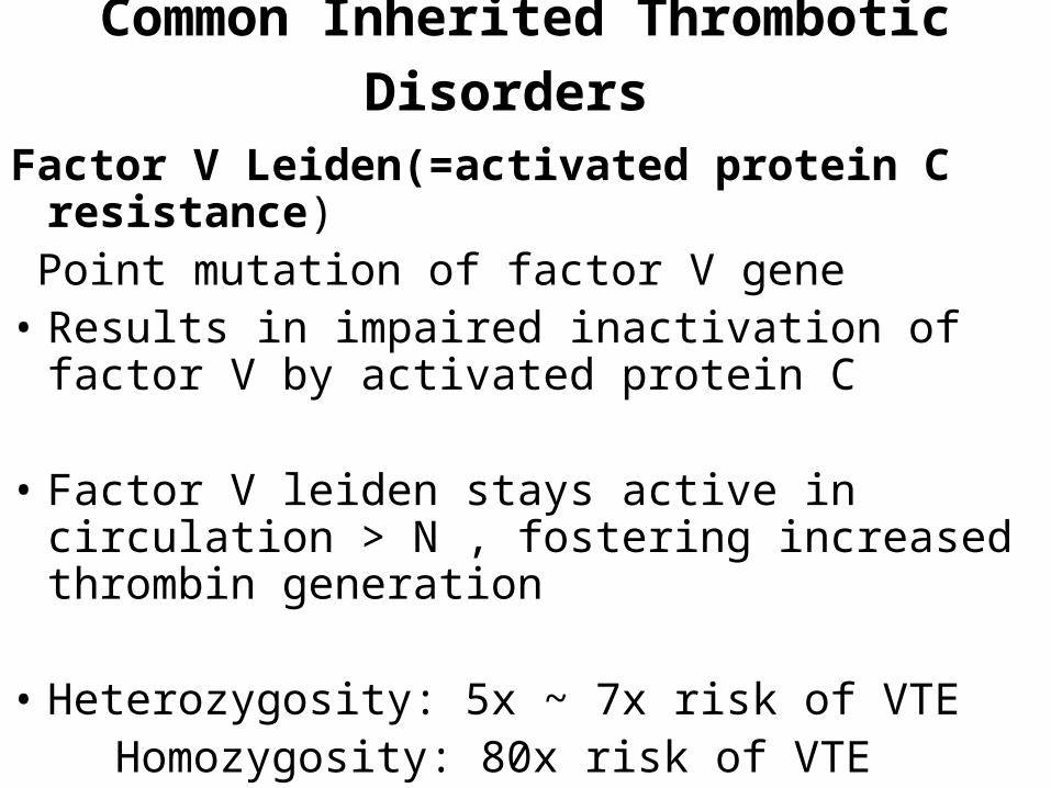
Common Inherited Thrombotic Disorders
Factor V Leiden(=activated protein C resistance)
Point mutation of factor V gene• Results in impaired inactivation of factor V by
activated protein C
• Factor V leiden stays active in circulation > N , fostering increased thrombin generation
• Heterozygosity: 5x ~ 7x risk of VTE Homozygosity: 80x risk of VTE
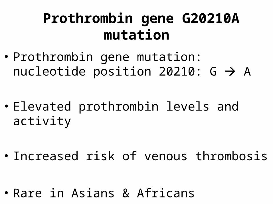
Prothrombin gene G20210A mutation
• Prothrombin gene mutation: nucleotide position 20210: G A
• Elevated prothrombin levels and activity
• Increased risk of venous thrombosis
• Rare in Asians & Africans
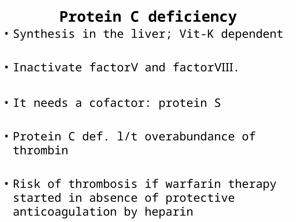
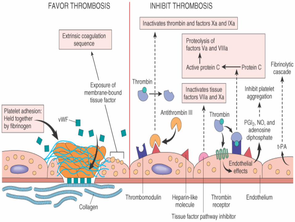
Protein C deficiency• Synthesis in the liver; Vit-K dependent
• Inactivate factor and factor .Ⅴ Ⅷ
• It needs a cofactor: protein S
• Protein C def. l/t overabundance of thrombin
• Risk of thrombosis if warfarin therapy started in absence of protective anticoagulation by heparin
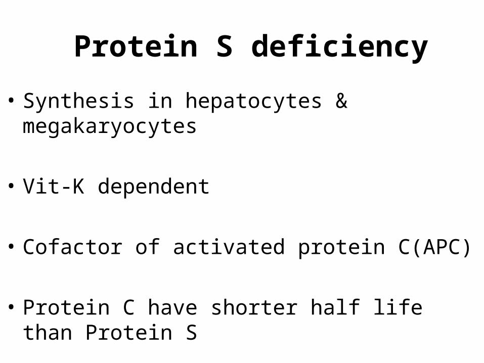
Protein S deficiency
• Synthesis in hepatocytes & megakaryocytes
• Vit-K dependent
• Cofactor of activated protein C(APC)
• Protein C have shorter half life than Protein S
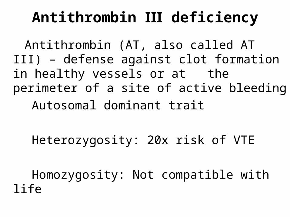
Antithrombin Ⅲ deficiency
Antithrombin (AT, also called AT III) – defense against clot formation in healthy vessels or at the perimeter of a site of active bleeding
Autosomal dominant trait
Heterozygosity: 20x risk of VTE
Homozygosity: Not compatible with life
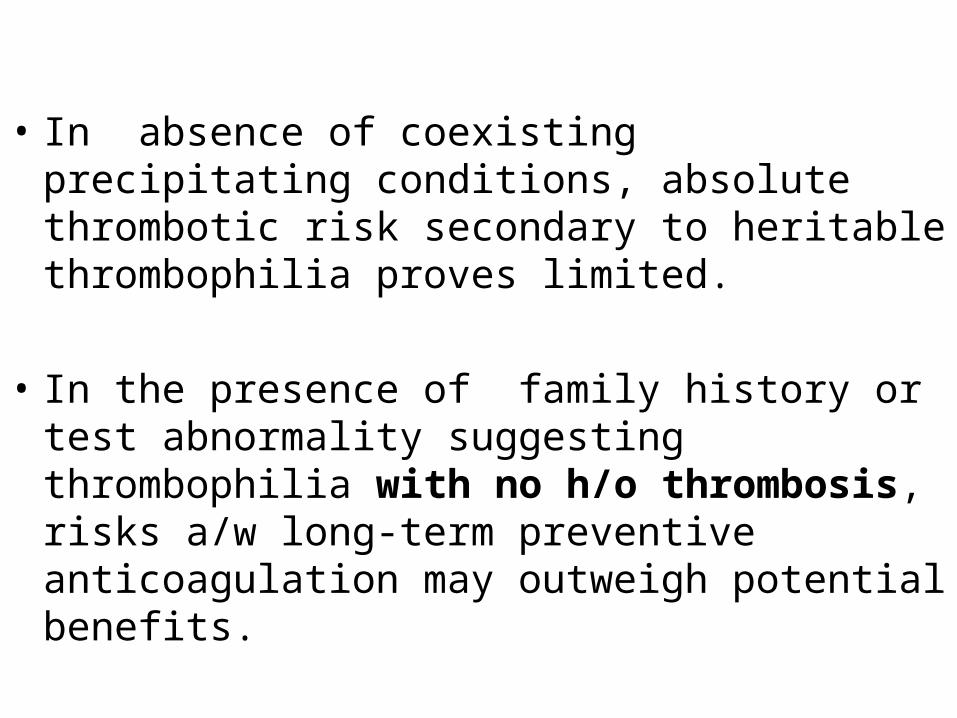
• In absence of coexisting precipitating conditions, absolute thrombotic risk secondary to heritable thrombophilia proves limited.
• In the presence of family history or test abnormality suggesting thrombophilia with no h/o thrombosis, risks a/w long-term preventive anticoagulation may outweigh potential benefits.
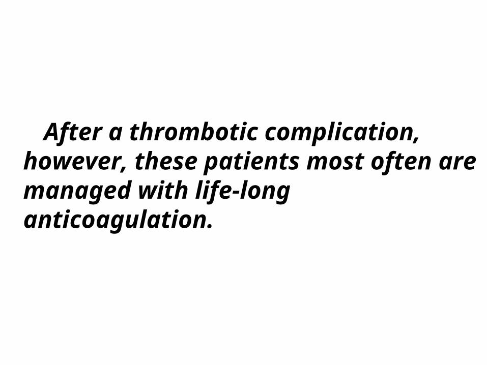
After a thrombotic complication, however, these patients most often are managed with life-long anticoagulation.
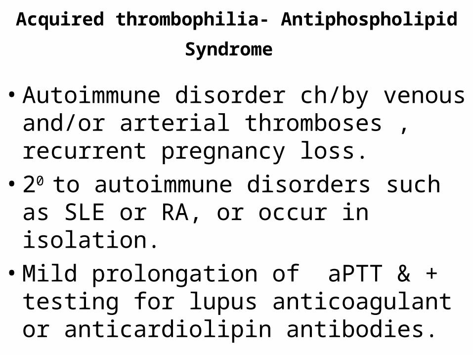
Acquired thrombophilia- Antiphospholipid
Syndrome
• Autoimmune disorder ch/by venous and/or arterial thromboses , recurrent pregnancy loss.
• 20 to autoimmune disorders such as SLE or RA, or occur in isolation.
• Mild prolongation of aPTT & + testing for lupus anticoagulant or anticardiolipin antibodies.
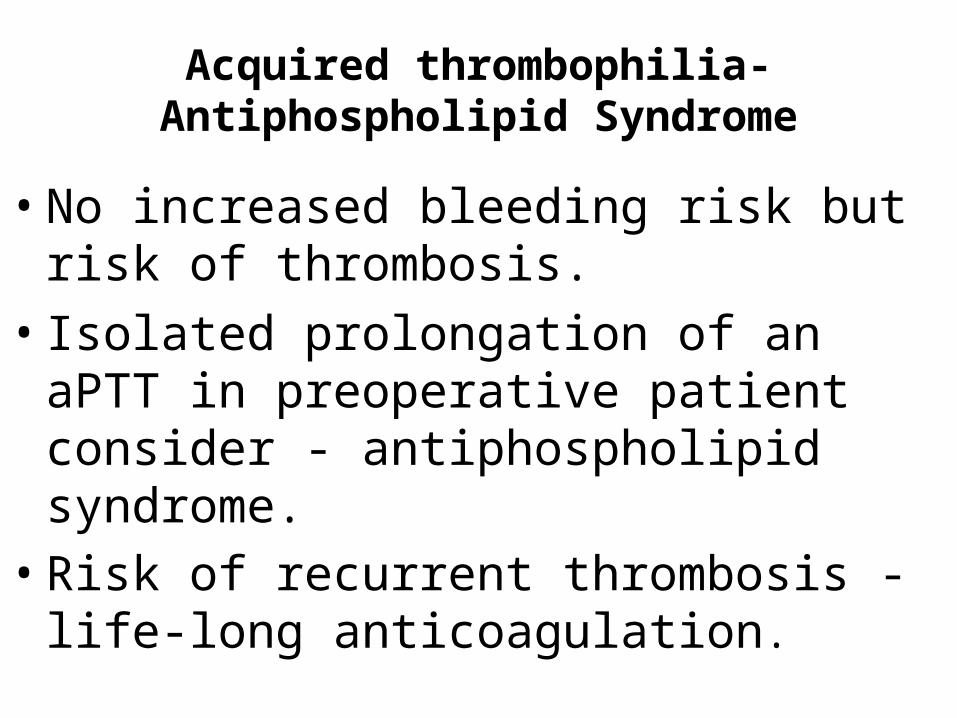
Acquired thrombophilia- Antiphospholipid Syndrome
• No increased bleeding risk but risk of thrombosis.
• Isolated prolongation of an aPTT in preoperative patient consider - antiphospholipid syndrome.
• Risk of recurrent thrombosis - life-long anticoagulation.
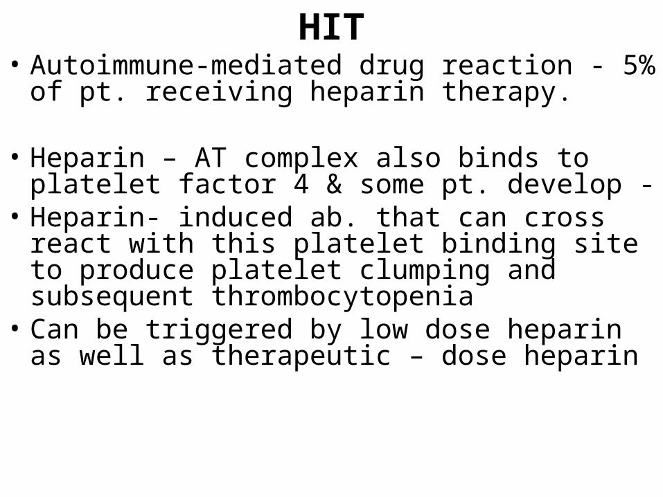
HIT• Autoimmune-mediated drug reaction - 5% of pt.
receiving heparin therapy.
• Heparin – AT complex also binds to platelet factor 4 & some pt. develop -
• Heparin- induced ab. that can cross react with this platelet binding site to produce platelet clumping and subsequent thrombocytopenia
• Can be triggered by low dose heparin as well as therapeutic – dose heparin
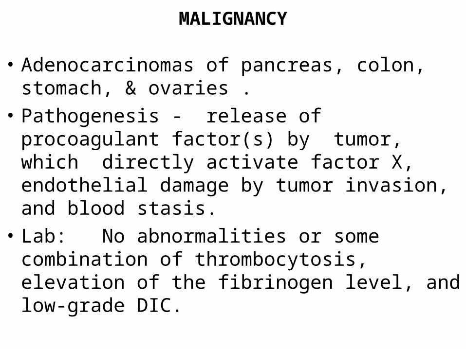
MALIGNANCY
• Adenocarcinomas of pancreas, colon, stomach, & ovaries .
• Pathogenesis - release of procoagulant factor(s) by tumor, which directly activate factor X, endothelial damage by tumor invasion, and blood stasis.
• Lab: No abnormalities or some combination of thrombocytosis, elevation of the fibrinogen level, and low-grade DIC.
Pregnancy and OCP Use
• Incidence - 1 in 1500 pregnancies
• Risk of PE highest during 3rd trimester & immediate postpartum period
• Antithrombin III–deficient women high risk -anticoagulated throughout pregnancy.
• Factor V Leiden and the prothrombin G20201A mutation a/w less risk.
• Women with one of these inherited traits not anticoagulated unless – h/o PE or recurrent DVT
Pregnancy and OCP Use
• Since low-dose estrogen OCP introduction -incidence decreased.
• Women - smoke, h/o migraine headaches, inherited hypercoagulable defect at increased risk (30-fold) of venous thrombosis, PE, & cerebrovascular thrombosis.
Nephrotic Syndrome Patients
• Risk of thromboembolic disease including renal vein thrombosis.
• D/t < N levels of antithrombin III or PC 20 renal loss of coagulation protein, factor XII deficiency, platelet hyperactivity, abnormal fibrinolytic activity, & > N levels of other coagulation factors.
• Hyperlipidemia and hypoalbuminemia - also possible etiologic factors
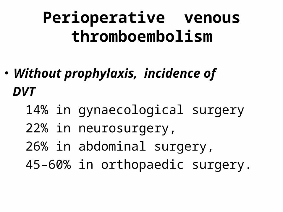
Perioperative venous thromboembolism
• Without prophylaxis, incidence of
DVT
14% in gynaecological surgery
22% in neurosurgery,
26% in abdominal surgery,
45–60% in orthopaedic surgery.

• Agency for health care research and quality have issued report stating that :
Prophylaxis for venous thromboembolism is single most important measure for ensuring patient safety in hospitalized patients
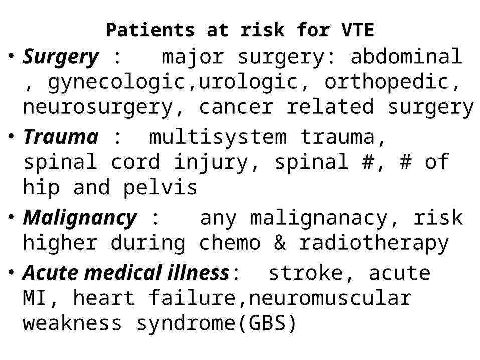
Patients at risk for VTE
• Surgery : major surgery: abdominal , gynecologic,urologic, orthopedic, neurosurgery, cancer related surgery
• Trauma : multisystem trauma, spinal cord injury, spinal #, # of hip and pelvis
• Malignancy : any malignanacy, risk higher during chemo & radiotherapy
• Acute medical illness: stroke, acute MI, heart failure,neuromuscular weakness syndrome(GBS)
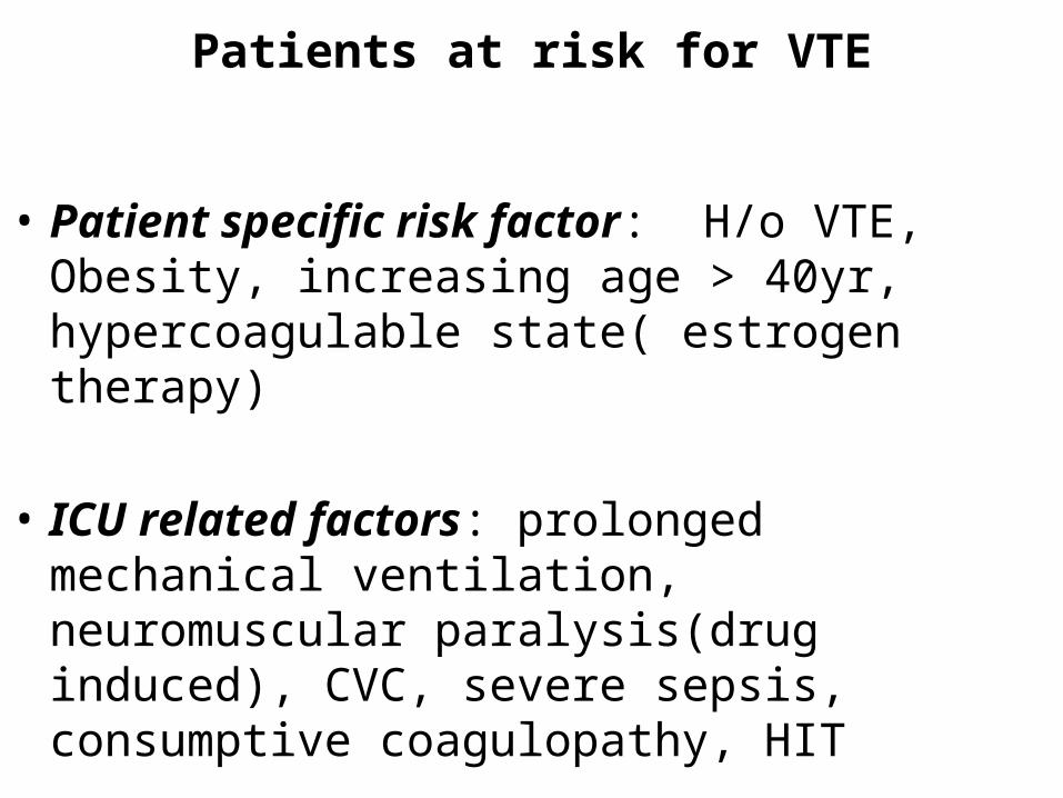
Patients at risk for VTE
• Patient specific risk factor: H/o VTE, Obesity, increasing age > 40yr, hypercoagulable state( estrogen therapy)
• ICU related factors: prolonged mechanical ventilation, neuromuscular paralysis(drug induced), CVC, severe sepsis, consumptive coagulopathy, HIT
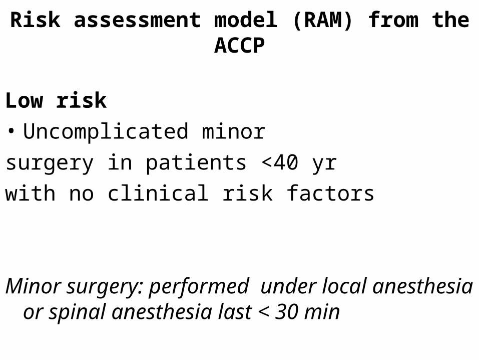
Risk assessment model (RAM) from the ACCP
Low risk
• Uncomplicated minor
surgery in patients <40 yr
with no clinical risk factors
Minor surgery: performed under local anesthesia or spinal anesthesia last < 30 min
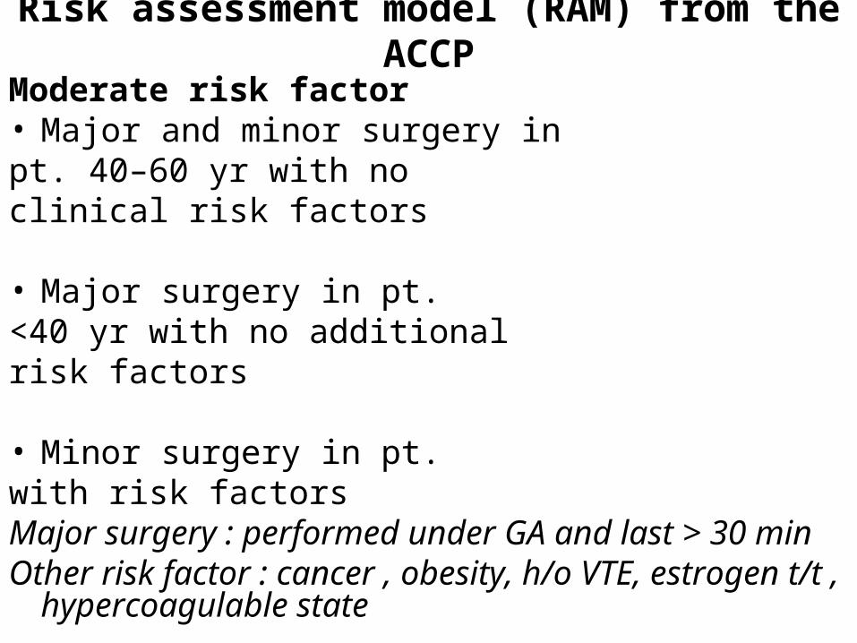
Risk assessment model (RAM) from the ACCP
Moderate risk factor• Major and minor surgery inpt. 40–60 yr with noclinical risk factors
• Major surgery in pt.<40 yr with no additionalrisk factors
• Minor surgery in pt.with risk factorsMajor surgery : performed under GA and last > 30 minOther risk factor : cancer , obesity, h/o VTE, estrogen
t/t , hypercoagulable state
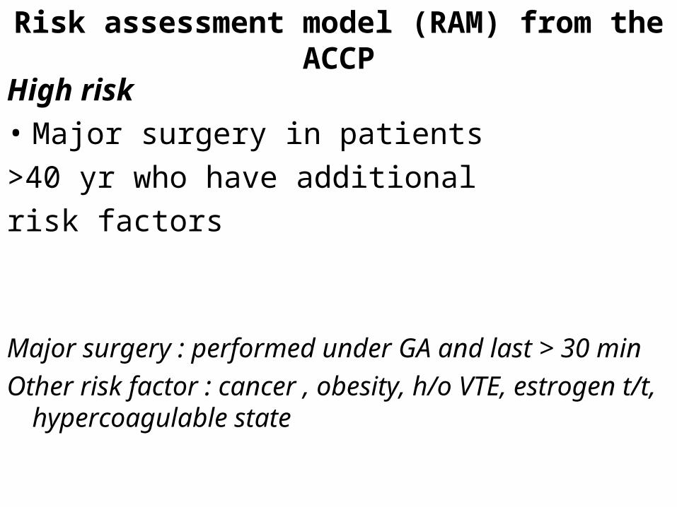
Risk assessment model (RAM) from the ACCP
High risk
• Major surgery in patients
>40 yr who have additional
risk factors
Major surgery : performed under GA and last > 30 min
Other risk factor : cancer , obesity, h/o VTE, estrogen t/t, hypercoagulable state
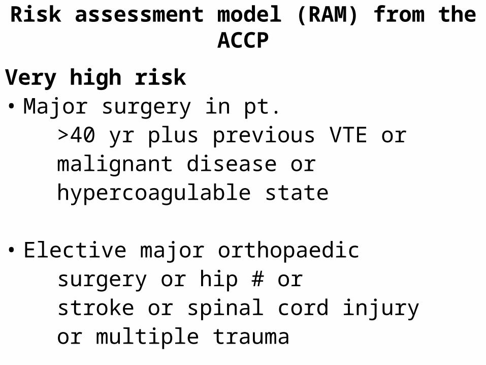
Risk assessment model (RAM) from the ACCP
Very high risk • Major surgery in pt. >40 yr plus previous VTE or malignant disease or hypercoagulable state
• Elective major orthopaedic surgery or hip # or stroke or spinal cord injury or multiple trauma
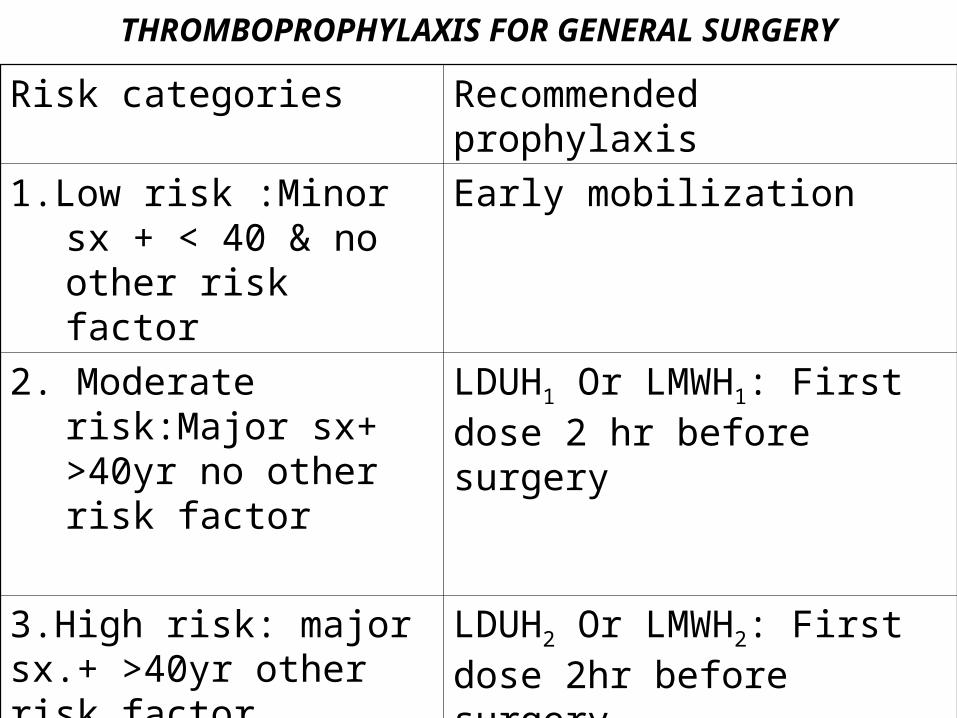
THROMBOPROPHYLAXIS FOR GENERAL SURGERY
Risk categories Recommended prophylaxis
1.Low risk :Minor sx + < 40 & no other risk factor
Early mobilization
2. Moderate risk:Major sx+ >40yr no other risk factor
LDUH1 Or LMWH1: First dose 2 hr before surgery
3.High risk: major sx.+ >40yr other risk factor
LDUH2 Or LMWH2: First dose 2hr before surgery
4.Very high risk :major sx.+ >40yr other risk factor
LDUH2 Or LMWH2 as above plus mechanical aid
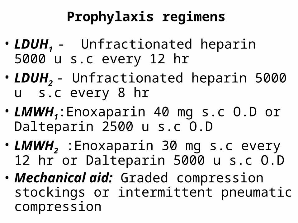
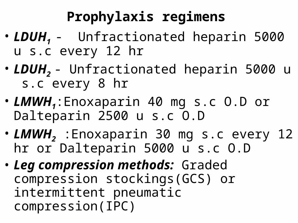
Prophylaxis regimens
• LDUH1 - Unfractionated heparin 5000 u s.c every 12 hr
• LDUH2 - Unfractionated heparin 5000 u s.c every 8 hr
• LMWH1:Enoxaparin 40 mg s.c O.D or Dalteparin 2500 u s.c O.D
• LMWH2 :Enoxaparin 30 mg s.c every 12 hr or Dalteparin 5000 u s.c O.D
• Mechanical aid: Graded compression stockings or intermittent pneumatic compression
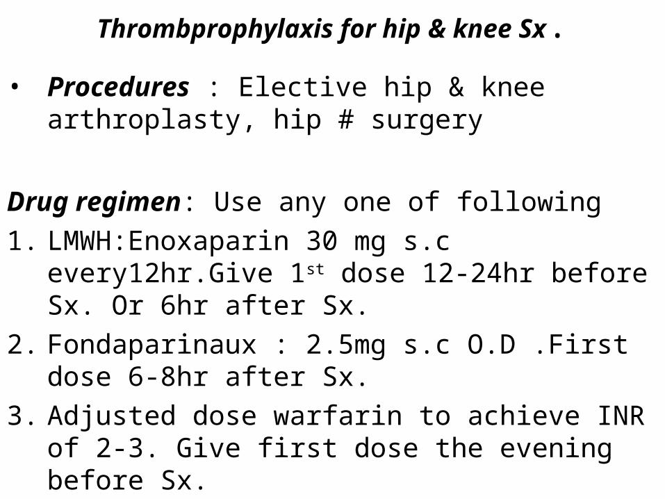
Thrombprophylaxis for hip & knee Sx.
• Procedures : Elective hip & knee arthroplasty, hip # surgery
Drug regimen: Use any one of following
1. LMWH:Enoxaparin 30 mg s.c every12hr.Give 1st dose 12-24hr before Sx. Or 6hr after Sx.
2. Fondaparinaux : 2.5mg s.c O.D .First dose 6-8hr after Sx.
3. Adjusted dose warfarin to achieve INR of 2-3. Give first dose the evening before Sx.
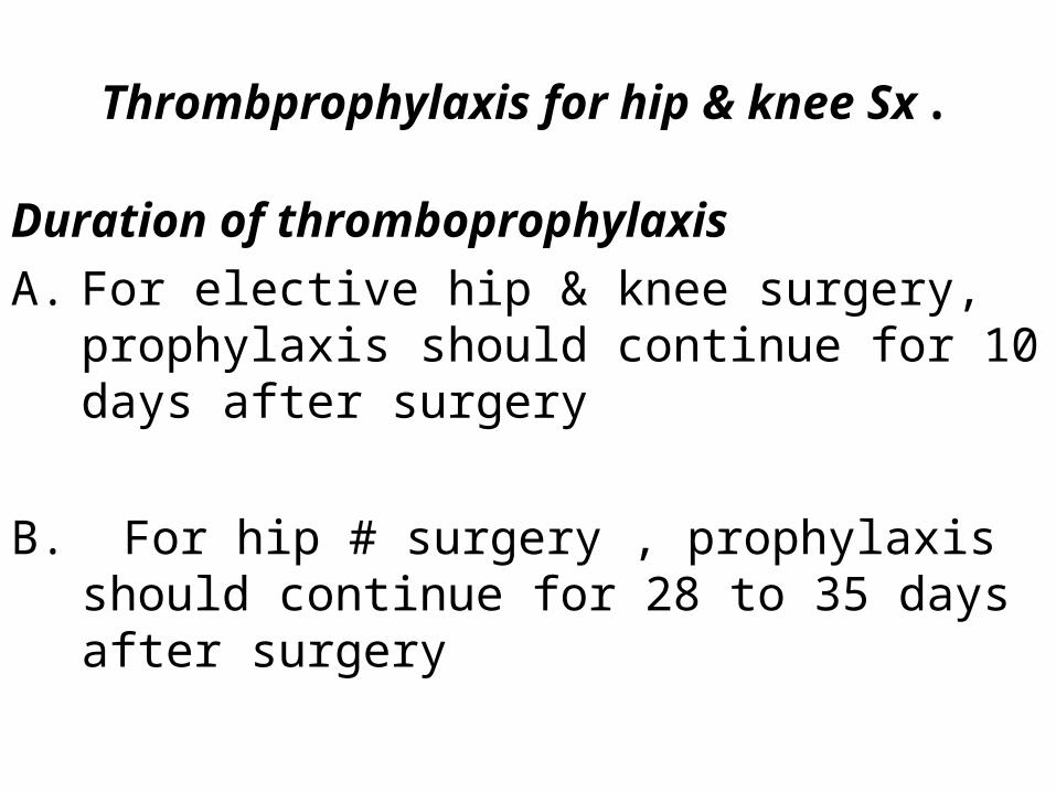
Thrombprophylaxis for hip & knee Sx.
Duration of thromboprophylaxis
A. For elective hip & knee surgery, prophylaxis should continue for 10 days after surgery
B. For hip # surgery , prophylaxis should continue for 28 to 35 days after surgery
• Thromboprophylaxis for other conditions
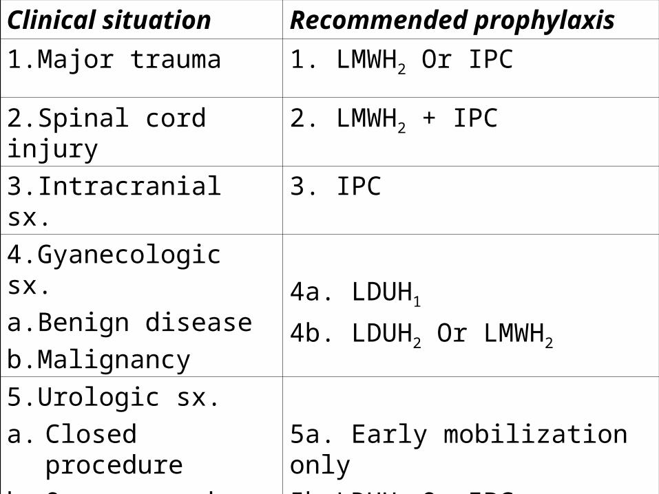
Clinical situation Recommended prophylaxis
1.Major trauma 1. LMWH2 Or IPC
2.Spinal cord injury 2. LMWH2 + IPC
3.Intracranial sx. 3. IPC
4.Gyanecologic sx.
a.Benign disease
b.Malignancy
4a. LDUH1
4b. LDUH2 Or LMWH2
5.Urologic sx.
a. Closed procedure
b. Open procedure
5a. Early mobilization only
5b.LDUH1 Or IPC
6.High risk medical condition
6.LDUH1 Or LMWH1
Prophylaxis regimens
• LDUH1 - Unfractionated heparin 5000 u s.c every 12 hr
• LDUH2 - Unfractionated heparin 5000 u s.c every 8 hr
• LMWH1:Enoxaparin 40 mg s.c O.D or Dalteparin 2500 u s.c O.D
• LMWH2 :Enoxaparin 30 mg s.c every 12 hr or Dalteparin 5000 u s.c O.D
• Leg compression methods: Graded compression stockings(GCS) or intermittent pneumatic compression(IPC)
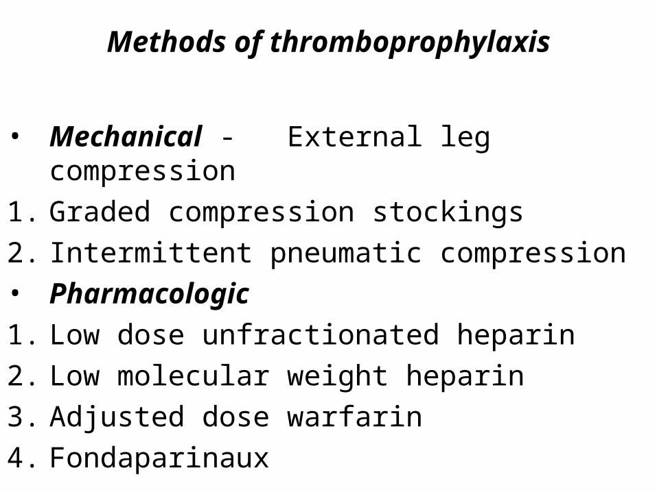
Methods of thromboprophylaxis
• Mechanical - External leg compression
1. Graded compression stockings
2. Intermittent pneumatic compression
• Pharmacologic
1. Low dose unfractionated heparin
2. Low molecular weight heparin
3. Adjusted dose warfarin
4. Fondaparinaux
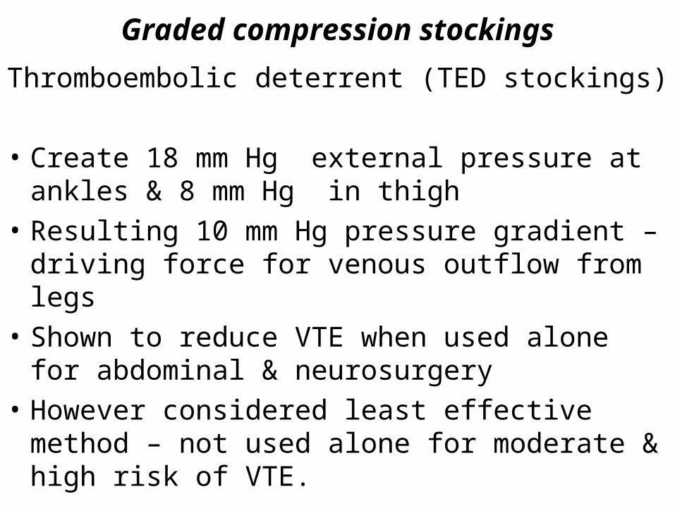
Graded compression stockings
Thromboembolic deterrent (TED stockings)
• Create 18 mm Hg external pressure at ankles & 8 mm Hg in thigh
• Resulting 10 mm Hg pressure gradient – driving force for venous outflow from legs
• Shown to reduce VTE when used alone for abdominal & neurosurgery
• However considered least effective method – not used alone for moderate & high risk of VTE.
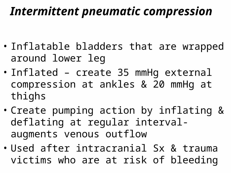
Intermittent pneumatic compression
• Inflatable bladders that are wrapped around lower leg
• Inflated – create 35 mmHg external compression at ankles & 20 mmHg at thighs
• Create pumping action by inflating & deflating at regular interval- augments venous outflow
• Used after intracranial Sx & trauma victims who are at risk of bleeding
Low – dose unfractionated heparin
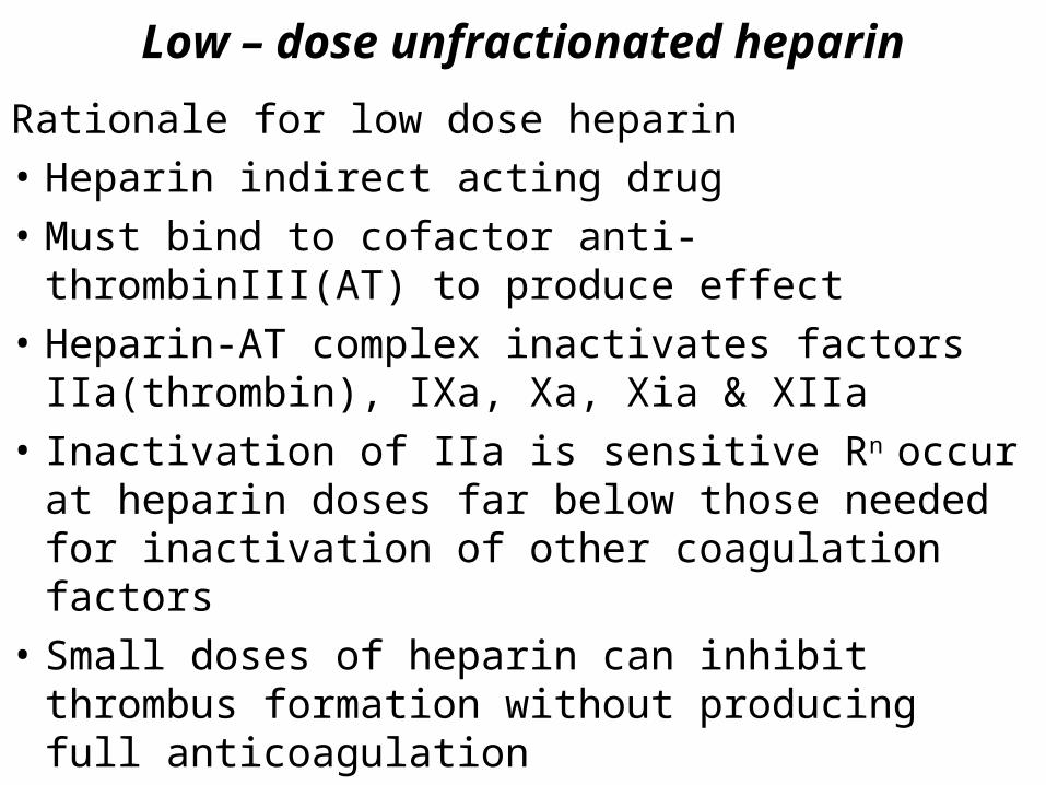
Rationale for low dose heparin
• Heparin indirect acting drug
• Must bind to cofactor anti-thrombinIII(AT) to produce effect
• Heparin-AT complex inactivates factors IIa(thrombin), IXa, Xa, Xia & XIIa
• Inactivation of IIa is sensitive Rn occur at heparin doses far below those needed for inactivation of other coagulation factors
• Small doses of heparin can inhibit thrombus formation without producing full anticoagulation
Low – dose unfractionated heparin
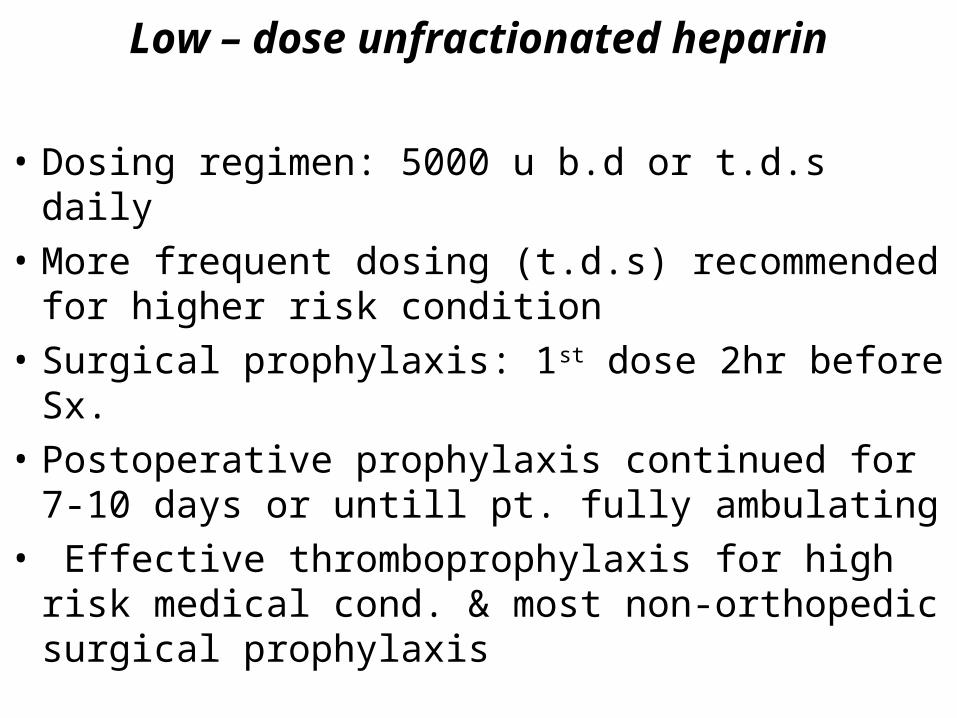
• Dosing regimen: 5000 u b.d or t.d.s daily
• More frequent dosing (t.d.s) recommended for higher risk condition
• Surgical prophylaxis: 1st dose 2hr before Sx.
• Postoperative prophylaxis continued for 7-10 days or untill pt. fully ambulating
• Effective thromboprophylaxis for high risk medical cond. & most non-orthopedic surgical prophylaxis
Low molecular weight heparin
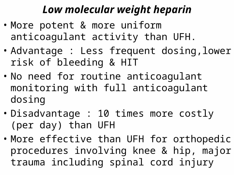
• More potent & more uniform anticoagulant activity than UFH.
• Advantage : Less frequent dosing,lower risk of bleeding & HIT
• No need for routine anticoagulant monitoring with full anticoagulant dosing
• Disadvantage : 10 times more costly (per day) than UFH
• More effective than UFH for orthopedic procedures involving knee & hip, major trauma including spinal cord injury
Low molecular weight heparin
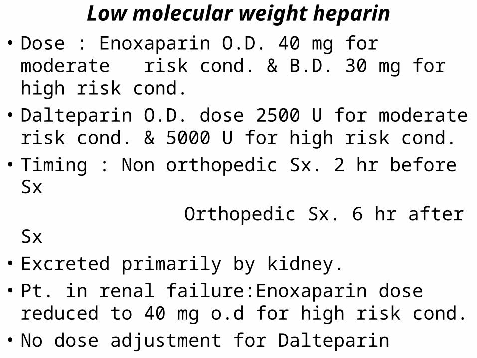
• Dose : Enoxaparin O.D. 40 mg for moderate risk cond. & B.D. 30 mg for high risk cond.
• Dalteparin O.D. dose 2500 U for moderate risk cond. & 5000 U for high risk cond.
• Timing : Non orthopedic Sx. 2 hr before Sx
Orthopedic Sx. 6 hr after Sx
• Excreted primarily by kidney.
• Pt. in renal failure:Enoxaparin dose reduced to 40 mg o.d for high risk cond.
• No dose adjustment for Dalteparin
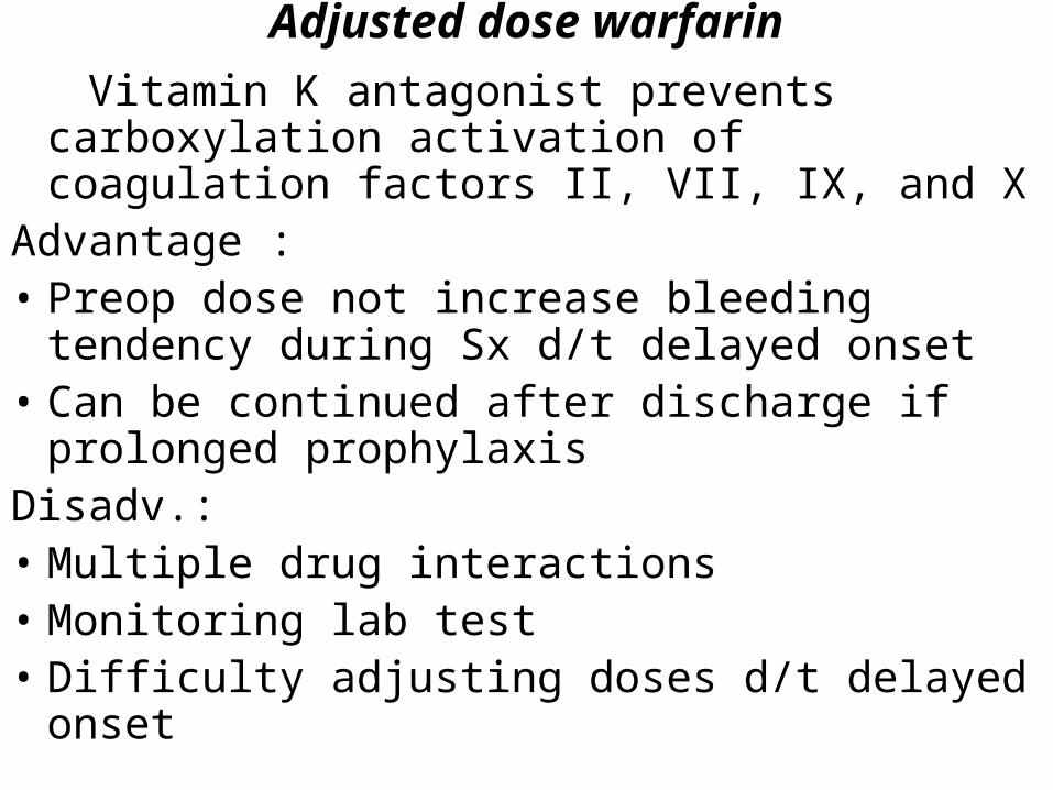
Adjusted dose warfarin
Vitamin K antagonist prevents carboxylation activation of coagulation factors II, VII, IX, and X
Advantage :• Preop dose not increase bleeding tendency
during Sx d/t delayed onset• Can be continued after discharge if prolonged
prophylaxisDisadv.:• Multiple drug interactions• Monitoring lab test • Difficulty adjusting doses d/t delayed onset
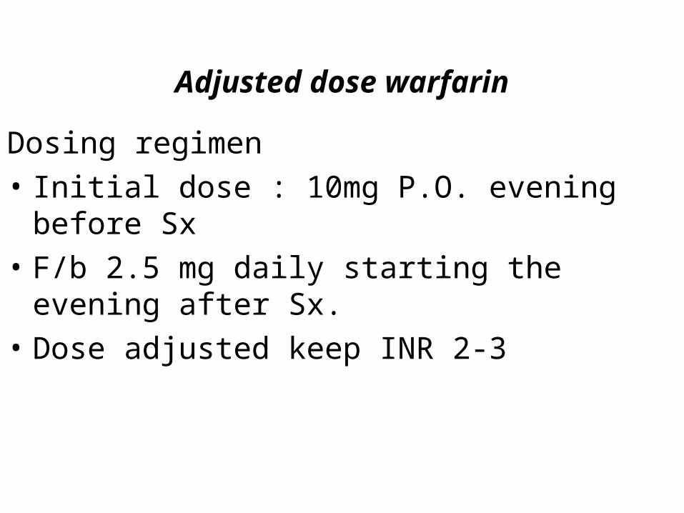
Adjusted dose warfarin
Dosing regimen
• Initial dose : 10mg P.O. evening before Sx
• F/b 2.5 mg daily starting the evening after Sx.
• Dose adjusted keep INR 2-3
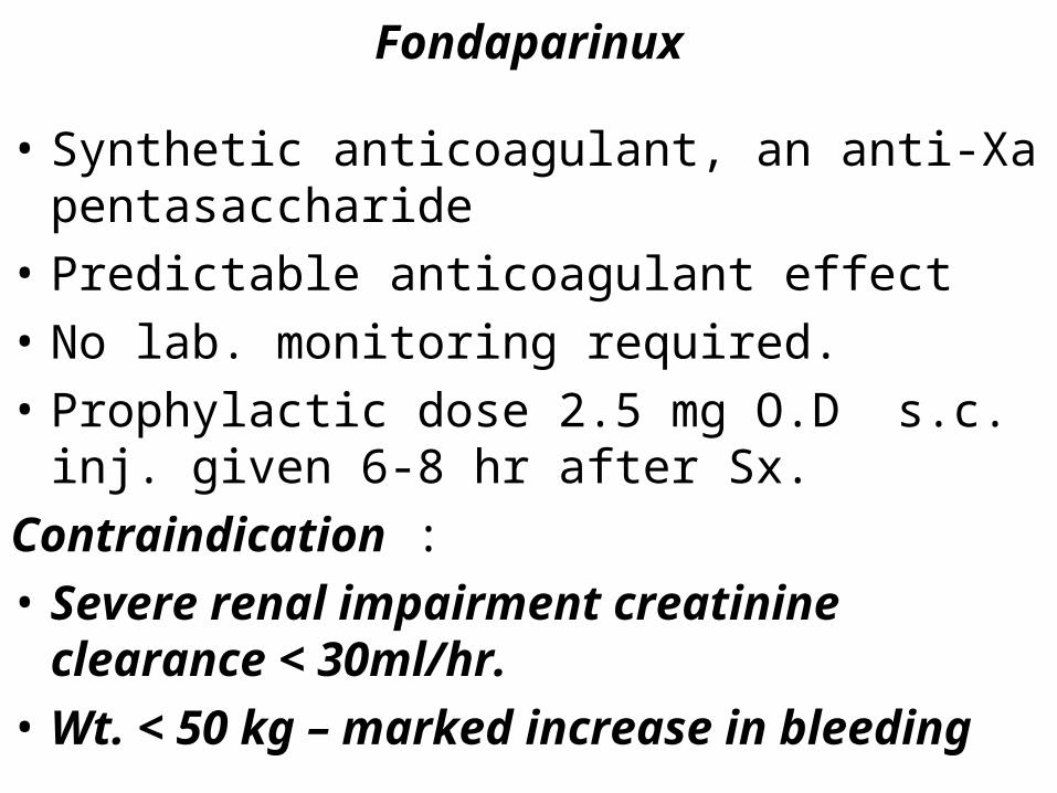
Fondaparinux
• Synthetic anticoagulant, an anti-Xa pentasaccharide
• Predictable anticoagulant effect
• No lab. monitoring required.
• Prophylactic dose 2.5 mg O.D s.c. inj. given 6-8 hr after Sx.
Contraindication :
• Severe renal impairment creatinine clearance < 30ml/hr.
• Wt. < 50 kg – marked increase in bleeding
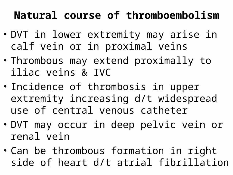
Natural course of thromboembolism
• DVT in lower extremity may arise in calf vein or in proximal veins
• Thrombous may extend proximally to iliac veins & IVC
• Incidence of thrombosis in upper extremity increasing d/t widespread use of central venous catheter
• DVT may occur in deep pelvic vein or renal vein
• Can be thrombous formation in right side of heart d/t atrial fibrillation
• Most clinically important & fatal pulmonary embolism occurs from proximal than distal DVT in leg
• PE occur in 50% of pt. with proximal DVT, while
asymptomatic thrombosis of leg vein is observed in 70% pt with PE
• On early ambulation, thrombus in deep veins may resolve completely
• Post-thrombotic syndrome may develop in 25% of patients, 2yrs after initial diagnosis and proper t/t of DVT
• Inadequate t/t of DVT result in 20-30% risk of recurrent VTE & collaterals develop parallel to thrombosed segment of vein
Diagnostic approach to thromboembolism
The Clinical evaluationClinical presentation of acute pulmonary
embolism is nonspecific
No clinical or lab findings that will confirm or exclude diagnosis of pulmonary embolism
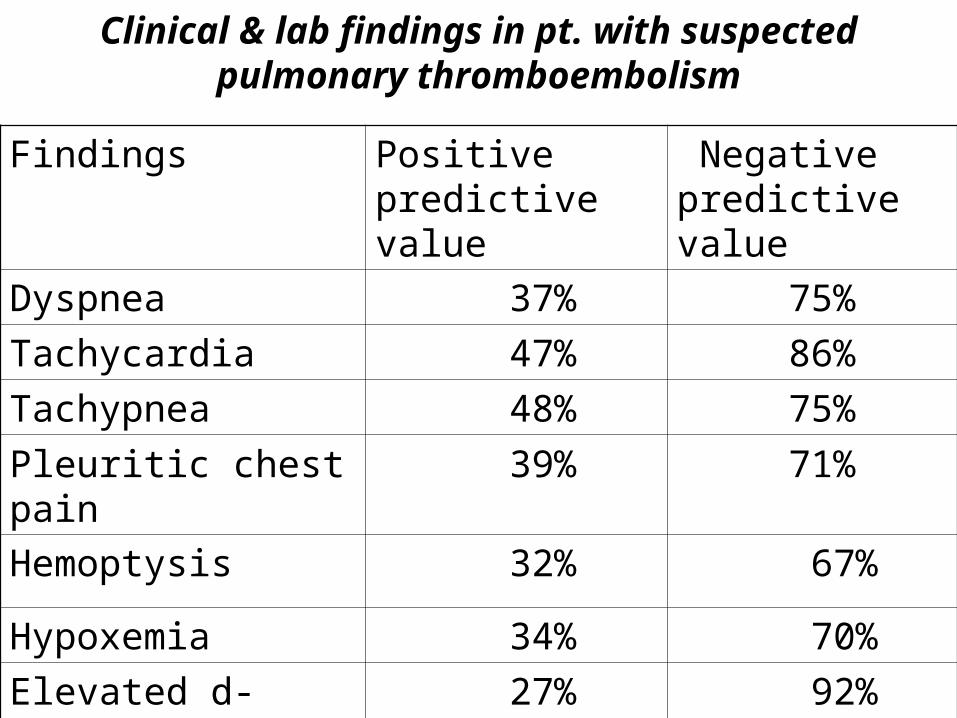
Clinical & lab findings in pt. with suspected pulmonary thromboembolism
Findings Positive predictive value
Negative predictive value
Dyspnea 37% 75%
Tachycardia 47% 86%
Tachypnea 48% 75%
Pleuritic chest pain 39% 71%
Hemoptysis 32% 67%
Hypoxemia 34% 70%
Elevated d- dimer 27% 92%
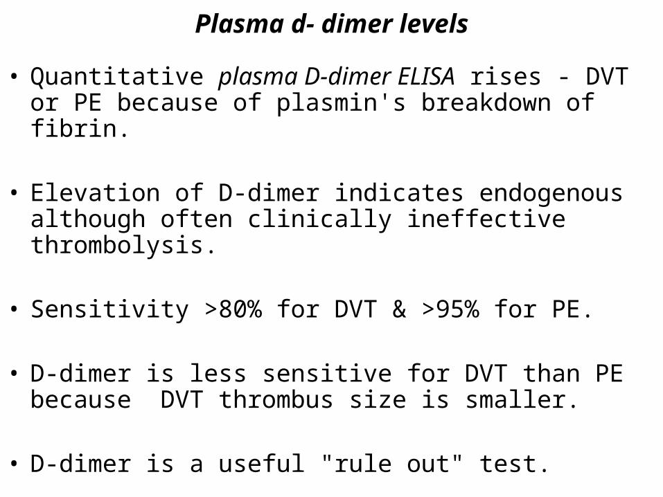
Plasma d- dimer levels
• Quantitative plasma D-dimer ELISA rises - DVT or PE because of plasmin's breakdown of fibrin.
• Elevation of D-dimer indicates endogenous although often clinically ineffective thrombolysis.
• Sensitivity >80% for DVT & >95% for PE.
• D-dimer is less sensitive for DVT than PE because DVT thrombus size is smaller.
• D-dimer is a useful "rule out" test.
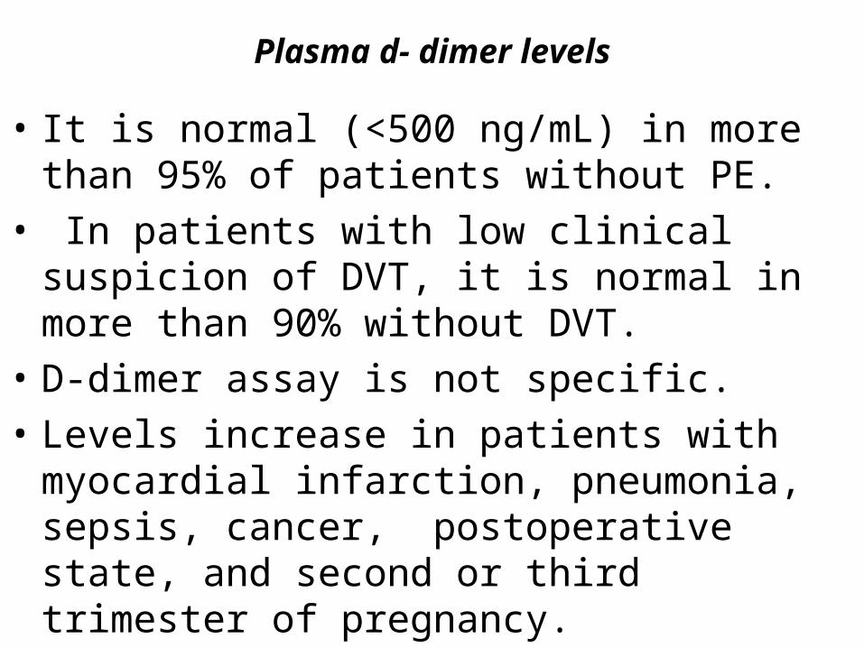
Plasma d- dimer levels
• It is normal (<500 ng/mL) in more than 95% of patients without PE.
• In patients with low clinical suspicion of DVT, it is normal in more than 90% without DVT.
• D-dimer assay is not specific.
• Levels increase in patients with myocardial infarction, pneumonia, sepsis, cancer, postoperative state, and second or third trimester of pregnancy.
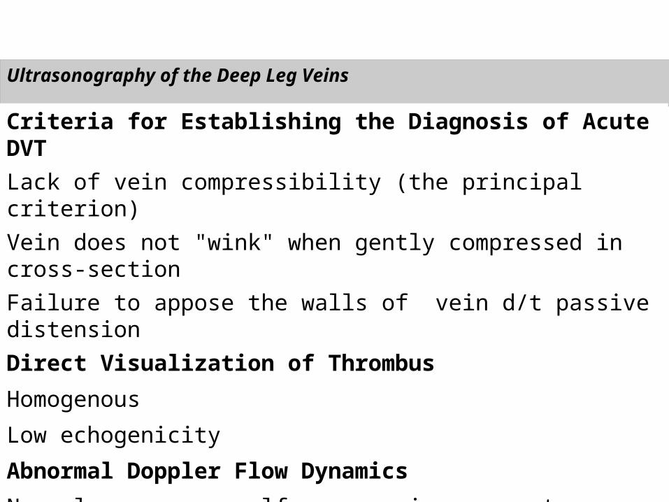
Ultrasonography of the Deep Leg Veins
Criteria for Establishing the Diagnosis of Acute DVT
Lack of vein compressibility (the principal criterion)
Vein does not "wink" when gently compressed in cross-section
Failure to appose the walls of vein d/t passive distension
Direct Visualization of Thrombus
Homogenous
Low echogenicity
Abnormal Doppler Flow Dynamics
Normal response: calf compression augments Doppler flow signal and confirms vein patency proximal and distal to Doppler
Abnormal response: flow blunted rather than augmented with calf compression
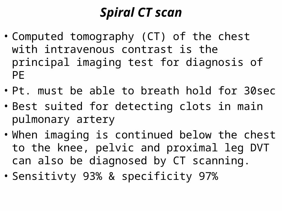
Spiral CT scan
• Computed tomography (CT) of the chest with intravenous contrast is the principal imaging test for diagnosis of PE
• Pt. must be able to breath hold for 30sec
• Best suited for detecting clots in main pulmonary artery
• When imaging is continued below the chest to the knee, pelvic and proximal leg DVT can also be diagnosed by CT scanning.
• Sensitivty 93% & specificity 97%
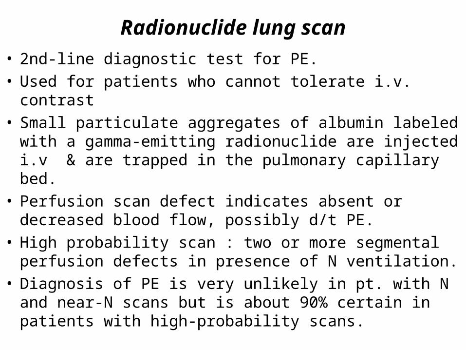
Radionuclide lung scan
• 2nd-line diagnostic test for PE.• Used for patients who cannot tolerate i.v. contrast• Small particulate aggregates of albumin labeled with a
gamma-emitting radionuclide are injected i.v & are trapped in the pulmonary capillary bed.
• Perfusion scan defect indicates absent or decreased blood flow, possibly d/t PE.
• High probability scan : two or more segmental perfusion defects in presence of N ventilation.
• Diagnosis of PE is very unlikely in pt. with N and near-N scans but is about 90% certain in patients with high-probability scans.
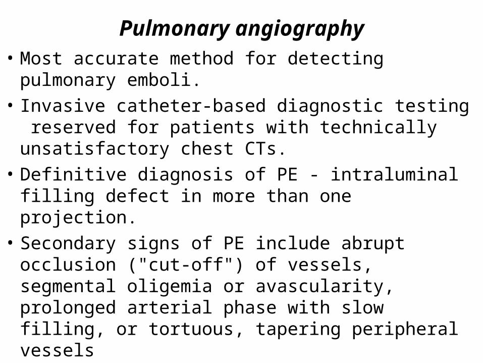
Pulmonary angiography
• Most accurate method for detecting pulmonary emboli.
• Invasive catheter-based diagnostic testing reserved for patients with technically unsatisfactory chest CTs.
• Definitive diagnosis of PE - intraluminal filling defect in more than one projection.
• Secondary signs of PE include abrupt occlusion ("cut-off") of vessels, segmental oligemia or avascularity, prolonged arterial phase with slow filling, or tortuous, tapering peripheral vessels
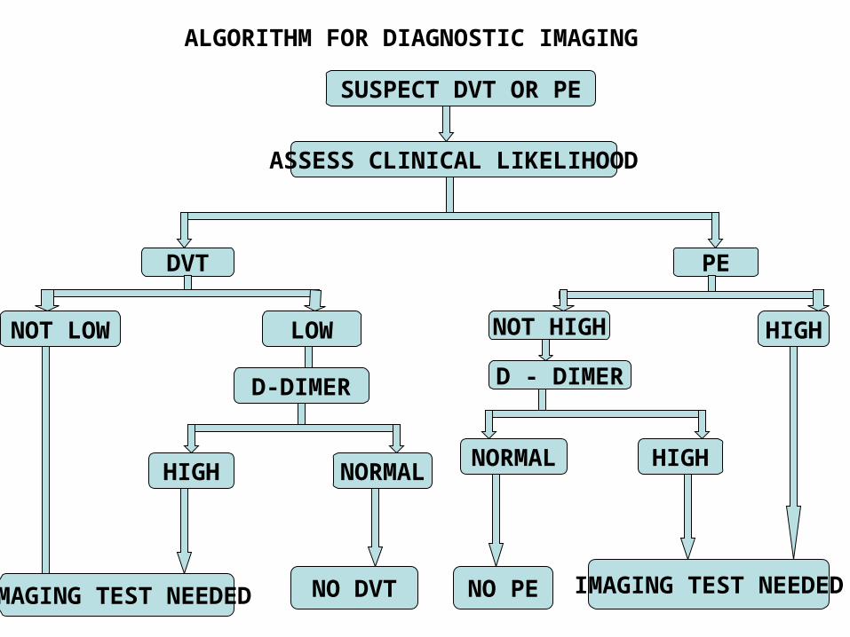
ALGORITHM FOR DIAGNOSTIC IMAGING
SUSPECT DVT OR PE
ASSESS CLINICAL LIKELIHOOD
DVT PE
NOT LOW LOW NOT HIGH HIGH
D-DIMER
IMAGING TEST NEEDED
HIGH NORMAL
NO DVT
D - DIMER
NORMAL HIGH
IMAGING TEST NEEDEDNO PE
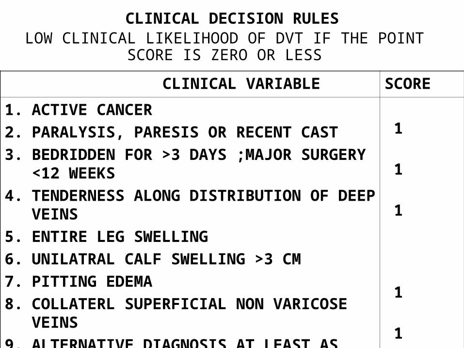
CLINICAL DECISION RULESLOW CLINICAL LIKELIHOOD OF DVT IF THE POINT SCORE
IS ZERO OR LESS
CLINICAL VARIABLE SCORE
1. ACTIVE CANCER
2. PARALYSIS, PARESIS OR RECENT CAST
3. BEDRIDDEN FOR >3 DAYS ;MAJOR SURGERY <12 WEEKS
4. TENDERNESS ALONG DISTRIBUTION OF DEEP VEINS
5. ENTIRE LEG SWELLING
6. UNILATRAL CALF SWELLING >3 CM
7. PITTING EDEMA
8. COLLATERL SUPERFICIAL NON VARICOSE VEINS
9. ALTERNATIVE DIAGNOSIS AT LEAST AS LIKELY AS DVT
1
1
1
1
1
1
1
1
- 2

CLINICAL DECISION RULES
HIGH CLINICAL LIKELIHOOD OF PE IF POINT SCORE EXCEEDS 4
CLINICAL VARIABLE SCORE
1. SIGN AND SYMPTOMS OF DVT
2. ALTERNATIVE DIAGNOSIS LESS LIKELY THAN PE
3. HEART RATE >100 / min
4. IMMOBILISATION >3 DAYS ;SURGERY WITHIN 4 WEEKS
5. PRIOR PE OR DVT
6. HEMOPTYSIS
7. CANCER
3.0
3.0
1.5
1.5
1.5
1.0
1.0
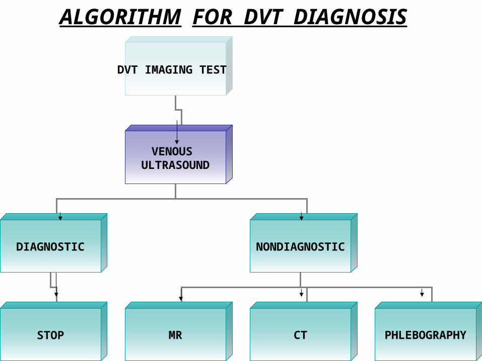
ALGORITHM FOR DVT DIAGNOSIS
DVT IMAGING TEST
VENOUS ULTRASOUND
DIAGNOSTIC NONDIAGNOSTIC
STOP MR CT PHLEBOGRAPHY
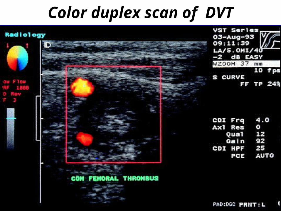
Color duplex scan of DVT
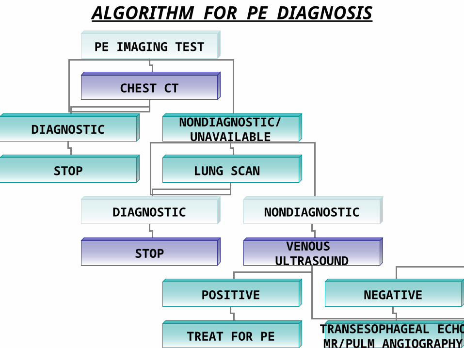
ALGORITHM FOR PE DIAGNOSIS
PE IMAGING TEST
CHEST CT
DIAGNOSTICNONDIAGNOSTIC/
UNAVAILABLE
STOP LUNG SCAN
DIAGNOSTIC NONDIAGNOSTIC
STOPVENOUS
ULTRASOUND
POSITIVE NEGATIVE
TREAT FOR PETRANSESOPHAGEAL ECHOMR/PULM ANGIOGRAPHY
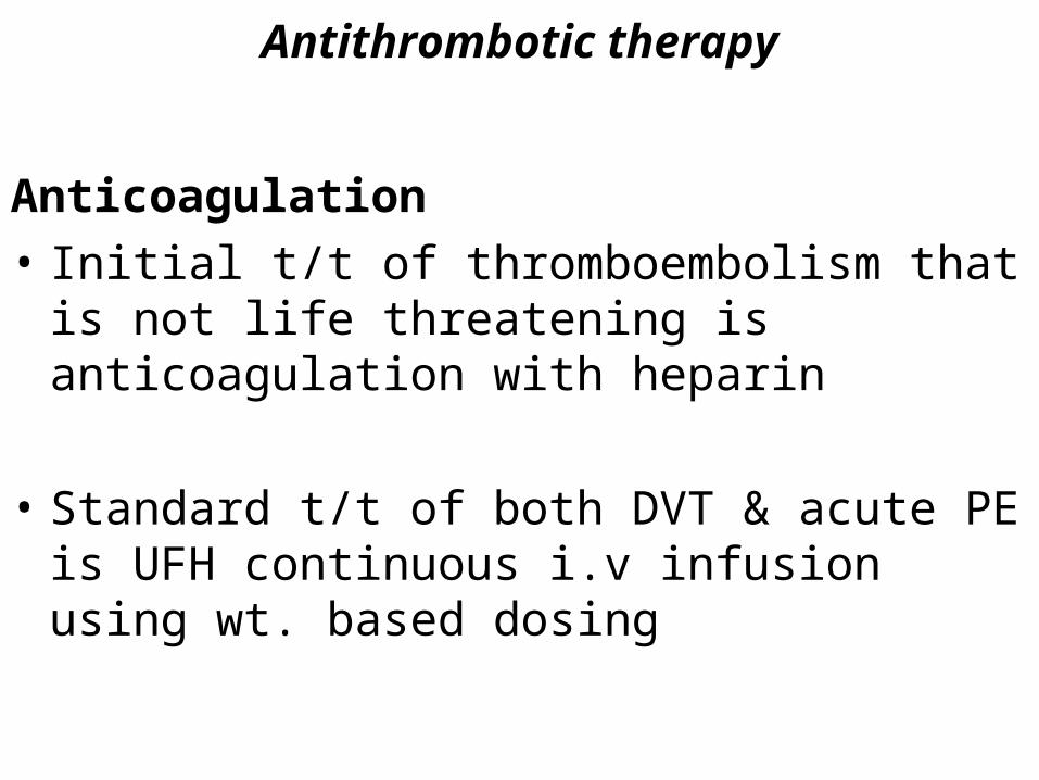
Antithrombotic therapy
Anticoagulation
• Initial t/t of thromboembolism that is not life threatening is anticoagulation with heparin
• Standard t/t of both DVT & acute PE is UFH continuous i.v infusion using wt. based dosing
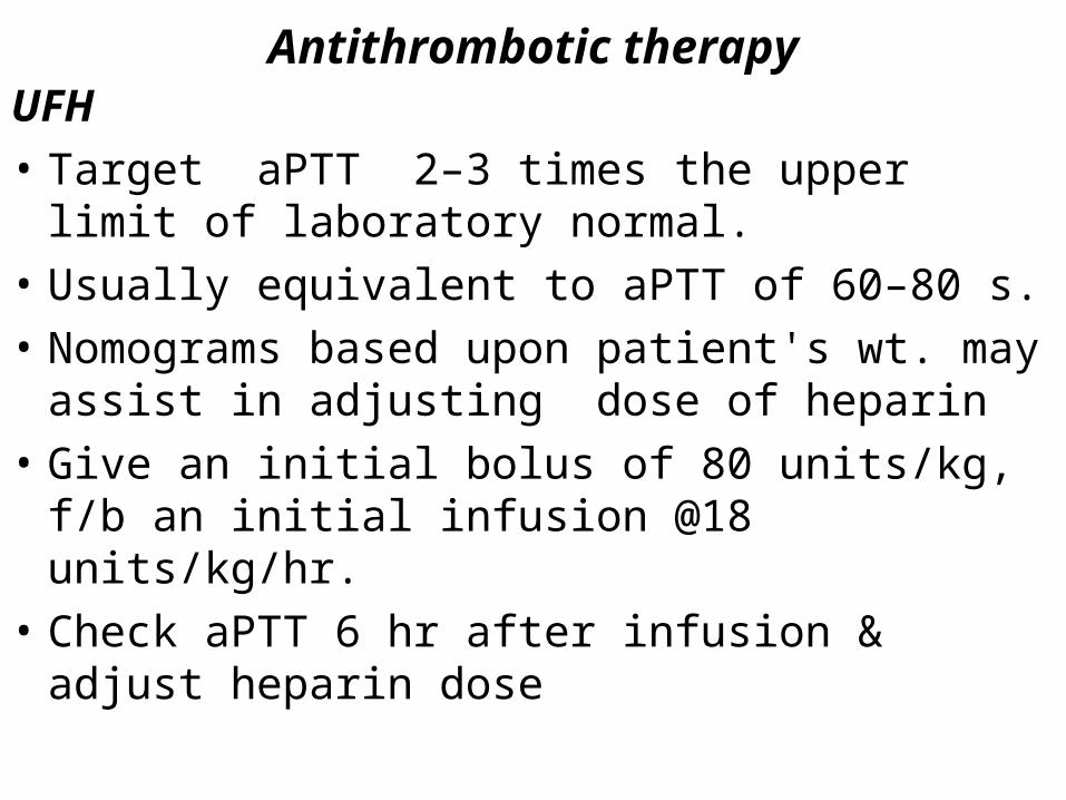
Antithrombotic therapyUFH
• Target aPTT 2–3 times the upper limit of laboratory normal.
• Usually equivalent to aPTT of 60–80 s.
• Nomograms based upon patient's wt. may assist in adjusting dose of heparin
• Give an initial bolus of 80 units/kg, f/b an initial infusion @18 units/kg/hr.
• Check aPTT 6 hr after infusion & adjust heparin dose
Antithrombotic therapy
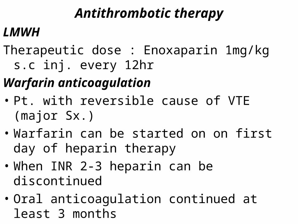
LMWH
Therapeutic dose : Enoxaparin 1mg/kg s.c inj. every 12hr
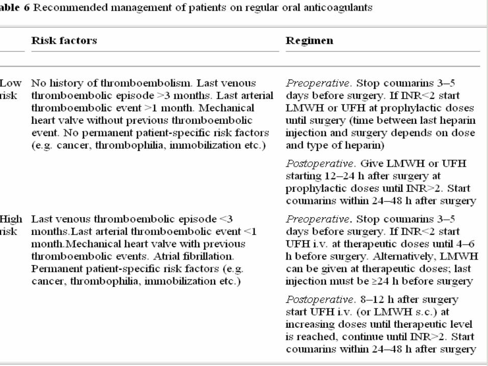
Warfarin anticoagulation
• Pt. with reversible cause of VTE (major Sx.)
• Warfarin can be started on on first day of heparin therapy
• When INR 2-3 heparin can be discontinued
• Oral anticoagulation continued at least 3 months
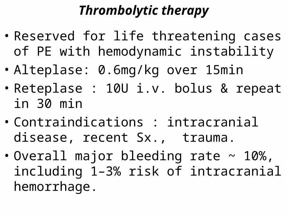
Thrombolytic therapy
• Reserved for life threatening cases of PE with hemodynamic instability
• Alteplase: 0.6mg/kg over 15min
• Reteplase : 10U i.v. bolus & repeat in 30 min
• Contraindications : intracranial disease, recent Sx., trauma.
• Overall major bleeding rate ~ 10%, including 1–3% risk of intracranial hemorrhage.
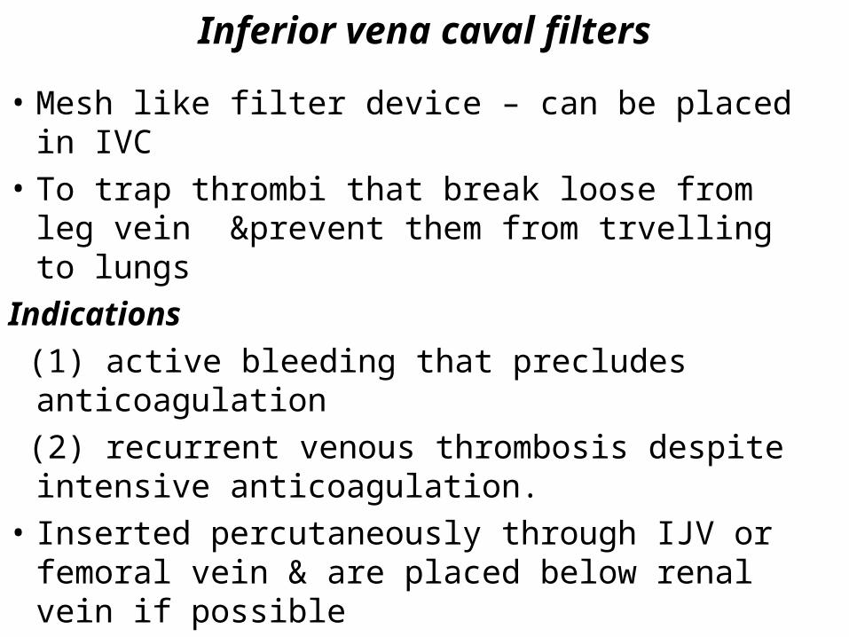
Inferior vena caval filters
• Mesh like filter device – can be placed in IVC
• To trap thrombi that break loose from leg vein &prevent them from trvelling to lungs
Indications
(1) active bleeding that precludes anticoagulation
(2) recurrent venous thrombosis despite intensive anticoagulation.
• Inserted percutaneously through IJV or femoral vein & are placed below renal vein if possible
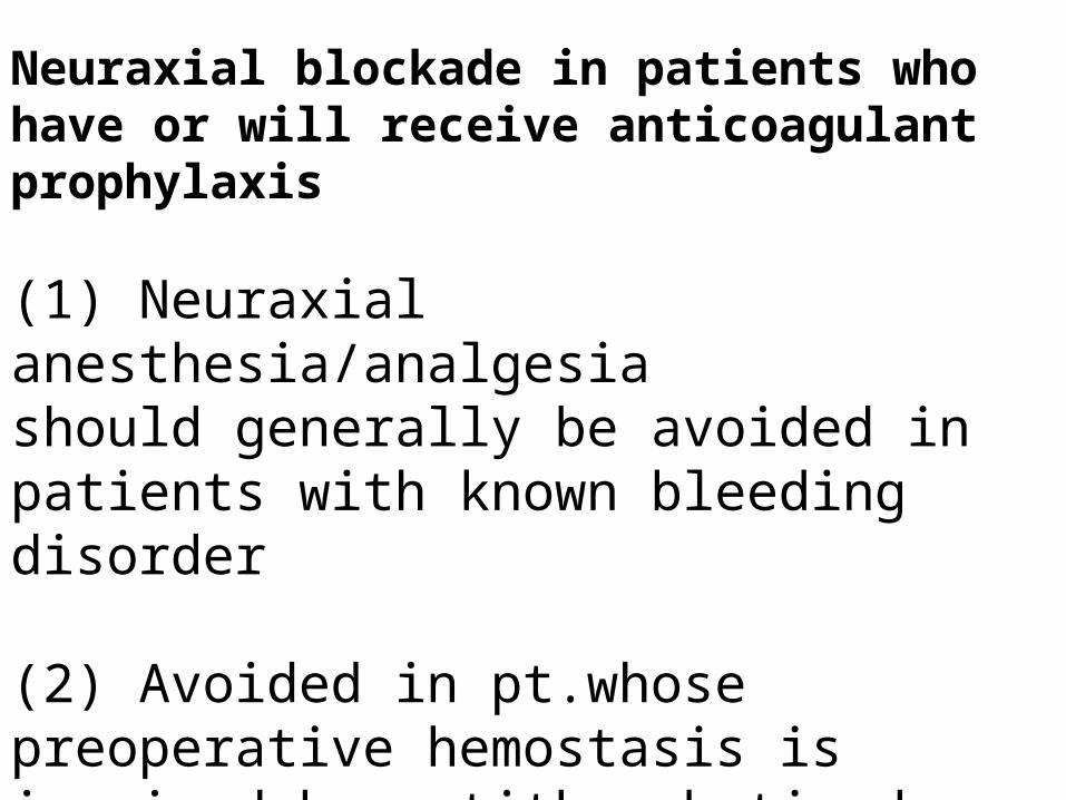
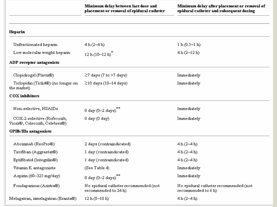
Neuraxial blockade in patients who have or will receive anticoagulant prophylaxis
(1) Neuraxial anesthesia/analgesiashould generally be avoided in patients with known bleeding disorder
(2) Avoided in pt.whose preoperative hemostasis is impaired by antithrombotic drugs
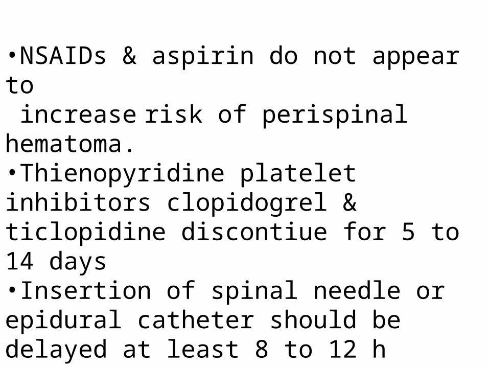
•NSAIDs & aspirin do not appear to increase risk of perispinal hematoma. •Thienopyridine platelet inhibitors clopidogrel & ticlopidine discontiue for 5 to 14 days•Insertion of spinal needle or epidural catheter should be delayed at least 8 to 12 h after s.c. dose of heparin or a twice daily prophylactic dose of LMWH•At least 18 h after O.D. LMWH injection
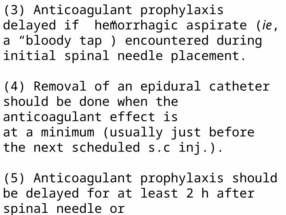
(3) Anticoagulant prophylaxis delayed if hemorrhagic aspirate (ie, a “bloody tap”) encountered during initial spinal needle placement.
(4) Removal of an epidural catheter should be done when the anticoagulant effect isat a minimum (usually just before the next scheduled s.c inj.). (5) Anticoagulant prophylaxis should be delayed for at least 2 h after spinal needle orepidural catheter removal.
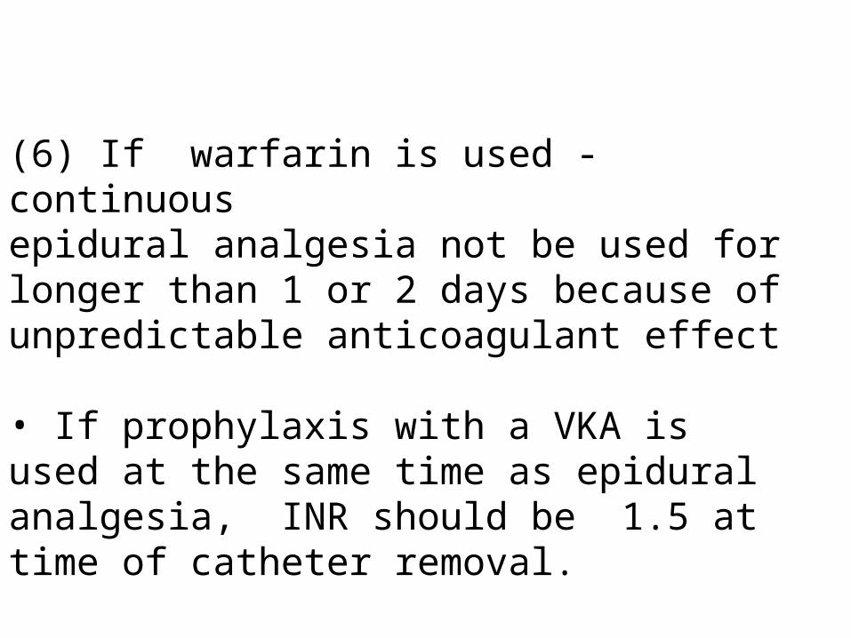
(6) If warfarin is used - continuousepidural analgesia not be used for longer than 1 or 2 days because of unpredictable anticoagulant effect
• If prophylaxis with a VKA isused at the same time as epidural analgesia, INR should be 1.5 at time of catheter removal.
(7)Postoperative prophylaxis with fondaparinux - safe in pt. who have received a spinal anesthetic
• No safety data about its use along with postoperative continuous epidural analgesia.
• Long half-life of fondaparinux & renalmode of excretion raise concerns about potential foraccumulation ,in elderly d/t associated impairment of renal function.
•With concurrent use of epidural analgesia & anticoagulant prophylaxis, all patients should be monitored for s/s of cord compression.
•Progression of lower extremity numbness or weakness, bowel or bladder dysfunction, & new onset of back pain.
• If spinal hematoma is suspected, diagnostic imaging and definitive surgical therapy performed rapidly to reduce the risk of permanent paresis.
•