Embed Size (px)
Citation preview
EMBOopen
Three-dimensional structure of basal body tripletrevealed by electron cryo-tomography
Sam Li1,3, Jose-Jesus Fernandez2,Wallace F Marshall3 and David A Agard1,3,*1The Howard Hughes Medical Institute, University of California,San Francisco, CA, USA, 2Centro Nacional de Biotecnologia—CSIC,Campus Universidad Autonoma, Madrid, Spain and 3Department ofBiochemistry and Biophysics, University of California, San Francisco,CA, USA
Basal bodies and centrioles play central roles in micro-
tubule (MT)-organizing centres within many eukaryotes.
They share a barrel-shaped cylindrical structure composed
of nine MT triplet blades. Here, we report the structure of
the basal body triplet at 33 A resolution obtained by
electron cryo-tomography and 3D subtomogram aver-
aging. By fitting the atomic structure of tubulin into the
EM density, we built a pseudo-atomic model of the tubulin
protofilaments at the core of the triplet. The 3D density
map reveals additional densities that represent non-tubu-
lin proteins attached to the triplet, including a large inner
circular structure in the basal body lumen, which func-
tions as a scaffold to stabilize the entire basal body barrel.
We found clear longitudinal structural variations along
the basal body, suggesting a sequential and coordinated
assembly mechanism. We propose a model in which
d-tubulin and other components participate in the assem-
bly of the basal body.
The EMBO Journal (2012) 31, 552–562. doi:10.1038/
emboj.2011.460; Published online 13 December 2011
Subject Categories: cell & tissue architecture; structural biology
Keywords: basal body; centriole; Chlamydomonas reinhardtii;
electron cryo-tomography; three-dimensional reconstruction
Introduction
The basal body, as a cellular organelle for organizing micro-
tubules (MTs), is composed of nine MT triplet blades.
It carries out multiple essential functions in cellular pro-
cesses. During cell division, it functions as a centriole to
recruit pericentriolar material to form the centrosome, which
in turn is responsible for establishing the bipolar spindle
during mitosis. Although centrioles are dispensable for mi-
tosis in certain organisms and cell lines (Debec et al, 2010),
ample evidence shows that in many other cases, over-ampli-
fication or depletion of centrioles leads to a delay in cell
division, aneuploidy and cell death, suggesting their impor-
tance for normal mitosis (Hinchcliffe et al, 2001; Piel et al,
2001; Mikule et al, 2007). More importantly, the centriole is
responsible for establishing cell polarity during cell division
(Yamashita et al, 2007; Wang et al, 2009). In many quiescent
cells, the centriole will migrate to the cell periphery, and
anchor beneath the cell membrane as a basal body that
templates cilium formation. The basal body also provides a
docking site for intraflagellar transport particles that move
bidirectionally along the cilium (Deane et al, 2001). Recent
studies show that, in addition to its role in motility, the cilium
also functions as an antenna for communicating between the
intracellular and external environment of the cell and plays
essential roles during cell regulation, tissue differentiation
and embryonic development (Nigg and Raff, 2009).
Mutations in cilium or basal body genes result in various
forms of human disease, collectively known as ciliopathies
(Gerdes et al, 2009).
Biogenesis of the centriole and basal body has been
studied in a variety of model organisms (Ringo, 1967;
Dippell, 1968; Allen, 1969; Anderson and Brenner, 1971;
Anderson, 1972; Cavalier-Smith, 1974; Gonzalez et al, 1998;
Pelletier et al, 2006; Giddings et al, 2010; Guichard et al,
2010). Genes involved in centriole biogenesis, either regula-
tory or structural, are generally well conserved, suggesting a
common assembly pathway across species (Carvalho-Santos
et al, 2010). The biogenesis of basal bodies or centrioles is
tightly coupled to the cell cycle, suggesting a highly regulated
assembly mechanism (Doxsey et al, 2005).
Proteomic studies of purified basal bodies or centrioles have
identified a list of bona fide centriole components and basal
body specific proteins many of whose function and structure
remain unknown (Andersen et al, 2003; Li et al, 2004; Keller
et al, 2005; Kilburn et al, 2007). Despite extensive descriptions
of the centriole and basal body ultrastructure, the lack of high-
resolution structural information significantly limits our under-
standing of the assembly process and the full repertoire of
cellular functions for this complex organelle.
Here, we used electron cryo-tomography (cryo-ET)
to visualize basal bodies purified from Chlamydomonas
reinhardtii in a near-native state. In combination with a
subtomogram averaging strategy, it has been possible to
obtain a high-quality 3D reconstruction of the basal body
that offers insight on how tubulin protofilaments (PFs) and
accessory components assemble into the triplet. Based on our
structural findings and previous studies, we propose potential
locations for several basal body components. Our structure
provides a framework for understanding the molecular me-
chanism of basal body and centriole biogenesis, and for
integrating new information as the protein composition of
the non-MT components are elucidated.
Results
Overall structure of the basal body triplet
Purified basal bodies in near-native state were visualized by
cryo-ET. In most of the tomograms, two cylindrically shapedReceived: 14 August 2011; accepted: 15 November 2011; publishedonline: 13 December 2011
*Corresponding author. Department of Biochemistry and Biophysics,Howard Hughes Medical Institute, University of California at SanFrancisco, 600 16th Street, Room S412D, San Francisco, CA 94158-2517,USA. Tel.: þ 1 415 476 2521; Fax: þ 1 415 476 1902;E-mail: [email protected]
The EMBO Journal (2012) 31, 552–562 | & 2012 European Molecular Biology Organization | Some Rights Reserved 0261-4189/12
www.embojournal.org
The EMBO Journal VOL 31 | NO 3 | 2012 &2012 European Molecular Biology Organization
EMBO
THE
EMBOJOURNAL
THE
EMBOJOURNAL
552

basal bodies were visible since they are tethered laterally
by the distal striated fibres (Hoops et al, 1984). The basal
bodies have a reproducible size, with an average diameter
of 260 nm and an overall length of 600 nm (Figure 1A). The
MT triplets start from the proximal end (the bottom in
Figure 1A) and span about 400 nm longitudinally towards
the distal end where the C-tubules of the triplets terminate.
The A- and B-tubules continue as a doublet for about 150 nm
before they reach the transitional plate, a hallmark of the
transition zone where the axoneme will assemble (Cavalier-
Smith, 1974; O’Toole et al, 2003; Geimer and Melkonian,
2004). We did not observe the cartwheel structures in our
tomograms, presumably because they were lost during pur-
ification. Electron dense structures, known as ‘A-tubule feet’,
are consistently visible in our tomograms along the wall of
the triplets projecting towards the lumen (Cavalier-Smith,
1974; Geimer and Melkonian, 2004), showing characteristic
8- and 16-nm periodicity (Figure 1A). The region decorated
with A-tubule feet starts at about 100 nm from the triplet
minus (proximal) end, spans about 250 nm longitudinally
and terminates at about 45 nm before the triplets become
doublets. This section forms the central core of the basal
body and was used for tomogram subvolume averaging
in order to obtain a higher-resolution structure of the
basal body.
We obtained an averaged MT triplet at 33 A resolution
(Figure 1B; Supplementary Figure S1). The resolution of the
averaged structure is sufficiently high to allow us to discern
each PF in the longitudinal projection of the triplet (Figure 1B
and C). The A-tubule is a complete MT composed of 13 PFs,
numbering starts clockwise from the basal body luminal side
as A1 to A13 following the Tilney-Linck convention (Linck
and Stephens, 2007). PF A10 is the site where the B-tubule
joins with the A-tubule. PFs A10 to A13 are referred to as
partition PFs shared between A- and B-tubule. The B-tubule is
composed of 10 PFs that are numbered from the outside
surface of the triplet clockwise as B1 to B10. The C-tubule
also has 10 PFs. PF C1 starts as a branch from PF B4 and they
Figure 1 Cryo-ET reconstruction of the basal body triplet. (A) A cross-section of tomographic reconstructed volume containing a basal body.The section is through the centre of the basal body barrel. The proximal end is at the bottom and the distal end is at the top. The ‘A-tubule feet’are marked with *. The terms of probasal body, A-tubule feet and transitional plate follow conventions of Geimer and Melkonian (2004).(B) The averaged 3D structure of the basal body triplet viewed from the distal end. The MT triplet density map has been deposited in theElectron Microscopy Data Bank with accession code EMD-5252. (C) A longitudinal projection of a 16-nm section of the averaged triplet. Everyother PFs in the triplet are labelled. (D) Docking of crystal structure of tubulin into the triplet density map. Selected PFs are labelled. Thelocations of the highest curvature in the A-tubule are marked with *.
Electron cryo-tomography of basal body tripletS Li et al
&2012 European Molecular Biology Organization The EMBO Journal VOL 31 | NO 3 | 2012 553
are numbered clockwise as C1 to C10. PFs B5 to B8 form the
partition shared by the B- and C-tubules.
The A-tubule has an elliptical shape with a substantial
variation of curvature along the MT wall (Figure 1B and C).
The longest axis running across the internal diameter of the
A-tubule is between PFs A3 and A10 with a diameter of
B247 A. The shortest axis is B201 A between PFs A6 and
A13. This change of diameters is equivalent to a 10% distor-
tion of an intact 13-PF MTand is similar to the A-tubule in the
axoneme doublet where B8% distortion has been observed
(Sui and Downing, 2006). Interestingly, the variation of
curvature is non-isotropic along the wall of the A-tubule.
The highest curvature is at PFs A9 and A10, followed by the
next-highest curvature at PFs A2 and A3, where large lateral
gaps are observed between PFs. The smallest curvature is at
the partition site from A11 to A13. In contrast to the A-tubule,
the B- and C-tubules have a rather smooth and uniform
curvature with diameters about 260 A. The diameter and
the curvature of the B- and C-tubules indicate that both
would form a circular MT with 15 PFs if they formed
complete rings, similar to the B-tubule in the axoneme
doublet (Sui and Downing, 2006).
Building a pseudo-atomic model of the triplet
In our structure, the 4-nm periodicity between individual
tubulin monomers can be easily resolved along most of the
PFs of the basal body (Supplementary Figure S2). The z-rise
(longitudinal rise) of tubulins between adjacent PFs, varies
from 10 to 12 A, consistent with previous theoretical and
experimental data from MTs with different PF numbers
(Chretien and Wade, 1991; Sui and Downing, 2010). This
allowed us to fit the atomic structure of a/b tubulin into the
EM density map and build a pseudo-atomic model of the
tubulin core of the triplet (Figure 1D). Previous studies have
shown that in a/b tubulin, the M-loop and H1-S2/H2-S3
loops provide the main lateral interactions between adjacent
PFs and we have kept these contacts in our model (Nogales
et al, 1999; Li et al, 2002; Sui and Downing, 2010). The overall
fit is excellent at the current resolution. In the A-tubule, due
to the variation in local curvature, the closest lateral interac-
tions are within partition PFs A11 to A13. Conversely, the
interfaces between PFs A2/A3 and PFs A9/A10 have less
contact due to large local curvature.
The fitting of tubulin PFs into the B- and C-tubule density
resulted in uniform tubulin lateral interactions. We have
modelled PF B1 as a tubulin PF that makes unusual lateral
contact with the outside surface of PF A10. Since the distal
half of PF C1 exhibits an 8-nm interval with a gap (Figure 4A
and B), it is most likely occupied by non-tubulin protein in
this region. Therefore, we did not fit tubulin monomers into
the PF C1 position anywhere along its length. This long-
itudinal change in PF C1 will be described in detail below.
Non-tubulin components associated with the triplet
Proteomic studies have shown that, besides tubulin, the basal
body contains nearly 50 non-tubulin components (Keller
et al, 2005). In order to find where they bind, how the tubules
are associated to form a triplet and how the triplets are
connected in the basal body, we used the pseudo-atomic
model of the tubulins within the triplet as a mask to subtract
its density from the 3D density map. The resulting difference
map shows novel density features that must correspond to
the locations of most of the non-tubulin accessory proteins
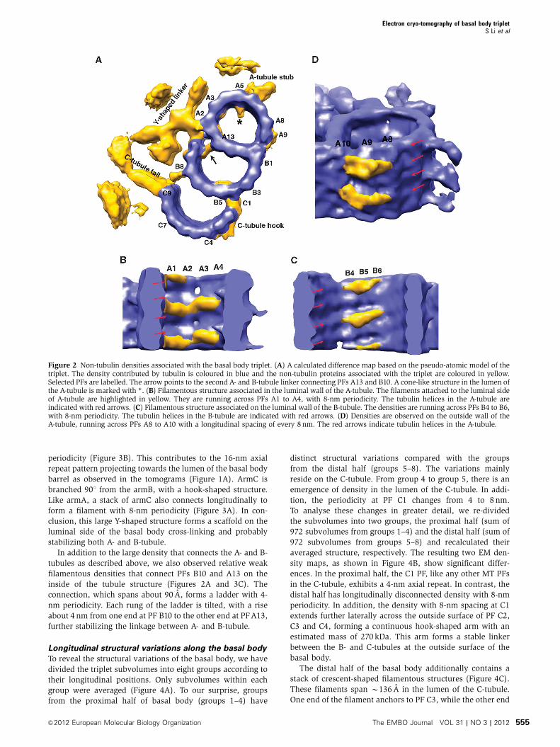
(Figure 2A).
Just as in the axoneme doublet structure (Nicastro et al,
2006; Sui and Downing, 2006), we also observed densities
decorating on the internal wall of A- and B-tubules in the
triplet, but in a more asymmetric manner. In the A- and B-
tubule, there are filamentous densities running across PFs A1
to A4 and B4 to B6, respectively, with an 8-nm periodicity
(Figure 2B and C). The densities follow the rise of the tubulin
helical repeat inside the lumen, forming lateral cross-links
between neighbouring tubulin monomers at the lumen side.
Likely, these are Tektin family proteins that stabilize adjacent
PFs and fine-tune local curvature of the tubule (Amos, 2008).
We also observed a cone-shaped density attached on the
luminal side of PF A5 with 8-nm periodicity (* in
Figure 2A), likely to stabilize the A-tubule or as the part of
structure connecting to the neighbouring triplet. Interestingly,
similar density has been consistently observed in the axo-
neme doublet (Nicastro et al, 2006; Sui and Downing, 2006;
Movassagh et al, 2010). Across from the cone-shaped density,
at PF A6 a stub-like density projects out every 8 nm on the
outside wall of the A-tubule (Figure 2A). The stub connects to
the C-tubule from the neighbouring triplet and will be de-
scribed in detail in the next section. Meanwhile, density was
observed on the outside wall of the A-tubule running across
PFs A8 to A10 with an 8-nm longitudinal spacing (Figure 2D).
Since both PFs A10 and B1 are at the junction of A- and B-
tubules, this density presumably will stabilize the linkage
between the A- and B-tubules at the outside wall of triplet.
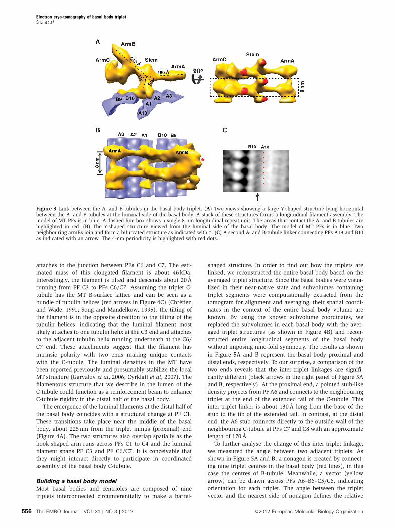
Remarkably, a large Y-shaped density was observed lying
horizontally as a ridge along the luminal side of the A- and B-
tubules (Figures 2A and 3A). It spans about 380 A, nearly the
entire inner circumference of the A- and B-tubules. It projects
radially about 170 A towards the centre of the basal body
barrel. The estimated mass of this structure is 1.1 MDa. We
have divided this structure into four parts and assigned them
as a central stem with three arms, termed armA, armB and
armC (Figure 3A and B). The central stem binds directly to
both A- and B-tubule. On the right side in Figure 3A, it fits into
the groove between PFs A1 and A2, which is likely the position
of the seam in the A-tubule (Song and Mandelkow, 1995).
After rising longitudinally B40 A, the left side of the stem
binds to PF B10 in the B-tubule. Here, the stem extends
laterally, running across the entire outer surface of PF B10
and partial surface of PF B9. Together, this central stem fills in
the gap between the A- and B-tubule and cross-links the two
tubules at their closest distance (B82 A). It makes substantial
interactions with both tubules at their luminal joint, with an
estimated total contact area between the stem and the A- and
B-tubule of B5200 A2. Multiple copies of the stem stack long-
itudinally with an axial repeat of 8 nm, forming a left-handed
spiral-shaped filament viewed from the outside of the basal
body (Figure 3A). This filament might account for the 11th PF
of the B-tubule as previously observed (Tilney et al, 1973).
ArmA rotates about 1081 counterclockwise relative to the
stem and extends 190 A towards the direction of A-tubule
(Figure 3A and B). At its end, armA connects longitudinally to
the neighbouring armAs, resulting in a filament along the
basal body barrel with an 8-nm periodicity. ArmB is 210 A
long and it is about 1151 clockwise relative to the stem
(Figure 3A). At the end of armB, the neighbouring two
arms join, forming in a bifurcated structure with 16-nm
Electron cryo-tomography of basal body tripletS Li et al
The EMBO Journal VOL 31 | NO 3 | 2012 &2012 European Molecular Biology Organization554
periodicity (Figure 3B). This contributes to the 16-nm axial
repeat pattern projecting towards the lumen of the basal body
barrel as observed in the tomograms (Figure 1A). ArmC is
branched 901 from the armB, with a hook-shaped structure.
Like armA, a stack of armC also connects longitudinally to
form a filament with 8-nm periodicity (Figure 3A). In con-
clusion, this large Y-shaped structure forms a scaffold on the
luminal side of the basal body cross-linking and probably
stabilizing both A- and B-tubule.
In addition to the large density that connects the A- and B-
tubules as described above, we also observed relative weak
filamentous densities that connect PFs B10 and A13 on the
inside of the tubule structure (Figures 2A and 3C). The
connection, which spans about 90 A, forms a ladder with 4-
nm periodicity. Each rung of the ladder is tilted, with a rise
about 4 nm from one end at PF B10 to the other end at PFA13,
further stabilizing the linkage between A- and B-tubule.
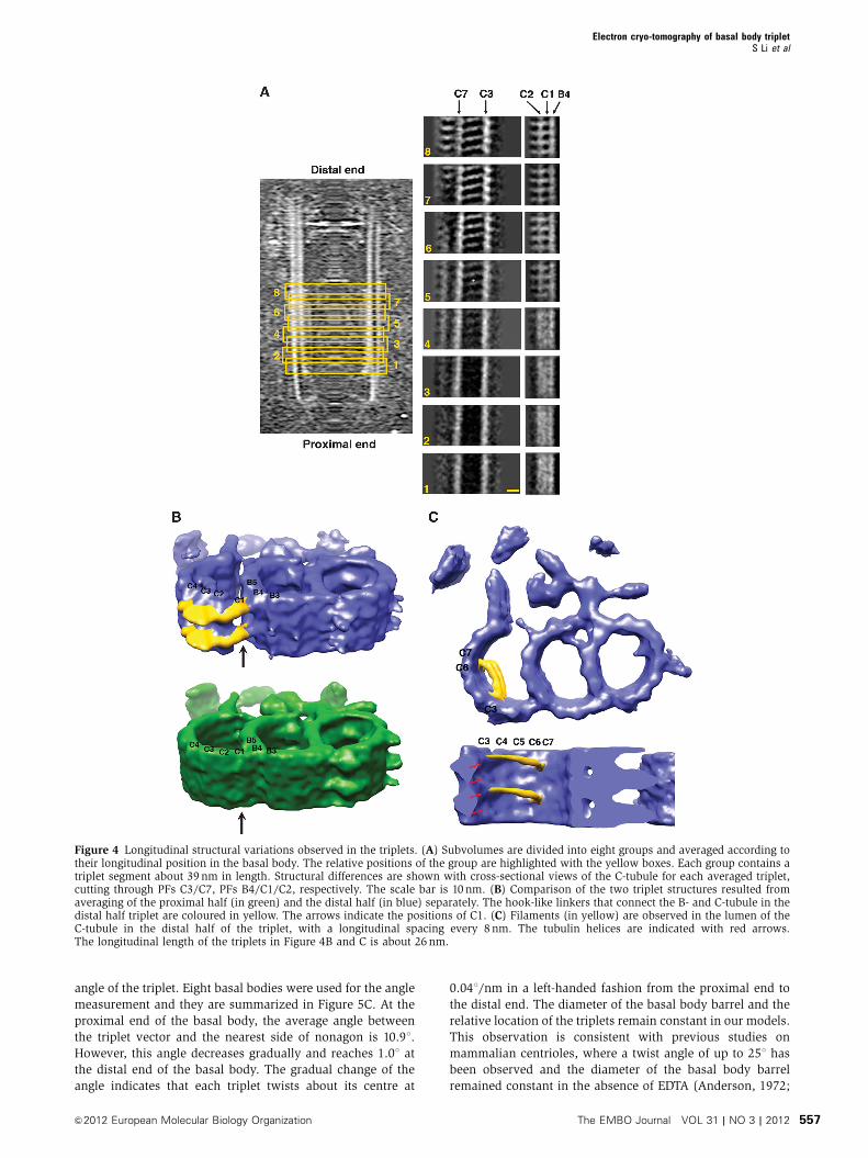
Longitudinal structural variations along the basal body
To reveal the structural variations of the basal body, we have
divided the triplet subvolumes into eight groups according to
their longitudinal positions. Only subvolumes within each
group were averaged (Figure 4A). To our surprise, groups
from the proximal half of basal body (groups 1–4) have
distinct structural variations compared with the groups
from the distal half (groups 5–8). The variations mainly
reside on the C-tubule. From group 4 to group 5, there is an
emergence of density in the lumen of the C-tubule. In addi-
tion, the periodicity at PF C1 changes from 4 to 8 nm.
To analyse these changes in greater detail, we re-divided
the subvolumes into two groups, the proximal half (sum of
972 subvolumes from groups 1–4) and the distal half (sum of
972 subvolumes from groups 5–8) and recalculated their
averaged structure, respectively. The resulting two EM den-
sity maps, as shown in Figure 4B, show significant differ-
ences. In the proximal half, the C1 PF, like any other MT PFs
in the C-tubule, exhibits a 4-nm axial repeat. In contrast, the
distal half has longitudinally disconnected density with 8-nm
periodicity. In addition, the density with 8-nm spacing at C1
extends further laterally across the outside surface of PF C2,
C3 and C4, forming a continuous hook-shaped arm with an
estimated mass of 270 kDa. This arm forms a stable linker
between the B- and C-tubules at the outside surface of the
basal body.
The distal half of the basal body additionally contains a
stack of crescent-shaped filamentous structures (Figure 4C).
These filaments span B136 A in the lumen of the C-tubule.
One end of the filament anchors to PF C3, while the other end
Figure 2 Non-tubulin densities associated with the basal body triplet. (A) A calculated difference map based on the pseudo-atomic model of thetriplet. The density contributed by tubulin is coloured in blue and the non-tubulin proteins associated with the triplet are coloured in yellow.Selected PFs are labelled. The arrow points to the second A- and B-tubule linker connecting PFs A13 and B10. A cone-like structure in the lumen ofthe A-tubule is marked with *. (B) Filamentous structure associated in the luminal wall of the A-tubule. The filaments attached to the luminal sideof A-tubule are highlighted in yellow. They are running across PFs A1 to A4, with 8-nm periodicity. The tubulin helices in the A-tubule areindicated with red arrows. (C) Filamentous structure associated on the luminal wall of the B-tubule. The densities are running across PFs B4 to B6,with 8-nm periodicity. The tubulin helices in the B-tubule are indicated with red arrows. (D) Densities are observed on the outside wall of theA-tubule, running across PFs A8 to A10 with a longitudinal spacing of every 8 nm. The red arrows indicate tubulin helices in the A-tubule.
Electron cryo-tomography of basal body tripletS Li et al
&2012 European Molecular Biology Organization The EMBO Journal VOL 31 | NO 3 | 2012 555
attaches to the junction between PFs C6 and C7. The esti-
mated mass of this elongated filament is about 46 kDa.
Interestingly, the filament is tilted and descends about 20 A
running from PF C3 to PFs C6/C7. Assuming the triplet C-
tubule has the MT B-surface lattice and can be seen as a
bundle of tubulin helices (red arrows in Figure 4C) (Chretien
and Wade, 1991; Song and Mandelkow, 1995), the tilting of
the filament is in the opposite direction to the tilting of the
tubulin helices, indicating that the luminal filament most
likely attaches to one tubulin helix at the C3 end and attaches
to the adjacent tubulin helix running underneath at the C6/
C7 end. These attachments suggest that the filament has
intrinsic polarity with two ends making unique contacts
with the C-tubule. The luminal densities in the MT have
been reported previously and presumably stabilize the local
MT structure (Garvalov et al, 2006; Cyrklaff et al, 2007). The
filamentous structure that we describe in the lumen of the
C-tubule could function as a reinforcement beam to enhance
C-tubule rigidity in the distal half of the basal body.
The emergence of the luminal filaments at the distal half of
the basal body coincides with a structural change at PF C1.
These transitions take place near the middle of the basal
body, about 225 nm from the triplet minus (proximal) end
(Figure 4A). The two structures also overlap spatially as the
hook-shaped arm runs across PFs C1 to C4 and the luminal
filament spans PF C3 and PF C6/C7. It is conceivable that
they might interact directly to participate in coordinated
assembly of the basal body C-tubule.
Building a basal body model
Most basal bodies and centrioles are composed of nine
triplets interconnected circumferentially to make a barrel-
shaped structure. In order to find out how the triplets are
linked, we reconstructed the entire basal body based on the
averaged triplet structure. Since the basal bodies were visua-
lized in their near-native state and subvolumes containing
triplet segments were computationally extracted from the
tomogram for alignment and averaging, their spatial coordi-
nates in the context of the entire basal body volume are
known. By using the known subvolume coordinates, we
replaced the subvolumes in each basal body with the aver-
aged triplet structures (as shown in Figure 4B) and recon-
structed entire longitudinal segments of the basal body
without imposing nine-fold symmetry. The results as shown
in Figure 5A and B represent the basal body proximal and
distal ends, respectively. To our surprise, a comparison of the
two ends reveals that the inter-triplet linkages are signifi-
cantly different (black arrows in the right panel of Figure 5A
and B, respectively). At the proximal end, a pointed stub-like
density projects from PF A6 and connects to the neighbouring
triplet at the end of the extended tail of the C-tubule. This
inter-triplet linker is about 130 A long from the base of the
stub to the tip of the extended tail. In contrast, at the distal
end, the A6 stub connects directly to the outside wall of the
neighbouring C-tubule at PFs C7 and C8 with an approximate
length of 170 A.
To further analyse the change of this inter-triplet linkage,
we measured the angle between two adjacent triplets. As
shown in Figure 5A and B, a nonagon is created by connect-
ing nine triplet centres in the basal body (red lines), in this
case the centres of B-tubule. Meanwhile, a vector (yellow
arrow) can be drawn across PFs A6–B6–C5/C6, indicating
orientation for each triplet. The angle between the triplet
vector and the nearest side of nonagon defines the relative
Figure 3 Link between the A- and B-tubules in the basal body triplet. (A) Two views showing a large Y-shaped structure lying horizontalbetween the A- and B-tubules at the luminal side of the basal body. A stack of these structures forms a longitudinal filament assembly. Themodel of MT PFs is in blue. A dashed-line box shows a single 8-nm longitudinal repeat unit. The areas that contact the A- and B-tubules arehighlighted in red. (B) The Y-shaped structure viewed from the luminal side of the basal body. The model of MT PFs is in blue. Twoneighbouring armBs join and form a bifurcated structure as indicated with *. (C) A second A- and B-tubule linker connecting PFs A13 and B10as indicated with an arrow. The 4-nm periodicity is highlighted with red dots.
Electron cryo-tomography of basal body tripletS Li et al
The EMBO Journal VOL 31 | NO 3 | 2012 &2012 European Molecular Biology Organization556
angle of the triplet. Eight basal bodies were used for the angle
measurement and they are summarized in Figure 5C. At the
proximal end of the basal body, the average angle between
the triplet vector and the nearest side of nonagon is 10.91.
However, this angle decreases gradually and reaches 1.01 at
the distal end of the basal body. The gradual change of the
angle indicates that each triplet twists about its centre at
0.041/nm in a left-handed fashion from the proximal end to
the distal end. The diameter of the basal body barrel and the
relative location of the triplets remain constant in our models.
This observation is consistent with previous studies on
mammalian centrioles, where a twist angle of up to 251 has
been observed and the diameter of the basal body barrel
remained constant in the absence of EDTA (Anderson, 1972;
Figure 4 Longitudinal structural variations observed in the triplets. (A) Subvolumes are divided into eight groups and averaged according totheir longitudinal position in the basal body. The relative positions of the group are highlighted with the yellow boxes. Each group contains atriplet segment about 39 nm in length. Structural differences are shown with cross-sectional views of the C-tubule for each averaged triplet,cutting through PFs C3/C7, PFs B4/C1/C2, respectively. The scale bar is 10 nm. (B) Comparison of the two triplet structures resulted fromaveraging of the proximal half (in green) and the distal half (in blue) separately. The hook-like linkers that connect the B- and C-tubule in thedistal half triplet are coloured in yellow. The arrows indicate the positions of C1. (C) Filaments (in yellow) are observed in the lumen of theC-tubule in the distal half of the triplet, with a longitudinal spacing every 8 nm. The tubulin helices are indicated with red arrows.The longitudinal length of the triplets in Figure 4B and C is about 26 nm.
Electron cryo-tomography of basal body tripletS Li et al
&2012 European Molecular Biology Organization The EMBO Journal VOL 31 | NO 3 | 2012 557
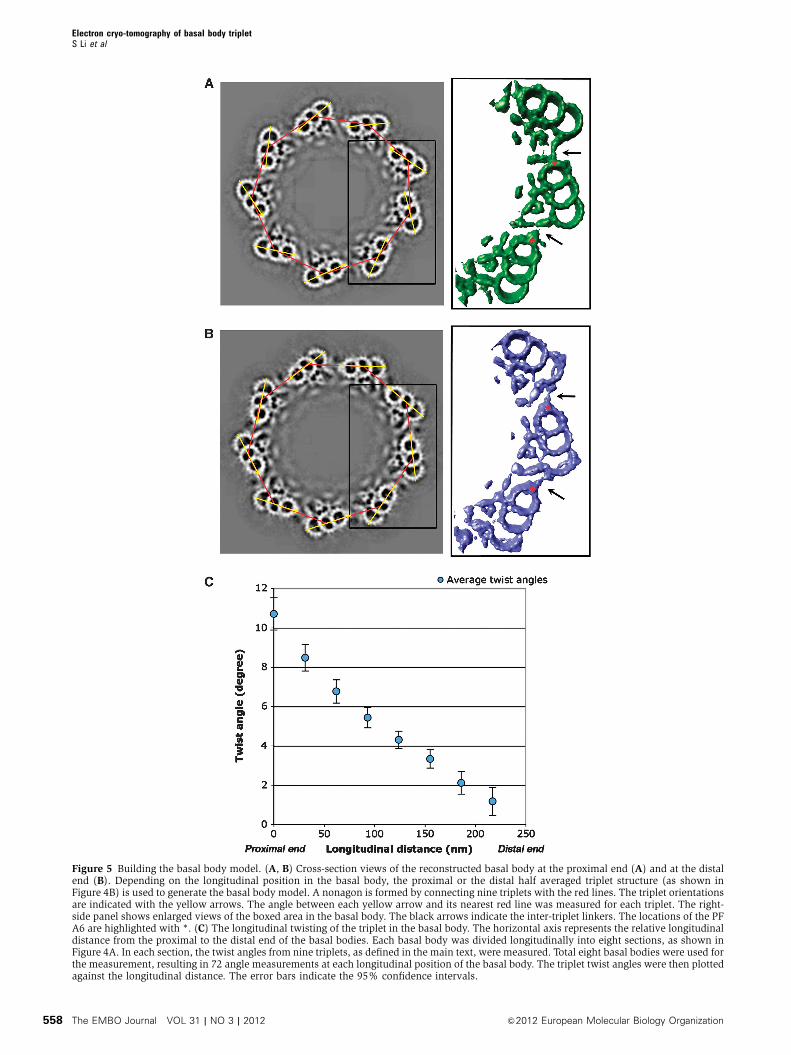
Figure 5 Building the basal body model. (A, B) Cross-section views of the reconstructed basal body at the proximal end (A) and at the distalend (B). Depending on the longitudinal position in the basal body, the proximal or the distal half averaged triplet structure (as shown inFigure 4B) is used to generate the basal body model. A nonagon is formed by connecting nine triplets with the red lines. The triplet orientationsare indicated with the yellow arrows. The angle between each yellow arrow and its nearest red line was measured for each triplet. The right-side panel shows enlarged views of the boxed area in the basal body. The black arrows indicate the inter-triplet linkers. The locations of the PFA6 are highlighted with *. (C) The longitudinal twisting of the triplet in the basal body. The horizontal axis represents the relative longitudinaldistance from the proximal to the distal end of the basal bodies. Each basal body was divided longitudinally into eight sections, as shown inFigure 4A. In each section, the twist angles from nine triplets, as defined in the main text, were measured. Total eight basal bodies were used forthe measurement, resulting in 72 angle measurements at each longitudinal position of the basal body. The triplet twist angles were then plottedagainst the longitudinal distance. The error bars indicate the 95% confidence intervals.
Electron cryo-tomography of basal body tripletS Li et al
The EMBO Journal VOL 31 | NO 3 | 2012 &2012 European Molecular Biology Organization558

Paintrand et al, 1992). This longitudinal left-handed twist of
the triplet along the basal body results in a structural change
at the inter-triplet linker. Since this twist proceeds gradually,
it suggests that the inter-triplet linker is flexible. Alternatively,
there might be multiple attachment sites for the inter-triplet
linker running across the wall of the C-tubule. The mechan-
ism for this transition awaits molecular details achieved only
from higher-resolution structures.
Discussion
Comparison with the axoneme doublet
The basal body provides a template for axoneme assembly.
To compare the two structures, we superimposed the
triplet EM density with one of the available axoneme
doublet structures at a comparable resolution (EMD-1696)
(Movassagh et al, 2010). As shown in Figure 6, the overall
fitting of the two structures is excellent. However, there are
notable differences in the shape of the A-tubule and striking
differences in non-MT densities. In both structures, the
A-tubules are elliptically deformed. The A-tubule ring in the
doublet is radially elongated B8% where the long axis of the
elliptical ring runs across PFs A2/3 and A9. The distortion is
likely due to accessory proteins, such as Tektins, attached to
the luminal wall of the A-tubule (Sui and Downing, 2006).
In the triplet, the A-tubule ring is elongated B10% in the
same direction as the doublet. However, the PFA3, A4, A5 are
stretched further outwards compared with the doublet
(Figure 6). This results in an asymmetric elliptical shape of
the A-tubule in the triplet. The variation of the local curvature
demonstrates the intrinsic property of MTwith flexible lateral
PF contacts. This flexibility is consistent with the results
from the EM reconstruction of in vitro assembled MTs with
various PF numbers (Sui and Downing, 2010). Interestingly,
the association patterns of the accessory proteins are also
different in these two structures. In the doublet, substantial
density has been observed in the lumen of the A-tubule and
on both sides of the partition bridge, presumably to stabilize
the doublet in resistance to the bending force generated by
axonemal dynein (Nicastro et al, 2006; Sui and Downing,
2006). In contrast, the triplet has fewer accessory proteins
attached to the luminal wall of A- and B-tubule, likely
because the basal body bears less mechanical stress from
its cellular environment and the A- and B-tubule are further
bolstered by the C-tubule.
Luminal structure cross-linking the A- and B-tubules in
the triplet
One of the striking features presented in our structure is the
horizontal Y-shaped structure circumferentially bound to the
triplet on the luminal side of the basal body. This is signifi-
cantly different from the axoneme doublet, where a flexible
linker density with 16-nm periodicity connects the A- and
B-tubules (Sui and Downing, 2006). In the triplet, the A- and
B-tubules are tightly linked by a spiral-shaped filament
formed by the stems that contact the A- and B-tubules
alternately with a 4-nm rise (Figure 3A and B). In addition,
the structure also has three extended arms emanating from
the stem. Both armA and armC form longitudinal filaments
with an 8-nm periodicity, while two neighbouring armBs
join to form a bifurcated structure with a 16-nm repeat.
Furthermore, in our basal body models, nine of these struc-
tures nearly fill the entire inner circumference of the basal body
(Figure 5A and B). It is likely that they are connected by
flexible linkers and form a cylindrical structure as observed
previously (Geimer and Melkonian, 2004). In additional to
the cartwheel and other basal body components, this might
provide another scaffold inside the barrel of the basal body.
Several functions might be carried out by this scaffold
structure. First, it links the A- and B-tubules and reinforces
the triplets. Second, it provides another inter-triplet linker
besides the link between neighbouring A- and C-tubules.
Third, the longitudinal interactions in these filaments will
further stabilize the basal body barrel. Finally, this scaffold
structure might recruit other luminal components, such as
filaments and electron dense materials often seen in the
central luminal region of the basal body (Cavalier-Smith,
1974; Geimer and Melkonian, 2004).
Similar luminal structures have previously been observed in
centrioles and basal bodies. For example, luminal disks were
observed in human centrioles (Paintrand et al, 1992; Ibrahim
et al, 2009), and luminal rims were observed in the centrioles
and basal bodies in other vertebrate cells (Fais et al, 1986).
Although their detailed structures might be different from what
we presented here, it is likely that luminal scaffold is
a common feature of basal bodies and centrioles in many
organisms, presumably playing a role both in stabilizing the
assembly and in recruiting additional components. This lumi-
nal scaffold emerges as a rigid structure about 100 nm from the
minus (proximal) end of the basal body and spans about
250 nm longitudinally (Figure 1A). This is concomitant with
the transition of the probasal body to the basal body, suggest-
ing that it assembles during elongation and early maturation of
the basal body in G2 phase in Chlamydomonas (Piasecki et al,
2008). However, in mammalian cells, this might take place in
early S phase when the procentrioles start to elongate
(Vorobjev and Chentsov, 1982).
Figure 6 Comparing the surface-rendered map of the basal bodytriplet with the previously published map of the axoneme doublet.The basal body triplet is coloured in blue. The doublet map (EMD-1696) is coloured in red (Movassagh et al, 2010).
Electron cryo-tomography of basal body tripletS Li et al
&2012 European Molecular Biology Organization The EMBO Journal VOL 31 | NO 3 | 2012 559
Longitudinal variations and its implications for the basal
body assembly
Perhaps the most remarkable finding from our structure is the
structural changes between the proximal and the distal half of
the basal body. These changes are mainly confined to the
C-tubule, but effectively alter assembly of the whole basal
body. One of the Chlamydomonas genes that affect the
C-tubule formation is the UNI3 gene encoding d-tubulin
(Dutcher and Trabuco, 1998). Previous studies have shown
that deletion of d-tubulin led to a basal body with mostly
doublets and occasional short stretches of triplet at the distal
end, suggesting that the d-tubulin is required for extension
and stability of the C-tubule (Garreau de Loubresse et al,
2001; Fromherz et al, 2004). Additional defects of the
d-tubulin deletion include duplicated and misplaced transi-
tion zones and a misaligned cell cleavage furrow (O’Toole
et al, 2003). Since one of the major structural changes we
have observed is at PF C1, it strongly suggests the PF C1 is
composed of d-tubulin. Comparing to a/b tubulins, d-tubulin
has a unique and highly conserved insertion sequence near
the M-loop located at the tubulin lateral interface. This
insertion will be ideally suited to make an unusual lateral
contact with PF B4 from the B-tubule (Chang and Stearns,
2000; Inclan and Nogales, 2001). Interestingly, an a-tubulin
mutant encoded by TUA2 could suppress the d-tubulin dele-
tion phenotype (Fromherz et al, 2004). This a-tubulin mutant
might partially replace d-tubulin at the proximal half of PF C1
and partly restore triplet formation. As the assembly extends
further towards the middle of the basal body, d-tubulin will
be replaced by an unknown protein or complex with a mass
of about 270 kDa. It binds to PF B4 every 8 nm and laterally
cross-links PF C2, C3 and C4. This complex extends long-
itudinally along the PF C1 as a filament until the C-tubule
terminates. In the absence of d-tubulin, this complex may
only partially bind to PF B4 and assembles as PF C1, resulting
in short stretches of triplet as observed at the distal end of the
basal body in the d-tubulin deletion mutant (O’Toole et al,
2003), further supporting the notion that the extension of PF
C1 and C-tubule is a cooperative assembly process.
In contrast to d-tubulin, a conserved centriole protein
POC5 is localized mainly at the distal portion of the centriole
and is essential for procentriole elongation to its full length
(Azimzadeh et al, 2009). Like d-tubulin, depletion of POC5
resulted in centrioles with MT doublets, suggesting that POC5
might be involved in the extension of C-tubule during the
centriole assembly. Interestingly, both d-tubulin and POC5
genes are absent in organisms such as Caenorhabditis elegans
and Drosophila melanogaster, where only singlets or doublets
are observed in the centriole or basal body in the somatic
cells (Dutcher, 2001; Azimzadeh et al, 2009), further suggest-
ing their unique roles in assembly of the C-tubule of the
triplet. However, triplets have been observed in the basal
bodies and centrioles in germ cells in Drosophila (Mahowald
and Strassheim, 1970; Riparbelli and Callaini, 2011). It will be
interesting to investigate the tissue-specific expression levels
of different tubulin isoforms, as this might provide insight
into their specific roles during the basal body/centriole
assembly.
Detailed analysis of POC5 also revealed that procentriole
elongation consists of two sequential and distinct steps
coupled to cell-cycle progression (Azimzadeh et al, 2009).
The structural variations observed in our study of the basal
body, in which the proximal half exhibits distinct differences
from the distal half, have provided a clear structural basis to
support a two-step assembly mechanism. Similar longitudi-
nal structural variation has previously been observed in the
axoneme, such as the 1–2 bridge concentrated at the proximal
end (Hoops and Witman, 1983), and is presumably used to
regulate the waveform of the flagella (Bui et al, 2009).
Overall, there are a number of features which, taken together,
suggest that basal body assembly is a tightly regulated and
coordinated process. First, there is the change at the C-tubule
including the structural transition at PF C1. Second, there is
the attachment of luminal filaments spanning C3 to C6/7 in
the C-tubule, and finally there is the relative rotation of the
adjacent triplet, resulting in a change at the inter-triplet
linker. In addition to key roles in assembly, these longitudinal
structural variations might provide the axial polarity and
spatial specificity for subsequent addition of accessory struc-
tures, such as the subdistal and distal appendages, during
basal body and centriole maturation. Our structural analysis
of the basal body reported here has paved the way for
identification of major basal body components within the
context of a 3D structure. This enables us to address funda-
mental questions concerning centriole and basal body
biogenesis in molecular detail.
Materials and methods
Basal body purification cryo-ET data collectionBasal bodies were purified following previous published method(Snell et al, 1974; Keller et al, 2005).
The purified basal body in solution was mixed with colloidalgold (10 nm) and was applied to 300 mesh holey carbon grids(Quantifoil, Germany) and flash-frozen in liquid ethane using aVitrobot (FEI, Inc., The Netherlands). A typical vitreous icethickness is about 300 nm. The grids were stored in liquid nitrogenat �1801C.
ET tilt series were collected on an FEG microscope (Polara, FEI,Inc.) operating at 300 KV. The microscope was equipped with apost-column energy filter (GIF, Gatan, Inc.) and the slit width wasset at 25 eV. UCSF Tomography software (Zheng et al, 2004) wasused for automatic data collection. Single-axis tilt series werecollected at a nominal magnification of 34 000 and images with sizeof 2032� 2032 were recorded on a CCD camera (UltraCam, Gatan,Inc.) and the final pixel size of image is 6.5 A. The specimen wastilted from �601 to þ 601 in 1.51 increment. The microscopedefocus values were set between 9 and 24mm. To avoid excessiveradiation damage, care was taken to limit the cumulative dose onthe specimen o80 e�/A2.
3D image processing, volume average and model buildingTomographic tilt series were aligned in IMOD (Kremer et al, 1996)by using 10-nm colloid gold beads as the fiducial markers. The CTFfor each tilt series was determined and corrected (Fernandez et al,2006). 3D reconstructed volumes were calculated with an iterativereconstruction algorithm (TAPIR) in Priism (Chen et al, 1996).To average sections of MT triplets from different tomogram datasets, each triplet volume is divided into a number of smallsegments, which are then aligned to a single common origin beforeaveraging. We found that the longest longitudinal repeat in thetriplet is 16 nm; therefore, we limited our final averaged volume to39 nm in the longitudinal direction to contain slightly more thantwo repeats. Subvolumes with pixel dimension of 200 by 60 by 200were boxed out along the basal body axis containing tripletsegments about 39 nm in length and 10% overlapped with adjacentsegments. Iterative subvolume alignment was carried out in Spider(Frank et al, 1996). Initially, a symmetrized triplet volume was usedas a reference for alignment followed by using averaged volumefrom the previous round as reference for the next round ofalignment. The average of the aligned subvolumes was carriedout in Fourier space following an algorithm proposed by Schmid
Electron cryo-tomography of basal body tripletS Li et al
The EMBO Journal VOL 31 | NO 3 | 2012 &2012 European Molecular Biology Organization560
and Booth (2008). The two repeats in the averaged subvolume werefurther aligned and averaged to obtain the final triplet structure. Atotal of 1644 subvolumes from 27 tilt series were used for averaging.To assess the resolution of the final averaged structure, subvolumeswere randomly split into two groups and their FSC was calculated.Since the basal body has intrinsic nine-fold symmetry where thenine triplets have different orientations, the resulting averagedtriplet structure has isotropic resolution with no missing wedgeartefacts. The estimated resolution is at 33 A by using FSC¼ 0.143criterion (Supplementary Figure S1) (Rosenthal and Henderson,2003). The pseudo-atomic model of the MT triplet was built basedon an atomic structure of tubulin (PDB ID: 1JFF) by manually fittinginto the EM density in program O (Jones et al, 1991). Care wastaken to have H1-S2/H2-S3 loop and M-loop at vicinity and to avoidsteric clash between laterally interacting PF. The fitting was furtherrefined in UCSF Chimera (Pettersen et al, 2004). UCSF Chimera wasused for 3D volume display and volume segmentation analysis. Thecontact area was estimated by assuming two density maps havingsurfaces within 5 A in distance. The masses of EM densities wereestimated by assuming protein density 1.41 g/cm3 (Fischer et al,2004) after setting the density threshold by using the pseudo-atomicMT triplet model as a standard.
Supplementary dataSupplementary data are available at The EMBO Journal Online(http://www.embojournal.org).
Acknowledgements
We thank Michael Braunfeld and Shawn Zheng for advice ontomography data collection, Lani Keller for suggestions on basalbody purification, Tom Goddart for using UCSF Chimera program,Juliette Azimzadeh and members of Agard lab for discussion andcomments. We also thank Richard Henderson (MRC-LMB) for criticalreading of this manuscript. This work was supported by HHMI andby the Keck Laboratory for Advanced Electron Microscopy.
Author contributions: SL, WFM and DAA designed the research;SL prepared the specimen and collected the cryo-EM data; SL andJJF carried out all the image processing and map analysis; SL, JJF,WFM and DAA analysed the final result and wrote the paper.
Conflict of interest
The authors declare that they have no conflict of interest.
References
Allen RD (1969) The morphogenesis of basal bodies and accessorystructures of the cortex of the ciliated protozoan Tetrahymenapyriformis. J Cell Biol 40: 716–733
Amos LA (2008) The tektin family of microtubule-stabilizing pro-teins. Genome Biol 9: 229
Andersen JS, Wilkinson CJ, Mayor T, Mortensen P, Nigg EA, MannM (2003) Proteomic characterization of the human centrosomeby protein correlation profiling. Nature 426: 570–574
Anderson RG (1972) The three-dimensional structure of thebasal body from the rhesus monkey oviduct. J Cell Biol 54:246–265
Anderson RG, Brenner RM (1971) The formation of basal bodies(centrioles) in the Rhesus monkey oviduct. J Cell Biol 50: 10–34
Azimzadeh J, Hergert P, Delouvee A, Euteneuer U, Formstecher E,Khodjakov A, Bornens M (2009) hPOC5 is a centrin-bindingprotein required for assembly of full-length centrioles. J CellBiol 185: 101–114
Bui KH, Sakakibara H, Movassagh T, Oiwa K, Ishikawa T (2009)Asymmetry of inner dynein arms and inter-doublet links inChlamydomonas flagella. J Cell Biol 186: 437–446
Carvalho-Santos Z, Machado P, Branco P, Tavares-Cadete F,Rodrigues-Martins A, Pereira-Leal JB, Bettencourt-Dias M (2010)Stepwise evolution of the centriole-assembly pathway. J Cell Sci123(Part 9): 1414–1426
Cavalier-Smith T (1974) Basal body and flagellar developmentduring the vegetative cell cycle and the sexual cycle ofChlamydomonas reinhardii. J Cell Sci 16: 529–556
Chang P, Stearns T (2000) Delta-tubulin and epsilon-tubulin: twonew human centrosomal tubulins reveal new aspects of centro-some structure and function. Nat Cell Biol 2: 30–35
Chen H, Hughes DD, Chan TA, Sedat JW, Agard DA (1996) IVE(Image Visualization Environment): a software platform forall three-dimensional microscopy applications. J Struct Biol 116:56–60
Chretien D, Wade RH (1991) New data on the microtubule surfacelattice. Biol Cell 71: 161–174
Cyrklaff M, Kudryashev M, Leis A, Leonard K, Baumeister W,Menard R, Meissner M, Frischknecht F (2007) Cryoelectrontomography reveals periodic material at the inner side of sub-pellicular microtubules in apicomplexan parasites. J Exp Med204: 1281–1287
Deane JA, Cole DG, Seeley ES, Diener DR, Rosenbaum JL (2001)Localization of intraflagellar transport protein IFT52 identifiesbasal body transitional fibers as the docking site for IFT particles.Curr Biol 11: 1586–1590
Debec A, Sullivan W, Bettencourt-Dias M (2010) Centrioles:active players or passengers during mitosis? Cell Mol Life Sci67: 2173–2194
Dippell RV (1968) The development of basal bodies in paramecium.Proc Natl Acad Sci USA 61: 461–468
Doxsey S, Zimmerman W, Mikule K (2005) Centrosome control ofthe cell cycle. Trends Cell Biol 15: 303–311
Dutcher SK (2001) The tubulin fraternity: alpha to eta. Curr OpinCell Biol 13: 49–54
Dutcher SK, Trabuco EC (1998) The UNI3 gene is required forassembly of basal bodies of Chlamydomonas and encodes delta-tubulin, a new member of the tubulin superfamily. Mol Biol Cell9: 1293–1308
Fais DA, Nadezhdina ES, Chentsov YS (1986) The centriolar rim.The structure that maintains the configuration of centrioles andbasal bodies in the absence of their microtubules. Exp Cell Res164: 27–34
Fernandez JJ, Li S, Crowther RA (2006) CTF determination andcorrection in electron cryotomography. Ultramicroscopy 106:587–596
Fischer H, Polikarpov I, Craievich AF (2004) Average protein density is amolecular-weight-dependent function. Protein Sci 13: 2825–2828
Frank J, Radermacher M, Penczek P, Zhu J, Li Y, Ladjadj M, Leith A(1996) SPIDER and WEB: processing and visualization of imagesin 3D electron microscopy and related fields. J Struct Biol 116:190–199
Fromherz S, Giddings TH, Gomez-Ospina N, Dutcher SK (2004)Mutations in alpha-tubulin promote basal body maturation andflagellar assembly in the absence of delta-tubulin. J Cell Sci117(Part 2): 303–314
Garreau de Loubresse N, Ruiz F, Beisson J, Klotz C (2001) Role ofdelta-tubulin and the C-tubule in assembly of Paramecium basalbodies. BMC Cell Biol 2: 4
Garvalov BK, Zuber B, Bouchet-Marquis C, Kudryashev M, GruskaM, Beck M, Leis A, Frischknecht F, Bradke F, Baumeister W,Dubochet J, Cyrklaff M (2006) Luminal particles within cellularmicrotubules. J Cell Biol 174: 759–765
Geimer S, Melkonian M (2004) The ultrastructure of theChlamydomonas reinhardtii basal apparatus: identification ofan early marker of radial asymmetry inherent in the basal body.J Cell Sci 117(Part 13): 2663–2674
Gerdes JM, Davis EE, Katsanis N (2009) The vertebrate primarycilium in development, homeostasis, and disease. Cell 137: 32–45
Giddings TH, Meehl JB, Pearson CG, Winey M (2010) Electrontomography and immuno-labeling of Tetrahymena thermophilabasal bodies. Methods Cell Biol 96: 117–141
Gonzalez C, Tavosanis G, Mollinari C (1998) Centrosomes andmicrotubule organisation during Drosophila development. J CellSci 111(Part 18): 2697–2706
Guichard P, Chretien D, Marco S, Tassin A-M (2010) Procentrioleassembly revealed by cryo-electron tomography. EMBO J 29:1565–1572
Hinchcliffe EH, Miller FJ, Cham M, Khodjakov A, Sluder G (2001)Requirement of a centrosomal activity for cell cycle progressionthrough G1 into S phase. Science 291: 1547–1550
Electron cryo-tomography of basal body tripletS Li et al
&2012 European Molecular Biology Organization The EMBO Journal VOL 31 | NO 3 | 2012 561

Hoops HJ, Witman GB (1983) Outer doublet heterogeneity revealsstructural polarity related to beat direction in Chlamydomonasflagella. J Cell Biol 97: 902–908
Hoops HJ, Wright RL, Jarvik JW, Witman GB (1984) Flagellarwaveform and rotational orientation in a Chlamydomonas mu-tant lacking normal striated fibers. J Cell Biol 98: 818–824
Ibrahim R, Messaoudi C, Chichon FJ, Celati C, Marco S (2009)Electron tomography study of isolated human centrioles. MicroscRes Tech 72: 42–48
Inclan YF, Nogales E (2001) Structural models for the self-assemblyand microtubule interactions of gamma-, delta- and epsilon-tubulin. J Cell Sci 114(Part 2): 413–422
Jones TA, Zou JY, Cowan SW, Kjeldgaard M (1991) Improvedmethods for building protein models in electron density mapsand the location of errors in these models. Acta Cryst A 47(Part2): 110–119
Keller LC, Romijn EP, Zamora I, Yates JR, Marshall WF(2005) Proteomic analysis of isolated chlamydomonascentrioles reveals orthologs of ciliary-disease genes. Curr Biol15: 1090–1098
Kilburn CL, Pearson CG, Romijn EP, Meehl JB, Giddings TH, CulverBP, Yates JR, Winey M (2007) New Tetrahymena basal bodyprotein components identify basal body domain structure. J CellBiol 178: 905–912
Kremer JR, Mastronarde DN, McIntosh JR (1996) Computer visua-lization of three-dimensional image data using IMOD. J Struct Biol116: 71–76
Li H, DeRosier DJ, Nicholson WV, Nogales E, Downing KH(2002) Microtubule structure at 8 A resolution. Structure 10:1317–1328
Li JB, Gerdes JM, Haycraft CJ, Fan Y, Teslovich TM, May-Simera H,Li H, Blacque OE, Li L, Leitch CC, Lewis RA, Green JS, Parfrey PS,Leroux MR, Davidson WS, Beales PL, Guay-Woodford LM, YoderBK, Stormo GD, Katsanis N et al (2004) Comparative genomicsidentifies a flagellar and basal body proteome that includes theBBS5 human disease gene. Cell 117: 541–552
Linck RW, Stephens RE (2007) Functional protofilament numberingof ciliary, flagellar, and centriolar microtubules. Cell MotilCytoskeleton 64: 489–495
Mahowald AP, Strassheim JM (1970) Intercellular migration ofcentrioles in the germarium of Drosophila melanogaster. Anelectron microscopic study. J Cell Biol 45: 306–320
Mikule K, Delaval B, Kaldis P, Jurcyzk A, Hergert P, Doxsey S (2007)Loss of centrosome integrity induces p38-p53-p21-dependent G1-S arrest. Nat Cell Biol 9: 160–170
Movassagh T, Bui KH, Sakakibara H, Oiwa K, Ishikawa T (2010)Nucleotide-induced global conformational changes of flagellardynein arms revealed by in situ analysis. Nat Struct Mol Biol17: 761–767
Nicastro D, Schwartz C, Pierson J, Gaudette R, Porter ME, McIntoshJR (2006) The molecular architecture of axonemes revealed bycryoelectron tomography. Science 313: 944–948
Nigg EA, Raff JW (2009) Centrioles, centrosomes, and cilia in healthand disease. Cell 139: 663–678
Nogales E, Whittaker M, Milligan RA, Downing KH (1999) High-resolution model of the microtubule. Cell 96: 79–88
O’Toole ET, Giddings TH, McIntosh JR, Dutcher SK (2003) Three-dimensional organization of basal bodies from wild-type anddelta-tubulin deletion strains of Chlamydomonas reinhardtii.Mol Biol Cell 14: 2999–3012
Paintrand M, Moudjou M, Delacroix H, Bornens M (1992)Centrosome organization and centriole architecture: their sensi-tivity to divalent cations. J Struct Biol 108: 107–128
Pelletier L, O’Toole E, Schwager A, Hyman AA, Muller-Reichert T(2006) Centriole assembly in Caenorhabditis elegans. Nature 444:619–623
Pettersen EF, Goddard TD, Huang CC, Couch GS, Greenblatt DM,Meng EC, Ferrin TE (2004) UCSF Chimera—a visualizationsystem for exploratory research and analysis. J Comput Chem25: 1605–1612
Piasecki BP, LaVoie M, Tam L-W, Lefebvre PA, Silflow CD (2008)The Uni2 phosphoprotein is a cell cycle regulated component ofthe basal body maturation pathway in Chlamydomonas reinhard-tii. Mol Biol Cell 19: 262–273
Piel M, Nordberg J, Euteneuer U, Bornens M (2001) Centrosome-dependent exit of cytokinesis in animal cells. Science 291:1550–1553
Ringo DL (1967) Flagellar motion and fine structure of the flagellarapparatus in Chlamydomonas. J Cell Biol 33: 543–571
Riparbelli MG, Callaini G (2011) Male gametogenesis withoutcentrioles. Dev Biol 349: 427–439
Rosenthal PB, Henderson R (2003) Optimal determination of parti-cle orientation, absolute hand, and contrast loss in single-particleelectron cryomicroscopy. J Mol Biol 333: 721–745
Schmid MF, Booth CR (2008) Methods for aligning and for aver-aging 3D volumes with missing data. J Struct Biol 161: 243–248
Snell WJ, Dentler WL, Haimo LT, Binder LI, Rosenbaum JL (1974)Assembly of chick brain tubulin onto isolated basal bodies ofChlamydomonas reinhardi. Science 185: 357–360
Song YH, Mandelkow E (1995) The anatomy of flagellar micro-tubules: polarity, seam, junctions, and lattice. J Cell Biol 128:81–94
Sui H, Downing KH (2006) Molecular architecture of axonemalmicrotubule doublets revealed by cryo-electron tomography.Nature 442: 475–478
Sui H, Downing KH (2010) Structural basis of interprotofilamentinteraction and lateral deformation of microtubules. Structure 18:1022–1031
Tilney LG, Bryan J, Bush DJ, Fujiwara K, Mooseker MS, MurphyDB, Snyder DH (1973) Microtubules: evidence for 13 protofila-ments. J Cell Biol 59(2 Part 1): 267–275
Vorobjev IA, Chentsov Y (1982) Centrioles in the cell cycle. I.Epithelial cells. J Cell Biol 93: 938–949
Wang X, Tsai J-W, Imai JH, Lian W-N, Vallee RB, Shi S-H (2009)Asymmetric centrosome inheritance maintains neural progenitorsin the neocortex. Nature 461: 947–955
Yamashita YM, Mahowald AP, Perlin JR, Fuller MT (2007)Asymmetric inheritance of mother versus daughter centrosomein stem cell division. Science 315: 518–521
Zheng QS, Braunfeld MB, Sedat JW, Agard DA (2004) An improvedstrategy for automated electron microscopic tomography. J StructBiol 147: 91–101
The EMBO Journal is published by NaturePublishing Group on behalf of European
Molecular Biology Organization. This work is licensedunder a Creative Commons Attribution-Noncommercial-No Derivative Works 3.0 Unported License. [http://creativecommons.org/licenses/by-nc-nd/3.0]
Electron cryo-tomography of basal body tripletS Li et al
The EMBO Journal VOL 31 | NO 3 | 2012 &2012 European Molecular Biology Organization562