Embed Size (px)
Citation preview
JOURNAL OF BACTERIOLOGY, Aug. 2004, p. 5052–5061 Vol. 186, No. 150021-9193/04/$08.00�0 DOI: 10.1128/JB.186.15.5052–5061.2004Copyright © 2004, American Society for Microbiology. All Rights Reserved.
Three-Dimensional Electron Microscopic Imaging of MembraneInvaginations in Escherichia coli Overproducing
the Chemotaxis Receptor TsrJonathan Lefman,1† Peijun Zhang,1† Teruhisa Hirai,1 Robert M. Weis,1,2 Jemma Juliani,1
Donald Bliss,1 Martin Kessel,1 Erik Bos,3 Peter J. Peters,3 and Sriram Subramaniam1*Laboratory of Cell Biology, National Cancer Institute, National Institutes of Health, Bethesda, Maryland 208171;
Department of Chemistry, University of Massachusetts, Amherst, Massachusetts 010032; and Division ofTumor Biology, The Netherlands Cancer Institute, Amsterdam, The Netherlands3
Received 24 December 2003/Accepted 30 March 2004
Electron tomography is a powerful method for determining the three-dimensional structures of largemacromolecular assemblies, such as cells, organelles, and multiprotein complexes, when crystallographicaveraging methods are not applicable. Here we used electron tomographic imaging to determine the moleculararchitecture of Escherichia coli cells engineered to overproduce the bacterial chemotaxis receptor Tsr. Tomo-grams constructed from fixed, cryosectioned cells revealed that overproduction of Tsr led to formation of anextended internal membrane network composed of stacks and extended tubular structures. We present aninterpretation of the tomogram in terms of the packing arrangement of Tsr using constraints derived fromprevious X-ray and electron-crystallographic studies of receptor clusters. Our results imply that the interac-tion between the cytoplasmic ends of Tsr is likely to stabilize the presence of the membrane networks in cellsoverproducing Tsr. We propose that membrane invaginations that are potentially capable of supporting axialinteractions between receptor clusters in apposing membranes could also be present in wild-type E. coli andthat such receptor aggregates could play an important role in signal transduction during bacterial chemotaxis.
Over the last three decades, methods for three-dimensionalreconstruction of objects (5) imaged with an electron micro-scope have been used to determine the structures of a varietyof biological assemblies by two types of approaches. One ap-proach, which has been used extensively in analyses of largemacromolecular assemblies, involves three-dimensional recon-struction of a structure by averaging images recorded fromseveral identical copies oriented randomly relative to the elec-tron beam (11, 31). The other approach, which has been usedfor reconstruction of objects that cannot be easily averaged,such as whole cells, involves tomographic reconstruction bycombining projection images of an object recorded with anelectron microscope over a range of tilt angles (4). Electrontomography is therefore a potentially powerful tool for three-dimensional imaging of the spatial arrangement of proteinsthat make up complex and dynamic assemblies, such as thoseinvolved in bacterial chemotaxis.
At least 12 proteins act in concert to convert the signal ofligand binding at the periplasmic end of a chemotaxis receptorinto rotation of the flagellar motor (6, 27). The principal pro-tein components at the input end include one of the chemo-taxis receptors (Tsr, Tar, Trg, Tap, or Aer), and the cytoplas-mic signaling proteins CheA and CheW, which are thought toform a noncovalent complex with the chemotaxis receptors.Knowledge of the structure and spatial arrangement of thechemotaxis receptors is therefore fundamental to understand-
ing the structural biology of signaling. X-ray crystallographicstudies of the periplasmic fragments of the aspartate receptorfragments have revealed the dimeric organization of the ligandbinding domain, in which the ligand binding pocket is locatedat the dimer interface (20). Structural studies of a cytoplasmicfragment of the related Tsr receptor have shown that it alsoforms a dimer of extended coiled-coil hairpins (13). The cyto-plasmic fragments pack in the three-dimensional crystals usedfor X-ray crystallographic analysis as trimers of dimers, andthis has led to a proposal by Kim et al. (14) that such anarrangement may also be representative of full-length recep-tors in cell membranes. Figure 1a shows the spatial arrange-ment of the periplasmic and cytoplasmic domains in the con-text of an atomic model for the full-length receptor (14) thatincludes plausible structures for the transmembrane regionand the linker region between the membrane domain and thecytoplasmic domain.
Ultrastructural studies of bacterial cells in combination withimmunoelectron microscopy have revealed that chemotaxis re-ceptors, as well as CheA and CheW, are predominantly local-ized to the poles of the cells (18, 25). Although biochemicalstudies have implicated the involvement of extended interac-tions among receptor subunits in the membrane during signal-ing (1) and many computational models for clustering havebeen proposed (19, 24), there is only limited direct structuralevidence that such interactions occur under physiological con-ditions. Approaches such as electron tomography provide toolswhich can be used to begin to unravel the molecular arrange-ment of these protein components in the cell because of thepotential for integrating structural information from X-raycrystallography with the lower-resolution data obtained fromelectron microscopic imaging. As a first step in this direction,
* Corresponding author. Mailing address: Laboratory of Cell Biol-ogy, National Cancer Institute, National Institutes of Health, Be-thesda, MD 20817. Phone: (301) 594-2062. Fax: (301) 480-3834. E-mail: [email protected].
† J.L. and P.Z. contributed equally to this work.
5052
Dow
nloa
ded
from
http
s://j
ourn
als.
asm
.org
/jour
nal/j
b on
13
Nov
embe
r 20
21 b
y 79
.134
.37.
135.

FIG. 1. (a) Structural model for the full-length Tsr dimer based on structures of the periplasmic and cytoplasmic domains, adapted from thework of Kim et al. (14). (b and c) Projection images of thin (70-nm) sections of fixed specimens of either wild-type E. coli (b) or an E. coliTsr-overexpressing strain (HCB721/pHSe5.tsrQEQE) induced with 1 mM isopropyl-�-D-thiogalactopyranoside (IPTG) (c). Scale bars � 0.5 �m. (d)Higher magnification of cells overproducing Tsr. Scale bar � 50 nm. The dark spots in the images are from 10-nm gold-conjugated protein A usedto locate Tsr.
VOL. 186, 2004 MEMBRANE INVAGINATIONS IN E. COLI 5053
Dow
nloa
ded
from
http
s://j
ourn
als.
asm
.org
/jour
nal/j
b on
13
Nov
embe
r 20
21 b
y 79
.134
.37.
135.

we obtained three-dimensional images of the assemblies andnetworks formed in cells by overexpression of the full-lengthchemotaxis receptor Tsr. Projection images recorded fromnegatively stained membrane extracts obtained from cells haveshown that Tsr is organized in receptor arrays that have theappearance of either zipper-like or micelle-like entities (32).The novel feature of these assemblies was the evidence thatthere are direct interactions between Tsr molecules both in theplane of the membrane and in the axial direction involving thecytoplasmic domains. In this study, we extended this analysisinto the third dimension with a description of the overall ar-chitecture of cells engineered to overproduce Tsr. The three-dimensional structural analyses demonstrated that the recep-tor arrays form an extended membrane network that extendsinto the cytoplasm, resulting in the formation of extendedsheets and tubules, which are converted into the zippered androunded assemblies seen in the isolated membrane extracts.We also describe similar analyses performed with wild-typecells to evaluate the presence of local invaginations in thecytoplasmic membrane.
MATERIALS AND METHODS
Fixation and embedding of cells. Escherichia coli strain RP437 was used as thecontrol for wild-type experiments. Plasmids pHSe5.tsrQQQQ and pHSe5.tsrQEQE
were used to produce Tsr in HCB721 cells, which do not express the chemotaxis-related proteins Tar, Tsr, Trg, Tap, CheA, CheW, CheR, and CheB, as describedpreviously (32). Harvested cells were fixed at room temperature for 2.5 h in amixture of 2% paraformaldehyde and 0.2% glutaraldehyde in the presence of 60mM piperazine-N,N�-bis(2-ethanesulfonic acid) (PIPES), 50 mM HEPES (pH6.9), 4 mM MgCl2, and 20 mM EGTA. The fixed cells were collected by cen-trifugation and resuspended in prewarmed 0.1 M phosphate buffer containing12% gelatin. After the cell-containing gelatin pellets were solidified on ice, theywere cut into 1-mm cubes and incubated with a solution containing 2.3 M sucroseand 0.1 M sodium phosphate buffer (pH 7.4). Cubes of gelatin were frozen on thesurfaces of aluminum pins by plunging them into liquid nitrogen and weresectioned with a cryoultramicrotome at �120°C. Labeling with anti-Tsr antibody(which specifically reacts with the conserved signaling domain) and proteinA-gold and subsequent embedding in methyl cellulose with uranyl acetate withreagents were carried out as described previously (22).
Electron microscopy. The projection images shown in Fig. 1 were recordedwith a Gatan 2K charge-coupled device camera mounted on a Tecnai 12 electronmicroscope (FEI Corporation, Hillsboro, Oreg.) equipped with an LaB6 filamentoperating at 120 kV. For tomography, a series of images were recorded at roomtemperature with the aid of a Gatan 2K charge-coupled device (magnification,��47,500) by tilting the specimen from �70° to 70° in increments of 0.5° in aTecnai F30 microscope equipped with a field emission gun tip operating at 300kV. Images were recorded at underfocus values that were between 2 and 3 �malong the tilt axis. A back-projection algorithm, as implemented in the IMODreconstruction package (15), was used to convert the information present in theseries of tilted projection images into three-dimensional density maps.
Preparation of Tween 80-extracted membranes. Membrane preparations (16)isolated on sucrose gradients were typically incubated with Tween 80 at a pro-tein/Tween 80 molar ratio of 0.004 for about 4 h as described previously (32).
Segmentation and rendering. The tomogram was segmented in the environ-ment of the program Amira (TGS Inc., San Diego, Calif.) by marking all regionsin the volume where the bilayer (white lines in the slice) could be visualizedclearly in three dimensions. An isosurface was created by tracing the path of thebilayer in each slice of the tomogram. Structural models of the two types ofreceptor assemblies shown in Fig. 3d and e were docked onto the isosurface byusing the program 3dsmax (DISCREET, Montreal, Quebec, Canada). The co-ordinates for the receptor dimer were the coordinates in the model described byKim et al. (14) and kindly provided by Sung-Hou Kim. Starting from the modelof the dimer, a variety of plausible arrangements for higher-order arrangements,such as the trimer of dimers shown in Fig. 3d and e, were then generated by usingthe electron microscopic images as a guide. One set of the plausible arrange-ments is shown in Fig. 3.
RESULTS
We carried out a three-dimensional structural analysis of E.coli cells engineered to overexpress the 60-kDa membraneprotein Tsr (16) in order to explore whether the novel assem-blies observed in membrane preparations isolated from dis-rupted cells (32) could also be detected in intact cells andimaged. Immunolabeling of fixed cryosections of wild-type E.coli (Fig. 1b) showed that Tsr was predominantly located at thepolar region(s) of the cells, which is consistent with previouslyreported data (18). In contrast, in cells overproducing Tsr, thereceptor was located throughout the periphery and in someinterior regions of the cell (Fig. 1c). In all areas where theimmunolabel appeared to be localized to the interior of thecell, there was evidence of membranous material, implying thatthe high level of Tsr resulted in extension of the membrane sothat it was no longer confined to the periphery of the cell. Acloser examination of a region of the cell displaying invagina-tions (Fig. 1d) confirmed that the immunolabel, which wasdirected against the C-terminal region of Tsr, was localizedprimarily to the interior portions of the zippered regions.
In order to investigate the three-dimensional structure of thecells overproducing Tsr, we recorded a series of projectionimages by tilting the specimen relative to the electron beam,typically over a range of 70° (Fig. 2a). The images were thencombined by using weighted back-projection methods to re-construct the three-dimensional structure. Representativeslices from an electron tomogram constructed from imagessuch as those shown in Fig. 2a are shown in Fig. 2b and c.These slices provide a glimpse of the extensive internal orga-nization of the membranes and the improved clarity relative tothe projection images. The slice in Fig. 2b is close to the upperedge of the section on the grid and shows extensive labeling ofTsr, as expected. The slice in Fig. 2c is from an interior regionof the section, and the absence of immunolabeling reflects thelack of penetration of the anti-Tsr antibody and/or the proteinA-gold conjugates into the section. An MPEG movie of thetomogram is available at http://hrem.nci.nih.gov/Lefman_et_al_J.Bact_2004/E.coli_tomogram.
A measure of the quality of both the collected data and thereconstruction procedure was obtained from analysis of a smallregion extracted from the central region of the tomogram usedto obtain the images shown in Fig. 2. The Fourier transforms(Fig. 2d and e) computed from individual horizontal (x-y) andvertical (x-z) slices in the tomogram showed that there werewell-defined Thon rings (30). The first zero crossing of thecontrast transfer function was at �40 A�1, and the spacing ofthe rings matched the values expected for the defocus valuesused (�2.6-�m underfocus along the tilt axis). The overallcontrast in the slice arises primarily from the presence of theuranyl acetate used for staining. However, the intrinsic reso-lution limit in a given slice of the tomogram relevant for de-tection of molecular structures is expected to be limited by thedamage that occurred during specimen preparation and by theradiation-induced damage that occurs upon illumination of thespecimen. For these reasons, the tomograms were not cor-rected for the effects of the contrast transfer function.
Inspection of the tomographic slice in Fig. 2c suggests thatthe internal volume is laced with an extensive network oftwisted sheet-like features that have the appearance of stacked
5054 LEFMAN ET AL. J. BACTERIOL.
Dow
nloa
ded
from
http
s://j
ourn
als.
asm
.org
/jour
nal/j
b on
13
Nov
embe
r 20
21 b
y 79
.134
.37.
135.
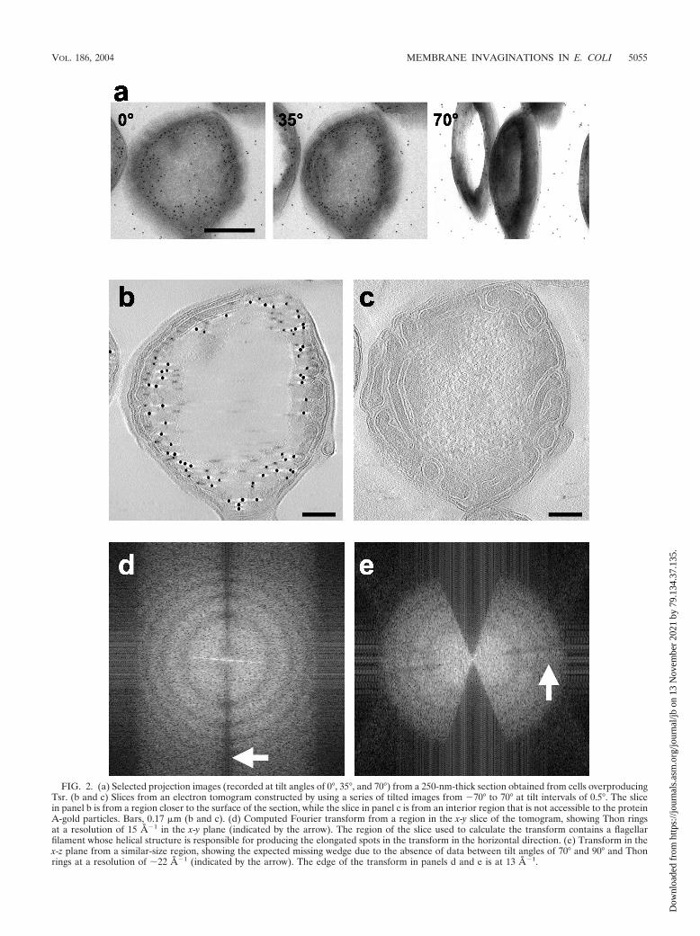
FIG. 2. (a) Selected projection images (recorded at tilt angles of 0°, 35°, and 70°) from a 250-nm-thick section obtained from cells overproducingTsr. (b and c) Slices from an electron tomogram constructed by using a series of tilted images from �70° to 70° at tilt intervals of 0.5°. The slicein panel b is from a region closer to the surface of the section, while the slice in panel c is from an interior region that is not accessible to the proteinA-gold particles. Bars, 0.17 �m (b and c). (d) Computed Fourier transform from a region in the x-y slice of the tomogram, showing Thon ringsat a resolution of 15 A�1 in the x-y plane (indicated by the arrow). The region of the slice used to calculate the transform contains a flagellarfilament whose helical structure is responsible for producing the elongated spots in the transform in the horizontal direction. (e) Transform in thex-z plane from a similar-size region, showing the expected missing wedge due to the absence of data between tilt angles of 70° and 90° and Thonrings at a resolution of �22 A�1 (indicated by the arrow). The edge of the transform in panels d and e is at 13 A�1.
VOL. 186, 2004 MEMBRANE INVAGINATIONS IN E. COLI 5055
Dow
nloa
ded
from
http
s://j
ourn
als.
asm
.org
/jour
nal/j
b on
13
Nov
embe
r 20
21 b
y 79
.134
.37.
135.
layers in certain regions and rounded features that appear tobe vesicular in cross section in other regions. From their loca-tion and continuity with the cytoplasmic membrane, it is clearthat the white lines defining the boundaries of the stacks andthe rounded structures represent the bilayer membrane. Theformation of these structures is a consequence of invaginationsof the surface of the cytoplasmic membrane in response to thehigh level of expression of Tsr. The uniformity of the spacingbetween the membrane layers (there was 5% variation acrossthe length of an individual stretch) in the stacks is consistentwith the idea that the membrane network is stabilized by spe-cific interactions between protein components in the bilayermembrane. Since these extended stacks and rounded struc-
tures were not observed in either wild-type E. coli or controlcells not expressing Tsr, the simplest explanation is that theyare formed in response to the high concentration of Tsr in themembrane and are stabilized by interactions between Tsr mol-ecules located in adjacent bilayer membranes.
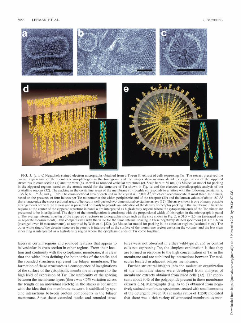
Further structural insights into the molecular organizationof the membrane stacks were developed from analyses ofmembrane extracts obtained from lysed cells (32). Tsr repre-sents about 90% of the polypeptide present in these membraneextracts (16). Micrographs (Fig. 3a to c) obtained from nega-tively stained membrane specimens treated with small amountsof the detergent Tween 80 (at molar ratios of 1:250) indicatedthat there was a rich variety of connected membranous mor-
FIG. 3. (a to c) Negatively stained electron micrographs obtained from a Tween 80 extract of cells expressing Tsr. The extract preserved theoverall appearance of the membrane morphologies in the tomogram, and the images show in more detail the organization of the zipperedstructures in cross section (a) and top view (b), as well as rounded vesicular structures (c). Scale bars � 50 nm. (d) Molecular model for packingin the zippered regions based on the atomic model for the structure of Tsr shown in Fig. 1a and the electron crystallographic analysis of thecrystalline regions (32). The packing in the crystalline areas of the membrane (b) roughly corresponds to a lattice with the following constants: a,�75 A; b, �75 A; and �, �60°. The cross-sectional area of each unit in the crystal is �5,000 A2, which can accommodate at most three Tsr dimers,based on the presence of four helices per Tsr monomer at the wider, periplasmic end of the receptor (20) and the known values of about 180 A2
that characterize the cross-sectional areas of helices in well-packed two-dimensional crystalline arrays (12). The array shown is one of many possiblearrangements of the three dimers and is presented primarily to provide an indication of the density of receptor packing in the membrane. The whiteregions at the center of the zippered structure in panel a are interpreted as high-density regions where the cytoplasmic ends of the Tsr trimer arepresumed to be interdigitated. The depth of the interdigitation is consistent with the proportional width of this region in the micrograph in panela. The average internal spacing of the zippered structures in tomographic slices such as the slice shown in Fig. 2c is 31.5 � 2.5 nm (averaged over26 separate measurements). This compares well with the value for the same internal spacing in these negatively stained specimens (31.3 0.6 nm[averaged over 10 measurements], as reported by Weis et al. [32]). (e) Molecular model for packing in the vesicular regions (sectional view). Theouter white ring of the circular structure in panel c is interpreted as the surface of the membrane region enclosing the volume, and the less clearinner ring is interpreted as a high-density region where the cytoplasmic ends of Tsr come together.
5056 LEFMAN ET AL. J. BACTERIOL.
Dow
nloa
ded
from
http
s://j
ourn
als.
asm
.org
/jour
nal/j
b on
13
Nov
embe
r 20
21 b
y 79
.134
.37.
135.

FIG. 4. (a) Segmented representation of a small region from a cellular tomogram. (b) Slice of the tomogram. The region indicated by the box(width, 230 nm) corresponds to a slice in the region that was segmented. Aside from the interaction between the cytoplasmic domains (yellow) inapposing bilayers, a complementary interaction between the periplasmic domains (blue) also appears to be likely. The uniformity in spacingbetween the rounded vesicular region and the invaginated membrane surrounded by it suggests that the entire membrane network is stabilized byinteractions at both ends of the receptor. The segmented region is composed of two sets of stacked membranes. The lower membrane sheet iswrapped around a tubular invagination (see panel 4 in Fig. 5 for a schematic representation of the geometry). The segmentation was carried outto be faithful to the density in the tomogram and is therefore noisy, as seen, for example, in the patchy appearance of the rounded end of thetubular invagination.
VOL. 186, 2004 MEMBRANE INVAGINATIONS IN E. COLI 5057
Dow
nloa
ded
from
http
s://j
ourn
als.
asm
.org
/jour
nal/j
b on
13
Nov
embe
r 20
21 b
y 79
.134
.37.
135.

phologies (see the legend to Fig. 3 for details of the dimen-sions), with the principal components being represented by (i)zipper-like features (Fig. 3a) with dimensions matching thoseobserved for the cross sections of the sheet-like regions de-tected in the tomogram, (ii) small crystalline arrays (Fig. 3b)with pseudohexagonal ordering, and (iii) rounded structures(Fig. 3c) which had a micellar appearance in the projectionimages and had a radius of curvature similar to that observedfor the rounded structures in the tomogram. Previous electroncrystallographic analyses (32) have suggested that the packingof the small crystalline patches in the plane is consistent withan arrangement in which the repeating unit has three Tsrdimers with a packing density of approximately 200 A2/helix.The overall appearance of these structures is thus similar to theappearance of structures seen in cells, although the detergenttreatment may have resulted in a slight enhancement of thepacking density of protein in the membranes.
The constraints for receptor packing in the isolated mem-brane preparations suggest how the interactions between Tsrcytoplasmic domains from two adjacent bilayers could lead tothe formation of a pair of stacked membranes whose appear-ance (Fig. 3d) could correspond to that of a sheet (membranestack) in a tomogram (Fig. 2c) and a zipper (Fig. 3a) or qua-
sicrystalline array (Fig. 3b) in a projection view. Similarly, it ispossible to derive a plausible molecular model for the roundedstructures (Fig. 3e) in which the lengths of the cytoplasmicdomain of Tsr are well-matched with the experimentally mea-sured radii in the electron micrographs (Fig. 3c) and withpacking densities of the cytoplasmic ends of Tsr in its interiorcomparable to the densities observed in the zipper-like struc-tures. Construction of plausible models for these assembliesallowed placement of the assemblies into the tomographicvolume, thereby providing a powerful tool for interpreting thelarge amount of information contained in the tomogram at themolecular level (Fig. 4).
What is the mechanism underlying the formation of thesestructures? In wild-type E. coli, the surface area covered by thecytoplasmic membrane is adequate to accommodate normallevels of expressed membrane proteins and lipids. In the cellsoverproducing Tsr, as the Tsr levels began to exceed theamount of membrane proteins normally present in the cell, themembrane invaginates in order to accommodate the excessprotein (Fig. 5, panels 1 and 2). Evidence for these interme-diate stages is clearly present in other regions of the tomo-graphic slice shown in Fig. 2c. The interaction between theinvaginated membranes can thus lead to the formation of two
FIG. 5. Schematic illustration of the formation of membrane networks such as the network shown in Fig. 3. (Panel 1) Cytoplasmic membraneof wild-type cell or overproducing cell when the levels of Tsr expression are not very high. (Panel 2) Invaginations of the cytoplasmic membraneat different regions to accommodate the increased levels of Tsr by increasing the ratio of membrane surface to cytoplasmic volume. (Panel 3)Formation of two kinds of interactions between proximal invaginated membranes. The interactions between the cytoplasmic domains (yellow) inadjacent membranes lead to a zipper-like structure with wider spacing (about 270 A), while the interactions between the periplasmic domains leadto the zipper-like structure with narrower spacing (about 150 A). Note that the diagram is drawn so that it indicates that not all regions of themembrane are part of the zippered structures. (Panel 4) The rounded regions at the ends of the invaginations are occasionally large enough toaccommodate an invagination from a different region of the membrane, as shown by the rounded tubule beginning to emerge from the backside.(Panel 5) The secondary invagination can become large enough that its growth is limited by the boundaries of other surrounding membranes. Across-sectional view through such a region would have the appearance of a free-standing vesicular region. This diagram corresponds to thetomogram shown in Fig. 4b (rotated clockwise about 70°).
5058 LEFMAN ET AL. J. BACTERIOL.
Dow
nloa
ded
from
http
s://j
ourn
als.
asm
.org
/jour
nal/j
b on
13
Nov
embe
r 20
21 b
y 79
.134
.37.
135.

kinds of stacking interactions (Fig. 5, panels 3 and 4) depend-ing on whether they involve the periplasmic side (narrowerspacing) or the cytoplasmic side (wider spacing). The interac-tions at the periplasmic domain may help accommodate theunique packing interaction between complementary curvedsurfaces (i.e., between the rounded ends at the ends of twoseparately invaginated regions). As shown in Fig. 5, panel 5,these interdigitated surfaces can sometimes suggest the pres-ence of free-standing vesicular regions in a given slice. How-ever, all such regions inspected by tomography were found torepresent fortuitous cross sections of tubular invaginationsarising from a different plane of the membrane.
Are the modes of interaction which we observed in cellsoverproducing Tsr relevant to chemotaxis receptors in wild-type cells? While the resolution of electron tomograms is notadequate at present to detect individual Tsr dimers in wild-type cells, we reasoned that a prerequisite for the formation ofaxially interacting receptor clusters would be local curvaturevariations in the cytoplasmic membrane which might be de-tectable in tomograms. Tomograms recorded from wild-typecells (RP437) indeed showed the presence of small local in-vaginations, as indicated in two representative slices shown inFig. 6. These invaginations were about five times more abun-dant in the polar regions of the cell, and their overall frequencyof occurrence was low; fewer than 10 such invaginations weretypically observed in tomograms of 1,000-A-thick slices of wild-type cells. This is not surprising since the level of expression ofTsr in the overproducing strain is �2 orders of magnitudehigher than the combined levels of expression of all chemotaxisreceptors in wild-type E. coli. The dimensions of some of theinvaginations (Fig. 6a) appear to be too small to accommodatethe zippered assemblies, such as those shown in Fig. 4, or thereceptor-CheA-CheW complexes observed in vitro by mixingpurified components (10). However, other invaginations (Fig.6b) resulted in membrane geometries that were, in principle,long enough to accommodate such a longitudinal receptor-transducer assembly. Invaginations such as those seen in wild-type cells were also observed in control cells (HCB721) lackingchemotaxis receptors, indicating that chemoreceptors are notrequired in the formation of these invaginations. At present, itis not possible to establish whether some of the invaginationsin wild-type cells contain axially interacting clusters of chemo-receptors. We also cannot exclude the possibility that the in-vaginations in the wild-type cells were an artifact induced bychemical fixation. Nevertheless, we think that it is conceivablethat the initial formation of the different shapes could be anintrinsic property of the cytoplasmic membrane that is prefer-ential to polar regions of the such cells. Once they are formed,such invaginations could provide a structural context for axialinteraction signaling between receptors which may already beclustered (1) in the plane of the membrane. Structural analysesof cells fixed by rapid freezing instead of chemical methods, aswell as resolution improvements in tomography, are likely toprovide a greater understanding of the structure and arrange-ment of chemoreceptor assemblies in bacterial cells.
DISCUSSION
It is instructive to identify specific features revealed by thetomographic analysis that are not evident in projection images
recorded from isolated membrane assemblies. One of the goalsof the tomographic studies was to understand the nature ofmembrane continuity in the cells overproducing Tsr. Two-dimensional images do not allow determination of whether thestriations and rounded regions observed in images such asthose in Fig. 3a to c result from free or connected entities inthe cell. Our tomographic analysis revealed that in all casesexamined, the invaginations showed evidence of physical con-tinuity with the cytoplasmic membrane. An illustration of theinsight gained by this approach is shown in Fig. 5, panel 5,which suggests how a series of sectional views alone could leadto an incorrect interpretation. Another new insight from thetomography analysis is the identification of a well-preservedshorter spacing corresponding to the face-to-face interactionsbetween the periplasmic sides of the membrane (Fig. 4). Thisinteraction, which is presumably weaker than the interdigitat-ing interaction on the cytoplasmic side, was an unexpectedfinding and explains how a three-dimensional network of mem-branes could be stabilized in the cell by combining the cytoplasmicand periplasmic pairs of interactions. Finally, it is important tonote that there is significant improvement in resolution in a
FIG. 6. Representative slices from tomograms of sections obtainedfrom wild-type cells processed in the same way that the strain over-producing Tsr was processed. Invaginations in the cytoplasmic mem-brane are evident in both tomograms. The invagination in panel aappears to be too small to accommodate an axially interacting clusterof chemotaxis receptors, while the invagination in panel b has thepotential to accommodate such an interdigitated receptor cluster atthe locations indicated by the arrow (width, �50 nm). Scale bars � 50nm.
VOL. 186, 2004 MEMBRANE INVAGINATIONS IN E. COLI 5059
Dow
nloa
ded
from
http
s://j
ourn
als.
asm
.org
/jour
nal/j
b on
13
Nov
embe
r 20
21 b
y 79
.134
.37.
135.

tomographic slice compared to the resolution in a projectionimage. For example, the image in Fig. 2c, which corresponds toa 1.4-nm-thick computational slice, shows more structural de-tail than one would expect from a conventional thin sectionthat is 30 to 50 nm thick.
The accumulation of membrane-rich structures as a result ofoverproduction of membrane proteins has been documentedpreviously for both prokaryotic cells (3) and eukaryotic cells(2). There are also examples of membrane deposits that havebeen found naturally, such as the deposits found in certaincyanobacteria (23). Nevertheless, the observation of these ex-tended structures provides an interesting view of the effects ofoverexpression of membrane proteins in a bacterial cell. It isvery likely that high levels of expression of a membrane proteinmay be required before invaginations form, but it is equallypossible that high levels of expression can be sustained in thiscase because of the stabilization of the membrane by the in-teractions which we describe here. The receptor arrangementsreflect the intrinsic tendency of Tsr to associate, and extensionof these studies to analysis of cells overproducing Tsr, CheA,and CheW is likely to provide further insights into the organi-zation of receptor-transducer assemblies in chemotaxis.
The presence of the invaginations in wild-type cells couldprovide a new dimension to considerations of the molecularmechanism of receptor signaling. The propensity of Tsr tointeract axially suggests that the interactions might stabilize theformation of complexes of CheA and CheW with the receptorin which these signaling proteins bind at the junction of the twocytoplasmic tail regions. Such an arrangement has indeed beenexperimentally observed in mixtures prepared from purifiedCheA, CheW, and a Tar receptor in which the transmembraneregion was replaced by a stretch of mostly nonpolar residues(10). What is especially striking is that the combined length(�30 nm) of the apposing cytoplasmic portions in the pair ofaxially interacting receptors in this ternary complex is essen-tially the same as the length observed in the zippered Tsrassemblies reported here (Fig. 3) which did not contain eitherCheA or CheW. It is therefore reasonable to suppose that thetype of interaction observed here in the cells overproducingTsr in the absence of CheA and CheW may also be relevant tosignaling in wild-type bacteria. In cells that lack expression ofchemotaxis receptors, CheA and CheW do not localize to thepoles, while in cells with normal levels of chemotaxis receptorsbut without CheA and CheW the extent of polar localization ofreceptors is considerably reduced (17). The idea that invagi-nations near the polar regions may provide a mechanism forthe selective clustering of an assembly of chemoreceptors,CheA, and CheW is fully consistent with these findings.
The zippered and rounded assemblies that we describe herehave some parallels to structural features observed in eukary-otic cells. Weak homotypic interactions between cytoplasmicdomains of membrane proteins expressed in the endoplasmicreticulum are known to induce the formation of membranestacks known as crystalloid (33) or organized smooth (26)endoplasmic reticulum. All eukaryotic cells also have molecu-lar machinery that is capable of triggering the conversion of flatregions of membrane into rounded structures that can be usedfor transporting soluble and membrane-bound components. Atleast four eukaryotic proteins have been identified that caninduce changes in membrane curvature: dynamin (28), am-
phiphysin (29), endophilin (7), and epsin (8). Furthermore, therecruitment of clathrin to membranes via epsin (8) or Ap180(9) can trigger bud formation, an event that precedes vesicleformation. The novel aspect of our work is the discovery thatwhen suitable protein-protein interactions are present, phe-nomena strongly resembling curvature induction and tubula-tion (21) may be mimicked even in a simple prokaryotic modelsystem. The rounded regions in the cell appear to be poised forrelease as small vesicles, but they stay attached in the absenceof a mechanism such as detergent addition (Fig. 2c) or anenergy source that triggers their release. We expect that themethods used here to reveal the organization of the bacterialmembrane networks will be directly applicable to imaging sim-ilar structures in eukaryotic cells and that the strategy of com-bining tomographic, electron, and X-ray crystallographic ap-proaches can be a tool that is generally applicable forconstructing plausible three-dimensional models for proteinnetworks in cells at molecular resolution.
ACKNOWLEDGMENTS
We thank Anas Chalah for assistance with preparation of mem-branes and Stanton Lee for generous assistance with recording tomo-grams.
This work was supported by the intramural program at the NationalCancer Institute.
REFERENCES
1. Ames, P., C. A. Studdert, R. H. Reiser, and J. S. Parkinson. 2002. Collabo-rative signaling by mixed chemoreceptor teams in Escherichia coli. Proc.Natl. Acad. Sci. USA 99:7060–7065.
2. Anderson, R. G., L. Orci, M. S. Brown, L. M. Garcia-Segura, and J. L.Goldstein. 1983. Ultrastructural analysis of crystalloid endoplasmic reticu-lum in UT-1 cells and its disappearance in response to cholesterol. J. CellSci. 63:1–20.
3. Arechaga, I., B. Miroux, S. Karrasch, R. Huijbregts, B. de Kruijff, M. J.Runswick, and J. E. Walker. 2000. Characterisation of new intracellularmembranes in Escherichia coli accompanying large scale over-production ofthe b subunit of F(1)F(0) ATP synthase. FEBS Lett. 482:215–219.
4. Baumeister, W., R. Grimm, and J. Walz. 1999. Electron tomography ofmolecules and cells. Trends Cell Biol. 9:81–85.
5. DeRosier, D. J., and A. Klug. 1968. Reconstruction of three-dimensionalstructures from electron micrographs. Nature 217:130–134.
6. Falke, J. J., and G. L. Hazelbauer. 2001. Transmembrane signaling in bac-terial chemoreceptors. Trends Biochem. Sci. 26:257–265.
7. Farsad, K., N. Ringstad, K. Takei, S. R. Floyd, K. Rose, and P. De Camilli.2001. Generation of high curvature membranes mediated by direct endophi-lin bilayer interactions. J. Cell Biol. 155:193–200.
8. Ford, M. G., I. G. Mills, B. J. Peter, Y. Vallis, G. J. Praefcke, P. R. Evans, andH. T. McMahon. 2002. Curvature of clathrin-coated pits driven by epsin.Nature 419:361–366.
9. Ford, M. G., I. G. Mills, B. J. Peter, Y. Vallis, G. J. Praefcke, P. R. Evans, andH. T. McMahon. 2001. Simultaneous binding of PtdIns(4,5)P2 and clathrinby AP180 in the nucleation of clathrin lattices on membranes. Science291:1051–1055.
10. Francis, N. R., M. N. Levit, T. R. Shaikh, L. A. Melanson, J. B. Stock, andD. J. DeRosier. 2002. Subunit organization in a soluble complex of Tar,CheW, and CheA by electron microscopy. J. Biol. Chem. 277:36755–36759.
11. Frank, J. 1996. Three-dimensional electron microscopy of macromolecularassemblies. Academic Press, New York, N.Y.
12. Heymann, J., R. Sarker, T. Hirai, D. Shi, J. L. S. Milne, P. C. Maloney, andS. Subramaniam. 2001. Projection structure and molecular architecture ofOxlT, a membrane transporter. EMBO J. 20:4408–4413.
13. Kim, K. K., H. Yokota, and S. H. Kim. 1999. Four-helical-bundle structureof the cytoplasmic domain of a serine chemotaxis receptor. Nature 400:787–792.
14. Kim, S. H., W. Wang, and K. K. Kim. 2002. Dynamic and clustering modelof bacterial chemotaxis receptors: structural basis for signaling and highsensitivity. Proc. Natl. Acad. Sci. USA 99:11611–11615.
15. Kremer, J. R., D. N. Mastronarde, and J. R. McIntosh. 1996. Computervisualization of three-dimensional image data using IMOD. J. Struct. Biol.116:71–76.
16. Li, G., and R. M. Weis. 2000. Covalent modification regulates ligand bindingto receptor complexes in the chemosensory system of Escherichia coli. Cell100:357–365.
5060 LEFMAN ET AL. J. BACTERIOL.
Dow
nloa
ded
from
http
s://j
ourn
als.
asm
.org
/jour
nal/j
b on
13
Nov
embe
r 20
21 b
y 79
.134
.37.
135.

17. Lybarger, S. R., and J. R. Maddock. 2001. Polarity in action: asymmetricprotein localization in bacteria. J. Bacteriol. 183:3261–3267.
18. Maddock, J. R., and L. Shapiro. 1993. Polar location of the chemoreceptorcomplex in the Escherichia coli cell. Science 259:1717–1723.
19. Mello, B. A., and Y. Tu. 2003. Quantitative modeling of sensitivity in bacte-rial chemotaxis: the role of coupling among different chemoreceptor species.Proc. Natl. Acad. Sci. USA 100:8223–8228.
20. Milburn, M. V., G. G. Prive, D. L. Milligan, W. G. Scott, J. Yeh, J. Jancarik,J. D. E. Koshland, and S. H. Kim. 1991. Three-dimensional structures of theligand-binding domain of the bacterial aspartate receptor with and without aligand. Science 254:1342–1347.
21. Nossal, R., and J. Zimmerberg. 2002. Endocytosis: curvature to the ENTHdegree. Curr. Biol. 12:R770–R772.
22. Peters, P. J., and W. Hunziker. 2001. Subcellular localization of Rab17 bycryo-immunogold electron microscopy in epithelial cells grown on polycar-bonate filters. Methods Enzymol. 329:210–225.
23. Porta, D., R. Rippka, and M. Hernandez-Marine. 2000. Unusual ultrastruc-tural features in three strains of Cyanothece (cyanobacteria). Arch. Micro-biol. 173:154–163.
24. Shimizu, T. S., S. V. Aksenov, and D. Bray. 2003. A spatially extendedstochastic model of the bacterial chemotaxis signalling pathway. J. Mol. Biol.329:291–309.
25. Skidmore, J. M., D. D. Ellefson, B. P. McNamara, M. M. Couto, A. J. Wolfe,and J. R. Maddock. 2000. Polar clustering of the chemoreceptor complex in
Escherichia coli occurs in the absence of complete CheA function. J. Bacte-riol. 182:967–973.
26. Snapp, E. L., R. S. Hegde, M. Francolini, F. Lombardo, S. Colombo, E.Pedrazzini, N. Borgese, and J. Lippincott-Schwartz. 2002. Formation ofstacked ER cisternae by low affinity protein interactions. J. Cell Biol. 163:257–269.
27. Stock, J. B., M. N. Levit, and P. M. Wolanin. 2002. Information processingin bacterial chemotaxis. Sci. STKE 2002:PE25.
28. Sweitzer, S. M., and J. E. Hinshaw. 1998. Dynamin undergoes a GTP-dependent conformational change causing vesiculation. Cell 93:1021–1029.
29. Takei, K., V. I. Slepnev, V. Haucke, and P. De Camilli. 1999. Functionalpartnership between amphiphysin and dynamin in clathrin-mediated endo-cytosis. Nat. Cell Biol. 1:33–39.
30. Thon, F. 1966. On the defocusing dependence of phase contrast in electronmicroscopical images. Z. Naturforsch. 21a:476–478.
31. van Heel, M., B. Gowen, R. Matadeen, E. V. Orlova, R. Finn, T. Pape, D.Cohen, H. Stark, R. Schmidt, M. Schatz, and A. Patwardhan. 2000. Single-particle electron cryo-microscopy: towards atomic resolution. Q. Rev. Bio-phys. 33:307–369.
32. Weis, R. M., T. Hirai, A. Chalah, M. Kessel, P. J. Peters, and S. Subrama-niam. 2003. Electron microscopic analysis of membrane assemblies formedby the bacterial chemotaxis receptor Tsr. J. Bacteriol. 185:3636–3643.
33. Yamamoto, A., R. Masaki, and Y. Tashiro. 1996. Formation of crystalloidendoplasmic reticulum in COS cells upon overexpression of microsomalaldehyde dehydrogenase by cDNA transfection. J. Cell Sci. 109:1727–1738.
VOL. 186, 2004 MEMBRANE INVAGINATIONS IN E. COLI 5061
Dow
nloa
ded
from
http
s://j
ourn
als.
asm
.org
/jour
nal/j
b on
13
Nov
embe
r 20
21 b
y 79
.134
.37.
135.


![PCR CHARACTERIZATION OF ESCHERICHIA COLIcrcooper01.people.ysu.edu/microlab/pcr-ecoli.pdf · • Escherichia coli, isolated from the environment [abbreviated as ECENV] • Escherichia](https://img.pdfslide.us/doc/110x75/5e6ee29ee0ed112b0c6f544d/pcr-characterization-of-escherichia-a-escherichia-coli-isolated-from-the-environment.jpg)