Embed Size (px)
Citation preview
University of ConnecticutOpenCommons@UConn
Master's Theses University of Connecticut Graduate School
6-30-2014
Three-dimensional Analysis of the ImpactedMaxillary Canine: Localization and Assessment ofSeverityGreg RossUConn Ortho, [email protected]
This work is brought to you for free and open access by the University of Connecticut Graduate School at OpenCommons@UConn. It has beenaccepted for inclusion in Master's Theses by an authorized administrator of OpenCommons@UConn. For more information, please [email protected].
Recommended CitationRoss, Greg, "Three-dimensional Analysis of the Impacted Maxillary Canine: Localization and Assessment of Severity" (2014). Master'sTheses. 680.https://opencommons.uconn.edu/gs_theses/680
i
Three-Dimensional Analysis of the
Impacted Maxillary Canine:
Localization and Assessment of Severity
Greg Ross
D.M.D., Nova Southeastern University, School of Dental Medicine, 2011
B.A., University of Texas at Austin, 2005
A Thesis
Submitted in Partial Fulfillment of the
Requirements for the Degree of
Master of Dental Science
At the
University of Connecticut
2014
ii
Approval Page
Masters of Dental Science
Three-Dimensional Analysis of the Impacted
Maxillary Canine:
Localization and Assessment of Severity
Presented by
Greg Ross, D.M.D.
Major Advisor________________________________________________________________ Madhur Upadhyay, B.D.S., M.D.S., M.Dent.Sc
Associate Advisor_____________________________________________________________ Aditya Tadinada, B.D.S., M. Dent. Sc.
Associate Advisor_____________________________________________________________ Sumit Yadav, B.D.S., M.D.S, Ph. D.
Associate Advisor_____________________________________________________________ Ravindra Nanda, B.D.S., M.D.S., Ph.D.
Program Director _____________________________________________________________ Flavio Uribe, D.D.S., M.D.S.
University of Connecticut
2014

iii
Acknowledgements
I would like to thank my advisors and committee members who guided and supported me
throughout my research. In particular, I would like to express special appreciation to Dr.
Upadhyay for being a great mentor and providing me with encouragement and direction during
the entire project. He was always available to offer any advice or guidance on the topic and his
positive encouragement made him a pleasure to work with. In addition, I would like to thank Dr.
Tadinada for his support and expertise in radiology and CBCT imaging. His experience proved
to be invaluable and I could not have accomplished this without his support. I would also like to
show gratitude to Dr. Yadav for his direction and encouragement as well as his help in acquiring
the images for the project. I am especially appreciative to Dr. Nanda for accepting me as a
resident in such a highly esteemed Orthodontic program. His instruction and advice in
Orthodontics and other matters has been invaluable. I would also like to express thanks to Dr.
Uribe for providing me with a wonderful education and driving me to improve in all aspects of
Orthodontics.
I am thankful to Dr. Hatcher for allowing me to visit his imaging center to acquire images
to review for my Thesis. I am also grateful to Dr. Scott Ross for his cooperation and for sharing
CBCTs from his private office. In addition, I would like to show appreciation to Sath Purush for
processing my data and helping with the statistical analysis.
I would like to thank my co-residents, especially the Class of 2014, for their support and
encouragement throughout my residency. Most importantly, I am deeply grateful to my family
for their never ending support and assistance. Without them I would have never been able to
accomplish this.
iv
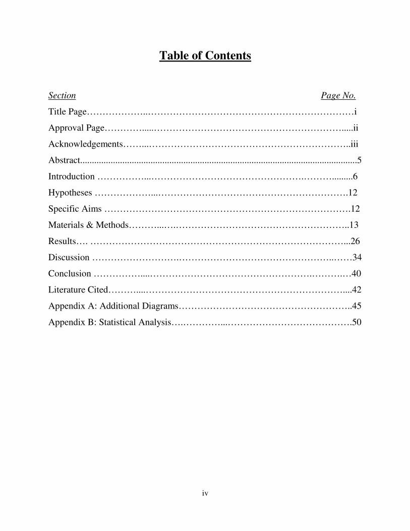
Table of Contents
Section Page No.
Title Page………………..…………………………………………………………i
Approval Page………….....…………………………………………………….....ii
Acknowledgements……...………………………………………………………..iii
Abstract......................................................................................................................5
Introduction ……………...………………………………………….……….........6
Hypotheses ………………...…………………………………………………….12
Specific Aims …………………………………………………………………….12
Materials & Methods………...….………………………………………………..13
Results…. ………………………………………………………………………...26
Discussion ……………………….…………………………………………..……34
Conclusion ……………....…………………………………………….……….…40
Literature Cited………....………………………………………………………....42
Appendix A: Additional Diagrams………………………………………………..45
Appendix B: Statistical Analysis….…………...………………………………….50
5
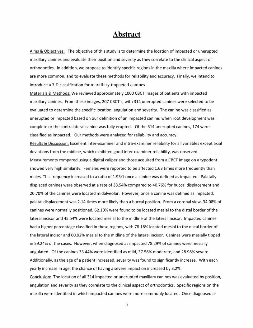
Abstract
Aims & Objectives: The objective of this study is to determine the location of impacted or unerupted
maxillary canines and evaluate their position and severity as they correlate to the clinical aspect of
orthodontics. In addition, we propose to identify specific regions in the maxilla where impacted canines
are more common, and to evaluate these methods for reliability and accuracy. Finally, we intend to
introduce a 3-D classification for maxillary impacted canines.
Materials & Methods: We reviewed approximately 1000 CBCT images of patients with impacted
maxillary canines. From these images, 207 CBCT’s, with 314 unerupted canines were selected to be
evaluated to determine the specific location, angulation and severity. The canine was classified as
unerupted or impacted based on our definition of an impacted canine: when root development was
complete or the contralateral canine was fully erupted. Of the 314 unerupted canines, 174 were
classified as impacted. Our methods were analyzed for reliability and accuracy.
Results & Discussion: Excellent inter-examiner and intra-examiner reliability for all variables except axial
deviations from the midline, which exhibited good inter-examiner reliability, was observed.
Measurements compared using a digital caliper and those acquired from a CBCT image on a typodont
showed very high similarity. Females were reported to be affected 1.63 times more frequently than
males. This frequency increased to a ratio of 1.93:1 once a canine was defined as impacted. Palatally
displaced canines were observed at a rate of 38.54% compared to 40.76% for buccal displacement and
20.70% of the canines were located midalveolar. However, once a canine was defined as impacted,
palatal displacement was 2.14 times more likely than a buccal position. From a coronal view, 34.08% of
canines were normally positioned, 62.10% were found to be located mesial to the distal border of the
lateral incisor and 45.54% were located mesial to the midline of the lateral incisor. Impacted canines
had a higher percentage classified in these regions, with 78.16% located mesial to the distal border of
the lateral incisor and 60.92% mesial to the midline of the lateral incisor. Canines were mesially tipped
in 59.24% of the cases. However, when diagnosed as impacted 78.29% of canines were mesially
angulated. Of the canines 33.44% were identified as mild, 37.58% moderate, and 28.98% severe.
Additionally, as the age of a patient increased, severity was found to significantly increase. With each
yearly increase in age, the chance of having a severe impaction increased by 3.2%.
Conclusion: The location of all 314 impacted or unerupted maxillary canines was evaluated by position,
angulation and severity as they correlate to the clinical aspect of orthodontics. Specific regions on the
maxilla were identified in which impacted canines were more commonly located. Once diagnosed as
6
impacted, females were observed to be affected 1.93 times more frequently than males. Impacted
canines were palatally positioned 2.14 times more commonly than buccal displacement. From a frontal
perspective, impacted canines were located mesial to the distal border of the lateral incisor in 78.16% of
cases and 60.92% were located mesial to the midline of the lateral incisor. As the age of a patient
increased, the chance of having a severe impaction increased by 3.2%. The methods outlined were
found to be reliable and accurate. In addition, a classification for impacted maxillary canines examined
by CBCT imaging was introduced.
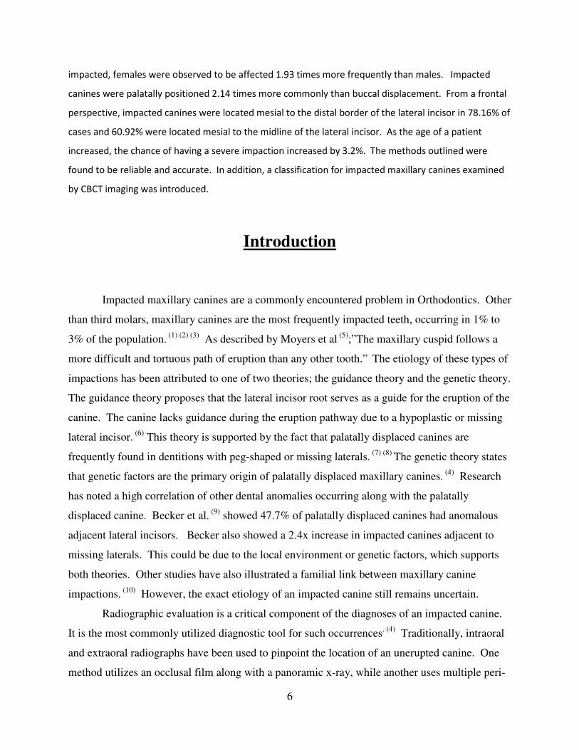
Introduction
Impacted maxillary canines are a commonly encountered problem in Orthodontics. Other
than third molars, maxillary canines are the most frequently impacted teeth, occurring in 1% to
3% of the population. (1) (2) (3)
As described by Moyers et al (5)
;”The maxillary cuspid follows a
more difficult and tortuous path of eruption than any other tooth.” The etiology of these types of
impactions has been attributed to one of two theories; the guidance theory and the genetic theory.
The guidance theory proposes that the lateral incisor root serves as a guide for the eruption of the
canine. The canine lacks guidance during the eruption pathway due to a hypoplastic or missing
lateral incisor. (6)
This theory is supported by the fact that palatally displaced canines are
frequently found in dentitions with peg-shaped or missing laterals. (7) (8)
The genetic theory states
that genetic factors are the primary origin of palatally displaced maxillary canines. (4)
Research
has noted a high correlation of other dental anomalies occurring along with the palatally
displaced canine. Becker et al. (9)
showed 47.7% of palatally displaced canines had anomalous
adjacent lateral incisors. Becker also showed a 2.4x increase in impacted canines adjacent to
missing laterals. This could be due to the local environment or genetic factors, which supports
both theories. Other studies have also illustrated a familial link between maxillary canine
impactions. (10)
However, the exact etiology of an impacted canine still remains uncertain.
Radiographic evaluation is a critical component of the diagnoses of an impacted canine.
It is the most commonly utilized diagnostic tool for such occurrences. (4)
Traditionally, intraoral
and extraoral radiographs have been used to pinpoint the location of an unerupted canine. One
method utilizes an occlusal film along with a panoramic x-ray, while another uses multiple peri-

7
apical images to locate the impaction. (1) (4)
Although these tools have aided us in the past, new
technology has been shown to be more accurate in determining the location of impactions as well
as the extent of resorption caused by the condition. Two-dimensional imaging has many well
documented limitations, including magnification, geometric distortion, superimpositions, and
elongation and foreshortening of objects. (20) (21)
In contrast, 3-D imaging has come to the
forefront in the diagnosis and treatment planning of “the anatomical truth.” (22)
Numerous studies
have exemplified the diagnostic advantage of 3-D imaging over traditional methods of 2-D
imaging. Boticelli et al. (21)
, showed significant differences between 2-D and 3-D imaging when
determining the location of an unerupted canine. This was attributed to distortion, magnification,
and the superimposition of anatomic structures that commonly occurs in the two-dimensional
images. Wreidt et al. (23)
showed that with panoramic x-rays, resorptions were overlooked in
20% of the patients evaluated, and the canine was located properly in only 64% of patients. In a
study by Alqerban et al. (24)
, two-dimensional and three-dimensional images were taken on a
cadaver skull with an impacted canine. Root resorption was detected 90-91% of the time
compared with 70% when using CBCT (Cone beam computed tomography) vs. panoramic
imaging. (24)
Ericson and Kurol in 1987 (25)
demonstrated that 1/3 of the resorbed teeth in their
study had a normal appearance on the peri-apical film. They attributed this to the fact that buccal
and lingual resorptions occur in 50% of the cases, and a midroot lesion is common. (18)
(25)
Resorption occurring in these regions may be undetectable using two-dimensional radiographs.
The above mentioned studies substantiate the advantages of 3-D imaging in regards to impacted
canines.
There are several clinical signs that indicate the possibility of a canine impaction.
Delayed eruption of the canine, prolonged retention of the deciduous canine, absence of a normal
labial bulge, presence of a palatal bulge, and delayed eruption, tipping, or migration of the lateral
incisor may be clinical findings that signify impaction. (1)
The presence of an impacted canine
may cause no harmful effects, however numerous consequences have been associated with this
anomaly. If left untreated, the migration of adjacent teeth, loss of arch length, and most
significantly, resorption of neighboring teeth may occur. (1) (4)
Resorption of adjacent teeth has
been observed 40.5% to 48% of the time, with even 77.8% being reported in some studies. (11) (12)
(13) Identifying the precise location of an impacted maxillary canine can be an essential part of
both diagnosis and treatment planning. According to Bedoya et al. (4)
, “Assessing the position of
8
the impacted canine is key to determining the feasibility of and proper access for a surgical
procedure, as well as the best direction for application of orthodontics forces.” Without proper
diagnosis, the direction of forces as well as the method of surgical exposure may be incorrectly
determined. Various diagnostic aids can analyze the numerous factors that may lead to a canine
becoming impacted. Radiographically, position and angulation have been shown to accurately
predict the likelihood of a canine becoming impacted. Studies have demonstrated that in 78% to
82% of canines destined to be impacted, the cusp tip crossed the distal aspect of the lateral
incisor root. (14) (15)
Sajnani and King (16)
illustrated the importance of angulation as a tool for
predicting whether a cuspid is destined to be impacted. Their findings show that after age 9, the
horizontal angulation increases 20° to 40° in relation to the midline compared to a normally
erupting canine.
Locating an impacted canine may also aid in distinguishing the possibility of causing
damage to adjacent structures. Resorption of the maxillary incisors has been shown to occur in
48% of cases with ectopic erupting maxillary canines. As the canine cusp tip is positioned more
mesially, a higher rate of resorption was observed. (11)(18)(41)
A study using CT (Computed
tomography) imaging demonstrated that resorption is mainly caused by contact and
physiological pressure from the ectopic canine. (12)
Resorption occurred 94.3% of the time when
the impacted canine was in close contact with the incisors. (11)
Angulation may also be a factor in
determining whether an ectopic canine will cause resorption to adjacent teeth. The risk of
resorption increases by 50% when the inclination relative to the midline exceeds 25° from the
frontal view. (18)
In addition, they found that impacted canines that caused resorption had an
increase in horizontal angulation of 18.1° from an occlusal perspective compared to normally
erupting canines. The identification of root resorption may lead to modifications in treatment
planning, such as extracting a resorbed lateral incisor over a premolar in an extraction case. (19)
The precise location of an impacted canine has a direct effect on the management and
treatment of the abnormality. In many cases, it can be beneficial to extract the deciduous canine
as an interceptive treatment allowing the impacted canine to erupt. Early extraction of a
deciduous canine when the succeeding tooth is impacted resulted in normalization of eruption in
78% of cases. (17)
However, this result was significantly different when the canine was positioned
mesial to the midline of the permanent lateral incisor. In fact, a normal eruption pattern was seen
in 91% of the cases when the canine was distal to this midline, compared to 64% when it was
9
positioned mesial. (17)
Angulation also plays a role in extracting deciduous canines to increase the
chance of normal eruption of the impacted canine. As the horizontal angulation of the canine
increases compared to the midline, the probability of successful eruption decreases. (26)
When the
angulation exceeds 31 degrees relative to the midline, the chance of normal eruption after
extraction decreased significantly. (26)
In the absence of prevention, surgical and orthodontic treatment should be considered in
order to bring the ectopic tooth into occlusion. (4) As stated by Bishara (1)
, “The diagnosis and
treatment of this problem usually requires the expertise and cooperation of the general
practitioner, the pediatric dentist, the oral surgeon, and the periodontist, as well as the
orthodontist.” From a surgical perspective, localization of the impacted canine aids in
establishing the method utilized to uncover the tooth. If the inappropriate surgical technique is
selected by the surgeon, the esthetic result may be unpredictable. (27)
It may also lead to a more
difficult and time consuming task for the orthodontist in aligning the impacted tooth within the
maxillary arch. (27)
The surgical method chosen depends on whether the canine is located in a
labial or palatal position. It has also been proven that periodontal conditions of the impacted
canine and adjacent teeth after surgical and orthodontic treatment are dependent on the initial
vertical and horizontal position of the canine. (28)
With this in mind, Kokich established four
criteria for determining the method of surgically exposing an impacted canine. These include:
the labiolingual position of the impacted crown; the vertical position of the tooth relative to the
mucogingival junction; the amount of gingiva surrounding the impacted cuspid; and the
mesiodistal position of the canine crown. (27)
Based on these factors, an appropriate surgical
technique can be chosen in order to optimize the esthetic outcome and reduce the difficulty of
orthodontic treatment.
From an orthodontic perspective, the location of the canine will influence the direction
and type of force utilized to align the canine. As previously stated, it may also modify the
treatment plan and extraction pattern. One investigation demonstrated that when evaluating case
difficulty and the direction of treatment, a significant difference was noted between 2-D and 3-D
imaging. (21)
In 29.5% of cases reviewed, a CBCT led the examiner to recommend a more active
approach focused on expansion and space maintenance. (21)
A separate study showed that in 18%
of their patients, treatment plans varied dependent upon whether they were diagnostically viewed
with a CBCT or a Panorex. (23)
Evidence has also indicated that orthodontic treatment time
10
increased 3.4 months when a patient has a unilateral impaction, and 9.9 months with bilateral
impactions. (29)
Treatment time was found to be dependent upon the distance the impacted
canine was from the occlusal plane. If it was less than 14mm, treatment time was on average
23.8 months, compared to 31.1 months if it was more than 14mm from the occlusal plane.
Another study exhibited that treatment time was 9.8 months longer when the impaction was
located mesial to the lateral incisor. (30)
The same study also observed an increase in treatment
time if the cusp tip of the impacted tooth was located further from the occlusal plane. Lastly,
they detected a significant association between the amount of angulation and duration of
treatment. As the ectopically erupting tooth was more horizontally angulated, the time in
treatment increased. (30)
One of the many risks we are exposed to throughout life is the exposure to radiation from
everyday activities. Medical and dental devices increase the amount of radiation we are exposed
to. As practitioners it is our responsibility to determine if the risk of radiation exposure is a
medically necessary diagnostic tool to benefit the patient during treatment. The average
individual is exposed to 2400 µSv each year from normal background radiation. (32)
That breaks
down to 6.58 µSv per day. Panoramic x-rays and lateral cephalograms are commonly used
diagnostic tools in the orthodontic practice. Each has been reported to have an effective dose of
anywhere from 2.7 to 23 µSv and 10 µSv respectively. (33) (34) (35) (36)
. Intra-oral radiographs have
an effective dose of 8.3 µSv according to the European Commission in 2004 (32)
and a full mouth
series has a radiation exposure of 13-100 µSv. (33)
Currently available CBCT units have been
reported to have radiation exposure in the range of 30 to 206 µSv for a full craniofacial scan.
(33)(36) (37) Even lower effective doses have been observed when using a smaller dentoalveolar
field of view. (36)
Although the radiation from a CBCT is slightly higher in most instances, the
accuracy and resolution of the image is more reliable. One study showed that measurement error
was significantly lower using CBCT images as compared to a cephalogram when evaluating 76
measurements against a gold standard. (20)
In fact, the 2-D image in one measurement showed an
average error of 13.61mm, while the 3-D image had less than 1mm of error on average. The
amount of error has been shown to be reduced when viewing images in the multiplanar (MPR)
view, as compared to volume rendered (VR) and shaded surface displace (SSD) view modes. (19)
The error seen in the VR and SSD modes may be attributed to surface contours being estimated
in these perspectives. (19)
Korbmacher et al. (38)
conducted a study that demonstrated CBCT
11
provided more information regarding cleft lip and palate, impacted and retained teeth, root
resorption, and third molars.
Bishara (1)
stated, “The proper localization of the impacted tooth plays a crucial role in
determining the feasibility of, as well as the proper access for, the surgical approach, and the
proper direction for the application of orthodontic forces.” Evidence has clearly shown the
diagnostic value of a 3-D image when evaluating an impacted canine. Bjerklin and Ericson (39)
showed that after viewing a CT image of a patient with an impacted canine, examiners changed
their treatment plan almost 44% of the time based on the findings in the image. When
comparing treatment planning using 2-D vs. 3-D imaging, Haney et al. (40)
demonstrated that not
only were Orthodontists more confident when utilizing a 3-D image, they modified their
treatment plans 27% of the time. While there may be additional radiation exposure for patients,
the benefits of CBCT images for diagnosis and adequate treatment planning of impacted
maxillary canines has been well documented.
Rationale:
Breakthroughs in technology lead us to new ways to evaluate information. Previously,
our diagnostic methods for locating impacted canines in 3 planes of space were limited. With
the use of multiple two-dimensional radiographs, one could determine if a canine was located
palatally or buccally to an adjacent tooth. (4) (31)
However, the distance from that tooth was
impossible to ascertain. In one study, they were only able to project the lateral incisor image
away from that of the canine 37% of the time. (3)
Accuracy was also a concern as the images
could be distorted by magnifications and many structures were superimposed on one another. (21)
With advancements in radiographic imaging, our diagnostic accuracy has greatly improved.
However, protocols and standards on how to properly utilize this improved technology to
determine the location and severity of an unerupted canine must be established. These
innovations allow us to improve our diagnostic capacity as well as how we implement our
treatment.
By viewing an impaction in three-dimensions we can locate and assess an impacted
canine with great accuracy. Surgical planning of the exposure of the impacted canine as well as
the proposed orthodontic forces needed to erupt the impacted canine into alignment with the
dentition becomes more precise. The surgeon and orthodontist, as a team, now have more
12
significant information prior to active treatment. This can only lead to a better result and less
potential for damage to adjacent teeth as well as the impacted tooth. However, once the three-
dimensional location of a canine is established on a CBCT image, there does not exist adequate
language to convey the entirety of its position and/or the severity of the impaction.
The objective of this study is to determine the location of impacted or unerupted
maxillary canines and evaluate their position and severity as they correlate to the clinical aspect
of orthodontics. This study hopes to establish a method to reliably locate an impacted canine
from a sagittal, axial and a coronal view. The level and severity of the impaction will be
measured by location and angulation related to other teeth and adjacent structures. Then with the
impaction correlated to all three planes of space, a severity index can be utilized to help the
clinician better determine the degree of impaction. The method described will be evaluated for
reliability and accuracy.
Hypothesis
1. The maxillary canine tooth is generally impacted only at specific sites in the maxilla and
CBCT imaging can accurately locate impacted canines in all 3 planes of space.
2. The method outlined to locate an impacted maxillary canine is reliable.
Specific Aims
1. To identify the specific regions which have a greater likelihood of canine impaction
2. To create an index along with a nomenclature to assist in classifying the location and
severity of impacted maxillary canines.
3. Evaluate the methods of localization for reliability and accuracy.
.

13
Materials & Methods
Previous studies have shown ways to identify or assess the severity of maxillary impacted
canines. Although these methods are effective, we propose to localize the impacted canines in a
fashion that provides more information that is valuable to the clinician. Our methods will allow
one to assess the severity of the impaction in terms of surgical exposure and biomechanical
maneuvering of the tooth into the arch.
We reviewed approximately 1000 CBCT images of patients with impacted maxillary
canines. From these images, 207 CBCT’s, with 314 unerupted canines were selected to be
analyzed. The images were collected from a diagnostic imaging center (Courtesy of Dr. David
Hatcher and Dr. Francisco Eraso) and one office that specializes in Periodontics (Dr. Scott Ross),
both located in the United States. No information regarding treatment or the reason the image
was taken was known. The only information collected was the patient age and sex.
Individual images were classified as bilateral or unilateral and each impaction was treated
as its own entity. The impaction was then characterized as unerupted or impacted. We defined
an unerupted canine as impacted when its root development was complete or the contralateral
canine was fully erupted. The presence of a primary canine on the side of impaction was
recorded, as well as whether the patient was in appliances or a palatal expansion device. If any
additional pathology, i.e. resorption, peg laterals, supernumerary teeth was observed it was
documented as well. Each CBCT was then evaluated in the 3 planes of space as described
below. Slice thickness was increased to 5mm to allow greater visibility of adjacent structures.
14
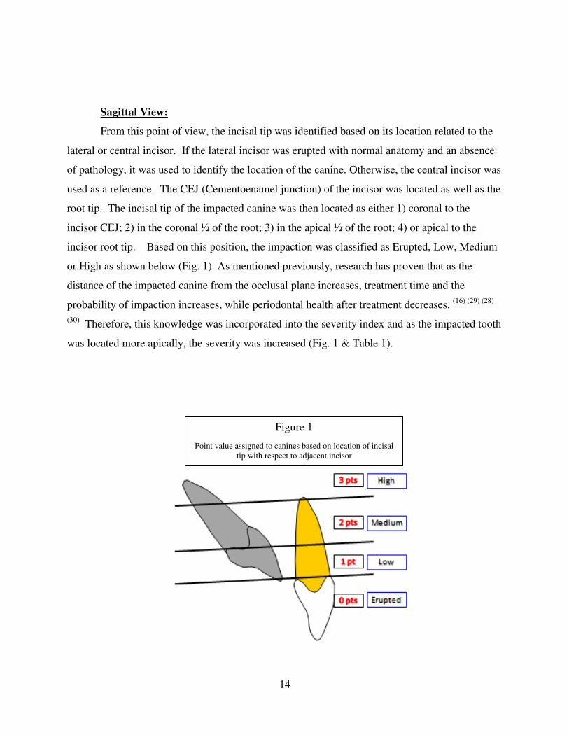
Sagittal View:
From this point of view, the incisal tip was identified based on its location related to the
lateral or central incisor. If the lateral incisor was erupted with normal anatomy and an absence
of pathology, it was used to identify the location of the canine. Otherwise, the central incisor was
used as a reference. The CEJ (Cementoenamel junction) of the incisor was located as well as the
root tip. The incisal tip of the impacted canine was then located as either 1) coronal to the
incisor CEJ; 2) in the coronal ½ of the root; 3) in the apical ½ of the root; 4) or apical to the
incisor root tip. Based on this position, the impaction was classified as Erupted, Low, Medium
or High as shown below (Fig. 1). As mentioned previously, research has proven that as the
distance of the impacted canine from the occlusal plane increases, treatment time and the
probability of impaction increases, while periodontal health after treatment decreases. (16) (29) (28)
(30) Therefore, this knowledge was incorporated into the severity index and as the impacted tooth
was located more apically, the severity was increased (Fig. 1 & Table 1).
Figure 1
Point value assigned to canines based on location of incisal
tip with respect to adjacent incisor

15
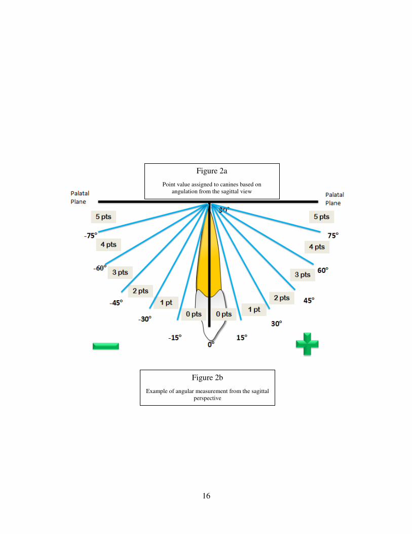
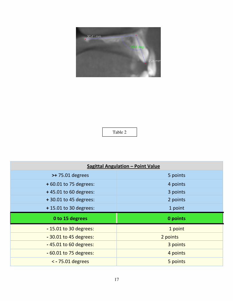
The angle created by the long axis of the tooth and a perpendicular line from the palatal
plane was measured (Fig, 2) and labeled as torque. Based on this angle, the below point system
was allotted to the impacted tooth (Table 2). For every 15 degrees away from the perpendicular
plane, 1 point was assigned. The tooth angulation was also classified as positive or negative in
relation to the perpendicular line.
Sagittal Location – Value & Classification
Coronal to the CEJ: 0 points Erupted
In the Coronal ½ of
the root: 1 point Low
In the Apical ½ of the
root: 2 points Medium
Apical to the root tip: 4 points High
Table 1
16
Figure 2a
Point value assigned to canines based on
angulation from the sagittal view
Figure 2b
Example of angular measurement from the sagittal
perspective
17
Sagittal Angulation – Point Value
>+ 75.01 degrees 5 points
+ 60.01 to 75 degrees: 4 points
+ 45.01 to 60 degrees: 3 points
+ 30.01 to 45 degrees: 2 points
+ 15.01 to 30 degrees: 1 point
0 to 15 degrees 0 points
- 15.01 to 30 degrees: 1 point
- 30.01 to 45 degrees: 2 points
- 45.01 to 60 degrees: 3 points
- 60.01 to 75 degrees: 4 points
< - 75.01 degrees 5 points
Table 2

18
Axial View:
From this view, the cusp tip was located and then the buccal and palatal alveolar borders
were identified. A line bisecting the alveolus was constructed and the distance from the closest
alveolar border, palatal or buccal, to the incisal tip was measured. (Fig. 3) This measurement
allowed us to calculate the distance from the midline of the alveolus. The cusp tip was
designated as buccal, palatal or mid-alveolar based on its distance from the center of the alveolar
bone. The mid-alveolus is defined as 1.5mm buccal or palatal to the midpoint between the
alveolar borders. Severity increased in 1.5mm increments as the distance increased from the
buccal and palatal cortical borders (Fig. 4). Since angulation in a buccal/palatal aspect was
established in the sagittal view, it was unnecessary to do so in this view again. The following
scale was assigned based on the findings in relation to the alveolar bisecting line in either a
buccal or palatal direction (Table 3):
Figure 3
Example of the distance of a canine from the midline of
the alveolus viewed from the axial perspective
Figure 4
Increments in 1.5mm based on distance from the
midline of the alveolus

19
Table 3
Axial Location
20
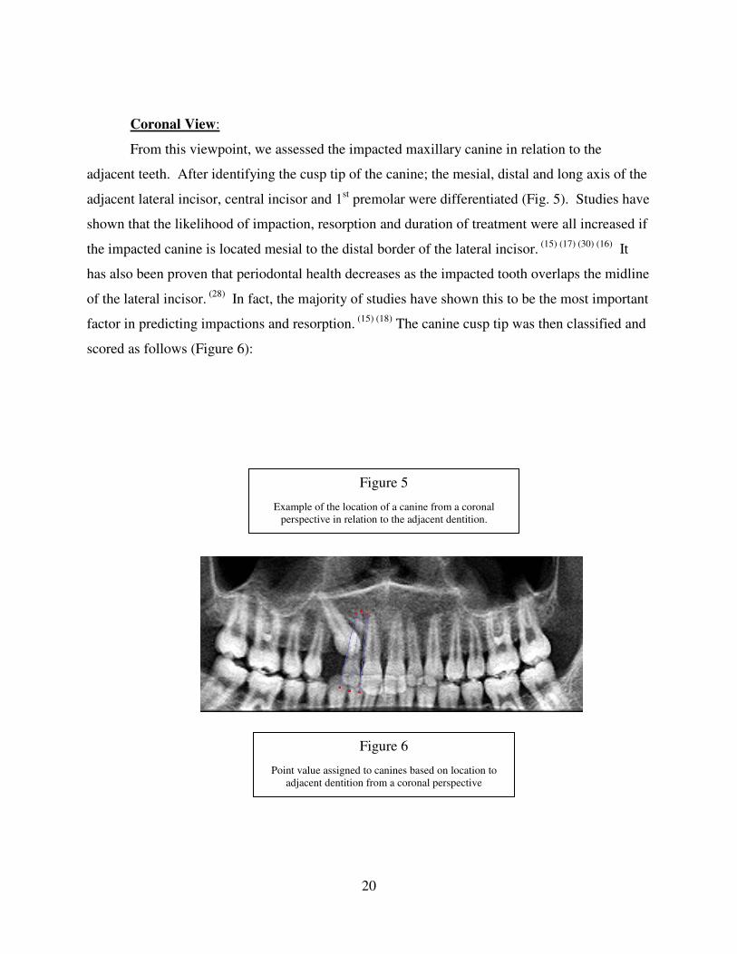
Coronal View:
From this viewpoint, we assessed the impacted maxillary canine in relation to the
adjacent teeth. After identifying the cusp tip of the canine; the mesial, distal and long axis of the
adjacent lateral incisor, central incisor and 1st premolar were differentiated (Fig. 5). Studies have
shown that the likelihood of impaction, resorption and duration of treatment were all increased if
the impacted canine is located mesial to the distal border of the lateral incisor. (15) (17) (30) (16)
It
has also been proven that periodontal health decreases as the impacted tooth overlaps the midline
of the lateral incisor. (28)
In fact, the majority of studies have shown this to be the most important
factor in predicting impactions and resorption. (15) (18)
The canine cusp tip was then classified and
scored as follows (Figure 6):
Figure 5
Example of the location of a canine from a coronal
perspective in relation to the adjacent dentition.
Figure 6
Point value assigned to canines based on location to
adjacent dentition from a coronal perspective
21
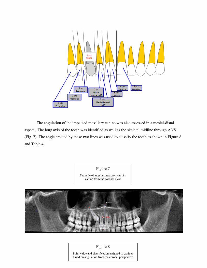
The angulation of the impacted maxillary canine was also assessed in a mesial-distal
aspect. The long axis of the tooth was identified as well as the skeletal midline through ANS
(Fig. 7). The angle created by these two lines was used to classify the tooth as shown in Figure 8
and Table 4:
Figure 7
Example of angular measurement of a
canine from the coronal view
Figure 8
Point value and classification assigned to canines
based on angulation from the coronal perspective

22
Coronal Location – Value & Classification
0 to - 15 degrees: 0 points Vertical
0 to 15 degrees: 0 points
15.01 to 30 degrees: 1 point
30.01 to 45 degrees: 2 points Mesial
45.01 to 60 degrees: 3 points
60.01o 75 degrees: 4 points
>75.01 degrees: 5 points
-15.01 to -30 degrees: 1 point
Mesial Distal
Table 4

23
-30.01 to -45 degrees: 2 points Distal
-45.01 to -60 degrees: 3 points
-60.01 to -75 degrees: 4 points
< -75.01 degrees: 5 points
Classification
Once the images were evaluated as noted above, the scores were combined to measure
severity. The classification index is designed to allow the clinician to be able to visualize the
impacted maxillary canine. The impaction was identified as follows based on the above
guidelines.
1. From an axial view the tooth was designated as:
Buccal
Palatal
Midalveolar
A number followed this nomenclature to designate how many increments of 1.5mm the
canine tip was located from the middle of the alveolus.
2. From the sagittal view, the tooth was classified as:
Erupted: Coronal to the CEJ of the adjacent incisor
Low: Coronal to the midpoint of the root of the adjacent incisor
Medium: In the apical ½ of the root of the adjacent incisor
High: Apical to the root tip of the adjacent incisor
It was also given a positive (+) or negative (-) classification to illustrate the
angulation. A number followed this sign to depict the amount of torque the canine
displayed.
3. From a coronal point of view the impaction was identified as:
Normally Erupting: Distal to the lateral incisor and mesial to the premolar
D-Lateral: Mesial to the distal border of the lateral incisor, but distal to its midpoint
M-Lateral: Mesial to the midpoint of lateral, but distal to the central incisor
Central: Mesial to the distal border of central incisor
24
Midline: Crossing the maxillary dental midline
Premolar: Distal to the mesial border of the premolar
4. Also from the coronal view, the canine was classified as:
Vertical: Angulation was between -15° to + 30° from the skeletal midline through
ANS
Mesial: Angle was greater than 30° in a mesial direction
Distal: Angle was less than -15° in a distal direction
5. Finally, using the point system outlined above, a severity was determined.
0 to 5 points: Mild
6 to 10 points: Moderate
More than 10 points: Severe
An impacted canine was then classified as:
Buccal 2; High, +3 torque; M-Lat; Mesially tipped; Moderate impaction.
A simplified version of this index was also created to portray a basic classification:
Buccal; High; M-Lat; mesially tipped; Moderate impaction.
Following are the inclusion and exclusion criteria for patient selection:
Inclusion criteria:
1. CBCT images of unilateral or bilateral impacted or unerupted maxillary canines.
Exclusion criteria:
1. Missing Central incisor
2. Impacted Central incisor
Forty canine images were reviewed 30 days apart for intrarater reliability. The same
images were evaluated by a separate examiner (Dr. Vishwanath) for interater reliability. The
examiner underwent minimal training (30 min) regarding measurement of the CBCT images.
A typodont setup with an impacted canine was also utilized to assess our methods.
Measurements were made on the typodont with a digital caliper. A CBCT image of the typodont
25
was also evaluated in the same fashion as described above. These results were compared with
each other to determine accuracy.
Statistical Analysis:
Simple descriptive statistics were used to summarize the data. Intra- and inter-examiner
reliability was examined by using Cohen-Kappa values for categorical variables and Cronbach
Alpha (intra-class correlation coefficients) for continuous variables. Kappa values were
computed for 10 variables and intra-class correlation coefficients for 3 variables. Outcomes
were compared between right and left sides and also by gender. For categorical variables, Chi-
square tests were used while Mann-Whitney U test was used for continuous variables. One of
the primary dependent variables was severity of case (mild, moderate, and severe). The effects of
age (each 1 year increase in age), gender, axial class, and location on severity were examined by
multivariable logistic regression models. Since severity of case was a polynomial variable, two
regression models were used to examine the outcome. In the first regression model the odds of
having a severe case compared to mild or moderate was examined. In the second regression
model, the odds of having a mild case compared to a moderate or severe case was examined.
The maximum likelihood methods were used to fit the multivariable logistic regression model.
Model fitness was examined by Hosmer and Lemeshow Goodness of fit test statistic. The effects
of age, gender, use of orthodontic appliances, use of maxillary expansion appliance, and
presence of primary canine on total points was examined by a multivariable linear regression
model. Ordinary least squares approach was used to fit the regression model. All statistical tests
were two-sided and a p-value of <0.05 was deemed to be statistically significant. All statistical
analyses were conducted using SPSS Version 22.0 software (IBM Inc, Research Triangle Park,
NC).

26
Results
Our study evaluated 207 patients with 314 unerupted maxillary canines. Of these, 140
were defined as unerupted, while 174 were classified as impacted. Of the patients, 57.49% (119)
were female, 35.27% (73) male, and 7.25% (15) were unreported, giving females a 1.63 greater
chance than males of having an unerupted/impacted canine (Table 5, Chart 1). Bilateral
impactions were present in 51.69% of patients, while right and left presentation appeared to be
equally distributed at 51.91% and 48.09%, respectively. Males and females demonstrated
similar patterns of bilateral impactions; 50.68% of males and 49.37% of females exhibited the
trait. Males were found to have a greater likelihood of having a moderate impaction, 43.64% to
36.16% of females; however this finding was not significant. Females were more likely to have
a mild impaction, 36.16% vs. 23.35% in males; this was also not statistically significant (Chart
2).
Table 5 Chart 1

Overall, palatal and buccal impactions seemed to be observed at the same prevalence,
38.54% and 40.76%, respectively
6, Chart 3). There was no difference seen between males and females
However, it was observed that as age increased, palatal impactions became more common.
Patients above the age of 13 had an occurrence of 63.57% of palatally displaced canine, with
23.26% buccally displaced and 13.18% midalveolar. When
21.08% were palatal, 52.97% were buccal and 25.95% were midalveolar
0.00%
10.00%
20.00%
30.00%
40.00%
50.00%
60.00%
Male Female
Prevalence of Unerupted Canines
0.00%
10.00%
20.00%
30.00%
40.00%
50.00%
27
buccal impactions seemed to be observed at the same prevalence,
respectively. 20.70% of the canines were classified as midalveolar
. There was no difference seen between males and females and this measurement
as age increased, palatal impactions became more common.
Patients above the age of 13 had an occurrence of 63.57% of palatally displaced canine, with
23.26% buccally displaced and 13.18% midalveolar. When compared to patients 13 or younger,
21.08% were palatal, 52.97% were buccal and 25.95% were midalveolar (Chart 4)
Unlisted
Prevalence of Unerupted Canines
MildModerate
Severe
Prevalence of Severity & Gender
Male
Female
Prevalence of Unerupted Canines
Male
Female
Unlisted
Bilateral
Unilateral
Right
Left
Chart 2
Chart 3
buccal impactions seemed to be observed at the same prevalence,
. 20.70% of the canines were classified as midalveolar (Table
measurement.
as age increased, palatal impactions became more common.
Patients above the age of 13 had an occurrence of 63.57% of palatally displaced canine, with
to patients 13 or younger,
(Chart 4).
Male
Female
Prevalence of Unerupted Canines
Patients Percentage
73 35.27%
119 57.49%
15 7.25%
107 51.69%
100 49.31%
56 56.00%
44 44.00%
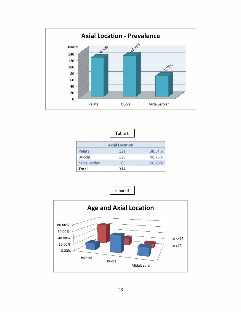
Palatal
Buccal
Midalve
Total
0
20
40
60
80
100
120
140
Palatal
Canines
Axial Location
0.00%
20.00%
40.00%
60.00%
80.00%
Palatal
Age and Axial Location
28
Axial Location
Palatal 121 38.54%
128 40.76%
Midalveolar 65 20.70%
314
Palatal Buccal Midalverolar
Axial Location - Prevalence
PalatalBuccal
Midalveolar
Age and Axial Location
<=13
>13
Table 6
Chart 4
<=13
>13
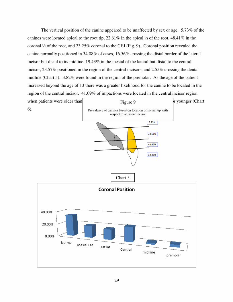
The vertical position of the canine appeared to be unaffected by sex or age. 5.73% of the
canines were located apical to the root tip, 22.61% in the apical ½ of the root, 48.41% in the
coronal ½ of the root, and 23.25% coronal to the CEJ
canine normally positioned in 34.08% of cases, 16.56% crossing the distal
incisor but distal to its midline, 19.43% in the mesial of the lateral
incisor, 23.57% positioned in the region of
midline (Chart 5). 3.82% were found in the region of the premolar. As the age of the patient
increased beyond the age of 13 there was a greater likelihood for the canine to be located in the
region of the central incisor. 41.09% of impactions were located in the central incisor region
when patients were older than 13, compared to 11.35%
6).
0.00%
20.00%
40.00%
NormalMesial Lat
29
vertical position of the canine appeared to be unaffected by sex or age. 5.73% of the
canines were located apical to the root tip, 22.61% in the apical ½ of the root, 48.41% in the
coronal ½ of the root, and 23.25% coronal to the CEJ (Fig. 9). Coronal position revealed the
34.08% of cases, 16.56% crossing the distal border
, 19.43% in the mesial of the lateral but distal to the central
, 23.57% positioned in the region of the central incisors, and 2.55% crossing the
. 3.82% were found in the region of the premolar. As the age of the patient
13 there was a greater likelihood for the canine to be located in the
region of the central incisor. 41.09% of impactions were located in the central incisor region
older than 13, compared to 11.35% when patients were 13 or younger
Mesial LatDist lat
Centralmidlline
premolar
Coronal Position
Figure 9
Prevalence of canines based on location of incisal tip with
respect to adjacent incisor
Chart 5
vertical position of the canine appeared to be unaffected by sex or age. 5.73% of the
canines were located apical to the root tip, 22.61% in the apical ½ of the root, 48.41% in the
sition revealed the
border of the lateral
but distal to the central
the central incisors, and 2.55% crossing the dental
. 3.82% were found in the region of the premolar. As the age of the patient
13 there was a greater likelihood for the canine to be located in the
region of the central incisor. 41.09% of impactions were located in the central incisor region
when patients were 13 or younger (Chart
premolar
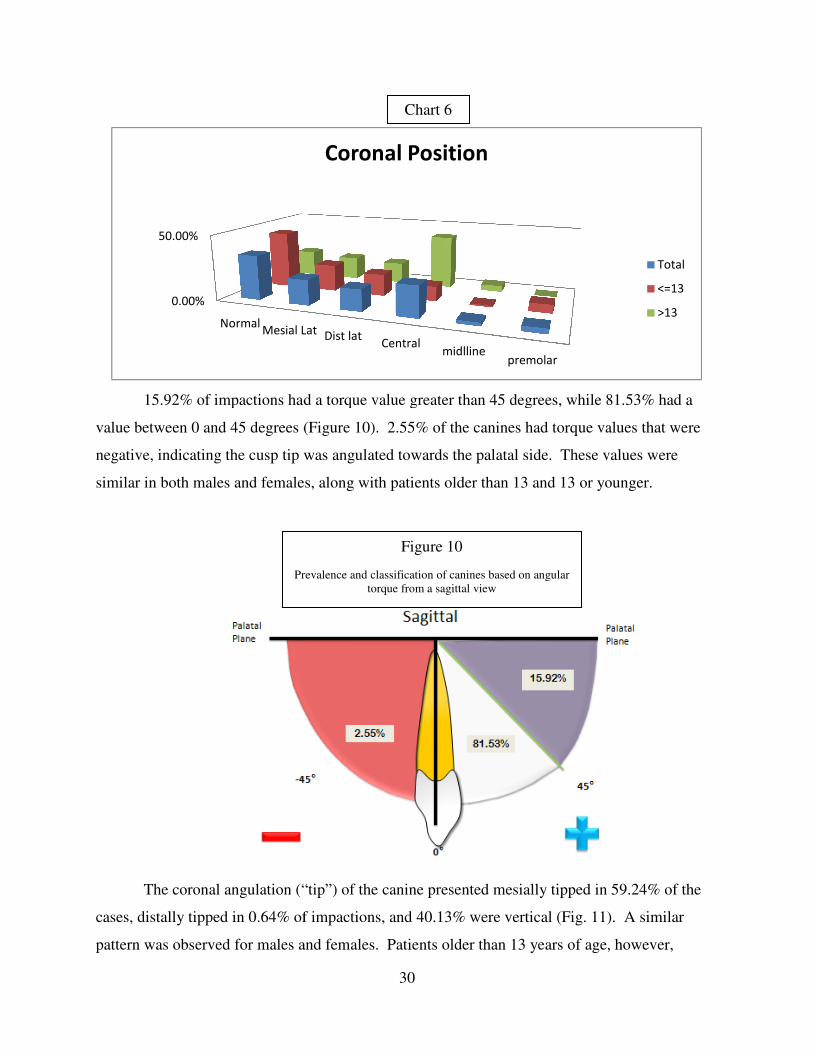
15.92% of impactions had a torque value greater than 45 degrees, while 81.53% had a
value between 0 and 45 degrees (Figure 10)
negative, indicating the cusp tip was angulated towards the palatal side. These values were
similar in both males and females, along with patients older than 13 and 13 or younger.
The coronal angulation (“tip”) of the canine presented mesially tipped in 59.24% of the
cases, distally tipped in 0.64% of impactions, and 40.13% were
pattern was observed for males and females. Patients older than 13 years of age, however,
0.00%
50.00%
NormalMesial Lat
Prevalence and classification of canines based on angular
30
15.92% of impactions had a torque value greater than 45 degrees, while 81.53% had a
(Figure 10). 2.55% of the canines had torque values that were
tip was angulated towards the palatal side. These values were
similar in both males and females, along with patients older than 13 and 13 or younger.
The coronal angulation (“tip”) of the canine presented mesially tipped in 59.24% of the
cases, distally tipped in 0.64% of impactions, and 40.13% were vertical (Fig. 11)
pattern was observed for males and females. Patients older than 13 years of age, however,
Mesial Lat Dist latCentral
midllinepremolar
Coronal Position
Chart 6
Figure 10
Prevalence and classification of canines based on angular
torque from a sagittal view
15.92% of impactions had a torque value greater than 45 degrees, while 81.53% had a
. 2.55% of the canines had torque values that were
tip was angulated towards the palatal side. These values were
similar in both males and females, along with patients older than 13 and 13 or younger.
The coronal angulation (“tip”) of the canine presented mesially tipped in 59.24% of the
11). A similar
pattern was observed for males and females. Patients older than 13 years of age, however,
Total
<=13
>13

31
showed a greater chance of being mesially angulated, with 78.29% mesially angulated, and
21.71% vertical (Chart 7). Patients 13 or younger were more likely to have a vertically tipped
canine, occurring in 52.97% of the cases, while 45.95% were mesially tipped.
Figure 11
Prevalence and classification of canines based
on angulation from a coronal view

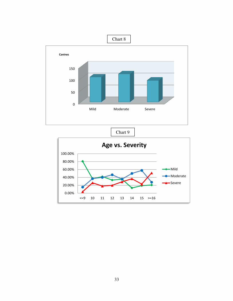
33.44% of the canines were classified as mild, 37.58%
(Chart 8, Table 7). As stated before the sex of the patient did not have a significant effect on t
severity of the impaction. It was observed that as the age of a patient
increased. With each yearly increase in age, the chance of having a severe impaction increased
by 3.2% (OR 1.032, P-value .041)
prevalence of mild impactions 43.24% compared
Severe impactions were observed in 41.86% of canines in the older age group compared to 20%
of those 13 or younger (Chart 10
is greater than that observed for bilateral impactions (21.03%). The buccal/palatal location also
affected the severity, as one would expect. If the canine was positioned buccally the impaction
was severe 4.947 times more than if it was positioned midalveolar (p
palatally, it was 3.767 times more likely to be severe than if located midalveolar (p
Mild
Moderate
Severe
Total
0.00%
10.00%
20.00%
30.00%
40.00%
50.00%
60.00%
70.00%
80.00%
<=13
Age vs. Sagittal Angulation
32
33.44% of the canines were classified as mild, 37.58% moderate and 28.98% severe
. As stated before the sex of the patient did not have a significant effect on t
It was observed that as the age of a patient increased, severity also
increased. With each yearly increase in age, the chance of having a severe impaction increased
value .041), (Chart 9). Patients 13 years or younger had a greater
prevalence of mild impactions 43.24% compared to 19.38% in patients that were older than 13.
Severe impactions were observed in 41.86% of canines in the older age group compared to 20%
10). 46% of unilateral impactions were classified as severe, which
at observed for bilateral impactions (21.03%). The buccal/palatal location also
affected the severity, as one would expect. If the canine was positioned buccally the impaction
was severe 4.947 times more than if it was positioned midalveolar (p-value .001). If positioned
palatally, it was 3.767 times more likely to be severe than if located midalveolar (p
Severity
105 33.44%
Moderate 118 37.58%
Severe 91 28.98%
314
<=13 >13
Age vs. Sagittal Angulation
>30°
-15°> & <30
<-15°
Table 7
Chart 7
and 28.98% severe
. As stated before the sex of the patient did not have a significant effect on the
increased, severity also
increased. With each yearly increase in age, the chance of having a severe impaction increased
. Patients 13 years or younger had a greater
to 19.38% in patients that were older than 13.
Severe impactions were observed in 41.86% of canines in the older age group compared to 20%
). 46% of unilateral impactions were classified as severe, which
at observed for bilateral impactions (21.03%). The buccal/palatal location also
affected the severity, as one would expect. If the canine was positioned buccally the impaction
1). If positioned
palatally, it was 3.767 times more likely to be severe than if located midalveolar (p-value .006).
> & <30°
0
50
100
150
Canines
0.00%
20.00%
40.00%
60.00%
80.00%
100.00%
<=9
33
Mild Moderate Severe
10 11 12 13 14 15 >=16
Age vs. Severity
Mild
Moderate
Severe
Chart 8
Chart 9
Mild
Moderate
Severe
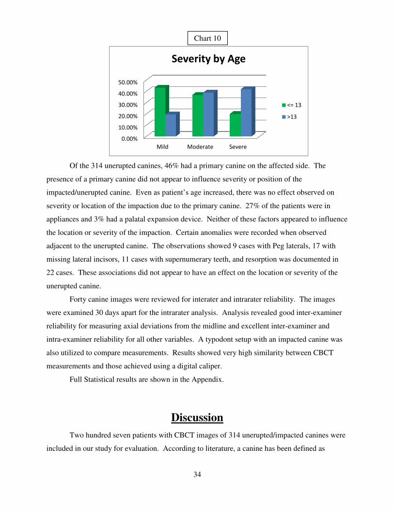
Of the 314 unerupted canines, 46% had a primary canine on the affected side.
presence of a primary canine did not appear to influence
impacted/unerupted canine. Even as
severity or location of the impaction due to the primary canine. 27% of the patients were in
appliances and 3% had a palatal expansion device. Neither of these factors appeared to influence
the location or severity of the impaction. Certain anomali
adjacent to the unerupted canine.
missing lateral incisors, 11 cases with supernumerary teeth, and resorption was documented in
22 cases. These associations did not
unerupted canine.
Forty canine images were reviewed for interater and intrarater reliability. The images
were examined 30 days apart for the intrarater analysis. Analysis revealed good inter
reliability for measuring axial deviations from the midline and excellent inter
intra-examiner reliability for all other variables. A typodont setup with an impacted canine was
also utilized to compare measurements. Results showed v
measurements and those achieved using a digital caliper.
Full Statistical results are shown in the Appendix.
Two hundred seven patients with CBCT images of 314
included in our study for evaluation.
0.00%
10.00%
20.00%
30.00%
40.00%
50.00%
34
Of the 314 unerupted canines, 46% had a primary canine on the affected side.
imary canine did not appear to influence severity or position of the
impacted/unerupted canine. Even as patient’s age increased, there was no effect observe
tion due to the primary canine. 27% of the patients were in
appliances and 3% had a palatal expansion device. Neither of these factors appeared to influence
the location or severity of the impaction. Certain anomalies were recorded when observed
adjacent to the unerupted canine. The observations showed 9 cases with Peg laterals,
, 11 cases with supernumerary teeth, and resorption was documented in
22 cases. These associations did not appear to have an effect on the location or severity of the
canine images were reviewed for interater and intrarater reliability. The images
were examined 30 days apart for the intrarater analysis. Analysis revealed good inter
reliability for measuring axial deviations from the midline and excellent inter-examiner and
examiner reliability for all other variables. A typodont setup with an impacted canine was
also utilized to compare measurements. Results showed very high similarity between CBCT
measurements and those achieved using a digital caliper.
Full Statistical results are shown in the Appendix.
Discussion
patients with CBCT images of 314 unerupted/impacted canines were
included in our study for evaluation. According to literature, a canine has been defined
Mild Moderate Severe
Severity by Age
<= 13
>13
Chart 10
Of the 314 unerupted canines, 46% had a primary canine on the affected side. The
severity or position of the
age increased, there was no effect observed on
tion due to the primary canine. 27% of the patients were in
appliances and 3% had a palatal expansion device. Neither of these factors appeared to influence
es were recorded when observed
Peg laterals, 17 with
, 11 cases with supernumerary teeth, and resorption was documented in
appear to have an effect on the location or severity of the
canine images were reviewed for interater and intrarater reliability. The images
were examined 30 days apart for the intrarater analysis. Analysis revealed good inter-examiner
examiner and
examiner reliability for all other variables. A typodont setup with an impacted canine was
ery high similarity between CBCT
unerupted/impacted canines were
defined as
<= 13
>13
35
impacted when the tooth was unerupted after complete root development and when the
contralateral canine was fully erupted.(42)
Another study considered canines impacted when their
roots were fully developed but the teeth were still covered with bone or mucosa.(43)
Since our
study included patients with bilateral presentation of impacted/unerupted canines, we defined an
unerupted canine as impacted when its root development was complete or the contralateral
canine was fully erupted. From this description we characterized 140 as unerupted and 174 were
classified as impacted.
Females are reported to be more commonly affected by canine impaction, occurring
approximately twice as frequently as males. (44)(1)(9)
Our study showed similar results, with
females 1.63 times more likely to be affected by an unerupted/impacted canine. When diagnosed
as impacted, that ratio increased to 1.93:1. Bilateral impactions have been shown to occur in
approximately 8% to 20% of patients with impacted canines. (11)(45)(1)
The results of our study
concluded 51.69% of patients had bilateral unerupted canines. This frequency is regarded as
high compared to some studies, however numerous others studies have shown similar rates of
bilateral impactions.(9)(13)(46)
This effect may be due to the population sample, as Asian
populations appear to have a lower frequency of bilateral impactions. (45(11)
Studies showing
more frequent bilateral impactions have Middle-Eastern populations or patients from the United
States, as in our study. (13)(46)(9)
It has been reported that palatal impactions are approximately 2 to 3 times more common
than labial displaced canines. (17)(46)(13)
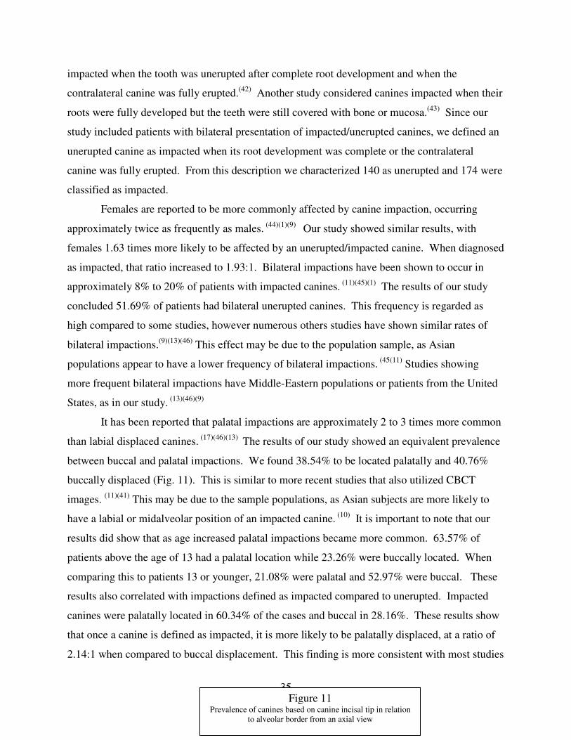
The results of our study showed an equivalent prevalence
between buccal and palatal impactions. We found 38.54% to be located palatally and 40.76%
buccally displaced (Fig. 11). This is similar to more recent studies that also utilized CBCT
images. (11)(41)
This may be due to the sample populations, as Asian subjects are more likely to
have a labial or midalveolar position of an impacted canine. (10)
It is important to note that our
results did show that as age increased palatal impactions became more common. 63.57% of
patients above the age of 13 had a palatal location while 23.26% were buccally located. When
comparing this to patients 13 or younger, 21.08% were palatal and 52.97% were buccal. These
results also correlated with impactions defined as impacted compared to unerupted. Impacted
canines were palatally located in 60.34% of the cases and buccal in 28.16%. These results show
that once a canine is defined as impacted, it is more likely to be palatally displaced, at a ratio of
2.14:1 when compared to buccal displacement. This finding is more consistent with most studies
Figure 11 Prevalence of canines based on canine incisal tip in relation
to alveolar border from an axial view
36
of European and American populations as well as with the accepted wisdom within
Orthodontics.
Previous studies have shown that the vertical position of an erupting canine can
significantly influence the likelihood of impaction.(16)
Periodontal health and treatment time have
also been proven to be significantly affected by the distance of an impacted canine to the
occlusal plane. In our study, 28.34% of the unerupted canines were located in the apical ½ of the
root or above the root tip (Fig. 12). Based on previous studies, we can infer canines positioned
above the midpoint of the adjacent incisor root would have a greater probability for adverse
effects and an extended duration of treatment. It was also observed that this frequency did not
change with age or when comparing impacted vs. unerupted canines.
Figure 12
Prevalence of canines apical and coronal to the midpoint of the
adjacent incisor root based on location of the canine incisal tip
37
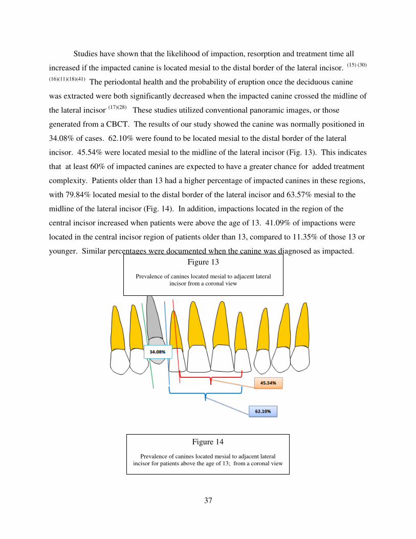
Studies have shown that the likelihood of impaction, resorption and treatment time all
increased if the impacted canine is located mesial to the distal border of the lateral incisor. (15) (30)
(16)(11)(18)(41) The periodontal health and the probability of eruption once the deciduous canine
was extracted were both significantly decreased when the impacted canine crossed the midline of
the lateral incisor. (17)(28)
These studies utilized conventional panoramic images, or those
generated from a CBCT. The results of our study showed the canine was normally positioned in
34.08% of cases. 62.10% were found to be located mesial to the distal border of the lateral
incisor. 45.54% were located mesial to the midline of the lateral incisor (Fig. 13). This indicates
that at least 60% of impacted canines are expected to have a greater chance for added treatment
complexity. Patients older than 13 had a higher percentage of impacted canines in these regions,
with 79.84% located mesial to the distal border of the lateral incisor and 63.57% mesial to the
midline of the lateral incisor (Fig. 14). In addition, impactions located in the region of the
central incisor increased when patients were above the age of 13. 41.09% of impactions were
located in the central incisor region of patients older than 13, compared to 11.35% of those 13 or
younger. Similar percentages were documented when the canine was diagnosed as impacted.
Figure 14
Prevalence of canines located mesial to adjacent lateral
incisor for patients above the age of 13; from a coronal view
Figure 13
Prevalence of canines located mesial to adjacent lateral
incisor from a coronal view

38
Angulation from a sagittal perspective demonstrated that 15.92% of impactions had a
value greater than 45 degrees when compared to the palatal plane, indicating a less than ideal
path to eruption. While no literature has shown significant implications of this angle, it can be
assumed that an impaction positioned less vertically may have difficulty erupting. From a
clinical perspective, the initial torque of the canine may create challenges in achieving the proper
angulation in the 3rd
order and possibly compromise esthetics. Prescriptions for modern straight-
wire appliances have torque values for maxillary canines ranging from 0 to -7 degrees.(49)
Some
clinicians have advocated using brackets with excessive negative torque value in order to
accomplish the proper 3rd
order angulation. (48)
From a surgical perspective, the angulation may
dictate where the bonded attachment is placed on the crown of the canine. Based on these
clinical implications, the torque of an unerupted canine must be taken into consideration during
diagnosis.
The angulation of an unerupted canine from a frontal perspective has been shown to
influence the likelihood of impaction, risk of resorption, the duration of treatment, and the
chances of normal eruption if the deciduous canine is extracted. (16)(18)(26)(30)
Our study classified
the unerupted canine as mesially tipped, vertical, or distally tipped in relation to the skeletal
midline through ANS. A mesially tipped canine was defined as an angle greater than 30 degrees.
This angle is based on 2 previous studies showing: 1) The risk of resorption increased by 50%
when this angle exceeded 25 degrees; (18)
2) The probability of normal eruption after extraction
of the deciduous canine significantly decreased when the angulation surpassed 31 degrees. (26)
In
39
addition, similar studies have shown that as this angle increased, the duration of treatment and
the likelihood of impaction also increased.(16)(30)
We observed mesially tipped canines in 59.24%
of the cases, distally tipped in 0.64%, and 40.13% were classified as vertically angulated.
Patients older than 13 years of age were shown to have a greater chance of a mesially angulated
canine, with 78.29% mesially angulated, and 21.71% vertical. Similar findings were recorded
for canines that were diagnosed as impacted vs. unerupted. Patients 13 or younger were more
likely to have a vertically tipped canine, occurring in 52.97% of the cases, while 45.95% were
mesially tipped. This indicates that severity and probability of adverse effects increases with age
and diagnosis of impaction.
Based on our classification system 33.44% of the canines were identified as mild, 37.58%
moderate, and 28.98% severe. The sex of the patient did not have a significant effect on the
severity of the impaction. Age, however, was shown to significantly affect severity. As the age
of a patient increased, severity also increased. With each yearly increase in age, the chance of
having a severe impaction increased by 3.2% (OR 1.032, P-value .041). In addition, severe
impactions were observed in 41.86% of canines in the patients above the age of 13 compared to
20% in those 13 or younger. 46% of unilateral impactions were classified as severe, which is
greater than that observed for bilateral impactions (21.03%). As expected a buccal or palatal
position was considered more severe than a midalveolar impaction, since this position directly
influences the calculation of severity. If the canine was positioned buccally the impaction was
severe 4.947 times more than if it was positioned midalveolar (p-value .001). If positioned
palatally, it was 3.767 times more likely to be severe than if located midalveolar (p-value .006).
Inter-examiner and Intra-examiner reliability were found to be excellent for almost all
measurements. This reflects the reproducibility of the method described to locate an
unerupted/impacted canine using CBCT. While previous studies have evaluated impacted
canines with three-dimensional images, the diagnostic accuracy of their methods was not
described. (47)
By comparing our measurements using the CBCT image of a typodont with an
impacted canine, to those calculated using a digital caliper, we were able to evaluate the
accuracy of our method. The results showed very high similarity between the CBCT
measurements and those achieved using a digital caliper.
Lastly, measurements recorded for each canine were utilized to develop an index of
nomenclature. All 314 canines were classified according to this index. Clinically, this allows for
40
improved communication between Orthodontists, Surgeons and other dental specialties. The
index will assist in more accurate description of the specific location, angulation and severity of
an unerupted or impacted maxillary canine. As a result, more ideal treatment and surgical
techniques can be utilized. This may lead to enhanced esthetics, reduced treatment time and
decreased adverse effects.
Conclusion
Our analysis of 204 patients with 314 unerupted maxillary canines showed 140 defined as
unerupted and 174 classified as impacted. The method outlined to analyze impacted canines
using CBCT exhibited excellent inter-examiner and intra-examiner reliability for all variables
except axial deviations from the midline which exhibited good inter-examiner reliability.
Measurements using a digital caliper on a typodont were compared to those acquired from a
CBCT image of the same typodont. Results showed very high similarity between the image and
caliper measurements.
Females were reported to be affected 1.63 times more frequently than males. 119
(57.49%) of the patients were female, 73 (35.27%) were male, and 15 (7.25%) were unreported.
Bilateral expression was observed in 51.69% of patients. No significant difference was observed
between right and left unerupted canines. Once a canine was defined as impacted, females were
observed to be affected 1.93 times more frequently than males.
41
Palatally displaced canines were observed at a rate of 38.54% compared to 40.76% for
buccal displacement and 20.70% of the canines were located midalveolar. However, once a
canine was defined as impacted, palatal impaction was 2.14 times more likely.
The results of our study demonstrated that 34.08% of canines were normally positioned,
62.10% were found to be located mesial to the distal border of the lateral incisor and 45.54%
were located mesial to the midline of the lateral incisor. One could conclude that at least 60% of
impacted canines would be expected to have a greater chance for adverse effects. When the
canine was defined as impacted or the patient was older than 13 years of age this percentage
increased.
The torque value of an unerupted canine may influence treatment difficulty and esthetic
outcome. 15.92% of canines evaluated exhibited angulation greater than 45 degrees when
compared to the palatal plane, indicating excessive positive torque. Clinicians should consider
the torque value of an unerupted canine during diagnosis and treatment planning. When
evaluating the angulation from a frontal view, mesially tipped canines were observed in 59.24%
of the cases. It was also observed that 78.29% of canines were mesially angulated once defined
as impacted or when the patients were older than 13. This indicates that severity and probability
of adverse effects increases with age or diagnosis of impaction.
Our evaluation classified the unerupted canines by severity based on their location and
angulation. 33.44% of the canines were identified as mild, 37.58% moderate, and 28.98%
severe. As the age of a patient increased, severity was found to significantly increase. With each
yearly increase in age, the chance of having a severe impaction increased by 3.2% (OR 1.032, P-
value .041). Buccal or palatal position was considered more severe than a midalveolar
impaction. If the canine was positioned buccally the impaction was severe 4.947 times more
than if it was positioned midalveolar (p-value .001). If positioned palatally, it was 3.767 times
more likely to be severe than if located midalveolar (p-value .006).
The presence or absence of active appliances, a primary canine, or a palatal expansion
device did not appear to influence severity or position of the impacted/unerupted canine. Other
anomalies such as peg laterals, a missing lateral incisor, supernumerary teeth or resorption did
not seem to influence the degree of impaction.

42
All 314 canines were classified according to the index described. The index was
designed to improve the quality of communication between dental specialists when diagnosing
the location and severity of an unerupted or impacted maxillary canine.
References
Bibliography
1. Impacted maxillary canines: A review. SE, Bishara. 2, 1992, Am J Ortho Dentofacial Orthop,
Vol. 101, pp. 159-171.
2. Radiographlc assessment of maxillary canine eruption in children with clinical signs of
eruption disturbance. Ericson S, Kurol J. Aug 1986, Eur J Orthod, Vol. 8, pp. 133-140.
3. Radiograpic examination of ectopically erupting maxillary canines. Ericson S, Kurol J. 6,
June 1987, Am J Orthod Dentofacial Orthop, Vol. 91, pp. 483-92.
4. A review of the diagnosis and management of impacted maxillary canines. Bedoya MM, Park
JH. 12, 2009, JADA, Vol. 140, pp. 1485-1493.
5. Standards of human occlusal development. Moyers RE, van der Linden FP, Riolo ML,
McNamara Jr. Ann Arbor, Mich: Center for human growth and development, The University of
Michigan : s.n., 1976. Vols. monograph 5, craniofacial growth series.
6. A Review of Early Displaced Maxillary Canines: Etiology, Diagnosis and Interceptive
Treatment. Litsas G, Acar A. March 2011, Open Dent J, Vol. 5, pp. 39-47.

43
7. A, Becker. The Orthodontic Treatment of Impacted Teeth. United Kingdom : Informa
Healthcare, 2007. Vol. Second Edition.
8. A controlled study of associated dental anomalies. T, Baccetti. 3, Jun 1998, Angle Orthod,
Vol. 68, pp. 267-74.
9. The incidence of anomalous maxillary lateral incisors in relation to palatally-displaced
cuspids. BeckerA, Smith P, Behar R. 1, Jan 1981, Angle Orthod, Vol. 51, pp. 24-29.
10. The palatally displaced canine as a dental anomaly of genetic origin. Peck S, Peck L,
Kataja M. 4, 1994, Angle Orthod, Vol. 64, pp. 249-256.
11. Localization of impacted maxillary canines and observation of adjacent incisor resorption
with cone-beam computed tomography. Liu DG, Zhang WL, Zhang ZY, Wu YT, Ma XC. 1,
Beijing, China : s.n., 2008, Oral Surg, Oral Med, Oral Path, Oral Rad and Endod, Vol. 105, pp.
91-98.
12. Resorption of Incisors After Ectopic Eruption of Maxillary Canines: A CT Study. Ericson S,
Kurol J. 6, December 2000, Angle Orthod, Vol. 70, pp. 415-423.
13. Three-dimensional localization of maxillary canines with cone-beam computed tomography.
Walker L, Enciso R, Mah J. 4, 2005, Am J, Vol. 128, pp. 418-23.
14. Canine impaction identified early with panoramic radiographs. Lindauer SJ, Rubenstein
LK, Hang WM, Andersen WC, Isaacson RJ. 3, March 1992, J Am Dent Assoc, Vol. 123, pp.
91-2, 95-7.
15. Prediction of maxillary canine impaction using sectors and angular measurement. Warford
JH Jr, Grandhi RK, Tira DE. 6, December 2003, Am J Orthod Dentofacial Orthop, Vol. 124,
pp. 651-5.
16. Early prediction of maxillary canine impaction from panoramic radiographs. Sajnani AK,
King NM. 1, July 2012, Am J Orthod Dentofacial Orthop, Vol. 142, pp. 45-51.
17. Early treatment of palatally erupting maxillary canines by extraction of the primary canines.
Ericson S, Kurol J. 1, 1988, Eur J Orthod, Vol. 10, pp. 283-295.
18. Resorption of maxillary lateral incisors caused by ectopic eruption of the canines. Ericson S,
Kurol J. 6, December 1988, Am J Orthod Dentofacial Orthop, Vol. 94, pp. 503-513.
19. The current status of cone beam computed tomography imaging in orthodontics. Kapila S,
Conley RS, Harrell WE Jr. 1, Jan 2011, Dentomaxillofac Radiol, Vol. 40, pp. 24-34.
20. Comparison between traditional 2-dimensional cephalometry and a 3-dimensional approach
on human dry skulls. Adams GL, Gansky SA, Miller AJ, Harrell WE Jr, Hatcher DC. 4, Oct
2004, Am J Orthod Dentofacial Orthop, Vol. 126, pp. 397-409.
21. Two- versus three-dimensional imaging in subjects with unerupted maxillary canines.
Botticelli S, Verna C, Paolo CM, Jens H, Melsen B. December 2011, Eur J Orthod, Vol. 33,
pp. 344-349.
22. In search of anatomic truth: 3-dimensional digital modeling and the future of orthodontics.
Harrell WE Jr, Hatcher DC, Bolt RL. 3, Sept 2002, Am J Orthod Dentofacial Orthop, Vol.
122, pp. 325-30.
23. Impacted upper canines: examination and treatment proposal based on 3D versus 2D
diagnosis. Wriedt S, Jaklin J, Al-Nawas B, Wehrbein H. 1, Jan 2012, J Orofac Orthop, Vol.
73, pp. 28-40.
24. In-vitro comparison of 2 cone-beam computed tomography systems and panoramic imaging
for detecting simulated canine impaction-induced external root resorption in maxillary lateral
incisors. Alqerban A, Jacobs R, Souza PC, Willems G. 4, December 2009, Am J Orthod
Dentofacial Orthop, Vol. 126, pp. 764-5.

44
25. Incisor resorption caused by maxillary cuspids. A radiographic study. Ericson S, Kurol J. 4,
Oct 1987, Angle Orthod, Vol. 57, pp. 332-46.
26. An investigation into the response of palatally displaced canines to the removal of deciduous
canines and an assessment of factors contributing to favourable eruption. Power SM, Short
MB. 3, Aug 1993, Br J Orthod, Vol. 20, pp. 215-23.
27. Surgical and orthodontic management of impacted maxillary canines. V, Kokich. 3, 2004,
Am J Orthod Dentofacial Orthop, Vol. 126, pp. 278-283.
28. Initial vertical and horizontal position of palatally impacted maxillary canine and effect on
periodontal status following surgical-orthodontic treatment. Zasciurinskiene E, Bjerklin K,
Smailiene D, Sidlauskas A, Puisys A. 2, Mar 2008, Angle Orthod, Vol. 78, pp. 275-80.
29. Factors that relate to treatment duration for patients with palatally impacted maxillary
canines. Stewart J.A., Heo G., Glover K.E., Williamson P.C., Lam E.W.N., Major P.W. 3,
March 2001, Am J Orthod Dentofacial Orthop, Vol. 119, pp. 216-225.
30. Palatally displaced maxillary canines: factors influencing duration and cost of treatment.
Bazargani F, Magnuson A, Dolati A, Lennartsson B. 2012, Eur J Orthod.
31. Radiographic localization techniques. JJ, Keur. 2, April 1986, Aust Dent J, Vol. 31, pp. 86-
90.
32. Radiation Protection 125: Low dose ionizing radiation and cancer risk. Commission,
European. Luxembourg : s.n., 2001.
33. Cone-beam computed tomography for routine orthodontic treatment planning: a radiation
dose evaluation. Silva MA, Wolf U, Heinicke F, Bumann A, Visser H, Hirsch E. 2008, Am J
Orthod Dentofacial Orthop, Vol. 133.
34. Dosimetry of 3 CBCT devices for oral and maxillofacial radiology: CB Mercuray, NewTom
3G and i-CAT. Ludlow JB, Davies-Ludlow LE, Brooks SL, Howerton WB. 2006,
Dentomaxillofac Radiol, Vol. 35, pp. 219-226.
35. Radiation absorbed in maxillofacial imaging with a new dental computed tomography
device. Mah JK, Danforth RA, Bumann A, Hatcher D. 2003, Oral Surg Oral Med Oral Pathol
Oral Radiol Endod, Vol. 96, pp. 508-513.
36. Manchester, University of. Radiation doses and risks of CBCT. [Online] 2009.
http://www.sedentexct.eu/content/radiation-doses-and-risks-cbct.
37. CBCT dosimetry: orthodontic considerations. SL, Brooks. 2009, Semin in Orthod, Vol. 15,
pp. 14-18.
38. Value of two cone-beam computed tomography systems from an orthodontic point of view.
Korbmacher H, Kahl-Nieke B, Schöllchen M, Heiland M. 4, July 2007, J Orofac Orthop.,
Vol. 68, pp. 278-89.
39. How a computerized tomography examination changed the treatment plans of 80 children
with retained and ectopically positioned maxillary canines. Bjerklin K, Ericson S. 1, Jan 2006,
Angle Orthod, Vol. 76, pp. 43-51.
40. Comparative analysis of traditional radiographs and cone-beam computed tomography
volumetric images in the diagnosis and treatment planning of maxillary impacted canines.
Haney E, Gansky SA, Lee JS, Johnson E, Maki K, Miller AJ, Huang JC. 5, May 2010, Am J
Orthod Dentofacial Orthop, Vol. 137, pp. 590-7.
41. Maxillary canine impaction increases root resorption risk of adjacent teeth: A problem of
physical proximity. Yan B, Sun Z, Fields H, Wang L. Dec 2012, Am J Orthod Dentofacial
Orthop, Vol 142, pp. 750-757.
45
42. Early Predictive Variables for Upper Canine Impaction as Derived from Posteroanterior
Cephalograms. Sambataro S, Bacetti T, Franchi L, Antonini F. Jan 2005, Angle Orthod, Vol
75, No 1, pp. 28-34.
43. Effect of surgical exposure technique, age, and grade of impaction on ankylosis of an
impacted canine, and the effect of rapid palatal expansion on eruption: A prospective clinical
study. Koutzogloua S, Kostakib A. March 2013, Am J Orthod Dentofacial Orthop, Vol 143,
pp. 342-52.
44. Canine impactions: incidence and management. Int J Periodontics Restorative Dent. Cooke
J, Wang HL. Sept/Oct 2006; 26(5), pp. 483-491.
45. The assessment of impacted maxillary canine position with panoramic radiography and cone
beam computed tomography. Jung YH, Liang H, Benson BW, Flint D, Cho BH.
Dentomaxillofacial Rad, 201, 000, pp 1–5.
46. The use of panoramic radiographs to localize displaced maxillary canines. Chaushu S,
Chaushu G, Becker A. Oral Surg Oral Med Oral Pathol Oral Radiol Endod 1999;85:511-6.
47. Localization of impacted maxillary canines using cone beam computed tomography. Review
of the literature. Rossini G,Cavallini C,Cassetta M, Galluccio G, Barbato E. Ann Stomatol,
Jan-Mar 2012; 3(1), pp 14-18.
48. Use of Cantilever Mechanics for Impacted Teeth: Case Series. Paduano S, Spagnuolo
G, Franzese G, Pellegrino G, Valletta R, Cioffi I. Open Dent J, 2013 Dec, 30;7:186-97.
49. Contemporary Orthodontics. Proffit W. Mosby, St. Louis, 2007.
Appendix A
Chart 11
Prevalence of “impacted” canines by Gender
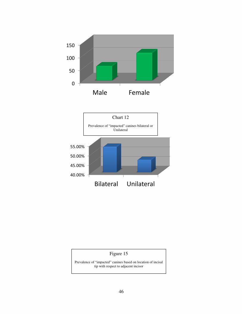
0
50
100
150
40.00%
45.00%
50.00%
55.00%
Prevalence of “impacted” canines bilateral or
Prevalence of “impacted” canines based on location of incisal
46
Male Female
Bilateral Unilateral
Chart 12
Prevalence of “impacted” canines bilateral or
Unilateral
Figure 15
Prevalence of “impacted” canines based on location of incisal
tip with respect to adjacent incisor
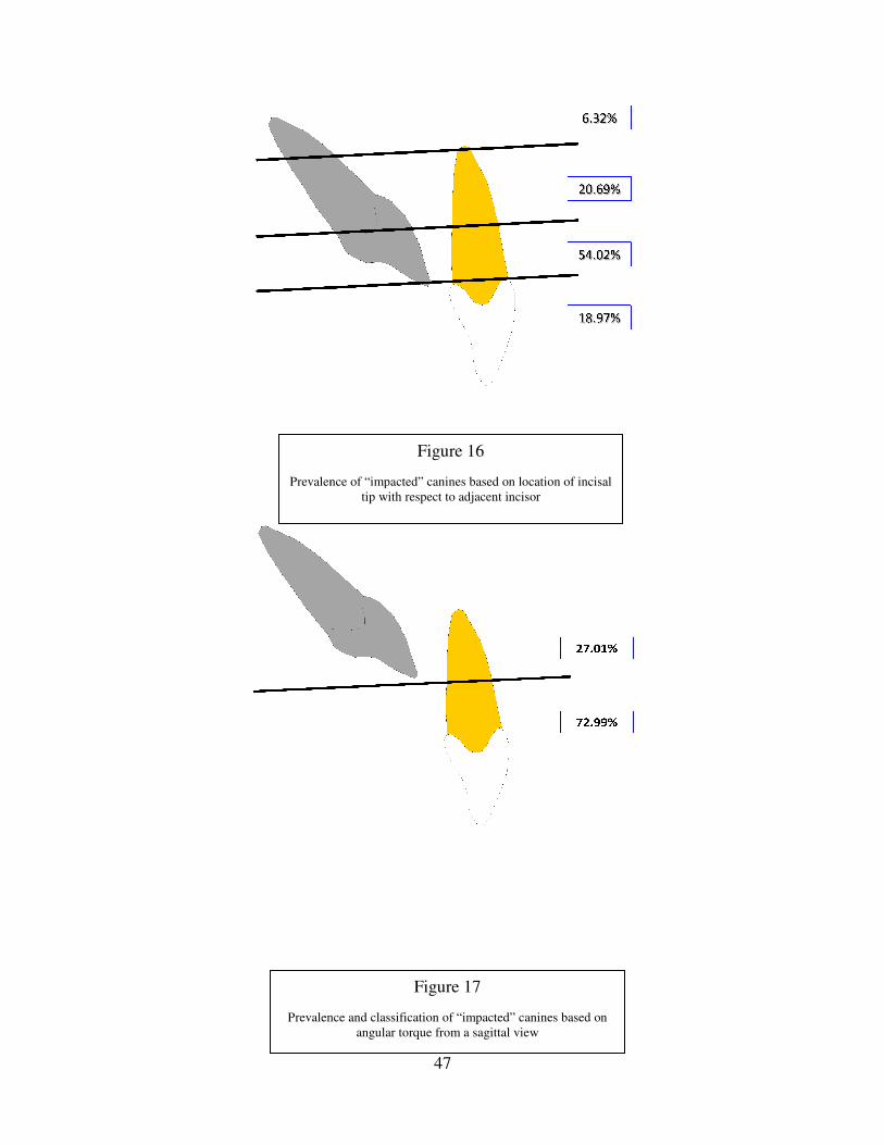
Prevalence of “impacted” canines based on location of incisal
Prevalence and classification of “impacted” canines based on
47
Figure 16
Prevalence of “impacted” canines based on location of incisal
tip with respect to adjacent incisor
Figure 17
Prevalence and classification of “impacted” canines based on
angular torque from a sagittal view
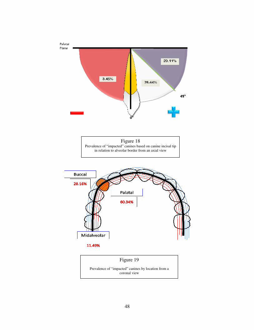
Prevalence of “impacted” canines based on canine incisal tip
in relation to alveolar border from an axial view
Prevalence of “impacted” canines by location from a
48
Figure 18 Prevalence of “impacted” canines based on canine incisal tip
in relation to alveolar border from an axial view
Figure 19
Prevalence of “impacted” canines by location from a
coronal view
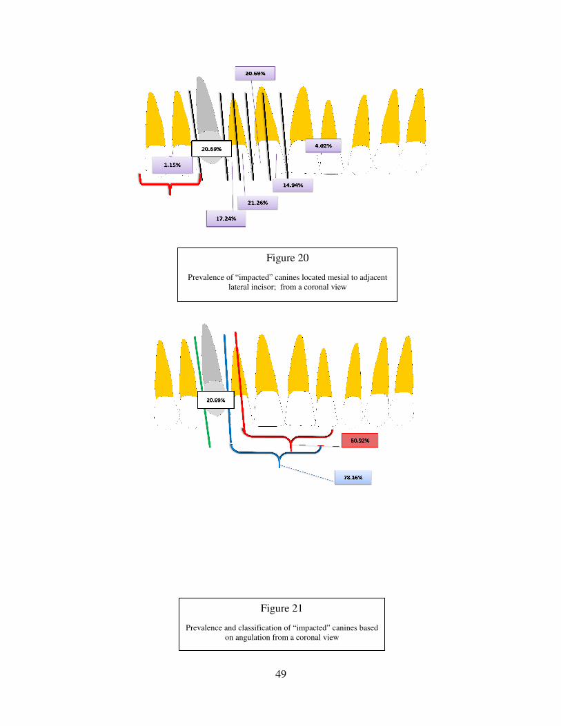
Prevalence
Prevalence of “impacted” canines located mesial to adjacent
49
Figure 21
Prevalence and classification of “impacted” canines based
on angulation from a coronal view
Figure 20
Prevalence of “impacted” canines located mesial to adjacent
lateral incisor; from a coronal view
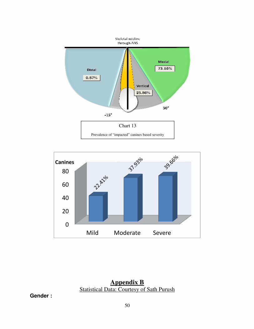
Statistical DataGender :
0
20
40
60
80
Mild
Canines
Prevalence of “impacted” canines based severity
50
Appendix B
Statistical Data: Courtesy of Sath Purush
Mild Moderate Severe
Chart 13
Prevalence of “impacted” canines based severity
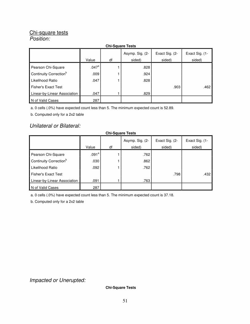
51
Chi-square tests Position:
Chi-Square Tests
Value df
Asymp. Sig. (2-
sided)
Exact Sig. (2-
sided)
Exact Sig. (1-
sided)
Pearson Chi-Square .047a 1 .828
Continuity Correctionb .009 1 .924
Likelihood Ratio .047 1 .828
Fisher's Exact Test .903 .462
Linear-by-Linear Association .047 1 .829
N of Valid Cases 287
a. 0 cells (.0%) have expected count less than 5. The minimum expected count is 52.89.
b. Computed only for a 2x2 table
Unilateral or Bilateral:
Chi-Square Tests
Value df
Asymp. Sig. (2-
sided)
Exact Sig. (2-
sided)
Exact Sig. (1-
sided)
Pearson Chi-Square .091a 1 .762
Continuity Correctionb .030 1 .862
Likelihood Ratio .092 1 .762
Fisher's Exact Test .798 .432
Linear-by-Linear Association .091 1 .763
N of Valid Cases 287
a. 0 cells (.0%) have expected count less than 5. The minimum expected count is 37.18.
b. Computed only for a 2x2 table
Impacted or Unerupted:
Chi-Square Tests

52
Value df
Asymp. Sig. (2-
sided)
Exact Sig. (2-
sided)
Exact Sig. (1-
sided)
Pearson Chi-Square 2.830a 1 .093
Continuity Correctionb 2.433 1 .119
Likelihood Ratio 2.823 1 .093
Fisher's Exact Test .111 .060
Linear-by-Linear Association 2.820 1 .093
N of Valid Cases 287
a. 0 cells (.0%) have expected count less than 5. The minimum expected count is 47.14.
b. Computed only for a 2x2 table
Appliances:
Chi-Square Tests
Value df
Asymp. Sig. (2-
sided)
Exact Sig. (2-
sided)
Exact Sig. (1-
sided)
Pearson Chi-Square 4.691a 1 .030
Continuity Correctionb 4.114 1 .043
Likelihood Ratio 4.619 1 .032
Fisher's Exact Test .039 .022
Linear-by-Linear Association 4.675 1 .031
N of Valid Cases 287
a. 0 cells (.0%) have expected count less than 5. The minimum expected count is 29.13.
b. Computed only for a 2x2 table
Expander:
Chi-Square Tests
Value df
Asymp. Sig. (2-
sided)
Exact Sig. (2-
sided)
Exact Sig. (1-
sided)
Pearson Chi-Square 4.682a 1 .030
Continuity Correctionb 3.222 1 .073
Likelihood Ratio 4.575 1 .032
Fisher's Exact Test .058 .038
Linear-by-Linear Association 4.666 1 .031
N of Valid Cases 287
a. 2 cells (50.0%) have expected count less than 5. The minimum expected count is 3.07.
b. Computed only for a 2x2 table
Primary Canine:
Chi-Square Tests
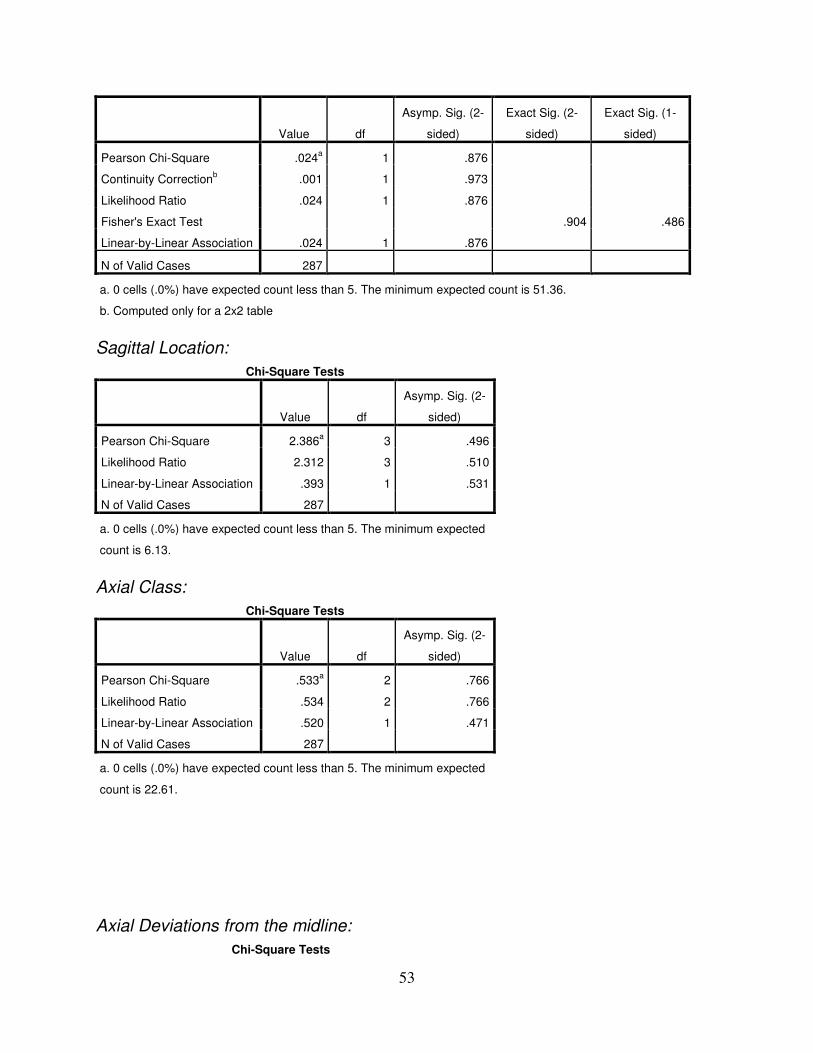
53
Value df
Asymp. Sig. (2-
sided)
Exact Sig. (2-
sided)
Exact Sig. (1-
sided)
Pearson Chi-Square .024a 1 .876
Continuity Correctionb .001 1 .973
Likelihood Ratio .024 1 .876
Fisher's Exact Test .904 .486
Linear-by-Linear Association .024 1 .876
N of Valid Cases 287
a. 0 cells (.0%) have expected count less than 5. The minimum expected count is 51.36.
b. Computed only for a 2x2 table
Sagittal Location:
Chi-Square Tests
Value df
Asymp. Sig. (2-
sided)
Pearson Chi-Square 2.386a 3 .496
Likelihood Ratio 2.312 3 .510
Linear-by-Linear Association .393 1 .531
N of Valid Cases 287
a. 0 cells (.0%) have expected count less than 5. The minimum expected
count is 6.13.
Axial Class:
Chi-Square Tests
Value df
Asymp. Sig. (2-
sided)
Pearson Chi-Square .533a 2 .766
Likelihood Ratio .534 2 .766
Linear-by-Linear Association .520 1 .471
N of Valid Cases 287
a. 0 cells (.0%) have expected count less than 5. The minimum expected
count is 22.61.
Axial Deviations from the midline:
Chi-Square Tests
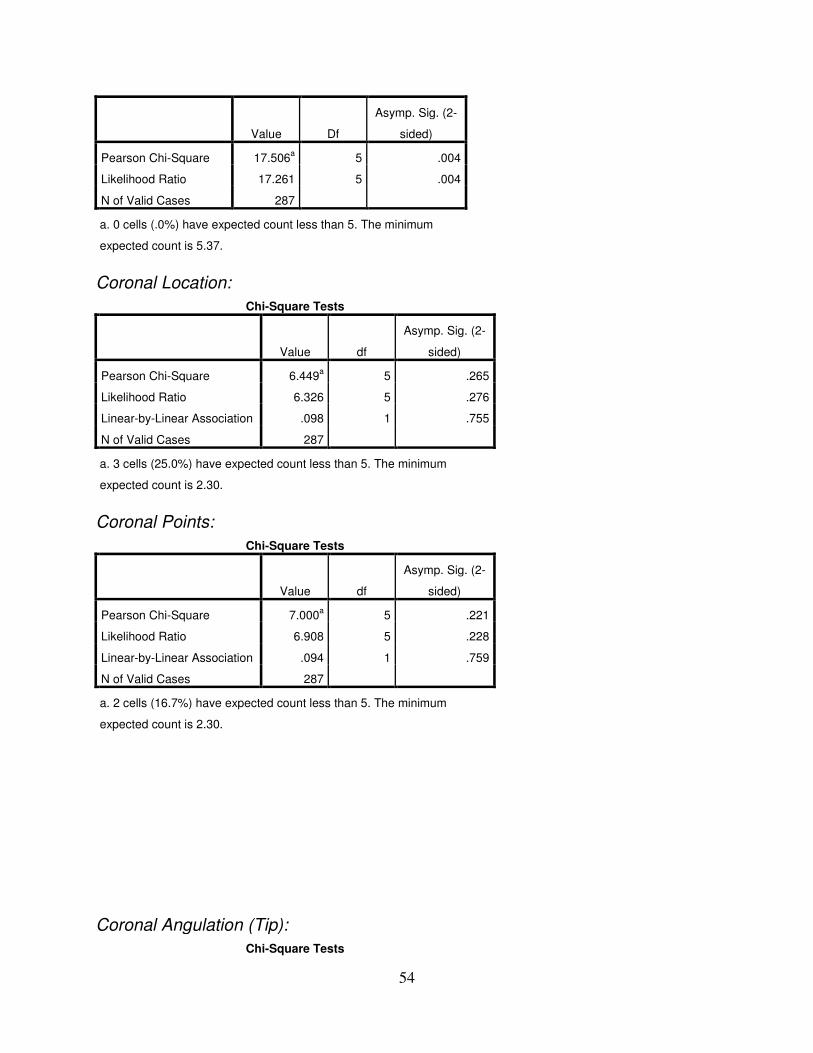
54
Value Df
Asymp. Sig. (2-
sided)
Pearson Chi-Square 17.506a 5 .004
Likelihood Ratio 17.261 5 .004
N of Valid Cases 287
a. 0 cells (.0%) have expected count less than 5. The minimum
expected count is 5.37.
Coronal Location:
Chi-Square Tests
Value df
Asymp. Sig. (2-
sided)
Pearson Chi-Square 6.449a 5 .265
Likelihood Ratio 6.326 5 .276
Linear-by-Linear Association .098 1 .755
N of Valid Cases 287
a. 3 cells (25.0%) have expected count less than 5. The minimum
expected count is 2.30.
Coronal Points:
Chi-Square Tests
Value df
Asymp. Sig. (2-
sided)
Pearson Chi-Square 7.000a 5 .221
Likelihood Ratio 6.908 5 .228
Linear-by-Linear Association .094 1 .759
N of Valid Cases 287
a. 2 cells (16.7%) have expected count less than 5. The minimum
expected count is 2.30.
Coronal Angulation (Tip):
Chi-Square Tests
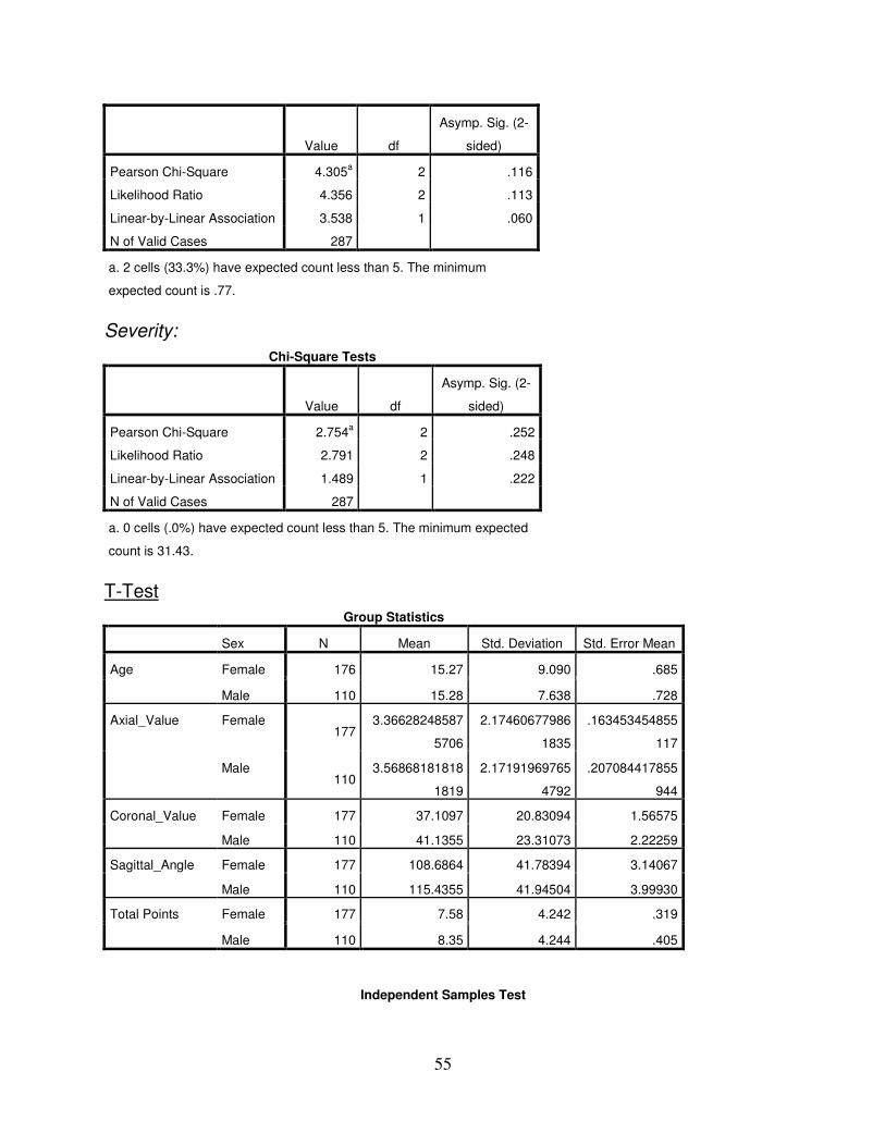
55
Value df
Asymp. Sig. (2-
sided)
Pearson Chi-Square 4.305a 2 .116
Likelihood Ratio 4.356 2 .113
Linear-by-Linear Association 3.538 1 .060
N of Valid Cases 287
a. 2 cells (33.3%) have expected count less than 5. The minimum
expected count is .77.
Severity:
Chi-Square Tests
Value df
Asymp. Sig. (2-
sided)
Pearson Chi-Square 2.754a 2 .252
Likelihood Ratio 2.791 2 .248
Linear-by-Linear Association 1.489 1 .222
N of Valid Cases 287
a. 0 cells (.0%) have expected count less than 5. The minimum expected
count is 31.43.
T-Test
Group Statistics
Sex N Mean Std. Deviation Std. Error Mean
Age Female 176 15.27 9.090 .685
Male 110 15.28 7.638 .728
Axial_Value Female 177
3.36628248587
5706
2.17460677986
1835
.163453454855
117
Male 110
3.56868181818
1819
2.17191969765
4792
.207084417855
944
Coronal_Value Female 177 37.1097 20.83094 1.56575
Male 110 41.1355 23.31073 2.22259
Sagittal_Angle Female 177 108.6864 41.78394 3.14067
Male 110 115.4355 41.94504 3.99930
Total Points Female 177 7.58 4.242 .319
Male 110 8.35 4.244 .405
Independent Samples Test
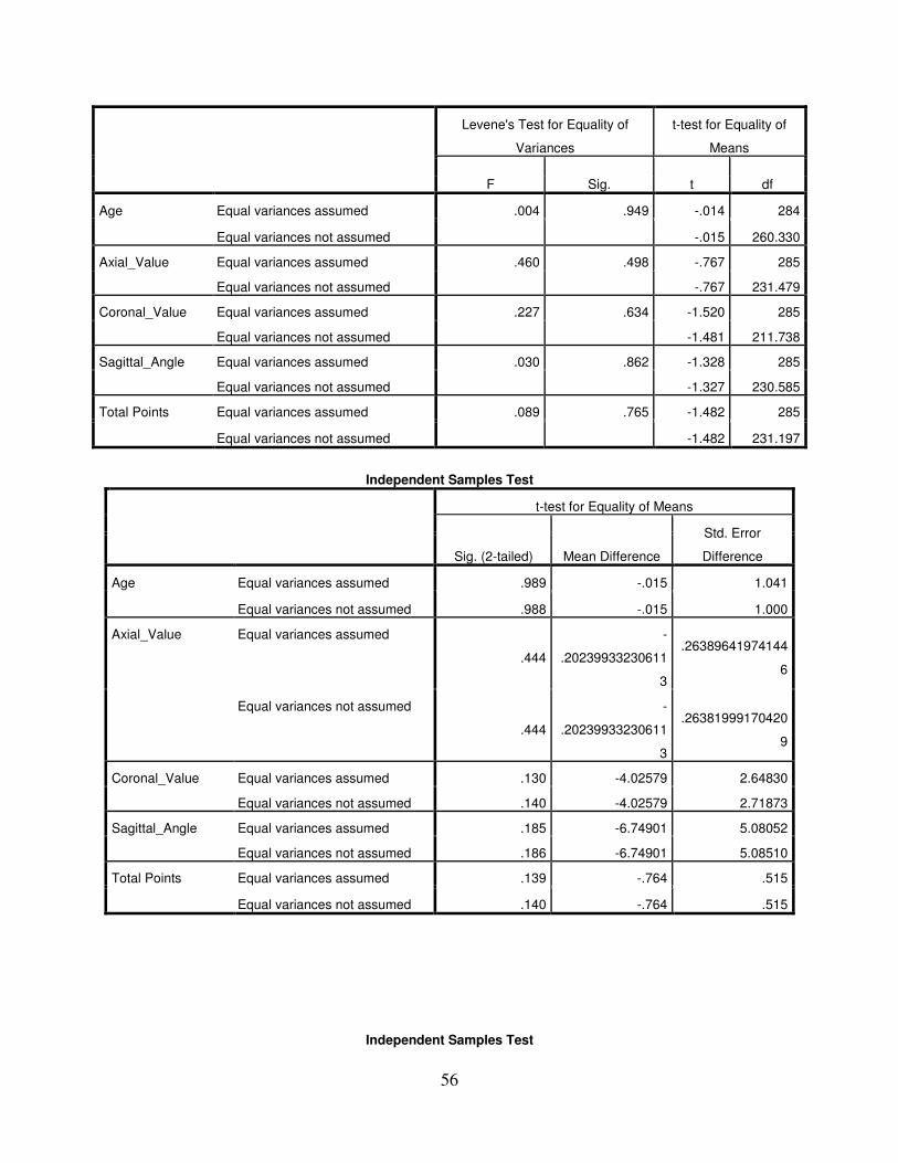
56
Levene's Test for Equality of
Variances
t-test for Equality of
Means
F Sig. t df
Age Equal variances assumed .004 .949 -.014 284
Equal variances not assumed -.015 260.330
Axial_Value Equal variances assumed .460 .498 -.767 285
Equal variances not assumed -.767 231.479
Coronal_Value Equal variances assumed .227 .634 -1.520 285
Equal variances not assumed -1.481 211.738
Sagittal_Angle Equal variances assumed .030 .862 -1.328 285
Equal variances not assumed -1.327 230.585
Total Points Equal variances assumed .089 .765 -1.482 285
Equal variances not assumed -1.482 231.197
Independent Samples Test
t-test for Equality of Means
Sig. (2-tailed) Mean Difference
Std. Error
Difference
Age Equal variances assumed .989 -.015 1.041
Equal variances not assumed .988 -.015 1.000
Axial_Value Equal variances assumed
.444
-
.20239933230611
3
.26389641974144
6
Equal variances not assumed
.444
-
.20239933230611
3
.26381999170420
9
Coronal_Value Equal variances assumed .130 -4.02579 2.64830
Equal variances not assumed .140 -4.02579 2.71873
Sagittal_Angle Equal variances assumed .185 -6.74901 5.08052
Equal variances not assumed .186 -6.74901 5.08510
Total Points Equal variances assumed .139 -.764 .515
Equal variances not assumed .140 -.764 .515
Independent Samples Test

57
t-test for Equality of Means
95% Confidence Interval of the Difference
Lower Upper
Age Equal variances assumed -2.063 2.033
Equal variances not assumed -1.984 1.954
Axial_Value Equal variances assumed -.721832620141435 .317033955529210
Equal variances not assumed -.722194672720169 .317396008107943
Coronal_Value Equal variances assumed -9.23850 1.18691
Equal variances not assumed -9.38503 1.33345
Sagittal_Angle Equal variances assumed -16.74911 3.25109
Equal variances not assumed -16.76822 3.27019
Total Points Equal variances assumed -1.777 .250
Equal variances not assumed -1.779 .251
Nonparametric Tests Mann-Whitney Test
Ranks
Sex N Mean Rank Sum of Ranks
Age Female 176 142.49 25077.50
Male 110 145.12 15963.50
Total 286
Sagittal_Angle Female 177 137.81 24392.00
Male 110 153.96 16936.00
Total 287
Axial_Value Female 177 139.56 24702.50
Male 110 151.14 16625.50
Total 287
Coronal_Value Female 177 138.89 24584.00
Male 110 152.22 16744.00
Total 287
Test Statisticsa
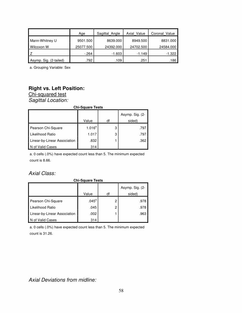
58
Age Sagittal_Angle Axial_Value Coronal_Value
Mann-Whitney U 9501.500 8639.000 8949.500 8831.000
Wilcoxon W 25077.500 24392.000 24702.500 24584.000
Z -.264 -1.603 -1.149 -1.322
Asymp. Sig. (2-tailed) .792 .109 .251 .186
a. Grouping Variable: Sex
Right vs. Left Position: Chi-squared test Sagittal Location:
Chi-Square Tests
Value df
Asymp. Sig. (2-
sided)
Pearson Chi-Square 1.016a 3 .797
Likelihood Ratio 1.017 3 .797
Linear-by-Linear Association .832 1 .362
N of Valid Cases 314
a. 0 cells (.0%) have expected count less than 5. The minimum expected
count is 8.66.
Axial Class:
Chi-Square Tests
Value df
Asymp. Sig. (2-
sided)
Pearson Chi-Square .045a 2 .978
Likelihood Ratio .045 2 .978
Linear-by-Linear Association .002 1 .963
N of Valid Cases 314
a. 0 cells (.0%) have expected count less than 5. The minimum expected
count is 31.26.
Axial Deviations from midline:

59
Chi-Square Tests
Value df
Asymp. Sig. (2-
sided)
Pearson Chi-Square 4.547a 5 .474
Likelihood Ratio 4.671 5 .457
N of Valid Cases 314
a. 0 cells (.0%) have expected count less than 5. The minimum
expected count is 8.18.
Coronal Location:
Chi-Square Tests
Value df
Asymp. Sig. (2-
sided)
Pearson Chi-Square 3.695a 5 .594
Likelihood Ratio 3.709 5 .592
Linear-by-Linear Association 1.527 1 .217
N of Valid Cases 314
a. 2 cells (16.7%) have expected count less than 5. The minimum
expected count is 3.85.
Coronal Points:
Chi-Square Tests
Value df
Asymp. Sig. (2-
sided)
Pearson Chi-Square 3.775a 5 .582
Likelihood Ratio 3.792 5 .580
Linear-by-Linear Association 2.284 1 .131
N of Valid Cases 314
a. 2 cells (16.7%) have expected count less than 5. The minimum
expected count is 3.85.
Coronal Angulation:
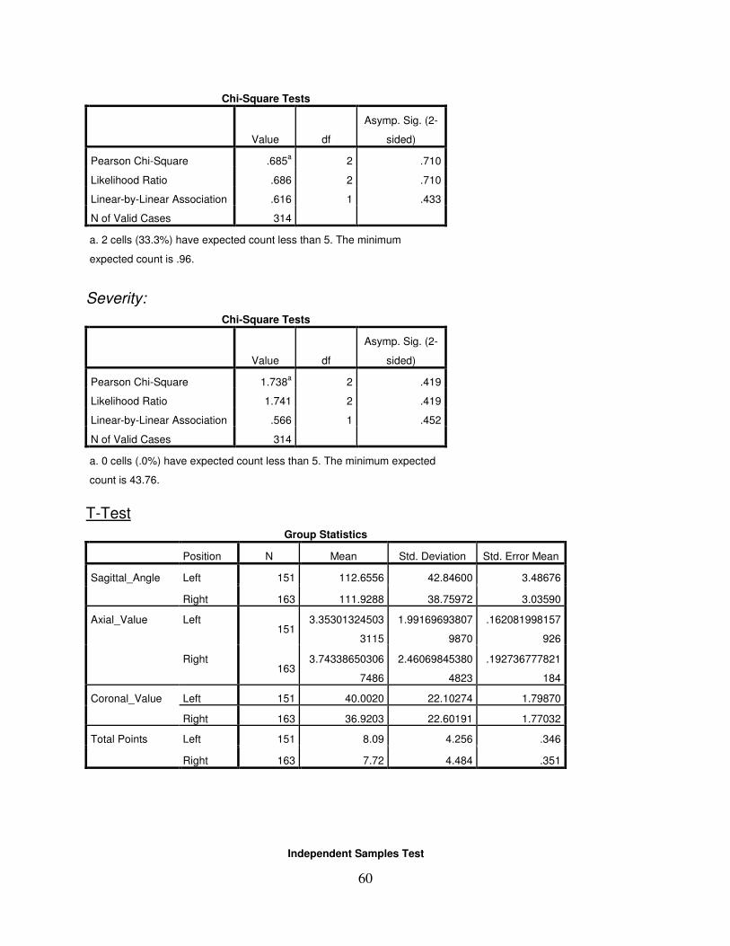
60
Chi-Square Tests
Value df
Asymp. Sig. (2-
sided)
Pearson Chi-Square .685a 2 .710
Likelihood Ratio .686 2 .710
Linear-by-Linear Association .616 1 .433
N of Valid Cases 314
a. 2 cells (33.3%) have expected count less than 5. The minimum
expected count is .96.
Severity:
Chi-Square Tests
Value df
Asymp. Sig. (2-
sided)
Pearson Chi-Square 1.738a 2 .419
Likelihood Ratio 1.741 2 .419
Linear-by-Linear Association .566 1 .452
N of Valid Cases 314
a. 0 cells (.0%) have expected count less than 5. The minimum expected
count is 43.76.
T-Test
Group Statistics
Position N Mean Std. Deviation Std. Error Mean
Sagittal_Angle Left 151 112.6556 42.84600 3.48676
Right 163 111.9288 38.75972 3.03590
Axial_Value Left 151
3.35301324503
3115
1.99169693807
9870
.162081998157
926
Right 163
3.74338650306
7486
2.46069845380
4823
.192736777821
184
Coronal_Value Left 151 40.0020 22.10274 1.79870
Right 163 36.9203 22.60191 1.77032
Total Points Left 151 8.09 4.256 .346
Right 163 7.72 4.484 .351
Independent Samples Test
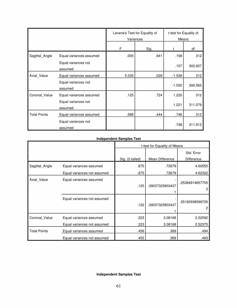
61
Levene's Test for Equality of
Variances
t-test for Equality of
Means
F Sig. t df
Sagittal_Angle Equal variances assumed .005 .941 .158 312
Equal variances not
assumed .157 302.607
Axial_Value Equal variances assumed 5.035 .026 -1.538 312
Equal variances not
assumed -1.550 306.565
Coronal_Value Equal variances assumed .125 .724 1.220 312
Equal variances not
assumed 1.221 311.079
Total Points Equal variances assumed .588 .444 .746 312
Equal variances not
assumed .748 311.812
Independent Samples Test
t-test for Equality of Means
Sig. (2-tailed) Mean Difference
Std. Error
Difference
Sagittal_Angle Equal variances assumed .875 .72679 4.60555
Equal variances not assumed .875 .72679 4.62322
Axial_Value Equal variances assumed
.125
-
.39037325803437
1
.25384914657705
3
Equal variances not assumed
.122
-
.39037325803437
1
.25182938599726
2
Coronal_Value Equal variances assumed .223 3.08168 2.52592
Equal variances not assumed .223 3.08168 2.52375
Total Points Equal variances assumed .456 .369 .494
Equal variances not assumed .455 .369 .493
Independent Samples Test
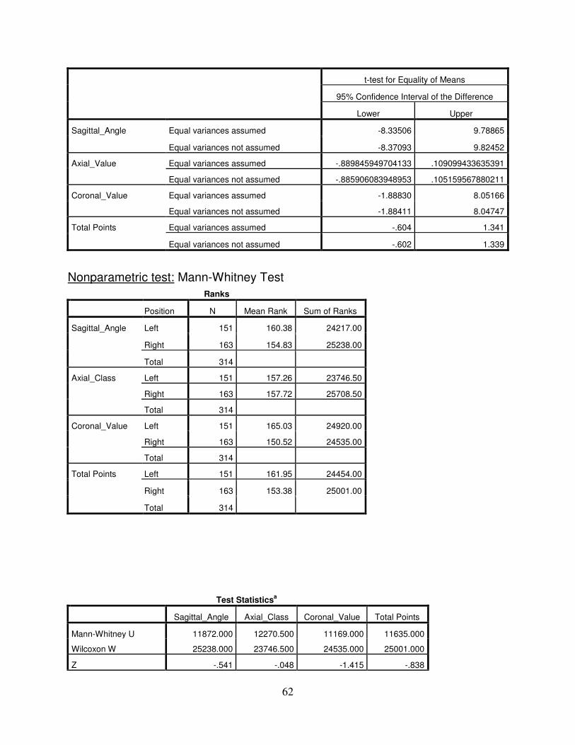
62
t-test for Equality of Means
95% Confidence Interval of the Difference
Lower Upper
Sagittal_Angle Equal variances assumed -8.33506 9.78865
Equal variances not assumed -8.37093 9.82452
Axial_Value Equal variances assumed -.889845949704133 .109099433635391
Equal variances not assumed -.885906083948953 .105159567880211
Coronal_Value Equal variances assumed -1.88830 8.05166
Equal variances not assumed -1.88411 8.04747
Total Points Equal variances assumed -.604 1.341
Equal variances not assumed -.602 1.339
Nonparametric test: Mann-Whitney Test
Ranks
Position N Mean Rank Sum of Ranks
Sagittal_Angle Left 151 160.38 24217.00
Right 163 154.83 25238.00
Total 314
Axial_Class Left 151 157.26 23746.50
Right 163 157.72 25708.50
Total 314
Coronal_Value Left 151 165.03 24920.00
Right 163 150.52 24535.00
Total 314
Total Points Left 151 161.95 24454.00
Right 163 153.38 25001.00
Total 314
Test Statisticsa
Sagittal_Angle Axial_Class Coronal_Value Total Points
Mann-Whitney U 11872.000 12270.500 11169.000 11635.000
Wilcoxon W 25238.000 23746.500 24535.000 25001.000
Z -.541 -.048 -1.415 -.838
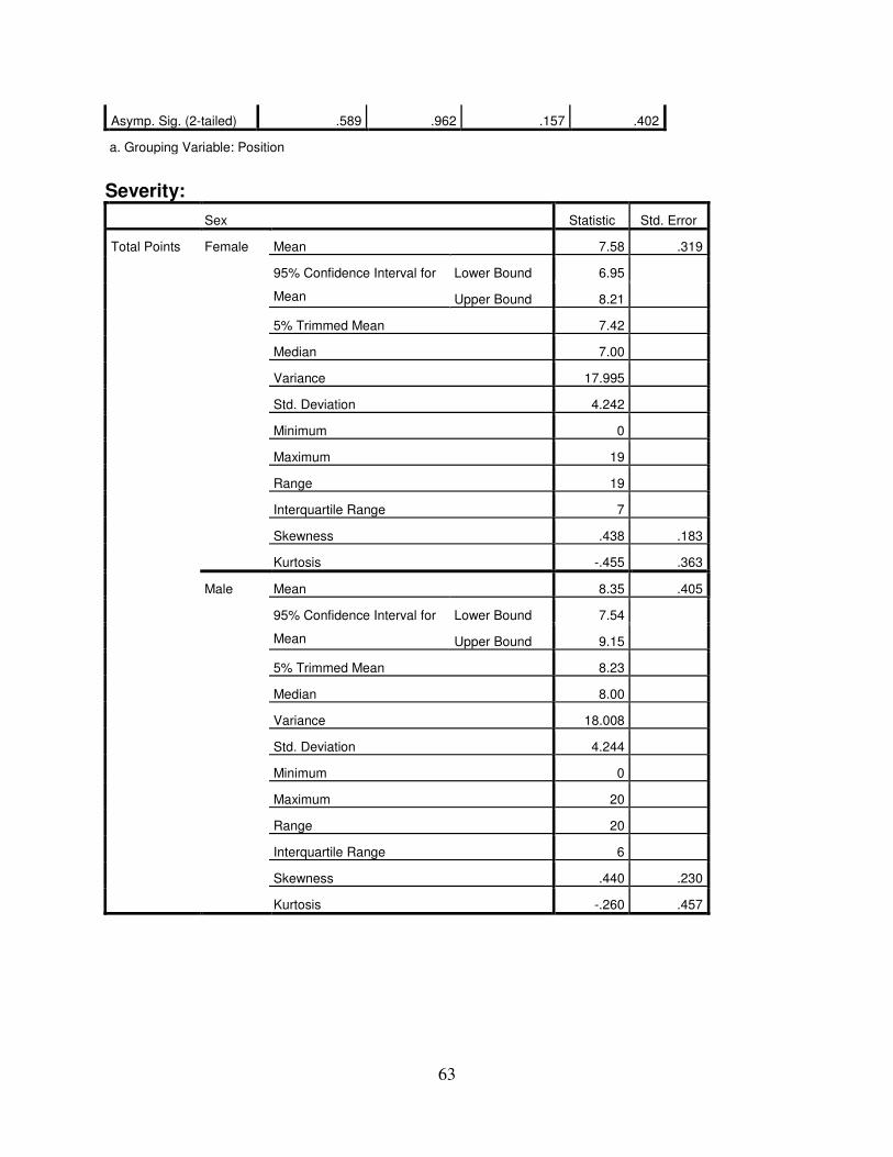
63
Asymp. Sig. (2-tailed) .589 .962 .157 .402
a. Grouping Variable: Position
Severity:
Sex Statistic Std. Error
Total Points Female Mean 7.58 .319
95% Confidence Interval for
Mean
Lower Bound 6.95
Upper Bound 8.21
5% Trimmed Mean 7.42
Median 7.00
Variance 17.995
Std. Deviation 4.242
Minimum 0
Maximum 19
Range 19
Interquartile Range 7
Skewness .438 .183
Kurtosis -.455 .363
Male Mean 8.35 .405
95% Confidence Interval for
Mean
Lower Bound 7.54
Upper Bound 9.15
5% Trimmed Mean 8.23
Median 8.00
Variance 18.008
Std. Deviation 4.244
Minimum 0
Maximum 20
Range 20
Interquartile Range 6
Skewness .440 .230
Kurtosis -.260 .457
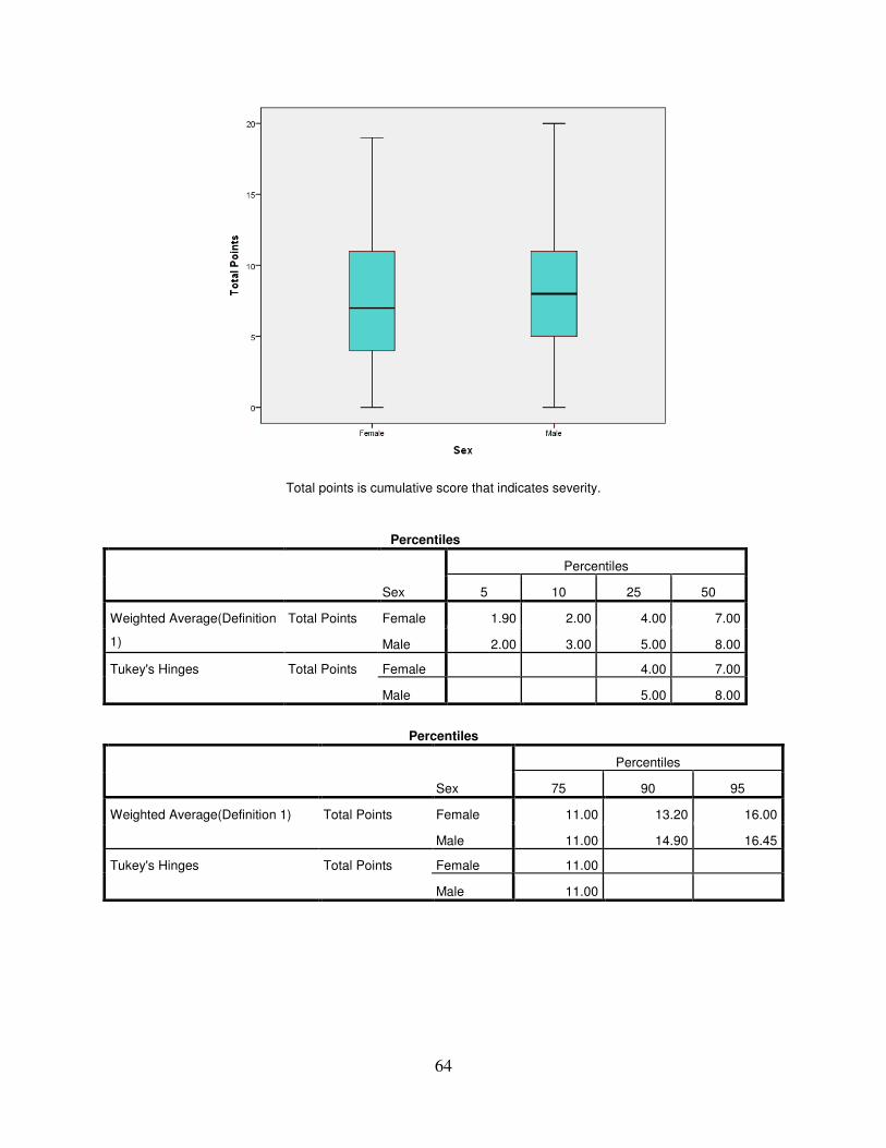
64
Total points is cumulative score that indicates severity.
Percentiles
Sex
Percentiles
5 10 25 50
Weighted Average(Definition
1)
Total Points Female 1.90 2.00 4.00 7.00
Male 2.00 3.00 5.00 8.00
Tukey's Hinges Total Points Female 4.00 7.00
Male 5.00 8.00
Percentiles
Sex
Percentiles
75 90 95
Weighted Average(Definition 1) Total Points Female 11.00 13.20 16.00
Male 11.00 14.90 16.45
Tukey's Hinges Total Points Female 11.00
Male 11.00
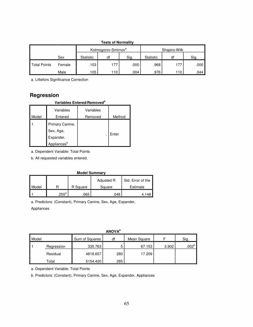
65
Tests of Normality
Sex
Kolmogorov-Smirnova Shapiro-Wilk
Statistic df Sig. Statistic df Sig.
Total Points Female .103 177 .000 .968 177 .000
Male .105 110 .004 .976 110 .044
a. Lilliefors Significance Correction
Regression
Variables Entered/Removeda
Model
Variables
Entered
Variables
Removed Method
1 Primary Canine,
Sex, Age,
Expander,
Appliancesb
. Enter
a. Dependent Variable: Total Points
b. All requested variables entered.
Model Summary
Model R R Square
Adjusted R
Square
Std. Error of the
Estimate
1 .255a .065 .048 4.148
a. Predictors: (Constant), Primary Canine, Sex, Age, Expander,
Appliances
ANOVAa
Model Sum of Squares df Mean Square F Sig.
1 Regression 335.763 5 67.153 3.902 .002b
Residual 4818.657 280 17.209
Total 5154.420 285
a. Dependent Variable: Total Points
b. Predictors: (Constant), Primary Canine, Sex, Age, Expander, Appliances
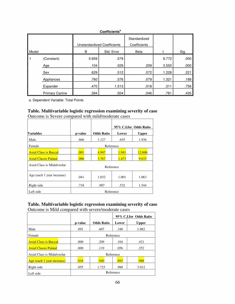
66
Coefficientsa
Model
Unstandardized Coefficients
Standardized
Coefficients
t Sig. B Std. Error Beta
1 (Constant) 5.659 .579 9.772 .000
Age .104 .029 .209 3.550 .000
Sex .629 .512 .072 1.228 .221
Appliances .760 .576 .079 1.321 .188
Expander .470 1.513 .018 .311 .756
Primary Canine .394 .504 .046 .781 .435
a. Dependent Variable: Total Points
Table. Multivariable logistic regression examining severity of case
Outcome is Severe compared with mild/moderate cases
Variables p-value Odds Ratio
95% C.I.for Odds Ratio
Lower Upper
Male .666 1.127 .655 1.936
Female Reference
Axial Class is Buccal .001 4.947 1.941 12.606
Axial Classis Palatal .006 3.767 1.473 9.633
Axial Class is Midalveolar Reference
Age (each 1 year increase) .041 1.032 1.001 1.063
Right side .718 .907 .532 1.544
Left side Reference
Table. Multivariable logistic regression examining severity of case
Outcome is Mild compared with severe/moderate cases
p-value Odds Ratio
95% C.I.for Odds Ratio
Lower Upper
Male .091 .607 .340 1.082
Female Reference
Axial Class is Buccal .000 .209 .104 .421
Axial Classis Palatal .000 .119 .056 .252
Axial Class is Midalveolar Reference
Age (each 1 year increase) .016 .940 .893 .988
Right side .055 1.725 .988 3.012
Left side Reference

67