Embed Size (px)
Citation preview
Thorax 1995;50(Suppl 1):S53-S58
Oxidantslantioxidants in idiopathicpulmonary fibrosis
W MacNee, I RahmanUnit of Respiratory Medicine, Department of Medicine, Royal Infirmary,Edinburgh, UK
Introductory article
The effect of oral N-acetylcysteine on lung glutathione levels in idiopathicpulmonary fibrosis
A Meyer, R Buhl, H Magnussen
Idiopathic pulmonary fibrosis (IPF) is characterized by an increased oxidant burden and by a deficiencyof glutathione, a major antioxidant, in the lung epithelial lining fluid (ELF). Therefore, a rationaltherapeutic approach is to reverse the imbalance between oxidants and antioxidants in the lung byenhancing the antioxidant screen. With this background, the aim of our study was to evaluate oralN-acetylcysteine (NAC) as a strategy to augment lung glutathione levels in patients with IPFConcentrations of total glutathione in bronchoalveolar lavage fluid (BALF) were quantifiedspectrophotometrically, before and following oral therapy with 3 x 600mg NAC per day for 5 days, in17 nonsmoking patients with biopsy-proven IPF The volume of ELF recovered by BAL was determinedusing the urea method. Pretherapy, total glutathione levels in ELF in IPF patients were significantly lessthan normal (187+36 vs 368+601y/), in contrast to levels in BALF (0.99+0-12 vs 1.18+ 19,uM). Followingtherapy with oral NAC, glutathione levels in BALF were 1.54 + 0.24 jiM (a significant increase comparedto pretherapy), whereas the increase in ELF levels (319+ 92,M) did not reach significance. The therapywas well-tolerated, and all routine clinical and bronchoscopic parameters remained unchanged. It isthus feasible and safe to augment deficient lung glutathione levels in patients with IPF; thereby,potentially augmenting pulmonary antioxidant protection. (Eur Respir J 1994;7:431-6)
Idiopathic pulmonary fibrosis (IPF) is thought to ariseas a response to persistent lung injury and in-flammation.' The early response to injury to the alveolarepithelium and/or the vascular endothelium results inthe influx ofneutrophils to the interstitium and airspaceswhich may persist.2 The hypothesis that the condition isimmunologically mediated arises from the mononuclearphase of the disease in which the recruitment of mono-cytes/macrophages and lymphocytes in the lungs occurs,presumably in response to an as yet unidentified antigenor immunological target. These cells are thought torelease fibrogenic cytokines such as tumour necrosisfactor alpha, transforming growth factors alpha andbeta, and eicosanoids such as leukotrienes B4 and C4which recruit and activate fibroblasts and stimulatedeposition of connective tissue.3 Treatment strategiesfor pulmonary fibrosis, whether old or new, can thereforebe categorised in terms of the stage of the fibroticresponse in the lung to which they are targeted - whetherto the inflammatory/immune response or tissue injury,
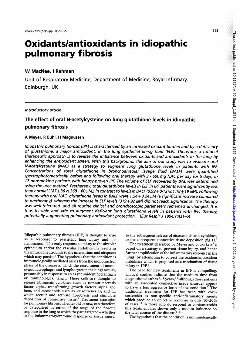
to the subsequent release of eicosanoids and cytokines,or the consequent connective tissue deposition (fig 1).'The treatment described by Meyer and coworkers5 is
based on a strategy to prevent tissue injury, and hencefurther amplification ofthe inflammatory response in thelungs, by attempting to correct the oxidant/antioxidantimbalance which is proposed as a mechanism of tissueinjury in IPF.1The need for new treatments in IPF is compelling.
Clinical studies indicate that the medium time fromdiagnosis to death is 3-5 years,68 although those patientswith an associated connective tissue disorder appearto have a less aggressive form of the condition.9 Thetraditional treatment for IPF has been with corti-costeroids as non-specific anti-inflammatory agentswhich produce an objective response in only 10-20%of cases.'0 In those who do respond to corticosteroidsthis treatment has shown only a modest influence onthe fatal course of the disease.610"The hypothesis that the condition is immunologically
S53
on February 5, 2022 by guest. P
rotected by copyright.http://thorax.bm
j.com/
Thorax: first published as 10.1136/thx.50.S
uppl_1.S53 on 1 S
eptember 1995. D
ownloaded from
MacNee, Rahman
Cytokine imbalance * Eicosanoids
Antioxidants 4--------------- Inflammation/immune response
Tissue injury
Connective tissue deposition
Figure 1 Major steps in the pathogenesis of pulmonaryfibrosis, interconnected by double headed arrows toindicate that each of these processes mutually regulates theother. The putative steps which may be influenced byantioxidant therapy are shown by the broken arrows.Modified from ref 4.
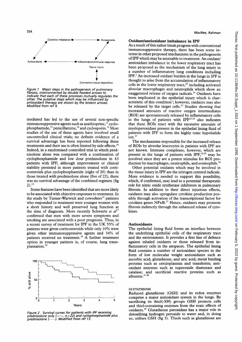
mediated has led to the use of several non-specificimmunosuppressive agents such as azathioprine,'2 cyclo-phosphamide,'3 penicillamine,'4 and cyclosporin.'5 Moststudies of the use of these agents have involved smalluncontrolled clinical trials; no definite evidence of asurvival advantage has been reported following thesetreatments and their use is often limited by side effects. 6Indeed, in a randomised controlled trial in which pred-nisolone alone was compared with a combination ofcyclophosphamide and low dose prednisolone in 43patients with IPF, although improvement or clinicalstability persisted in more patients treated with corti-costeroids plus cyclophosphamide (eight of 20) than inthose treated with prednisolone alone (five of 22), therewas no survival advantage of the combined regimen (fig2).13Some features have been identified that are more likely
to be associated with objective responses to treatment. Inthe study by Turner-Warwick and coworkers'0 patientswho responded to treatment were younger women witha short history and well preserved lung function atthe time of diagnosis. More recently Schwartz et al'7confirmed that men with more severe symptoms andsmoking are associated with a poor prognosis. Thus, ina recent survey of treatment for IPF in the UK 55% ofpatients were given corticosteroids while only 10% weregiven other immunosuppressive agents and 34% ofpatients received no treatment.'8 A further treatmentoption in younger patients is, of course, lung trans-plantation. '9
100
.0)
=
4)
o-0
50
Figureprednipredni
~~~~~~~~~-~~~~~~~~~~~I
- l_______
, !
Oxidant/antioxidant imbalance in IPFAs a result ofthis rather bleak progress with conventionalimmunosuppressive therapy, there has been some in-terest in other proposed mechanisms in the pathogenesisofIPF which may be amenable to treatment. An oxidant/antioxidant imbalance in the lower respiratory tract hasbeen proposed as the mechanism of the lung injury ina number of inflammatory lung conditions includingIPF. l An increased oxidant burden in the lungs in IPF isthought to arise from the accumulation of inflammatorycells in the lower respiratory tract,20 including activatedalveolar macrophages and neutrophils which show anexaggerated release of oxygen radicals.2' Oxidants havebeen implicated in the epithelial injury which is char-acteristic of this condition'; however, oxidants may alsobe released by the target cells.22 Studies showing thatincreased amounts of reactive oxygen intermediates(ROI) are spontaneously released by inflammatory cellsin the lungs of patients with IPF2324 also indicatesthat these ROIs react with the excessive amounts ofmyeloperoxidase present in the epithelial lining fluid ofpatients with IPF to form the highly toxic hypohalide
d23ion."The mechanisms responsible for the increased release
of ROIs by alveolar leucocytes in patients with IPF arenot known. Immune complexes, however, which arepresent in the lungs of patients with IPF,2425 may beinvolved since they are a potent stimulus for ROI pro-duction by macrophages, neutrophils, and eosinophils.26
Other potential oxidants which may be involved inthe tissue injury in IPF are the nitrogen centred radicals.More evidence is needed to support this possibility,which, if confirmed, may lead to a potential therapeuticrole for nitric oxide synthetase inhibitors in pulmonaryfibrosis. In addition to their direct injurious effects,oxidants may also upregulate cytokine production pos-sibly through activation of the transcriptional factor forcytokine genes NFKB.27 Hence, oxidants may promotefibrosis indirectly through the enhanced release of cyto-kines.
AntioxidantsThe epithelial lining fluid forms an interface betweenthe underlying epithelial cells of the respiratory tractand the environment. It provides a first line of defenceagainst inhaled oxidants or those released from in-flammatory cells in the airspaces. The epithelial liningfluid contains a number of antioxidant species in theform of low molecular weight antioxidants such asascorbic acid, glutathione, and uric acid; metal bindingproteins such as ceruloplasmin and transferrin; anti-oxidant enzymes such as superoxide dismutase andcatalase; and sacrificial reactive proteins such asalbumin.2728
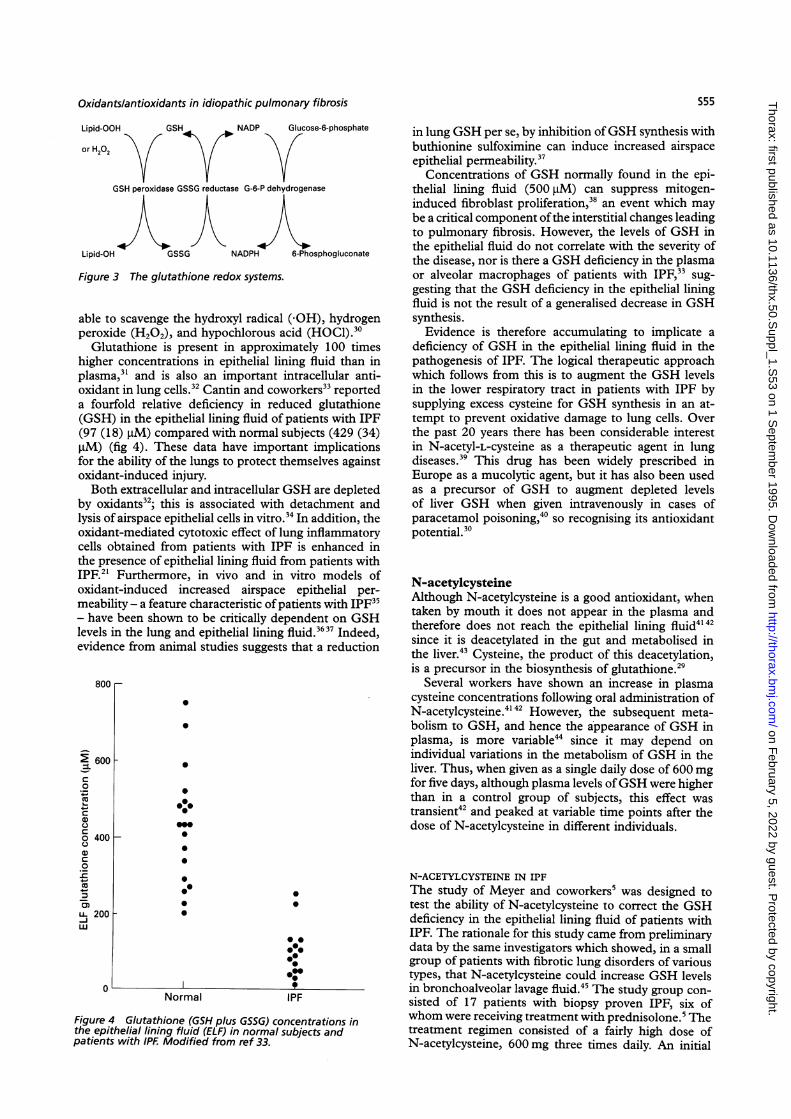
GLUTATHIONEReduced glutathione (GSH) and its redox enzymes
D0 comprise a major antioxidant system in the lungs. By0 1 2 3 4 5 sacrificing its thiol(-SH) groups GSH protects cellsYears and thiol-containing enzymes from the toxic effects of
oxdns2' Glutathione peroxidase has a major role in!2 Survival curves for patients with IPF receiving oxidants.isolone only ( , n =22) and cyclophosphamide plus detoxifying hydrogen peroxide to water and, in doingisolone (---- ). Modified from ref 13. so, utilises GSH (fig 3). Thiols such as glutathione are
S54
c
I 1- -- -
on February 5, 2022 by guest. P
rotected by copyright.http://thorax.bm
j.com/
Thorax: first published as 10.1136/thx.50.S
uppl_1.S53 on 1 S
eptember 1995. D
ownloaded from
Oxidantslantioxidants in idiopathic pulmonary fibrosis
Lipid-OOH GSH NADP
or H202
Glucose-6-phosphate
GSH peroxidase GSSG reductase G-6-P dehydrogenase
Lipid-OH GSSG NADPH 6-Phosphogluconate
Figure 3 The glutathione redox systems.
able to scavenge the hydroxyl radical (-OH), hydrogenperoxide (H202), and hypochlorous acid (HOCI).'0
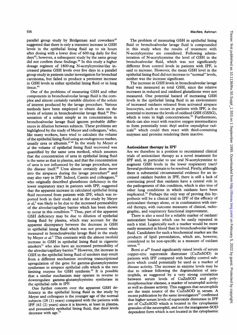
Glutathione is present in approximately 100 timeshigher concentrations in epithelial lining fluid than inplasma,"3 and is also an important intracellular anti-oxidant in lung cells.32 Cantin and coworkers33 reporteda fourfold relative deficiency in reduced glutathione(GSH) in the epithelial lining fluid of patients with IPF(97 (18) jiM) compared with normal subjects (429 (34),uM) (fig 4). These data have important implicationsfor the ability of the lungs to protect themselves againstoxidant-induced injury.Both extracellular and intracellular GSH are depleted
by oxidants32; this is associated with detachment andlysis of airspace epithelial cells in vitro.'4 In addition, theoxidant-mediated cytotoxic effect of lung inflammatorycells obtained from patients with IPF is enhanced inthe presence of epithelial lining fluid from patients withIPF." Furthermore, in vivo and in vitro models ofoxidant-induced increased airspace epithelial per-meability - a feature characteristic of patients with IPF"3- have been shown to be critically dependent on GSHlevels in the lung and epithelial lining fluid.'637 Indeed,evidence from animal studies suggests that a reduction
800 r
2 600
C
0
._
a,
O 400
0
a)
CN
0 0
0. 200-Jw)
0
_
0
00
*:00
0090
0
F
u-,'
00
0 @
00.00;0
Normal IPF
in lung GSH per se, by inhibition ofGSH synthesis withbuthionine sulfoximine can induce increased airspaceepithelial permeability.37
Concentrations of GSH normally found in the epi-thelial lining fluid (500 jM) can suppress mitogen-induced fibroblast proliferation,38 an event which maybe a critical component ofthe interstitial changes leadingto pulmonary fibrosis. However, the levels of GSH inthe epithelial fluid do not correlate with the severity ofthe disease, nor is there a GSH deficiency in the plasmaor alveolar macrophages of patients with IPF,33 sug-gesting that the GSH deficiency in the epithelial liningfluid is not the result of a generalised decrease in GSHsynthesis.Evidence is therefore accumulating to implicate a
deficiency of GSH in the epithelial lining fluid in thepathogenesis of IPF. The logical therapeutic approachwhich follows from this is to augment the GSH levelsin the lower respiratory tract in patients with IPF bysupplying excess cysteine for GSH synthesis in an at-tempt to prevent oxidative damage to lung cells. Overthe past 20 years there has been considerable interestin N-acetyl-L-cysteine as a therapeutic agent in lungdiseases.39 This drug has been widely prescribed inEurope as a mucolytic agent, but it has also been usedas a precursor of GSH to augment depleted levelsof liver GSH when given intravenously in cases ofparacetamol poisoning,40 so recognising its antioxidantpotential.'0
N-acetylcysteineAlthough N-acetylcysteine is a good antioxidant, whentaken by mouth it does not appear in the plasma andtherefore does not reach the epithelial lining fluid414'since it is deacetylated in the gut and metabolised inthe liver.4' Cysteine, the product of this deacetylation,is a precursor in the biosynthesis of glutathione.'9
Several workers have shown an increase in plasmacysteine concentrations following oral administration ofN-acetylcysteine.414' However, the subsequent meta-bolism to GSH, and hence the appearance of GSH inplasma, is more variable44 since it may depend onindividual variations in the metabolism of GSH in theliver. Thus, when given as a single daily dose of 600 mgfor five days, although plasma levels ofGSH were higherthan in a control group of subjects, this effect wastransient4' and peaked at variable time points after thedose of N-acetylcysteine in different individuals.
N-ACETYLCYSTEINE IN IPFThe study of Meyer and coworkers5 was designed totest the ability of N-acetylcysteine to correct the GSHdeficiency in the epithelial lining fluid of patients withIPF. The rationale for this study came from preliminarydata by the same investigators which showed, in a smallgroup of patients with fibrotic lung disorders of varioustypes, that N-acetylcysteine could increase GSH levelsin bronchoalveolar lavage fluid.45 The study group con-sisted of 17 patients with biopsy proven IPF, six ofwhom were receiving treatment with prednisolone.5 Thetreatment regimen consisted of a fairly high dose ofN-acetylcysteine, 600 mg three times daily. An initial
Figure 4 Glutathione (GSH plus GSSG) concentrations inthe epithelial lining fluid (ELF) in normal subjects andpatients with IPF Modified from ref 33.
S55
on February 5, 2022 by guest. P
rotected by copyright.http://thorax.bm
j.com/
Thorax: first published as 10.1136/thx.50.S
uppl_1.S53 on 1 S
eptember 1995. D
ownloaded from
MacNee, Rahman
parallel group study by Bridgeman and coworkers4"suggested that there is only a transient increase in GSHlevels in the epithelial lining fluid up to six hoursafter dosing with a lower dose of 600 mg daily for fivedays42; however, a subsequent study by the same groupdid not confirm these findings.44 In this study a higherdosage regimen of 1800 mg N-acetylcysteine/day in-creased plasma GSH levels over five days in a parallelgroup study in patients under investigation for bronchialcarcinoma, but failed to produce a persistent increasein GSH levels in either epithelial lining fluid or in lungtissue.44One of the problems of measuring GSH and other
components in bronchoalveolar lavage fluid is the com-plex and almost certainly variable dilution of the soluteof interest produced by the lavage procedure. Variousmethods have been employed to obtain data on thelevels of solutes in bronchoalveolar lavage fluid.46 Pre-sentation of a solute simply as its concentration inbronchoalveolar lavage fluid ignores probable differ-ences in dilution between subjects. These problems arehighlighted by the study of Meyer and colleagues,5 who,like many workers, have tried to calculate the volumeofthe epithelial lining fluid using an endogenous marker,usually urea or albumin.4748 In the study by Meyer etal the volume of epithelial lining fluid recovered wasquantified by the same urea method, which assumesthat the concentration of urea in epithelial lining fluidis the same as that in plasma, and that the concentrationof urea is not influenced by the lavage procedure, northe disease itself.48 Urea almost certainly does moveinto the airspaces during the lavage procedure49 andmay also vary in IPF. Indeed, Cantin and colleagues,33who originally described glutathione deficiency in thelower respiratory tract in patients with IPF, suggestedthat the apparent increase in calculated epithelial liningfluid recovered from patients with IPF, which was re-ported both in their study and in the study by Meyeret al,5 was likely to be due to the increased permeabilityof the alveolar/capillary barrier which has been shownto occur in this condition.35 Thus, part of the apparentGSH deficiency may be due to dilution of epitheliallining fluid by plasma, which may account for theapparent discrepancy between the decrease in GSHin epithelial lining fluid which was not present whenmeasured in bronchoalveolar lavage fluid in the studyby Meyer et al.5 This contrasts with the almost twofoldincrease in GSH in epithelial lining fluid in cigarettesmokers31 who also have an increased permeability ofthe alveolar/capillary barrier.50 However, the increase inGSH in the epithelial lining fluid of smokers may resultfrom a different mechanism involving transcriptionalupregulation of the gene for gamma-glutamylcysteinesynthetase in airspace epithelial cells which is the ratelimiting enzyme for GSH synthesis."9 It is possiblethat a similar mechanism may operate in reverse todownregulate gamma-glutamylcysteine synthetase inthe epithelial cells in IPF.One further concern over the apparent GSH de-
ficiency in the epithelial lining fluid in the study byMeyer and colleagues is the younger age of the normalsubjects (28 (1) years) compared with the patients withIPF (62 (2) years) since it is known, at least in plasma,and presumably epithelial lining fluid, that thiol levelsdecrease with age.5"
The problem of measuring GSH in epithelial liningfluid or bronchoalveolar lavage fluid is compoundedin this study when the results of treatment withN-acetylcysteine are considered. Following admin-istration of N-acetylcysteine the level of GSH in thebronchoalveolar fluid, which was not significantlydifferent from control levels in patients with IPF, issaid to increase. However, the mean GSH level in theepithelial lining fluid did not increase to "normal" levels,neither was the increase significant.The increase in GSH levels in bronchoalveolar lavage
fluid was measured as total GSH, since the relativeincreases in reduced and oxidised glutathione were notmeasured. One potential hazard of increasing GSHlevels in the epithelial lining fluid in an environmentof increased oxidants released from activated airspaceleucocytes, such as occurs in patients with IPF, is thepotential to increase the levels ofoxidised GSH (GSSG),which is toxic in high concentrations."2 Furthermore,thiols can also react with reactive oxygen intermediatesto form potentially toxic thiyl and/or oxysulphur rad-icals53 which could then react with thiol-containingenzymes and proteins rendering them inactive.
Antioxidant therapy in IPFAre we therefore in a position to recommend clinicaltrials of antioxidant therapy as a novel treatment forIPF and, in particular to use oral N-acetylcysteine toaugment GSH levels in the lower respiratory tract?Several problems remain to be resolved. Firstly, althoughthere is substantial circumstantial evidence for an in-creased oxidant burden in IPF, there is still a lack ofconvincing proof that oxidants have a central role inthe pathogenesis of this condition, which is also true ofother lung conditions in which oxidants have beenimplicated.54 Perhaps the only way of proving this hy-pothesis will be a clinical trial in IPF of the efficacy ofantioxidant therapy alone, or in combination with ster-oid therapy, with outcome measures of clinical, radio-graphic, and respiratory functional improvement.There is also a need for a reliable marker of oxidant/
antioxidant balance which can be easily repeated insuch a trial. Logistically such a marker would be moreeasily measured in blood than in bronchoalveolar lavagefluid. Candidates for such a biochemical marker are theproducts of lipid peroxidation, which are, however,considered to be non-specific as a measure of oxidantstress.55
Borzi et ar56 found significantly raised levels of serumcopper-zinc superoxide dismutase (CuZnSOD) inpatients with IPF compared with healthy control sub-jects which could potentially be used as a marker ofdisease activity. The increase in enzyme levels may bedue to release following the degranulation of neu-trophils, as suggested by a very strong correlationbetween serum levels of CuZnSOD and poly-morphonuclear elastase, a marker of neutrophil activityas well as disease activity. This suggests that neutrophilsare the main source of the CuZnSOD in serum. Arecent publication by the same investigators57 confirmedthat higher serum levels of superoxide dismutase in IPFare of CuZnSOD which is located in the cytoplasmicgranules ofthe neutrophil, and not the manganese-SODdependent form which is not located in the cytoplasmic
S56
on February 5, 2022 by guest. P
rotected by copyright.http://thorax.bm
j.com/
Thorax: first published as 10.1136/thx.50.S
uppl_1.S53 on 1 S
eptember 1995. D
ownloaded from
Oxidants/antioxidants in idiopathic pulmonary fibrosis
IL-j
UL)
0
CY)
0H-
GSH aerosol600 mg/12 hours
1000
800
600 [-
400
200
00 1 2 3
Days
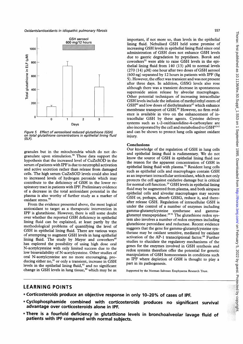
Figure 5 Effect of aerosolised reduced glutathione (GSH)on total glutathione concentrations in epithelial lining fluid(ELF).
granules but in the mitochondria which do not de-granulate upon stimulation.58 These data support thehypothesis that the increased level of CuZnSOD in theserum ofpatients with IPF is due to neutrophil activationand active secretion rather than release from damagedcells. The high serum CuZnSOD levels could also leadto increased levels of hydrogen peroxide which maycontribute to the deficiency of GSH in the lower re-spiratory tract in patients with IPF. Preliminary evidenceof a decrease in the total antioxidant potential in theplasma is also worthy of further study as a marker ofoxidant stress.59From the evidence presented above, the most logical
antioxidant to target as a therapeutic intervention inIPF is glutathione. However, there is still some doubtover whether the reported GSH deficiency in epitheliallining fluid can be explained, at least partly by themethodological problems of quantifying the level ofGSH in epithelial lining fluid. There are various waysof attempting to augment GSH levels in lung epitheliallining fluid. The study by Meyer and coworkers45has explored the possibility of using high dose oralN-acetylcysteine with only limited success due to thelow bioavailability of N-acetylcysteine. Other studies oforal N-acetylcysteine are no more encouraging, pro-ducing either no,4' or only a transient, increase in GSHlevels in the epithelial lining fluid,42 and no significantchange in GSH levels in lung tissue,44 which may be as
important, if not more so, than levels in the epitheliallining fluid. Nebulised GSH held some promise ofincreasing GSH levels in epithelial lining fluid since oraladministration of GSH does not enhance GSH levelsdue to gastric degradation by peptidases. Borok andcoworkers52 were able to raise GSH levels in the epi-thelial lining fluid from 140 (13) j1M to normal levels(270 (14) ,uM) one hour after two doses ofGSH aerosol(600 ng) separated by 12 hours in patients with IPF (fig5). However, the effect was transient and was not presentafter three days. In addition, GSSG levels also rosealthough there was a transient decrease in spontaneoussuperoxide anion release by alveolar macrophages.Other potential techniques of increasing intracellularGSH levels include the infusion of methyl/ethyl esters ofGSH60 and low doses of diethylmaleate6' which enhancemembrane transport of GSH.60 However, no firm evid-ence is available in vivo on the enhancement of in-tracellular GSH by these agents. Cysteine deliverysystems such as L-2-oxithiazolidine-4-carboxylate arealso incorporated by the cell and metabolised to GSH62 63and can be shown to protect lung cells against oxidantinjury.
ConclusionsOur knowledge of the regulation of GSH in lung cellsand epithelial lining fluid is rudimentary. We do notknow the source of GSH in epithelial lining fluid northe reason for the apparent concentration of GSH inepithelial lining fluid with plasma.3' Resident lung cellssuch as epithelial cells and macrophages contain GSHas an important intracellular antioxidant, which not onlyprotects the cell against oxidative damage but is criticalfor normal cell function.29 GSH levels in epithelial liningfluid may be augmented from plasma, and both airspaceepithelial cells and alveolar macrophages may secreteGSH or, perhaps, absorb GSSG, reduce it, and there-after release GSH. Regulation of intracellular GSH isunder the control of a number of enzymes includinggamma-glutamylcysteine synthetase and gamma-glutamyl transpeptidase.2931 The glutathione redox sys-tem also involves a number of redox enzymes includingglutathione peroxidase and reductase. Recent evidencesuggests that the gene for gamma-glutamylcysteine syn-thetase may be oxidant sensitive, mediated by oxidantactivation of the AP-1 transcriptional factor.64 Furtherstudies to elucidate the regulatory mechanisms of thegenes for the enzymes involved in GSH synthesis andredox systems therefore offer the potential for geneticmanipulation of GSH homoeostasis in conditions suchas IPF where depletion of GSH is thought to play a
part in its pathogenesis.
Supported by the Norman Salvesen Emphysema Research Trust.
LEARNING POINTS* Corticosteroids produce an objective response in only 10-20% of cases of IPF.* Cyclophosphamide combined with corticosteroids produces no significant survivaladvantage over corticosteroids alone in IPF.
* There is a fourfold deficiency in glutathione levels in bronchoalveolar lavage fluid ofpatients with IPF compared with normal subjects.
S57
on February 5, 2022 by guest. P
rotected by copyright.http://thorax.bm
j.com/
Thorax: first published as 10.1136/thx.50.S
uppl_1.S53 on 1 S
eptember 1995. D
ownloaded from
MacNee, Rahman
1 Crystal RG, Ferrans VJ, Basset F. Biologic bases of pulmonary fibrosis.In: Crystal RG, West JB, eds. The lung: scientific foundations. NewYork: Raven Press, 1991:2031-46.
2 Strieter RM, Lukacs NW, Standiford TJ, Kunkel SL. Cytokines andlung inflammation: mechanisms of neutrophil recruitment to the lung.Thorax 1993;48:765-9.
3 Gauldie J, Jordana M, Cox G. Cytokines and pulmonary fibrosis. Thorax1993;48:931-5.
4 Phan SH. New strategies for treatment of pulmonary fibrosis. Thorax1995;50:415-21.
5 Meyer A, Buhl R, Magnussen H. The effect of oral N-acetylcysteineon lung glutathione levels in idiopathic pulmonary fibrosis. Eur Respir
1994;7:431-6.6 Stack BHR, Choo-Kang YEJ, Heard BE. The prognosis of cryptogenic
fibrosing alveolitis. Thorax 1972;27:535-42.7 Carrington CB, Gaensler EA, Coutu RE, Fitzgerald MX, Gupta RA.
Natural history and treated course of usual and desquamative in-terstitial pneumonia. N Engl JfMed 1978;298:801-9.
8 Turner-Warwick M, Burrows SB, Johnson A. Cryptogenic fibrosingalveolitis: clinical features and their effects on survival. Thorax 1980;35:171-80.
9 Agustini C, Xaubet A, Roca J, Agustini AGN, Rodriguez-Roisin R.Interstitial pulmonary fibrosis with and without associated collagenvascular disease: results of a 2 year follow up. Thorax 1992;47:1035-40.
10 Turner-Warwick M, Burrows B, Johnson B. Cryptogenic fibrosingalveolitis: response to corticosteroid treatment and its effect on sur-vival. Thorax 1980;35:593-9.
11 Meir-Sydow J, Rust M, Dronenberger H. Diagnosis and therapy in
idiopathic pulmonary fibrosis. Allergol Ininnunopathol 1980;8(Suppl):101-9.
12 Winterbaur RH, Hammer SP, Hallman KO, Hayes JE, Pardee NE,Morgan EH, et al. Diffuse interstitial pneumonitis. Clinicopathologicalcorrelation in 20 patients treated with prednisone/azothiaprine. Ant .7
Med 1978;65:661-72.13 Johnstone MA, Kwan S, Snell SJC. Randomised control, trial comparing
prednisolone alone with cyclophosphamide and low dose prednisolonein combination in cryptogenic fibrosing alveolitis. Thorax 1989;44:280-8.
14 Meir-Sydow J, Rust M, Dronenberger H, Theil C, Amthor M, RiemannH. Long term follow-up of lung function parameters in patientswith idiopathic pulmonary fibrosis treated with prednisolone andazothiaprine or d-penicillamine. Prax Klin Pnieuinol/ 1979;33:680-8.
15 Moolman JA, Bardin PG, Possoum DJ, Jobert JR. Cyclosporin as a
treatment for interstitial lung disease of unknown aetiologv. Thorax1991;46:592-5.
16 Cherniack RM, Crystal RG, Kalica AR. NHLBI workshop summary.Current concepts in IPF; a road map for the future. Ant Rev RespirDis 1991;143:680-3.
17 Schwartz DA, Helmers RA, Galvin JR, van Fossen DS, Frees KL,Dayton CS, et al. Determinants of survival in idiopathic pulmonarvfibrosis. Am _7 Respir Crit Care Med 1994;149:445-54.
18 Johnstone IDA, Gomm SA, Kalrs S, Woodcock AA, Evans CC, HindCRK. The management of cryptogenic fibrosing alveolitis in threeregions of the United Kingdom. Eur Respir]f 1993;6:891-3.
19 Editorial. Lung transplantation. Lancet 1992;339:1021-2.20 Crystal RG, Bitterman PB, Rennard SI, Hance AJ, Keogh BA. Interstitial
lung diseases of unknown cause: disorders characterised by chronicinflammation of the lower respiratory tract. N Engli Med 1984;310:
154-66.21 Cantin AM, North SL, Fells GA, Hubbard RC, Crystal RG. Oxidant
mediated epithelial injury in idiopathic pulmonarv fibrosis. .7 ClitInvest 1987;79:1665-73.
22 Phan SH, Gannon DE, Ward PA, Karmiol S. Mechanism of xanthine/xanthine oxidase conversion in endothelial cells: evidence of a rolefor elastase. Am I Respir Cell Mol Biol 1992;6:270-8.
23 Strausz J, Muller-Quernheim J, Steppling H, Ferlinz R. Oxygen radicalproduction by alveolar inflammatory cells in idiopathic pulmonaryfibrosis. Am Rev Respir Dis 1990;141:124-8.
24 Dreisen RB, Schwartz MI, Theofilapoulos AN, Stanford RE. Circulatingimmune complexes in the idiopathic interstitial pneumonias. N EnglJfMed 1978;298:353-7.
25 Hunninghake GW, Gadek JE, Lawley TJ, Crystal RG. Mechanisms of
neutrophil accumulation in the lungs of patients with idiopathicpulmonary fibrosis. _7 Clin Invest 1981;68:259-69.
26 Ward PA, Dugue RE, Sulavik MC, Johnson KJ. In vitro and in vivo
stimulation of rat neutrophils and alveolar macrophages by immune
complexes: production of 0° and H,O.. Ant _7 Pathol 1983;11O:297-309.
27 Cross CE, Van der Vliet A, O'Neill CA, Louie S, Halliwell B. Oxidants,antioxidants, and respiratory tract lining fluids. Environi Health Perspec1994,102(Suppl 10):185-91.
27 Schreck R, Rieber P, Baeuerle PA. Reactive oxygen intermediates as
apparently widely used messengers in the activation of NFKB
transcription factor and HIV. EMBO_7 199 1;10:2247-58.28 Halliwell B, Gutterige JMC. The antioxidants of human extracellular
fluid. Arch Biochent Biophys 1990;280:1-8.29 Griffith OW. Glutathione. In: The encyclopaedia of huntan biology. Lon-
don: Academic Press, 1991;3:907-19.30 Aruoma OI, Halliwell B, Hoey BM, Butlen J. The antioxidant action
of N-acetylcysteine: its reaction with hydrogen peroxide, hydroxylradical, superoxide and hypochloric acid. Free Radic Biol Med 1989;6:593-7.
31 Cantin AM, North SL, Hubbard RC, Crystal RG. Normal alveolar
epithelial lining fluid contains high levels of glutathione. JAppl Physiol1987;63:152-7.
32 Seis H. Oxidative stress: oxidattts attd antioxidants. New York: AcademicPress, 1991.
33 Cantin AM, Hubbard RC, Crystal RG. Glutathione deficiency in theepithelial lining fluid of the lower respiratory tract of patients withidiopathic pulmonary fibrosis. Ant Rev Respir Dis 1989;139:370-2.
34 Lannan S, Donaldson K, Brown D, MacNee W. Effect of cigarettesmoke and its condensates on alveolar epithelial cell injury in vitro.Ant. 7 Physiol: Lung Cell Mol Physiol 1993;266:L92-100.
35 Rinderknecht J, Shapiro L, Krauthammer M, Taplin G, Wasserman K,Uszler JM, et al. Accelerated clearance of small solutes from the lungsin interstitial lung disease. Atit Rev Respir Dis 1980;121:105-17.
36 Li XY, Donaldson K, Rahman I, MacNee W. An investigation ofthe mechanism of the increased epithelial permeability induced bycigarette smoke in vivo and in vitro models. Ant .7 Respir Cnit CareMed 1994;149:1518-25.
37 Li XY, Donaldson K, Broxsn D, MacNee W. The role of tumournecrosis factor in increased airspace epithelial permeability in acutelung inflammation. Anit7 Respir Cell Mol Biol 1995 (in press).
38 Cantin AM, Larivee P, Begin RO. Extracellular glutathione suppresseshuman lung fibroblast proliferation. Anti _7 Respir Cell Mol Biol 1990;3:79-85.
39 Ziment I. Acetylcysteine: a drug with an interesting past and a fascinatingfuture. Respirationi 1986;50:26-30.
40 Smilkstein MJ, Knapp GL, Kulig KW, Rumack BH. Efficacy of oralN-acetylcysteine in the treatment of acetaminophen overdose. Analysisof the National Multicenter Study (1976-1985) N Engl Med 1988;319:1557-62.
41 Cotgreave IA, Eklund A, Larsson K, Moldeus PW. No penetration oforally administered N-acetvlcysteine into bronchoalveolar lavage. ElurJRespir Dis 1987;70:73-7.
42 Bridgeman MME, Marsden M, MacNee W, Flenley DC, Ryle AP.Cysteine and glutathione concentrations in plasma and bron-choalveolar lavage fluid after treatment with N-acetylcysteine. Thorax1991;46:39-42.
43 Borgstrom L, Kagedal B, Paulsen 0. Pharmacokinetics of N-acetyl-cysteine in man. Elur Jf Pharttnacol 1986;31:217-22.
44 Bridgeman MME, Marsden M, Selbv C, Morrison D, MacNee W.Effect of N-acetylcvsteine on the concentrations of thiols in plasma,bronchoalveolar lavage fluid, and lung tissue. Thorax 1994;49:670-5.
45 Meyer A, Magnussen H. Der Effekt von oralem N-Acetylcystein aufdie Glutathion-Konzentration in der bronchoalveolaren Lavage vonPatienten mit fibrosieren-den Lugenerkrankungen. Med K/itt 1991;6:279-83.
46 Walters EH, Gardiner PV. Bronchoalveolar lavage as a research tool.Thorax 199 1;46:613-8.
47 Reynolds HY. Bronchoalveolar lavage. Ant Rev Respir Dis 1987;135:250-63.
48 Rennard S, Basset G, Lecossier D, O'Donnell K, Martin D, CrystalRG. Estimation of volume of epithelial lining fluid recovered by lavageusing urea as a marker of dilution. _7 Appl Physiol 1986;60:532-8.
49 Marcy TW, Merrill WM, Rankin JA, Reynolds HY. Limitation of usingurea to quantify epithelial lining fluid recovered by bronchoalveolar
lavage. Ant Rev Respir Dis 1987;135:1276-80.50 Jones JG, Royston D, Minty BD. Changes in alveolar-capillary barrier
function in animals and humans. Anti Rev RespirDis 1983;127:S51-9.51 Bridges AB, Scott NA, Parrv GJ, Belch JJF. Age, sex, cigarette smoking
and indices of free radical activity in healthy humans. Eur 7 Med1993;2:205-8.
52 Borok Z, Buhl R, Grimes GJ, Bokser AD, Hubbard RC, Holroyd KJ,et al. Effect of glutathione aerosol on oxidant-antioxidant imbalance
in idiopathic pulmonary fibrosis. Lanicet 1991;338:215-6.53 Munday R. Toxicity of thiols and disulphides: involvement of free
radical species. Free Radic Biol Med 1989;7:659-73.54 Halliwell B, Gutteridge JMC, Cross CE. Free radicals antioxidants and
human disease: where are we now?.7 Lab Clin Med 1992;119:598-620.55 Halliwell B, Chirico S. Lipid peroxidation: its mechanism, measurement
and significance. An _7 Clitt Nutr 1993;57:S715-25.56 Borzi RM, Grigolo B, Meliconi R, Fasano L, Sturani C, Fabbri M, et
al. Elevated serum superoxide dismutase levels correlate with disease
severity and neutrophil degranulation in idiopathic pulmonary fibrosis.
Clin Sci 1993;85:353-9.57 Grigolo B, Borzi RM, Fasano L, Fabbri M, Meliconi R, Facchini A.
Superoxide dismutases in idiopathic pulmonary fibrosis. Clin Sci 1995;88:371.
58 Pham Huu T, Marquettv C, Amit N, Hakin J. Effect of degranulationon superoxide dismutase activity in human neutrophils. .7 Free Radic
Riol Med 1986;2:213-7.59 Wehbe LA, Rahman I, Drost E, MacNee W. Oxidant/antioxidant im-
balance in acute exacerbations of COPD. Ant Rev Respir Crit Care
Med 1994;149:A458.60 Puri RN, Meister A. Transport of glutathione, as gamma-glutamyl-
cysteinyl glycyl ester, into liver and kidney. Proc Natl Acad Sci USA
1993;80:5258-60.61 Deneke SM, Baxter DF, Phelps DT, Fanburg BL. Increase in endothelial
cell glutathione and precursor amino acids uptake by diethylmaleateand hyperoxia. An _7 Physiol 1989;257:L265-71.
62 Williamson JM, Boettcher B, Meister A. Intracellular cysteine deliverysystem that protects against toxicity by promoting glutathione syn-thesis. Proc Natl Acad Sci USA 1982;79:6246-9.
63 Porta P, Aebi S, Summer K, Lauterburgh BH. L-2-oxothiazolidine-4-
carboxylic acid, a cysteine pro-drug. Pharmakinetics and effects on
thiols in plasma and lymphocytes in human. .7 Pharntacol Exp Ther
199 1;257:331-4.64 Rahman I, MacNee W. Glutathione and its redox system
epithelial cells in response to oxidative stress. Am Rev Respir CritMed 1994;149:A457.
S58
on February 5, 2022 by guest. P
rotected by copyright.http://thorax.bm
j.com/
Thorax: first published as 10.1136/thx.50.S
uppl_1.S53 on 1 S
eptember 1995. D
ownloaded from

![FEN VE MÜHENDİSLİK DERGİSİ - web.deu.edu.trweb.deu.edu.tr/fmd/s53/S53-m7.pdf · bağlı polimer ağlardır [19, 20] ... 4/glisin elat kompleksi varlığında, arap sakızı ve](https://img.pdfslide.us/doc/110x75/5d0758a788c993ea1b8c6014/fen-ve-muehendislik-dergisi-webdeuedutrwebdeuedutrfmds53s53-m7pdf.jpg)