Embed Size (px)
Citation preview

Thoracic wall - structure, blood
supply and innervation
Ingrid Hodorová
UPJŠ LF, Dept. of Anatomy
MediTec training for students1.-15.9.2019, Kosice, Slovakia

Thoracic borders
external -Upper:jugular notch,
clavicule, acromionscapulae, spine of C7 (vertebra
prominens)
Lower:xiphoid process,
costal arches (right and left),
Th12
internal -
Upper:superior thoracic aperture:
jugular notch, 1. pair of ribs, Th1
Lower:inferior thoracic aperture:diaphragm (right side - to 4. ICS
left side - to 5. ICS)

Lines of orientation
Anterior median line (midsternal)
Sternal line
Parasternal l.
Midclavicular l.
Anterior axillary l.
Middle axillary l.
Posterior axillary l.
Scapular l.
Paravertebral l.
Posterior median line

Layers of thoracic wall
► Deep layer - osteothorax, muscles of proper thoracic wall + intrinsic muscles of the back, deep
structures, endothoracic fascia
► Middle layer - thoracohumeral mm., spinohumeral mm., spinocostal mm., (fascie, vessels,
nerves)
► Superficial layer - skin, subcutaneous tissue, superficial structures, mammary gland
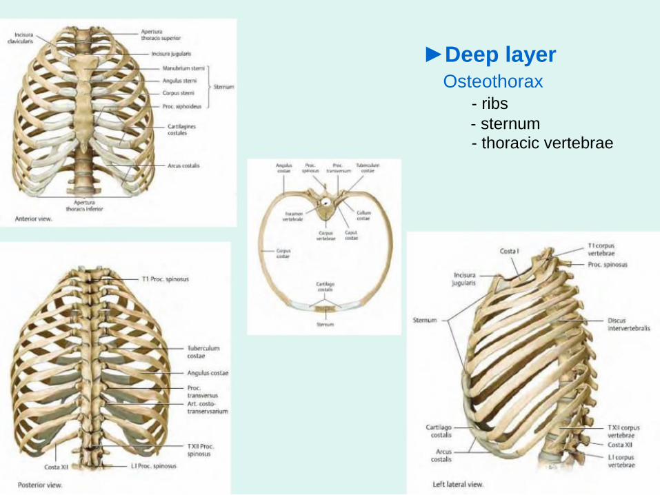
►Deep layerOsteothorax
- ribs
- sternum- thoracic vertebrae

OsteothoraxRibs
Types of ribs:

Sternum- manunbrium of sternum
- body of sternum
- xiphoid process
- manunbriosternal and xiphisternal
synchondrosis(synostosis)

Movement of the ribs and sternum
during breathing

Thoracic vertebrae
- body
- arch
(lamina+pedicles)
- spinous process
- transverse processes
- superior and inferior
articular processes

Joints of the ribs
anteriorly
►sternocostal joints (2nd-5th ribs)
posteriorly
►costovertebral joints
- joints of the heads of the ribs
- costotransverse joints

Ligaments of vertebral column
(Syndesmosis)
► long ligaments:
anterior et posterior longitudinal
ligaments
► short ligaments:
flava ligaments
interspinous ligaments
intertransverse ligaments

Ligaments of vertebral column

Intervertebral disk
(synchondrosis)
● number: 23
1st i.d.: C2-C3
last i.d.: L5-S1
• nucleus pulposus
et annulus fibrosus
Os sacrum
(synostosis)

Joints of vertebral column
- superior and inferior articular
processes

Curvatures of vertebral column
-sagital plane● Lordoses - cervical and lumbar
● Kyfoses - thoracic and sakral
-frontal plane
● Skoliosis

Radiograph of thorax(postero-anterior projection)

Radiograph of vertebral
column, thoracic part(antero-posterior projection)

►Deep layer
Mm. of the proper thoracic wall
1. intercostals external - inspiration
2. intercostals internal - expiration
3. intercostals intimi (innermoust) - exp.
4. subcostals - exp.
5. transversus thoracis - exp.
Blood supply: post. intercostal aa. vv.,
internal thoracic a.+v.,
supreme intercostal a.+v.
Nerve supply: intercostal nn.

►Deep layerIntrinsic mm. of the back
Erector spinae - in the osteofibrous canal
lateral to spinous processes
- in cervical region by lig. nuchae
- in lumbar region - is covered by
thoracolumbar fascia
-in sacral region - starts by tendon
Origin: median sacral crest, spinous procc. of
lumbar, 11. and 12. Th, supraspinal lig.
- divided into 2 tracts:
Lateral tract - iliocostalis,longissimus
Medial tract - spinalis
Innervation:
- dorsal rr. of spinal nerves

►Middle layer (thoracohumeral muscles + mm. of the back)
Thoracohumeral muscles
1. pectoralis major - pectoral br. (from thoracoacromial a.), med.+later.pectoral nn. (brachial pl.)2. pectoralis minor - #
3. serratus anterior - lateral thoracic a. (axillary a.), long thoracic n. (from brachial pl.)
4. subclavius - clavicular br. (a. thoracoacromialis), suclavius n. (brachial pl.)- help during inspirationPectoral fascia
Clavipectoral fascia - attaches to clavicle

Diaphragm

Mm. of the back●1st layer - spinohumeral muscles
1. TrapeziusOrigin: spinous process of Th12-1, C7-1, nuchal
lig., external occipital protuberanceInsertion: spine of scapula, acromion, clavicle
Function: turns, lifts up, adducts scapula to
spine,
turns the headInnervation: accessory n.
2. Latissimus dorsiOrigin: thoracolumbar fascia, spinous process of Th7-
12, L1-5,
sacrum, lower ribs
Insertion: crest of lesser tubercleFunction: extension, internal rotation of humerusInnervation: thoracodorsal n.
Mm. of the back●2nd layer - spinohumeral muscles
1. Levator scapulaeOrigin: transverse proc. C1-4
Insertion: sup. angle of scapula
2. Rhomboid minor
Origin: spinous proc. C6-7
3. Rhomboid majorOrigin: spinous proc. Th1-4
Insertion: medial margin of scapula
Function: lift up scapulaInsertion: med. margin of scapula
Innervation: dorsal scapular n.

Mm. of the back●3rd layer - spinocostal mm.
1. Serratus posterior superior
Function:- pulls the ribs upward
- inspiration
2. Serratus posterior inferior
Function:- pulls the ribs down
- expiration
Innervation: - Intercostal nn.
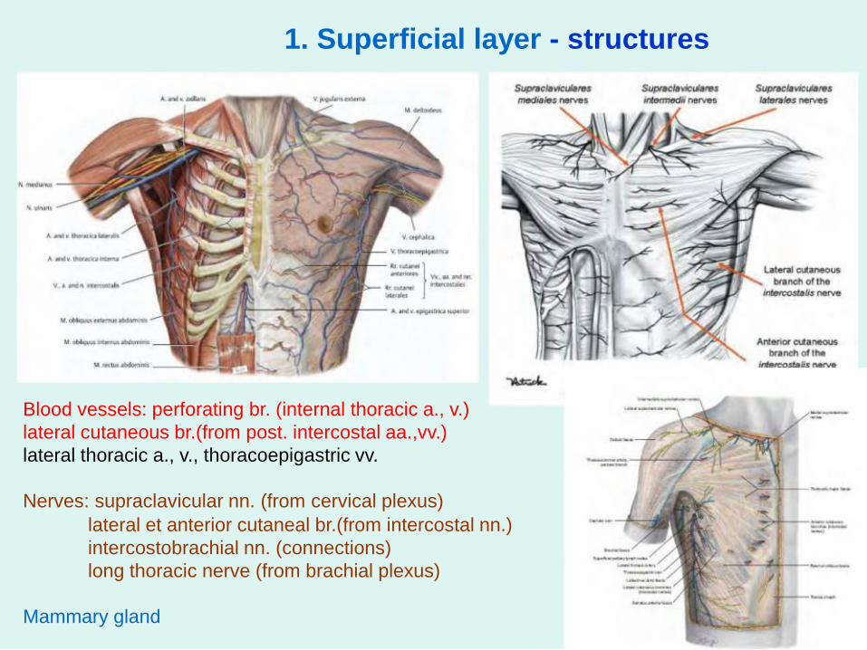
1. Superficial layer - structures
Blood vessels: perforating br. (internal thoracic a., v.)
lateral cutaneous br.(from post. intercostal aa.,vv.)
lateral thoracic a., v., thoracoepigastric vv.
Nerves: supraclavicular nn. (from cervical plexus)
lateral et anterior cutaneal br.(from intercostal nn.)
intercostobrachial nn. (connections)
long thoracic nerve (from brachial plexus)
Mammary gland

Arteries of the thoracic wallThree sources:
axillary artery
subclavian artery
thoracic aorta

Arteries of the thoracic wall - continuation
Axillary artery
Superior thoracic artery
Thoracoacromial artery
Lateral thoracic artery
Subscapular artery
thoracodorsal artery
circumflex scapular artery

Arteries of the thoracic wall -
(Arterial blood supply of ICS)
Thoracic aorta
3.-12. posterior intercostal aa (aa.ICP), a.subcostal a.
Subclavian artery
costocervical trunk
supreme IC a.
1.-2. aa.ICP
╔► branches of posterior intercostal a.

Arteries of the thoracic wall
Subclavian artery
internal thoracic a.
internal thoracic v.
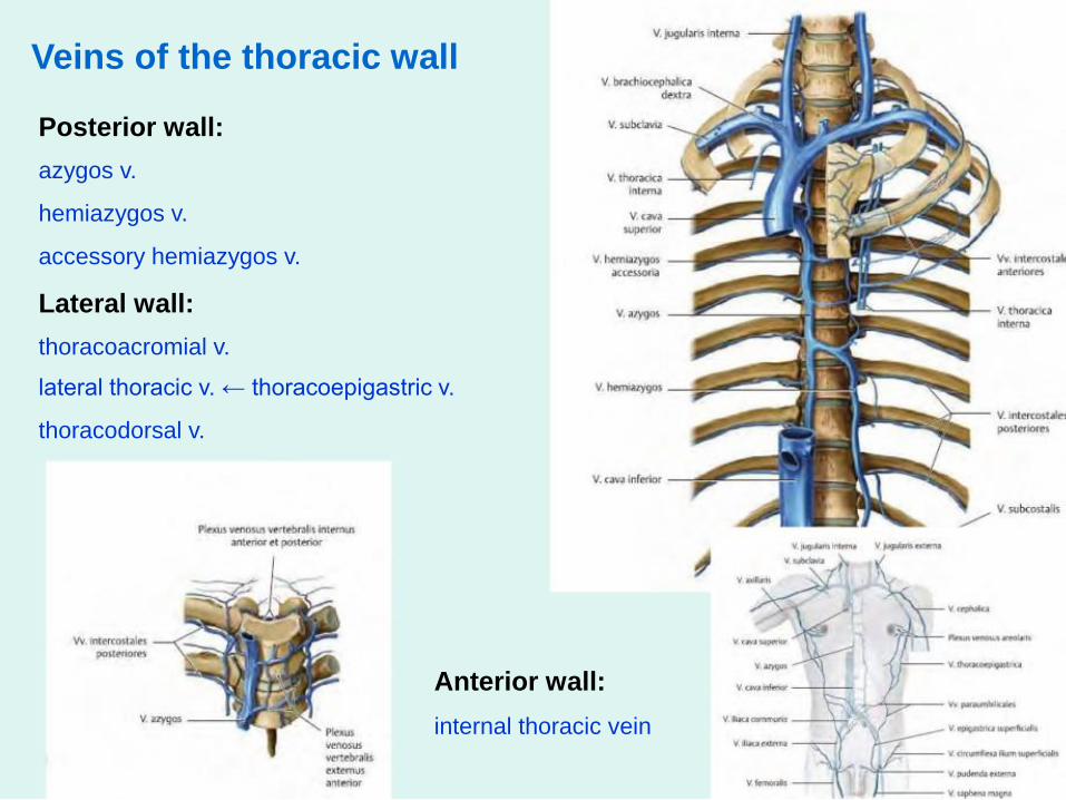
Veins of the thoracic wall
Posterior wall:
azygos v.
hemiazygos v.
accessory hemiazygos v.
Lateral wall:
thoracoacromial v.
lateral thoracic v. ← thoracoepigastric v.
thoracodorsal v.
Anterior wall:
internal thoracic vein

Innervation of the thoracic wall
Motor innervation:
intercostals nerves (12 pairs)
dorsal r. of spinal nerves
long thoracic nerve
thoracodorsal nerve
pectoral medial et lateral nerves
phrenic nerve
accessory nerve

Innervation of the thoracic wall - continuation
Sensory innervation:
intercostals nerves
supraclavicular nerves
intercostobrachial nerves
╔►branches of intercostal nerve

Neurovascular bundle
- VAN
3 parts in intercostal space: posterior intercostal vein, artery and intercostal nerve = VAN
1. Between endothoracic fascia and internal intercostal
2. Between internal intercostals mm. > separate innermoust (intimi) intercostals3. Between endothoracic fascia and internal intercostals

Lymphatic drainage of thoracic wall
Lymph nodes: visceral and parietal
Parietal:- Parasternal l.n. (from mammary gland, liver, diaphragm,pericardium, intercostal spaces)
- Intercostal (from intercostal spaces, deep muscles of the back, vertebral canal)
- Prevertebral l. n.- Phrenic (from diaphragm, liver)
- Axillary (from mammary gland, thoracic wall, upper limb)

Mammary gland - lymphatic drainage
1. Axillary lymph nodes(interpectoral l.n. - Sorgius l.n.)
2. Parasternal l.n.
3. Supraclavicular l.n.
4. Superfical inguinal l.n.

Mammary gland - blood supply, innervation
Blood supply:
1. Thoracoacromial a. - pectoral br.
2. Lateral thoracic a. - lateral mammary br.
3. Internal thoracic a. - medial mammary br.
(perforating br.)
Nerve supply:
1. Lateral mammary br. (intercostal nn.)
2. Medial mammary br. (intercostal nn.)
3. Medial supraclavicular nn. (cervical pl.)

Projection and auscultation of heart
Testute´s points
A. Right 2.ICS - 1cm from
sternum - aortic valve
B. Right 5. ICS next to sternum
- tricuspid valve
C. Left 5. ICS - 5cm from sternum
- bicuspid valve
D. Left 2. ICS - 2cm from sternum
- valve of pulmonary trunk
A D
B
C

Projection of lungs, pleura Lungs, pleura
Anterior border:
- major supraclavic. fossa, caudallyto 2. pair of ribs, 2.-4. ribs - together,
Right side- to 6. rib
Left-to 6.rib, curves laterally
Lower border:Parastern.l. - cartilage of 6.rib
Midcalvicular l. - 6. rib
Axillary l.- 8. rib
Scapular l.-10.rib
Posterior border:
In paravertebr.l. to apex of lung
Pleura: inferior - 1rib lower than lungs
Sup. interpl. area - thymic
Inf. interpl. area - pericardiac

Take home messageStudy!- Bones (costae, sternum, vertebrae)- Joints of ribs (sternocostal et vertebrocostal art.)
- Continuous and discontinuous joints of vertebral column (ligaments,intervertebral disk, intervertebral art.)
- Mm. of the thorax ( mm. of proper thoracic wall+ thoracohumeral mm.)
- Mm. of the back (spinohumeral mm. + spinocostal mm. + deep mm. of theback)
- Arteries of thoracic wall (thoracic aorta, internal thoracic a., repeat branchesof axillary a.)
- Veins of thoracic wall (internal thoracic v., lat. thoracic v.,
thoracoepigastric v., thoracoacromial v., azygos v., hemiazygos v.,accesory hemiazygos v.)
- Lymph nodes of thoracic wall - parietal- Inervation of thoracic wall (intercostal nn., repeat nerves of brachial plexus
- supraclavicular part, supraclavicular nn.)
- Mammary gland

Thank you for your attention


![Muscle Innervation Chart II[1]](https://img.pdfslide.us/doc/110x75/55241db64a7959da488b45f0/muscle-innervation-chart-ii1.jpg)