Embed Size (px)
Citation preview

77:222 Spring 2005 Free Radicals in Biology and Medicine Page 0
This student paper was written as an assignment in the graduate course
Free Radicals in Biology and Medicine
(77:222, Spring 2005)
offered by the
Free Radical and Radiation Biology Program
B-180 Med Labs The University of Iowa
Iowa City, IA 52242-1181 Spring 2005 Term
Instructors:
GARRY R. BUETTNER, Ph.D. LARRY W. OBERLEY, Ph.D.
with guest lectures from:
Drs. Freya Q . Schafer, Douglas R. Spitz, and Frederick E. Domann The Fine Print: Because this is a paper written by a beginning student as an assignment, there are no guarantees that everything is absolutely correct and accurate. In view of the possibility of human error or changes in our knowledge due to continued research, neither the author nor The University of Iowa nor any other party who has been involved in the preparation or publication of this work warrants that the information contained herein is in every respect accurate or complete, and they are not responsible for any errors or omissions or for the results obtained from the use of such information. Readers are encouraged to confirm the information contained herein with other sources. All material contained in this paper is copyright of the author, or the owner of the source that the material was taken from. This work is not intended as a threat to the ownership of said copyrights.

S. Jetawattana Thalassemias, disorders of hemoglobin synthesis 1
Thalassemias, disorders of hemoglobin synthesis
By
Suwimol Jetawattana
Department of Radiation Oncology
Free Radical and Radiation Biology
The University of Iowa
Iowa City, IA 52242-1181
For 77:222, Spring 2005
5 May 2005
Abbreviations:
Epo: erythropoietin; Fe2+: ferrous iron; Fe3+: ferric iron; GSH: reduced glutathione; Hb: hemoglobin; H2O2:
hydrogen peroxide; LDL: low density lipoprotein; MDA: malondialdehyde; MnSOD: manganese superoxide
dismutade; O2: oxygen; O2●-: superoxide anion radical; ●OH: hydroxyl radicals; PMN: polymorphonuclear
neutrophil
S. Jetawattana Thalassemias, disorders of hemoglobin synthesis 2
Outline Page
1. Abstract 3
2. Introduction 4
3. Normal structure and expression of globin gene clusters 4
4. Normal hemoglobin 6
5. Classification of thalassemia 6
Beta thalassemia 6
Alpha thalassemia 7
6. Genetic and molecular basis for the thalassemia 7
Globin synthesis in beta thalassemia 7
Globin synthesis in alpha thalassemia 8
7. Prevalence and geographic distribution 10
8. Free radicals and oxidative stress in thalassemia 11
9. Role of free radical reactions and mechanism of injury 13
10. Future research direction 15
11. Bone marrow and stem cell transplantation 15
12. Oxygen-regulated gene therapy 16
13. Antioxidant therapy 17
14. Experiment design to test the hypothesis 18
Experiments using animal model 18
Experiment using human model 19
15. Summary 20
16. References 21
S. Jetawattana Thalassemias, disorders of hemoglobin synthesis 3
Abstract
Thalassemias are a group of hereditary diseases of abnormal hemoglobin synthesis where
the normal hemoglobin protein is produced in lower amounts than usual. These conditions cause
varying degrees of anemia, which can range from insignificant to life threatening. Thalassemia
can be classified according to the deficient globin chain, alpha or beta thalassemia. Patients with
severe anemia who receive regular blood transfusions become iron overloaded, which increases
damaging free radical activity and lowers antioxidant levels in their bodies. Several investigators
have provided evidence supporting the hypothesis that oxidative stress plays an important role in
development of clinical complications in thalassemia. Increasing antioxidant activity is then
expected to bring oxidative stress down to minimal level. Therefore, it is important to
understand the mechanisms of free radical-induced oxidative stress damage in thalassemia before
using it as a potential tool in diagnosis and treatment of the disease.

S. Jetawattana Thalassemias, disorders of hemoglobin synthesis 4
Introduction
Thalassemia is described as a heterogenous group of inherited anemias characterized by a
reduced or absent amount of hemoglobin. Thalassemia can be classified according to the
deficient globin chain, alpha or beta thalassemia. The symptoms vary from relatively mild
anemia to life-threatening. Many studies have shown that reactive oxygen species are generated
in increased amounts in thalassaemic red cells. Conditions such as rupture of erythrocytes, iron
overload, and depletion of antioxidants in tissues (e. g. blood circulation) are considered in the
promotion of oxidative stress. This implies the possible alteration of redox status in thalassaemic
patients, which may adversely affect their health. Specific treatments for thalassemia are
employed based on many factors such as age of the patients and severity of the disease.
Currently, studies on the correlation between lipid peroxidation and plasma levels of antioxidants
such as vitamin A, C, and E as well as correlations with antioxidant enzymes to hemoglobin
disorders have been reported (5, 12, 13). An effective form of gene therapy and stem cell
transplantation are now being used to improve conventional treatment and enhanced the
prognosis of thalassaemia (20, 22,23). The goal of this paper is to provide information about
thalassemia and will focus on its correlation with oxidative stress.
Normal structure and expression of globin gene clusters
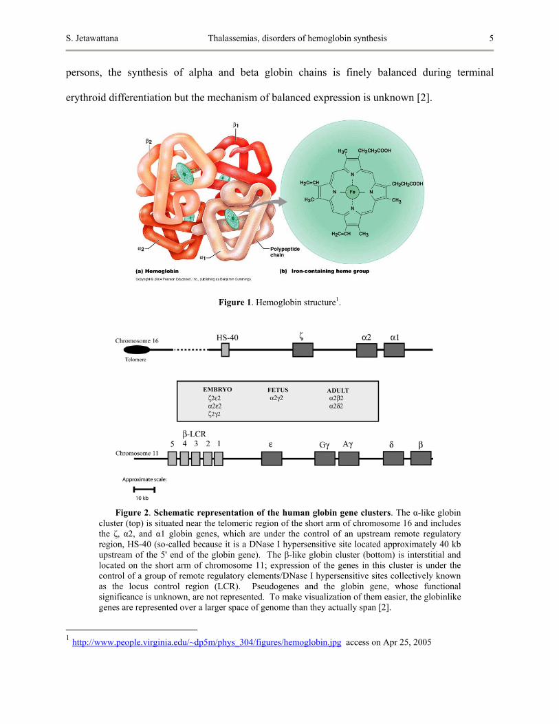
Human hemoblobin is a heterotetramer protein, compose of two alpha and two beta
subunits as shown in Figure 1. Each subunit contains a heme group, an iron containing
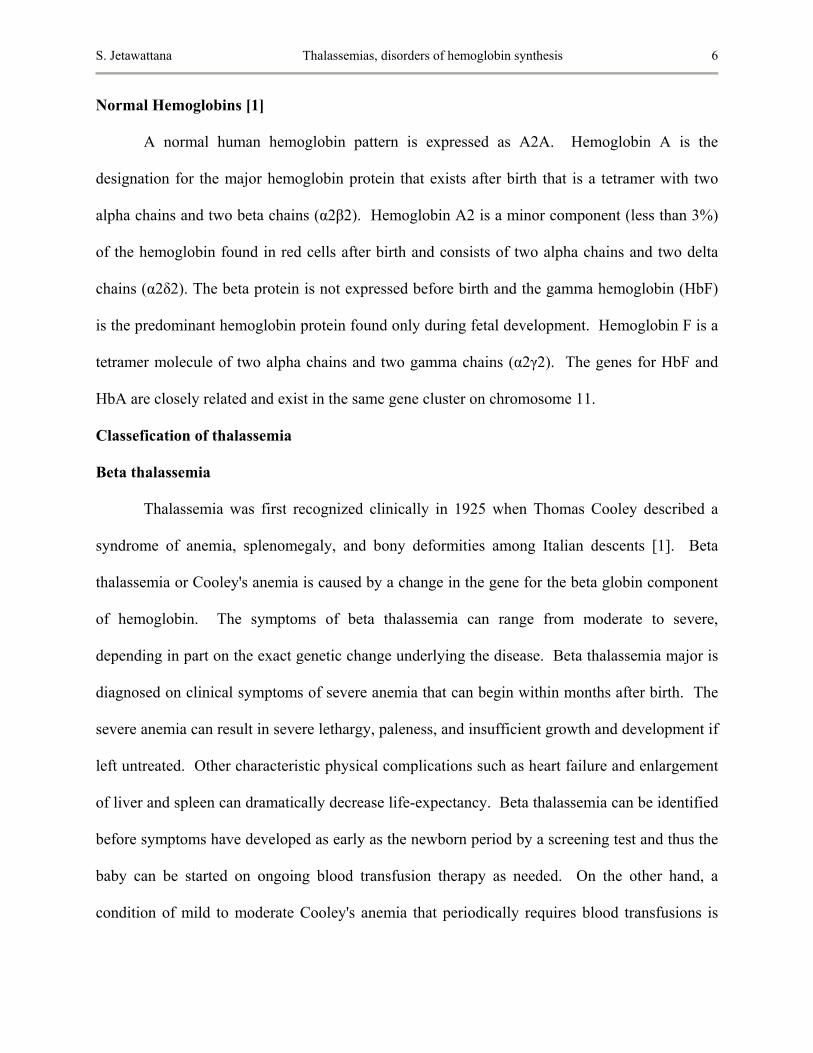
compound that binds to oxygen. The synthesis of hemoglobin is controlled by two
developmentally regulated multigene clusters: the alpha-like globin cluster on chromosome 16
and the beta-like globin cluster on chromosome 11 as demonstrated in Figure 2. In healthy
S. Jetawattana Thalassemias, disorders of hemoglobin synthesis 5
persons, the synthesis of alpha and beta globin chains is finely balanced during terminal
erythroid differentiation but the mechanism of balanced expression is unknown [2].
Figure 1. Hemoglobin structure1.
Figure 2. Schematic representation of the human globin gene clusters. The α-like globin cluster (top) is situated near the telomeric region of the short arm of chromosome 16 and includes the ζ, α2, and α1 globin genes, which are under the control of an upstream remote regulatory region, HS-40 (so-called because it is a DNase I hypersensitive site located approximately 40 kb upstream of the 5' end of the globin gene). The β-like globin cluster (bottom) is interstitial and located on the short arm of chromosome 11; expression of the genes in this cluster is under the control of a group of remote regulatory elements/DNase I hypersensitive sites collectively known as the locus control region (LCR). Pseudogenes and the globin gene, whose functional significance is unknown, are not represented. To make visualization of them easier, the globinlike genes are represented over a larger space of genome than they actually span [2].
1 http://www.people.virginia.edu/~dp5m/phys_304/figures/hemoglobin.jpg access on Apr 25, 2005
S. Jetawattana Thalassemias, disorders of hemoglobin synthesis 6
Normal Hemoglobins [1]
A normal human hemoglobin pattern is expressed as A2A. Hemoglobin A is the
designation for the major hemoglobin protein that exists after birth that is a tetramer with two
alpha chains and two beta chains (α2β2). Hemoglobin A2 is a minor component (less than 3%)
of the hemoglobin found in red cells after birth and consists of two alpha chains and two delta
chains (α2δ2). The beta protein is not expressed before birth and the gamma hemoglobin (HbF)
is the predominant hemoglobin protein found only during fetal development. Hemoglobin F is a
tetramer molecule of two alpha chains and two gamma chains (α2γ2). The genes for HbF and
HbA are closely related and exist in the same gene cluster on chromosome 11.
Classefication of thalassemia
Beta thalassemia
Thalassemia was first recognized clinically in 1925 when Thomas Cooley described a
syndrome of anemia, splenomegaly, and bony deformities among Italian descents [1]. Beta
thalassemia or Cooley's anemia is caused by a change in the gene for the beta globin component
of hemoglobin. The symptoms of beta thalassemia can range from moderate to severe,
depending in part on the exact genetic change underlying the disease. Beta thalassemia major is
diagnosed on clinical symptoms of severe anemia that can begin within months after birth. The
severe anemia can result in severe lethargy, paleness, and insufficient growth and development if
left untreated. Other characteristic physical complications such as heart failure and enlargement
of liver and spleen can dramatically decrease life-expectancy. Beta thalassemia can be identified
before symptoms have developed as early as the newborn period by a screening test and thus the
baby can be started on ongoing blood transfusion therapy as needed. On the other hand, a
condition of mild to moderate Cooley's anemia that periodically requires blood transfusions is

S. Jetawattana Thalassemias, disorders of hemoglobin synthesis 7
described as beta thalassemia intermedia while beta thalassemia minor (also called thalassemia
trait) may cause no symptoms, but changes in the blood do occur.
Alpha thalassemia
Alpha thalassemia is the result of reduction in the synthesis of the alpha globin chains.
Two main types of alpha thalassemia are described as alpha thalassemia major and hemoglobin
H disease. Alpha thalassemia major is a very serious disease of severe anemia characterized by
hypochromic microcytic anemia that begins even before birth. Most affected babies do not
survive full gestation or die shortly after birth. In contrast, patients with HbH disease can
experience events of hemolytic anemia, which is caused by the rapid breakdown of the red blood
cells. These events are thought to be triggered by various environmental causes, such as
infection and/or exposure to certain chemicals. Hemoglobin H disease is milder than beta
thalassemia and does not generally require transfusion therapy [1]. However, the combination of
the very low production of alpha chains and destruction of red cells in HbH disease can produce
a severe, life-threatening anemia.
Genetic and molecular basis for thalassemia
The inheritance is recessive in nearly all types of thalassemia. The rare exception is most
commonly found in beta thalassemia where globin gene mutations exhibit a dominant pattern of
inheritance in which only one gene needs to be altered in order to see disease expression2.
Globin synthesis in beta thalassemia
Healthy persons make the beta globin component of normal adult hemoglobin, HbA,
from two normal copies of the beta globin gene, which is located on chromosome 11. The main
pathophysiologic feature of beta thalassemia is the accumulation of unpaired alpha globin chains
in erythrocyte precursors and red blood cells. This accumulation alters cell membrane function 2 http://www.healthatoz.com/healthatoz/Atoz/ency/thalassemia.jsp accessed on Apr 25, 2005

S. Jetawattana Thalassemias, disorders of hemoglobin synthesis 8
and results in ineffective erythropoiesis and early cell destruction. In contrast to alpha
thalassemia, most of the beta thalassemia syndromes are caused by mutations affecting gene
regulation or expression rather than gene deletion [1]. The mutations are designated as β0 where
no beta globin is produced while only small fraction of the normal amount of beta globin is
produced with a β+ mutation. When one normal beta globin gene and one beta thalassemia
mutation are present, it is said that a person carries the beta thalassemia trait, which is thought
not to cause health problems.
There are other thalassemia-like mutations that can affect the beta globin gene.
Hemoglobin E is a hemoglobin variant which has structural defect in hemoglobin molecule
resulting from a single nucleotide base substitution in the hemoglobin beta chain. The
combination of HbE and beta thalassemia manifests a condition more severe than is seen with
either HbE trait or beta-thalassemia trait. Large deletions around and including the beta globin
gene can lead to delta/beta thalassemia or hereditary persistence of fetal hemoglobin (HPFH).
Clinical manifestations of the delta/beta thalassemia trait behave very similarly to beta
thalassemia trait, while the HPFH trait is not likely to cause hemoglobin disease when co-
inherited with other beta globin mutation or a second thalassemia.
Globin synthesis in alpha thalassemia
Alpha thalassemia occurs when there is a reduction in the synthesis of the alpha globin
chains of adult hemoglobin (HbA, α2β2) relative to beta globin synthesis. Two alpha
thalassemia phenotypes are recognized; one is associated with a total absence of alpha globin
synthesis and is designated α0 thalassemia while the other phenotype, designated as α+
thalassemia, is only a reduction in alpha globin synthesis [1]. The severe imbalance between the
alpha chain and beta chain production causes an accumulation of beta chains inside the red blood

S. Jetawattana Thalassemias, disorders of hemoglobin synthesis 9
cells. The excess beta chains then form homotetramers (HbH, β4) that are characterized by
erythrocyte inclusions and can be detected by supravital staining, chromatography, or gel
electrophoresis. These tetramers of beta globin subunits do not transport oxygen properly,
making it functionally useless to the cell. Moreover, HbH protein damages the membrane that
surrounds the red cell, accelerating cell destruction. The α0 thalassemia phenotype is caused by
several deletions affecting both alpha globin genes. Some of the α+ thalassemias result from a
deletion involving one of the two alpha globin genes. The other result from nondeletion
mutations, give rise to structurally abnormal hemoglobin and limit alpha gene expression.
Interactions of the mutations causing deficient alpha globin synthesis produce a spectrum of
phenotypes that can be grouped into four clinical syndromes [1]. In each syndrome, the severity
of symptoms correlates closely with the deficiency of alpha globin chains relative to beta chains.
The loss of one alpha globin structural gene diminishes the production of the alpha protein only
slightly. This condition is very close to normal and a person with this condition is called a silent
carrier. The syndrome of alpha thalassemia minor is characterized by the two-gene deletion that
produces a condition with small red blood cells, but anemia is mild or absent. The three-gene
deletion of alpha thalassemia produces a serious hematological problem with severe anemia and
often requires blood transfusions to survive. The loss of all four alpha genes causes the gamma
chains produced during fetal life to form homotetramers hemoglobin Bart’s. Most individual
with four-gene deletion alpha thalassemia die in utero or shortly after birth.
In addition, abnormalities of hemoglobin synthesis may also arise as a secondary
manifestation of another disease. Acquired alpha thalassemia is reported to be associated with a
myelodysplastic syndrome (MDS) and other hematologic malignancies such as x-linked alpha
thalassemia mental retardation syndrome (ATR-X). Two molecular mechanisms for acquired
S. Jetawattana Thalassemias, disorders of hemoglobin synthesis 10
alpha thalassemia are now recognized: acquired deletion of the alpha globin gene cluster limited
to the neoplastic clone and, more commonly, inactivating somatic mutations of the trans-acting
chromatin-associated factor ATRX. This causes dramatic down regulation of the alpha globin
gene expression. The relationship between ATRX mutations that lead to reduced expression of
alpha globin gene and a thalassemia through epigenetic mechanisms is still unclear [2].
Prevalence and geographic distribution of thalassemia [1]
The thalassemias are among the most common genetic diseases worldwide. Both alpha
and beta thalassemia have been described in individuals of almost every ancestry, but the
conditions are more common among certain ethnic groups. Beta thalassemia trait is seen most
commonly in people with the following ancestry: Mediterranean (including North African, and
particularly Italian and Greek), Middle Eastern, Indian, African, Chinese, and Southeast Asian
(including Vietnamese, Laotian, Thai, Singaporean, Filipino, Cambodian, Malaysian, Burmese,
and Indonesian). All types of alpha thalassemia disease are most common among people of
Southeast Asian and Chinese descent. Unaffected carriers of all types of thalassemia traits do
not experience health problems. Many different mutant alleles of globin genes have been
selected for many generations to reach high frequencies in tropical and subtropical regions of the
world. Coincidentally, these mutations increased the likelihood that carriers would survive
malaria infection and allowed survivors to pass the mutation onto their offspring. Thus the trait
became established throughout areas where malaria is common. The geographic distribution of
thalassemia trait population increased when populations migrated. However, it is difficult to
obtain accurate prevalence for various types of thalassemia within different populations due to
limitations in diagnostic testing, as well as the fact that many studies have focused on small,
biased hospital populations.

S. Jetawattana Thalassemias, disorders of hemoglobin synthesis 11
Free radicals and oxidative stress in thalassemia
Oxidative stress, from the generation of reactive oxygen species (ROS), is believed to
play a role in the pathophysiology of thalassemia. One study has reported that significant ROS
and lipid peroxidation were found to be higher, and GSH lower in beta thalassemic red blood
cells compared to their normal counterparts [6, 7]. Similar results were found in
polymorphonuclear neutrophils (PMN) and platelets obtained from beta thalassemia patients
which indicated a state of oxidative stress [8]. In addition, the conditions such as rupture of
erythrocytes, iron overload and depletion of antioxidants in tissues and blood circulation have
been reported to be common in beta thalassemia [3, 4]. The levels of cellular antioxidant vitamin
such as vitamin A, C and E, as well as the activities of enzymatic antioxidants such as catalase,
glutathione peroxidase, and glutathione reductase, were found to be considerably lower in
thalassemic patients compared to normal subjects. These results suggest a major consumption of
antioxidants under iron overload from continuous blood transfusions or oxidative stress in
thalassemia [12, 13, 14]. It has also been reported that superoxide dismutase and catalase
activities increased significantly in beta thalassemia patients seminal plasma but total antioxidant
status values were unaltered. However, seminal lipoperoxidation did increase. These results
also suggested an oxidative stress in semen of these patients and it could have contributed to the
impairment of sperm motility [15]. These imply the possible alteration of redox status in
thalassemic patients, which may unfavorably affect their health.
In order to prevent severe anemia and allow for normal growth and development, patients
with beta thalassemia major usually receive regular blood transfusions on a monthly basis.
While transfusions can prevent many of the complications of severe anemia, the body is unable
to eliminate the iron overload that accompanies each transfusion. This excess iron deposits in

S. Jetawattana Thalassemias, disorders of hemoglobin synthesis 12
tissues and organs, resulting in damage and organ failure [1]. The mechanism of iron-induced
organ dysfunction was reported to be part of a free radical-mediated process [11]. Additional
chelation therapy, usually with the iron-binding agent desferrioxamine (Desferal), is needed to
help the body get rid of excess iron and to prevent death from complications by tissue iron
toxicity. The peroxidative status generated by iron overload in thalassemia patients was
confirmed when free and total malondialdehyde (MDA) and nontransferrin-bound iron levels
were found to be higher in transfused beta thalassemia major patients than in untransfused beta
thalassemia intermedia patients. In transfused beta thalassemia major patients, the free MDA
levels were positively correlated with serum iron, while the total MDA positively correlated with
nontransferrin-bound iron. However, a negative correlation was observed between the total
peroxyl radical-trapping antioxidant parameter and nontransferrin-bound iron determined by
spectrophotometry. The marked amounts of free MDA were also rapidly formed despite the
chelation therapy. In contrast, no significant correlations between free or total MDA and the
total peroxyl radical-trapping antioxidant parameter or nontransferrin-bound iron were observed
in untransfused beta thalassemia intermedia patients [16]. The elevated level of endogenous
hemin, a denaturative product of hemoglobin (iron (III)- protoporphyrin IX), was reported to be a
reliable cause of oxidative stress in blood circulation of beta thalassemia/HbE disease and may
contribute a major pro-oxidant in blood circulation [5]. Oxidative stress, therefore, is thought to
be an important mechanism for development of clinical complications.

S. Jetawattana Thalassemias, disorders of hemoglobin synthesis 13
Role of free radical reactions and mechanisms of injury
It is generally accepted that iron, released from macromolecules which normally
sequester it, represents the source of iron-catalyzed oxidative stress, such as DNA and protein
oxidation, and lipid peroxidation. Under physiological conditions, iron is not available to
catalyze the conversion of molecular oxygen to the highly reactive radical species by Fenton
chemistry, because ferric iron (Fe3+) is bound to proteins which prevent it from participating in
reactions that could lead to cell injury [3]. One of the mechanisms involved in the oxidative
stress of thalassemia diseases is due to the key reactions involving oxidation and reduction of
hemoglobin that take place in the presence of hydrogen peroxide (H2O2) and superoxide anion
radicals (O2●-) according to Fenton-like reactions as shown in reactions (1) and (2):
Haem-Fe2+ + H2O2 → Fe3+ + OH− + ●OH (1)
Fe3+ + O2●- → Fe2+ + O2 (2)
In general, the O2 carried by hemeproteins is only bound directly to the ferrous iron with
an oxidation state of two (Fe2+). In the presence of H2O2, ferrous iron and H2O2 are going to
react to generate ferric iron (Fe3+) and highly reactive hydroxyl radicals (●OH). Addition of a
reducing agent, such as ascorbate, leads to a cycle which increases the damage to biological
molecules.
According to the Haber–Weiss reactions, consisting of the following reactions shown in
reactions (4) and (5), ●OH and molecular oxygen can be formed. Reaction (4) can be catalyzed
by Fe3+ and is a possible source of ●OH; however, its rate constant is negligible.
O2●- + H2O2 → ●OH + OH− + O2 (4)
●OH + H2O2 → O2●- + H2O + H+ (5)

S. Jetawattana Thalassemias, disorders of hemoglobin synthesis 14
It was demonstrated that the release of Fe3+ from the porphyrin ring increased as a
function of O2 concentration as shown in reactions (6) and (7):
Heme-Fe2+ + O2 → Heme-Fe3+ + O2●- (6)
Heme-Fe3+ → Fe3+ + iron-free Heme (7)
The above oxidation and subsequent ejection of Fe3+ from the porphyrin ring could also be done
by ●OH, as shown in reactions (8) and (9). When Fe3+ is released, it could generate reactive
oxygen species that would, in turn, damage cellular compartments [17]. Thus, iron supplements
should be avoided by patients with thalassemia unless iron deficiency is diagnosed.
Heme-Fe2+ + ●OH → Heme-Fe3+ + OH− (8)
Heme-Fe3+ → Fe3+ + iron-free Heme (9)
In addition, since the modification of low density lipoprotein (LDL) was observed in
patients with the severe hemolytic anemia beta thalassemia [9], it was suggested that under
oxidative stress extracellular chains of normal hemoglobin (HbA) can trigger oxidation of LDL
in the presence of H2O2. The modified LDL particles observed in beta thalassemia may reflect
lipoprotein oxidation by alpha chains in circulation. A possible explanation of the high
peroxidative activity of alpha chains is their ability to undergo appreciable autoxidation yielding
hydrogen peroxide as a byproduct, enhancing LDL peroxidation [10].
Interestingly, in order to investigate the role of oxidant injury in the pathophysiology of
human thalassemias, one study has shown that the apoptotic programs turned on in beta
thalassemic erythroid precursor cells were not triggered by oxidative damage but were dependent
on activation of FAS/FAS-Ligand cell surface interaction. The authors suggested that the
destruction of thalassemic erythroid precursors may involve different mechanisms other than
oxidant injury that cause red blood cells hemolysis [18].

S. Jetawattana Thalassemias, disorders of hemoglobin synthesis 15
Future research directions
The goal for future research in thalassemia is not only to introduce new strategies of
diagnosis and treatment of thalassemia but also to discover ways to prevent the occurrence of
oxidative damage in thalassemic patients. Current work deals with specific complications in
thalassemia patients such as endocrine, cardiopulmonary, thrombophilic, and osteopenic
problems, as well as iron overload. The effectiveness of oral iron-chelating drugs are being
developed and tested, which could greatly simplify treatment of this disease. Studies conduced
on in oxidative status and correlations between antioxidants can also provide insights
accomplishing this purpose. In addition, the thalassemias are likely to benefit from specific gene
therapy designed to overcome the adversity in the synthesis of a specific globin chain [19].
Bone marrow and stem cell transplantation
Specific treatments for thalassemia are employed based on many factors such as age of
the patients and severity of the disease. Red blood cell transfusion, splenectomy, iron chelation
are among the general methods used for thalassemia treatments [1]. A major event in the area
was the discovery that allogeneic bone marrow (stem cell) transplantation could succeed in
severely affected thalassemia patients [19]. However, bone marrow transplants have cured some
cases of thalassemia but they are not widely used due to less successful of the methods [20].
Currently, scientists are investigating the use of umbilical cord cell transplantation3. Cord blood
is an excellent source of stem cells that have remarkable potential to develop into many different
cell types in the body. If it continues to be successful, this could eliminate the need for long-
term transfusion therapy or iron chelation treatment.
3 http://web1.tch.harvard.edu/cfapps/A2ZtopicDisplay.cfm?Topic=Beta%20Thalassemia access Apr 25, 2005
S. Jetawattana Thalassemias, disorders of hemoglobin synthesis 16
Oxygen-regulated gene therapy
Many concepts of gene therapy have been proposed4. Gene therapy may involve
inserting a normal beta or alpha globin gene into the patient's stem cells. The new alpha or beta
globin genes should be designed to insert with appropriate copy number into the hematopoietic
stem cells, but they should be expressed in normal amounts only in erythroid precursors and not
in any of the other nine stem-cell-derived blood cell lines [19]. Presently, significant advances
have been made toward this goal using lentiviral vectors to obtain high-level expression of
complex globin gene cassettes. Therapeutic correction in murine models of both beta-
thalassemia and sickle cell anemia has been achieved using this approach [22, 23]. However,
further refinement of the gene therapy approach in thalassemia requires more animal modeling,
particularly with primate models, before planning a phase one human study. Another form of
gene therapy may concern using drugs or other methods to reactivate the patient's genes for fetal
hemoglobin (HbF). Then they can make the blood cells of patients with thalassemia produce
more fetal hemoglobin to compensate for their deficiency of adult hemoglobin. Initial studies of
rare individuals with genetic traits that allow them to produce only fetal hemoglobin show that
they are generally healthy, demonstrating that fetal hemoglobin can be a fine substitute for adult
hemoglobin [21].
Recently, the development of a physiologically regulated gene therapy that can deliver
physiologically regulated expression of Epo has been demonstrated. One of the study reported
that the continuous delivery of high amounts of autologous erythropoietin, via a recombinant
adeno-associated virus-cytomegalovirus vector into skeletal muscles, induced a sustained
stimulation of beta minor globin synthesis and a stable improvement of erythropoiesis in the beta
thalassemic mouse model [24]. However, the relevance of the study to chronic anemia has been 4 http://www.icomm.ca/geneinfo/thalass.htm access on Apr 25, 2005
S. Jetawattana Thalassemias, disorders of hemoglobin synthesis 17
limited because the Epo gene has been delivered to beta thalassemic mice. Recently, the
potential of genetic delivery of the Epo gene in a more relevant model of chronic anemia was
performed. It was reported that an intramuscular delivery of the constructed promoter, Oxford
Biomedica hypoxia response element (OBHRE), used in treating homozygous erythropoietin-
SV40 T antigen (Epo-TAg(h)) mice with relative erythropoietin deficiency, gives rise to
physiologically regulated erythropoietin secretion. The long-term delivery of Epo expression
can correct the hematocrit level in anemic mice to a normal physiologic level that stabilized
without resulting in polycythemia (an expanded red cell mass) but had no significant effect on
the hematocrit of control mice. The OBHRE-Epo-treated Epo-TAg(h) mice also shown a
significant reversal of cardiac hypertrophy, which is a common adaptive response in patients
with chronic state of anemia. This establishes that a hypoxia regulatory mechanism similar to the
natural mechanism can be achieved, and it makes Epo gene therapy more attractive and safer in
clinical settings [25].
Antioxidant therapy
It should be noticed that many studies are now focusing on the correlations between
antioxidant activities and severity of the disease in thalassemic patients. It has been reported that
oxidative stress-induced changes in thalassemic erythrocytes can be attenuated by vitamin E
[13]. N-Acetylcysteine amide, a novel cell-permeating thiol was reported to restore cellular
glutathione and protect human red blood cells from oxidative stress [26]. Oxidative stress is
greatly increased in thalassemia because of the prolonged exposure to iron accumulation. Thus,
the measurement of ROS or other pro-oxidant activity levels in thalassemic patient’s serum will
be useful for the management of the disease in preventing oxidative damage by appropriate
antioxidant treatment.
S. Jetawattana Thalassemias, disorders of hemoglobin synthesis 18
Experimental design to test the hypothesis
From the evidence of several studies, it is clearly indicated that oxidative stress, at least
some parts, plays an important role in pathophysiologic status of thalassemia disease. In order to
prove this hypothesis and explore the potential combination therapies that would allow for
attenuation of ROS in thalassemia, some of the following experiment designs might provide
promising results for further investigation.
Experiments using animal models
1. In order to reveal and understand whether free radical initiated oxidative damage plays an
important role in thalassemia, the research strategy should directly measure the levels of
free radials and oxidative damage in blood components of normal and thalassemic mice.
The correlations between free radical levels and oxidative damage will provide a clue
underlying pathophysiology of the disease.
2. More information underlying the mechanism of oxidative damage in thalassemia is
needed. A long-term study of free radicals exposure to normal and thalassemic mice may
help to determine the adverse effects of a certain levels of free radicals that develop
severity of anemia and other complications.
3. It might be useful to measure the levels of free radicals, oxidative products in vivo in
thalassemia transgenic mice at different age to see whether free radicals and oxidative
stress levels are influenced by age.
4. A decrease in antioxidant enzyme has been detected in thalassemic patients suggesting
that a deficient activity of antioxidant enzymes may play important role in at lease some
thalassemia case. Increasing antioxidant activity will hopefully reduce oxidative stress
down to minimal level. For this particular study, the overexpression of antioxidant
S. Jetawattana Thalassemias, disorders of hemoglobin synthesis 19
enzymes such as MnSOD and catalase in mice will provide a rational basis for
development of gene therapy as a method of protection against free radical-induced
oxidative stress in thalassemic mice.
Experiments using human model
1. The mechanism of oxidative damage in thalassemia is likely to be complex, involving
iron overload. Utilizing a case-control study design, determine the status of antioxidant,
lipid peroxides in the presence or absence of iron chelating agent might provide
information of mechanism which contributes to iron overload.
2. Vitamin E is a major lipid-soluble antioxidant, and is the most effective chain-breaking
antioxidant within the cell membrane where it protects membrane fatty acids from lipid
peroxidation. Preliminary studies have found that oral supplements of 200 to 600 IU per
day of vitamin E reduce free radical damage to red blood cells in thalassemia patients
[27], while an in vitro study has shown that propionyl-L-carnitine protects red blood cells
of thalassemia patients against free radical damage [28]. These results suggest that
providing appropriate nutritional supplements to the patients may prevent any
complications from hemolytic red blood cells including significantly decreased need for
blood transfusions. Therefore, it might be useful to investigate whether various kinds of
supplementary antioxidants, such as vitamin C, E, beta-carotene, and the resultant
antioxidant plasma status of these nutrients are associated with regression of oxidative
damage of red blood cells.
S. Jetawattana Thalassemias, disorders of hemoglobin synthesis 20
Summary
The fundamental abnormality in thalassemia is impaired production of either the alpha or
beta hemoglobin chain. Several theories of pathogenesis and severity of thalassemia disease
have emerged including free radicals induce oxidative stress. Patients with severe thalassemia
who receive regular blood transfusions become iron overloaded, which increases damaging free
radical activity and lowers antioxidant levels in their bodies. General treatment strategies of
thalassemia are blood transfusion and iron chelation. Besides the promising treatment of stem
cell transplantation and gene therapy, an imbalance in the antioxidant protective mechanisms
leading to oxidative stress in blood components suggests a new era of antioxidant therapy for
thalassemia disease. However, more attempts to put forth the evidence for involvement of free
radicals in pathophysiology of thalassemia and the potential of treatment with antioxidant and
scavenger substances are still needed and required a combination of a multitude of disciplines.

S. Jetawattana Thalassemias, disorders of hemoglobin synthesis 21
References
1.Lukens JN. (1993) The thalassemias and related disorders: quantitative disorders of hemoglobin synthesis. In: Lee GR, ed. Wintrobe’s Clinical Hematology 9th edition Vol. 1. Lea & Febiger, PA Press; 1102-1145.
2. Steensma DP, Gibbons RJ, Higgs DR. (2005) Acquired α-thalassemia in association with myelodysplastic syndrome and other hematologic malignancies. Blood. 105(2): 443-452. 3. Livrea MA, Tesoriere L, Pintaudi AM, Calabrese A, Maggio A, Freisleben HJ, D'Arpa D, D'Anna R, Bongiorno A. (1996) Oxidative stress and antioxidant status in beta-thalassemia major: iron overload and depletion of lipid-soluble antioxidants. Blood. 88(9):3608-3014. 4. Luca C, Filosa A, Grandinetti M, Maggio F, Lamba M, Passi S. (1999) Blood antioxidant status and urinary levels of catecholamin metabolites in ß-thalassemia. Free Radic Res. 30:453-462. 5. Phumala N, Porasuphatana S, Unchern S, Pootrakul P, Fucharoen S, Chantharaksri U. (2003) Hemin: a possible cause of oxidative stress in blood circulation of beta halassemia/hemoglobin E disease. Free Radic Res. 37(2):129-135. 6. Amer J, Goldfarb A, Fibach E. (2004) Flow cytometric analysis of the oxidative status of normal and thalassemic red blood cells. Cytometry A. 60:73-80. 7. Amer J, Fibach E. (2004) Oxidative status of platelets in normal and thalassemic blood.
Thromb Haemost. 92:1052-1059. 8. Amer J, Fibach E. (2005) Chronic oxidative stress reduces the respiratory burst response of
neutrophils from beta-thalassaemia patients. Br J Haematol. 129(3):435-441. 9. Altamentova SM, Marva E, Shaklai N. (1997) Oxidative interaction of unpaired hemoglobin chains with lipids and proteins: a key for modified serum lipoproteins in thalassemia. Arch Biochem Biophys. 345:39-46. 10. Altamentova SM, Shaklai N. (1998) Oxidative stress in beta-thalassemia: hemoglobin alpha-
chains activate peroxidation of low density lipoproteins. Biofactors. 8:169-172. 11. Bartfay WJ, Bartfay E. (2000) Iron-overload cardiomyopathy: evidence for a free radical- mediated mechanism of injury and dysfunction in a murine model. Biol Res Nurs. 2(1):49-59. 12. Kassab-Chekir A, Laradi S, Ferchichi S, Haj Khelil A, Feki M, Amri F, Selmi H, Bejaoui M,
Miled A. (2003) Oxidant, antioxidant status and metabolic data in patients with beta-thalassemia. Clin Chim Acta. 338(1-2):79-86.
13. Das N, Das Chowdhury T, Chattopadhyay A, Datta AG. (2004) Attenuation of oxidative
stress-induced changes in thalassemic erythrocytes by vitamin E. Pol J Pharmacol. 56:85-96.

S. Jetawattana Thalassemias, disorders of hemoglobin synthesis 22
14. Cheng ML, Ho HY, Tseng HC, Lee CH, Shih LY, Chiu DT. (2005) Antioxidant deficit and enhanced susceptibility to oxidative damage in individuals with different forms of alpha-thalassaemia. Br J Haematol. 128(1):119-127.
15. Carpino A, Tarantino P, Rago V, De Sanctis V, Siciliano L. (2004) Antioxidant capacity in
seminal plasma of transfusion-dependent beta-thalassemic patients. Exp Clin Endocrinol Diabetes. 112(3):131-134.
16. Cighetti G, Duca L, Bortone L, Sala S, Nava I, Fiorelli G, Cappellini MD. (2002) Oxidative
status and malondialdehyde in beta thalassaemia patients. Eur J Clin Invest. 32 Suppl 1:55-60. 17. Anastassopoulou J, Anifantakis B, Anifantakis ZA, Dovas A, Theophanides T. (2000) The role of free radical reactions with haemoglobin and thalassaemia. J Inorg Biochem. 79:327-329. 18. Schrier SL, Centis F, Verneris M, Ma L, Angelucci E. (2003) The role of oxidant injury in the pathophysiology of human thalassemias. Redox Rep. 8(5):241-245. 19. Schrier SL, Angelucci E. (2005) New strategies in the treatment of the thalassemias. Annu
Rev Med. 56:157-171. 20. Gaziev J, Lucarelli G. (2005) Stem cell transplantation for thalassaemia. Reprod Biomed Online. 10(1):111-115. 21. Persons DA, Tisdale JF. (2004) Gene therapy for the hemoglobin disorders. Semin Hematol. 41(4):279-286. 22. Puthenveetil G, Scholes J, Carbonell D, Qureshi N, Xia P, Zeng L, Li S, Yu Y, Hiti AL, Yee
JK, Malik P. (2004) Successful correction of the human beta-thalassemia major phenotype using a lentiviral vector. Blood. 104(12):3445-3453.
23. Imren S, Fabry ME, Westerman KA, Pawliuk R, Tang P, Rosten PM, Nagel RL, Leboulch P,
Eaves CJ, Humphries RK. (2004) High-level beta-globin expression and preferred intragenic integration after lentiviral transduction of human cord blood stem cells. J Clin Invest. 114(7):953-962.
24. Bohl D, Bosch A, Cardona A, Salvetti A, Heard JM. (2000) Improvement of erythropoiesis
in beta-thalassemic mice by continuous erythropoietin delivery from muscle. Blood. 95(9):2793-2798.
25. Binley K, Askham Z, Iqball S, Spearman H, Martin L, de Alwis M, Thrasher AJ, Ali RR,
Maxwell PH, Kingsman S, Naylor S. (2002) Long-term reversal of chronic anemia using a hypoxia-regulated erythropoietin gene therapy. Blood. 100:2406-2413.

S. Jetawattana Thalassemias, disorders of hemoglobin synthesis 23
26. Grinberg L, Fibach E, Amer J, Atlas D. (2005) N-acetylcysteine amide, a novel cell-permeating thiol, restores cellular glutathione and protects human red blood cells from oxidative stress. Free Radic Biol Med. 38:136-145.
27. Suthutvoravut U, Hathirat P, Sirichakwal P, Sasanakul W, Tassaneeyakul A, Feungpean B.
(1993) Vitamin E status, glutathione peroxidase activity and the effect of vitamin E supplementation in children with thalassemia. J Med Assoc Thai. 76 Suppl 2:146-152.
28. Palmieri L, Ronca F, Malengo S, Bertelli A. (1994) Protection of beta-thalassaemic
erythrocytes from oxidative stress by propionyl carnitine. Int J Tissue React. 16(3):121-129.