Embed Size (px)
Citation preview

This report by the Australian Institute of Health andWelfare Dental Statistics and Research Unit presents theresults of the Child Dental Health Survey for 1996 andhighlights the continued reduction in the average burdenof dental decay among AustraliaÕs school-age children.
The Child Dental Health Survey 1996 describes the state oforal health in AustraliaÕs school-age children, includingage-specific and age-standardised measures of dentaldecay and treatment by State and Territory, and nationalestimates of these measures for 1996.
Aging and
WM ThomsonAJ Spencer
Th
e c
hild
de
nta
l he
alth
surve
y Au
stralia
19
96
DEN
TALS
TATI
STIC
S&RE
SEAR
CHSE
RIE
SDE
NTA
LSTA
TIST
ICS&
RESE
ARCH
SER
IES
DEN
TALS
TATI
STIC
S&RE
SEAR
CHSE
RIE
SDE
NTA
LSTA
TIST
ICS&
RESE
ARCH
SER
IES
dentalhealth
RL EttingerJM Chalmers

The Australian Institute of Health and Welfare (AIHW) is an independent health and welfarestatistics and information agency in the Commonwealth Health and Family Services portfolio. TheInstitute’s mission is to inform community discussion and decision making through nationalleadership in the development and provision of authoritative and timely information on the healthand welfare of Australians.
The AIHW Dental Statistics and Research Unit (DSRU) is a collaborative unit of the AIHWestablished in 1988 at The University of Adelaide. The DSRU aims to improve the oral health ofAustralians through the collection, analysis and reporting of dental statistics and research on dentalhealth status, dental practices and the use of dental services, and the dental labour force.
DSRU Staff:
Director: Professor A John SpencerActing Deputy Director: Dr Kaye F Roberts-ThomsonResearch Officers: Mr Jason M Armfield
Mr David S BrennanMr Knute D CarterDr Jane M ChalmersDr Janet M FussMs Liana LuzziMrs Judy F Stewart
Research Associates: Ms Kelly JonesDr Rachel Mathew
General Staff: Mrs Leonie F JefferyMrs Lorna LucasMrs D Ruth Wass
Consultants: Dr Mike Morgan (University of Melbourne)Dr Gary D Slade (University of North Carolina)
Any comments or information relevant to the subject matter of this report would be welcome.Correspondence should be directed to:
Dr Jane ChalmersResearch OfficerAIHW Dental Statistics and Research UnitDental SchoolThe University of AdelaideSOUTH AUSTRALIA 5005
Tel: +61 8/(08) 8303 4051Fax: +61 8/(08) 8303 4858
E-mail: [email protected]: http://www.adelaide.edu.au/socprev-dent/dsru
Australian Institute of Health and Welfare
Board ChairProfessor Janice Reid
DirectorDr Richard Madden

DENTAL STATISTICS AND RESEARCH SERIES
Number 19
Aging and Dental Health
JM ChalmersRL Ettinger
WM ThomsonAJ Spencer
1999
AIHW Catalogue No. DEN 45

© Commonwealth of Australia 1999
This work is copyright. Apart from any use as permitted under the Copyright Act 1968, no part maybe reproduced by any process without written permission from The University of Adelaide.Requests and inquiries concerning reproduction and rights should be directed to the Director, AIHWDental Statistics and Research Unit, The University of Adelaide, South Australia 5005.
This is the nineteenth publication in the Australian Institute of Health and Welfare's Dental Statisticsand Research Series. A complete list of the Institute's publications is available from the PublicationsUnit, Australian Institute of Health and Welfare, GPO Box 570, Canberra ACT 2601.
ISSN 1321-0254
Suggested citation
Chalmers JM, Ettinger RL, Thomson WM, Spencer AJ (1999). Aging and Dental Health.AIHW Dental Statistics and Research Series No. 19, The University of Adelaide, Adelaide.
Acknowledgements
The assistance provided by Mrs Lorna Lucas and Mr Knute Carter of the AIHW Dental Statisticsand Research Unit in the preparation of this report is acknowledged and appreciated.
Disclaimer
This publication contains a collection of papers presented at a symposium titled ‘Aging and DentalHealth’ at The World Congress of Gerontology, held in Adelaide, South Australia, August 1997. Thesymposium was coordinated by staff of the AIHW Dental Statistics and Research Unit. It is beingpublished by the AIHW to put the information that they contain into the public domain. The viewsexpressed in this report are those of the authors and do not necessarily reflect the views of theAustralian Institute of Health and Welfare.
Printed in Australia by The University of Adelaide, Adelaide 3101/WINDOC97\CHALMERS\GERWSHOP\CONGRESS.DOC

Aging and Dental Health 3
Contents
Oral diseases in older adults (by Dr Jane Chalmers) .................................................................... 5
Clinical dental care for older adults (by Professor Ron Ettinger) ............................................. 19
Medications and oral health (by Dr W Murray Thomson)......................................................... 30
Dental services for older adults (by Professor John Spencer) ..................................................... 41

4 Aging and Dental Health
Contact details
Professor A John Spencer (Convenor)Professor of Social and Preventive DentistryDental SchoolThe University of AdelaideSOUTH AUSTRALIA 5005
Phone: (08) 8303 5438Fax: (08) 8303 4858E-mail: [email protected]
Dr Ron L EttingerProfessorDepartment of Prosthodontics and Dows Institute for Dental ResearchCollege of DentistryThe University of Iowa418 Dental Science Building NorthIowa City IA 52242USA
Phone: (319) 335 7385Fax: (319) 335 8895E-mail: [email protected]
Dr W Murray ThomsonSenior Lecturer in Dental Public HealthDepartment of Oral HealthSchool of DentistryThe University of Otago280 Great King StreetDunedinNEW ZEALAND
Phone: 64 (3) 479 7116Fax: 64 (3) 479 7113E-mail: [email protected]
Dr Jane M ChalmersResearch OfficerAIHW Dental Statistics and Research UnitDental SchoolThe University of AdelaideSOUTH AUSTRALIA 5005
Phone: (08) 8303 4048Fax: (08) 8303 4858E-mail: [email protected]

Aging and Dental Health 5
Oral diseases in older adultsDr Jane Chalmers
ABSTRACT
PURPOSE – This presentation describes: (1) the relationship of oral diseases to the general healthof older adults; (2) oral diseases and conditions found in geriatric populations, their causes andrelationship to ‘aging’; (3) the incidence of oral diseases and conditions found in geriatricpopulations, using data from longitudinal dental studies of older adults; and (4) changing diseaseprevalence and incidence, and identification of ‘high-risk’ older adult populations.
DISCUSSION – Management of oral diseases and conditions is needed: (1) to maintain quality oflife, ensuring older adults can eat and talk comfortably and are pain free; and (2) for medicalreasons, to prevent infections and manage medication side-effects. Data from laboratory studiesand dental longitudinal investigations of older adult populations have improved our knowledgeconcerning the natural history and prevention of oral diseases, and indicate that oral diseases arenot true ‘aging’ changes in all older adults. Oral diseases such as coronal and root caries andperiodontal diseases, and conditions such as tooth-loss and oral mucosal problems are ‘age-related’changes, reflecting both the accumulation of oral diseases over time and the influence of factorssuch as stress, trauma, medications, and psychological, neurological and medical conditions. As aconsequence of demographic and oral health status changes, there will be a marked growth in thenumber of older adults retaining their natural teeth. High levels of oral diseases and conditionshave been found in specific geriatric populations, such as older adults who are functionallydependent, institutionalised, have chronic mental illnesses, or have neurological disorders such asParkinson’s disease and dementia.
PRESENTATION
This presentation will discuss four aspects of oral diseases found in older adults:
1. The relationship of oral diseases to the general health of older adults.
2. Oral diseases and conditions found in geriatric populations, their causes and relationship to‘aging’.
3. The incidence of oral diseases and conditions found in geriatric populations.
4. Identification of older adults at high risk for dental diseases and conditions.
1. The relationship of oral diseases to the general health of older adults
Oral diseases, although not life-threatening or seriously impairing for most older adults, can affectquality of life and the management of medical conditions.
Management of oral diseases is needed for older adults to maintain quality of life; it ensures they can:
• eat and talk comfortably;
• feel happy with their appearance;
• maintain social interaction and self-esteem;

6 Aging and Dental Health
• stay pain free;
• maintain habits/standards they have had throughout their life; and
• stay as healthy as possible.
Management of oral diseases is also needed for medical reasons:
• to minimise oral sources of pathogens (especially those that are blood-borne, or can be aspiratedinto the lungs);
• to manage medication side-effects and interactions (such as salivary gland hypofunction,xerostomia, gingival overgrowth, lichenoid reactions, tardive dyskinesia, and problems withspeech, swallowing and taste);
• to manage systemic diseases and their treatments that affect salivary, oral motor and oralsensory functions (e.g., radiation, chemotherapy and other medications);
• to detect dental pain that may be masked by analgesic and sedative medications;
• to minimise behavioural problems due to dental pain (such as disinterest in food and not eating,‘pulling’ at the mouth or face, and chewing of the lip or tongue);
• to assist with nutritional intake.
Older adults’ use of general health/physician services is the highest of all age groups (over age5 years) in the population. However, older adults’ use of dental services is the lowest of all agegroups (over age 5 years). Many barriers do exist for the current cohorts of older adults when theyare accessing dental services, and maintaining adequate oral health can be difficult for many olderadults.
Longitudinal studies combining cohort and regression techniques have shown that successivecohorts of older adults have higher dental contact rates, suggesting that dental utilisation by thesenew cohorts of older adults will increase (Dolan & Atchison, 1993).
2. Oral diseases and conditions found in geriatric populations, their causes andrelationship to ‘aging’
Previously, there have been common misconceptions held concerning aging of the oral cavity,including the beliefs that:
• tooth loss was an inevitable part of the normal aging process;
• most teeth were lost as people became ‘long in the tooth’ because of advancing periodontaldisease;
• all adults were susceptible to severe periodontal disease;
• dental caries was not a common oral disease in older adults, and occurred mainly in the young;and
• salivary flow decreased in all older adults.
Data from laboratory studies and dental longitudinal investigations of older adult populations haveimproved our knowledge concerning the natural history and prevention of oral diseases, andindicate that all oral diseases and conditions are not true ‘aging’ changes in all older adults. Oraldiseases such as coronal and root caries and periodontal diseases, and conditions such as tooth-lossand oral mucosal problems are ‘age-related’ changes, reflecting both the accumulation of oral diseases

Aging and Dental Health 7
over time and the influence of factors such as stress, trauma, medications, and psychological,neurological and medical conditions.
Tooth appearance
However, some aging changes do occur in terms of the appearance of teeth. Occlusal attrition mayresult in the appearance of ‘shortening’ of the tooth crown. Also, teeth darken, become ‘yellower’and are less translucent because of:
1. decreased enamel permeability, altering the optical density of the enamel; and
2. increased ‘thickness’ of dentine, change in the type of dentine and dentine sclerosis fromsedimentation of crystals.
Oral mucosa
There are differing reports concerning oral mucosal changes and aging, with some reportingthinning of the epithelium and clinical appearance changes. However, it seems likely from theinvestigations to date that oral mucosal disorders are age-related, and are associated with disease,nutritional status and medications.
Tooth loss
Table 1. Tooth loss in the US and Australia
US 1958 60% of 65+ were edentulous1985 41% of 65+ were edentulous
Australia 1979 67% of 65+ were edentulous1996 39% of 65+ were edentulous
(Dolan & Atchison, 1993; Carter, 1997)
The complete loss of all teeth, or edentulism, is decreasing in industrialised countries (Table 1). Inthe US, in 1958 more than 60% of the 65+ population were edentulous. By 1985 this percentage haddecreased to 41% (Dolan & Atchison, 1993).
8 Aging and Dental Health
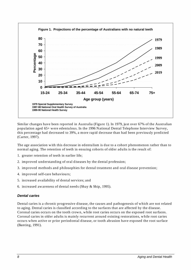
Figure 1. Projections of the percentage of Australians with no natural teeth
1979 Special Supplementary Survey1987-88 National Oral Health Survey of Australia1989-90 National Health Survey
0
10
20
30
40
50
60
70
80
15-24 25-34 35-44 45-54 55-64 65-74 75+
Age group (years)
Per
cen
tag
e1979
1989
1999
2009
2019
Similar changes have been reported in Australia (Figure 1). In 1979, just over 67% of the Australianpopulation aged 65+ were edentulous. In the 1996 National Dental Telephone Interview Survey,this percentage had decreased to 39%, a more rapid decrease than had been previously predicted(Carter, 1997).
The age association with this decrease in edentulism is due to a cohort phenomenon rather than tonormal aging. The retention of teeth in ensuing cohorts of older adults is the result of:
1. greater retention of teeth in earlier life;
2. improved understanding of oral diseases by the dental profession;
3. improved methods and philosophies for dental treatment and oral disease prevention;
4. improved self-care behaviours;
5. increased availability of dental services; and
6. increased awareness of dental needs (Shay & Ship, 1995).
Dental caries
Dental caries is a chronic progressive disease, the causes and pathogenesis of which are not relatedto aging. Dental caries is classified according to the surfaces that are affected by the disease.Coronal caries occurs on the tooth crown, while root caries occurs on the exposed root surfaces.Coronal caries in older adults is mainly recurrent around existing restorations, while root cariesoccurs when active or prior periodontal disease, or tooth abrasion have exposed the root surface(Banting, 1991).

Aging and Dental Health 9
Periodontal disease
Periodontal disease is the term used to describe a cluster of diseases that involve the periodontaltissues. The understanding of periodontal disease has advanced in the last two decades, so thatprevious concepts have been dramatically revised. Some loss of periodontal attachment andalveolar bone is to be expected in older adults, but age alone in a healthy adult does not lead to acritical loss of periodontal support. Although mild gingivitis and mild/moderate loss of alveolarbone and periodontal attachment is common in the elderly, severe loss of periodontal attachment isnot a natural consequence of aging. Gingivitis precedes periodontitis, but few sites with gingivitislater develop periodontitis. Thus, periodontitis is not the major cause of tooth loss in adults(Burt, 1994).
Saliva
The misconception concerning reduced salivary flow in older adults has also been revised. Healthy,non-medicated older adults do not have functionally decreased salivary flow rates or alteredsalivary composition due to aging (Baum, 1986). While medications have been implicated ascontributing to decreased salivary flow and xerostomia in some older adults, their role is still beinginvestigated. However, diseases such as Sjogren’s syndrome and radiation therapy andchemotherapy do have direct effects on the salivary glands (Shay & Ship, 1995).
3. The incidence of oral diseases and conditions
Geriatric dental epidemiological research has focused on two main groups of older adults:
1. community-dwelling functionally independent older adults; and
2. institutionalised functionally dependent older adults (residing in nursing facilities).
There has also been some limited research concerning specific groups of older adults with medicalconditions such as dementia, Parkinson’s disease, and chronic mental illness.
This research has revealed that most community-dwelling functionally independent older adultshave generally good oral health while they can access dental services; however, there appear to besome individuals who are at higher risk for some oral diseases and conditions, such as thesocioeconomically disadvantaged. As older adults’ functional status and medical status decline,and other complications of declining cognitive status, declining affective functioning, increasedneed for social support, increased caregiver burden, and increased barriers to dental care areencountered, oral health status also appears to become compromised. Institutionalised functionallydependent older adults have many complex oral problems, as do functionally dependentindividuals with dementia, Parkinson’s disease and chronic mental illness.
Community-dwelling functionally independent older adults
Tooth loss
While older adults continue to share the disproportionate burden of problems associated with toothloss, successive cohorts of older adults are maintaining a greater proportion of teeth into later years(Dolan & Atchison, 1993). Higher rates of edentulism have been found in disadvantagedpopulations of community-dwelling older adults: (1) the oldest ‘old’; (2) women; (3) those of lowersocioeconomic status, with less education and lower incomes, and social security card holders; and(4) several subgroups such as black Americans, Native American Indians and AustralianAboriginals.

10 Aging and Dental Health
Table 2. Tooth loss incidence (from longitudinal study data)
Study Time period Subjects losing one or more teeth
Iowa 18 months 21%5 years 39%
North Carolina 18 months 19%3 years 29%
Ontario 3 years 28.5%
South Australia 2 years 19.5%
(Hunt et al., 1988; Hand et al., 1991; Drake et al., 1995; Hunt et al., 1995; Locker et al., 1996; Slade et al., 1997)
Tooth loss incidence, in longitudinal studies, was approximately 20% during the first 18 monthsand approximately 29% at 3 years. In the one study reporting 5 year data, tooth loss incidence wasreported as 39% (Table 2) (Hunt et al., 1988; Hand et al., 1991; Drake et al., 1995; Hunt et al., 1995;Locker et al., 1996; Slade et al., 1997).
Table 3. Tooth loss and edentulism incidence
Tooth loss incidence Iowa 0.4 teeth/18 monthsSouth Australia 0.4 teeth/2 years
Annualised incidence edentulism (very low) Ontario 1.2%South Australia 0.7%
In these longitudinal investigations, similar patterns of tooth loss have been found (Table 3). Themean incidence of tooth loss was 0.4 teeth/18 months in Iowa and 0.4 teeth/2 years in SouthAustralia. The annualised incidence of edentulism was very low, approximately 1% of subjects.Thus, although tooth loss occurred in many subjects, the number of teeth lost per subject was smalland few subjects lost all of their teeth (Hunt et al., 1988; Locker et al., 1996; Slade et al., 1997).
Caries
Cross-sectional and longitudinal studies have demonstrated a substantial burden of cumulativedisease from dental caries among community-dwelling older adults, with coronal and root cariesevident in all age groups.
Cross-sectional studies have reported:
1. coronal caries prevalence ranging from 20–30% of subjects;
2. the minority of subjects had the majority of disease (McGuire et al., 1993);
3. among decayed teeth, recurrent coronal decay ranged from 14–22%; and
4. caries experience was higher for molars and premolars, with up to 80% of these teeth havingrestorations on two or more surfaces (Dolan & Atchison, 1993).

Aging and Dental Health 11
Table 4. Caries incidence (from longitudinal study data)
Study Timeperiod
Coronalcaries
Rootcaries
Iowa 36 months 30.6% both coronal and root47.5% coronal or root
North Carolina 18 months 43% whites30% blacks
Tufts 18 months 50%
Caries incidence rates are shown in Table 4, and have ranged from:
1. 30.6% developing both coronal and root caries and 47.5% developing one or the other over36 months in the Iowa study (Hand et al., 1988);
2. 43% of North Carolina whites and 30% of blacks developing coronal caries (Drake et al., 1994);and
3. 50% of subjects in the Tufts study developing root caries (Joshi et al., 1993).
In Iowa, the combined 18 and 36 month net coronal and root caries annualised incidence was1.2 surfaces, with only 14% due to recurrent caries, suggesting that community-dwellingfunctionally independent older adults do continue to develop new carious lesions.
Periodontal disease
Loss of periodontal attachment is now used to measure the cumulative effects of periodontaldiseases.
Loss of Attachment = recession from cemento-enamel junction + probing depth.
Mild gingivitis and also mild to moderate loss of periodontal attachment are common in olderadults; however, severe periodontal disease is not common. Longitudinal and cross-sectionalstudies have revealed that deep probing depths of >6mm are no more extensive in adults aged 65+than in middle-aged adults and have occurred in no more than 9–20% of the populations studied(Burt, 1994).
In the Iowa study, periodontal diseases were found to be weak predictors of tooth loss. Also, theneed for routine scaling and cleaning for mild gingivitis and mild to moderate periodontitis wasfound to be very high (in approximately 90% of dentate subjects), compared to only 15% requiringmore complex periodontal treatment for severe loss of attachment.
Oral mucosal lesions
The prevalence of oral mucosal lesions in community-dwelling older adults is high, having beenfound in up to two-thirds of the subjects in some investigations (Galan et al., 1995). In the Iowastudy, nearly one-quarter of all subjects had oral mucosal lesions, with 27.0% of denture wearershaving denture-related lesions. Lesions were more commonly found on the palate and then on thelips. Risk predictors for denture-related lesions were: less education, use of tobacco, use of alcoholand increased time since last dental visit (Hand & Whitehill, 1986).

12 Aging and Dental Health
Dentures
A large percentage of community-dwelling denture-wearers have been evaluated by examiners ashaving denture-related problems, with up to 40% of those with complete maxillary dentures and upto 60% of those with mandibular dentures in the Iowa study presenting problems. The risk ofproblems and the need for prosthodontic treatment was greater for:
1. complete dentures than for partial dentures; and
2. for mandibular dentures than for maxillary dentures.
Also, perceived prosthodontic treatment need by subjects has been lower than the observed needreported by dental examiners (Hunt et al., 1985).
Institutionalised functionally dependent older adults (residents of nursing facilities)
There is little national-level or longitudinal information available concerning the oral health ofnursing facility residents. Most investigations have presented descriptive data and used smallcross-sectional convenience samples of single or several nursing facilities. They have also excludedresidents that are deemed ‘behaviourally difficult’ or ‘uncommunicative’, such as those withdementia. This is problematic, as often many nursing home residents have cognitive impairment.However, from the several more comprehensive investigations conducted, we can gain someinsights into the oral diseases found in nursing home residents (Empey et al., 1983; CaliforniaDental Association, 1986; Homan et al., 1988; Vigild, 1988; Veteran’s Administration, 1989;Kiyak et al., 1993; MacEntee et al., 1990; Thomson et al., 1991 and 1992).
Table 5. Prevalence of oral diseases and conditions in nursing facility residents
Disease or condition Percentage of residents
Edentulism 45–71Coronal caries 13–70Root caries up to 85Poor oral hygiene nearly allProbing depths ≥4mm 11–27Oral mucosal lesions 33–45Denture treatment 67–75
In Table 5, the prevalence of oral diseases and conditions in nursing facility residents is presented.Edentulism rates in these populations were high, ranging from 45–71%. Clinical examinationsrevealed that the great majority had dental diseases, oral problems and treatment needs (Dolan &Atchison, 1993). The prevalence estimates of coronal caries ranged from 13–70% of residents, withone longitudinal study finding new carious lesions appearing in 33% of subjects at 1 year and in78% of subjects at 2 years (MacEntee et al., 1990). Evidence of root caries was found in the majorityof residents. Poor oral hygiene of natural teeth and dentures was found in the great majority of bothdentate and edentulous residents. Periodontal probing depths of >4mm were found in 11–27% ofdentate residents, and in one study, 25% of residents had spontaneous gingival bleeding (Vigild,1988). Oral mucosal lesions were observed in 33–45% of those examined. Most lesions were relatedto dentures, with denture stomatitis the most prevalent condition observed. Prosthodontictreatment needs were high, ranging from 67–75%.

Aging and Dental Health 13
4. Identification of older adults at high risk for dental diseases and conditions
Identification is needed of those older adults who are at high-risk for developing oral diseases andconditions. Some research has been conducted in longitudinal investigations to identify specific riskfactors for tooth loss and caries, while cross-sectional data has been used to identify risk indicatorsfor caries and periodontal disease. There have been very few reports concerning risk indicators orrisk factors for oral diseases and conditions in nursing facility residents; this discussion of risk willtherefore focus on community-dwelling functionally independent older adults.
Community-dwelling functionally independent older adults
Tooth loss
In four longitudinal investigations, risk factors identified for tooth loss were:
1. clinical indicators of existing disease (root caries, tooth mobility, >4mm probing depths, >4mmrecession, retained roots);
2. a history of tooth loss; and
3. other factors such as smoking, race, marital status, and education (Locker et al., 1996; Slade, 1997;Drake et al., 1995; Hunt et al., 1995).
Table 6. Risk factors and indicators for coronal caries
(Locker et al.,1993a)Ontario
(McGuireet al., 1993)
New England
(Drake & Beck,1992)
North Carolina
(Drake et al., 1994)
North Carolina
blacks whites
Less education Less education Race (black) andfinancial statuspoor
Lowersocioeconomicindex score
No regularannualpreventive visits
Irregular dentalcare attendance
Increasedbaselinecoronal caries
Toothsensitivity
Increasednumber of teeth
Presence ofdecayed roots
Morepreviouslyfilled coronalsurfaces
Antihistaminemedication atbaseline
Born in Canada Sex (males) Decreasedsalivary flow rate(<1.5 ml/min)
Higher countslactobacillus
Perception ofmore problemsthan before
Caries
In the longitudinal and cross-sectional data collected, a range of risk factors and risk indicators forcaries have been identified (Table 6). People at high risk for coronal caries included those of lowersocioeconomic status and educational level, less regular attenders for dental care, and those whohad more previous experience of dental caries.
14 Aging and Dental Health
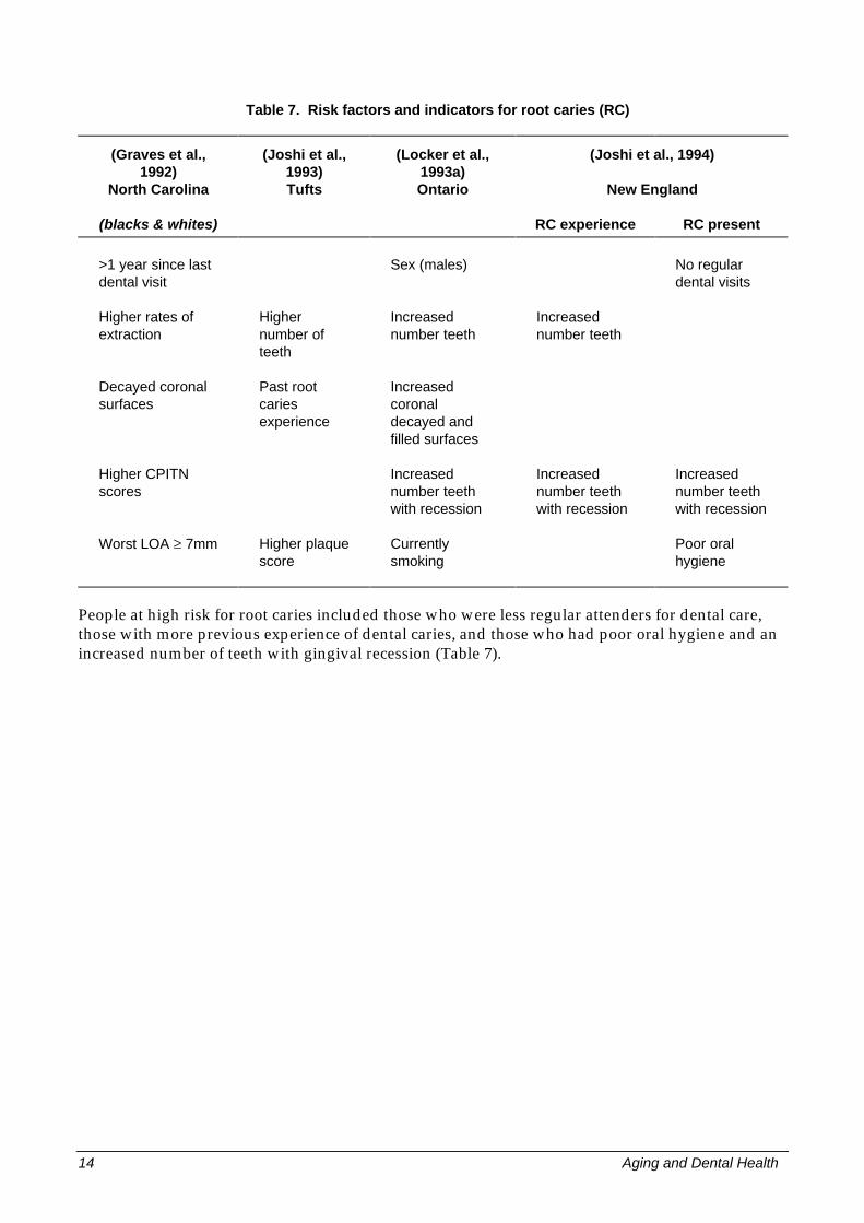
Table 7. Risk factors and indicators for root caries (RC)
(Graves et al.,1992)
North Carolina
(Joshi et al.,1993)Tufts
(Locker et al.,1993a)Ontario
(Joshi et al., 1994)
New England
(blacks & whites) RC experience RC present
>1 year since lastdental visit
Sex (males) No regulardental visits
Higher rates ofextraction
Highernumber ofteeth
Increasednumber teeth
Increasednumber teeth
Decayed coronalsurfaces
Past rootcariesexperience
Increasedcoronaldecayed andfilled surfaces
Higher CPITNscores
Increasednumber teethwith recession
Increasednumber teethwith recession
Increasednumber teethwith recession
Worst LOA ≥ 7mm Higher plaquescore
Currentlysmoking
Poor oralhygiene
People at high risk for root caries included those who were less regular attenders for dental care,those with more previous experience of dental caries, and those who had poor oral hygiene and anincreased number of teeth with gingival recession (Table 7).

Aging and Dental Health 15
Periodontal Disease
Cross-sectional data has been used to investigate risk indicators for periodontal disease (Table 8).Age, sex, race, socioeconomic status, smoking and diabetes have been investigated previously, andmore recently general health status, and behavioural, psychosocial, and oral health variables havebeen used. This table presents indicators identified for severe periodontal disease, with thosesmoking, those with fewer teeth and those with irregular dental attendance being identified mostfrequently.
Table 8. Risk indicators for severe periodontal disease
(Levy et al., 1987)Iowa
(Beck et al., 1990)North Carolina
(Locker et al., 1993)Ontario
Number cigarettes smoked andpipe smoking
Tobacco use Currently smoking
Irregular attendance fordental visits
Irregular attendance fordental visits
Fewer teeth remaining Fewer number teethremaining
Increased number teeth withcalculus
Age (cumulative diseaseexperience)
Previous periodontalinvolvement (recession)
Biological indicators Less education
Summary
As a consequence of demographic and oral health status changes, there will be a marked growth inthe number of older adults retaining their natural teeth. The focus of oral diseases and conditionswill move from dentures to natural teeth. High levels of oral diseases and conditions have beenidentified in specific geriatric populations, such as:
1. those functionally dependent, residing in nursing homes;
2. the functionally dependent with neurological conditions; and
3. sub-groups of community-dwelling functionally independent older adults, for example thesocioeconomically disadvantaged, tobacco and alcohol users, those with greater experience ofcaries and periodontal diseases, and those who do not attend regularly for dental care.

16 Aging and Dental Health
References
Banting DW 1991. Management of caries in the older patient. In: Geriatric dentistry: aging and oralhealth. Papas AS, Niessen LC & Chauncy HH. St Louis, Mosby, pp 141.
Baum BJ 1986. Salivary gland function during aging. Gerodontics 2:61.
Beck JD, Koch GG, Rozier RG & Tudor GE 1990. Prevalence and risk indicators for periodontalattachment loss in a population of older community-dwelling blacks and whites. J Periodontol61:521–8.
Burt BA 1994. Periodontitis and aging: reviewing recent evidence. JADA 125(3):273–9.
California Dental Association 1986. California skilled facilities’ residents: a survey of dental needs.Sacramento CA: California Dental Association.
Carter KD 1997. National Dental Telephone Interview Survey. AIHW Dental Statistics and ResearchUnit. Personal Communication.
Dolan TA & Atchison KA 1993. Implications of access, utilization and need for oral health care bythe institutionalized and non-institutionalized elderly on the dental delivery system. J Dent Educ57(12):876–87.
Drake CW & Beck JD 1992. Models for coronal caries and root fragments in an elderly population.Caries Res 26:402–7.
Drake CW, Hunt RJ, Beck JD & Koch GG 1994. Eighteen-month coronal caries incidence in NorthCarolina Adults. J Pub Health Den 54(1):24–30.
Drake CW, Hunt RJ & Koch GG 1995. Three-year tooth loss among black and white older adults inNorth Carolina. J Dent Res 74(2):675–80.
Empey G, Kiyak HA & Milgrom P 1983. Oral health in nursing homes. Spec Care Dent 3(2):65–7.
Galan D, Brecx M & Heath MR 1995. Oral health status of a population of community-dwellingolder Canadians. Gerodontology 12(1):41–8.
Graves RC, Beck JD, Disney JA & Drake CW 1992. Root caries prevalence in black and white NorthCarolina adults over age 65. J Pub Health Dent 52(2):94–101.
Hand JS & Whitehill JM 1986. The prevalence of oral mucosal lesions in an elderly population.JADA 112:73–6.
Hand JS, Hunt RJ & Beck JD 1988. Coronal and root caries incidence in older Iowans: 36-monthincidence. Gerodontics 4:136–9.
Hand JS, Hunt RJ & Kohout FJ 1991. The five-year incidence of tooth loss in Iowans aged 65 andolder. Community Dent Oral Epidemiol 19:48–51.
Homan BT, Lam B & Larsen RG 1988. The oral needs and demands of a geriatric population atMt Olivet, Brisbane, 1986. Aust Dent J 33(5):424–32.
Hunt RJ, Hand JS, Kohout FT, Beck JD 1988. Incidence of tooth loss in elderly Iowans. Am J PubHealth 78(10):330–2.

Aging and Dental Health 17
Hunt RJ, Srisilapanan P & Beck JD 1985. Denture-related problems and prosthodontic treatmentneeds in the elderly. Gerodontics 1:226–30.
Hunt RJ, Levy SM & Beck JD 1990. The prevalence of periodontal attachment loss in an Iowapopulation aged 70 and older. J Pub Health Dent 50(4):251–6.
Hunt RJ, Drake CW & Beck JD 1995. Eighteen-month incidence of tooth loss among older adults inNorth Carolina. Am J Pub Health 85(4):561–3.
Joshi A, Papas AS & Giunta J 1993. Root caries incidence and associated risk factors in middle-agedand older adults. Gerodontology 10(2):84–91.
Joshi A, Douglass CW, Jette A & Feldman H 1994. The distribution of root caries in community-dwelling elders in New England. J Pub Health Dent 54(1):15–23.
Kiyak HA, Grayston MN & Crinean CL 1993. Oral health problems and needs of nursing homeresidents. Community Dent Oral Epidemiol 12:49–52.
Levy SM, Heckert DA, Beck JD & Kohout FJ 1987. Multivariate correlates of periodontally healthyteeth in an elderly population. Gerodontics 3:85–8.
Locker D & Leake JL 1993a. Coronal and root decay experience in older adults in Ontario, Canada.J Pub Health Dent 53(3):158–64.
Locker D & Leake JL 1993b. Risk indicators and risk markers for periodontal disease experience inolder adults living independently in Ontario, Canada. J Dent Res 72(1):9–17.
Locker D, Ford J & Leake JL 1996. Incidence of and risk factors for tooth loss in a population ofolder Canadians. J Dent Res 75(2):783–9.
MacEntee MI, Wyatt CCL & McBride BC 1990. Longitudinal study of caries and cariogenic bacteriain an elderly disabled population. Gerodontics 18:149–52.
MacEntee MI, Hill PM, Wong G, Mojon P, Berkowitz J & Glick N 1991. Predicting concerns for themouth among institutionalized elders. J Pub Health Dent 51(2):82–90.
McGuire SM, Fox CH & Douglass CW 1993. Beneath the surface of coronal caries: primary decay,recurrent decay and failed restorations in a population-based survey of New England elders. J PubHealth Dent 53(2):76–82.
Shay K & Ship JA 1995. The importance of oral health in the older patient. J American Ger Soc43:1414–22.
Slade GD & Spencer AJ 1995. Periodontal attachment loss among adults aged 60+ in SouthAustralia. Community Dent Oral Epidemiol 23:237–42.
Slade GD, Gansky S & Spencer AJ 1997. Two year incidence of tooth loss among South Australiansaged 60+ years. Community Dent Oral Epidemiol 25:429–37.
Thomson WM, Brown RH & Williams SM 1991. Dental status and treatment needs of a NewZealand institutionalized elderly population. New Zealand Dental J 87:119–23.

18 Aging and Dental Health
Thomson WM, Brown RH & Williams SM 1992. Dentures, prosthetic treatment needs and mucosalhealth in an institutionalized elderly population. New Zealand Dental J 88:51–5.
Veterans Administration 1989. Oral health survey of nursing home residents. Report to the ACMDfor Dentistry. Washington DC: Veterans Administration.
Vigild M 1988. Oral hygiene and periodontal conditions among 201 dentate institutionalizedelderly. Gerodontics 4:140–5.

Aging and Dental Health 19
Clinical dental care for older adultsProfessor Ron Ettinger
ABSTRACT
In dentistry, for treatment purposes it is possible to divide the aging population into three broadgroups based upon their functional status.
This presentation will use longitudinal case histories to explore the decision-making processrequired to develop a treatment plan which adequately addresses the oral health needs offunctionally independent older adults, frail older adults and functionally dependent older adults.
In the recent past, older adults utilised dental services infrequently because they were edentulousand felt ‘no need’ to seek care. In the last decade a new dentate elderly consumer has evolved.These older adults tend to have extensive dental problems which are complicated by their chronicdiseases and varied pharmacotherapies. Many of these persons, even when faced with bone lossthrough periodontal disease, caries or unresolved periapical pathology, will no longer accept thesimple solutions of the past, such as the extraction of their remaining natural dentition and theconstruction of complete dentures.
PRESENTATION
Introduction
Although the number and the percentage of adults over the age of 65 is growing dramatically,(Davies, 1985; United Nations, 1991; Ettinger, 1993a; Department of Commerce, Bureau of Census,1994) it is important to remember that heterogeneity among persons aged 65 years and older isprobably greater than at any other time period in the life span (Nelson & Dannefer, 1991). It hasbeen common practice for all age groups older than 65 years to be grouped together when planningprograms or analysing data. However, elderly persons are a complex combination and expressionof their individual genetic predispositions, lifestyles, socialisation and environments. All of thesefactors influence their health beliefs and, therefore, their health behaviour. In order to understand apatient’s attitudes, the dentists must evaluate the cultural, psychological, educational, social,economic, dietary and chronologically specific cohort experiences which may have influenced thatpatient’s life. Similarly, oral status is also affected by these same factors, as well as including anindividual’s life experience with dental care, with caries, periodontal disease and iatrogenic disease.Consequently, oral status reflects the history of the person’s behavioural attitudes and expectationsfor their own oral health. Finally, the skills, attitudes and philosophies of the various dentists thatpeople have encountered during their life must also affect their oral status (Ettinger, 1990).
In the not-so-recent past, the elderly population comprised a relatively small proportion of the totalpopulation and the majority of these older adults were edentulous. They utilised dental careinfrequently and then only when their previously unmet needs could no longer be ignored (Burt,1978; Ettinger, 1971). However, there is now ample evidence to show that a new elderly dentalconsumer has emerged. These older adults are better educated, more politically aware and mostimportantly many of them have some remaining teeth. As patients, these older people have a widerrange of needs and expectations, and are demanding a greater variety of services than the pastemphasis on complete dentures by previous cohorts (Ettinger & Beck, 1982).

20 Aging and Dental Health
The elderly have been defined as a cohort of people aged 65 years or older. It became clear to usthat a chronological definition of the aging population was not particularly useful in dentistry.Thus, Ettinger and Beck (1984) developed a functional definition which categorised the agingpopulation into three distinct groups based upon their ability to seek services. The groups wereidentified as:
a. the functionally independent older adult;
b. the frail older adult; and
c. the functionally dependent older adult.
It also became apparent that different older adults had different needs and that their functionaldisabilities affected their ability to accept and receive dental treatment. In this paper a case historyof a frail older adult will be presented to illustrate some specific oral needs, problems and issues inclinical geriatric dentistry.
Case history
Mrs J.M. was a 76-year-old married woman who lived at home with her husband, some 20 milesaway. Mrs J.M. was of Bohemian origin, spoke English poorly, had less than a high schooleducation and worked as a cook’s assistant in a restaurant in her youth. She needed the aid of awalking stick to walk independently. Her husband had brought her to our clinic because severallocal dentists, after examining her, told her that they could not treat her. Her chief complaint wasthat she had a lot of oral discomfort and could not eat hard foods and that she wanted to becomfortable.
Medical history
After talking to her husband we found that she had a history of hypertension, osteoarthritis,especially of the knees, and a history of psychiatric disease, including depression for which she hadbeen hospitalised in the past and had received electroconvulsive therapy (ECT). She also had ahistory of pernicious anaemia.
Drug history
She carried a little card on which her physician had written her medications:
Elavil 50 mg bidRestoril 20 mg hs.Moduretic 1 tab/day (with food)K-Tabls 20 mEq tidTylenol #3 1 tab q 6 hVitamin B12 (injection: 1/month)

Aging and Dental Health 21
Oral examination
The patient had uncontrolled rhythmic movements of the mouth and tongue, suggestive ofextra-pyramidal symptoms. She could control the movements for brief seconds, thus suggestingthat it was tardive dyskinesia (Kamen, 1975; Asnis et al., 1977; Bell & Smith, 1978; Blair & Dauner,1993). Intraorally, her mouth was very dry. The following teeth were present:
R L
X X 6* 5 4* 3* 2 X 11 2 3 X 5 X X X
X X X X X 3 2 X X 2* 3* 4* X X X
* caries1 root only
The teeth had the lower 1/3 covered with plaque and there was subgingival calculus, but no teethexcept the upper left premolar had probing depths greater than 3mm. There were no occludingpairs of teeth in the retruded position, and the patient functioned by protruding her jaw to findcontact with the remaining lower teeth.
Treatment plan
To develop a treatment plan we needed to answer the following questions:
1. What were patient’s desires and expectations?
2. What were her dental needs and how complex would her treatment need to be?
3. What would be the impact of her dental needs on her quality of life?
4. How would her medical problems impact on her treatment?
a. How stable was her hypertension and could we use vasoconstrictor in her local anaesthetic?
b. How severe was her osteoarthritis and how long could she sit in a dental chair?
c. Was her depression under control and could she take responsibility for her daily oralhygiene?
d. When was the last episode of her psychiatric disease and what medication had caused thetardive dyskinesia?
e. How severe was her tardive dyskinesia and would it limit our ability to deliver clinicaldental care?
5. Could we discuss her medications with her physician with regard to her dry mouth?
6. How compliant would she be with any oral health care regimen?
7. What would be her ability to tolerate the stress of treatment? How tolerant would her soft tissuesbe of the physiological trauma created by dentures, especially a mandibular denture?
8. Were there any financial limitations on her being able to receive treatment?

22 Aging and Dental Health
Patient management issues
Patient’s desires and expectations
The patient wanted to be comfortable and to be able to chew. She was prepared to do what wasnecessary and had the support of her family.
Communications
Mrs J.M.’s English was so poor that we needed to make sure her husband was with her when wepresented any treatment plan. If he did not seem to understand the informed consent, then weneeded to have her daughter, who lives near to the parents, come to the appointment as well.
Dental needs
She had no opposing pairs of teeth and so had poor masticatory function which would require amaxillary and mandibular prosthesis. As she had difficulty chewing and pain when she did, wewould need to remove some teeth and restore the remaining teeth.
An evaluation of her radiograph showed caries on most of the teeth on the upper right quadrantand lower left quadrant. There was a broken central incisor on the left side with only the root left.
Quality of life
Her current oral status impacted on the quality of her life in that she was uncomfortable and haddifficulty chewing. Any improvement would also improve the quality of her life.
Medical consultation
a. We recorded her vital signs: her blood pressure was 155/90 mm Hg, with a pulse of 72. It seemsher hypertension was controlled by her medications. Even if her blood pressure was controlledby medication, the blood pressure should be monitored and recorded prior to and at the end ofevery dental appointment, especially if any invasive procedures were to be used (Malamed,1980). The accepted norm for blood pressure for an older adult is 160/95 mm Hg and a protocolfor treatment is shown in Figure 1 (Ettinger, 1993b).
The American Heart Association guidelines for use of epinephrine as a vasoconstrictor in localanaesthetic is not more than 0.04 mg (Mulligan, 1985). This translates to not more than 2½
carpules of 1.8 ccs of lidocaine 2% with 1
100 000, epinephrine at any one time, making sure that it
is not placed in a blood vessel by deliberate aspiration (Jastak & Yagiela, 1983).
b. She had severe osteoarthritis of the knees and some problems with her neck. However, herhands and fingers were not severely affected. We felt that if we could get her in a comfortableposition, sitting in a dental chair was not going to be a problem.
c. We found out that Mrs J.M. had been treated with chloropromazine for several years and shehad had an acute episode of depression with psychotic features and been hospitalised 15 yearsago. The treatment was ECT. The follow-up treatment for the depression was with amytriptiline.Because amytriptiline is so drying, we asked the physician to change her to another tricyclic,desipramine, which is much less xerogenic (Sreebny & Schwarz, 1986; Atkinson & Wu, 1994;

Aging and Dental Health 23
Ettinger, 1996). According to her physician, Mrs J.M.’s depression has been stable for the last fewyears.
d. There is an increased risk of myocardial infarction and stroke between 6 a.m. and 9 a.m., so wedo not want to see her before 9 a.m. This risk is due to a diurnal increase in platelet aggregabilitywhich increases the risk of thrombus formation (Willich et al., 1987; Ridker et al., 1990; Panzaet al., 1991).
Daily medications
The influence of her daily medications on Mrs J.M.’s oral condition and on her dental managementis shown in Table 1.
Ability to maintain oral hygiene
This is a key factor in decision making as plaque control is essential to maintenance of any teethwithin the arch (Berkey et al., 1996). The compliance with this preventive behaviour depends uponthe patient having:
a. Adequate knowledge on why she needs to clean her teeth, and understanding that her dry mouthputs her into the high risk category for caries and periodontal disease. Because of the languageproblems we need to keep the explanations as simple and as practical as possible.
b. Adequate motivation is needed if people are to change behaviours. In Mrs J.M.’s case, she statedthat she wanted to keep as many teeth as possible so the prognosis for her cooperation is good.
c. Adequate neuromuscular skills are necessary to hold a toothbrush. One must have adequate visionto see and enough fine hand–eye coordination to be able to put the brush where it will be mosteffective. The tardive dyskinesia seems to only affect her oral cavity, her hand–eye coordinationseems to be relatively good as she crochets while sitting in the dental chair.
As Mrs J.M. has such a dry mouth, we wanted to put her on a home care regimen for persons with ahigh risk of caries. That is, we suggested that she try an artificial saliva and used a fluoridetoothpaste and to begin with a neutral fluoride rinse before going to bed. The one we prescribedwas a 0.2% sodium fluoride rinse. We would have liked to use a chlorhexidine rinse but none wasavailable which did not have an alcohol base. Alcohol is contraindicated for patients who have avery dry mouth. We will have Mrs J.M. on 3-month recall; if we see any new carious lesions we willprescribe a neutral sodium fluoride brush-on gel with 5,000 ppm fluoride.
Tardive dyskinesia
It is our experience that with patients with neurological problems such as Parkinson’s disease ormyasthenia gravis, movement disorders such as tardive dyskinesia, etc., or cognitive disorders suchas Alzheimer’s disease and other dementias, it is imperative to try and prevent them becomingedentulous in the mandibular arch. One can make a complete maxillary denture or a maxillaryremovable partial denture (RPD) with very little cooperation from the patient; however, that is nottrue for the mandible. Therefore, it is important to maintain some key teeth in these at-risk patients.
Moreover, there are a group of individuals who do not accommodate well to a completemandibular denture because their tissues are very fragile. These persons include those:
1. whose soft tissue has degenerated with extensive denture wearing;
2. with significant reduction in salivary flow rates;

24 Aging and Dental Health
3. whose pain threshold has been altered;
4. whose neuromuscular skills have deteriorated;
5. whose cognitive skills have deteriorated;
6. whose physical health has deteriorated and who are physically frail.
To do restorative care in Mrs J.M.’s mouth we used a mouth prop made out of cotton rolls andtaped together to keep her mouth open, and to protect her tongue we used a suction tip with a largemetal tongue deflector.
Probability of success
Provided the patient’s health remains stable at the present level, then she can cooperate and we cantreat her, however:
a. Communication may be a problem.
b. Transportation will not be a problem as she has a good support system.
c. Her xerostomia will increase the risk of caries and periodontal disease and cause problems withwearing dentures. It is important to avoid a complete denture especially on the mandible.
d. Her tardive dyskinesia will be a management problem and the clenching may hinder her abilityto wear a mandibular denture. Also, the ability to record vertical dimension of occlusion andcentric relation will be a problem. The use of interim RPD’s will help diagnostically.
e. Her motivation seems to be good and her hand–eye coordination adequate.
The overall probability of success is fair to good and since the patient is in pain it is imperative todeliver emergency care as soon as possible.
The treatment plan
Initial
After evaluating all the modifying factors the following treatment plan was offered to Mrs J.M.:
Maxilla:
Extractions: right side: 1st molar, 1st and 2nd premolarleft side: central incisor and 2nd premolar
Restorations: right side: canine
Mandible:
Extractions: left side, 1st premolar and lateral incisor
Restorations: canine cut down as a vital overdenture abutment
Construction of interim transitional RPD’s for the maxilla and mandible to determine verticaldimension of occlusion, neuromuscular skills and home care regimen. After delivery andadjustments the patient was placed on 3-month recall.

Aging and Dental Health 25
At 3-month recall
Oral hygiene was much improved, no caries were seen and Mrs J.M. was functionally comfortablewith the dentures. Her husband had suffered a stroke and was in a nursing home; however,Mrs J.M. had excellent community support including neighbours who were prepared to drive her.
Treatment plan (after 3-month recall)
Because Mrs J.M. was doing so well we suggested the following treatment plan:
Maxilla:
surveyed crowns on the left and right caninesrestore the incisal of left lateral with composite resincast chrome cobalt RPD to restore aesthetic and occlusal contacts
Mandible:
a surveyed crown on the right canine, cast chrome cobalt RPD to restore the dentine cast chromecobalt RPD to restore occlusal contacts
The patient accepted this treatment plan but during its early phase her husband died whichdelayed the treatment. However, the treatment was completed and Mrs J.M. was placed on4-month recall. Because she had no occluding pairs of teeth she was given a night guard to wear onthe mandibular arch. She was asked to place some neutral sodium fluoride gel in the night guardeach evening.
Conclusions
This case history illustrates that it is possible to do extensive restorative work for an at-risk patientif one understands the influence the social and medical problems have upon the oral cavity anddental treatment. It is imperative that a step-wise approach is used and that no irreversible step istaken until an adequate risk-assessment of potential for success has been made. The key tosuccessful treatment is to understand how the patient functions in his/her environment and howdentistry fits into his/her overall needs.

26 Aging and Dental Health
Table 1: Mrs JM – Influence of daily medication on oral condition and her dental management
Disease Drug generic name Potential oral sideeffects
Management issues
Depression Amitriptyline
(Antidepressive)
Xerostomia
Changes in taste
Swelling of the tongue
Parkinson’s-like movement
1. Caries prevention
2. Artificial saliva
3. Potential effect onepinephrine
4. Poor healing
5. Orothostatichypotension
6. Avoid alcohol
Insomnia Temazepam
(Sedative-hypnotic)
Xerostomia 1. Drowsiness
2. Risk of falling
Hypertension Amiloride HCL 5 mg
With hydrochlorothiazide50 mg (Diuretic)
Xerostomia 1. Caries prevention
2. Postural hypotension
3. Poor healing
4. Limit saline products
5. Avoid alcohol
Hypokalemia Potassium chloride
(Anti-hypokalemic)
None Cold extremities
Osteoarthritis Acetominophen 300 mgwith codeine phosphate30 mg
(Analgesic)
Gingival bleeding
Xerostomia
1. Bleeding withextractions or deepscaling
2. Risk of falling
3. Orthostatichypotension
4. Caries prevention
Pernicious anaemia Vitamin B12 None None

Aging and Dental Health 27
Figure 1. Guidelines for treatment decisions regarding persons who have blood pressurereadings greater than 160mm Hg systolic or 95mm Hg diastolic
(Modified from: Ettinger, RL 1993. Management of elderly patients in the private practice system.Int Dent J 43:29–40.)

• R
efer
imm
edia
tely
• N
o tr
eatm
ent
• C
arot
id p
ulsa
tion
• H
eada
ches
(fr
eque
nt)
• D
izzi
ness
• V
ertig
o•
Pal
pita
tions
• N
o sy
mpt
oms
• C
ompl
ete
exam
inat
ion
and
cont
act p
hysi
cian
HIS
TO
RY
OF
HY
PE
RT
EN
SIO
N
MA
NO
ME
TR
ICR
EA
DIN
G>
160/
95 m
m H
g
NO
HIS
TO
RY
OF
HY
PE
RT
EN
SIO
N
SY
ST
OL
IC /
DIA
ST
OL
IC16
5 to
190
/ 90
to
105
SY
ST
OL
IC /
DIA
ST
OL
IC19
0 to
220
/ 10
5 to
120
SY
ST
OL
IC /
DIA
ST
OL
IC>
220
/ > 1
29
• C
ompl
ete
exam
inat
ion
and
trea
tmen
t pla
n•
Em
erge
ncy
care
onl
y•
Che
ck b
lood
pre
ssur
e at
nex
tap
poin
tmen
t; if
BP
stil
l hig
h, r
efer
to p
hysi
cian
for
eval
uatio
n
• E
xam
ine
• E
mer
genc
y ca
re o
nly
• R
eapp
oint
to c
ompl
ete
trea
tmen
tpl
an a
fter
cons
ulta
tion
with
phy
sici
an
• N
o tr
eatm
ent
• C
all p
hysi
cian
& r
efer
imm
edia
tely

28 Aging and Dental Health
References
Asnis GM, Leopold M, Duvoisin RC & Schwartz AH 1977. A survey of tardive dyskinesia inpsychiatric outpatients. Amer J Psychiat 134:1367–70.
Atkinson JC & Wu AJ 1994. Salivary gland dysfunction causes, symptoms, treatment. J Am DentAssoc 125:409–16.
Bell RC & Smith RC 1978. Tardive dyskinesia: characterization and prevalence in a statewidesystem. J Clin Psychiat 39:39–47.
Berkey DB, Berg RG, Ettinger RL, Mersel A & Mann J 1996. The old-old dental patient: thechallenges of clinical decision-making. J Am Dental Assoc 127:321–32.
Blair DT & Dauner A 1993. Non-neuroleptic etiologies of extrapyramidal symptoms. Clinic NurseSpec 7:225–31.
Burt RA 1978. Influences for change in the dental health status of populations: a historicalperspective. J Pub Hlth Dent 38:272–8.
Davies AM 1985. Epidemiology and the challenge of aging. Int J Epidemiol 14:9–21.
Department of Commerce, Bureau of Census 1994. Projections for states by age, sex, race, andHispanic origin 1993–2020. In: Current population reports, p. 25–111. US Government PrintingOffice, Washington, DC.
Ettinger RL 1971. An evaluation of the attitudes of a group of elderly edentulous patients todentists, dentures, and dentistry. Dent Pract Dent Rec 22:85–91.
Ettinger RL & Beck JD 1982. The new elderly: what can the dental profession expect? Spec CareDent 2:62–9.
Ettinger RL & Beck JD 1984. Geriatric dental curriculum and the needs of the elderly. Spec Care Dent4:207–13.
Ettinger RL 1990. Restoring the aging dentition: repair or replacement. Int Dent J 40:275–82.
Ettinger RL 1993a. Demography and dental needs, an international perspective. Gerodontology10:3–9.
Ettinger RL 1993b. Management of elderly patients in the private practice system. Int Dent J43:29–40.
Ettinger RL 1996. Review: Xerostomia: a symptom which acts like a disease. Age and Ageing25:409–12.
Jastak JT & Yagiela JA 1983. Vasoconstrictors and local anaesthesia: a review and rationale for use.J Am Dent Assoc 107:623–30.
Kamen S 1975. Tardive dyskinesia: a significant syndrome for geriatric dentistry. Oral Surg 39:52–7.
Malamed SF 1980. Blood pressure evaluation and the prevention of medical emergencies in dentalpractice. J Prev Dent 6:183–8.

Aging and Dental Health 29
Mulligan R 1985. Pretreatment for the cardiovascularly compromised geriatric dental patient. SpecCare Dent 5:116–23.
Nelson EA & Dannefer D 1991. Aged heterogeneity: fact or fiction? The fate of diversity ingerontological research. The Gerontologist 32:17–23.
Panza JA, Epstein SE & Quyyumi AA 1991. Circadian variation in vascular tone and its relation toα-sympathetic vasoconstrictor activity. N Engl J Med 325:986–90.
Ridker PM, Manson JE, Buring JE, Miller JE & Henneksens CH 1990. Circadian variation of acutemyocardial infarction and effect of low-dose aspirin in a randomized trial of physicians. Circulation82:897–902.
Sreebny LM & Schwartz SS 1986. A reference guide to drugs and dry mouth. Gerodontology 5:75–99.
United Nations 1991. Sex and age distribution of populations. Dept of International, Economic andSocial Affairs (Population Studies No. 112) New York.
Willich SN, Levy D, Rocco MB, Tofler GH, Stone PH & Muller JE 1987. Circadian variation in theincidence of sudden cardiac death in the Framingham Heart Study population. Am J Cardiol60:801–6.

30 Aging and Dental Health
Medications and oral healthDr W Murray Thomson
ABSTRACT
PURPOSE – The purpose of the presentation is to examine occurrence of dry mouth and themedication – dry mouth findings from a cohort study of older people which is the first study to uselongitudinal medication-exposure data.
METHODS – The presentation uses data from the South Australian Dental Longitudinal Study atbaseline in 1991 and again five years later in 1996. At the five-year data collection, resting wholesalivary flow rates were measured and xerostomia was estimated using (1) the XerostomiaInventory and (2) the standard single item ‘How often does your mouth feel dry?’, with theresponse options ‘never’, ‘occasionally’, ‘frequently’ or ‘always’. For the 20 most prevalent classesof medication, participants were divided into continuous users, starters, stoppers and non-usersaccording to whether each medication type was taken at baseline and five years. The groups’salivary flow-rates and Xerostomia Inventory scores were compared using analysis of variance andlinear regression modelling.
RESULTS – The prevalence of xerostomia in the sample was 20.5%, and that of salivary glandhypofunction (SGH) 22.1%. Some 65% of the sample suffered neither xerostomia nor SGH, and thetwo conditions coincided in only 5.7% of individuals. The multivariate analyses showed thatsalivary flow rates were significantly reduced in participants who were on (or who had been on)antidepressants, and were higher in those who were taking hypolipidaemic drugs. XerostomiaInventory scores were higher in those who were continuous users of narcotic analgesics or anginals,or who had started or stopped taking hormone replacement therapy (HRT) in the preceding fiveyears. The findings of this study suggest: (1) that xerostomia and SGH are discrete conditions andusually occur independently; (2) that current concepts of the association of medication and drymouth need to be modified; and (3) that dentate individuals taking certain classes of medicationmay be at higher risk of oral diseases.
PRESENTATION
Older people are thought to be more susceptible to medications which may produce dry mouth.The purpose of this paper is to examine the relationship between dry mouth and particular classesof medication in the first study to use longitudinal medication-exposure data.
Dry mouth
Dry mouth is common among older populations, with a reported prevalence of between 10 and38%, depending upon the group being studied; around 20% is the most frequently reportedestimate. While it is known to be more prevalent in the ‘older old’ and in females, there is noevidence to suggest that this is a feature of the aging process itself; rather, it is thought that olderpeople’s salivary systems are more vulnerable to exogenous factors which may act to reducesalivary flow. Examples of these factors are: certain medications, some chronic inflammatoryconditions, and radiation therapy for head and neck tumours.
Should we be concerned about dry mouth? The answer is definitely yes: it can significantly affectthe quality of life for affected individuals, and there is also the suggestion from prevalence studies –

Aging and Dental Health 31
as yet unsubstantiated by cohort studies – that it is associated with an increased occurrence ofdental caries, and can also affect denture function. Locker (1995) has shown that the prevalence ofxerostomia in an older population increases over time, and it is imperative that our understandingof the aetiology, epidemiology and natural history of the condition is rapidly improved.
Before proceeding any further in describing the relationship of medications and dry mouth, it is agood idea to examine the two conceptually distinct manifestations of dry mouth.
1. Salivary gland hypofunction (SGH), or hyposalivation, is an observable, chronic reduction insalivary flow rate; as such, it is a sign.
2. Xerostomia is the subjective feeling of dry mouth, and commonly involves a varied collection ofassociated symptoms.
The relationship between these two manifestations is currently unclear: they may be discrete,occurring separately in the population; they may always occur together in certain affectedindividuals; or there may be a variable degree of overlap between them. Two circumstances havecontributed to the lack of understanding. First, most studies have used either convenience samplesof xerostomia clinic patients (Fox et al., 1987), compliant elders (Billings et al., 1996), or dentalstudents (Dawes, 1987); second, the population-based approach has only been used in a smallnumber of studies (Nederfors et al., 1997; Locker, 1993; Gilbert et al., 1993; Osterberg et al., 1984),none of which has measured both conditions. It is axiomatic that a comprehensive understanding ofthe epidemiology and public health significance of a condition requires not only accurate anddetailed epidemiological and clinical study, but also unbiased estimates of its occurrence.
1. Salivary gland hypofunction (SGH)
SGH is most commonly defined as a resting whole-salivary flow rate of <0.1 ml/min, or a reducedstimulated flow of <0.5 ml/min. Flow rate is estimated by using sialometry; there is the choice ofcollecting resting or stimulated flows, or whole or glandular saliva. It is thought that the restingwhole-saliva flow is probably most important in the long-term protection of the dentition. There area number of ways in which saliva can be collected, ranging from the relatively unsophisticated‘spit’ or ‘drain’ methods, through to cannulating individual glands and using negative pressure towithdraw secretions (Navazesh & Christensen, 1982).
2. Xerostomia
The measurement of xerostomia is more problematic, as the only way to find out about symptomsis to ask the individual concerned. Two approaches have been used to date: (1) the single globalquestion; and (2) a mix of questions covering various experiential and behavioural aspects of drymouth. The former has been more common and is easier to interpret, while the latter approach hasbeen faced with the problem of analysing the responses to the different questions separately andfinding that each gives a different prevalence estimate.
In the past, the single-item approach has been most commonly used, employing questions such as:
Does your mouth feel distinctly dry?
Do you sip liquids to aid in swallowing dry foods?
Does your mouth feel dry when eating a meal?
Do you have difficulties swallowing any foods?

32 Aging and Dental Health
Does the amount of saliva in your mouth seem to be too little, too much, or you don’t noticeit?
Do you feel dryness in the mouth at any time?
Do you have mouth dryness?
Is your mouth sometimes dry?
How often does your mouth feel dry?
Does your mouth feel dry?
Each of the population-based studies of xerostomia has used the single global question approach.Osterberg et al. (1984) used the question ‘Does your mouth feel distinctly dry?’ (binary responseoption ‘yes’, ‘no’), and conducted measurements of salivary flow on only a small subsample of theirstudy participants. Locker (1993) used the single question ‘During the last four weeks have you hadany of the following – dryness of mouth?’ (binary response option ‘yes’, ‘no’) but did not measuresalivary flow. Nederfors et al. (1997) used the question ‘Does your mouth usually feel dry?’ (binaryresponse option ‘yes’, ‘no’), and did not measure salivary flow either. None of those studiesprovided evidence for the validity of the questions used. Gilbert et al. (1993) reported from a studyof a stratified random sample of community-dwelling older Florida residents, which used a numberof questions on mouth dryness, but did not measure salivary flow. Their main outcome measurewas the question ‘Is your mouth sometimes dry?’, to which 39% of respondents answeredaffirmatively. The latter question can be criticised on the grounds that it is probably too inclusivefor use as a tool for categorising individuals as ‘xerostomic’; certainly, the obtained prevalenceestimate is unusually high in comparison to those from the other studies, and indicates thatperhaps the word ‘often’ should have been substituted for ‘sometimes’.
There are a number of problems with these past approaches: (1) they have not been used in amulti-item inventory approach to date – the user of any single item must face the inevitableproblem that what he/she thinks is being measured actually isn’t; and (2) qualitative work hasshown that xerostomia has both experiential and behavioural dimensions. A method is neededwhich will take both types of symptom into account. The Xerostomia Inventory (Thomson et al.,1999) was developed to answer this need, and to enable measurement of the severity of xerostomiasymptoms on a continuous scale.
Medications and dry mouth
Almost every dentist is aware of the association between certain medications and dry mouth.However, most of the ‘evidence’ for the relationship is anecdotal and based largely upon casereports. That which is available from systematic science comes to us mostly from studies ofparticular subgroups of older people, or from convenience samples of attenders at dry mouthclinics. Lists of xerogenic (‘dry-mouth-causing’) drugs are available: three which have been widelycited are those by Grad et al. (1985), Handelman et al. (1986) and Sreebny & Schwartz (1986). Thelist produced by Sreebny & Schwartz (1986) has recently been updated (Sreebny & Schwartz, 1997).These were so inclusive as to be almost useless to anyone wishing to conduct systematicinvestigation into the relationship between drugs and dry mouth; for example, the Sreebny listalone held 400 different preparations. The utility of such classifications is limited for a number ofreasons. First, there is no indication of whether the medications listed are purported to causexerostomia or SGH, when it is now realised that distinguishing between the two is becomingincreasingly important in studying the natural history of those conditions. Second, the variationmanifested across such classifications is itself testimony to their arbitrary nature and shakyempirical foundations. Third, even if such a classification is successfully used in a research project,

Aging and Dental Health 33
the question which the researcher will ultimately be obliged to answer is: which specific medicationswere associated with the occurrence of xerostomia/SGH?
In recent years, a number of prevalence studies have provided circumstantial evidence for anassociation of certain medication types and dry mouth in populations. These are listed in Table 1.
Table 1: Medication types reported to be associated with xerostomia and/or SGH
Medication type Associated with xerostomia Associated with SGH
Antihypertensives Narhi et al. (1992) Streckfus et al. (1994)
Antipsychotics Osterberg et al. (1984)
Antidepressants Loesche et al. (1995) ohnson et al. (1984)Loesche et al. (1995)
Antihistamines Osterberg et al. (1984) ohnson et al. (1984)
Anticholinergics Osterberg et al. (1984)Thomson et al. (1993)
Anxiolytics Persson et al. (1991)Osterberg et al. (1984)
oesche et al. (1995)
Diuretics Atkinson et al. (1989) ohnson et al. (1984)Persson et al. (1991)Osterberg et al. (1984)
Analgesics Narhi (1994)
Cardiac agents (including angina medications) Narhi et al. (1992)Loesche et al. (1995)
Bronchodilators Loesche et al. (1995)Narhi (1994)
Antiulcer drugs Loesche et al. (1995) oesche et al. (1995)
The cross-sectional nature of these studies – with exposure and outcome being measuredsimultaneously – means that their evidence must be regarded as hypothesis-generating, at best; thecurrent state of knowledge on drugs and dry mouth in older populations was most appropriatelysummarised by Atkinson and Wu (1994), who wrote: ‘the relationship of salivary function andindividual medications in the unhealthy elderly is largely untested’.
Nonetheless, there is some evidence beginning to emerge from longitudinal studies thatmedications are determinants of dry mouth. Locker (1995) examined the predictors for thedevelopment of xerostomia over a five-year period in older Toronto people (aged 50 or more), andfound that poorer self-rated health status and a greater number of medications being taken at thefive-year follow-up predicted the development of xerostomia. Unfortunately, that study lookedonly at the total number of medications taken, and did not examine particular medicationcategories.

34 Aging and Dental Health
So what is needed to ensure an adequate investigation of the relationship between medications anddry mouth?
First, we need to be able to adequately capture and analyse the medication data. A numeric,hierarchically based classification such as the recently published MedCap system (Thomson, 1997)has greatly simplified this task. Second, we need a method of measuring xerostomia – preferably asa continuous variable – which takes its multidimensional nature into account. Third, we need alongitudinal design, in which repeated observations are made of participants – this allows thecharacterisation of exposure prior to the measurement of the outcome which is of interest. Fourth,we need a sufficiently large sample to provide enough numbers in medication subgroups forstatistical efficiency. Finally, the requisite degree of scientific scepticism is essential: anevidence-based approach demands nothing less.
The South Australian Dental Longitudinal Study
The South Australian Dental Longitudinal Study (SADLS) is a cohort study of older people who areliving in Adelaide and Mount Gambier. It commenced in 1991 with the recruitment of 1645participants, and the five-year data collection was completed in 1996, with 939 (57.1%) of theoriginal sample still involved.
Methods
Dry mouth in the SADLS study was measured in three ways:
1. Sialometry was used to estimate resting whole-salivary flow rate, using the spit method.
2. A standard dry mouth question was included as part of the household questionnaire which wasadministered by telephone. The question was How often does your mouth feel dry? and had theresponse options ‘never’, ‘occasionally’, ‘frequently’ and ‘always’.
3. The multidimensional, 6-item Xerostomia Inventory (XI)1 was used to obtain a point estimate onan ordinal scale of each individual’s symptoms of dry mouth. It was mailed to each participantfor completion before the dental examination.
Scores for the XI’s six items were summed to give a single XI score, with a theoretical range from6–30.
At five years in SADLS, the mean score among the 649 individuals who supplied XI data was 9.89(sd, 4.13), and scores ranged from 6 to 27. There were no sex differences in scores, and they wereonly weakly correlated with age. The correlation of XI scores with responses to the standardquestion was moderate, but significant.
Medication data in SADLS were collected at both baseline and at five years, thus giving theopportunity for an unprecedented look at their longitudinal effects. At both stages, medicationinformation was coded into numeric, analysable form using the hierarchical MedCap system(Thomson, 1997).
1 The version of the XI used in this paper was a 6-item precursor to the full 19-item inventory which has been accepted for publication in
Community Dental Health. The associations with medications are similar for both versions.

Aging and Dental Health 35
The approach of Psaty et al. (1995) was used to allocate participants into one of four groups for eachmedication category:
• Continuous users of a particular category were those who were taking it at baseline and at fiveyears; the assumption was made that exposure between those two times was in fact continuousand not intermittent.
• Starters were those who were not taking it at baseline but were at five years.
• Stoppers were those who were taking it at baseline but were not at five years.
• Non-users were those who were taking it on neither occasion.
No previous analyses of medications and dry mouth have used this longitudinal approach toexposure classification.
Results
The mean resting flow rate among the sample was 0.27 ml/min (sd, 0.22), and 22.2% had a flowrate of less than 0.1 ml/min. These people would normally be labelled as ‘SGH cases’. The flow ratewas negatively correlated with the number of chronic medical conditions, and was higher in malesthan in females (0.29 and 0.25 ml/min respectively).
The mean XI score was 9.89 (sd, 4.13). The XI score was positively correlated with the number ofchronic medical conditions. Some 20.4% of individuals answered ‘frequently’ or ‘always’ to thestandard question and were thus defined as ‘xerostomic’. This raised the interesting question ofhow much overlap there was between the SGH and xerostomia cases: traditionally, the twoconditions have not been viewed as being different at all. However, there was almost no correlationbetween flow rate and either the standard question responses or the XI scores, and it was thereforehighly likely that the two conditions would be shown to be largely distinct (Table 2).
Table 2: Concurrence between xerostomia and salivary gland hypofunction (SGH)
Salivary gland hypofunctionb
Yes No Total
Xerostomiaa Yes 39 (5.7%) 101 (14.8%) 140 (20.5%)
No 112 (16.4%) 431 (63.1%) 543 (79.5%)
Total 151 (22.1%) 532 (77.9%) 683 (100.0%)
a Case definition: standard question response ‘frequently’ or ‘always’b Case definition: SGH resting flow rate <0.1 ml/min
This was the case, with only 5.7% of individuals experiencing both conditions – this equates toapproximately 15% of those who have either condition. It is interesting that, regardless of whatapproach is taken to estimating dry mouth prevalence, an estimate of just over 20% is obtained;perhaps this has contributed to some of the terminological confusion which has been apparent todate.
The 20 most prevalent medication categories were examined for their association with dry mouth.Among the individuals (n = 700) for whom both medication and flow rate data were available,

36 Aging and Dental Health
three categories were associated with changes in flow: the antidepressants, the ACE inhibitorsubclass of antihypertensives, and the hypolipidaemic drugs (Table 3).
The antidepressants showed a clearly lower flow rate in the continuous users than in the non-users,with the starters and stoppers in between them. Continuous users of the ACE inhibitors had amarkedly higher flow rate than either the starters or the non-users. As an aside, the emergence ofthese preparations in the 1990s is reflected here in the large number of starters. With thehypolipidaemics, it was apparent that chronic exposure was associated with increased flow rate,with substantially higher mean values in both the continuous users and the starters. This was atotally unexpected finding which has interesting implications for the prevention and treatment ofSGH if it can be confirmed in other studies.
Table 3: Mean resting flow rates among medication exposure groups
Medication category Number Mean flow rate (sd)
AntidepressantsContinuous users 12 0.12 (0.08)a
Starters 24 0.21 (0.15)Stoppers 9 0.20 (0.18)Non-users 654 0.28 (0.25)
ACE InhibitorsContinuous users 4 0.57 (0.46)b
Starters 122 0.27 (0.19)Stoppers 0 – (–)Non-users 573 0.27 (0.25)
HypolipidaemicsContinuous users 23 0.36 (0.57)Starters 35 0.37 (0.26)Stoppers 12 0.25 (0.16)Non-users 629 0.27 (0.22)
a Oneway ANOVA: continuous users and non-users differb Oneway ANOVA: continuous users differ from starters and non-users
The significant associations – together with age, gender and the number of chronic medicalconditions – were entered into a linear regression model (Table 4) with flow rate as the dependentvariable. This suggested the independence of the medication effects from those of the total numberof chronic medical conditions, and indicated that the use of hypolipidaemic drugs in the preventionof SGH might be worth pursuing. The model explained 4.1% of the variance around the mean flowrate.

Aging and Dental Health 37
Table 4: Multivariate model of flow rate
B T Signif. of T
Antidepressants (continuous users) -0.16 -2.29 <0.05
Hypolipidaemics (continuous users) 0.22 3.30 <0.05
Hypolipidaemics (starters) 0.12 2.77 <0.01
ACE inhibitors (continuous users) 0.31 2.59 <0.05
Female -0.04 -2.25 <0.05
No. of chronic medical conditions -0.01 -2.01 <0.05
Constant 0.35 11.27 <0.01
Turning now to xerostomia, two approaches were used. The association of a particular medicationwith the continuous XI score was examined using ANOVA, and its association with the standardquestion responses was examined using the Chi-square test. The former can be regarded asexamining the severity of xerostomia, while the latter examines the prevalence of the condition, albeitusing a largely unsubstantiated case definition. In the initial stages of the investigation at least, anassociation was deemed to be an important one if it was confirmed by both approaches. Only threecategories emerged using this method (Table 5): the diuretics, anginals and the bronchodilators.With the diuretics, those who had started taking them since baseline differed from the non-users.Only the anginals showed the anticipated gradient, with those with higher chronicity of exposurealso showing the higher scores. Bronchodilators also showed increased xerostomia with increasedexposure, but, with the highest scores among the stoppers, there is the suggestion of either somerebound effect, or that perhaps those who had stopped using those preparations were having towork harder in breathing, and this was manifested in more frequent symptoms of dry mouth.
Table 5: Xerostomia Inventory scores among medication exposure groups
Medication category Number Mean XI score (sd)
DiureticsContinuous users 78 10.50 (5.05)a
Starters 76 10.89 (4.61)Stoppers 37 9.54 (3.54)Non-users 433 9.63 (3.90)
AnginalsContinuous users 20 13.20 (5.69)b
Starters 29 11.52 (5.52)Stoppers 39 10.36 (4.28)Non-users 536 9.64 (3.92)
BronchodilatorsContinuous users 18 11.72 (3.63)c
Starters 30 11.07 (5.85)Stoppers 7 14.43 (6.40)Non-users 569 9.71 (3.98)
a Oneway ANOVA: starters and non-users differb Oneway ANOVA: continuous users differ from stoppers and non-users; starters differ from non-usersc Oneway ANOVA: non-users differ from continuous users and stoppers; starters and stoppers differ

38 Aging and Dental Health
The criteria for inclusion were then loosened, so that medications which showed a significantassociation with the XI but not with the standard question could be included in the model. Therewere three such medication categories: the antigout preparations, the narcotic analgesics, andthyroxine. Chronic exposure to the antigout drugs was associated with increased XI scores. Thenarcotic analgesics may have increased xerostomia, but this was not clear-cut, and there was asuggestion of a rebound effect, with stoppers having higher XI scores. Finally, chronic exposure tothyroxine was associated with greater XI scores. Unfortunately, there were insufficient numbers toallow investigation of commonly implicated medications such as the antihistamines,anticholinergics or the antipsychotics.
Along with age, sex and the number of chronic medical conditions, the significant associations wereentered into a linear regression model using the XI score as the dependent variable (Table 6).
Table 6: Multivariate model of xerostomia severity (XI score)
B T Signif. of T
Anginals (continuous users) 2.33 2.56 <0.05
Bronchodilators (stoppers) 3.38 2.39 <0.05
Narcotic analgesics (stoppers) 3.49 2.89 <0.01
No. of chronic medical conditions 0.63 5.70 <0.01
Constant 8.29 28.76 <0.01
Continuous use of anginals was associated with more severe dry mouth symptoms, as was havingceased taking bronchodilators or narcotic analgesics since baseline. As with the model for flow rate,the number of chronic medical conditions was a significant predictor.
Significance
This is the first study to report from a longitudinal analysis of individuals’ medication exposureand the occurrence of dry mouth; it is argued therefore that the reported associations are all themore compelling for that fact. Moreover, the decision at the design stage to measure xerostomiaand hyposalivation separately has been vindicated by the surprisingly clear-cut distinction betweenthe two conditions and their dissimilar associations with the examined medication types. Clearly,more terminological precision is needed in the study and description of dry mouth.
That certain medications were associated with dry mouth came as no surprise; rather, what wassurprising was the medication types themselves, particularly those which were associated withincreases in flow. However, a word of warning is appropriate: it should always be borne in mindthat what is being observed may actually be the effect of the underlying chronic medical condition(or conditions), notwithstanding the fact that the medication effects in both multivariate modelsemerged independently of the number of chronic medical conditions. It may yet be that themeasurement of these was inappropriate.
So what of the dental profession’s clinical advice for the individuals on these medication types? Itis apparent that its past inclination to give preventive advice to those on antidepressants wascorrect all along, given current understanding of the role of saliva in the protection of the dentition.Regarding the somewhat serendipitous finding that the hypolipidaemics may actually enhancesalivary flow, there may be a role for these agents in the treatment (or prevention) of salivary glandhypofunction. Clearly, further clinical, pharmacological and epidemiological investigation of these

Aging and Dental Health 39
agents is warranted. A final proposition is that individuals who are on xerostomic medications(such as anginals) may face at least the same degree of caries risk as those on hyposalivatorymedications, as their more severe symptoms of dry mouth are likely to drive them to indulge inxerostomia-alleviating behaviours such as sucking sweets or having frequent sips of sugary orerosive liquids. Perhaps it is these agents about which dentists should be far more concerned thanthose which reduce salivary flow, not least because of the much lower prevalence of the latter.There is therefore a need to direct intensive preventive advice and efforts to those individuals aswell.
Acknowledgements
Dr Mohammad Ketabi is thanked for his assistance with the study. The SADLS study was fundedby the National Health and Medical Research Council of Australia, and by the United StatesNational Institute of Dental Research Grant RO1-DEO9588.
References
Atkinson JC, Shiroky JB, Macynski A & Fox PC 1989. Effects of furosemide on the oral cavity.Gerodontol 8:23–26.
Atkinson JC & Wu AJ 1994. Salivary gland dysfunction: causes, symptoms, treatment. J Amer DentAssoc 125:409–16.
Billings RJ, Proskin HM & Moss ME 1996. Xerostomia and associated factors in acommunity dwelling adult population. Community Dent Oral Epidemiol 24:312–16.
Dawes C 1987. Physiological factors affecting salivary flow rate, oral sugar clearance, and thesensation of dry mouth in man. J Dent Res 66 (Spec Issue):648–53.
Fox PC, Busch KA & Baum BJ 1987. Subjective reports of xerostomia and objective measures ofsalivary gland performance. J Amer Dent Assoc 115:581–4.
Grad H, Grushka M & Yanover L 1985. Drug induced xerostomia: the effects and treatment.J Canad Dent Assoc 4:296–300.
Gilbert GH, Heft MW & Duncan RP 1993. Mouth dryness as reported by older Floridians.Community Dent Oral Epidemiol 21:390–7.
Handelman SL, Baric JM, Espeland MA & Berglund KL 1986. Prevalence of drugs causinghyposalivation in an institutionalised geriatric population. Oral Surg Oral Med Oral Pathol 62:26–31.
Johnson G, Barenthin I & Westphal P 1984. Mouthdryness among patients in longterm hospitals.Gerodontol 3:197–203.
Locker D 1993. Subjective reports of oral dryness in an older adult population. Community Dent OralEpidemiol 21:165–8.
Locker D 1995. Xerostomia in older adults: a longitudinal study. Gerodontol 12:18–25.
Loesche WJ, Bromberg J, Terpenning MS, Bretz WA, Dominguez BL, Grossman NS & Langmore SE1995. Xerostomia, xerogenic medications and food avoidance in selected geriatric groups. J AmerGeriat Soc 43:401–7.

40 Aging and Dental Health
Narhi TO, Meurman JH, Ainamo A, Nevalainen JM, Schmidt-Kaunisaho KG, Siukosaari P,Valvanne J, Erkinjuntti T, Tilvis R & Makila E 1992. Association between salivary flow rate and theuse of systemic medication among 76-, 81- and 86-year-old inhabitants in Helsinki, Finland. J DentRes 71:1875–80.
Narhi TO 1994. Prevalence of subjective feelings of dry mouth in the elderly. J Dent Res 73:20–5.
Navazesh M & Christensen CM 1982. A comparison of whole mouth resting and stimulatedsalivary measurement procedures. J Dent Res 61:1158–62.
Nederfors T, Isaksson R, Mornstad H & Dahlof C 1997. Prevalence of perceived symptoms of drymouth in an adult Swedish population – relation to age, sex and pharmacotherapy. Community DentOral Epidemiol 25:211–16.
Osterberg T, Landahl S & Hedegard B 1984. Salivary flow, saliva, pH and buffering capacity in70-year-old men and women. J Oral Rehab 11:157–70.
Persson RE, Izutsu KT, Truelove EL & Persson R 1991. Differences in salivary flow rates in elderlysubjects using xerostomatic medications. Oral Surg Oral Med Oral Pathol 72:42–6.
Psaty BM, Koepsell TD, Yanez ND, Smith NL, Manolio TA, Heckbert SR, Borhani NO, Gardin JM,Gottdiener JS, Rutan GH, Siscovick DS & Furberg CD 1995. Temporal patterns of antihypertensivemedication use among older adults, 1989 through 1992: an effect of the major clinical trials onclinical practice? J Amer Med Assoc 273:1436–8.
Sreebny LM & Schwartz SS 1986. A reference guide to drugs and dry mouth. Gerodontol 5:75–99.
Sreebny LM & Schwartz SS 1997. A reference guide to drugs and dry mouth, 2nd edition. Gerodontol14:33–47.
Streckfus CF, Wu AJ, Ship JA & Brown LJ 1994. Stimulated parotid salivary flow rates innormotensive, hypertensive, and hydrochlorothiazide-medicated African-Americans. J Oral PatholMed 23:280–3.
Thomson WM, Brown RH & Williams SM 1993. Medication and perception of dry mouth in apopulation of institutionalised elderly people. NZ Med J 106:219–21.
Thomson WM, Chalmers JM, Spencer AJ & Williams SM (1999). The Xerostomia Inventory: amulti-item approach to measuring dry mouth. Community Dent Health 16:12–17.
Thomson WM 1997. A medication capture and analysis system for use in epidemiology. DrugsAging 10:290–8.

Aging and Dental Health 41
Dental services for older adultsProfessor John Spencer
ABSTRACT
PURPOSE – The purpose of the presentation is to identify different risk groups among older adults;to examine their access to dental care; and to explore alternatives for addressing their requirementsfor dental care.
METHODS – The presentation draws on data from the National Oral Health Survey of Australia1987/88, the National Dental Telephone Interview Surveys 1994/95/96, the Adult Dental ProgramsSurvey 1995/96 and the South Australian Dental Longitudinal Study 1991/93/96. Livingarrangements, functional and financial independence, and dentate status are used as the keyparameters to identify the diversity in oral health status and dental care needs among older adults.
RESULTS – A range of dental treatment programs are required for the different risk groups formedfrom these parameters. At the simplest level this includes hygiene and maintenance care, but theremust also be a capacity to provide palliative and more complex care. Consequently there is a needfor specific and novel dental health programs for older adults. While the historical circumstances ofmost of the old (in relation to social and economic cycles, and the development of skills andtechnology within dentistry) have caused high levels of edentulism and tooth loss, subsequentcohorts of older adults present a substantial public dental health challenge both because of theirincreasing numbers and their retention of natural teeth. Recommendations have been supported foraddressing the challenge: improving advocacy; establishing new dental health targets andmonitoring; establishing and evaluating demonstration programs applying preventive strategies;and focusing dental education toward the empathy, understanding and skills required to improvedental services for older adults.
PRESENTATION
The provision of dental services to older adults has lacked attention. Need and demand for dentalservices has been low, leading to widespread neglect of the oral health of older adults. The highrates of edentulism of the past led to low rates of dental visits, and limited percentages of olderadults faced dental expenditures, which were usually not catastrophic or continuing. There has,therefore, been a tendency to under-estimate the challenge presented in the provision of dentalservices for older adults.
This situation is rapidly changing. A combination of demographic, oral epidemiologic and socialfactors are creating a significant challenge to dental and health personnel in the provision of dentalservices for older adults (NHMRC, 1994). These factors include:
• increasing numbers and proportion of older adults in the population;
• decreasing prevalence of edentulism;
• widespread and extensive experience of dental diseases and disorders;
• improved self-image and attitudes toward oral health;
• increasing infrastructure and political influence of representative groups;
• improved attitudes of the community toward older adults.

42 Aging and Dental Health
Together these factors are building a significant new challenge – the equitable provision of dentalservices so as to optimise the oral well-being of older adults.
The purposes of this presentation are:
• to identify changes in risk and different risk groups among older adults;
• to examine their access to dental care; and
• to explore policy responses for addressing their requirements for dental care.
Changes in risk and risk groups among older adults
Dental caries, whether coronal, root, or secondary caries, and periodontal diseases are the oraldiseases which have the most extensive impact among older adults (NHMRC, 1993). In the past, theburden of their impact in older adults was reduced by edentulism and missing teeth, but theretention of teeth is increasing (National Health Strategy, 1992).
Figure 1. Projections of the percentage of Australians with no natural teeth
1979 Special Supplementary Survey1987-88 National Oral Health Survey of Australia1989-90 National Health Survey
0
10
20
30
40
50
60
70
80
15-24 25-34 35-44 45-54 55-64 65-74 75+
Age group (years)
Per
cen
tag
e
1979
1989
1999
2009
2019
Edentulism is decreasing rapidly. It has gone from being the predominant oral presentation tobeing the status of a minority of older adults. Importantly, the prevalence of edentulism is projectedto decrease further across the next two decades (Figure 1).
Edentulous and dentate individuals have quite different oral health needs (Table 1).

Aging and Dental Health 43
Table 1. Oral health needs of dentate and edentulous individuals
Dentate Dental needs Incidence Skill,Status intensity,
duration ofservices
__________________________________________________________________________
Edentulous Replacement Low Low
Dentate Prevention/maintenance High High__________________________________________________________________________
Resource requirements = Incidence x Skill,intensity,duration ofservices
The edentulous require replacement of natural teeth with dentures and maintenance of oralhygiene. The incidence of this need is low and while the skill level required can vary, the intensityand duration of dental services required is low. This is in contrast to the dentate, who requireprevention of oral disease and maintenance of oral function. There is a high incidence of theseneeds and the skill, intensity and duration of the dental services required to satisfy them is high.The product of the incidence of need, and the skill, intensity and duration of services requireddetermines the resources required for dental services. The decreasing prevalence of edentulism iscreating an increasing requirement for resources to satisfy the dental needs of older adults.
These dental needs are unevenly distributed, adding to the heterogeneity of the older adultpopulation.

44 Aging and Dental Health
Figure 2. The older adult population
The older adult population can be divided into the community-dwelling and the institutionalised,and subsequently into the functionally independent, the frail who require support, and those whoare functionally dependent (Ettinger & Beck, 1984a) (Figure 2). The risk of dental diseases anddisorders increases across these groups as dependency increases. The nature and balance of dentalservices required to promote oral health, relieve symptoms, and restore and maintain oral function,and the educational focus for patients or caregivers, will also vary by the dentate status of olderadults in each group.
Access to dental services
The requirements for dental services should be managed by the equitable access to dental services.Dental services must be available, obtainable and appropriate to the needs of the older adult(Lewis et al., 1976).
Older adult population
Community-dwelling Institutionalized
Functionally Frail Functionally Functionally Independent dependent dependent

Aging and Dental Health 45
Figure 3. Use of dental services by community-dwelling older adults (65+ years)
60.3
14.3 12.5 13.016.0
10.0
22.0
52.0
<12 mths 1–<2 yrs 2–<5 yrs 5+ yrs
Dentate Edentulous
NDTIS 1994; community-dwelling older adults
Among dentate older adults, 60% have visited in the last 12 months and a further 14% in the lasttwo years. In contrast, only 18% of the edentulous have visited in the last 12 months and a further10% in the last two years (Carter et al., 1995) (Figure 3).
The decline in edentulism is, therefore, expected to be associated with an increase in the use ofservices. However, this will only occur if the factors associated with edentulism are not replaced byconcomitant individual, dentist or structural barriers to accessing dental services (Cohen, 1987; FDI,1986; Kiyak, 1989; Berkey et al., 1988; Dolan & Atchison, 1993).
For the patient, the potential barriers to accessing dental services include:
• unawareness of need for dental services because they
– feel that they have no problems
– think the problem is one of aging
– think the problem is not important;
• lack of transportation, or restrictions on mobility;
• co-morbidity;
• caregiver unavailability;
• expense of dental services; and
• dental fear.
At the dentist level, the potential barriers to accessing dental services include:
• concerns about adequacy of education and conflicting practice pressures and demands;
• lack of empathy; and
• low rate of return/reimbursement through longer times needed to provide services.
Percentage
Time since last dental visit

46 Aging and Dental Health
At the structural level, the potential barriers to accessing dental services include:
• lack of interest by carers or nursing staff;
• transportation difficulties;
• difficulty in finding dentists to attend the functionally dependent;
• lack of portable equipment; and
• poor working conditions in homes or institutions.
These potential barriers are important in shaping the policy responses to the emerging challenge ofdental services for older adults.
Policy responses
The dental requirements of older adults cannot be met through existing arrangements for adultdental services. Such services fail to address the needs of significant groups among older adults,such as the 10% of older adults who are functionally dependent, with approximately 5%community-dwelling and 5% institutionalised; or the 20% of older adults who are frail and needconsiderable long-term support (Ettinger & Beck, 1984b). Even among the 70% of older adults whoare functionally independent, many face potential barriers that will not be overcome withoutpurposeful program development. For example, nearly two-thirds of functionally independentolder adults are financially disadvantaged, creating affordability and financial hardship issues inthe use of the predominant private practice mode of service delivery.
The first requirement for any effort to address the challenge of providing dental services to olderadults is advocacy: pleading and supporting calls for more readily available and flexible dentalservices. Such advocacy needs to be built upon a knowledge of the demography, oral epidemiologyand social circumstances surrounding older adults (WHO, 1986; Oral Health Working Group, 1988).
Specifically, there is a need to document and analyse (Brown, 1991):
• the extent and combinations of oral diseases/disorders;
• co-existing medical or psychosocial factors;
• the extent of oral dysfunction; and
• the barriers to existing dental services.
Advocacy based on such documentation and analysis needs to be followed and supported by:
• establishing new dental health targets for older adults;
• monitoring progress toward them; and
• establishing standards of care.
Such targets and standards help define the priorities and provide leverage for obtaining funds(Horowitz, 1995). Targets and standards provide a focus for the efforts of community groups,dentists, researchers, administrators and policy-makers. That focus needs to be directed towardbuilding a range of programs that provide equitable access to appropriate dental services so as tooptimise the well-being of older adults.
The high incidence of dental caries and periodontal diseases in the vast majority of now dentateolder adults calls for much greater emphasis on disease prevention (Brastiris, 1987). Preventive

Aging and Dental Health 47
programs need to be inclusive of older adults (Gift, 1988). This includes population strategies likewater fluoridation and individual strategies like the use of fluoride toothpastes and mouthrinses, aswell as dietary control of the frequency of sugary food or acidic drink consumption.
Programs for dental services need to recognise the heterogeneity of the older adult population.Specific interventions need to be tailored to the needs and circumstances of the major subgroupsamong older adults. Among the functionally dependent there is a hierarchy of needs. Allfunctionally dependent older adults should have access to all levels of intervention, but theintensity of maintenance care provided should be the result of rational treatment planning for theindividual patient. The provision of services needs to be a cooperative effort by caregivers, dentalauxiliaries like dental hygienists practising under only general supervision, and formal domiciliarydental services.
For the frail aged, education and support needs to be provided to maximise their own capacity tomaintain their oral health. Preventive aids and transportation assistance when seeking routinedental care are essential. Such activities require an involvement of aged-care services in theidentification and provision of support for the frail aged.
The functionally independent should be encouraged to maintain periodic use of routine dentalservices. Such encouragement needs to be backed, as with the other groups, by an assurance ofaccess through government financial support, where appropriate, for care sought in the privatesector, or the provision of free public dental services (Spencer, 1993).
Such program directions might be pursued initially through demonstration projects for dentalservices for older adults. Demonstrations are projects carried out primarily to test or extend theapplicability of already existing knowledge (Sechrest, 1985). Much needs to be learnt, or wherethere have been existing services, documented, about the applicability of alternative arrangementsin effectively and efficiently providing the required dental services.
It is only after such demonstration projects have been conducted that access to dental servicesshould be assured by implementation of wider programs.
An increasing emphasis on delivery of dental services to older adults needs to be matched bychanges in dental education and research (Ettinger & Beck, 1984). Dental education should providea greatly improved understanding of and competency in the provision of geriatric dental services.To do so there needs to be:
• an adequate and integrated curriculum in geriatric oral health within both dentist and dentalauxiliary education;
• specific clinical exposure to provision of dental services to the homebound and institutionalised;
• identifiable teaching units that can give focus to teaching and research, and provide professionalleadership; and
• opportunities for postgraduate training, leading to specialisation.
While much of the necessary clinical understanding and competency for provision of geriatricdental services can be obtained in broader curriculum areas, there is a need to give focus to geriatricoral health through the establishment of academic posts specifically in this discipline. This wouldnot only provide the necessary focus for the responsibility of providing an adequate undergraduateexperience, but it also opens up the possibility of postgraduate training and systematic researchprograms. The range of research is potentially broad, leading naturally to a degree of segmentationof research interests across the university sector.

48 Aging and Dental Health
Summary and conclusion
Changing demographics and oral health are creating a significant challenge for the equitable andefficient provision of dental services to older adults. A consequence of the increased retention ofteeth is an expanded pool of people and teeth at risk of dental diseases and disorders among olderadults. However, this fundamental change in the dental needs of older adults is yet to beaddressed.
In examining the challenge, the heterogeneity of the older adult population offers a necessarystarting point. Variation in access to dental services and the range of individual, dentist andstructural barriers give direction to the possible policy responses. Advocacy, based ondocumentation and analysis of oral health and needs, supported by the establishment andmonitoring of oral health targets and standards of care, could lead to a range of demonstrationprojects from which much could be learnt about what works for whom. Such programs should betailored to the circumstances of subgroups of older adults. At the same time dental education needsto focus on the understanding and competency required to prepare dental personnel for thedelivery of dental services to older adults.
Such policy responses can sustain an informed environment for meeting the challenge of achievingequitable provision of dental services so as to optimise the oral well-being of older adults.
References
Berkey DB, Call RL, Gordon SR & Berkey KG 1988. Barriers influencing dental care in long-termcare facilities. Gerodontics 4:315–9.
Brastiris ED 1987. Preventive care for geriatric patients. Clin Prev Dent 9:23–6.
Brown LD 1991. Knowledge and power: health services research as a political resource.In E Ginzberg (ed.) Health services research; key to health policy. Harvard University Press,20–45. Cambridge, Mass.
Carter K, Stewart J, Davies M, Szuster F, Allister J, Slade G & Spencer J 1995. National DentalTelephone Interview Survey 1994. Adelaide: AIHW Dental Statistics and Research Unit.
Cohen LK 1987. Converting unmet need for care to effective demand. Int Dent J 37:114–6.
Dolan TA & Atchison KA 1993. Implications of access, utilization and need for oral health care bythe non-institutionalized and institutionalized elderly in the dental delivery system. J Dent Educ57:876–87.
Ettinger RL & Beck JD 1984a. Dental curriculum and the needs of the elderly. Spec Care Dent4:207–13.
Ettinger RL & Beck JD 1984b. Geriatric dentistry – is there such a discipline? Aust Dent J29:355–61.
Federation Dentaire Internationale 1986. Improving access to oral health care. Int Dent J 36:113–4.
Gift HC 1988. Issues of aging and oral health promotion. Gerodontics 4:194–206.
Horowitz AM 1995. The public’s oral health: the gaps between what we know and what wepractice. Adv Dent Res 9:91–5.

Aging and Dental Health 49
Kiyak HA 1989. Reducing barriers to older persons’ use of dental services. Int Dent J 39:95–102.
Lewis CE, Fein R & Mechanic D 1976. A right to health; the problem of access to primary medicalcare. New York: John Wiley and Sons, 11.
National Health and Medical Research Council 1993. The impact of change in oral health status ondental education, workforce, practices and services in Australia. Canberra: NHMRC.
National Health and Medical Research Council 1994. Oral health care for older adults. Canberra:NHMRC.
National Health Strategy 1992. Improving dental health in Australia. Melbourne: National HealthStrategy; Background Paper No. 9.
Oral Health Working Group 1988. Recommendations for oral health promotion activities with olderadults. Gerodontics 4:207–8.
Sechrest L 1985. Experiments and demonstrations in health service research. Med Care 23:677–95.
Spencer AJ 1993. Policy directions on dental care for Australian adults. Adelaide: AIHW DentalStatistics and Research Unit.
World Health Organization, Health and Welfare Canada, Canadian Public Health Association 1986.Ottawa charter for health promotion. Ottawa: the authors.