Embed Size (px)
Citation preview


This pageintentionally left
blank
Copyright © 2008 New Age International (P) Ltd., PublishersPublished by New Age International (P) Ltd., Publishers
All rights reserved.No part of this ebook may be reproduced in any form, by photostat, microfilm,xerography, or any other means, or incorporated into any information retrievalsystem, electronic or mechanical, without the written permission of the publisher.All inquiries should be emailed to [email protected]
PUBLISHING FOR ONE WORLD
NEW AGE INTERNATIONAL (P) LIMITED, PUBLISHERS4835/24, Ansari Road, Daryaganj, New Delhi - 110002Visit us at www.newagepublishers.com
ISBN (13) : 978-81-224-2867-4

In the recent past, the world has witnessed the smooth transitionof the ‘quantum state’ of a trapped Ca2+ ion to another Ca2+ ion viameticulously teleported means in a critically controlled manner therebymustering enough hope that in the near future ‘teleportation’ ofindividual atoms and molecules would pave the way to teleportation ofmolecules and ‘microorganisms’.
‘‘Dreams will only help you actualize your goals. I started dreamingwhen I was a child.....
I became a scientist because I dreamt’’.
—APJ Abdul KalamHon’ble President of India

This pageintentionally left
blank
PREFACE
The textbook of ‘Pharmaceutical Microbiology’ specifically aims at the ever demandingthoughtful need of an absolutely well-documented compilation of factual details related to : theoriticalprinciples, classifications, diagramatic profiles, graphic presentations, critical explanation, latest examplesfor the Pharmacy Degree (B. Pharm.,) throughout the Indian Universities, SAARC-countries, and similarcurricula adopted abroad.
Modern invigorative society, based on the overwhelming and overemphasized broad-spectrumimportance vis-a-vis utilities of ‘Microbiology’ profusely gets benefited from the intricate species ofscores of microorganisms in several ways and means, namely : antibiotics, vaccines, enzymes, vitaminsetc. Nevertheless, a quantum-leap-forward in the field of ‘Modern Biotechnology’ rests predominantlyupon reasonably sound microbiological foundation. Besides, microorganisms do modulate a plethoraof vital and critical functionalities, such as : (a) enable completion of cycles of C, O, N and S whichessentially occur in both terrestrial and aquatic systems ; (b) provide absolutely indispensablecomponents of prevailing ecosystem ; and (c) serve as a critical source of ‘nutrients’ occurring at thegrass-root of practically a large segment of ecological food webs and chains.
The entire course-content presented in ‘Pharmaceutical Microbiology’ has been meticulouslyand painstakingly developed and expanded as per the AICTE-Approved Syllabus–2000. Each chapterhas been duly expatiated in a simple, lucid, and crisp language easily comprehensible by its augustreaders. A unique largely acceptable style of presentation has been adopted, viz., brief introduction,principles, labeled figures, graphics, diagrams of equipments, descriptions, explanations, pharmaceuticalapplications, and selected classical examples. Each chapter is duly elaborated with adequate foot-notes,references, and ‘further reading references’ at the end.
An exhaustive ‘Glossary of Important Microbiological Terminologies’ has been duly annexedat the end of the textbook. A fairly up to date computer-generated ‘Index’ in the textbook will surelyenlarge the vision of its readers in gaining an easy access of subject enriched well documented textmaterials.
Pharmaceutical Microbiology consists of Ten Chapters : (1) Introduction and Scope ;(2) Structure and Function : Bacterial Cells ; (3) Characterization, Classification and Taxonomy ofMicrobes ; (4) Identification of Microorganisms ; (5) Nutrition, Cultivation and Isolation : Bacteria-Actinomycetes-Fungi-Viruses ; (6) Microbial Genetics and Variations ; (7) Microbial Control by Physicaland Chemical Methods ; (8) Sterility Testing : Pharmaceutical Products ; (9) Immune Systems ; and(10) Microbiological (Microbial) Assays : Antibiotics–Vitamins–Amino Acids.
The text material essentially embodies not only an ample emphasis on the vivid coverage offundamental principles of microbiology as a scientific discipline but also maintains a manageable lengthfor the apprehension of brilliant students.
(vii)
Microbial assays for antibiotics, vitamins, and amino acids have been treated at length withsufficient experimental details to enable students, research scholars, and budding scientists to pursuetheir objectives in the field of Pharmaceutical Microbiology.
The author earnestly believes that ‘Pharmaceutical Microbiology’ may prove to be of paramountimportance for B. Pharm. (Pharmacy Degree), M. Sc., (Food Microbiology), M. Sc., (Microbiology),and M. Sc., (Environmental Science) students as well.
I extend my sincere thanks to Shri Saumya Gupta, MD and his excellent production wing to havethe project completed in a record time frame.
Gurgaon Ashutosh Kar
(viii)

CONTENTS
1. Introduction and Scope ... 1
1.1 Introduction ... 1
1.2 Historical Development of Microbiology ... 3
1.2.1. The Microscope ... 3
1.2.2. Spontaneous Generation Vs Biogenesis ... 4
1.2.3. Fermentation ... 6
1.2.4. Germ Theory ... 6
1.2.5. Classical Laboratory Methods and Pure Cultures ... 7
1.2.6. Immunity ... 8
1.2.7. Medical Microbiology ... 9
1.2.8. Pharmaceutical Microbiology ... 10
1.2.9. Industrial Microbiology ... 14
1.2.10. Emergence of Molecular Biology ... 15
1.2.11. Emergence of Virology ... 17
1.2.12. Microorganisms as Geochemical Agents ... 19
1.2.13. Microbiology in the New Millennium ... 19.
2. Structure and Function : Bacterial Cells ... 23
2.1 Introduction ... 232.2 Characteristic Features ... 23
2.2.1. Shape ... 23
2.2.2. Size ... 23
2.2.3. Reproduction ... 24
2.2.4. Formation of Colony ... 24
2.2.5. Mutation ... 24
2.2.6. Motility ... 24
2.2.7. Food and Oxygen Requirements ... 24
2.2.8. Temperature Requirements ... 24
2.3 Activities ... 25
2.4 Organization of Microbial Cells ... 25
2.4.1. Type of Cells ... 26
2.4.1.1. Eukaryotic Cells ... 27
2.4.1.2. Prokaryotic Cells ... 33
2.5 Archaeobacteria and Eubacteria ... 37
2.5.1. Methanogenic Bacteria [Methanogens] ... 38
2.5.2. Extreme Halophiles ... 40(ix)

2.5.3. Thermoacidophiles ... 412.5.3.1. Thermoplasma ... 412.5.3.2. Sulfolobus ... 41
2.6 The Bacterial Cells ... 42
2.6.1. Typical Bacterial Cells ... 432.6.2. Capsules and Slimes ... 442.6.3. Flagella and Fimbria ... 46
2.6.3.1. Flagella ... 46
2.6.3.2. Fimbria [or Pili] ... 482.6.4. Cell Envelope ... 492.6.5. Gram-Positive and Gram-Negative Bacteria ... 512.6.6. Significance of Teichoic Acids ... 53
2.6.7. The Cell Membrane ... 542.6.8. Bacterial Cytoplasm ... 552.6.9. Ribosomes ... 57
2.6.10. Cellular Reserve Materials ... 58
3. Characterization, Classification and Taxonomy of Microbes ... 62
3.1 Introduction ... 623.2 Characterization ... 62
3.2.1. Morphological Characteristics ... 63
3.2.2. Chemical Characteristics ... 643.2.3. Cultural Characteristics ... 643.2.4. Metabolic Characteristics ... 663.2.5. Antigenic Characteristics ... 66
3.2.6. Genetic Characteristics ... 673.2.6.1. DNA Base Composition ... 673.2.6.2. Sequence of Nucleotide Bases in DNA ... 68
3.2.7. Pathogenecity ... 69
3.2.8. Ecological Characteristics ... 693.3 Classificiation ... 70
3.3.1. Difficulties Encountered in Classification of Microorganisms ... 703.3.2. Objectives of Classification ... 70
3.3.3. Genetic Methods of Classifying Microbes ... 713.3.3.1. Genetic Relatedness ... 713.3.3.2. The Intuitive Method ... 723.3.3.3. Numerical Taxonomy ... 72
3.3.4. Systemetized Classification ... 753.3.4.1. Natural Classification ... 753.3.4.2. Phyletic Calssification ... 75
(x)
3.3.4.3. Linnear Binomial Scheme ... 76
3.3.4.4. Phenotypic Classification ... 77
3.3.4.5. Microscopic Examination ... 79
3.3.4.6. Cataloguing rRNA ... 80
3.3.4.7. Computer Aided Classification ... 81
3.3.4.8. Bacterial Classification ... 82
3.4 Taxonomy ... 87
3.5 The Kingdom Prokaryotae ... 88
3.5.1. Actinomyctes ... 89
3.5.1.1. General Characteristics ... 89
3.5.1.2. Significance of Actinomycetes ... 90
3.5.1.3. Classification ... 91
3.5.1.3.1. Whole Cell Carbohydrate Patterns of AerobicActinomycetes ... 91
3.5.1.3.2. Major Constituents of Cell Wall Types ofActinomycetes ... 91
3.5.1.3.3. Groups of Actinomycetes Based on WholeCell Carbohydrate Pattern and CellWall Type ... 92
3.5.1.3.4. Actinomycetes with Multiocular Sporangia ... 92
3.5.1.4. Actinomycetes and Related Organisms ... 933.5.1.4.1. Group ... 933.5.1.4.2. Genus ... 943.5.1.4.3. Order ... 97
3.5.1.4.4. Family ... 983.5.2. Bacteria ... 102
3.5.2.1. Salient Features ... 103
3.5.2.2. Structure and Form of the Bacterial Cell ... 104
3.5.2.2.1. Size and Shape ... 105
3.5.2.2.2. Structure ... 105
3.5.3. Rickettsia and Coxiella ... 1073.5.4. Spirochaetes ... 108
4. Identification of Microorganisms ... 112
4.1 Introduction ... 112
4.2 Morphology ... 113
4.3 Selective and Diagnostic Media ... 113
4.3.1. Differential Media ... 116
4.3.1.1. Eosin Methylene Blue Agar [EMB-Agar] ... 116
(xi)
4.3.1.2. MacConkey Agar ... 116
4.3.1.3. Hektoen Enteric Agar [HE-Agar] ... 1164.3.2. Enrichment Media ... 116
4.3.2.1. Blood Agar ... 1164.3.2.2. Chocolate Agar ... 117
4.3.3. Characteristic Media ... 117
4.3.3.1. Triple Sugar Iron Agar [TSI-Agar] ... 117
4.4 Cultural Characteristics ... 119
4.5 Biochemical Tests (or Properties) ... 120
4.5.1. Carbohydrate (Sugar) Fermentation ... 120
4.5.2. Litmus Milk ... 120
4.5.3. Indole Production ... 120
4.5.4. Methyl Red Test [MR-Test] ... 121
4.5.5. Voges-Proskauer Test [VP-Test] ... 121
4.5.6. Citrate Utilization ... 121
4.5.7. Nitrate Reduction ... 122
4.5.8. Ammonia Production ... 122
4.5.9. Urease Test ... 122
4.5.10. Production of Hydrogen Sulphide ... 123
4.5.11. Reduction of Methylene Blue ... 123
4.5.12. Production of Catalase [Tube Catalase Test] ... 123
4.5.13. Oxidase Reaction ... 123
4.5.14. Egg-Yolk Reaction ... 124
4.5.15. Growth in Presence of Potassium Cyanide ... 124
4.5.16. Composite Media ... 124
4.6 Profile of Microbial Stains ... 127
4.6.1. Preparation of Bacterial Specimens for Light Microscopy ... 128
4.6.1.1. Standard Preparations ... 128
4.6.1.2. Preparation of Smears for Staining ... 128
4.6.1.3. Gram Staining ... 129
4.6.1.4. Differential Staining ... 131
4.6.1.4.1. Gram’s Stain ... 131
4.6.1.4.2. Acid-Fast Stain ... 131
4.6.1.5. Miscellaneous Staining ... 131
4.6.1.5.1. Capsule Staining ... 132
4.6.1.5.2. Endospore Staining ... 132
4.6.1.5.3. Flagella Staining ... 133
(xii)

4.6.2. Microscopy : The Differential Instruments ... 133
4.6.2.1. Concepts ... 133
4.6.2.2. Microscope Variants ... 1344.6.2.2.1. Bright-Field Microscope ... 134
4.6.2.2.2. Dark-Field Microscope ... 136
4.6.2.2.3. Phase-Contrast Microscope ... 136
4.6.2.2.4. Differential Interference Contrast
(DIC) Microscope ... 139
4.6.2.2.5. Fluorescence Microscope ... 139
4.6.2.2.6. Electron Microscope ... 141
4.6.2.2.6.1. Transmission Electron
Microscope (TEM) ... 142
4.6.2.2.6.2. Scanning Electron
Microscope (SEM) ... 143
5. Nutrition, Cultivation and Isolation : Bacteria-Actinomycetes-Fungi-Viruses ... 146
5.1 Introduction ... 146
5.2 Bacteria ... 146
5.2.1. Nutrition of Microorganisms ... 146
5.2.2. Cultivation of Bacteria ... 147
5.2.2.1. Binary Fission ... 148
5.2.2.2. Normal Growth Curve of Microorganisms ... 149
5.2.2.3. The Lag Phase of Microbial Growth ... 150
5.2.2.4. Translational Periods Between Various Growth Phases ... 150
5.2.2.5. Synchronous Growth ... 151
5.2.2.6. Effect of Nutritional Concentration Vs Growth Rate of
Bacterial Culture ... 152
5.2.2.7. Growth Determining Techniques ... 152
5.2.3. Isolation of Bacteria ... 154
5.2.3.1. Selective and Diagnostic Media ... 154
5.2.3.2. Bismuth Sulphate Agar ... 154
5.2.3.3. Selective Media for Staphylococci ... 155
5.3 Actinomycetes ... 155
5.4 Fungi ... 156
5.4.1. Reproduction of Fungi ... 158
5.4.1.1. Asexual Reproduction ... 158
5.4.1.2. Sexual Reproduction ... 159
5.4.2. Industrial Importance of Fungi ... 159
(xiii)
5.4.2.1. Production of Wines and Beer ... 159
5.4.2.2. Production of Bakery Products ... 160
5.4.2.3. Production of Cheeses ... 160
5.5 Viruses ... 160
5.5.1. Bacteriophages ... 161
5.5.2. Growth of Bacteriophages in the Laboratory ... 164
5.5.3. Bacteriophage Lambda : The Lysogenic Cycle ... 164
6. Microbial Genetics and Variations ... 167
6.1 Introduction ... 167
6.2 Microbial Genetics ... 169
6.2.1. Structure and Function of Genetic Material ... 169
6.2.2. Genotype and Phenotype ... 170
6.2.3. Adaption and Mutation ... 170
6.2.4. DNA and Chromosomes ... 171
6.2.5. DNA Replication ... 172
6.2.6. Rate DNA Replication ... 174
6.2.7. Flow of Genetic Information ... 175
6.2.8. Bacterial Transformation ... 177
6.2.9. Bacterial Transcription ... 180
6.2.10. Bacterial Translation ... 182
6.2.11. Bacterial Conjugation ... 186
6.2.12. Bacterial Transduction ... 188
6.2.12.1. Generalized Transduction ... 189
6.2.12.2. Specialized Transduction ... 190
6.2.13. Bacterial Transfection ... 192
6.2.14. Phage Conversion ... 192
6.3 Microbial Variations [Genetic Manipulation in Microorganisms] ... 193
7. Microbial Control By Physical and Chemical Methods ... 198
7.1 Introduction ... 198
7.2 Physical Methods ... 198
7.2.1. Heat ... 199
7.2.2. Moist Heat ... 200
7.2.2.1. Boiling ... 201
7.2.2.2. Autoclaving ... 201
7.2.2.3. Pasteurization ... 204
7.2.2.4. Dry-Heat Sterilization ... 205
7.2.2.5. Filtration ... 205
(xiv)
7.2.2.6. Cold ... 2077.2.2.7. Desiccation ... 2087.2.2.8. Osomotic Pressure ... 2087.2.2.9. Radiation ... 209
7.2.2.9.1. Ionizing Radiation ... 2097.2.2.9.2. Nonionizing Radiation ... 210
7.3 Chemical Methods ... 2147.3.1. Effective Disinfection—Fundamentals ... 214
7.3.2. Disinfectant—Critical Evaluation ... 2157.3.2.1. Use-Dilution Tests ... 2157.3.2.2. Filter Paper Method ... 216
7.3.3. Disinfectant Variants ... 216
7.3.3.1. Alcohols ... 2167.3.3.2. Aldehydes ... 2177.3.3.3. Chlorohexidine ... 2187.3.3.4. Gaseous Chemosterilizers ... 218
7.3.3.5. Heavy Metals and Derivatives ... 2197.3.3.6. Halogens ... 2197.3.3.7. Organic Acids and Derivatives ... 2217.3.3.8. Oxidizing Agents ... 222
7.3.3.9. Phenol and Phenolics ... 2237.3.3.10. Quaternary Ammonium Compounds [QUATS] ... 224
7.3.3.11. Surface-Active Agents ... 226
7.4 Experimental Parameters Influencing the Antimicrobial Agent Activity ... 228
7.4.1. Population Size ... 228
7.4.2. Population Composition ... 228
7.4.3. Concentration of Antimicrobial Agent ... 229
7.4.4. Duration of Exposure ... 229
7.4.5. Temperature ... 229
7.4.6. Local Environment ... 229
8. Sterility Testing : Pharmaceutical Products ... 231
8.1 Introduction ... 231
8.2 Test for Sterility : Pharmaceutical Products ... 233
8.2.1. Membrane Filtration ... 233
8.2.2. Direct Inoculation ... 239
8.2.2.1. Nutrient Broth ... 239
8.2.2.2. Cooked Meat Medium and Thioglycollate Medium ... 240
8.2.2.3. Sabouraud Medium ... 240
(xv)

8.3 Sampling : Probability Profile ... 243
8.4 Overall Conclusions ... 245
9. Immune Systems ... 246
9.1 Introduction ... 246
9.1.1. Discrimination ... 247
9.1.2. Specificity ... 247
9.1.3. Anamnesis ... 247
9.1.4. Transferability by Living Cells ... 247
9.2 Types of Specific Immunity ... 248
9.2.1. Acquired Immunity ... 248
9.2.2. Active Immunity ... 249
9.2.3. Cell-Mediated Immunity ... 249
9.2.4. Congenital Immunity ... 249
9.2.5. Herd Immunity ... 249
9.2.6. Humoral Immuity [or B-Cell Mediated Immunity] ... 249
9.2.7. Local Immunity ... 250
9.2.8. Natural Immunity ... 250
9.2.9. Passive Immunity ... 250
9.3 Duality of Immune Systems ... 250
9.4 Immunological Memory ... 251
9.5 Natural Resistance and Nonspecific Defence Mechanisms ... 252
9.5.1. Natural Resistance ... 253
9.5.1.1. Species Resistance ... 253
9.5.1.2. Racial Resistance ... 253
9.5.1.3. Individual Resistance ... 254
9.5.1.4. External Defence Mechanisms ... 254
9.5.2. Internal Defense Mechanisms ... 256
9.5.3. Nonspecific Defense Mechanisms ... 256
9.5.3.1. Complement System ... 257
9.5.3.2. Phagocytosis ... 259
9.5.3.2.1. Functions of Phagocytes ... 260
9.5.3.2.2. Mechanism of Phagocytosis ... 260
9.5.3.3. Natural Killer Cells [NK Cells] ... 262
9.5.3.4. Interferons [IFNs] ... 263
9.5.3.4.1. Salient Features ... 263
9.5.3.4.2. Interferon : An Ideal Antiviral Substance ... 264
(xvi)
(xvii)
9.5.3.4.3. Interferon Based on Recombinant
DNA Technology ... 264
9.5.3.4.4. Classical Recombinant Interferons [r IFNs] ... 265
10. Microbiological (Microbial) Assays : Antibiotics–Vitamins–Amino Acids ... 268
10.1 Introduction ... 268
10.1.1. Importance and Usefulness ... 268
10.1.2. Principle ... 269
10.1.3. Methodologies ... 269
10.1.3.1. Cylinder-Plate Method ... 269
10.1.3.2. Turbidimetric (or Tube Assay) Method ... 269
10.1.4. Present Status of Assay Methods ... 270
10.2 Variants in Assay Profile ... 270
10.2.1. Calibration of Assay ... 270
10.2.2. Precision of Assay ... 271
10.2.3. Accuracy of Assay ... 272
10.2.4. Evaluation of Assay Performance ... 272
10.3 Types of Microbiological (Microbial) Assays ... 273
10.3.1. Agar-Plate Diffusion Assays (Method A) ... 273
10.3.1.1. One-Dimensional Assay ... 273
10.3.1.2. 2D-or 3D-Assay ... 274
10.3.1.3. Dynamics of Zone Formation ... 274
10.3.1.4. Management and Control of Reproducibility ... 275
10.3.1.5. Measurement of Zone of Inhibition ... 277
10.3.1.6. Calibration ... 277
10.3.1.6.1. Standard Curves ... 277
10.3.1.6.2. 2-By-2-Assay ... 278
10.3.2. Rapid-Reliable-Reproducible Microbial Assay Methods ... 279
10.3.2.1. Urease Activity ... 279
10.3.2.2. Luciferase Assay ... 280
10.4 Radioenzymatic [Transferase] Assays ... 281
10.4.1. Calibration ... 282
10.4.2. Non-Isotopic Modification ... 283
10.5 Analytical Methods for Microbial Assays ... 283
10.5.1. High Performance Liquid Chromatography [HPLC] ... 283
10.5.2. Reverse-Phase Chromatography [RPC] ... 286
10.5.3. Ion-Pair (or Paired-Ion) Chromatography [IPC] ... 286
10.6 Examples of Pharmaceutical Microbial Assays ... 287
10.6.1. Antibiotic Assays ... 287
10.6.1.2. Standard Preparation and Units of Activity ... 287
10.6.1.2. Preparation of Standard Solution ... 289
10.6.1.3. Preparation of Sample Solution ... 289
10.6.1.4. Test Organisms ... 291
10.6.1.5. Preparation of Inoculum ... 294
10.6.1.5.1. For Method A ... 294
10.6.1.5.2. For Method B ... 294
10.6.1.6. Temperature Control ... 294
10.6.1.7. Spectrophotometer ... 295
10.6.1.8. Cylinder-Plate Assay Receptacles ... 295
10.6.1.9. Turbidimetric Assay Receptacles ... 295
10.6.1.10. Assay Designs ... 295
10.6.1.10.1. Methods ... 296
[A] Cylinder-Plate or Cup-Plate Method ... 296
A-1. One Level Assay with Standard Curve ... 297
A-2. Two Level Factorial Assay ... 298
A-3. Other Designs ... 298
[B] Turbidimetric or Tube Assay Method ... 298
10.7 Assay of Antibiotics by Turbidimetric (or Nephelometric) Methods ... 300
10.7.1. Assay of Chlorotetracycline ... 300
10.7.2. Cognate Assays ... 301
10.7.3. Assay of Vitamins ... 301
10.7.3.1. Calcium Pantothenate ... 302
10.7.3.2. Niacin (or Niacinamide) ... 304
10.7.3.2. Vitamin B12 [or Cyanocobalamin] ... 306
10.7.4. Assay of Amino Acids ... 307
Glossary ... 308
Index ... 342
1
INTRODUCTION AND SCOPE
1.1. INTRODUCTION
Microbiology is the — ‘scientific study of the microorganisms’.
In fact, microorganism invariably refers to the minute living body not perceptible to the nakedeyes, especially a bacterium or protozoon.
Importantly, microorganisms may be carried from one host to another as follows :
(a) Animal Sources. Certain organisms are pathogenic for humans as well as animals and maybe communicated to humans via direct, indirect, or intermediary animal hosts.
(b) Airborne. Pathogenic microorganisms in the respiratory track may be discharged from themouth or nose into the air and usually settle on food, dishes or clothing. They may carryinfection if they resist drying.
(c) Contact Infections. Direct transmission of bacteria from one host to another viz., sexuallytransmitted diseases (STD).
(d) Foodborne. Food as well as water may contain pathogenic organisms usually acquired fromthe handling the food by infected persons or via fecal or insect contamination.
(e) Fomites. Inanimate objects e.g., books, cooking utensils, clothing or linens that can harbormicroorganisms and could serve to transport them from one location to another.
(f) Human Carriers. Persons who have recovered from an infectious disease do remain carri-ers of the organism causing the infection and may transfer the organism to another host.
(g) Insects. Insects may be the physical carriers, for instance : housefly (Musca domestica), oract as intermediate hosts, such as : the Anopheles mosquito.
(h) Soilborne. Spore-forming organisms in the soil may enter the body via a cut or wound.Invariably fruits and vegetables, particularly root and tuber crops, need thorough cleansingbefore being eaten raw.
Microbiology is the specific branch of ‘biology’ that essentially deals with the elaborated inves-tigation of ‘microscopic organisms’ termed as microbes, that are composed of only one cell. These aretypically either unicellular or multicellular microscopic organisms that are distributed abundantly bothin the living bodies of plants and animals and also in the air, water, soil, and marine kingdom.
1• Introduction
• Historical Development of Microbiology — Milestones
2 PHARMACEUTICAL MICROBIOLOGY
Interestingly, each and every microbe essentially bear both specific and special characteristicfeatures that enable it to survive adequately in a wide spectrum of environments, such as : streams,ponds, lakes, rivers, oceans, ice, water-borne pipes, hot-springs, gastro-intestinal tract (GIT), roots ofplants, and even in oil wells. In general, the microorganisms are usually characterized by very typicaland extremely high degree of adaptability. Microbes are invariably distributed over the entire biosphere*,lithosphere, hydrosphere, and above all the atmosphere.
One may also define microbiology as — ‘the study of living organisms of microscopic size, thatinclude essentially bacteria, fungi, algae, protozoa and the infectious agents at the very borderline oflife which are broadly known as viruses.
It is mainly concerned with a variety of vital and important aspects, such as : typical form, inher-ent structure, reproduction, physiological characteristics, metabolic pathways (viz., anabolism, and ca-tabolism), and logical classification. Besides, it includes the study of their :
• Distribution in nature,
• Relationship to each other and to other living organisms,
• Specific effects on humans, plants, and animals, and
• Reactions to various physical and chemical agents.
The entire domain of microbiology may be judiciously sub-divided into a plethora of diversified,well-recognized, and broadly accepted fields, namely :
Bacteriology : the study of organism (bacteria),
Mycology : the study of fungi,
Phycology : the study of algae,
Protozoology : the study of protozoans, and
Virology : the study of viruses.
Advantages : The advantageous fields of microbiology are essentially the ones enumerated below :
1. Aero-Microbiology — helps in the overall preservation and preparation of food, food-pronediseases, and their ultimate prevention.
2. Beverage Microbiology — making of beer, shandy, wine, and a variety of alcoholic bever-ages e.g., whisky, brandy, rum, gin, vodka. etc.
3. Exomicrobiology — to help in the exploration of life in the outerspace.
4. Food Microbiology — making of cheese, yogurt.
5. Geochemical Microbiology — to help in the study of coal, mineral deposits, and gas forma-tion ; prospecting the deposits of gas and oil, coal, recovery of minerals from low-grade ores.
6. Industrial Microbiology — making of ethanol, acetic acid, lactic acid, citric acid, glucosesyrup, high-fructose syrup.
7. Medical Microbiology — helps in the diagnostic protocol for identification of causativeagents of various human ailments, and subsequent preventive measures.
8. Pharmaceutical Microbiology — making of life-saving drugs, ‘antibiotics’ e.g., penicillins,ampicillin, chloramphenicol, ciprofloxacin, tetracyclines, streptomycin.
* The parts of earth’s land, water, and atmosphere in which living organisms can exist.

INTRODUCTION AND SCOPE 3
9. Soil and Agricultural Microbiology — helps in the maintenance of a good farm land bykeeping and sustaining a reasonable and regular presence of microbes in it.
10. Waste-Treatment Microbiology — treatment of domestic and industrial effluents or wastesby lowering the BOD*, and COD**.
Disadvantages : The apparently disadvantageous and detrimental manner whereby the microor-ganisms may exhibit their effects are, namely : disease-producing organisms viz., typhus fever causedby Rickettsia prowazekii, malaria caused by Plasmodium falciparum ; food-spoilage microbes ; and ahost of organisms that essentially deteriorate materials like optical lenses (in microscopes andspectrophotometers), iron-pipes, and wood filings.
1.2. HISTORICAL DEVELOPMENT OF MICROBIOLOGY — MILESTONES
It is more or less a gospel truth that in science the ultimate credit, glory, and fame goes to the onewho actually succeeds to convince the world, and not to the one who first had conceived the originalconcept and idea. Hence, in the development of microbiology the most popular and common namesare invariably of those researchers/scientists who not only convinced the world in general, but alsodeveloped a tool or a specific technique or an idea (concept) which was virtually adopted or who expa-tiated their observations/findings rather vividly or astronomically that the science grew and prospered inparticular.
Evidence from the literature reveals that Antony van Leeuwenhoek’s (1632-1723) lucid expla-nations with regard to the ubiquitous (i.e., found everywhere) nature of the microbes practically enabledLouis Pasteur (1822–1895) almost after two centuries to discover the involvement of these microorgan-isms in a variety of fermentation reaction procedures that eventually permitted Robert Koch (1843-1910), Theobald Smith, Pasteur and many others to establish and ascertain the intimate relationship ofthe various types of microbes with a wide range of dreadful human diseases. In fact, Robert Kochbagged the most prestigious Nobel prize in the year 1905 for his spectacular and wonderful discoveryfor the isolation and characterization of the bacteria that cause anthrax*** and tuberculosis.****
With the passage of time the ‘mankind’ has won several gruesome battles with dreadful micro-organisms quite successfully and have adequately mustered the knack not only to make them work in anuseful and beneficial manner but also to control and prevent some of those that are rather dangerous andharmful in nature.
1.2.1. The Microscope
The evolution of microscope gathered momentum in the year 1674, when a Dutch cloth mer-chant Antony van Leeuwenhoek first of all had a glimpse at a drop of lake-water via a lens made of glassthat he had ground himself. Through this simple device using a magnifying lens Leeuwenhoek first andforemost ever had an ‘amazing sight’ of the most fascinating world of the microbes.
* BOD : Biological oxidation demand.
** COD : Chemical oxidation demand.
*** Anthrax : Acute infectious disease caused by Bacillus anthracis, usually attacking cattle sheep, horses, andgoats. Humans contract it from contact with animal hair, hides or waste.
**** Tuberculosis [TB]. An infectious disease caused by the tubercle bacillus, Mycobacterium tuberculosis, andcharacterized pathologically by inflammatory infiltrations, formation of tubercles, necrosis, abscesses,fibrosis, and calcification.

4 PHARMACEUTICAL MICROBIOLOGY
Later on, Leeuwenhoek critically and explicitly described the finer details of a plethora of micro-organisms viz., protozoa, algae, yeast, and bacteria to the august Royal Society of London (UK) in aseries of letters. It is worthwhile to mention here that the entire description was so precise and accuratethat as to date it is now quite possible to assign them into each particular genera without any additionaldescription whatsoever.
The earlier observations of microorganisms were made duly by several researchers chronologi-cally as given below :
Roger Bacon (1220–1292) : first ever postulated that a disease is caused by invisible livingcreatures.
Girolamo Fracastoro (1483–1553) and Anton von Plenciz (1762) : these two reseachers alsomade similar observations, assertions, and suggestions but without any experimental concrete evidences/proofs.
Athanasius Kircher (1601–1680) : made reference of these ‘worms’ that are practically invis-ible to the naked eyes and found in decaying meat, milk, bodies, and diarrheal secretions. Kircher was,in fact, the pioneer in pronouncing the cognizance and significance of bacteria and other microbes indisease(s).
Antony van Leeuwenhoek (1632–1723) : initiated the herculian task of ‘microscope making’through his inherent hobby of ‘lens making’. During his lifespan stretching over to 89 years he meticu-lously designed more than 250 microscopes ; of which the most powerful one could magnify about 200-300 times only. However, these microscopes do not have any resemblance to the present day ‘com-pound light microscope’ that has the ability to even magnify from 1,000-3,000 times.
1.2.2. Spontaneous Generation Vs Biogenesis
The wonderful discovery of microbes both generated and spurred enough interest not only in thefundamental origin of ‘living things’ but also augmented argument and speculation alike.
Based upon the various experimental evidences the following observations were duly made byscientists as enumerated below :
John Needham (1713-1781) : Precisely in the year 1749, while experimenting with raw meatbeing exposed to hot ashes, he observed meticulously the appearance of organisms that were not presentat the initial stages; and, therefore, inferred that the bacteria virtually originated from the raw meat itself.
Lazaro Spallanzani (1729-1799) : actually boiled ‘beef broth’ for a duration of 60 minutes,and subsequently sealed the flasks tightly. After usual incubation for a certain length of time, practi-cally no microbes appeared. However, Needham never got convinced with Spallanzani’s findings, andvehemently insisted that ‘air’ happened to be an essential component to the process of spontaneousgeneration of the microbes, and that it had been adequately excluded from the flasks by sealing themprecisely by the later.
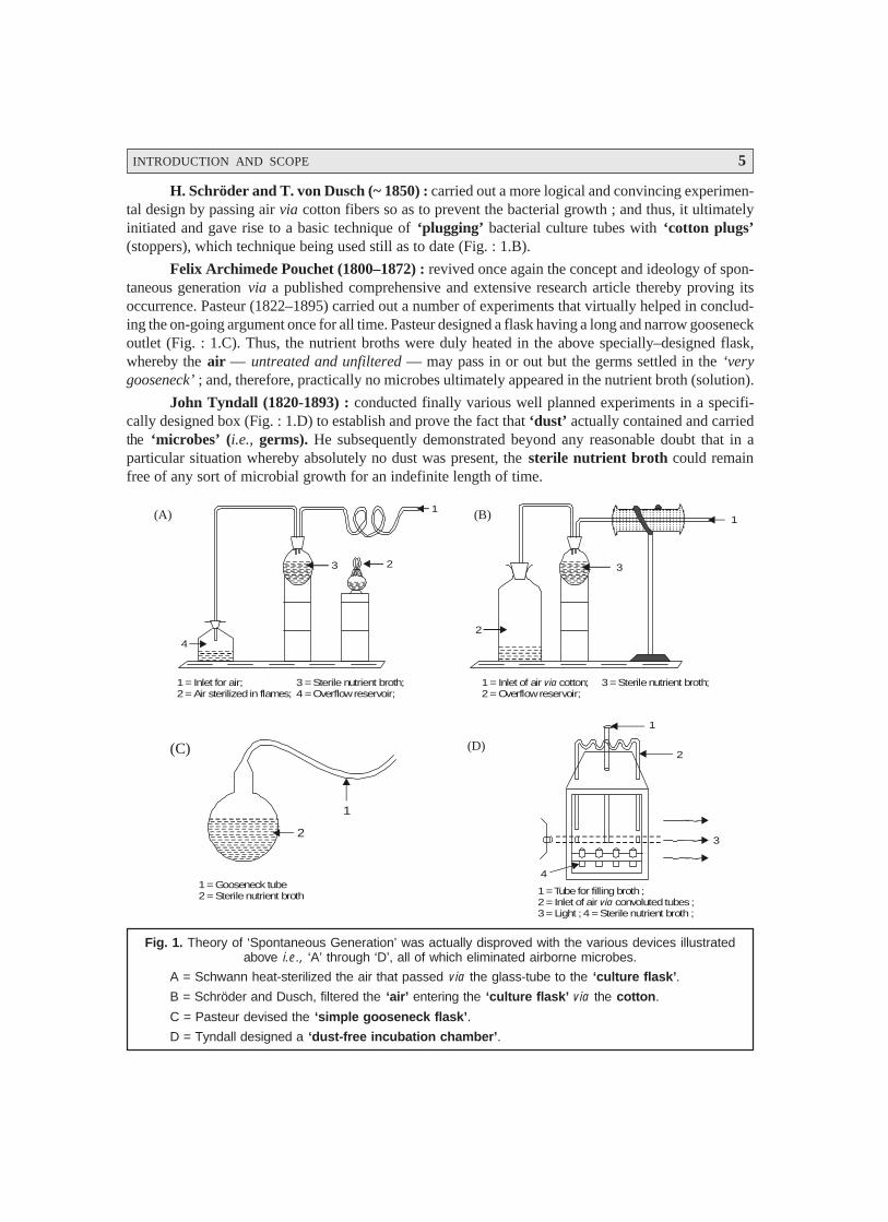
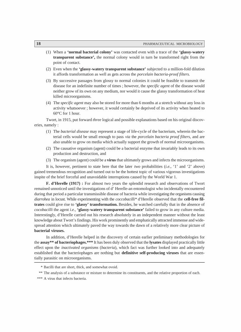
Franz Schulze (1815-1873) and Theodor Schwann (1810–1882) : these two scientists inde-pendently fully endorsed and justified the earlier findings of Spallanzani by allowing air to pass throughstrong acid solutions into the boiled infusions, and by passing air into the flasks via red-hot tubesrespectively (Fig. 1.A). In neither instance did microorganisms appear.
Special Note : The stubbornly conservative advocates of the theory of ‘spontaneous generation’were hardly convinced by the aforesaid experimental evidences.
INTRODUCTION AND SCOPE 5
H. Schröder and T. von Dusch (~ 1850) : carried out a more logical and convincing experimen-tal design by passing air via cotton fibers so as to prevent the bacterial growth ; and thus, it ultimatelyinitiated and gave rise to a basic technique of ‘plugging’ bacterial culture tubes with ‘cotton plugs’(stoppers), which technique being used still as to date (Fig. : 1.B).
Felix Archimede Pouchet (1800–1872) : revived once again the concept and ideology of spon-taneous generation via a published comprehensive and extensive research article thereby proving itsoccurrence. Pasteur (1822–1895) carried out a number of experiments that virtually helped in conclud-ing the on-going argument once for all time. Pasteur designed a flask having a long and narrow gooseneckoutlet (Fig. : 1.C). Thus, the nutrient broths were duly heated in the above specially–designed flask,whereby the air — untreated and unfiltered — may pass in or out but the germs settled in the ‘verygooseneck’ ; and, therefore, practically no microbes ultimately appeared in the nutrient broth (solution).
John Tyndall (1820-1893) : conducted finally various well planned experiments in a specifi-cally designed box (Fig. : 1.D) to establish and prove the fact that ‘dust’ actually contained and carriedthe ‘microbes’ (i.e., germs). He subsequently demonstrated beyond any reasonable doubt that in aparticular situation whereby absolutely no dust was present, the sterile nutrient broth could remainfree of any sort of microbial growth for an indefinite length of time.
Fig. 1. Theory of ‘Spontaneous Generation’ was actually disproved with the various devices illustratedabove i.e., ‘A’ through ‘D’, all of which eliminated airborne microbes.
A = Schwann heat-sterilized the air that passed via the glass-tube to the ‘culture flask’.
B = Schröder and Dusch, filtered the ‘air’ entering the ‘culture flask’ via the cotton.
C = Pasteur devised the ‘simple gooseneck flask’.
D = Tyndall designed a ‘dust-free incubation chamber’.
1 = Inlet for air; 3 = Sterile nutrient broth;2 = Air sterilized in flames; 4 = Overflow reservoir;
3 2
1
4
(A)
1 = Inlet of air cotton; 3 = Sterile nutrient broth;2 = Overflow reservoir;
via
(B)
3
1
2
1 = Gooseneck tube2 = Sterile nutrient broth
2
1
(C)
1 = Tube for filling broth ;2 = Inlet of air convoluted tubes ;3 = Light ; 4 = Sterile nutrient broth ;
via
1
2
3
4
(D)

6 PHARMACEUTICAL MICROBIOLOGY
* HTST-Pasteurizer. It makes use of high-temperature short-time pasteurization process employing high-temperature live steam.
** Septicemia following childbirth [SYN : childbed Fever ; Puerperal Sepsis ;]
1.2.3. Fermentation
France having the strategical geographical location developed the commercial manufacture of alarge variety of wines and beer as a principal industry. Pasteur played a critical and major role in theproper standardization of various processes and techniques intimately associated with the said two‘alcoholic beverages’ in order to obtain a consistently good product. Pasteur used his God gifted won-derful skill and wisdom to explore and exploit the unique capabilities of microbes in the fermentationindustry exclusively using fruits and grains resulting in alcohol-based table wines, dry-wines, cham-pagne, whiskies, etc. Pasteur meticulously isolated, typified, and characterized ‘certain microbes’ ex-clusively responsible for the ‘good batches’ predominantly in comparison to the ones found solely inthe ‘poor products’.
In fact, the overall net outcome of such extensive as well as intensive investigations helped in along way for the assured and successful production of consistently good and uniform ultimate product.Pasteur vehemently argued and suggested that the unwanted/undesirable types of microbes must bedestroyed and removed by heating not enough to alter the original and authentic inherent flavour/aromaof the fruit juice, but just sufficient to cause and afford the legitimate destruction of a relatively very highpercentage of the ‘bad microbial population’. This ‘destructive microbial phenomenon’ could be ac-complished successfully by holding the juices at a temperature of 145°F (≡ 62.8°C) for a duration of 30minutes.
Pasteurization. Nowadays, the large-scale handling of such destructive microbial process may beachieved by ‘pasteurization’* in commercial fermentation industries using either ‘malt wort’ (having ~10% solids) or molasses (~ 10% solids) or even fruit-juices.
1.2.4. Germ Theory
A plethora of observant researchers had already conceptualized and opined rather vehementlythe much applauded and widely accepted ‘germ theory’ of disease even before Pasteur establishedexperimentally that microbes (or bacteria) happen to be the root cause of several human dreadfuldiseases. Later on various other scientists supported and proved the aforesaid ‘germ theory’ in one wayor the other as stated under :
Girolamo Fracastro (1483–1553) : advocated that certain diseases might be caused by virtue ofinvisible organisms transmitted from one subject to another.
Plenciz (1762) : stated that the living microbes (or agents) are the ultimate cause of disease butat the same time aired his views that different germs were responsible for different ailments.
Oliver Wendell Holmes (1809–1894) : suggested that puerperal fever** was highly conta-gious in nature ; besides, it was perhaps caused by a germ carried eventually from one mother to anothereither by midwives or physicians.
Ignaz Philipp Semmelweis (1818–1865) : pioneered the usage of antiseptics specifically in theobstetrical practices.
Joseph Lister (1890) : made known in England the importance of antisepsis, which was subse-quently fully appreciated by the medical profession all and sundry.
INTRODUCTION AND SCOPE 7
* Acute, infectious disease caused by Bacillus anthracis, usually attacking cattle, sheep, horses, andgoats. First ever proved that a bacterium to be the cause of an animal disease.
Robert Koch (1843–1910) : discovered the typical bacilli having squarish ends in the bloodsample of cattle that had died due to anthrax.*
Koch’s Modus Operandi — Koch adopted the following steps to isolate microbes causing an-thrax :
(1) First of all these bacteria were duly grown in cultures in the laboratory.
(2) Bacteria examined microscopically so as to ascertain only one specific type was present.
(3) Injected bacteria into other animals to observe whether they got also infected, and subse-quently developed clinical symptoms of anthrax.
(4) Isolated microbes from experimentally infected animals squarely matched with those ob-tained originally from sheep that died due to infection of anthrax.
Koch’s Postulates : The series of vital observations ultimately led to the establishment of Koch’spostulates, that essentially provided four vital guidelines to identify the particular causative agent for aninfectious disease, namely :
(a) A particular microbe (organism) may invariably be found in association with a given disease.
(b) The organism may be isolated and cultivated in pure culture in the laboratory.
(c) The pure culture shall be able to cause the disease after being duly inoculated into a suscep-tible animal.
(d) It should be quite possible to recover conveniently the causative organism in its pure cultureright from the infected experimental animal.
1.2.5. Classical Laboratory Methods and Pure Cultures
Microorganisms are abundantly found in nature in sufficiently large populations invariably com-prised of a plethora of different species. It is, however, pertinent to state here that to enable one to carryout an elaborated study with regard to the characteristic features of a specific species it is absolutelynecessary to have it separated from all the other species.
Laboratory Methods : Well defined, articulated, and explicite laboratory methods have beenadequately developed which enable it to isolate a host of microorganisms representing each species,besides to cultivate each of the species individually.
Pure Culture : Pure culture may be defined as — ‘the propogation of microorganisms or ofliving tissue cells in special media that are conducive to their growth’.
In other words it may also be explained as the growth of mass of cells belonging to the samespecies in a laboratory vessel (e.g., a test tube). It was indeed Joseph Lister, in 1878, who first andforemost could lay hand on pure cultures of bacteria by the aid of ‘serial dilution technique’ in liquidmedia.
Example : Lister diluted milk, comprising of a mixture of bacteria, with a specially designedsyringe until a ‘single organism’ was strategically delivered into a container of sterile milk. The con-tainer on being subjected to incubation for a definite period gave rise to a bacteria of a single type, verymuch akin to the parent cell. Lister termed it as Bacterium lactis.

8 PHARMACEUTICAL MICROBIOLOGY
Colonies : Koch meticulously devised methods for the specific study of microorganism. Hesmeared bacteria on a sterile glass slide, followed by addition of certain specific dyes so as to observethe individual cells more vividly under a microscope. Koch carefully incorporated some specific solidi-fying agents, such as : gelatin, agar into the media in order to obtain characteristic isolated growths oforganisms usually called as colonies. Importantly, each colony is essentially comprised of millions ofindividual bacterial cells packed tightly together.
Now, from these identified colonies one may transfer pure cultures to other sterile media. How-ever, the development of a liquefiable solid-culture medium proved to be of immense fundamentalimportance.
Example : Koch thoroughly examined material obtained from subjects suffering from pulmo-nary tuberculosis, and succeeded in the isolation of the tubercle bacillus Mycobacterium tuberculosis.
In conclusion, one may summarize the remarkable importance of ‘pure cultures’ toward theoverwhelming development in the field of microbiology, because by the help of pure-culture tech-niques several intricate and complicated problems could be answered with reasonable clarification andcomplete satisfaction, namely :
Microorganisms causing a large number of infections,
Certain specific fermentative procedures,
Nitrogen-fixation in soil,
High-yielding alcohol producing strains from ‘malt wort’, and ‘molasses’,
Selected good cultures for making top-quality wines, and
Specific cultures for manufacturing dairy products viz., cheeses, yogurt.
Futuristic Goals
The futuristic goals of ‘pure cultures’ are exclusively based upon the following two cardinalaspects, namely :
(a) better understanding of the physiology of individual microorganisms present in the pureculture, and
(b) ecological relationships of the entire microbial populations in a given environment.
Thus, the following new horizons in the domain of microbiology may be explored with greatzeal and gusto :
Advancements in marine microbiology,
Rumen microbiology,
Microbiology of the gastro-intestinal tract (GIT), and
Several other systems.
1.2.6. Immunity
Immunity refers to the state of being immune to or protected from a disease, especially aninfectious disease. This state is invariably induced by having been exposed to the antigenic marker onan organism that invades the body or by having been immunized with a vaccine capable of stimulatingproduction of specific antibodies.*
* K Sambamurthy and Ashutosh Kar, Pharmaceutical Biotechnology, New Age International (P) Ltd., Pub-lishers, New Delhi, 2006.

INTRODUCTION AND SCOPE 9
* Microorganisms that have been rendered thin or made less virulent (infectious).
** Any of the complex glycoproteins produced by lymphocytes in response to the presence of an antigen.
*** Infectious organisms.
**** A substance that neutralizes toxin.
Interestingly, Pasteur’s practical aspects and Koch’s theoretical aspects jointly established thefact that the attenuated microorganisms* retained their capacity and capability for stimulating therespective host to produce certain highly specific substances i.e., antibodies** which critically protectagainst subsequent exposure to the virulent organisms.***
Examples :
(a) Edward Jenner’s successful cowpox vaccine (in 1798) : Jenner’s epoch-making successfulattempts in vaccinating (innoculating) patients with cowpox vaccine, that ultimately re-sulted in the development of resistance to the most dreadful smallpox infection.
(b) Pasteur’s successful rabies vaccine : Pasteur’s charismatic fame and reputation becamewell known throughout France when he successfully prepared rabies vaccine by innoculatinga rabbit with the saliva from a rabid dog. The healthy rabbit contracted the rabies virus anddied later on. The extract of dead rabbit’s brain and spinal cord were duly attenuated andinjected into rabies patient who eventually survived later on. Thus, the vaccine for rabies orhydrophobia — a disease transmitted to humans through bites of dogs, cats, monkeys, andother animals.
1.2.7. Medical Microbiology
Interestingly, the ‘germ theory’ of disease was very much in existence for a long duration ;however, the direct implication and involvement of germs in causing disease was not well established,and hence recognized and widely accepted.
The magnificent and remarkable success of Louis Pasteur and Robert Koch not only earnedthem befitting honours and accolades from their beloved countrymen, but also rewarded them bybestowing their gratitude in establishing the famous and prestigious Pasteur Institute in Paris (1888),and Professor of Hygiene and Director of the Institute for Infective Diseases in the University ofBerlin respectively.
At this point in time altogether newer microorganisms (bacteria) were being discovered with anever-increasing speed and momentum, and their disease-producing capabilities were adequately estab-lished and proved by Koch’s four cardinal postulates as stated earlier (see section 1.2.4).
In this manner, the domain of ‘medical microbiology’ gradually received a progressive advance-ment through the meaningful researches conducted by several scientists and scholars as enumeratedbelow :
Edwin Klebs (1883) and Frederick Loeffler (1884) : discovered the diphtheria bacillus,corynebacterium diphtheriae ; and showed that it produced its toxins (poisons) in a laboratory flask.
Emil von Behring and Shibasaburo Kitasato : devised an unique technique of producing im-munity to infections caused by C. diphtheriae by injecting the toxins into healthy animals so that anantitoxin**** gets developed.
Shibasaburo Kitasato and Emil von Behring : cultivated (grown) the microorganism respon-sible for causing tetanus (lockjaw), Chlostridium titani ; and Behring prepared the corresponding anti-toxin for the control, prevention, treatment, and management of this fatal disease.
10 PHARMACEUTICAL MICROBIOLOGY
* An antibody is a water-soluble protein produced from globulins (e.g., γ-globulin) in the spleen, lymph nodes,thymus gland, liver or bone marrow in response to an antigen (foreign protein). Antibodies attack antigens torender them inactive and no longer infective.
** A therapeutic concept developed by Paul Ehrlich (1854–1915) wherein a specific chemical or drug is used totreat an infectious disease or cancer ; ideally, the chemical should destroy the pathogen or the cancer cellswithout harming the host.
*** A chemical produced by a microorganism or prepared partially or totally by synthetic means that inhibitsgrowth or kills other microorganisms at low concentration.
Emil von Behring bagged the Nobel Prize in 1901 in physiology or medicine.
De Salmon and Theobald Smith : proved amply that immunity to a plethora of infectiousdiseases may be produced quite effectively and efficiently by proper timely innoculation with the killedcultures of the corresponding microorganisms.
Elie Metchnikoff : described for the first time the manner certain specific leukocytes (i.e., whiteblood cells) were able to ingest (eat up) the disease-producing microorganisms present in the body. Hebaptized these highly specific defenders and crusaders against bacterial infections known as phagocytes(‘eating cells’), and the phenomenon is termed as phagocytosis.
Metchnikoff’s Theory : Based of the aforesaid explanations Metchnikoff put forward a theorythat — ‘the phagocytes were the body’s first and most important line of defense against a variety ofinfection’.
Paul Ehrlich : Paul Ehrlich (Robert Koch’s brilliant student) put forward two altogether newerconcepts with regard to the modus operandi whereby the body aptly destroys microorganisms (bacteria),namely :
(a) Antibody* : The logical explanation of immunity based upon certain antibodies in theblood, and
(b) Chemotherapy** and Antibiotics*** : Both these aspects virtually opened the flood gatesto the enormous future developments in combating the growth and destruction of pathogenicbacteria.
Example : Arsphenamine [Salvarsan(R)] : A light yellow organo-metallic compound (powder)containing about 30% Arsenic (As), was formerly used in the treatment of syphilis.
The two decades stretching between 1880–1900 proved to be indeed a golden era for the ‘sci-ence of microbiology’ to step into adolescence from the stage of infancy. In fact, during this specificperiod many researchers have gainfully identified the causative microorganisms duly responsible for theeruption of a host of infectious human diseases, such as :
Anthrax, Gonorrhea, Typhoid fever, Malaria, Wound infections, Tuberculosis, Cholera, Diph-theria, Tetanus, Meningitis, Gas gangarene, Plague, Dysentery, Syphilis, Whooping cough, and RockyMountain spotted fever.
1.2.8. Pharmaceutical Microbiology
The remarkable and spectacular breakthroughs accomplished by Pasteur, Koch, Jenner, and ahost of others more or less paved the way towards several miraculous discoveries in curing fatal anddreadful human ailments thereby minimising their immense sufferings. Many meaningful and wonder-ful researches also led to the discovery of a good number of causative agents of diseases and altogethernewer techniques for diagnosis, which ultimately rendered the diagnosis of these ailments rather rapidand precise.

INTRODUCTION AND SCOPE 11
Examples : (a) Widal Test* — for typhoid fever, and
(b) Wasserman Test** — for syphilis.
Importantly, a plethora of dreadful diseases were duly identified and characterized by the pres-ence of their specific causative microorganisms, such as : Hensen (1874) leprosy (Mycobacteriumleprae) ; Neisser (1879) gonorrhea (Neisseria gonorrhoeae) ; Ogston (1881) wound infections(Staphylococcus aureus) ; Nicolaier (1885) tetanus (Clostridium titani) ; Kitasato and Yersin (1894)plague (Yersinia pestis) ; Shiga (1898) dysentry (Shigella dysenteriae) ; Schaudin and Hoffmann (1905)syphilis (Treponema pallidum) ; Bordet and Gengou (1906) whooping cough (Bordetella pertussis) ;Ricketts (1909) rocky mountain spotted fever (Rickettsia ricketsii) ;
Some of the important events that mark the history of pharmaceutical microbiology are enu-merated below in a chronological arrangement :
Era Discoverer Important Events
Eighteenth Edward Jenner Discovery of small pox vaccine.
Century (1729–1799)
Nineteenth Justus von Liebig Conceptualized the physico-chemical theory of fermen-
Century (1803–1873) tation.
Ignaz Philipp First and foremost introduced the application of antiseptics.
Semmelweis
(1818–1865)
Joseph Lister Developed aseptic techniques : isolated bacteria in pure
(1827–1912) culture.
Fanny Hesse Suggested use of agar as a solidifying material for the
(1850–1934) preparation of microbiological media.
Paul Ehrlich Developed modern concept of chemotherapy and
(1854–1915) chemotherapeutic agents.
Hans Christian Invented vital and important procedure for differential
Gram staining of microorganisms i.e., the well-known Gram Stain.
(1853–1933)
Twentieth August von Developed complement-fixation test for syphilis.
Century Wassermann
(1866–1925)
Martinus Willem Employed the principles of enrichment cultures :
Beijerinck confirmed finding of the very first virus.
(1851–1931)
Frederick W. Twort Discovered independently the bacteriophages i.e., viruses
(1877–1950) ; and that destroy bacteria.
Felix H.d’ Herelle
(1873–1949)
* An agglutination test for typhoid fever.** A complement fixation test for syphilis.
12 PHARMACEUTICAL MICROBIOLOGY
* The living together in close association of two organisms of different species. If neither organism is harmed,this is called commensalism ; if the association is beneficial to both, mutualism ; if one is harmed and theother benefits, parasitism.
** An association or relationship between two organisms in which one is harmful to the other.
*** An enzyme found in phagocytes, neutrophils, and macrophages, and in tears, saliva, sweat and other bodysecretions, that destroys bacteria by breaking down their walls.
Antibiotics : Antibiotic refers to a natural or synthetic substance that destroys microorganismsor inhibits their growth. Antibiotics are employed extensively to treat infectious, diseases in humans,animals, and plants. In fact, the terminology ‘antibiotic’ etymologically evidently signifies anythingagainst life. Obviously, in the event when the microorganisms are critically present in a naturalmedium two situations may arise invariably viz., (a) favouring the growth of bacteria usually termed as‘symbiosis’ ;* and (b) antagonizing the growth of bacteria normally called as ‘antibiosis’.**
Charles Robert Darwin (1809–1882), a British naturalist) aptly commenced scientific and me-thodical investigative explorations into the fundamental problems of natural selection and struggle amongstthe interspecies ; and later on came up with his famous doctrine — ‘Survival of the fittest’. LouisPasteur (1822–1895) observed for the first time the characteristic antagonistic interrelations prevailingbetween the microorganisms of different species.
Joubert and Pasteur first observed the critical destruction of cultures of Bacillus anthracis bymeans of certain air-borne microbes. A follow up by Sirotinin (1888) emphatically proved the antago-nistic action of Bacillus anthracis upon the enteric fever, and Blagoveshchensky (1890) carefully ascer-tained the antagonistic effect of the blue-pus organism on the Bacillus anthracis. It was ultimately themiraculous discovery of Lashchenkov (1909) and Alexander Fleming (1922) who meticulously isolatedthe enzyme lysozyme***, that was chiefly capable of inhibiting a relatively larger segment of microor-ganisms. Chain, Florey, and co-workers (1929) made the epoch making historical development in theemerging field of antibiotics with the remarkable discovery of wonderful therapeutic and interestingpharmacological properties of the extracts obtained from the cultures of the mold Penicillium notatumthat eventually gave rise to the formation of the wonder drug ‘penicillin’.
Specifically the antibiotics are extremely useful in the control, management and treatment of agood number of human infectious diseases but their diversified applications are found to be equallyuseful in the meticulous curing and controlling of plant and animal diseases as well. Penicillin has beeneffectively employed in the management and control of pests. Antibiotics, in general, are invariablyemployed in animal husbandry as ‘feed additive’ to cause enhancement in the fattening of food animals.Food handling and processing industries extensively make use of antibiotics to critically minimise in-evitable spoilage of fish, vegetables, and poultry products. Present day modern scientific researchesbeing conducted across the globe do make use of antibiotics as useful and indispensable tools for theelaborated study of biochemical cellular mechanisms.
Since the discovery of penicillin many more antibiotics came into being as stated under :Waksman (1944) : Streptomycin — [Streptomyces griseus] — a soil microbe ;
— (1945) : Bacitracin — [Bacillus subtilis] ;— (1947) : Chloramphenicol (Chloromycetin) — [Streptomyces venezuelae] ;— (1947) : Polymixin — [Bacillus polymixa] — and various designated polymixins
A, B, C, D, and E.
— (1948) : Chlorotetracycline — [Streptomyces aureofaciens] — a broad-spectrumantibiotic.
— (1948) : Neomycin — [a species of Streptomyces] — isolated from soil.

INTRODUCTION AND SCOPE 13
— (1950) : Oxytetracycline — [a strain of Streptomyces].
— (1952) : Erythromycin — [Streptomyces erythreus].
It is, however, pertinent to state here that the ‘antibiotics’ may be broadly classified into ninecategories as given below :
S.No. Class Designated Antibiotics
I Aminoglycosides Amikasin ; Gentamycin ; Kanamycin ; Neomycin ; Netilmicin ;
Streptomycin ; and Tobramycin ;
II Ansamycins Maytansine ; and Rifampicin ;
III Beta-lactam antibiotics Amoxycillin ; Ampicillin ; Cephalosporin ; Clavulanic acid ;
Cloxacillin ; Nocardicins ; Penicillins ; and Thienamycin ;
IV Cyclic polypeptides Gramicidin ; and Polymixins A, B, C, D and E ;
V Fluoroquinolones Ciprofloxacin ; Enoxacin ; Norfloxacin ; and Ofloxacin.
VI Macrolides Azithromycin ; Bacitracin ; Clarithromycin ; and Erythromycin ;
VII Polyenes Amphotericin B ; Griseofulvin ; and Nystatin ;
VIII Tetracyclines Aureomycin ; Doxycycline ; Minocycline ; Oxytetracycline ;
and Tetracycline ;
IX Miscellaneous Adriamycin ; Chloramphenicol (Chloromycetin) ; Clindamycin ;
Cycloserine ; and Mitomycins.
[Kar, Ashutosh : Pharmacognosy and Pharmacobiotechnology, New Age International (P)Ltd., Publishers, New Delhi, 2003].
Important Points : The various important points with respect to the development of antibioticsare summarized below :
in all approximately 5000 antibiotics have been prepared, characterized, and evaluated fortheir therapeutic efficacy till date.
nearly 1000 antibiotics belonging to only six genera of filamentous fungi i.e., includingCephelosporium and Penicillium have been reported successfully.
about 50 antibiotics have been synthesized from two genera and belonging to the class ofnon-filamentous bacteria.
nearly 3000 antibiotics have been prepared from a group of filamentous bacteria i.e., in-cluding streptomyces.
approximately 50 antibiotics are at present actively used in therapeutic treatment and veteri-nary medicine around the world.
Importantly, the most common bacteria that invariably attack the humans specifically, and thediseases they cause or organs of the body they attack, are listed as under :
S.No. Microorganism Disease/Place of Infection
1 Bacteroides Pelvic organs ;
2 Bordetella pertussis Whooping cough ;
3 Brucella abortus Brucellosis ;
4 Chlamydia trachomatis Vinereal disease ;
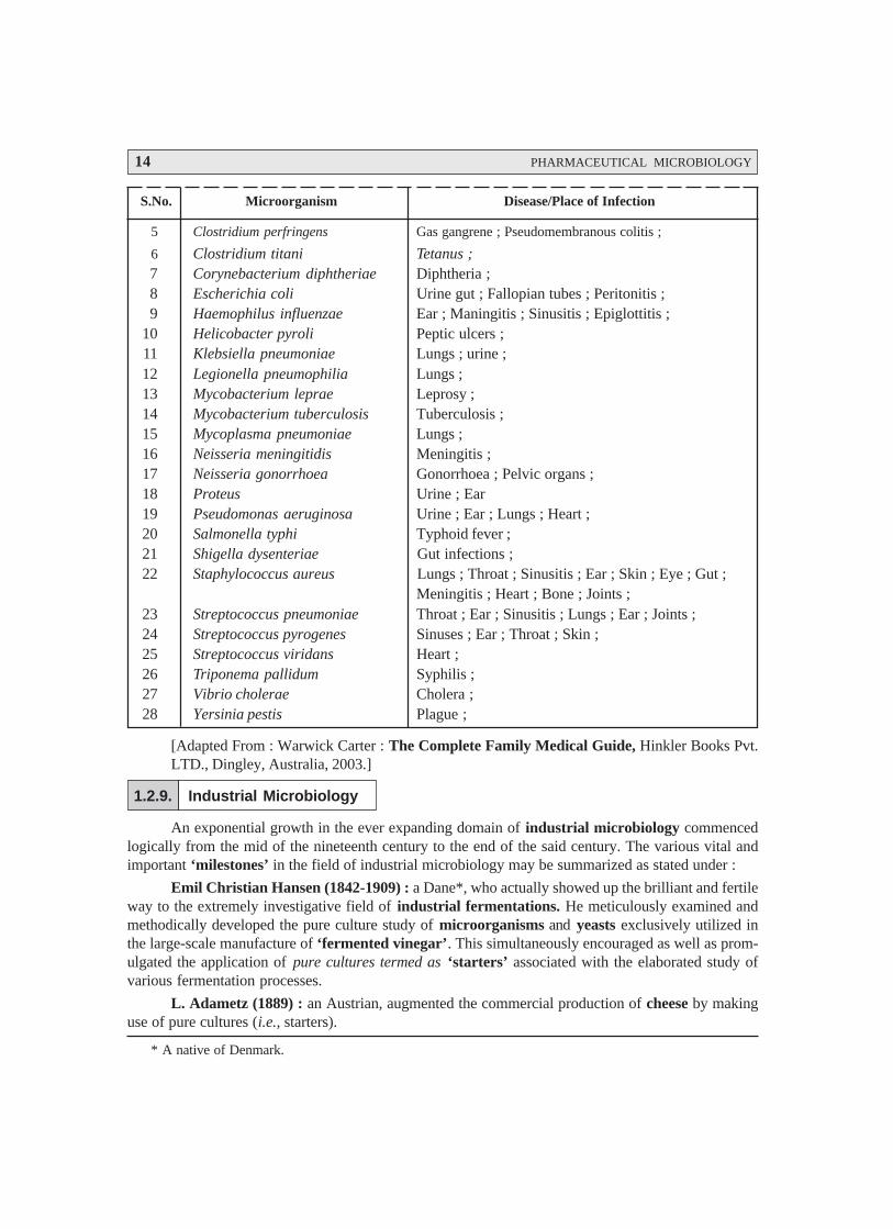
14 PHARMACEUTICAL MICROBIOLOGY
S.No. Microorganism Disease/Place of Infection
5 Clostridium perfringens Gas gangrene ; Pseudomembranous colitis ;
6 Clostridium titani Tetanus ;7 Corynebacterium diphtheriae Diphtheria ;8 Escherichia coli Urine gut ; Fallopian tubes ; Peritonitis ;9 Haemophilus influenzae Ear ; Maningitis ; Sinusitis ; Epiglottitis ;
10 Helicobacter pyroli Peptic ulcers ;11 Klebsiella pneumoniae Lungs ; urine ;12 Legionella pneumophilia Lungs ;13 Mycobacterium leprae Leprosy ;14 Mycobacterium tuberculosis Tuberculosis ;15 Mycoplasma pneumoniae Lungs ;16 Neisseria meningitidis Meningitis ;17 Neisseria gonorrhoea Gonorrhoea ; Pelvic organs ;18 Proteus Urine ; Ear19 Pseudomonas aeruginosa Urine ; Ear ; Lungs ; Heart ;20 Salmonella typhi Typhoid fever ;21 Shigella dysenteriae Gut infections ;22 Staphylococcus aureus Lungs ; Throat ; Sinusitis ; Ear ; Skin ; Eye ; Gut ;
Meningitis ; Heart ; Bone ; Joints ;23 Streptococcus pneumoniae Throat ; Ear ; Sinusitis ; Lungs ; Ear ; Joints ;24 Streptococcus pyrogenes Sinuses ; Ear ; Throat ; Skin ;25 Streptococcus viridans Heart ;26 Triponema pallidum Syphilis ;27 Vibrio cholerae Cholera ;28 Yersinia pestis Plague ;
[Adapted From : Warwick Carter : The Complete Family Medical Guide, Hinkler Books Pvt.LTD., Dingley, Australia, 2003.]
1.2.9. Industrial Microbiology
An exponential growth in the ever expanding domain of industrial microbiology commencedlogically from the mid of the nineteenth century to the end of the said century. The various vital andimportant ‘milestones’ in the field of industrial microbiology may be summarized as stated under :
Emil Christian Hansen (1842-1909) : a Dane*, who actually showed up the brilliant and fertileway to the extremely investigative field of industrial fermentations. He meticulously examined andmethodically developed the pure culture study of microorganisms and yeasts exclusively utilized inthe large-scale manufacture of ‘fermented vinegar’. This simultaneously encouraged as well as prom-ulgated the application of pure cultures termed as ‘starters’ associated with the elaborated study ofvarious fermentation processes.
L. Adametz (1889) : an Austrian, augmented the commercial production of cheese by makinguse of pure cultures (i.e., starters).
* A native of Denmark.
INTRODUCTION AND SCOPE 15
HW Conn (in Connecticut, USA) and H Weigmann (in Germany) (1890–1897) : developedmiraculously a host of pure culture starters for the commercial production of butter.
Alcohol Fermentations : Pure culture of yeasts were used to produce alcohol (ethanol) from avariety of fermentable carbohydrates such as : corn, molasses, potatoes, sugar beets, grapes etc., employedthroughout the world.
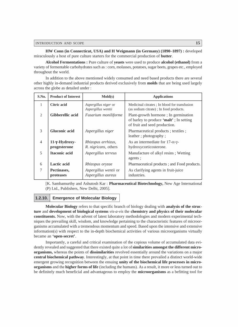
In addition to the above mentioned widely consumed and need based products there are severalother highly in-demand industrial products derived exclusively from molds that are being used largelyacross the globe as detailed under :
S.No. Product of Interest Mold(s) Applications
1 Citric acid Aspergillus niger or Medicinal citrates ; In blood for transfusionAspergillus wentii (as sodium citrate) ; In food products.
2 Gibberellic acid Fusarium moniliforme Plant-growth hormone ; In germinationof barley to produce ‘malt’ ; In settingof fruit and seed production.
3 Gluconic acid Aspergillus niger Pharmaceutical products ; textiles ;leather ; photography ;
4 11-γγγγγ-Hydroxy- Rhizopus arrhizus, As an intermediate for 17-α-γ-progesterone R. nigricans, others hydroxycorticosterone.
5 Itaconic acid Aspergillus terreus Manufacture of alkyl resins ; Wettingagents ;
6 Lactic acid Rhizopus oryzae Pharmaceutical products ; and Food products.
7 Pectinases, Aspergillus wentii or As clarifying agents in fruit-juiceproteases Aspergillus aureus industries.
[K. Sambamurthy and Ashutosh Kar : Pharmaceutical Biotechnology, New Age International(P) Ltd., Publishers, New Delhi, 2005].
1.2.10. Emergence of Molecular Biology
Molecular Biology refers to that specific branch of biology dealing with analysis of the struc-ture and development of biological systems vis-a-vis the chemistry and physics of their molecularconstituents. Now, with the advent of latest laboratory methodologies and modern experimental tech-niques the prevailing skill, wisdom, and knowledge pertaining to the characteristic features of microor-ganisms accumulated with a tremendous momentum and speed. Based upon the intensive and extensiveinformation(s) with respect to the in-depth biochemical activities of various microorganisms virtuallybecame an ‘open-secret’.
Importantly, a careful and critical examination of the copious volume of accumulated data evi-dently revealed and suggested that there existed quite a lot of similarities amongst the different micro-organisms, whereas the points of dissimilarities revolved essentially around the variations on a majorcentral biochemical pathway. Interestingly, at that point in time there prevailed a distinct world-wideemergent growing recognition between the ensuing unity of the biochemical life processes in micro-organisms and the higher forms of life (including the humans). As a result, it more or less turned out tobe definitely much beneficial and advantageous to employ the microorganisms as a befitting tool for
16 PHARMACEUTICAL MICROBIOLOGY
* Professor Salvador E Luria — at the Massachusetts Institute of Technology (MIT) as a Professor of Biologywas awarded the Nobel Prize in 1969 for his splendid research in the field of molecular biology.
** Viruses that infect bacteria.
deciphering and exploring the basic life phenomena. In order to accomplish the aforesaid aims andobjectives the microorganisms do offer invariably a plethora of advantages for this type of researchactivities, namely :
they reproduce (i.e., cultivate) extremely fast,
they may be cultured (grown) either in small or large quantum easily, conveniently, andquickly,
their growth may be manipulated and monitored in a not-so-difficult manner by means ofchemical and physical methods, and
their cells may be cleaved and torn apart, and the contents segregated into different fractionsof varying particle sizes.
Conclusively, the above cited characteristic features together with certain other vital factors helpto render the ‘microorganisms’ an extremely vulnerable and a very convenient research-role-modelin pin-pointing and establishing precisely the modus operandi of various life processes that essentiallyoccur with respect to certain particular chemical reactions, besides the specific structural featuresinvolved intimately.
In the light of the above statement of facts showing the enormous strengths of microorganisms inthe revelation of the intricacies of life processes various scientists and researchers of all disciplines viz.,physicists, chemists, geneticists, biologists, and microbiologists not only joined their hands together butalso put their intellectual resources and wisdom in a concerted manner to evolve an altogether newdiscipline christened as molecular biology. According to Professor Luria* molecular biology may bedefined as — ‘the programme of interpreting the specific structures and functions of organisms in termsof molecular structure’.
The outcome of the results obtained from the brilliant studies accomplished in the field of mo-lecular biology are numerous, such as :
Elucidation of enzyme structure and mode of action,
Cellular regulatory mechanisms,
Energy metabolism mechanisms,
Protein synthesis,
Structure of viruses,
Functionality of membranes, and
Structure and function of nucleic acids.
Significance of Discoveries : The major significance of discoveries with regard to molecularbiology may be ascertained by virtue of the following breakthroughs :
Fundamental information(s) regarding DNA and genetic processes at the molecular level viabacteria and bacteriophages**, and
Many Nobel Prizes bagged due to researches carried out in molecular biology related tovarious arms of biology.
INTRODUCTION AND SCOPE 17
* Genetic mutation wherein the tissues of an organism are of different genetic kinds even thoughthey were derived from the same cell.
** An alkaline fluid found in the lymphatic vessels and the cisterna chyli.
*** A substance introduced by innoculation.
**** A poisonous substance of animal or plant origin.
1.2.11. Emergence of Virology
Virology essentially refers to — ‘the study of viruses and viral diseases’.
Preamble : Towards the later part of the nineteenth century Pasteur and his co-workers werevigorously attempting to unfold the precise and exact mechanism of the phenomenon of disease devel-opment by examining meticulously a good number of infectious fluids (drawn from patients) for thepossible presence of specific disease producing agent(s) by allowing them to pass through filters with aview to retain the bacterial cells. An affirmative conclusion could be reached easily in the event whenthe filtrates (obtained above) failed to produce any infection, and the presence of the disease producingbacterial agent in the original (infectious) fluid.
The following researchers determined the presence of ‘virus’ in pathological fluids in the fol-lowing chronological order :
Chamberland (1884) : First and foremost developed the specially designed ‘porcelain filters’that exclusively permitted the passage of fluid but not the microorganisms ; and, therefore, could beused gainfully for the sterilization of liquids. Besides, the application of such devices may also suggestand ascertain if at all ‘infective agents’ smaller in dimensions than the bacteria could exit actually.
Iwanowski (1892) : Repeated the similar sort of test but employed an extract meticulouslyobtained from the infected tobacco plants, with ‘mosaic* disease’. Iwanowski observed that the clearfiltrate was found to be extremely infectious to the healthy tobacco plants.
Beijerinck (1898) : He confirmed Iwanowski’s findings and baptised the contents of the clearfiltrate as ‘virus’ (i.e., infectious poisonous agent). He further affirmed that the virus could be propogatedstrategically within the living host.
Loeffler and Frosch (1998) : They first and foremost demonstrated that the clear filtrate hap-pened to be the main culprit, virus, which had the capability of being transmitted from one infectedanimal to another. Later on they amply proved that the lymph** obtained from infected animals suffer-ing from ‘foot and mouth disease’, whether it was either filtered or unfiltered, both caused infection inhealthy animals almost to the same extent. From the above critical studies one may infer that sinceanimals infected with the filtered lymph served as a source of inoculum*** for the infection of healthyanimals thereby suggesting overwhelmingly that the infective filterable agent never was a toxin****,but an agent capable of undergoing multiplication.
FW Twort (1915) : Twort inoculated nutrient agar with smallpox vaccine fluid with a possibleexpectation that a virulent variant of vaccinia virus could grow up eventually into colonies. In fact, theonly colonies which actually showed up on the agar plates were nothing but bacteria that proved to becontaminants in the vaccine lymph. However, these bacterial colonies had undergone a transformationthat turned into a ‘glassy watery transparent substance’, which could not be subcultured anymore.
Salient Features of ‘Glassy-Watery Transparent Substance : The various salient features ofthe glassy-watery transparent substance are as given under :
18 PHARMACEUTICAL MICROBIOLOGY
* Bacilli that are short, thick, and somewhat ovoid.
** The analysis of a substance or mixture to determine its constituents, and the relative proportion of each.
*** A virus that infects bacteria.
(1) When a ‘normal bacterial colony’ was contacted even with a trace of the ‘glassy-waterytransparent substance’, the normal colony would in turn be transformed right from thepoint of contact.
(2) Even when the ‘glassy-watery transparent substance’ subjected to a million-fold dilutionit affords transformation as well as gets across the porcelain bacteria-proof filters.
(3) By successive passages from glossy to normal colonies it could be feasible to transmit thedisease for an indefinite number of times ; however, the specific agent of the disease wouldneither grow of its own on any medium, nor would it cause the glassy transformation of heatkilled microorganisms.
(4) The specific agent may also be stored for more than 6 months at a stretch without any loss inactivity whatsoever ; however, it would certainly be deprived of its activity when heated to60°C for 1 hour.
Twort, in 1915, put forward three logical and possible explanations based on his original discov-eries, namely :
(1) The bacterial disease may represent a stage of life-cycle of the bacterium, wherein the bac-terial cells would be small enough to pass via the porcelain bacteria proof filters, and arealso unable to grow on media which actually support the growth of normal microorganisms.
(2) The causative organism (agent) could be a bacterial enzyme that invariably leads to its ownproduction and destruction, and
(3) The organism (agent) could be a virus that ultimately grows and infects the microorganisms.
It is, however, pertinent to state here that the later two probabilities (i.e., ‘1’ and ‘2’ above)gained tremendous recognition and turned out to be the hottest topic of various vigorous investigationsinspite of the brief forceful and unavoidable interruptions caused by the World War 1.
F. d’Herelle (1917) : For almost two years the splendid research and observations of Twortremained unnoticed until the investigations of d’ Herelle-an entomologist who incidentally encounteredduring that period a particular transmissible disease of bacteria while investigating the organisms causingdiarrohea in locust. While experimenting with the coccobacilli* d’Herelle observed that the cell-free fil-trates could give rise to ‘glassy’ transformation. Besides, he watched carefully that in the absence ofcocobacilli the agent i.e., ‘glassy-watery transparent substance’ failed to grow in any culture media.Interestingly, d’Herelle carried out his research absolutely in an independent manner without the leastknowledge about Twort’s findings. His work prominently and emphatically attracted immense and wide-spread attention which ultimately paved the way towards the dawn of a relatively more clear picture ofbacterial viruses.
In addition, d’Herelle helped in the discovery of certain earlier preliminary methodologies forthe assay** of bacteriophages.*** It has been duly observed that the lysates displayed practically littleeffect upon the inactivated organisms (bacteria), which fact was further looked into and adequatelyestablished that the bacteriophages are nothing but definitive self-producing viruses that are essen-tially parasitic on microorganisms.
INTRODUCTION AND SCOPE 19
* A special type of virus-bacterial cell interaction maintained by a complex cellular regulatory mechanism.Bacterial strains freshly isolated from their natural environment may contain a low concentration ofbacteriophage. This phage will lyse other related bacteria. Cultures that contain these substances are said tobe lysogenic.
** A rod-shaped, Gram-negative, non pathogenic soil and water bacteria that fix atmospheric nitrogen ; thesingle genus of the family Azotobacteraceae.
A. Lwoff (1921) : Lwoff further ascertained and proved the fact that bacteria invariably carrybacteriophages without undergoing ‘any sort of clearance’, and it was termed as ‘lysogeny’*.
1.2.12. Microorganisms as Geochemical Agents
The mid of the nineteenth century witnessed an ever growing interest in the pivotal role ofmicroorganisms in carrying out not only the various processes related to fermentations but also tacklingsome of the human diseases. Nevertheless, Pasteur’s articulated contributions on fermentation evidentlyproved and established that microorganisms in particular may cater as highly specific entities in per-forming a host of chemical transformations.
Winogradsky and Beijerinck legitimately shared the overall merit and credibility for establishingthe precise role of microbes in the critical transformations of N and S.
Windogradsky (1856-1953) : He critically examined and observed that there exist a plethora ofdistinct and discrete categories of microorganisms each of which is invariably characterized by its inherentcapability to make use of a specific inorganic energy source.
Examples :(a) Sulphur Microbes : They oxidize inorganic sulphur containing entities exclusively.
(b) Nitrogen Microbes : They oxidize inorganic nitrogen containing compounds solely.Interestingly, Winogradsky caused to be seen that there are certain microorganisms which either
in association with free living or higher plants may exclusively make use of gaseous nitrogen for thesynthesis of the specific cell components.
Hellriegel and Wilfarth (1888) : They showed explicitely that a predominantly mutual andimmensely useful symbiosis does exist between bacteria and the leguminous plants particularly.
Beijerinck (1901) : He meticulously observed, described, and even enumerated the usefulnessof the very presence of the ‘free-living nitrogen fixing’ organism Azotobacter** in maintaining thefertility of the soil.
1.2.13. Microbiology in the New Millennium
The major thrust in the specialized domain of ‘microbiology’ got a tremendous boost in speedand momentum during the twentieth century towards the development of judicious control and manage-ment of infectious human diseases ; elaborated studies in immunity profile ; as exceptionally attractivemodels for investigating fundamental life processes viz., activities related to metabolizing, growing,reproducing, aging, and dying ; and microbes’ broad spectrum physiological and biochemical potenti-alities than all other organisms combined. In addition, the science of microorganisms have propogatedother allied disciplines, for instance : biochemistry, genetics, genetic engineering, molecular biology,and the like.
Historic revelation of DNA (deoxyribonucleic acid), which being the key to life and genetics,was duly discovered by two world famous biologists Watson and Crick. DNA forms the basic funda-
20 PHARMACEUTICAL MICROBIOLOGY
* This is like a twisted rubber ladder. Each rung of the ladder is formed by a set combination of amino acidsthat form a code. Segments of that code form a gene. Only four chemicals make up the code : adenine (A),thymine (T), guanine (G), and cytosine (C). A always pairs with T, and G with C, allowing exact reproduc-tion of the chromosome.
mental structure of each and every chromosome in the precise shape of a ‘double-helix’.* In fact,microorganisms helped extensively and intensively in the better understanding of the exact mechanismwhereby the most critical and valuable information meticulously stored in the ‘genetic material’ isultimately transcribed and subsequently translated into proteins. Later on, Escherichia coli i.e., a colonbacterium, served as a via-media or a common tool for the geneticists, microbiologists, and biochem-ists to decepher the intricacies of various cellular processes. The concerted research inputs made byNirenberg, Khorana, Holley, Jacob, Monod, and a plethora of others substantiated copious informationsto the present day knowledge of the living systems, of course, making use of the microorganisms. It is,however, pertinent to mention at this juncture that microbes are being skilfully and gainfully utilized tograsp the meaning with respect to the control mechanisms directly involved in cell division as well asreproduction.
As to date ‘microbiology’ has marked with a dent an altogether separate identity and distinctbranch of biology having an established close relationship with biochemistry and genetics. It has pro-gressively and aggressively emerged into an intriguing subject over the years because each and everyspecific area in microbiology has virtually expanded into a large specialized subject in itself, namely :dairy microbiology, environmental microbiology, food microbiology, industrial microbiology, medicalmicrobiology, sanitary microbiology, and soil microbiology. Importantly, newer techniques exploringand exploiting microorganisms for gainful and economically viable products of interest have alwaysbeen the focus of attention across the globe. In the same vein, the absolute control and management ofcertain non-productive and troublesome species have always remained another virile and fertile area ofinterest in ‘microbiology’, which ultimately yielding definitely not only a purer product but also aug-mented the end-product to a considerable extent.
There are ample evidences cited in the scientific literatures with respect to enormous utilizationof the microorganisms to understand both biology and the prevailing intricacies of various biologicalprocesses towards the last two decades of the twentieth century and the early part of the New Millen-nium. Besides, microbes have been adequately exploited particularly as ‘cloning vehicles’. In this con-text one may always bear in mind that E. coli and other microorganisms have been used extensively inorder to carry out the spectacular piece of most innovative inventions of the century, for instance : (a)cloning specific segments of DNA ; (b) large-scale production of vital chemicals hitherto synthesized bytedious high-cost chemical routes, e.g., acetic acid, ethanol, citric acid, a variety of antibiotics, andsteroids.
The microbiological transformations have beneficially led to the production of a good numberof steroid variants from progesterone as illustrated under :
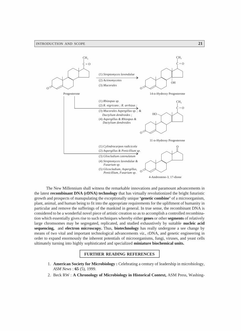
INTRODUCTION AND SCOPE 21
O O
O
O
CH3CH3
CH3
C = O C = O
C = O
O
OH
14 -Hydroxy Progesteroneα
11 -Hydroxy Progesteroneα
4-Androstene-3, 17-dione
Progesterone
HO
(1) Streptomyces lavendulae
(1) Rhizopus sp.
(1) Cylindrocarpon radicicola
(2) R. nigricans ; R. arrhizus ;
(2) Aspergillus & Penicillium sp.
(2) Actinomycetes
(3) Mucorales Aspergillus sp. ; &
(3) Gliocladium catenulatum
Dactylium dendroides ;
(3) Mucorales
(4) Aspergillus & Rhizopus & Dactylium dendroides
(5) Glicocladium, Aspergillus, Penicillium, Fusarium sp.
(4) Streptomyces lavendulae & Fusarium sp.
The New Millennium shall witness the remarkable innovations and paramount advancements inthe latest recombinant DNA (rDNA) technology that has virtually revolutionized the bright futuristicgrowth and prospects of manupulating the exceptionally unique ‘genetic combine’ of a microorganism,plant, animal, and human being to fit into the appropriate requirements for the upliftment of humanity inparticular and remove the sufferings of the mankind in general. In true sense, the recombinant DNA isconsidered to be a wonderful novel piece of artistic creation so as to accomplish a controlled recombina-tion which essentially gives rise to such techniques whereby either genes or other segments of relativelylarge chromsomes may be segregated, replicated, and studied exhaustively by suitable nucleic acidsequencing, and electron microscopy. Thus, biotechnology has really undergone a see change bymeans of two vital and important technological advancements viz., rDNA, and genetic engineering inorder to expand enormously the inherent potentials of microorganisms, fungi, viruses, and yeast cellsultimately turning into highly sophisticated and specialized miniature biochemical units.
FURTHER READING REFERENCES
1. American Society for Microbiology : Celebrating a century of leadership in microbiology,ASM News : 65 (5), 1999.
2. Beck RW : A Chronology of Microbiology in Historical Context, ASM Press, Washing-
22 PHARMACEUTICAL MICROBIOLOGY
ton DC, 2000.
3. Benacerraf B et. al. : A History of Bacteriology and Immunology, William Heinemann,London, 1980.
4. Brock T (ed). : Milestones in Microbiology, Prentice-Hall, Eaglewood Cliffs., NJ., 1961.
5. Collard P. : The Development of Microbiology, University Press, Cambridge, 1976.
6. Dowling HF : Fighting Infection, Conquests of the Twentieth Century, Harvard, Cam-bridge, Mass., 1977.
7. Hellemans A and Bunck B : The Timetables of Science, Simon and Schuster, New York,1988.
8. Lechevalier H. and Solotorovsky M. : Three Centuries of Microbiology, McGraw Hill,New York, 1965.
9. Parisch HJ : Victory with Vaccines — The Story of Immunization, Livingstone, London,1968.
10. Summers WC : History of Microbiology. In : Encylopedia of Microbiology, Vol. 2. J.Ledenberg, Ed., Academic Press, San Diego, 677–97, 2000.
11. Van Iterson et. al. : Martinus Bijerinck’s, His Life and Work, Science Tech, Madison,Wis., 1984.
12. Waterson AP and Wilkinson L. : An Introduction to the History of Virology, CambridgeUniversity Press, London, 1978.

23
STRUCTURE AND FUNCTION :BACTERIAL CELLS
2.1. INTRODUCTION
Bacterium (pl. bacteria) refers to a single-celled organism without having a true nucleus orfunctionally specific components of metabolism that belongs to the kingdom Prokaryotae (Monera).The internal cytoplasm is invariably surrounded by one-or two-layered rigid cell wall composed ofphospholipids. Some bacteria also produce a specific mucoid extracellular capsule for additional pro-tection, particularly from phagocytosis by white-blood cells (WBCs). Bacteria can synthesize nucleicacids (DNA, RNA), other important proteins and can reproduce independently, but may essentially needa host to supply food and also a supportive environment. In reality, millions of nonpathogenic bacterialive on the skin and mucous membranes of the human gastrointestinal tract (GIT) ; these are termed asnormal flora. Importantly, bacteria that cause disease are usually known as pathogens.
2.2. CHARACTERISTIC FEATURES
A few vital and cardinal characteristic features of ‘bacteria’ are as enumerated under :
2.2.1. Shape
There are three principal forms of bacteria, namely :
(a) Spherical or Ovoid — bacteria occur as single cells (micrococci), or in pairs (diplococci),clusters (staphylococci), chains (streptococci) or cubical groups (sarcinae) ;
(b) Rod-shaped — bacteria are termed as bacilli, more oval ones are known as coccobacilli, andthose forming a chain are called as streptobacilli ; and
(c) Spiral — bacteria are rigid (spirilla), flexible (spirochaetes) or curved (vibrios).
2.2.2. Size
An average rod-shaped bacterium measures approximately 1 μm in diameter and 4 μm in length.They usually vary in size considerably from < 0.5 to 1.0 μm in diameter to 10–20 μm in length in someof the longer spiral forms.
2• Introduction• Characteristic Features• Activities• Organization of Microbial Cells• Archaeobacteria and Eubacteria• The Bacterial Cells
24 PHARMACEUTICAL MICROBIOLOGY
2.2.3. Reproduction
It has been observed that simple cell division is the usual method of reproduction, whereas cer-tain bacteria give rise to buds or branches that eventually break off. The growth rate is substantiallyaffected on account of changes in temperature, nutrition, and other factors.
Importantly, bacilli can produce reproductive cells invariably termed as spores, whose relativelythick coatings are highly resistant to adverse environmental conditions. In the event of a better congenialenvironment the spores commence to grow. Besides, spores are difficult to kill as they are highlyresistant to heat as well as disinfectant action.
2.2.4. Formation of Colony
A group of bacteria growing in one particular place is known as a colony. A colony is invariablycomprised of the ‘descendants of a single cell’. It has been found that colonies differ in shape, size,colour, texture, type of margin, and several other characteristic features. Interestingly, each species ofbacteria has a characteristic type of colony formation.
2.2.5. Mutation
Evidently, a majority of bacteria, like all living organisms, do possess the ability to adapt theirshape or functions when encountered with distinct changes in their environment, but there are certaindegree of limits to this ability. However, they may also mutate to adapt to some potentially lethal sub-stances, for instance : antibiotics.
2.2.6. Motility
It has been duly observed that none of the ovoid or spherical cocci are capable of moving, butcertain bacilli and spiral forms do exhibit absolute independent movement. It is, however, pertinent tomention here that the power of locomotion exclusively depends on the possession one or more flagella,slender whiplike appendages which more or less work like propellars.
2.2.7. Food and Oxygen Requirements
Bacteria are of different types based upon their food and oxygen requirements as given below :
(a) Heterotrophic : require organic material as food,
(b) Parasites : feed on living organisms,
(c) Saprophytes : feed on non-living organic material,
(d) Autotrophic : i.e., self-nourishing–obtain their energy from inorganic substances, includingmost of the soil bacteria,
(e) Aerobes : essentially require oxygen for their very existence and growth, and
(f) Anaerobes : do not require oxygen for their existence and growth. e.g., most bacteria foundin the GIT.
2.2.8. Temperature Requirements
Although some bacteria live at very low temperature or very high temperature ; however, theoptimum temperature for a majority of pathogens is 37 °C (98.6 °F).
STRUCTURE AND FUNCTION : BACTERIAL CELLS 25
* It is the gradual decomposition of organic matter exposed to air by bacteria and fungi.
** It is the decomposition of nitrogenous and other organic materials in the absence of air thereby producingfoul odours.
*** One of more than 100 distinct proteins produced primarily by WBCs. They provide signals to regulateimmunological aspects of cell growth and function during both inflammation and specific common responses.
2.3. ACTIVITIES
Following are some of the predominant activities of bacteria, namely :
(a) Enzyme Production. Bacteria invariably give rise to the production of enzymes that act oncomplex food molecules, breaking them down into much simpler components ; they are theprincipal agents responsible for causing decay* and putrefaction.**
(b) Toxin Production. Special molecules called adhesins bind bacteria to the host cells. Oncethe attachment gets materialized, the bacteria may produce poisonous substances usuallyknown as toxins.
Toxins are commonly of two kinds, such as :
(i) Exotoxins — enzymes that virtually disrupt the cell’s function or kill it, and
(ii) Endotoxins — stimulate production of cytokines*** which may produce widespreadvasodilation and shock.
(c) Miscellaneous. A host of bacteria produce several chemical and physical characteristic prod-ucts, such as :
Pigments — colouring matter,
Light — exhibiting luminescent at night,
Chemical substances — e.g., acids, alcohols, aldehydes, ammonia, gases, carbohydrates,and indole, and
Hemolysins, leukocidins, coagulases, and fibrolysins — produced by pathogenic bacteria ;
The soil bacteria play a vital and important role in different phases of the nitrogen cycle viz.,nitrification, nitrogen fixation, and denitrification.
2.4. ORGANIZATION OF MICROBIAL CELLS
Irrespective of the very nature and complexity of an organism, the cell designates the fundamen-tal structural unit of life. In other words, all living cells are basically similar. The ‘cell theory’ i.e., theconcept of the cell as the structural unit of life, was duly put forward by Schleiden and Schwann.
In short, all biological systems essentially have the following characteristic features in common,namely :
(1) ability for reproduction,
(2) ability to assimilate or ingest food substances, and subsequently metabolize them for energyand growth,
(3) ability to excrete waste products,
(4) ability to react to unavoidable alterations in their environment — usually known as irritabil-ity, and
(5) susceptibility to mutation.

26 PHARMACEUTICAL MICROBIOLOGY
The plants and the animals were the two preliminary kingdoms of living organisms duly rec-ognized and identified by the earlier biologists. However, one may articulately distinguish these twogroups by means of a number of well-defined structural and functional characteristic features as givenin Table : 2.1.
Table 2.1. Certain Structural and Functional Differentiations between Plants and Animals.
S.No. Characteristic FeaturesKingdoms
Plants Animals
I Structural features
1 Cell wall Present Absent
2 Chloroplasts Present Absent
3 Growth Open Closed
II Functional features
Carbon source Carbon dioxide Organic compounds
Energy source Light (UV) Chemical energy
Growth factor needs None Complex
Active moment Absent Present
Soon after the discovery of the ‘microbial world’ — the immotile multicellular and photosyn-thetic algae were classified duly in the plant kingdom; whereas — the microscopic motile forms ofalgae were placed duly in the animal kingdom. Hence, a close and careful examination revealed thepresence of both plant — and animal-like characteristic features in the ‘microorganisms’. Further,supporting evidences and valuable informations strongly established that the ‘microorganisms’ couldnot fit reasonably into the two aforesaid kingdoms, namely : ‘plants’ and ‘animals’. Therefore, Haeckel(1866) legitimately and affirmatively proposed a ‘third kingdom’ termed as the ‘Protista’* to includethe ‘microorganisms’ exclusively.
Importantly, Protista group usually comprises of both the photosynthetic and non-photosyn-thetic microorganisms, of course, with certain members sharing the characteristic features of both theusual traditional plant and animal kingdoms. Nevertheless, the most prominent and predominant at-tribute of this particular group being the comparatively much simpler biological organization. Most ofthe representative members of this group are normally unicellular and undifferentiated unlike the ani-mals and the plants.
Noticeably, further categorization of this kingdom was exclusively dependent upon the extent ofcomplexity encountered by the cellular organization, substantial progress in microscopy, and the ‘bio-chemistry of various microorganisms’ has ultimately paved the way towards a much advanced andbetter understanding of the differences with regard to the ‘internal architectural design of the micro-bial cells’.
2.4.1. Types of Cells
As to date there are two types of cells that have been recognized duly, such as :
* In taxonomy, a kingdom of organisms that includes the protozoa, unicellular and multicellular algae, and theslime molds. The cells are eukaryotic.
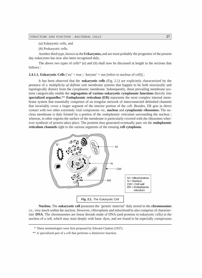
STRUCTURE AND FUNCTION : BACTERIAL CELLS 27
(a) Eukaryotic cells, and
(b) Prokaryotic cells.
Another third type, known as the Urkaryotes, and are most probably the progenitor of the presentday eukaryotes has now also been recognized duly.
The above two types of cells* (a) and (b) shall now be discussed at length in the sections thatfollows :
2.4.1.1. Eukaryotic Cells [‘eu’ = true ; ‘karyote’ = nut (refers to nucleus of cell)] ;
It has been observed that the eukaryotic cells (Fig. 2.1) are explicitely characterized by thepresence of a multiplicity of definite unit membrane systems that happen to be both structurally andtopologically distinct from the cytoplasmic membrane. Subsequently, these prevailing membrane sys-tems categorically enable the segregation of various eukaryotic cytoplasmic functions directly intospecialized organelles.** Endoplasmic reticulum (ER) represents the most complex internal mem-brane system that essentially comprises of an irregular network of interconnected delimited channelsthat invariably cover a larger segment of the interior portion of the cell. Besides, ER gets in directcontact with two other extremely vital components viz., nucleus and cytoplasmic ribosomes. The nu-cleus membrane is duly formed by a portion of the endoplasmic reticulum surrounding the nucleus ;whereas, in other regions the surface of the membrane is particularly covered with the ribosomes wher-ever synthesis of protein takes place. The proteins thus generated eventually pass via the endoplasmicreticulum channels right to the various segments of the ensuing cell cytoplasm.
M
N
CW
ERM = MitochondrionN = NucleusCW = Cell wall ER = Endoplasmic reticulum
Fig. 2.1. The Eukaryotic Cell
Nucleus. The eukaryotic cell possesses the ‘genetic material’ duly stored in the chromosomesi.e., very much within the nucleus. However, chloroplasts and mitochondria also comprise of character-istic DNA. The chromosomes are linear threads made of DNA (and proteins in eukaryotic cells) in thenucleus of a cell, which may stain deeply with basic dyes, and are found to be especially conspicuous
* These terminologies were first proposed by Edward Chatton (1937).
** A specialized part of a cell that performs a distinctive function.
28 PHARMACEUTICAL MICROBIOLOGY
during mitosis. The DNA happens to be the genetic code of the cell ; and specific sequences of DNAnucleotides are the genes for the cell’s particular proteins. However, the size and the number of thechromosome vary widely with various organisms. Nevertheless, the nucleus invariably contains a nu-cleolus that is intimately associated with a particular chromosomal segment termed as the ‘nucleolarorganizer’, which is considered to be totally involved in ribosomal RNA (rRNA) synthesis.
Mitosis. Mitosis refers to a type of cell division of somatic cells wherein each daughter cellcontains the same number of chromosomes as the parent cell. Mitosis is the specific process by whichthe body grows and dead somatic cells are replaced. In fact, mitosis is a continuous process divided intofour distinct phases, namely : prophase, metaphase, anaphase, and telophase.
A brief discussion of the aforesaid four phases shall be given in the sections that follows alongwith their illustrations in Fig. 2.2.
(a) Prophase. In prophase, the chromatin granules of the nucleus usually stain more denselyand get organized into chromosomes. These first appear as long filaments, each comprisingof two identical chromatids,* obtained as a result of DNA replication. As prophase progresses,the chromosomes become shorter and more compact and stain densely. The nuclear mem-brane and the nucleoli disappear. At the same time, the centriole divides and the two daugh-ter centrioles,** each surrounded by a centrosphere, move to opposite poles of the cell.They are duly connected by fine protoplasmic fibrils, which eventually form an achromaticspindle.
(b) Metaphase. The metaphase refers to the chromosomes (paired chromatids) that arrangethemselves in an equatorial plane midway between the two centrioles.
(c) Anaphase. In anaphase, the chromatids (now known as daughter chromosomes) divergeand move towards their respective centrosomes. The end of their migration marks the begin-ning of the next phase.
(d) Telophase. In telophase, the chromosomes at each pole of the spindle undergo changes thatare the reverse of those in the prophase, each becoming a long loosely spiraled thread. Thenuclear membrane re-forms and nucleoli reappear. Outlines of chromosomes disappear, andchromatin appears as granules scattered throughout the nucleus and connected by a highlystaining net. The cytoplasm gets separated into two portions, ultimately resulting in twocomplete cells. This is accomplished in animal cells by constriction in the equatorial region ; inplant cells, a cell plate that produces the cell membrane forms in a similar position. Theperiod between two successive divisions is usually known as interphase.
Mitosis is of particular significance wherein the genes are distributed equally to each daughtercell and a fixed number of chromosomes is maintained in all somatic cells of an organism.
Mitosis are of two kinds, namely :
(i) heterotypic mitosis : The first or reduction division in the maturation of germ cells, and
(ii) homeotypic mitosis : The second or equational division in the maturation of germ cells.
* One of the two potential chromosomes formed by DNA replication of each chromosome before mitosis andmeiosis. They are joined together at the centromere and separate at the end of metaphase ; then the newchromosomes migrate to opposite poles of the cell at anaphase.
** A minute organelle consisting of a hollow cylinder closed at one end and open at the other, found in the cellcentre or attraction sphere of a cell.

STRUCTURE AND FUNCTION : BACTERIAL CELLS 29
Centrioles
NucleolusCentromere
Chromosome
SpindleFibersFormed Attached
Centromere
Late Prophase :Pairs of ChromatidsMigrate to Equatorof Cell
Spindle FibresPull ChromosomesTowards the Polesof the cell
DiploidNumberof cell is 4
Late interphase : Chromosomesduplicate themselves. Each becomesa pair of Chromatids attachedat the Centromere
Early Prophase :Pairs of Chromatidsappear as short rods
Metaphase : Pairs of ChromatidsLine up on Equator of cell
Interphase: Centromeresside ; each Chromatidsshow a separateChromosome
Telophase : Chromosomesreach the poles ofthe cell and uncoil,Cytoplasm begins to divide
Only Interphase :Two identical daughtercells, each with the diploid number of Chromosomes
Fig. 2.2. Different Phases of Mitosis
Meiosis. Meiosis refers to a specific process of two successive cell divisions, giving rise to cells,egg or sperm, that essentially contain half the number of chromosomes in somatic cells. When fertiliza-tion takes place, the nuclei of the sperm and ovum fuse and produce a zygote with the full chromosomecomplement.
30 PHARMACEUTICAL MICROBIOLOGY
In other words, the phenomenon of meiosis may be duly expatiated in sexually reproducingorganisms, wherein the prevailing cellular fusion followed by a reduction in the ‘chromosome number’is an important and vital feature. The two cells which actually participate in the sexual reproduction aretermed as ‘gametes’, which fuse to form a ‘zygote’. The above process is subsequently followed by anuclear fusion and the resulting zygote nucleus contains two complete sets of genetic determinants[2N]. In order to adequately maintain the original haploid number in the succeeding generations, thereshould be a particular stage at which a definite reduction in the chromosome number takes place. Thisprocess that occurs after the fusion of gametes is known as meiosis.
Fig. 2.3 illustrates the schematic representation of meiosis, and the various steps involved maybe explained sequentially as follows :
(1) Meiosis comprises of two meiotic divisions viz., prophase I, and prophase II.
(2) Prophase-I. It represents the first meiotic division, whereby the homologous chromosomesbecome apparently visible as single strands that subsequently undergo pairing.
(3) Each chromosome renders visible as two distinct chromatids and thus crossing over takesplace.
(4) It is immediately followed by metaphase I, wherein the actual orientation of ‘paired chro-mosomes’ in an equatorial plane and the subsequent formation of a ‘spindle apparatus’takes place.
(5) It is followed by Anaphase I, and the homologous centromeres gradually move to the oppositepoles of the spindle.
(6) Telophase-I. It markedly represents the end of the first meiotic division, and formation oftwo nuclei takes place.
(7) Interphase-II. Telophase-I is followed by Interphase-I during which the chromosomes getelongated.
(8) Prophase-II and Metaphase-II. In prophase-II and metaphase-II the division of centromereand migration of the homologous chromatids occurs, which is duly followed by anaphase-II,and the desired second meiotic division resulting in the formation of four haploid* cells.
Eukaryotic Protist. It has been observed that in several eukaryotic protists belonging to higherploidy** (> 1) meiosis usually takes place after the formation of the zygote and prior to spore formation.In certain eukaryotes there may even be a critically pronounced alteration of haploid and diploid gen-erations as in the case of the yeast. Interestingly, in this particular instance, the diploid zygote producesa diploid individual that ultimately gives rise to haploid cells only after having undergone the phenom-enon of meiosis. Consequently, the haploid cell may either multiply as a haploid or get fused withanother haploid of the ‘opposite mating type’ to generate again a diploid.
* Possessing half the diploid or normal number of chromosomes found in somatic or body cells. Such is thecase of the germ cells–ova or sperm–following the reduction divisions in gametogenesis, the haploid numberbeing 23 in humans.
** The number of chromosome sets in a cell (e.g., haploidy, diploidy, and triploidy for one, two or three sets ofchromosomes respectively.

STRUCTURE AND FUNCTION : BACTERIAL CELLS 31
Fig. 2.3. Schematic Representation of Meiosis
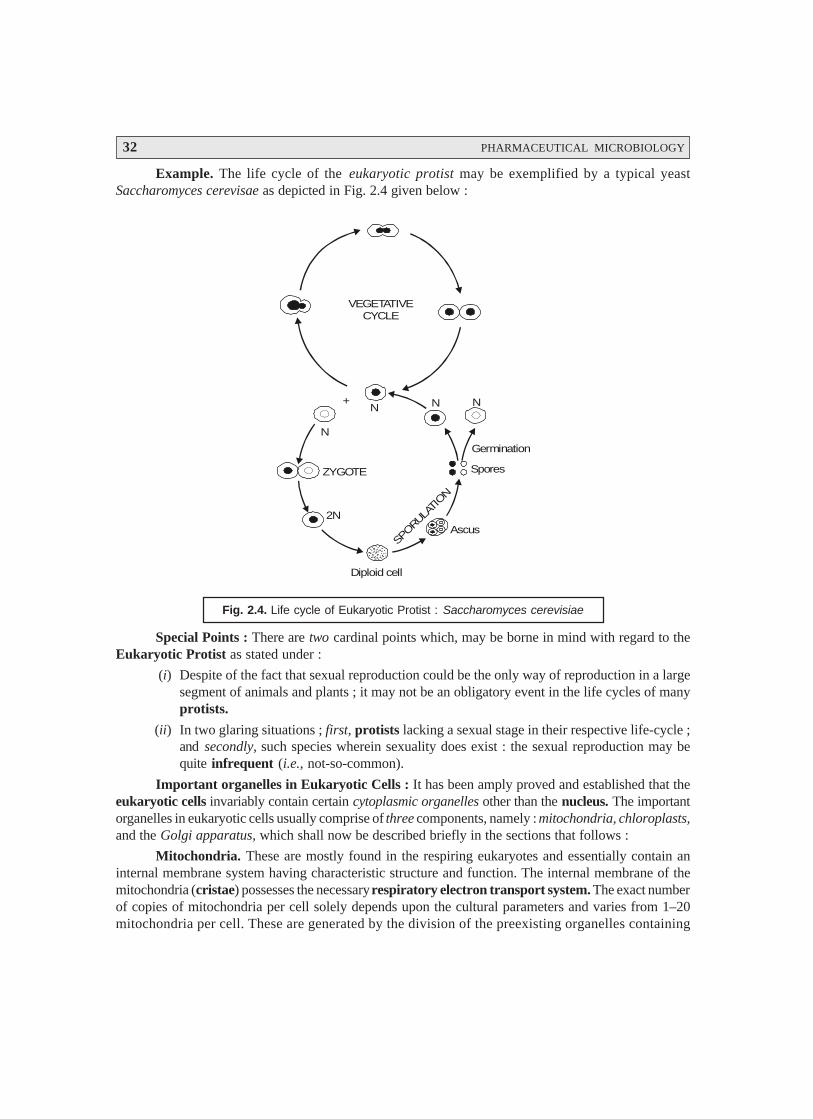
32 PHARMACEUTICAL MICROBIOLOGY
Example. The life cycle of the eukaryotic protist may be exemplified by a typical yeastSaccharomyces cerevisae as depicted in Fig. 2.4 given below :
+
N
N N N
2N
Diploid cell
Ascus
Spores
Germination
ZYGOTE
VEGETATIVECYCLE
SPORU
LATION
Fig. 2.4. Life cycle of Eukaryotic Protist : Saccharomyces cerevisiae
Special Points : There are two cardinal points which, may be borne in mind with regard to theEukaryotic Protist as stated under :
(i) Despite of the fact that sexual reproduction could be the only way of reproduction in a largesegment of animals and plants ; it may not be an obligatory event in the life cycles of manyprotists.
(ii) In two glaring situations ; first, protists lacking a sexual stage in their respective life-cycle ;and secondly, such species wherein sexuality does exist : the sexual reproduction may bequite infrequent (i.e., not-so-common).
Important organelles in Eukaryotic Cells : It has been amply proved and established that theeukaryotic cells invariably contain certain cytoplasmic organelles other than the nucleus. The importantorganelles in eukaryotic cells usually comprise of three components, namely : mitochondria, chloroplasts,and the Golgi apparatus, which shall now be described briefly in the sections that follows :
Mitochondria. These are mostly found in the respiring eukaryotes and essentially contain aninternal membrane system having characteristic structure and function. The internal membrane of themitochondria (cristae) possesses the necessary respiratory electron transport system. The exact numberof copies of mitochondria per cell solely depends upon the cultural parameters and varies from 1–20mitochondria per cell. These are generated by the division of the preexisting organelles containing
STRUCTURE AND FUNCTION : BACTERIAL CELLS 33
ribosomes that usually resemble the bacterial ribosomes. However, the process of protein synthesis inthe mitochondria are very much akin to that in the prokaryotic cells.
These cell organelles (rod/oval shape 0.5 μm in diameter) may be seen by employing a phase-contrast or electron microscopy. They mostly contain the enzymes for the aerobic stages of cell respi-ration and thus are the usual sites of most ATP synthesis chloroplasts [or Chloroplastids] :
Chloroplasts are found in the photosynthetic eukaryotic organisms. The internal membraneof the chloroplasts is termed as the ‘thylakoid’ which essentially has the three important components :(a) photosynthetic pigments, (b) electron transport system, and (c) photochemical reaction centres. Thenumber of copies of the chloroplasts depends exclusively upon the cultural conditions and varies from40 to 50 chloroplasts per cell. These are also produced by the division of the preexisting organelles.
Generally, chloroplasts are the sites of photosynthesis. They possess a stroma and contain fourpigments : chlorophyll a, chlorophyll b, carotene, and xanthophyll.
Golgi Apparatus : The Golgi apparatus is a lamellar membranous organelle invariably foundin the eukaryotic cells and consists of thickly packed mass of flattened vessels and sacks of differentsizes. The major functions of the Golgi apparatus are, namely :
• packaging of both proteinaceous and nonproteinaceous substances duly synthesized in theendoplasmic reticulum, and
• their adequate transport to other segments of the cell.
Golgi apparatus may be best viewed by the aid of electron microscopy. It contains curvedparallel series of flattened saccules that are often expanded at their ends. In secretory cells, the apparatusconcentrates and packages the secretory product. Its function in other cells, although apparently impor-tant, is poorly understood.
2.4.1.2. Prokaryotic Cells [‘pro’ = primitive ; ‘karyote’ = nut (refers to nucleus of cell)] :
Prokaryote : is an organism of the kingdom Monera with a single circular chromosome, withouta nuclear membrane, or membrane bound organelles. Included in this classification are bacteria andcyanobacteria (formerly the blue-green algae) [SYN : prokaryote].
In fact, the prokaryotic cell is characterized by the absence of the endoplasmic reticulum (ER)and the cytoplasmic membrane happens to be the only unit membrane of the cell. If has been observedthat the cytoplasmic membrane may be occasionally unfolded deep into the cytoplasm. An exhaustiveelectron microscopical studies would reveal that most prokaryotes {i.e., prokaryotic cells) only twodistinct internal regions, namely : (a) the cytoplasm ; and (b) the nucleoplasm, as shown in Fig. : 2.5.
Cytoplasm : Cytoplasm refers to the protoplasm cell outside the nucleus. It is granular in ap-pearance and contains ribosomes that are specifically smaller in size in comparison to the correspondingeukaryotic ribosomes.
Nucleoplasm : It refers to the protoplasm of a cell nucleus. It is fibrillar in character and containsDNA.
With mycoplasmas* as an exception, other prokaryotes invariably comprise of a defined andrigid cell wall. It has been observed that neither the membranous structures very much identical to the
mitochondria nor chloroplasts are present in the prokaryotes. Besides, the cytoplasmic membrane happens
to be the site of the respiratory electron in the prokaryotes usually. Interestingly, in the photosynthetic micro-
* A group of bacteria that lack cell walls and are highly pleomorphic.
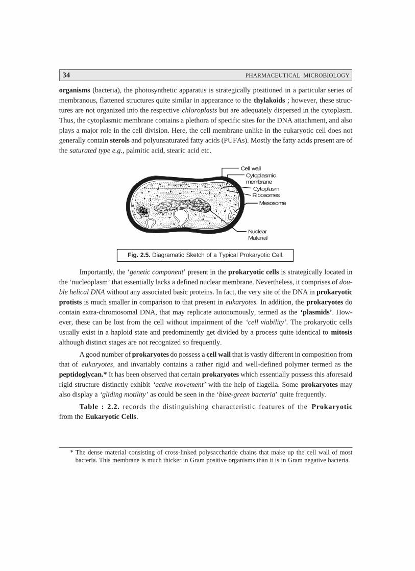
34 PHARMACEUTICAL MICROBIOLOGY
organisms (bacteria), the photosynthetic apparatus is strategically positioned in a particular series of
membranous, flattened structures quite similar in appearance to the thylakoids ; however, these struc-tures are not organized into the respective chloroplasts but are adequately dispersed in the cytoplasm.
Thus, the cytoplasmic membrane contains a plethora of specific sites for the DNA attachment, and also
plays a major role in the cell division. Here, the cell membrane unlike in the eukaryotic cell does notgenerally contain sterols and polyunsaturated fatty acids (PUFAs). Mostly the fatty acids present are of
the saturated type e.g., palmitic acid, stearic acid etc.
Cell wallCytoplasmicmembrane
CytoplasmRibosomes
Mesosome
NuclearMaterial
Fig. 2.5. Diagramatic Sketch of a Typical Prokaryotic Cell.
Importantly, the ‘genetic component’ present in the prokaryotic cells is strategically located in
the ‘nucleoplasm’ that essentially lacks a defined nuclear membrane. Nevertheless, it comprises of dou-
ble helical DNA without any associated basic proteins. In fact, the very site of the DNA in prokaryoticprotists is much smaller in comparison to that present in eukaryotes. In addition, the prokaryotes do
contain extra-chromosomal DNA, that may replicate autonomously, termed as the ‘plasmids’. How-ever, these can be lost from the cell without impairment of the ‘cell viability’. The prokaryotic cells
usually exist in a haploid state and predominently get divided by a process quite identical to mitosisalthough distinct stages are not recognized so frequently.
A good number of prokaryotes do possess a cell wall that is vastly different in composition fromthat of eukaryotes, and invariably contains a rather rigid and well-defined polymer termed as the
peptidoglycan.* It has been observed that certain prokaryotes which essentially possess this aforesaid
rigid structure distinctly exhibit ‘active movement’ with the help of flagella. Some prokaryotes mayalso display a ‘gliding motility’ as could be seen in the ‘blue-green bacteria’ quite frequently.
Table : 2.2. records the distinguishing characteristic features of the Prokaryoticfrom the Eukaryotic Cells.
* The dense material consisting of cross-linked polysaccharide chains that make up the cell wall of mostbacteria. This membrane is much thicker in Gram positive organisms than it is in Gram negative bacteria.
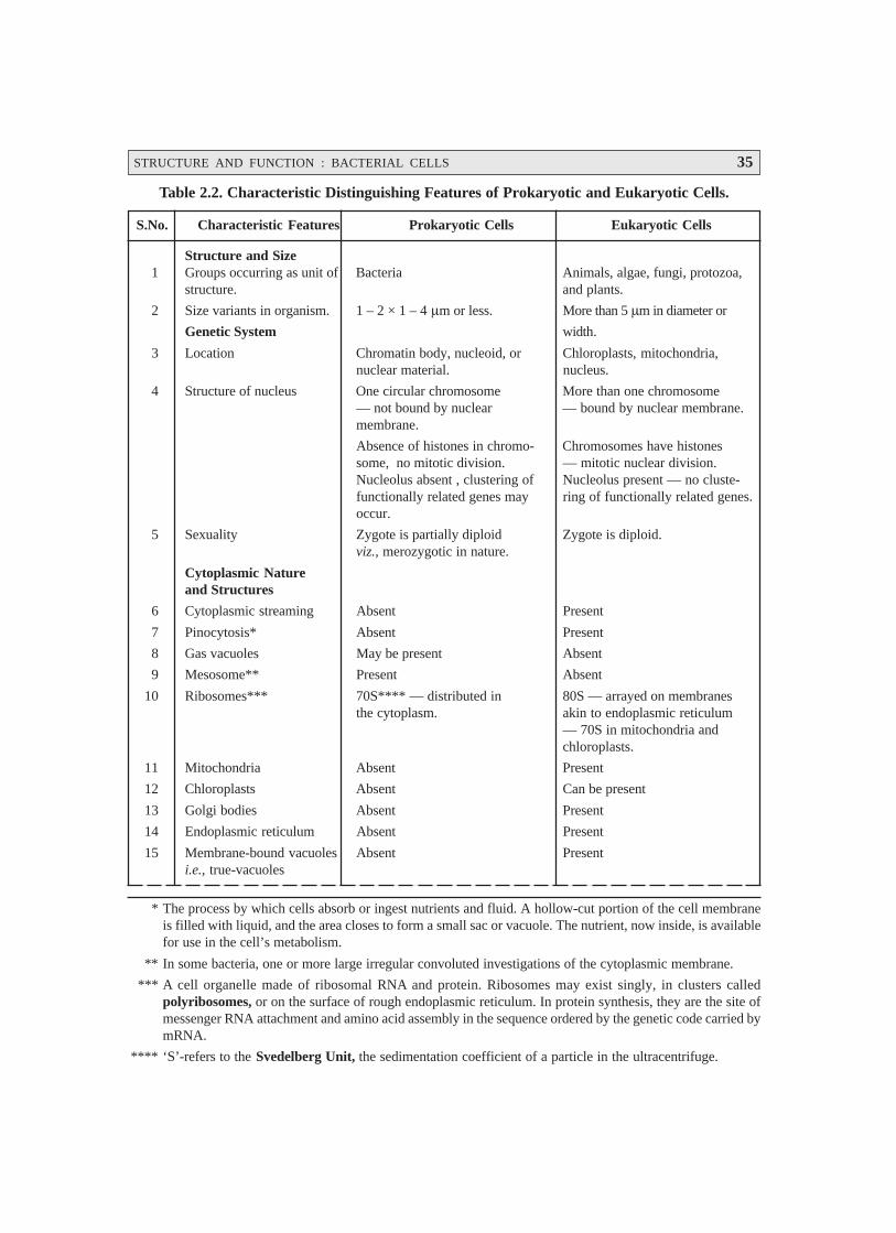
STRUCTURE AND FUNCTION : BACTERIAL CELLS 35
Table 2.2. Characteristic Distinguishing Features of Prokaryotic and Eukaryotic Cells.
S.No. Characteristic Features Prokaryotic Cells Eukaryotic Cells
Structure and Size1 Groups occurring as unit of Bacteria Animals, algae, fungi, protozoa,
structure. and plants.
2 Size variants in organism. 1 – 2 × 1 – 4 μm or less. More than 5 μm in diameter or
Genetic System width.
3 Location Chromatin body, nucleoid, or Chloroplasts, mitochondria,nuclear material. nucleus.
4 Structure of nucleus One circular chromosome More than one chromosome— not bound by nuclear — bound by nuclear membrane.membrane.
Absence of histones in chromo- Chromosomes have histonessome, no mitotic division. — mitotic nuclear division.Nucleolus absent , clustering of Nucleolus present — no cluste-functionally related genes may ring of functionally related genes.occur.
5 Sexuality Zygote is partially diploid Zygote is diploid.viz., merozygotic in nature.
Cytoplasmic Natureand Structures
6 Cytoplasmic streaming Absent Present
7 Pinocytosis* Absent Present
8 Gas vacuoles May be present Absent
9 Mesosome** Present Absent
10 Ribosomes*** 70S**** — distributed in 80S — arrayed on membranesthe cytoplasm. akin to endoplasmic reticulum
— 70S in mitochondria andchloroplasts.
11 Mitochondria Absent Present
12 Chloroplasts Absent Can be present
13 Golgi bodies Absent Present
14 Endoplasmic reticulum Absent Present
15 Membrane-bound vacuoles Absent Presenti.e., true-vacuoles
* The process by which cells absorb or ingest nutrients and fluid. A hollow-cut portion of the cell membraneis filled with liquid, and the area closes to form a small sac or vacuole. The nutrient, now inside, is availablefor use in the cell’s metabolism.
** In some bacteria, one or more large irregular convoluted investigations of the cytoplasmic membrane.
*** A cell organelle made of ribosomal RNA and protein. Ribosomes may exist singly, in clusters calledpolyribosomes, or on the surface of rough endoplasmic reticulum. In protein synthesis, they are the site ofmessenger RNA attachment and amino acid assembly in the sequence ordered by the genetic code carried bymRNA.
**** ‘S’-refers to the Svedelberg Unit, the sedimentation coefficient of a particle in the ultracentrifuge.
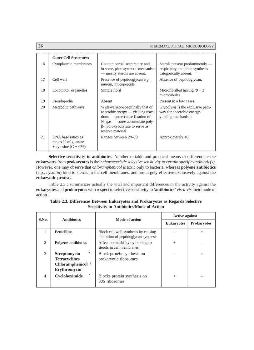
36 PHARMACEUTICAL MICROBIOLOGY
Outer Cell Structures
16 Cytoplasmic membranes Contain partial respiratory and, Sterols present predominently —in some, photosynthetic mechanism, respiratory and photosynthesis— mostly sterols are absent. categorically absent.
17 Cell wall Presence of peptidoglycan e.g., Absence of peptidoglycan.murein, mucopeptide.
18 Locomotor organelles Simple fibril Microfibrilled having ‘9 + 2’microtubules.
19 Pseudopodia Absent Present in a few cases.
20 Metabolic pathways Wide-variety-specifically that of Glycolysis is the exclusive path-anaerobic energy — yielding react- way for anaerobic energy-tions — some cause fixation of yielding mechanism.N2 gas — some accumulate polyβ-hydroxybutyrate to serve asreserve material.
21 DNA base ratios as Ranges between 28–73 Approximately 40.moles % of guanine+ cytosine (G + C%)
Selective sensitivity to antibiotics. Another reliable and practical means to differentiate theeukaryotes from prokaryotes is their characteristic selective sensitivity to certain specific antibiotic(s).However, one may observe that chloramphenicol is toxic only to bacteria, whereas polyene antibiotics(e.g., nystatin) bind to sterols in the cell membranes, and are largely effective exclusively against theeukaryotic protists.
Table 2.3 : summarizes actually the vital and important differences in the activity against theeukaryotes and prokaryotes with respect to selective sensitivity to ‘antibiotics’ vis-a-vis their mode ofaction.
Table 2.3. Differences Between Eukaryotes and Prokaryotes as Regards SelectiveSensitivity to Antibiotics/Mode of Action
S.No. Antibiotics Mode of actionActive against
Eukaryotes Prokaryotes
1 Penicillins Block cell wall synthesis by causing – +inhibition of peptidoglycan synthesis
2 Polyene antibiotics Affect permeability by binding to + –sterols in cell membranes
3 Streptomycin Block protein synthesis on – +Tetracyclines prokaryotic ribosomesChloramphenicolErythromycin
4 Cycloheximide Blocks protein synthesis on + –80S ribosomes
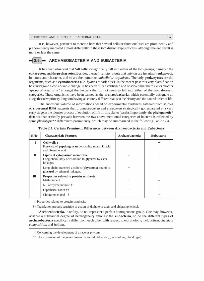
STRUCTURE AND FUNCTION : BACTERIAL CELLS 37
* Concerning the development of a race or phylum.
** The expression of the genes present in an individual (e.g., eye colour, blood type).
It is, however, pertinent to mention here that several cellular functionalities are prominently andpredominently mediated almost differently in these two distinct types of cells, although the end result ismore or less the same.
2.5. ARCHAEOBACTERIA AND EUBACTERIA
It has been observed that ‘all cells’ categorically fall into either of the two groups, namely : theeukaryotes, and the prokaryotes. Besides, the multicellular plants and animals are invariably eukaryoticin nature and character, and so are the numerous unicellular organisms. The only prokaryotes are theorganisms, such as : cyanobacteria (Gr. hyanos = dark blue). In the recent past this very classificationhas undergone a considerable change. It has been duly established and observed that there exists another‘group of organisms’ amongst the bacteria that do not seem to fall into either of the two aforesaidcategories. These organisms have been termed as the archaeobacteria, which essentially designate analtogether new primary kingdom having an entirely different status in the history and the natural order of life.
The enormous volume of informations based on experimental evidences gathered from studiesof ribosomal RNA suggests that archaeobacteria and eubacteria strategically got separated at a veryearly stage in the pioneer process of evolution of life on this planet (earth). Importantly, the phylogenetic*distance that critically prevails between the two above mentioned categories of bacteria is reflected bysome phenotypic** differences prominently, which may be summarized in the following Table : 2.4.
Table 2.4. Certain Prominent Differences between Archaeobacteria and Eubacteria
S.No. Characteristic Features Archaeobacteria Eubacteria
I Cell walls : – +Presence of peptidoglycan containing muramic acidand D-amino acid.
II Lipids of cytoplasmic membrane – +Long-chain fattly acids bound to glycerol by esterlinkages.
Long-chain branched alcohols (phytanols) bound to + –glycerol by ethereal linkages.
III Properties related to protein synthesisMethionine † + –
N-Formylmethionine † – +
Diphtheria Toxin †† + –
Chloramphenicol †† – +
† Properties related to protein synthesis.
†† Translation process sensitive to action of diphtheria toxin and chloramphenicol.
Archaeobacteria, in reality, do not represent a perfect homogeneous group. One may, however,observe a substantial degree of heterogeneity amongst the eubacteria, so do the different types ofarchaeobacteria specifically differ from each other with respect to morphology, metabolism, chemicalcomposition, and habitat.
38 PHARMACEUTICAL MICROBIOLOGY
* Pertaining to green plants and bacteria that essentially form protein and carbohydrate from inorganic saltsand CO2.
** An enzyme activator ; a diffusible, heat-stable substance of low molecular weight that, when combined withan inactive protein termed as apoenzyme, forms an inactive compound or a complete enzyme called aholoenzyme (e.g., adenylic acid, riboflavin, and coenzymes I and II).
*** The science of structure and form of organisms without regard to function.
**** A method of staining bacteria, important in their identification [SYN : Gram Stain ; Gram’s method]. Gram-negative : Losing the crystal-violet stain and taking the colour of the red counter stain in Gram’s method ofstaining — a primary characteristic of certain microorganisms ; Gram-positive : Retaining the colour of thecrystal-violet stain in Gram’s method of staining.
The ‘archaeobacteria’ are unusual organisms by nature, and this particular category is known tocomprise essentially of three different types of bacteria, namely :
(a) Methanogenic bacteria,
(b) Extreme halophiles, and
(c) Thermoacidophiles
These three groups of organisms shall now be treated individually in the sections that follows :
2.5.1. Methanogenic Bacteria [Methanogens]
The methanogenic bacteria are considered to be the hard-core anaerobes which, invariablypossess the capability of deriving energy for their progressive growth by certain particular oxidizingchemical entities, for instance : hydrogen (H2), formic acid (HCOOH) ; and actually exert their ‘action’by making use of the electrons thus produced to reduce ultimately carbon-dioxide (CO2) to give rise tothe formation of methane (CH4) gas :
CO2 + 4H2 ⎯⎯⎯→ CH4 + 2H2O Carbon Hydrogen Methane Water
dioxide
It has been observed that certain genera specifically may grow as autotrophs* — thereby utiliz-ing hydrogen and carbon dioxide as exclusive sources of carbon as well as energy ; whereas some othersdo need several additional components, for instance : organic-sulphur compounds, amino acids, aceticacid, and vitamins. Interestingly, a plethora of species actually grow quite abundantly and aggressivelyin a complex media viz., comprising of yeast extract in comparison to inorganic-salts containing media.
Coenzymes** : There are at least two uncommon coenzymes that invariably occur in all meth-anogenic bacteria (methanogens) that have not been noticed in other varieties of microorganisms.
Examples : Following are two typical examples of methanogenic coenzymes :(a) Coenzyme M — directly involved in methyl transfer reactions, and
(b) Coenzyme F420 — a flavin-like chemical entity intimately involved in the anaerobic elec-tron transport system of these microorganisms. It has the ability to fluoresce when exposedto UV light ; and, therefore, its presence may be detected by visualizing the organisms via afluorescence microscope conveniently (also used for its critical identification and examina-tion).
Differentiation of Methanogens : The genera of methanogens i.e., the methane-producing bac-teria may be clearly differentiated exclusively based upon their morphology*** and Gram reaction****.However, the glaring distinct differences occurring in the cell-wall composition have been duly observedto correlate specifically with these genera.

STRUCTURE AND FUNCTION : BACTERIAL CELLS 39
Table 2.5 : records the morphology, motility, and wall composition of several methanogenicorganisms with specific ‘genus’.
Table 2.5. Differentiation of Methanogens
S.no. (Methano) Genus Morphology Motility Wall Composition
1 Methanobacterium Gram +ve to Gram- — Pseudomureinvariable long rods
2 Methanobrevibacter Gram +ve lancet-shaped — —do—short rods or cocci
3 Methanococcus Gram –ve pleomor- + ; one flagellar tuft Protein with trace ofphic* cocci glucosamine
4 Methanogenium —do— + ; peritrichous flagella Protein
5 Methanomicrobium Gram –ve short-rods + ; single polar flagellum —do—
6 Methanosarcina Gram +ve cocci in — Heteropolysaccharideclusters
7 Methano spirillum Gram –ve long wavy + ; polar flagellafilaments or curved rods
Importantly, the cell walls of two genera essentially consist of pseudomurein, that prominentlydiffers from eubacterial peptidoglycan by the following two distinct structural features, namely :
(a) substitution of N-acetyltalosaminuronic acid for N-acetylmuramic acid, and
(b) presence of tetrapeptide composed totally of L-amino acids, having glutamic acid attachedduly at the C-terminal end.
Habitats : Interestingly, the methanogenic bacteria most commonly found in anaerobic habi-
tats that are eventually rich organic matter which ultimately produced by nonmethanogenic bacteria via
fermentation to yield H2 and CO2. A few such common as well as vital habitats are, namely : marine
sediments, swamps, marshes, pond and lake mud, intestinal tract of humans (GIT) and animals, rumenof cattle (e.g., cow, buffalow, sheep, pig, goat etc.), and anaerobic sludge digesters in sewage-treatment
plants.
Figure. 2.6 [A and B] depicts the diagramatic sketch of the cells commonly observed in various
kinds of methanogenic organisms (viz., methane-producing bacteria).
Figure 2.6 [A] evidently shows the typical cells of Methanosarcina barkeri and Methanospirillum
hungatei representing ideally the methane-producing bacteria.
Figure 2.6 [B] likewise illustrates the characteristic cells of Methanobacterium thermo-
autotrophicum and Methanobacterium ruminantium designating the methanogens.
* Having many shapes.
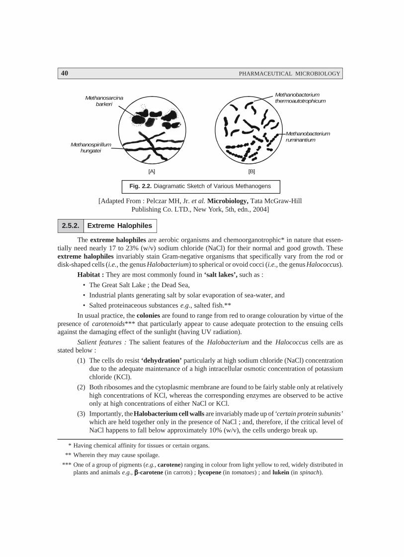
40 PHARMACEUTICAL MICROBIOLOGY
* Having chemical affinity for tissues or certain organs.
** Wherein they may cause spoilage.
*** One of a group of pigments (e.g., carotene) ranging in colour from light yellow to red, widely distributed inplants and animals e.g., βββββ-carotene (in carrots) ; lycopene (in tomatoes) ; and lukein (in spinach).
Methanobacteriumthermoautotrophicum
Methanobacteriumruminantium
Methanosarcinabarkeri
Methanospirillumhungatei
[A] [B]
Fig. 2.2. Diagramatic Sketch of Various Methanogens
[Adapted From : Pelczar MH, Jr. et al. Microbiology, Tata McGraw-HillPublishing Co. LTD., New York, 5th, edn., 2004]
2.5.2. Extreme Halophiles
The extreme halophiles are aerobic organisms and chemoorganotrophic* in nature that essen-tially need nearly 17 to 23% (w/v) sodium chloride (NaCl) for their normal and good growth. Theseextreme halophiles invariably stain Gram-negative organisms that specifically vary from the rod ordisk-shaped cells (i.e., the genus Halobacterium) to spherical or ovoid cocci (i.e., the genus Halococcus).
Habitat : They are most commonly found in ‘salt lakes’, such as :
• The Great Salt Lake ; the Dead Sea,
• Industrial plants generating salt by solar evaporation of sea-water, and
• Salted proteinaceous substances e.g., salted fish.**
In usual practice, the colonies are found to range from red to orange colouration by virtue of thepresence of carotenoids*** that particularly appear to cause adequate protection to the ensuing cellsagainst the damaging effect of the sunlight (having UV radiation).
Salient features : The salient features of the Halobacterium and the Halococcus cells are asstated below :
(1) The cells do resist ‘dehydration’ particularly at high sodium chloride (NaCl) concentrationdue to the adequate maintenance of a high intracellular osmotic concentration of potassiumchloride (KCl).
(2) Both ribosomes and the cytoplasmic membrane are found to be fairly stable only at relativelyhigh concentrations of KCl, whereas the corresponding enzymes are observed to be activeonly at high concentrations of either NaCl or KCl.
(3) Importantly, the Halobacterium cell walls are invariably made up of ‘certain protein subunits’which are held together only in the presence of NaCl ; and, therefore, if the critical level ofNaCl happens to fall below approximately 10% (w/v), the cells undergo break up.
STRUCTURE AND FUNCTION : BACTERIAL CELLS 41
(4) Interestingly, the Halococcus cell walls are usually comprised of a complexheteropolysaccharide which is found to be stable reasonably at comparatively lower NaClconcentrations.
Adenosine Triphosphate (ATP) Synthesis. It is worthwhile to mention here that generally the‘halobacteria’ are ‘aerobic’ in nature. It is amply established that in aerobic organisms, an electron-transport chain invariably gives rise to a specific protonmotive force that in turn helps to carry out thedesired ATP-Synthesis.
Salient Features : There are several salient features that are associated with the ATP-synthesis,namely :
(1) ATP-synthesis may alternatively be accomplished by halobacteria via fermentation ofarginine (an amino acid), which permits them to grow in an anaerobic environment.
(2) The third method of ATP formation is rather unique and extraordinary to the ‘halobacteria’.Predominently distinct patches of a purple pigment, known as bacteriorhodopsin*, are pro-duced in the cell membrane particularly at reasonably low O2 levels. Subsequently, whenthese cells containing the said pigments are exposed to the UV-light—the pigment getsbleached gradually. In the course of the ‘bleaching phenomenon’, the resulting protons**get duly extruded right into the outside portion of the membrane, thereby exerting an appre-ciable protonmotive force that in turn carries out the ATP synthesis strategically.
(3) Conclusively, halobacteria essentially follows the mechanism of light-monitored synthesisof ATP. Furthermore, these are actually devoid of bacteriochlorophyll.
2.5.3. Thermoacidophiles
The thermoacidophiles are generally the aerobic Gram –ve archaeobacteria prominently char-acterized by a remarkable tendency and capability to attain growth not only under extremely high acidicconditions, but also at considerably elevated temperatures.
There are two most prominent genera that belong to this particular category, namely :
(a) Thermoplasma, and
(b) Sulfolobus.
2.5.3.1 Thermoplasma
These chemoorganotrophic microorganisms very much look alike the mycoplasm (i.e., a groupof organisms that lack cell walls and are highly pleomorphic), and obviously varying from spherical inshape to filamentous in nature. The ideal and optimum temperature for their progressive growth rangesbetween 55 and 59 °C (minimum, 44 °C ; maximum, 62 °C), whereas the optimum pH is 2 (minimum, 1 ;maximum, 4). It has been duly observed that the cells of these thermoplasmas undergo abundant lysisvirtually at a neutral pH. In actual practice, the thermoplasmas have been duly isolated from the re-sidual heaps of burning coal refuse.
2.5.3.2 Sulfolobus
The cells of this particular genus are more or less lobe-shaped or spherical in shape and appear-ance. They have the definite cell walls that are essentially made up of protein. However, the optimumtemperature and optimum pH of different species of sulfolobus are as given below :
* The pigment is so named since its close similarity to the photosensitive pigment rhodopsin which is fre-quently seen in the retinal rods of higher vertebrates.
** H+ or Hydrogen ions.
42 PHARMACEUTICAL MICROBIOLOGY
* Capable of growing in the absence of organic compounds.
** The science that deals with the formation, structure, and function of cells.
Optimum temperature : 70–87 °C ;
Optimum pH : 2 [Min. 1 ; Max. 4].
Nevertheless, the sulfolobus are established to be autotrophic* facultatively. In fact, sulfolobusmay be grown in two different manners as stated under :
Method ‘A’ — as ‘chemolithotrophs’ when adequately provided with ‘S’ as an element and an electron donor, and
Method ‘B’ — as ‘chemoorganotrophs’ in the respective media comprising of organic substrates.
Interestingly, the natural occurrence of the sulfolobus species are prominently and predominentlyfound in sulphur (acidic) hot springs around the world.
2.6. THE BACTERIAL CELLS
The present section shall encompass briefly the major cellular structures usually encountered inthe bacteria. Nevertheless, the various functional anatomy of these cell types would throw an amplelight upon the various special activities that such cells perform normally.
The cellular structure should essentially provide the following three cardinal objectives, namely :
(a) a specific container to support the internal contents and to segregate it totally from the exter-nal medium,
(b) to store and replicate the genetic information, and
(c) to synthesize energy and other necessary cellular components for the replication of the cell.
In general, the bacterial cells grossly fulfil these requirements completely ; besides, they havedistinguishable characteristic features to help differentiation one from the other.
It is, however, pertinent to state here that extensive hurdles and difficulties were encountered bythe microbiologists across the globe in carrying out the detailed cytological studies** of bacteria onaccount of the following vital factors, such as :
(i) the extremely small size (dimension) of the microorganism, and
(ii) almost optically homogeneous nature of the cytoplasm.
As to date, the advent of the development of complex and precisely selective staining techniquesamalgamated with the magnificent discovery of electron microscope and phase-contrast microscopehave contributed enormously in obtaining a far better in-depth knowledge and understanding of the‘internal structures of bacterial cells’.
The various important aspects referring to the domain of the ‘bacterial cells’ shall be adequatelydealt with under the following heads stated as under :
(i) Typical bacterial cell
(ii) Capsules and slimes
(iii) Flagella and fimbria
(iv) Cell envelope
(v) Gram-positive and gram-negative bacteria

STRUCTURE AND FUNCTION : BACTERIAL CELLS 43
(vi) Significance of teichoic acids
(vii) The cell membrane
(viii) Bacterial cytoplasm
(ix) Ribosomes, and
(x) Cellular reserve materials
2.6.1. Typical Bacterial Cell
Bacteria being prokaryotic in nature are much simpler in comparison to the ‘animal cells’. Inaddition to this, they have three distinct characteristic features, namely : (a) an extensive endoplasmicreticulum* ; (b) essentially lack a membrane-bound nucleus ; and (c) mitochondria.
Nevertheless, bacteria do possess a rather complex surface structure having a rigid cell wall thatsurrounds the cytoplasmic membrane, as shown in Fig. 2.7, which essentially serves as the osmoticbarrier as well as the ‘active transport’ necessarily needed so as to sustain and maintain a suitableintracellular concentration of the specific ions and the metabolites.
Nuclear material
Cytoplasm
Ribosome
Cytoplasmic membrane
Mesosome
Cell wall
Nuclear material
Fig. 2.7. Diagramatic Sketch of Major Sructures of Typical Bacterial Cell Wall
Infact, the bacterial cell wall has two major roles to play :
(a) to protect the cell against osmotic rupture particularly in diluted media, and also againstcertain possible mechanical damage(s), and
(b) to assign bacterial shapes, their subsequent major division into Gram positive and Gramnegative microorganisms and their antigenic attributes.
* A complex network of membranous tubules between the nuclear and cell membranes of the cell and cyto-plasm, and sometimes of the cell exterior (visible via electron microscope only).
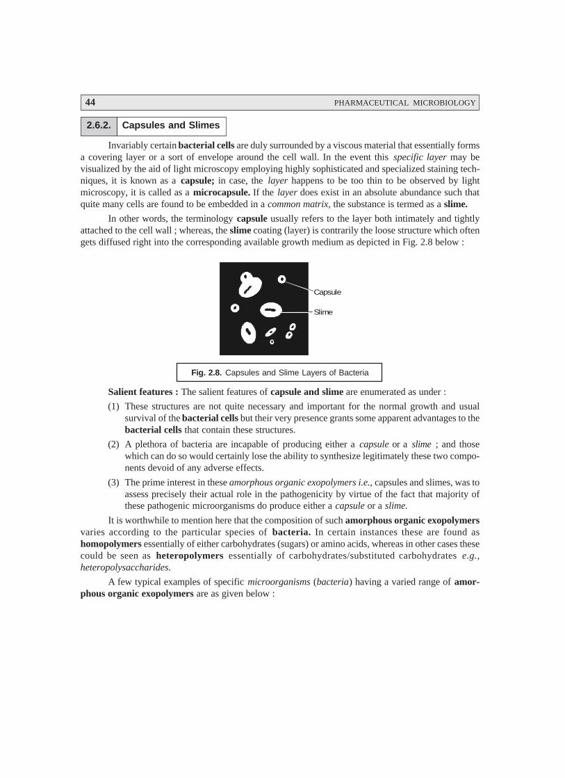
44 PHARMACEUTICAL MICROBIOLOGY
2.6.2. Capsules and Slimes
Invariably certain bacterial cells are duly surrounded by a viscous material that essentially formsa covering layer or a sort of envelope around the cell wall. In the event this specific layer may bevisualized by the aid of light microscopy employing highly sophisticated and specialized staining tech-niques, it is known as a capsule; in case, the layer happens to be too thin to be observed by lightmicroscopy, it is called as a microcapsule. If the layer does exist in an absolute abundance such thatquite many cells are found to be embedded in a common matrix, the substance is termed as a slime.
In other words, the terminology capsule usually refers to the layer both intimately and tightlyattached to the cell wall ; whereas, the slime coating (layer) is contrarily the loose structure which oftengets diffused right into the corresponding available growth medium as depicted in Fig. 2.8 below :
Capsule
Slime
Fig. 2.8. Capsules and Slime Layers of Bacteria
Salient features : The salient features of capsule and slime are enumerated as under :
(1) These structures are not quite necessary and important for the normal growth and usualsurvival of the bacterial cells but their very presence grants some apparent advantages to thebacterial cells that contain these structures.
(2) A plethora of bacteria are incapable of producing either a capsule or a slime ; and thosewhich can do so would certainly lose the ability to synthesize legitimately these two compo-nents devoid of any adverse effects.
(3) The prime interest in these amorphous organic exopolymers i.e., capsules and slimes, was toassess precisely their actual role in the pathogenicity by virtue of the fact that majority ofthese pathogenic microorganisms do produce either a capsule or a slime.
It is worthwhile to mention here that the composition of such amorphous organic exopolymersvaries according to the particular species of bacteria. In certain instances these are found ashomopolymers essentially of either carbohydrates (sugars) or amino acids, whereas in other cases thesecould be seen as heteropolymers essentially of carbohydrates/substituted carbohydrates e.g.,heteropolysaccharides.
A few typical examples of specific microorganisms (bacteria) having a varied range of amor-phous organic exopolymers are as given below :
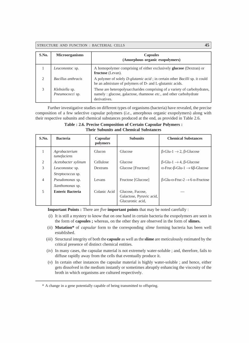
STRUCTURE AND FUNCTION : BACTERIAL CELLS 45
* A change in a gene potentially capable of being transmitted to offspring.
S.No. Microorganisms Capsules(Amorphous organic exopolymers)
1 Leuconostoc sp. A homopolymer comprising of either exclusively glucose (Dextran) orfructose (Levan).
2 Bacillus anthracis A polymer of solely D-glutamic acid ; in certain other Bacilli sp. it couldbe an admixture of polymers of D- and L-glutamic acids.
3 Klebsiella sp. These are heteropolysaccharides comprising of a variety of carbohydrates,Pneumococci sp. namely : glucose, galactose, rhamnose etc., and other carbohydrate
derivatives.
Further investigative studies on different types of organisms (bacteria) have revealed, the precisecomposition of a few selective capsular polymers (i.e., amorphous organic exopolymers) along withtheir respective subunits and chemical substances produced at the end, as provided in Table 2.6.
Table : 2.6. Precise Composition of Certain Capsular Polymers :Their Subunits and Chemical Substances
S.No. Bacteria Capsular Subunits Chemical Substancespolymers
1 Agrobacterium Glucon Glucose β-Glu-1 → 2, β-Glucosetumefaciens
2 Acetobacter xylinum Cellulose Glucose β-Glu-1 → 4, β-Glucose
3 Leuconostoc sp. Dextrans Glucose [Fructose] α-Fruc-β-Glu-1 → 6β-Glucose
Streptococcus sp.
4 Pseudomonas sp. Levans Fructose [Glucose] β-Glu-α-Fruc-2 → 6 α-Fructose
Xanthomonas sp.
5 Enteric Bacteria Colanic Acid Glucose, Fucose, —Galactose, Pyruvic acid,Glucuronic acid,
Important Points : There are five important points that may be noted carefully :
(i) It is still a mystery to know that on one hand in certain bacteria the exopolymers are seen inthe form of capsules ; whereas, on the other they are observed in the form of slimes.
(ii) Mutation* of capsular form to the corresponding slime forming bacteria has been wellestablished.
(iii) Structural integrity of both the capsule as well as the slime are meticulously estimated by thecritical presence of distinct chemical entities.
(iv) In many cases, the capsular material is not extremely water-soluble ; and, therefore, fails todiffuse rapidly away from the cells that eventually produce it.
(v) In certain other instances the capsular material is highly water-soluble ; and hence, eithergets dissolved in the medium instantly or sometimes abruptly enhancing the viscosity of thebroth in which organisms are cultured respectively.
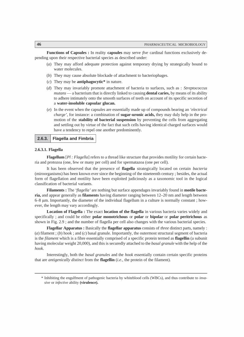
46 PHARMACEUTICAL MICROBIOLOGY
* Inhibiting the engulfment of pathogenic bacteria by whiteblood cells (WBCs), and thus contribute to inva-sive or infective ability (virulence).
Functions of Capsules : In reality capsules may serve five cardinal functions exclusively de-pending upon their respective bacterial species as described under:
(a) They may afford adequate protection against temporary drying by strategically bound towater molecules.
(b) They may cause absolute blockade of attachment to bacteriophages.
(c) They may be antiphagocytic* in nature.
(d) They may invariably promote attachment of bacteria to surfaces, such as : Streptococcusmutans — a bacterium that is directly linked to causing dental caries, by means of its abilityto adhere intimately onto the smooth surfaces of teeth on account of its specific secretion ofa water-insoluble capsular glucan.
(e) In the event when the capsules are essentially made up of compounds bearing an ‘electricalcharge’, for instance: a combination of sugar-uronic acids, they may duly help in the pro-motion of the stability of bacterial suspension by preventing the cells from aggregatingand settling out by virtue of the fact that such cells having identical charged surfaces wouldhave a tendency to repel one another predominently.
2.6.3. Flagella and Fimbria
2.6.3.1. Flagella
Flagellum [Pl : Flagella] refers to a thread like structure that provides motility for certain bacte-ria and protozoa (one, few or many per cell) and for spermatazoa (one per cell).
It has been observed that the presence of flagella strategically located on certain bacteria(miroorganisms) has been known ever since the beginning of the nineteenth century ; besides, the actualform of flagellation and motility have been exploited judiciously as a taxonomic tool in the logicalclassification of bacterial variants.
Filaments : The ‘flagella’ are nothing but surface appendages invariably found in motile bacte-ria, and appear generally as filaments having diameter ranging between 12–20 nm and length between6–8 μm. Importantly, the diameter of the individual flagellum in a culture is normally constant ; how-ever, the length may vary accordingly.
Location of Flagella : The exact location of the flagella in various bacteria varies widely andspecifically ; and could be either polar monotrichous or polar or bipolar or polar peritrichous asshown in Fig. 2.9 ; and the number of flagella per cell also changes with the various bacterial species.
Flagellar Apparatus : Basically the flagellar apparatus consists of three distinct parts, namely :(a) filament ; (b) hook ; and (c) basal granule. Importantly, the outermost structural segment of bacteriais the filament which is a fibre essentially comprised of a specific protein termed as flagellin (a subunithaving molecular weight 20,000), and this is securedly attached to the basal granule with the help of thehook.
Interestingly, both the basal granules and the hook essentially contain certain specific proteinsthat are antigenically distinct from the flagellin (i.e., the protein of the filament).
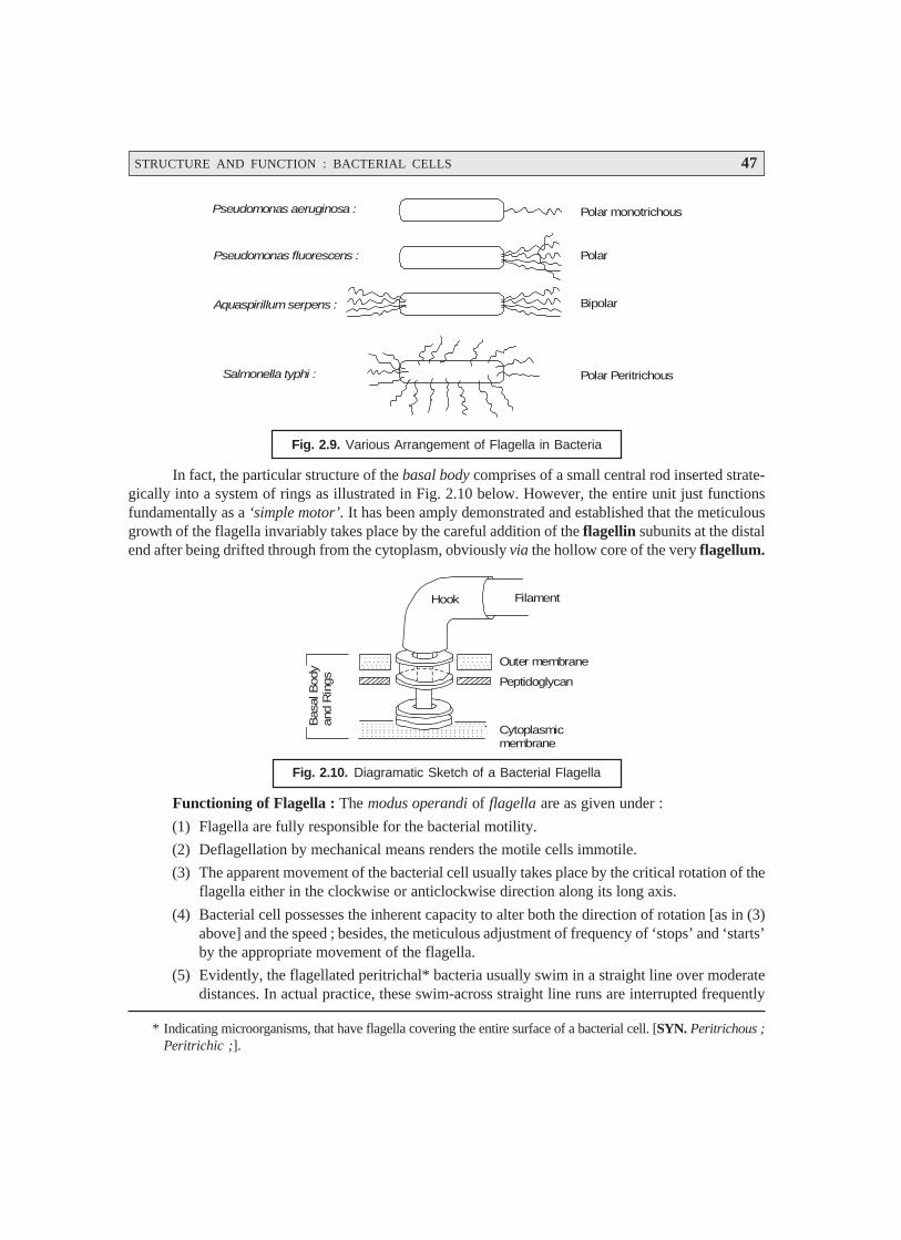
STRUCTURE AND FUNCTION : BACTERIAL CELLS 47
* Indicating microorganisms, that have flagella covering the entire surface of a bacterial cell. [SYN. Peritrichous ;Peritrichic ;].
Pseudomonas aeruginosa :
Pseudomonas fluorescens :
Aquaspirillum serpens :
Salmonella typhi :
Polar monotrichous
Polar
Bipolar
Polar Peritrichous
Fig. 2.9. Various Arrangement of Flagella in Bacteria
In fact, the particular structure of the basal body comprises of a small central rod inserted strate-gically into a system of rings as illustrated in Fig. 2.10 below. However, the entire unit just functionsfundamentally as a ‘simple motor’. It has been amply demonstrated and established that the meticulousgrowth of the flagella invariably takes place by the careful addition of the flagellin subunits at the distalend after being drifted through from the cytoplasm, obviously via the hollow core of the very flagellum.
FilamentHook
Outer membrane
Peptidoglycan
Cytoplasmicmembrane
Bas
al B
ody
and
Rin
gs
Fig. 2.10. Diagramatic Sketch of a Bacterial Flagella
Functioning of Flagella : The modus operandi of flagella are as given under :
(1) Flagella are fully responsible for the bacterial motility.
(2) Deflagellation by mechanical means renders the motile cells immotile.
(3) The apparent movement of the bacterial cell usually takes place by the critical rotation of theflagella either in the clockwise or anticlockwise direction along its long axis.
(4) Bacterial cell possesses the inherent capacity to alter both the direction of rotation [as in (3)above] and the speed ; besides, the meticulous adjustment of frequency of ‘stops’ and ‘starts’by the appropriate movement of the flagella.
(5) Evidently, the flagellated peritrichal* bacteria usually swim in a straight line over moderatedistances. In actual practice, these swim-across straight line runs are interrupted frequently
48 PHARMACEUTICAL MICROBIOLOGY
* Filamentous appendages of which there may be hundreds on a single cell. One function of pili is to attach thebacterium to cells of the host ; another to propel the bacterial cell.
** A coil or spiral.
by abrupt alterations in the direction that ultimately leads to tumbling. Therefore, the move-ment of the bacteria is believed to be zig-zag.
(6) It has been observed that the phenomenon of smooth swimming in a fixed direction is invari-ably mediated by the rotation of flagella in an anticlockwise direction ; whereas, the processof tumbling in a zig-zag direction is usually caused by the rotation of flagella in a clockwisedirection.
(7) The presence of ‘polar flagella’ in bacteria affords a distinct change in the direction thatusually takes place by the reciprocal alteration in the direction of rotation.
2.6.3.2 Fimbriae [or Pili*]
Fimbriae or Pili are hollow, non-helical, filamentous hair-like structures that are apparentlythinner, shorter, and more numerous than flagella. However, these structures do appear on the surface ofthe only Gram negative bacteria and are virtually distinct from the flagella.
Another school of thought rightly differentiates the terminology ‘fimbriae’ exclusively reservedfor all hair-like structures ; whereas, other structures that are directly and intimately involved in theactual transfer of genetic material solely are termed as ‘pili’. Likewise, the bacterial flagella that maybe visualized conveniently with the help of a light microscope after only suitable staining ; and thebacterial pili can be seen vividly only with the aid of an electron microscope.
Salient Features of Fimbriae : Some of the important salient features of ‘fimbriae’ are asenumerated under :
(1) At least 5 to 6 fimbriae variants have been recognized besides the sex pili.
(2) Type I fimbriae has been characterized completely.
(3) They contain a particular protein known as pilin having molecular weight of 17,000 daltons.
(4) The fimbriae are found to be spread over the entire cell surface. These have a diameter of7 nm and a length ranging between 0.5 to 2 μm ; besides, an empty core of 2 to 2.5 nm.
(5) The pilin subunits are duly arranged in a helical manner having the pitch of the helix**almost nearly at 2.3 μm.
(6) In addition to the Type-I fimbriae, the Gram-negative bacteria invariably own a specialcategory of pili termed as the sex pili (or F-pili), the synthesis of which is predominentlydirected by the sex factor (or F-factor). It has been observed that the sex pili do have auniform diameter of approximately 9 nm, and a length almost nearing between 1-20 μm.
(7) Very much akin to the flagella, both fimbriae and pili are observed to originate from thebasal bodies strategically located within the cytoplasm. Interestingly, neither fimbriae norpili seem to be essential for the survival of the bacteria.
Human Infection : It has been demonstrated that certain pili do play a major role in causing andspreading human infection to an appreciable extent by permitting the pathogenic bacteria to get strate-gically attached to various epithelial cells lining the genito urinary, intestinal, or respiratory tracts spe-cifically. It is worthwhile to mention here that this particular attachment exclusively checks and pre-vents the bacteria from being washed away critically by the incessent flow of either mucous or bodyfluids thereby allowing the infection to be established rather firmly.
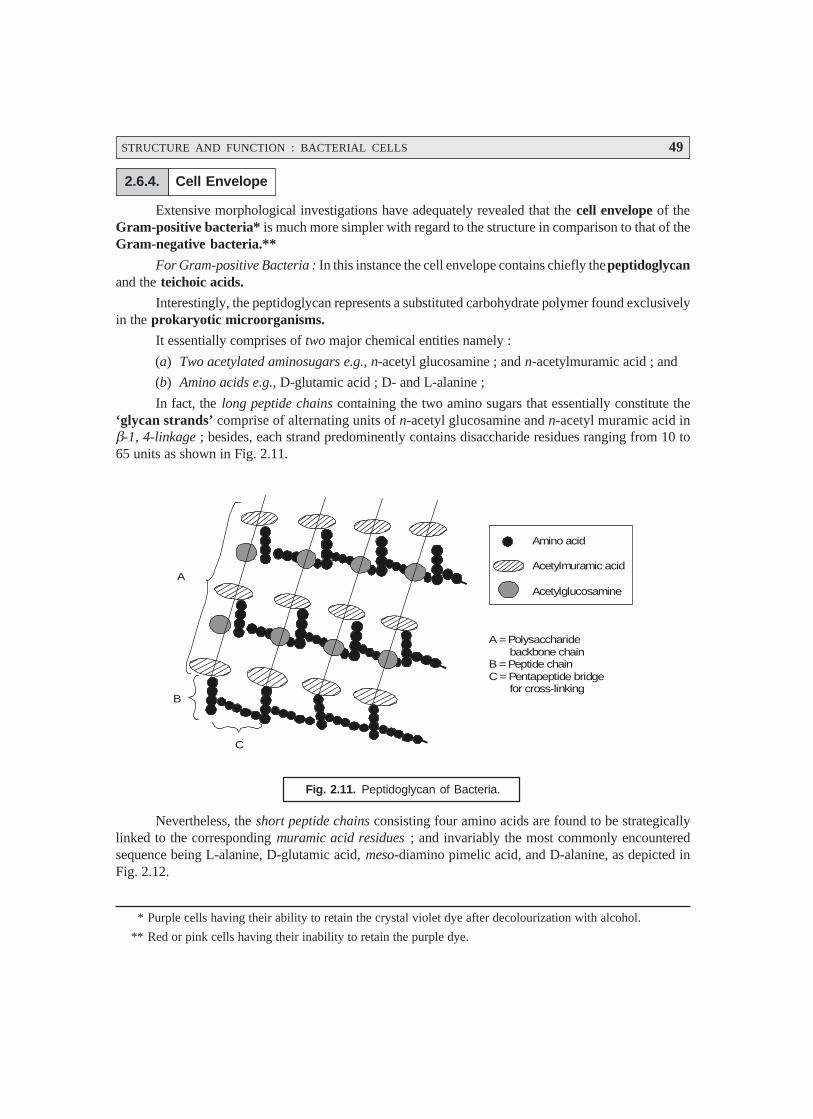
STRUCTURE AND FUNCTION : BACTERIAL CELLS 49
* Purple cells having their ability to retain the crystal violet dye after decolourization with alcohol.
** Red or pink cells having their inability to retain the purple dye.
2.6.4. Cell Envelope
Extensive morphological investigations have adequately revealed that the cell envelope of theGram-positive bacteria* is much more simpler with regard to the structure in comparison to that of theGram-negative bacteria.**
For Gram-positive Bacteria : In this instance the cell envelope contains chiefly the peptidoglycanand the teichoic acids.
Interestingly, the peptidoglycan represents a substituted carbohydrate polymer found exclusivelyin the prokaryotic microorganisms.
It essentially comprises of two major chemical entities namely :
(a) Two acetylated aminosugars e.g., n-acetyl glucosamine ; and n-acetylmuramic acid ; and
(b) Amino acids e.g., D-glutamic acid ; D- and L-alanine ;
In fact, the long peptide chains containing the two amino sugars that essentially constitute the‘glycan strands’ comprise of alternating units of n-acetyl glucosamine and n-acetyl muramic acid inβ-1, 4-linkage ; besides, each strand predominently contains disaccharide residues ranging from 10 to65 units as shown in Fig. 2.11.
Amino acid
Acetylmuramic acid
Acetylglucosamine
A = Polysaccharide backbone chainB = Peptide chainC = Pentapeptide bridge for cross-linking
A
B
C
Fig. 2.11. Peptidoglycan of Bacteria.
Nevertheless, the short peptide chains consisting four amino acids are found to be strategicallylinked to the corresponding muramic acid residues ; and invariably the most commonly encounteredsequence being L-alanine, D-glutamic acid, meso-diamino pimelic acid, and D-alanine, as depicted inFig. 2.12.
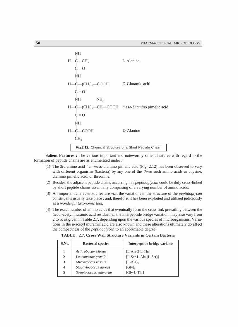
50 PHARMACEUTICAL MICROBIOLOGY
NH
H C CH⎯ ⎯ 3
C = O
NH
H C (CH ) COOH⎯ ⎯2⎯ 2
C = O
NH
H C (CH ) CH COOH⎯ ⎯3⎯ ⎯2
NH2
C = O
NH
H C COOH⎯⎯
CH3
L-Alanine
D-Glutamic acid
meso Diamino- pimelic acid
D-Alanine
Fig.2.12. Chemical Structure of a Short Peptide Chain
Salient Features : The various important and noteworthy salient features with regard to theformation of peptide chains are as enumerated under :
(1) The 3rd amino acid i.e., meso-diamino pimelic acid (Fig. 2.12) has been observed to varywith different organisms (bacteria) by any one of the three such amino acids as : lysine,diamino pimelic acid, or threonine.
(2) Besides, the adjacent peptide chains occurring in a peptidoglycan could be duly cross-linkedby short peptide chains essentially comprising of a varying number of amino acids.
(3) An important characteristic feature viz., the variations in the structure of the peptidoglycanconstituents usually take place ; and, therefore, it has been exploited and utilized judiciouslyas a wonderful taxonomic tool.
(4) The exact number of amino acids that eventually form the cross link prevailing between thetwo n-acetyl muramic acid residue i.e., the interpeptide bridge variation, may also vary from2 to 5, as given in Table 2.7, depending upon the various species of microorganisms. Varia-tions in the n-acetyl muramic acid are also known and these alterations ultimately do affectthe compactness of the peptidoglycan to an appreciable degree.
TABLE : 2.7. Cross Wall Structure Variants in Certain Bacteria
S.No. Bacterial species Interpeptide bridge variants
1 Arthrobacter citreus [L-Ala-2-L-Thr]
2 Leuconostoc gracile [L-Ser-L-Ala-(L-Ser)]
3 Micrococcus roseus [L-Ala]3
4 Staphylococcus aureus [Gly]5
5 Streptococcus salivarius [Gly-L-Thr]

STRUCTURE AND FUNCTION : BACTERIAL CELLS 51
2.6.5. Gram-Positive and Gram-Negative Bacteria
The various characteristic features of Gram-positive and Gram-negative bacteria shall be dis-cussed at length in this particular section.
For Gram-negative bacteria. There are two distinct layers that have been duly recognized inthe cell envelopes of Gram-negative bacteria, namely :
(a) An uniform inner layer approximately 2–3 mm wide, and
(b) A thicker outer layer nearly 8–10 nm wide.
Importantly, the peptidoglycan is prominently confined to the inner layer ; whereas, the outerlayer (membrane) essentially comprises of proteins, lipoproteins, and lipopolysaccharides.
The principal chemical differences that predominently occur between the cell walls of Gram-positive bacteria and the inner rigid wall layer and outer wall layer(s) of Gram-negative bacteria havebeen duly summarized in Table 2.8 given below :
Table 2.8. Principal Chemical Differences Existing Between Cell Walls of Gram-positive andGram-negative Bacteria
S.No. Chemical entities Gram-positive bacteria Gram-negative Bacteria
Inner rigid wall layer Outer wall layer(s)
1 Lipoprotein – + or – +
2 Lipopolysaccharide – – –
3 Peptidoglycan + + –
4 Polysaccharide + – –
5 Protein + or – – +
6 Teichoic acid + – –
Cardinal characteristic features of component variants in Gram +ve and Gram –ve micro-organisms : The various important characteristic features of component variants in Gram +ve and Gram–ve microbes are as stated under :
(1) Peptidoglycans belonging to the Gram –ve microorganisms exhibits a rather low extent ofcross linkages within the glycan strands.
(2) Outer-membrane. The fine structure of the outer membrane, very much akin to cell mem-brane, essentially comprises of a lipid bilayer wherein both phospholipids andlipopolysaccharides are definitely present. Besides, the lipopolysaccharide generates the majorcomponent of the outer membrane, and represents an extremely complex molecule varyingin chemical composition within/between the Gram –ve bacteria.
(3) Outer surface. The peptidoglycan of the wall has particular kinds of lipoproteins residingon its outer surface, that are strategically linked by peptide bonds to certain diaminopimelicacid residues present in the peptidoglycan.
(4) Lipoproteins evidently serve as a sort of bridge right from the peptidoglycan upto the outer-wall-layer.
(5) The total number of proteins definitely present, unlike in the inner membrane, are quite afew in number (approx. 10) ; and, therefore, these are markedly distinct from those invari-ably found in the inner membrane.
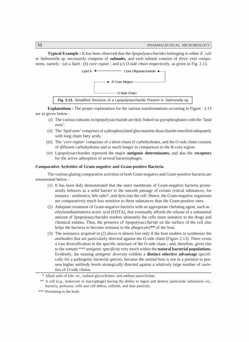
52 PHARMACEUTICAL MICROBIOLOGY
* Alkali salts of bile viz., sodium glycocholate, and sodium taurocholate.
** A cell (e.g., leukocyte or macrophage) having the ability to ingest and destroy particulate substances viz.,bacteria, protozoa, cells and cell debris, colloids, and dust particles.
*** Pertaining to the body.
Typical Example : It has been observed that the lipopolysaccharides belonging to either E. colior Salmonella sp. necessarily comprise of subunits, and each subunit consists of three vital compo-nents, namely : (a) a lipid ; (b) core region ; and (c) O-side chain respectively, as given in Fig. 2.13.
Lipid A Core Oligosaccharide
R Core Region
O Side Chain
Fig. 2.13. Simplified Structure of a Lipopolysaccharide Present in Salmonella sp.
Explanations : The proper explanations for the various transformations occurring in Figure : 2.13are as given below :
(i) The various subunits in lipopolysaccharide are duly linked via pyrophosphates with the ‘lipidzone’.
(ii) The ‘lipid zone’ comprises of a phosphorylated glucosamine disaccharide esterified adequatelywith long chain fatty acids.
(iii) The ‘core region’ comprises of a short-chain of carbohydrates, and the O-side chain consistsof different carbohydrates and is much longer in comparison to the R-core region.
(iv) Lipopolysaccharides represent the major antigenic determinants, and also the receptorsfor the active adsorption of several bacteriophages.
Comparative Activities of Gram-negative and Gram-positive Bacteria
The various glaring comparative activities of both Gram-negative and Gram-positive bacteria areenumerated below :
(1) It has been duly demonstrated that the outer membrane of Gram-negative bacteria promi-nently behaves as a solid barrier to the smooth passage of certain critical substances, forinstance : antibiotics, bile salts*, and dyes into the cell. Hence, the Gram-negative organismsare comparatively much less sensitive to these substances than the Gram-positive ones.
(2) Adequate treatment of Gram-negative bacteria with an appropriate chelating agent, such as :ethylenediaminetetra acetic acid (EDTA), that eventually affords the release of a substantialamount of lipopolysaccharides renders ultimately the cells more sensitive to the drugs andchemical entities. Thus, the presence of lipopolysaccharide on the surface of the cell alsohelps the bacteria to become resistant to the phagocytes** of the host.
(3) The resistance acquired in (2) above is almost lost only if the host enables to synthesize theantibodies that are particularly directed against the O-side chain (Figure 2.13). There existsa vast diversification in the specific structure of the O-side chain ; and, therefore, gives riseto the somatic*** antigenic specificity very much within the natural bacterial populations.Evidently, the ensuing antigenic diversity exhibits a distinct selective advantage specifi-cally for a pathogenic bacterial species, because the animal host is not in a position to pos-sess higher antibody levels strategically directed against a relatively large number of varie-ties of O-side chains.
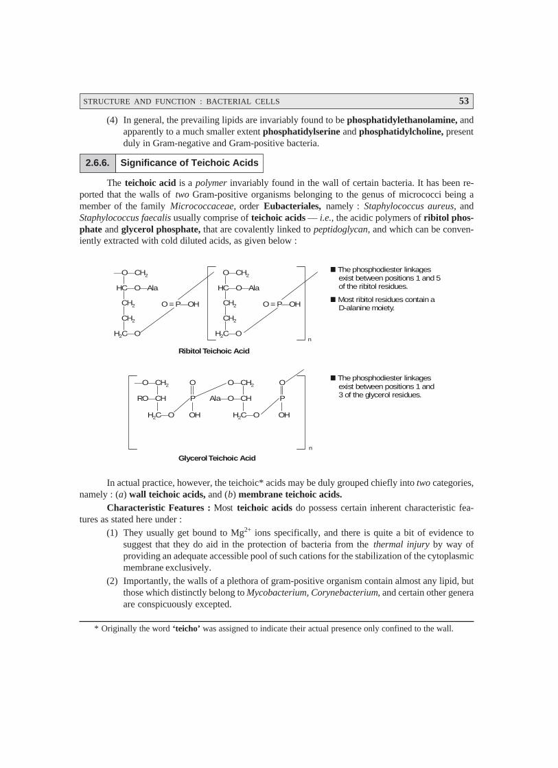
STRUCTURE AND FUNCTION : BACTERIAL CELLS 53
* Originally the word ‘teicho’ was assigned to indicate their actual presence only confined to the wall.
(4) In general, the prevailing lipids are invariably found to be phosphatidylethanolamine, andapparently to a much smaller extent phosphatidylserine and phosphatidylcholine, presentduly in Gram-negative and Gram-positive bacteria.
2.6.6. Significance of Teichoic Acids
The teichoic acid is a polymer invariably found in the wall of certain bacteria. It has been re-ported that the walls of two Gram-positive organisms belonging to the genus of micrococci being amember of the family Micrococcaceae, order Eubacteriales, namely : Staphylococcus aureus, andStaphylococcus faecalis usually comprise of teichoic acids — i.e., the acidic polymers of ribitol phos-phate and glycerol phosphate, that are covalently linked to peptidoglycan, and which can be conven-iently extracted with cold diluted acids, as given below :
⎯O CH⎯ 2
HC Ala⎯O⎯
CH2
CH2
H C O2 ⎯
O = P⎯OH
O CH⎯ 2
HC Ala⎯O⎯
CH2
CH2
H C O2 ⎯
O = P⎯OH
The phosphodiester linkages exist between positions 1 and 5 of the ribitol residues.
Most ribitol residues contain a D-alanine moiety.
n
⎯O CH⎯ 2
RO⎯CH
H2C O⎯
Ribitol Teichoic Acid
O
P
OH
O CH⎯ 2
Ala⎯O CH⎯
H2C O⎯
O
P
OH
n
The phosphodiester linkages exist between positions 1 and 3 of the glycerol residues.
Glycerol Teichoic Acid
In actual practice, however, the teichoic* acids may be duly grouped chiefly into two categories,namely : (a) wall teichoic acids, and (b) membrane teichoic acids.
Characteristic Features : Most teichoic acids do possess certain inherent characteristic fea-tures as stated here under :
(1) They usually get bound to Mg2+ ions specifically, and there is quite a bit of evidence tosuggest that they do aid in the protection of bacteria from the thermal injury by way ofproviding an adequate accessible pool of such cations for the stabilization of the cytoplasmicmembrane exclusively.
(2) Importantly, the walls of a plethora of gram-positive organism contain almost any lipid, butthose which distinctly belong to Mycobacterium, Corynebacterium, and certain other generaare conspicuously excepted.
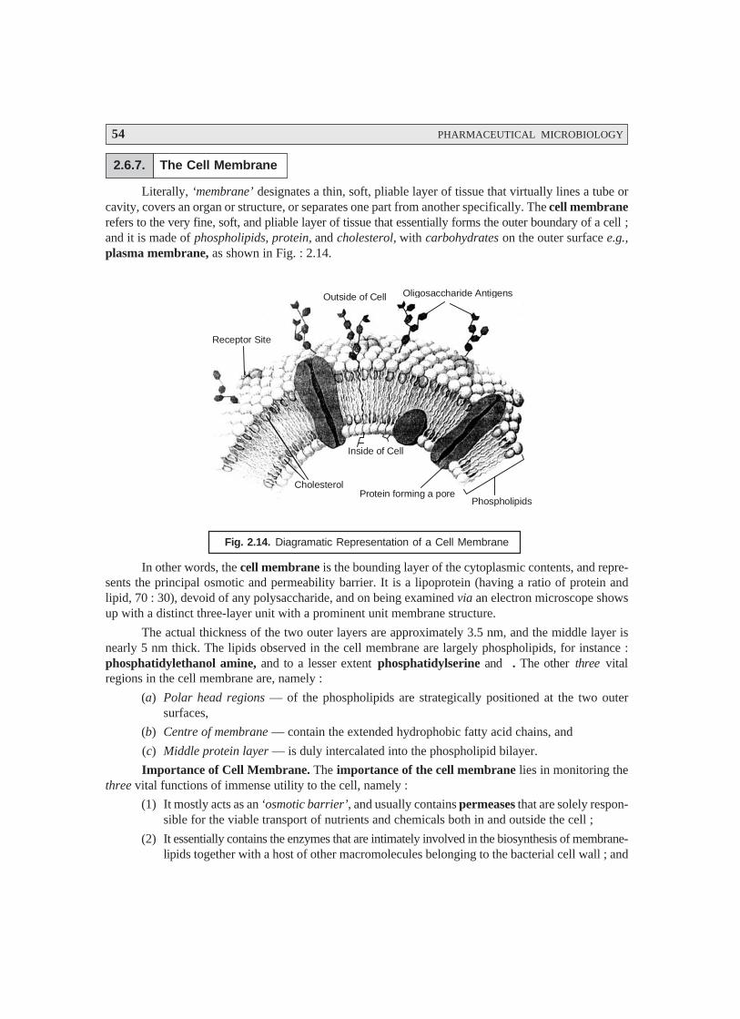
54 PHARMACEUTICAL MICROBIOLOGY
2.6.7. The Cell Membrane
Literally, ‘membrane’ designates a thin, soft, pliable layer of tissue that virtually lines a tube orcavity, covers an organ or structure, or separates one part from another specifically. The cell membranerefers to the very fine, soft, and pliable layer of tissue that essentially forms the outer boundary of a cell ;and it is made of phospholipids, protein, and cholesterol, with carbohydrates on the outer surface e.g.,plasma membrane, as shown in Fig. : 2.14.
Cholesterol
Inside of Cell
Protein forming a porePhospholipids
Oligosaccharide AntigensOutside of Cell
Receptor Site
Fig. 2.14. Diagramatic Representation of a Cell Membrane
In other words, the cell membrane is the bounding layer of the cytoplasmic contents, and repre-sents the principal osmotic and permeability barrier. It is a lipoprotein (having a ratio of protein andlipid, 70 : 30), devoid of any polysaccharide, and on being examined via an electron microscope showsup with a distinct three-layer unit with a prominent unit membrane structure.
The actual thickness of the two outer layers are approximately 3.5 nm, and the middle layer isnearly 5 nm thick. The lipids observed in the cell membrane are largely phospholipids, for instance :phosphatidylethanol amine, and to a lesser extent phosphatidylserine and . The other three vitalregions in the cell membrane are, namely :
(a) Polar head regions — of the phospholipids are strategically positioned at the two outersurfaces,
(b) Centre of membrane — contain the extended hydrophobic fatty acid chains, and
(c) Middle protein layer — is duly intercalated into the phospholipid bilayer.
Importance of Cell Membrane. The importance of the cell membrane lies in monitoring thethree vital functions of immense utility to the cell, namely :
(1) It mostly acts as an ‘osmotic barrier’, and usually contains permeases that are solely respon-sible for the viable transport of nutrients and chemicals both in and outside the cell ;
(2) It essentially contains the enzymes that are intimately involved in the biosynthesis of membrane-lipids together with a host of other macromolecules belonging to the bacterial cell wall ; and
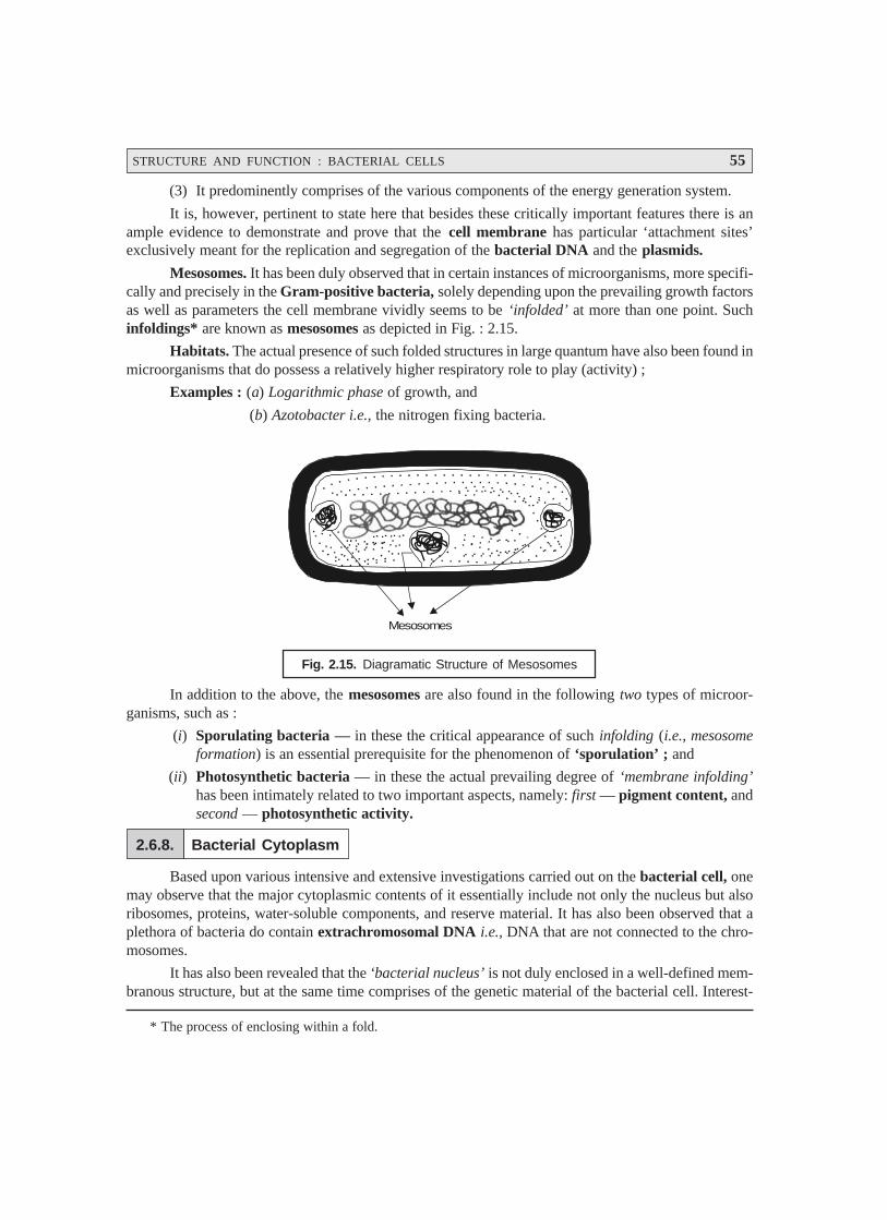
STRUCTURE AND FUNCTION : BACTERIAL CELLS 55
* The process of enclosing within a fold.
(3) It predominently comprises of the various components of the energy generation system.
It is, however, pertinent to state here that besides these critically important features there is anample evidence to demonstrate and prove that the cell membrane has particular ‘attachment sites’exclusively meant for the replication and segregation of the bacterial DNA and the plasmids.
Mesosomes. It has been duly observed that in certain instances of microorganisms, more specifi-cally and precisely in the Gram-positive bacteria, solely depending upon the prevailing growth factorsas well as parameters the cell membrane vividly seems to be ‘infolded’ at more than one point. Suchinfoldings* are known as mesosomes as depicted in Fig. : 2.15.
Habitats. The actual presence of such folded structures in large quantum have also been found inmicroorganisms that do possess a relatively higher respiratory role to play (activity) ;
Examples : (a) Logarithmic phase of growth, and
(b) Azotobacter i.e., the nitrogen fixing bacteria.
Mesosomes
Fig. 2.15. Diagramatic Structure of Mesosomes
In addition to the above, the mesosomes are also found in the following two types of microor-ganisms, such as :
(i) Sporulating bacteria — in these the critical appearance of such infolding (i.e., mesosomeformation) is an essential prerequisite for the phenomenon of ‘sporulation’ ; and
(ii) Photosynthetic bacteria — in these the actual prevailing degree of ‘membrane infolding’has been intimately related to two important aspects, namely: first — pigment content, andsecond — photosynthetic activity.
2.6.8. Bacterial Cytoplasm
Based upon various intensive and extensive investigations carried out on the bacterial cell, onemay observe that the major cytoplasmic contents of it essentially include not only the nucleus but alsoribosomes, proteins, water-soluble components, and reserve material. It has also been observed that aplethora of bacteria do contain extrachromosomal DNA i.e., DNA that are not connected to the chro-mosomes.
It has also been revealed that the ‘bacterial nucleus’ is not duly enclosed in a well-defined mem-branous structure, but at the same time comprises of the genetic material of the bacterial cell. Interest-
56 PHARMACEUTICAL MICROBIOLOGY
* Not connected to the chromosomes.
ingly, several altogether sophisticated meticulous and methodical investigations pertaining to the actualstatus/content(s) of the bacterial nucleus reveal amply that :
(a) Electron microscopy : Electron micrographs of the bacterial nucleus under investigationevidently depict it as a region very tightly and intimately packed with fibrillar DNA i.e., consisting ofvery small filamentous structure.
(b) Cytological, biochemical, physical, and genetic investigations : Such investigations withrespect to a large cross-section of bacterial species revealed that the ‘bacterial nucleus’ essentiallycontains a distinct singular molecule of definite circular shape, and having a double-stranded DNA.
The genome size of DNA i.e., the complete set of chromosomes, and thus the entire geneticinformation present in a cell, obtained painstakingly from a variety of bacterial species has been deter-mined and recorded in Table 2.9 below :
Table 2.9. The Genome Size of Certain Bacteria
S.No. Microorganisms (Bacteria) Genome size(Daltons × 109)
1 Bacillus subtilis 2.500
2 Escherichia coli 25.000 (± 0.5)
3 Micrococcus salivarius 3.300
4 Mycoplasma pneumoniae 0.480
5 Peptococcus aerogenes 0.816
6 Peptococcus saccharolydicus 1.250
7 Staphylococcus aureus 1.458
Specifications of E. coli: The size of DNA in E. coli together with certain other specificationsare as given below :
Average length : Approx. 1000 μm
Base pairs : 5 × 10³ kilo base pairs
Molecular weight : 2.5 × 10⁹ Daltons (± 0.5 × 10⁹)
• The ensuing DNA happens to be a highly charged molecule found to be dissociated with anybasic proteins as could be observed in higher organisms.
• Neutralization of charge is duly caused either by polyamines e.g., spermine, spermidine, orby bivalent cations e.g., Mg2+, Ca2+.
Plasmid DNA : Besides, the apparent and distinct presence of the bacterial ‘nuclear DNA’, theyinvariably contain extrachromosomal* DNA termed as plasmid DNA that replicates autonomously. Ithas been duly observed these plasmid DNAs exhibit different specific features, such as :
• confer on the bacterial cell,
• drug resistance,
• ability to generate bacteriocins i.e., proteinaceous toxins.
• ability to catabolize uncommon organic chemical entities (viz., in Pseudomonas).
STRUCTURE AND FUNCTION : BACTERIAL CELLS 57
Nevertheless, the actual size of plasmid DNA usually found in these specific structures may benearly 1/10th or even less in comparison to that invariably found in the bacterial nucleus ; however, theexact number of copies may change from one to several. Besides, these structures are not enclosed in amembrane structure. Importantly, the plasmid DNA is mostly circular in shape and double stranded inits appearance.
2.6.9. Ribosomes
Ribosome refers to a cell organelle made up of ribosomal RNA and protein. Ribosomes mayexist singly, in clusters called polyribosomes, or on the surface of rough endoplasmic reticulum. Inprotein synthesis, they are the most favoured site of messenger RNA attachment and amino acid assem-bly in the sequence ordered b the genetic code carried by mRNA.
In other words, the specific cytoplasmic area which is strategically located in the cell materialbound by the cytoplasmic membrane having granular appearance and invariably rich in the macromolecularRNA-protein bodies is termed as ribosome.
Characteristic Features : Following are some of the cardinal characteristic features of the‘ribosomes’, namely:
(1) Contrary to the animal or plant cells, there exists no endoplasmic reticulum to which ribosomesare bound intimately.
(2) Interestingly, there are certain ribosomes that are found to be virtually ‘free’ in the cyto-plasm ; whereas, there are some, particularly those critically involved in the synthesis ofproteins require to be transported out of the cell, get closely linked to the inner surface of thecytoplasmic membrane.
(3) The number of ‘ribosomes’ varies as per the ensuing ‘rate of protein synthesis’, and mayreach even upto 15,000 per cell. In fact, greater the rate of proteins synthesis, the greater isthe rate of prevailing ribosomes.
(4) Ribosomes represent ribonucleoprotein particles (comprising of 60 RNA ; 40 Protein) hav-ing a diameter of 200 Å, and are usually characterised by their respective sedimentationphysical properties as depicted in Fig. 2.16.
(5) Prokaryotic Ribosome. In the event when the ribosomes of the prokaryotes undergo ‘sedi-mentation’ in an ultra-centrifuge, they normally exhibit a sedimentation coefficient of 70 S(S = Svedberg Units), and are essentially composed of two subunits i.e., a 50 S and a 30 Ssubunit (almost fused as shown in Figure 2.16). Consequently, these two subunits get dis-tinctly separated into a 50 S and a 30 S units*. As a result the 50 S unit further gets segre-gated into a RNA comprised of two daughter subunits of 5 S and 23 S each together withthirty two (32) altogether different proteins [derived from 50 – (5 + 23) = 22 sub-units].
Likewise, the 30 S gets fragmented into two segments i.e., first, a RNA comprised of onlyone subunit having 16 S plus twenty one (21) precisely different proteins [derived from30 – 16 = 14 sub-units], (see Fig. : 2.16).
(6) Eukaryotic Ribosome : This is absolutely in contrast to the ribosomes of the correspondingprokaryotic organisms, that do possess a sedimentation coefficient of 80 S, and are essen-tially comprised of two subunits each of 60 S and 40 S, respectively.
* The ‘mother ribosomes’ when placed in a low concentration of Mg2+ ions get dissociated into two smaller‘daughter ribosomes’.
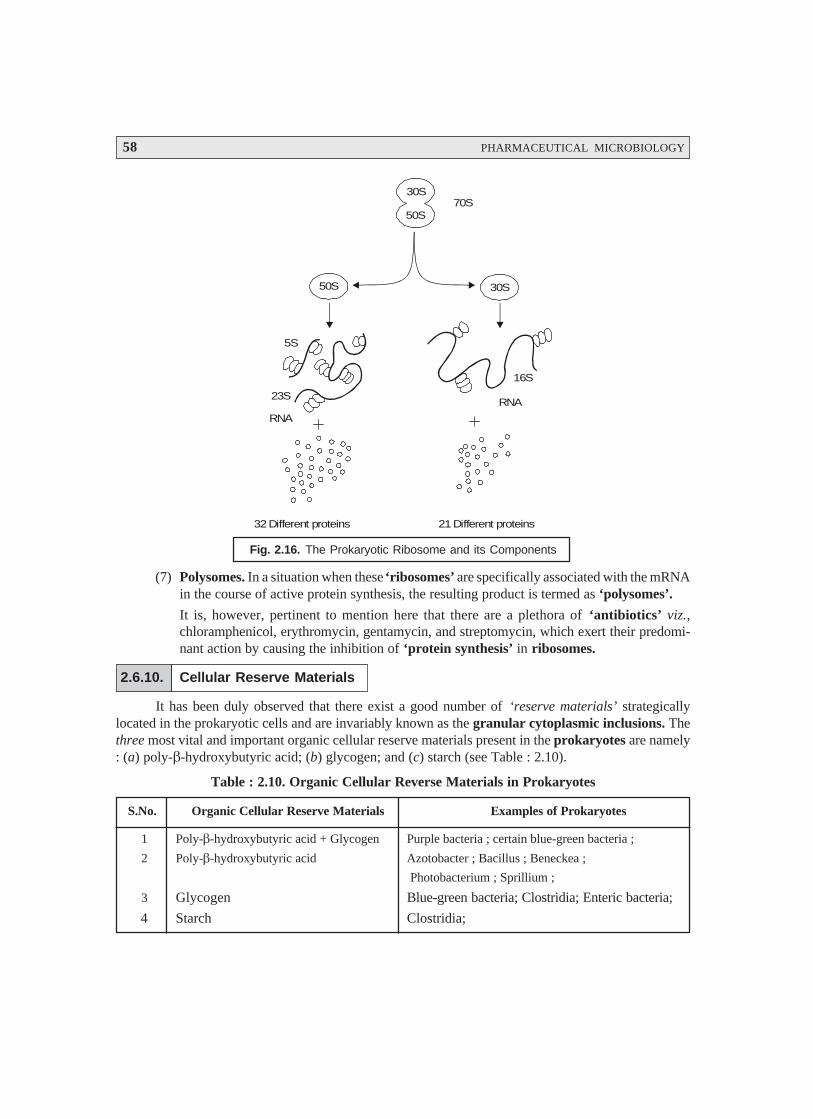
58 PHARMACEUTICAL MICROBIOLOGY
30S
50S
30S50S
5S
23S
RNA
RNA
16S
70S
32 Different proteins 21 Different proteins
Fig. 2.16. The Prokaryotic Ribosome and its Components
(7) Polysomes. In a situation when these ‘ribosomes’ are specifically associated with the mRNAin the course of active protein synthesis, the resulting product is termed as ‘polysomes’.
It is, however, pertinent to mention here that there are a plethora of ‘antibiotics’ viz.,chloramphenicol, erythromycin, gentamycin, and streptomycin, which exert their predomi-nant action by causing the inhibition of ‘protein synthesis’ in ribosomes.
2.6.10. Cellular Reserve Materials
It has been duly observed that there exist a good number of ‘reserve materials’ strategicallylocated in the prokaryotic cells and are invariably known as the granular cytoplasmic inclusions. Thethree most vital and important organic cellular reserve materials present in the prokaryotes are namely: (a) poly-β-hydroxybutyric acid; (b) glycogen; and (c) starch (see Table : 2.10).
Table : 2.10. Organic Cellular Reverse Materials in Prokaryotes
S.No. Organic Cellular Reserve Materials Examples of Prokaryotes
1 Poly-β-hydroxybutyric acid + Glycogen Purple bacteria ; certain blue-green bacteria ;
2 Poly-β-hydroxybutyric acid Azotobacter ; Bacillus ; Beneckea ;
Photobacterium ; Sprillium ;
3 Glycogen Blue-green bacteria; Clostridia; Enteric bacteria;
4 Starch Clostridia;
STRUCTURE AND FUNCTION : BACTERIAL CELLS 59
Salient Features. The salient features of the organic cellular reserve materials present in theprokaryotes are as stated under :
(1) Poly-βββββ-hydroxybutyric acid. It is found exclusively in the prokaryotes and invariably ca-ters as an equivalent of lipoidal content duly stored in the eukaryotic cells. It is observed inseveral species of Azotobacter, bacilli, and pseudomonads. Interestingly, certain specific or-ganisms viz., purple bacteria has the ability to synthesize even two types of reserve materi-als (e.g., glycogen and poly-β-hydroxybutyrate) simultaneously.
(a) Visibility — These organic cellular reserve materials are found to be deposited almostuniformly very much within the cytoplasm ; however, they may not be detected under alight microscope unless and until these are duly stained.
(b) Cellular content — The actively ‘growing cells’ do have these reserve materials presentin rather small quantum in the cellular content ; whereas, they get usually accumulatedexclusively in the C-rich culture medium under the influence of restricted amounts ofnitrogen.
(c) Availability — These reserve materials may sometimes represent even upto 50% of thetotal cellular content on dry weight basis.
(d) Utility — These reserve materials are fully utilized when the prevailing cells are ad-equately provided with a suitable source of N and the growth is resumed subsequently.
(2) Glycogen and Starch — It has been duly established that the synthesis of glycogen andstarch is usually accomplished via a proven mechanism for storing C in a form which isosmotically inert ; whereas, in the particular instance of poly-β-hydroxybutyric acid it pre-cisely designates a method of neutralizing an acidic metabolite.
(3) Cyanophycine (a copolymer of arginine and aspartic acid) :
In general, prokaryotes fail to store particularly the organic nitrogenous materials, but theblue-green bacteria is expected which essentially accumulate a nitrogenous reserve materialtermed as cyanophycine. It invariably represents as much as 8% of cellular dry weight; andmay be regarded as a copolymer of arginine and aspartic acid.
(4) Volutin (metachromatic) Granules. A plethora of prokaryotes acquire more and more ofvolutin granules that may be stained meticulously with a ‘basic dye’, for instance : methyl-ene blue. In fact, these prokaryotes appear as red on being stained with a ‘blue-dye’. Impor-tantly, the prevailing metachromatic nature of the ensuing ‘red complex’ is on account of thevery presence of a substantial quantum of ‘inorganic phosphates’. Evidently, the actualaccumulation of these substances in the prokaryotes takes place under critical parameters ofstarvation specifically during ‘sulphate starvation’. It has been observed that these instantlygenerated volutin granules disappear as soon as the cells are adequately made available witha ‘sulphur source’, and subsequently the phosphate moiety [PO4
3–] is incorporated strategi-cally into the nucleic acids i.e., DNA and RNA. From the above statement of facts one mayvividly infer that the ‘volutin granules’ definitely represent particularly the ‘intracellularphosphate reserve’ when the desired nucleic acid synthesis fails to materialize.
(5) Sulphur Bacteria [e.g., photosynthetic purple sulphur bacteria ; and filamentous non-photosynthetic bacteria (viz., Baggiatoa and Thiothrix)]. The aforementioned two sulphurbacteria specifically help in the accumulation of ‘Sulphur’ transiently in the course of hydro-gen sulphide [H2S] oxidation.
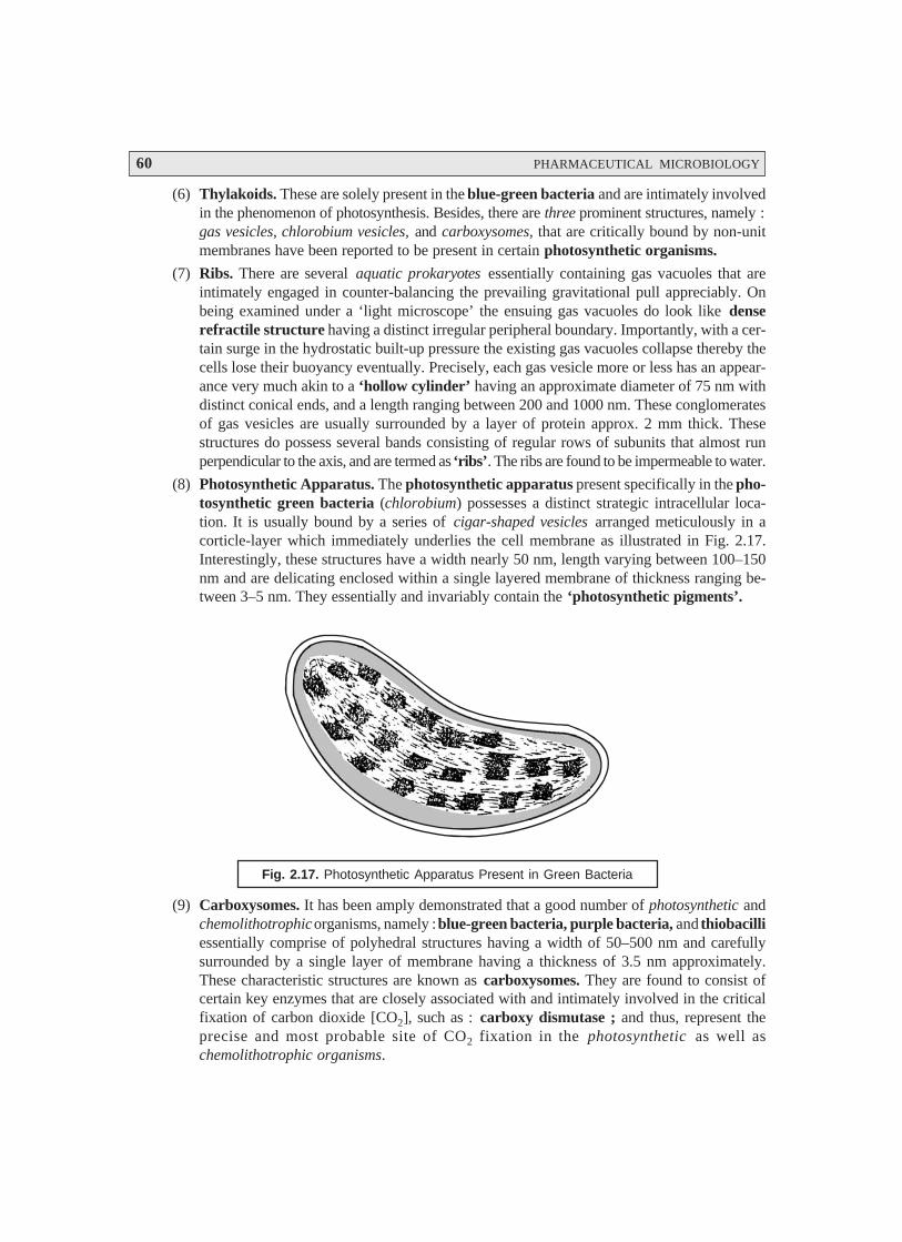
60 PHARMACEUTICAL MICROBIOLOGY
(6) Thylakoids. These are solely present in the blue-green bacteria and are intimately involvedin the phenomenon of photosynthesis. Besides, there are three prominent structures, namely :gas vesicles, chlorobium vesicles, and carboxysomes, that are critically bound by non-unitmembranes have been reported to be present in certain photosynthetic organisms.
(7) Ribs. There are several aquatic prokaryotes essentially containing gas vacuoles that areintimately engaged in counter-balancing the prevailing gravitational pull appreciably. Onbeing examined under a ‘light microscope’ the ensuing gas vacuoles do look like denserefractile structure having a distinct irregular peripheral boundary. Importantly, with a cer-tain surge in the hydrostatic built-up pressure the existing gas vacuoles collapse thereby thecells lose their buoyancy eventually. Precisely, each gas vesicle more or less has an appear-ance very much akin to a ‘hollow cylinder’ having an approximate diameter of 75 nm withdistinct conical ends, and a length ranging between 200 and 1000 nm. These conglomeratesof gas vesicles are usually surrounded by a layer of protein approx. 2 mm thick. Thesestructures do possess several bands consisting of regular rows of subunits that almost runperpendicular to the axis, and are termed as ‘ribs’. The ribs are found to be impermeable to water.
(8) Photosynthetic Apparatus. The photosynthetic apparatus present specifically in the pho-tosynthetic green bacteria (chlorobium) possesses a distinct strategic intracellular loca-tion. It is usually bound by a series of cigar-shaped vesicles arranged meticulously in acorticle-layer which immediately underlies the cell membrane as illustrated in Fig. 2.17.Interestingly, these structures have a width nearly 50 nm, length varying between 100–150nm and are delicating enclosed within a single layered membrane of thickness ranging be-tween 3–5 nm. They essentially and invariably contain the ‘photosynthetic pigments’.
Fig. 2.17. Photosynthetic Apparatus Present in Green Bacteria
(9) Carboxysomes. It has been amply demonstrated that a good number of photosynthetic andchemolithotrophic organisms, namely : blue-green bacteria, purple bacteria, and thiobacilliessentially comprise of polyhedral structures having a width of 50–500 nm and carefullysurrounded by a single layer of membrane having a thickness of 3.5 nm approximately.These characteristic structures are known as carboxysomes. They are found to consist ofcertain key enzymes that are closely associated with and intimately involved in the criticalfixation of carbon dioxide [CO2], such as : carboxy dismutase ; and thus, represent theprecise and most probable site of CO2 fixation in the photosynthetic as well aschemolithotrophic organisms.

STRUCTURE AND FUNCTION : BACTERIAL CELLS 61
FURTHER READING REFFERENCES
1. Becker W.M., Kleinsmith L, and Hardin J. The World of the Cell, 4th ed., Benjamin/Cummings, Redwood City, Calif., 2000.
2. Cruickshank R. et al.: Medical Microbiology, Vol. II. The Practice of Medical Microbiology,Churchill Livingstone, London, 12th edn., 1975.
3. Dawes I.W. and Sutherland W: Microbial Physiology, 2nd end., Blackwell, Oxford, 1991.
4. Gould G.W. and Hurst A. : The Bacterial Spore, Academic Press, London, 1983.
5. Harshey R.M. : Bacterial Motility on a Surface : Many Ways to a Common Goal, Annu.Rev. Microbiol. 57 : 249–73, 2003.
6. Kotra LP, Amro NA, Liu GY, and Mobashery S : Visualizing Bacteria at High Resolution,ASM News, 66 (11) : 675–81, 2000.
7. Lederberg J : Encyclopedia of Microbiology, 2nd ed. Academic Press, San Diego, 2000.
8. Lodish H, et al. Molecular Cell Biology, 4th ed., Scientific American Books, New York,1999.
9. Macnab R.M. : How Bacteria Assemble Flagella, Annu. Rev. Microbiol. 57, 77–100.
10. Mattick J.S. : Type IV Pili and Twitching Motility, Annu. Rev. Microbiol. 56 : 289–314,2002.
11. Moat A.G. and Foster J.W. : Microbial Physiology, 3rd. edn., Wiley-Liss, New York, 1995.
12. Pelczar M.J. et al.: Microbiology, 5th end., Tata McGraw Hill Publishing Co., Ltd. NewYork, 1993.
13. Roy C.R., Exploitation of the Endoplasmic Reticulum by Bacterial Pathogens, TrendsMicrobiol. 10 (9) : 418–24, 2002.
14. Schapiro L and Losick R : Protein Localization and Cell Fate in Bacteria, Science, 276 :712–18, 1997.
15. Schulz H.N. and Jorgensen B.B. : Big Bacteria, Annu. Rev. Microbiol. 55 : 105–37, 2001.
16. Schwartz R.M. and Dayhoff M.O.: Origins of Prokaryotes, Eukaryotes, and Chloroplasts,Science, 199 : 395, 1978.
17. Stainer R.Y. et al.: The Microbial World, 5th edn., Prentice-Hall, New Jersey, 1986.
18. Woese C.R.: Archaebacteria, Scientific American, 244 : 94, 1981.
19. Yonekura K. et al. The Bacterial Flagella Cap as the Rotary Promoter of Flagellin Self-assembly, Science, 290 : 2148–52, 2000.
62
CHARACTERIZATION, CLASSIFICATIONAND TAXONOMY OF MICROBES
3.1. INTRODUCTION
Microbiology is an integral part of ‘biological sciences’, and hence essentially encompasses thethree cardinal objectives, namely: characterization, classification, and identification. The entire‘microbial world’ enjoys the reputation for being an extremely complex and extraordinarily diversifieddomain with respect to their morphological, physiological, and genetical characteristic features. In thelight of the said glaring facts, it became almost necessary to afford a broad and critical classification asa means of bringing order to the puzzling diversity as well as variety of organisms in nature. Therefore,once the characteristic features of the various microbes existing in this universe have been duly established,one may compare it with other organisms quite conveniently in order to draw a line amongst theirsimilarities and dissimilarities in particular. It would be a lot easier task to segregate the microbes havingthe same features and subsequently group them together under a specific classified head or group knownas ‘classification’.
Based upon the enormous volume of researches made in the study of microorganisms, one has toknow their characteristics prior to their legitimate identification and classification. Because of theextremely minute and microscopical size of the microorganism, it may not be quite feasible to carry outan elaborated study of the characteristics of a single microorganism. In order to circumvent the abovedifficulties, one may conveniently study the characteristics of a culture i.e., a population of microor-ganisms or the propogation of microorganisms. Therefore, the meticulous investigation of the charac-teristics of a culture comprising a host of microorganisms,* it is as good as exploring the characteristicsof a single organism. Pure Culture [Axenic Culture] : It refers to a ‘culture’ that essentially becomposed of a single type of microorganism, irrespective of the number of individuals, in a surroundingabsolutely free of other living microbes (organisms). Summarily, the process of establishing the ‘char-acteristics’ of microorganisms is not only a cardinal prerequisite for classification but also play a vari-ety of vital, indeed essential, roles in nature.
3.2. CHARACTERIZATION
The microorganisms may be broadly characterized into the following categories, namely:
* Invariably millions or billions of cells specifically of one type.
3• Introduction• Characterization• Classificiation• Taxonomy• The Kingdom Prokaryotae

CHARACTERIZATION, CLASSIFICATION AND TAXONOMY OF MICROBES 63
* 1 μm = 0.001 millimeter (mm) or ≡ 0.00004 inches. It may be carried out either with the help of a high-power microscope or an electron microscope (which provides magnification of thousands of diametersand enables to see more refined/detailed cellular structure(s).
** Concerning the development of a race or phylum.
(i) Morphological characteristics
(ii) Chemical characteristics
(iii) Cultural characteristics
(iv) Metabolic characteristics
(v) Antigenic characteristics
(vi) Genetic characteristics
(vii) Pathogenicity, and
(viii) Ecological characteristics.
The aforesaid categories of characteristics shall now be treated individually in the sections thatfollow :
3.2.1. Morphological Characteristics
Morphology refers to the science of structure and form of organisms without any regard to theirfunction. The morphological determinations invariably require the intensive studies of the individualcells of a pure culture. The microorganisms being of very small size are usually expressed in microm-eters (μμμμμm)*.
Interestingly, the morphological characteristic features are relatively easier to analyze and study,specifically in the eukaryotic microorganisms as well as the more complex prokaryotes. However, themorphological comparisons amongst the microbes play an important and vital role by virtue of the factthat their major structural features exclusively depend upon the prevailing expression of several genes.In fact, they are found to be fairly stable genetically, and hence fail to undergo drastic variation inresponse to the environmental alterations. Therefore, morphological similarity serves as an essentialnovel indicator with regard to phylogenetic **relationship.
However, the ‘morphological characteristics’ frequently employed in the classification andidentification of certain microbial groups vis-a-vis their structural features are enumerated as under:
S. No. Characteristic Features Microbial Groups
1 Cell size Most major categories
2 Cell shape —do—
3 Cellular inclusions —do—
4 Cilia and flagella —do—
5 Colonial morphology —do—
6 Colour —do—
7 Endospore shape and location Endospore forming microbes
8 Mode of motility Spirochaetes, gliding microbes
9 Spore morphology and location Algae, fungi, microbes,
10 Pattern of staining Microbes, certain fungi
11 Ultrastructural characteristic features Most major categories
64 PHARMACEUTICAL MICROBIOLOGY
3.2.2. Chemical Characteristics
Interestingly, one may observe a broad spectrum of organic compounds critically located in themicrobial cells. These cells upon undergoing disintegration (broken apart) give rise to several differentchemical entities that are methodically subjected to vigorous chemical analysis. Thus, each type ofmicroorganism is observed to possess altogether specific and characteristic chemical composition. Thepresence of distinct qualitative and quantitative differences in composition does occur amongst thevarious prevailing microbial species.
Examples:
(a) Gram-positive Microorganisms—they essentially possess in their cell walls an organic acidknown as ‘teichoic acids’, and such compounds are not be seen in Gram-negativemicroorganisms.
(b) Gram-negative Microorganisms—they invariably contain ‘lipolysaccharide’ in their cellwalls, and this is distinctly absent in Gram-positive bacteria.
Note. (1) Both algal and fungal cell walls are found to be entirely different in compositionfrom those of microbes.
(2) In viruses, the most prominent point of difference is solely based upon the type ofnucleic acid they essentially possess, viz., RNA and DNA.
3.2.3. Cultural Characteristics
It has been amply established that each and every type of microorganism possesses specific aswell as definitive growth-requirements.
Salient Features. The salient features of the important and vital cultural characteristics are asstated under:
(1) A plethora of microbes may be grown either on or in a cultural medium*.
(2) A few microorganisms could be cultivated (grown) in a medium comprising specificallyorganic chemical entities**, whereas some others require solely inorganic chemical enti-ties.
(3) Certain microbes do require complex natural materials*** only for their normal growth.
(4) Importantly, there are certain critical microbes that may be carefully and meticulouslypropogated only in a living host or living cells, and cannot be grown in an usual artificiallaboratory medium.
Example: Rickettsias**** prominently require a definitive host in which they may growconveniently and generously, for instance: (a) an arthroped*****; (b) a chick embryo (i.e., a fertilized
* A mixture of nutrients employed in the laboratory to support growth as well as multiplication of microbes.** Amino acids, coenzymes, purines and pyrimidines, and vitamines.
*** Blood cell, Blood serum, Peptone, Yeast autolysate.**** Rickettsia: A genus of bacteria of the family Rickettsiaceae, order Rickettsiales. They are obligate
intracellular parasites (must be in living cells to reproduce) and are the causative agents of many diseases.***** A member of the phylum Arthropoda i.e., a phylum of invertebrate animals marked by bilateral symme-
try, a hard, jointed exoskeleton, segmented bodies, and jointed paired appendages viz., insects, myriapods,and the crustaceans.
CHARACTERIZATION, CLASSIFICATION AND TAXONOMY OF MICROBES 65
chicken egg); and (c) a culture of mammalian tissue cells. In reality, the host being employed as anextremely complex specified and articulated ‘medium’ essentially required for such nutritionallydemanding microorganisms.
(5) Specific physical parameters: Besides, certain highly critical and specific array of nutrients,each type of microorganism predominantly needs certain particular physical parameters forits natural and normal growth.
Examples :
(a) Microbes growing at high temperatures (e.g., Thermophilic bacteria): Some organisms doprefer to grow and thrive best at temperatures ranging between 40° and 70°C (104° and158°F) ; and hence, fail to grow below 40°C e.g., Thermoactinomyces vulgaris; Thermusaquaticus; and Streptococcus thermophilus.
(b) Microbes growing at low temperatures : Certain microorganisms grow best in the cold en-vironmental conditions and simply cannot grow above 20°C e.g., Vibrio marinus strain MP-1 ;and Vibrio psychoerythrus.
(c) Pathogenic bacteria: A host of organisms that are solely responsible for causing diseases inhumans do essentially require a temperature very close to that of the human body (i.e., 37°Cor 98.4°F) e.g., Salmonella typhi; Vibrio cholerae; Mycobacterium tuberculosis; Clostridiumtetani; Shigella dysenteriae; Treponema pallidum; Bordetella pertussis; Rickettsia rickettsiietc.
(d) Gaseous environment: It is equally important to have requisite gaseous environment for thesubstantial growth of the microorganisms.
Examples: (1) Aerobic Microbes: These are of two kinds, namely :
(i) Facultative Aerobes i.e., microbes that are able to live and grow preferably in an environ-ment devoid of oxygen, but has adapted so that it can live and grow in the presence of oxygen.
(ii) Obligate Aerobes i.e., microorganisms that can live and grow only in the presence ofoxygen.
(2) Anaerobic Microbes: These microorganisms can live and grow in the absence of oxgyen,and are of two types, namely :
(i) Facultative Anaerobes i.e., microbes that can live and grow with or without oxgyen.
(ii) Obligatory Anaerobes i.e., microorganisms that can live and grow only in the absence ofoxygen.
(e) Light (i.e., UV-Light): UV-Light provides a source of energy necessary for the growth ofcertain microbes e.g., cyanobacteria (blue green algae). Interestingly, some organisms may be indif-ferent to light or at times may even prove to be quite deleterious to their legitimate growth.
(f) Liquid Culture Medium: It has been observed that each and every type of microorganisminvariably grows in an absolutely typical characteristic manner in various liquid culture medium withvariant composition, such as:
(i) Sparse or abundant growth—as could be seen in a liquid medium.
(ii) Evenly distributed growth—as seen spread throughout the liquid medium.
(iii) Sedimented growth—as may be observed as a sediment usually at the bottom.
(iv) Thin-film growth—as could be seen on the surface of the liquid culture medium.

66 PHARMACEUTICAL MICROBIOLOGY
(v) Pellicle growth—as may be observed as a scum at the top.
Example: Salivary pellicle—The thin-layer of salivary proteins and glycoproteins that quicklyadhere to the tooth surface after the tooth has been cleaned; this amorphous, bacteria-free layer mayserve as an attachment medium for bacteria, which in turn form plaque.
(g) Solid Culture Medium: It has been amply demonstrated that microorganisms invariablygrow on solid culture medium as colonies* which are markedly distinct, compact masses of cellsevidently visible with a naked eye (macroscopically). In fact, the ensuing colonies are usually character-ized based upon, their particular shape, size, consistency, texture, colouration, compactness, and otherseveral vital characteristic features.
3.2.4. Metabolic Characteristics
Metabolism refers to the sum of all physical and chemical changes that take place within anorganism; all energy and material transformations that occur within living cells. It includes essentiallythe material changes (i.e., changes undergone by substances during all periods of life, for instance:growth, maturity, and senescence), and energy changes (i.e., all transformations of chemical energy offood stuffs to mechanical energy or heat). Metabolism involves two fundamental processes, namely:anabolism (viz., assimilation or building-up processes), and catabolism (viz., disintegration or tearing,down processes). Anabolism is the conversion of ingested substances into the constituents of protoplasm;Catabolism is the breakdown of substances into simpler substances, the end products usually beingexcreted.
The broad spectrum of these reactions gives rise to a plethora of excellent opportunities to char-acterize and differentiate categories of microorganisms.
Examples:
(a) Absorption of Light: Certain microbes may derive energy via absorption of light.
(b) Oxidation: A few microorganisms may obtain energy through oxidation of a host of inor-ganic and organic compounds.
(c) Redistribution of Atoms: Some organisms engage actively in the redistribution of atomswithin certain molecules thereby rendering the resulting molecules less stable.
(d) Synthesis of Cell Components: The microorganisms also vary a lot in the manner wherebythey invariably synthesize their prevailing cell components in the course of their usual growth.
(e) Role of Enzymes: The wide variety of chemical reactions of an organism are duly catalyzedby certain proteineous substances termed as enzymes. Interestingly, the complement of en-zymes invariably owned by one specific type of organism, and the manners whereby suchenzymes are meticulously modulated, may differ rather appreciably from that of other microbes.
3.2.5. Antigenic Characteristics
There are some chemical entities abundantly found in the microbial cells known as antigens. Infact, antigens refer to a protein or an oligosaccharide marker strategically located upon the surface ofcells which critically identifies the cell as self or non-self; identifies the type of cell, e.g., skin, kidney;stimulates the production of antibodies, by B lymphocytes which will neutralize or destroy the cell, ifnecessary; and stimulates cytotoxic responses by granulocytes, monocytes, and lymphocytes.
* A cluster of growth of microorganisms in a culture ; invariably considered to have grown from a single pureorganism.
CHARACTERIZATION, CLASSIFICATION AND TAXONOMY OF MICROBES 67
It is, however, pertinent to state here that the very antigenic characterization of a microorgan-ism bears an immense practical significance. It has been duly observed that as soon as the ‘microbialcells’ enter the animal body, the latter quickly responds to their respective antigens due to the formationof particular blood serum proteins known as antibodies, which eventually get bound to the correspond-ing antigens. Obviously, the antibodies are extremely specific for the respective antigens which cat-egorically persuade their actual formation. Taking critical advantage of the vital fact that various typesof microorganisms do significantly possess various types of antigens ; and, therefore, antibodies findtheir abundant utility and tremendous application as most vital tools for the precise as well as instantidentification of specific types of microbes.
In other words, one may regard this antigen-antibody reaction very much similar to the ‘lockand key arrangement’. Therefore, keeping in view the extremely critical as well as highly specificnature of the said reaction, if one is able to decipher one segment of the ensuing system (antigen orantibody) one may most conveniently identify the other with great ease.
Example: Identification of typhoid organism : The typhoid bacterium antibody when dulymixed with a suspension of unknown bacterial cells, and consequently a positive reaction takes place,one may safely infer that the bacterial cells are definitely those of the typhoid organism. In turn, ifthere is no definite reaction taking place, one may draw a conclusion that these ensuing bacterial cellsare not of the typhoid bacterium but may belong to certain other bacterial species.
3.2.6. Genetic Characteristics
It has been duly established that the double-stranded chromosomal DNA of each individualtype of microbe essentially inherits some typical characteristic features which remain not only constantand absolutely specific for that microorganism, but also quite beneficial for its methodical classificationas well.
However, there are two predominant criteria invariably employed for determining the ‘geneticcharacteristics’ of microbes, namely:
(a) DNA base composition, and
(b) Sequence of nucleotide bases in DNA.
These two aspects shall now be treated individually in the sections that follows:
3.2.6.1. DNA Base Composition
Importantly, one may evidently observe that the double-stranded DNA molecule is essentiallycomprised of base pairs, such as: adenine-thymine, and guanine-cytosine. However, the entire grossaggregate of the actual nucleotide bases present in the DNA, the relevant percentage articulately consti-tuted by guanine plus cytosine is known as the mole % G + C value (or more concisedly as mole %G + C). Such values usually vary from 23 to 75 for various organisms.
Table 3.1. Records the DNA base composition of certain typical microbial species.

68 PHARMACEUTICAL MICROBIOLOGY
Table 3.1. Certain Typical Examples of DNA Base Composition of Microorganisms*
S.No. Microbial Species Mole % G + C Content of DNA
1 Azospirillum brasilense 70–71
2 Azospirillum lipoferum 69–70
3 Klebsiella pneumoniae 56–58
4 Klebsiella terrigena 57
5 Neisseria gonorrhoeae 50–53
6 Neisseria elongata 53–54
7 Pseudomonas aeruginosa 67
8 Pseudomonas cichorii 59
9 Wolinella recta 42–46
10 Wolinella succinogenes 45–49
In other words, in a double-stranded DNA, one may observe that A pairs with T, and G pairs withC; and thus, the (G + C)/(A + T) ratio or G + C content i.e., the per cent of G + C in DNA, actuallyreflects the base sequence which in turn critically varies with the prevailing sequence changes as givenbelow:
Mole % G + C = G + C
G + C + A + T × 100
Chemical methods—are used frequently to ascertain the G + C content after due hydrolysis ofDNA and separation of its bases.
Physical methods—are employed more often and conveniently e.g., the melting temperature(Tm) of DNA.
3.2.6.2. Sequence of Nucleotide Bases in DNA
Based on intensive and extensive studies it has been duly revealed that the sequence of nucleotidebases in DNA is not only absolutely extraordinary for each type of organism, but also designates themost fundamental of all the characteristic features of a microorganism. As a result of this unique geneticcharacteristic feature it commands an immense significance for the legitimate classification of microbes.
Besides, there are two cardinal factors, namely : chromosomal DNA, and plasmid DNA thatmay occasionally show their very presence in the microbial cells.
Plasmids represent an altogether diverse category of extra-chromosomal genetic elements. Infact, these are circular double-stranded DNA molecules critically present intracellularly and symbioticallyin most microorganisms. They invariably reproduce inside the bacterial cell but are not quite essential toits viability. In addition, plasmids are responsible for carrying out the autonomous replication within thebacterial cells, and their presence would ably impart highly specific characteristic features upon thecells that essentially contain them, such as:
* Krieg NR (ed.): Bergey’s Manual of Systematic Bacteriology, Vol. 1, Williams and Wilkins, Baltimore,1984.

CHARACTERIZATION, CLASSIFICATION AND TAXONOMY OF MICROBES 69
• Capability of producing toxins
• Render resistance to different range of ‘antibiotics’
• Make use of ‘uncommon chemical entities’ as nutrients
• Ability to produce enzymes that specifically produce certain antibiotics
• Ability of the cell to detoxify harmful materials, and
• Production of bacteriocins*.
3.2.7. Pathogenicity
Pathogenicity refers to the particular state of producing or being able to produce pathologicalchanges and diseases. Therefore, the ability to cause pathogenicity of certain microorganisms is defi-nitely an unique noticeable characteristic feature that has virtually given a tremendous boost to theearlier researches carried out with the microbes. It has been observed that comparatively a few microbialvariants actually produce disease, some microorganisms prove to be pathogenic for plants and animals,and lastly certain microbes may bring about specific disease in other microbes.
Examples:(a) Bdellovibrio: A parasite that invades bacteria by forming a hole in the cell wall. It usually
lives and reproduces inside the cell.
(b) Bacteriophage: A virus that infects bacteria. Bacteriophages are widely distributed in na-ture, having been isolated from faeces, sewage, and polluted surface waters. They are re-garded as bacterial viruses, the phage particle consisting of a head composed of either RNAor DNA and a tail by which it attaches the host cells.
3.2.8. Ecological Characteristics
Exhaustive and meticulous studies have provided a substantial evidence that the habitat (i.e., amicrobe’s or an animal’s or plant’s natural environment) of a microorganism is extremely vital andimportant in the precise and definitive characterization of that particular organism.
Examples:(a) Microbes in Buccal Cavity: The population of the microorganisms present in the buccal
cavity (or oral cavity) distinctly differs from that of the gastrointestinal tract (GIT).
(b) Marine Microorganisms: Invariably the microorganisms located specifically in the marineenvironments differ predominantly from those found in the fresh water and terrestrial envi-ronments.
(c) Distribution in Nature: Quite often one may observe that certain microorganisms are abun-dantly and widely distributed in nature, whereas others, may be significantly restricted to aspecific environment.
Besides, a number of vital factors, such as : life-cycle patterns, the nature of symbiotic** rela-tionships, the capability for causing disease in a specific host, and preferential habitats e.g., pH, O2,temperature, osmotic concentration, do represent other befitting examples of taxonomically importantecological characteristic features.
* Protein produced by certain bacteria that exerts a lethal effect on closely related bacteria. In general,bacteriocins are more potential but have a narrower range of activity than antibiotics.
** Concerning symbiosis i.e., the living together in close association of two organisms of different species.

70 PHARMACEUTICAL MICROBIOLOGY
3.3. CLASSIFICATION
After having determined and established the characteristic variants of the microorganisms anddocumented methodically, the important task of their classification may be initiated and accomplishedultimately.
3.3.1. Difficulties Encountered in Classification of Microorganisms
A large cross section of microorganisms are found to be haploid* in nature, and they invariablyundergo reproduction by asexual methods. Perhaps that could be the most appropriate logical explana-tion that the concepts of the species, as it is widely applicable to the plant and animal kingdoms thatnormally reproduce sexually and wherein the species may be stated precisely either in genetic or inevolutionary terms, can never be made applicable very intimately and strictly to the microorganisms inthe right prespective. Importantly, the microbial species reasoning correctly can never be regarded as an‘interbreeding population’ ; and, therefore, the two ensuing offspring caused by the ultimate divisionof a microbial cell are virtually quite ‘free’ to develop in an altogether divergent fashion. It has been dulyobserved that the reduction in genetic isolation caused by following two recombination procedures,namely:
(a) Sexual or para sexual recombination, and
(b) Special mechanisms of recombination.
usually offer great difficulty in assessing accurately the genuine effect of these recombination phenom-ena by virtue of the fact that in nature the prevailing frequencies with which they take place remain to beestablished. Nevertheless, in the domain of microorganisms, the problem of reduction in ‘genetic isola-tion’ gets complicated by the legitimate presence of the extrachromosomal** elements that specifi-cally help in the chromosomal rearrangements and transfers as well.
In the recent past, systematic and articulated attempts have been affected to characterize themicrobial species by carrying out the exhaustive descriptive studies of both phenotype*** and geno-type****. Keeping in view the remarkable simplicity as observed in the structural variants in the micro-organisms these criteria or characteristics could not be used for their systematic classification on a soundbasis; and, therefore, one may resort to alternative characteristic features, namely: genetic, biochemical,physiological, and ecological aspects in order to supplement the structural data authentically. Thus, onemay infer conclusively that the bacterial classification is exclusively employed as a supporting evi-dence more predominantly upon the functional attributes in comparison to the structural attributes.
3.3.2. Objectives of Classification
Importantly, the researchers and scientists practising ‘taxonomy’ i.e., the laws and principles ofclassification of living organisms, do make great efforts to bring into being logical and justifiable clas-sifications of microorganisms that essentially possess the following two cardinal qualities, namely:
(a) Stability : It has been duly observed that such ‘classifications’ that are essentially liable toexperience rapid, radical alterations, practically tantamount to utter confusion. Hence, sincere and ear-
* Possessing half the diploid or normal number of chromosomes found in somatic or body cells.** Not connected to the chromosomes i.e., exerting an effect other than through chromosomal action.
*** The expression of the genes present in an individual microorganism.**** The total of the hereditary information present in an organism.
CHARACTERIZATION, CLASSIFICATION AND TAXONOMY OF MICROBES 71
nest efforts must be geared into action to put forward such universally acceptable classifications thatwould hardly require any major changes, whatsoever, as and when new streams of information(s) cropup.
(b) Predictability: It is ardently vital and important that by acquiring enough knowledge withrespect to the critical characteristic features of one specific bonafide member of a ‘taxonomic group’, itmust be quite possible and feasible to solemnly predict that the other members of the same identicalgroup presumably have almost similar characteristics as well. In case, the said objective is not accom-plished satisfactorily, the ‘classification’ could be considered as either invalid or of little value.
3.3.3. Genetic Methods of Classifying Microbes
There are three most prominent ‘genetic methods’ that are invariably employed for the methodi-cal arrangement of microbes based upon various taxonomic groups (i.e., Taxa), namely:
(i) Genetic relatedness
(ii) The intuitive method, and
(iii) Numerical taxonomy.
The aforesaid ‘genetic methods’ shall now be treated separately in the sections that follows.
3.3.3.1. Genetic Relatedness
It is regarded to be one of the most trustworthy and dependable method of classification basedsolely upon the critical extent of genetic relatedness occurring between different organisms. In addition
this particular method is considered not only to be the utmost objective of all other techniques based
upon the greatest extent pertaining to the fundamental aspect of organisms, but also their inherent he-reditary material (deoxyribonucleic acid, DNA).
It is, however, pertinent to state here that in actual practice the genetic relatedness may also be
estimated by precisely measuring the degree of hybridization taking place either between denatured
DNA molecules or between single stranded DNA and RNA species. The extent of homology* is as-sayed by strategically mixing two different, types of ‘single-stranded DNA’ or ‘single-stranded DNAwith RNA’ under highly specific and suitable experimental parameters; and subsequently, measuring
accurately the degree to which they are actually and intimately associated to give rise to the formation ofthe desired ‘double-stranded structures’ ultimately. The aforesaid aims and objectives may be accom-
plished most precisely and conveniently by rendering either the DNA or RNA radioactive and measur-
ing the radio activities by the help of Scintillation Counter or Geiger-Müller Counter.
Table 3.2, shows the extent of genetic relatedness of different microbes as assayed by the ensu-
ing DNA-RNA hybridization. Nevertheless, it has been duly demonstrated and proved that the geneticrelatedness can be estimated accurately by DNA-RNA hybridization; however, the DNA-DNA hybridi-zation affords the most precise results, provided adequate precautions are duly taken to ascertain and
ensure that the prevailing hybridization between the two strands is perfectly uniform.
* Similarity in structure but not necessarily in function.

72 PHARMACEUTICAL MICROBIOLOGY
Table 3.2 : DNA Homologies amongst Some Microorganisms
S.No. Source of DNA Percent Relatedness to E. coli (%)
1 Acetobacter aerogenes 45
2 Bacillus subtilis 1
3 Escherichia coli 100
4 Proteus vulgaris 14
5 Salmonella typhimurium 35
6 Serratia marcescens 7
7 Shigella dysenteriae 71
3.3.3.2. The Intuitive Method
Various ‘microbiologists’ who have acquired enormous strength of knowledge, wisdom, andhands-on experience in the expanding field of ‘microbiology’ may at a particular material time vehe-mently decide and pronounce their ultimate verdict whether the microorganisms represent one or morespecies or genera. The most predominant and utterly important disadvantage of this particular methodbeing that the characteristic features of an organism which may appear to be critical and vital to oneresearcher may not seem to be important to the same extent to another, and altogether different taxono-mists would ultimately decide on something quite different categorization at the end. Nevertheless,there are certain ‘classification schemes’ that are exclusively based upon the intuitive method anddefinitively proved to be immensely beneficial and useful in microbiology.
3.3.3.3. Numerical Taxonomy
The survey of literatures have amply proved that in the Nineteenth Century, microbes werecategorically grouped strictly in proportion to their evolutionary affinities. Consequently, the systematicand methodical segregation and arrangement of microorganisms into the various organized groups wasentirely on the specialized foundation of inherited and stable structural and physiological characteristicfeatures. This arrangement is termed as the ‘Natural Classification’ or the ‘Phylogenetic Classifiction’.Interestingly, this particular modus operandi for the classification of microorganisms has now almostturned out to be absolutely redundant, and hence abandoned outright quite in favour of a rather morerealistic empirical approach based exclusively on ‘precise quantification’ pertaining to close similaritiesand distinct dissimilarities prevailing amongst the various microbes. Michael Adanson was the firstand foremost microbiologist who unequivocally suggested this magnanimous approach, which was termedas Adansonian Taxonomy or Numerical Taxonomy.
Salient Features: The various salient features of the Numerical Taxonomy (or AdansonianTaxonomy) are as enumerated below:
(1) The fundamental basis of Numerical Taxonomy is the critical assumption, that in the eventwhen each phenotypic character is assigned even and equal weightage, it must be viable andfeasible to express numerically the explicit taxonomic distances existing between microor-ganisms, with regard to the number of actual characters which are shared in comparison tothe total number of characters being examined ultimately. The importance of the Numerical
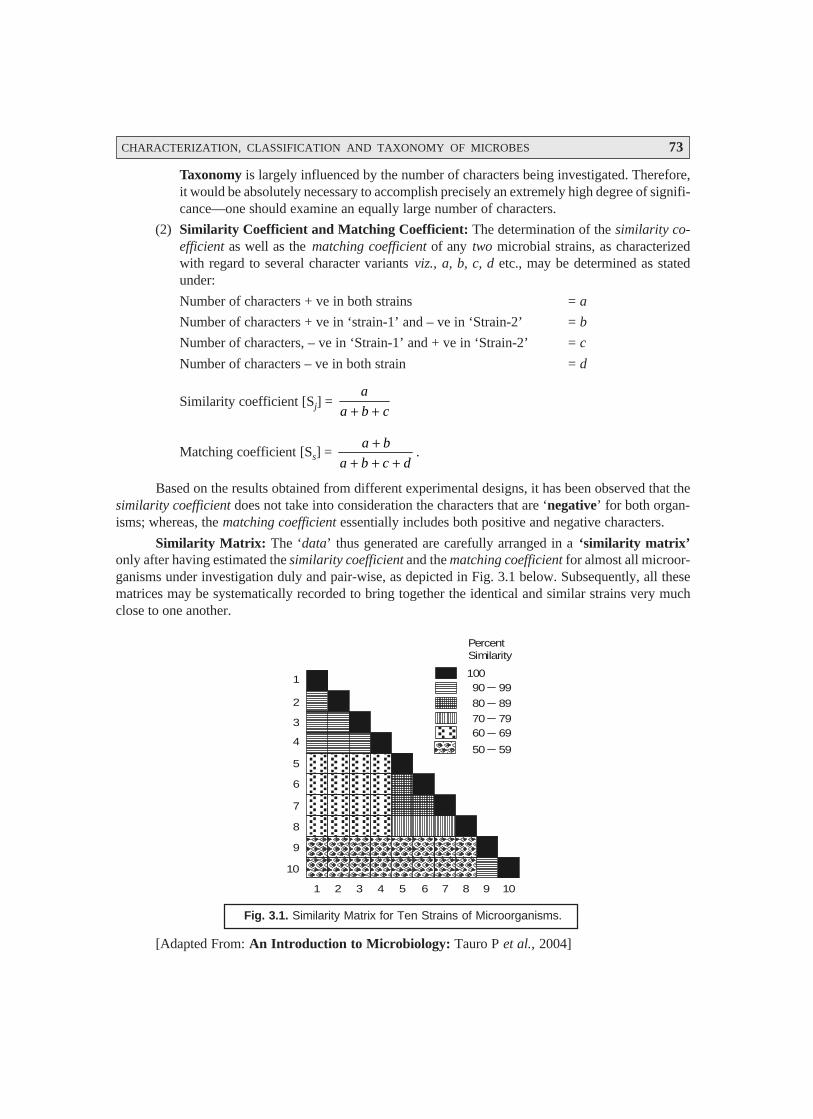
CHARACTERIZATION, CLASSIFICATION AND TAXONOMY OF MICROBES 73
Taxonomy is largely influenced by the number of characters being investigated. Therefore,it would be absolutely necessary to accomplish precisely an extremely high degree of signifi-cance—one should examine an equally large number of characters.
(2) Similarity Coefficient and Matching Coefficient: The determination of the similarity co-efficient as well as the matching coefficient of any two microbial strains, as characterizedwith regard to several character variants viz., a, b, c, d etc., may be determined as statedunder:
Number of characters + ve in both strains = a
Number of characters + ve in ‘strain-1’ and – ve in ‘Strain-2’ = b
Number of characters, – ve in ‘Strain-1’ and + ve in ‘Strain-2’ = c
Number of characters – ve in both strain = d
Similarity coefficient [Sj] = a
a b c+ +
Matching coefficient [Ss] = a b
a b c d
++ + +
.
Based on the results obtained from different experimental designs, it has been observed that thesimilarity coefficient does not take into consideration the characters that are ‘negative’ for both organ-isms; whereas, the matching coefficient essentially includes both positive and negative characters.
Similarity Matrix: The ‘data’ thus generated are carefully arranged in a ‘similarity matrix’only after having estimated the similarity coefficient and the matching coefficient for almost all microor-ganisms under investigation duly and pair-wise, as depicted in Fig. 3.1 below. Subsequently, all thesematrices may be systematically recorded to bring together the identical and similar strains very muchclose to one another.
1
2
3
4
5
6
7
8
9
10
1 2 3 4 5 6 7 8 9 10
PercentSimilarity
10090 99
80 89
70 7960 69
50 59
Fig. 3.1. Similarity Matrix for Ten Strains of Microorganisms.
[Adapted From: An Introduction to Microbiology: Tauro P et al., 2004]
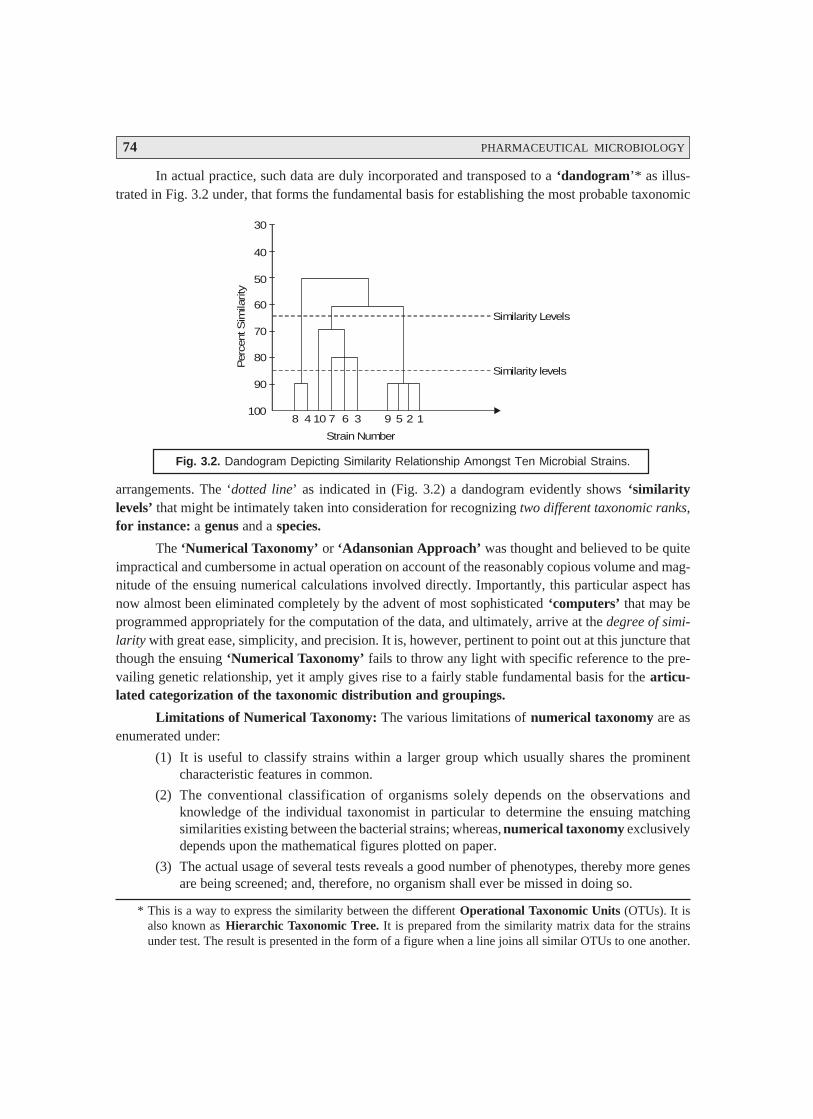
74 PHARMACEUTICAL MICROBIOLOGY
In actual practice, such data are duly incorporated and transposed to a ‘dandogram’* as illus-trated in Fig. 3.2 under, that forms the fundamental basis for establishing the most probable taxonomic
30
40
50
60
70
80
90
1008 4 10 7 6 3 9 5 2 1
Similarity Levels
Similarity levels
Strain Number
Per
cent
Sim
ilarit
y
Fig. 3.2. Dandogram Depicting Similarity Relationship Amongst Ten Microbial Strains.
arrangements. The ‘dotted line’ as indicated in (Fig. 3.2) a dandogram evidently shows ‘similaritylevels’ that might be intimately taken into consideration for recognizing two different taxonomic ranks,for instance: a genus and a species.
The ‘Numerical Taxonomy’ or ‘Adansonian Approach’ was thought and believed to be quiteimpractical and cumbersome in actual operation on account of the reasonably copious volume and mag-nitude of the ensuing numerical calculations involved directly. Importantly, this particular aspect hasnow almost been eliminated completely by the advent of most sophisticated ‘computers’ that may beprogrammed appropriately for the computation of the data, and ultimately, arrive at the degree of simi-larity with great ease, simplicity, and precision. It is, however, pertinent to point out at this juncture thatthough the ensuing ‘Numerical Taxonomy’ fails to throw any light with specific reference to the pre-vailing genetic relationship, yet it amply gives rise to a fairly stable fundamental basis for the articu-lated categorization of the taxonomic distribution and groupings.
Limitations of Numerical Taxonomy: The various limitations of numerical taxonomy are asenumerated under:
(1) It is useful to classify strains within a larger group which usually shares the prominentcharacteristic features in common.
(2) The conventional classification of organisms solely depends on the observations andknowledge of the individual taxonomist in particular to determine the ensuing matchingsimilarities existing between the bacterial strains; whereas, numerical taxonomy exclusivelydepends upon the mathematical figures plotted on paper.
(3) The actual usage of several tests reveals a good number of phenotypes, thereby more genesare being screened; and, therefore, no organism shall ever be missed in doing so.
* This is a way to express the similarity between the different Operational Taxonomic Units (OTUs). It isalso known as Hierarchic Taxonomic Tree. It is prepared from the similarity matrix data for the strainsunder test. The result is presented in the form of a figure when a line joins all similar OTUs to one another.
CHARACTERIZATION, CLASSIFICATION AND TAXONOMY OF MICROBES 75
(4) One major limitation of the numerical analysis is that in some instances, a specific strainmay be grouped with a group of strains in accordance to the majority of identical characteristicfeatures, but certainly not to all the prevailing characters. However, simultaneously the particu-lar strain may possess a very low ebb of similarity with certain other members of the cluster.
(5) The exact location of the taxon is not yet decided, and hence cannot be grouped or related toany particular taxonomic group, for instance : genes or species.
(6) Evidently, in the numerical analysis, the definition of a species is not acceptable as yet,whereas some surveys do ascertain that a 65% single-linkage cluster distincly provides a75% approximate idea of the specific species.
3.3.4. Systematized Classification
After having studied the various aspects of characterization of microbes followed by the prelimi-nary discussions on certain important features related to their classification, one may now have an ex-plicit broader vision on the systematized classification. An extensive and intensive survey of literaturewould reveal that the microorganisms may be classified in a systematized manner under the followingeight categories, namely:
(i) Natural classification,
(ii) Phyletic classification,(iii) Linnean binomial scheme,(iv) Phenotypic classification,(v) Microscopic examination,
(vi) Cataloguing rRNA,(vii) Computer-aided classification, and
(viii) Bacterial classification (Bergey’s Manual of Systematic Bacteriology).
The aforesaid eight categories in the systematized classification of microorganisms would nowbe dealt with individually in the sections that follows.
3.3.4.1. Natural Classification
The natural classification may be considered as one of the most desirable classification systemswhich is broadly based upon the anatomical characteristic features of the specific microorganisms. Inactual practice, the natural classification predominantly helps to organize and arrange the wide spec-trum of organisms into various categories (or groups) whose members do share several characteristics,and reflects to the greatest extent the intricate and complex biological nature of organisms. In reality, aplethora of taxonomists have concertedly opined that a larger segment of the so called natural classifi-cation is importantly and essentially the one having the maximum informations incorporated into it orthe emanated predicted values obtained thereof.
3.3.4.2. Phyletic* Classification
Phyletic classification usually refers to the evolutionary development of a species. Based uponthe most spectacular and master piece publication of Darwin’s—On the Origin of Species (1859),microbiologists across the globe started making an attempt much to sincere and vigorous, so as todevelop phyletic (or phylogenetic) classification systems. Interestingly, the present system serves
* Phyletic [Syn : Phylogentic] : Concerning the development of a phylum.
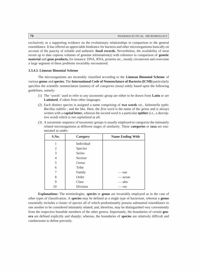
76 PHARMACEUTICAL MICROBIOLOGY
exclusively as a supporting evidence on the evolutionary relationships in comparison to the generalresemblance. It has offered an appreciable hindrance for bacteria and other microorganisms basically onaccount of the paucity of reliable and authentic fossil records. Nevertheless, the availability of mostrecent up to date copious volumes of genuine information(s) with reference to comparison of geneticmaterial and gene products, for instance: DNA, RNA, proteins etc., mostly circumvent and overcomea large segment of these problems invariably encountered.
3.3.4.3. Linnean Binomial Scheme
The microorganisms are invariably classified according to the Linnean Binomial Scheme ofvarious genus and species. The International Code of Nomenclature of Bacteria (ICNB) particularlyspecifies the scientific nomenclature (names) of all categories (taxa) solely based upon the followingguidelines, namely:
(1) The ‘words’ used to refer to any taxonomic group are either to be drawn from Latin or areLatinized, if taken from other languages.
(2) Each distinct species is assigned a name comprising of two words viz., Salmonella typhi;Bacillus subtilis ; and the like. Here, the first word is the name of the genus and is alwayswritten with a capital letter, whereas the second word is a particular epithet (i.e., a descrip-tive word) which is not capitalized at all.
(3) A taxonomic sequence of taxonomic groups is usually employed to categorize the intimatelyrelated microorganisms at different stages of similarity. These categories or taxa are enu-merated as under:
S.No. Category Name Ending With
1 Individual2 Species3 Series
4 Section5 Genus6 Tribe7 Family — eae
8 Order — aceae9 Class — ales
10 Division — ces
Explanations: The terminologies, species or genus are invariably employed as in the case ofother types of classification. A species may be defined as a single type of bacterium, whereas a genusessentially includes a cluster of species all of which predominantly possess substantial resemblance toone another to be considered intimately related; and, therefore, may be distinguished very convenientlyfrom the respective bonafide members of the other genera. Importantly, the boundaries of certain gen-era are defined explicitly and sharply; whereas, the boundaries of species are relatively difficult andcumbersome to define precisely.
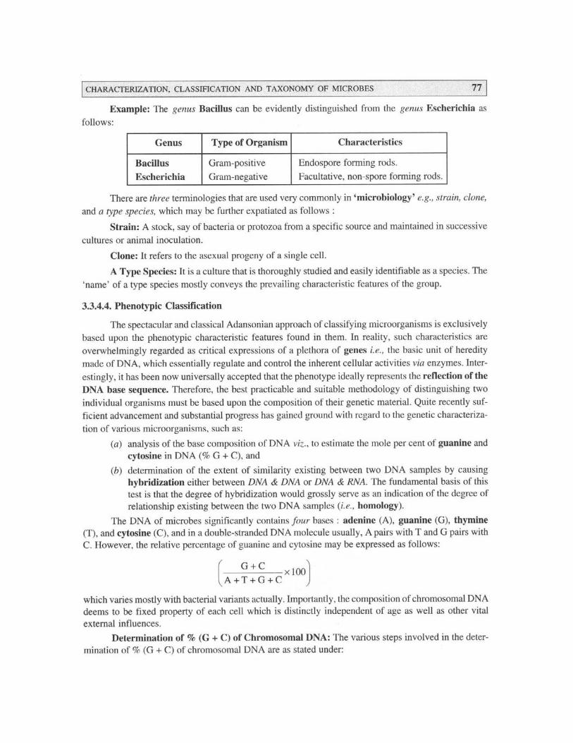
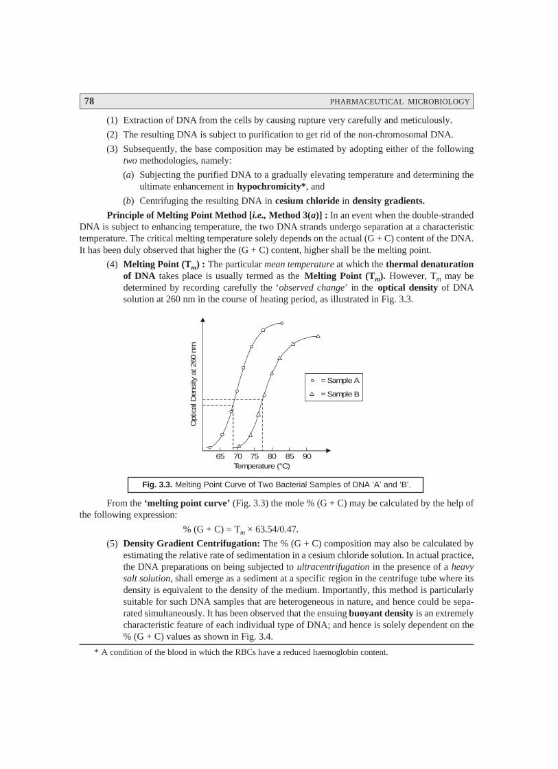
78 PHARMACEUTICAL MICROBIOLOGY
(1) Extraction of DNA from the cells by causing rupture very carefully and meticulously.
(2) The resulting DNA is subject to purification to get rid of the non-chromosomal DNA.
(3) Subsequently, the base composition may be estimated by adopting either of the followingtwo methodologies, namely:
(a) Subjecting the purified DNA to a gradually elevating temperature and determining theultimate enhancement in hypochromicity*, and
(b) Centrifuging the resulting DNA in cesium chloride in density gradients.
Principle of Melting Point Method [i.e., Method 3(a)] : In an event when the double-strandedDNA is subject to enhancing temperature, the two DNA strands undergo separation at a characteristictemperature. The critical melting temperature solely depends on the actual (G + C) content of the DNA.It has been duly observed that higher the (G + C) content, higher shall be the melting point.
(4) Melting Point (Tm) : The particular mean temperature at which the thermal denaturationof DNA takes place is usually termed as the Melting Point (Tm). However, Tm may bedetermined by recording carefully the ‘observed change’ in the optical density of DNAsolution at 260 nm in the course of heating period, as illustrated in Fig. 3.3.
= Sample A
= Sample B
65 70 75 80 85 90
Opt
ical
Den
sity
at 2
60 n
m
Temperature (°C)
Fig. 3.3. Melting Point Curve of Two Bacterial Samples of DNA ‘A’ and ‘B’.
From the ‘melting point curve’ (Fig. 3.3) the mole % (G + C) may be calculated by the help ofthe following expression:
% (G + C) = Tm × 63.54/0.47.
(5) Density Gradient Centrifugation: The % (G + C) composition may also be calculated byestimating the relative rate of sedimentation in a cesium chloride solution. In actual practice,the DNA preparations on being subjected to ultracentrifugation in the presence of a heavysalt solution, shall emerge as a sediment at a specific region in the centrifuge tube where itsdensity is equivalent to the density of the medium. Importantly, this method is particularlysuitable for such DNA samples that are heterogeneous in nature, and hence could be sepa-rated simultaneously. It has been observed that the ensuing buoyant density is an extremelycharacteristic feature of each individual type of DNA; and hence is solely dependent on the% (G + C) values as shown in Fig. 3.4.
* A condition of the blood in which the RBCs have a reduced haemoglobin content.
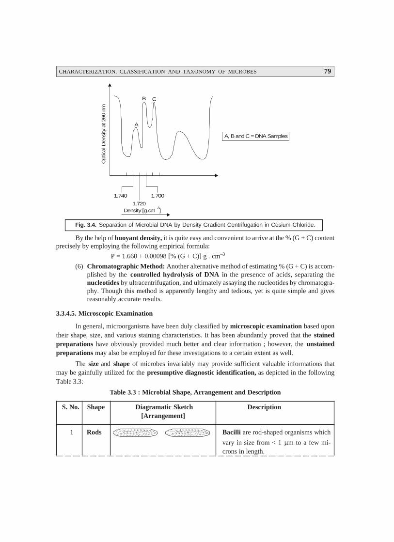
CHARACTERIZATION, CLASSIFICATION AND TAXONOMY OF MICROBES 79
A, B and C = DNA Samples
1.740 1.700
1.720Density [g.cm ]
– 3
Opt
ical
Den
sity
at 2
60 n
m
A
B C
Fig. 3.4. Separation of Microbial DNA by Density Gradient Centrifugation in Cesium Chloride.
By the help of buoyant density, it is quite easy and convenient to arrive at the % (G + C) contentprecisely by employing the following empirical formula:
P = 1.660 + 0.00098 [% (G + C)] g . cm–3
(6) Chromatographic Method: Another alternative method of estimating % (G + C) is accom-plished by the controlled hydrolysis of DNA in the presence of acids, separating thenucleotides by ultracentrifugation, and ultimately assaying the nucleotides by chromatogra-phy. Though this method is apparently lengthy and tedious, yet is quite simple and givesreasonably accurate results.
3.3.4.5. Microscopic Examination
In general, microorganisms have been duly classified by microscopic examination based upontheir shape, size, and various staining characteristics. It has been abundantly proved that the stainedpreparations have obviously provided much better and clear information ; however, the unstainedpreparations may also be employed for these investigations to a certain extent as well.
The size and shape of microbes invariably may provide sufficient valuable informations thatmay be gainfully utilized for the presumptive diagnostic identification, as depicted in the followingTable 3.3:
Table 3.3 : Microbial Shape, Arrangement and Description
S. No. Shape Diagramatic Sketch Description[Arrangement]
1 Rods Bacilli are rod-shaped organisms which
vary in size from < 1 μm to a few mi-crons in length.
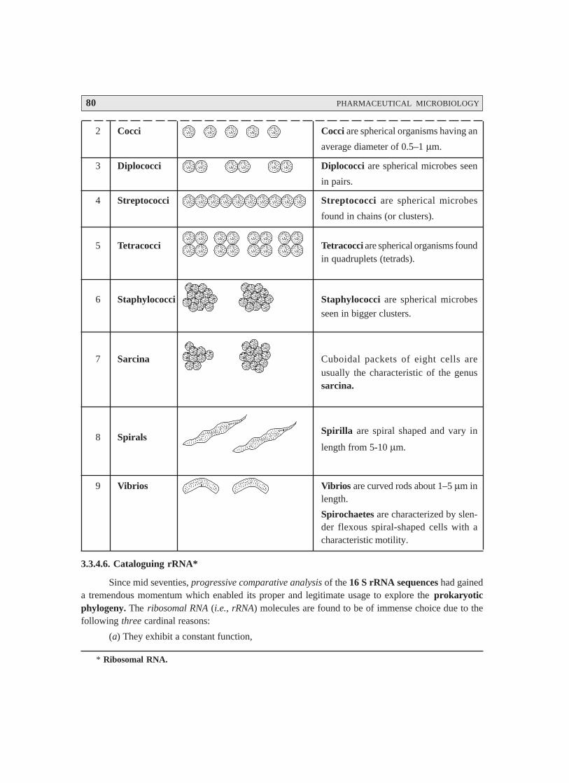
80 PHARMACEUTICAL MICROBIOLOGY
2 Cocci Cocci are spherical organisms having an
average diameter of 0.5–1 μm.
3 Diplococci Diplococci are spherical microbes seen
in pairs.
4 Streptococci Streptococci are spherical microbes
found in chains (or clusters).
5 Tetracocci Tetracocci are spherical organisms foundin quadruplets (tetrads).
6 Staphylococci Staphylococci are spherical microbes
seen in bigger clusters.
7 Sarcina Cuboidal packets of eight cells areusually the characteristic of the genussarcina.
8 SpiralsSpirilla are spiral shaped and vary in
length from 5-10 μm.
9 Vibrios Vibrios are curved rods about 1–5 μm inlength.
Spirochaetes are characterized by slen-der flexous spiral-shaped cells with acharacteristic motility.
3.3.4.6. Cataloguing rRNA*
Since mid seventies, progressive comparative analysis of the 16 S rRNA sequences had gaineda tremendous momentum which enabled its proper and legitimate usage to explore the prokaryoticphylogeny. The ribosomal RNA (i.e., rRNA) molecules are found to be of immense choice due to thefollowing three cardinal reasons:
(a) They exhibit a constant function,
* Ribosomal RNA.
CHARACTERIZATION, CLASSIFICATION AND TAXONOMY OF MICROBES 81
(b) They are universally present in all organisms, and
(c) They seem to have changed in sequence extremely slowly.
Salient Features. The various salient features in cataloguing rRNA are as enumerated under:
(1) 5S rRNA Molecule: Because of its relatively smaller size it has been taken as an accurateindicator of the phylogenetic relationship.
(2) 16S rRNA Molecule: It is sufficiently large ; and, therefore, quite easy to handle with areasonably high degree of precision.
(3) 23S rRNA Molecule: Because of its relatively much larger size it is rather more difficult tocharacterize, and hence used in the comparative analysis.
(4) In the last two decades, the 16 S rRNA has been critically examined, explored, and extractedfrom a large cross-section of microorganisms and duly digested with ribonuclease T1. Theresulting nucleotide are meticulously resolved by 2D-electrophoresis* technique, andsequenced appropriately.
(5) The advent of latest sophisticated instrument e.g., DNA-Probe** which may sequencenucleic acids have further aided in the phenomenon of sequencing of 16 S rRNA frommicroorganisms.
(6) The skilful comparison of rRNA catalogues predominantly designates genealogical rela-tionship existing amongst the wide range of microbes.
(7) The aforesaid genealogical relationship may be suitably quantified in terms of an associa-tion coefficient, designated as SAB, which proves to be a typical characteristic feature for apair of microorganisms. The association coefficient SAB may be expressed as follows :
SAB = AB
A B
2N
N + N
where, NAB = Number of residues existing in sequences common to two rRNA catalogues.
NA and NB = Total number of residues duly represented by oligomers of at least 6 nucleotidesin catalogues A and B respectively.
(8) As to date, the rRNA sequences of more than 200 species of microbes and eukaryotes havebeen duly characterized and documented adequately.
(9) It has been observed that most of the microorganisms strategically give rise to a coherent butalso a very large segment including the eubacteria. Importantly, the methanogens, halophiles,and thermoacidophiles do not necessarily fall within the domain of eubacteria***.
(10) The aforesaid kind of rRNA sequencing has in fact duly permitted the methodical and logi-cal characterization of archaeobacteria.
3.3.4.7. Computer Aided Classification
In the latest spectacular and astronomical growth in the field of computer technology, it hasinducted a tremendous impetus and great help in the proper grouping of microorganisms, and eventually
* Two dimensional electrophoresis.
** A single-strand DNA fragment used to detect the complementary fragment.
*** A genera of bacteria of the order Eubacteriales (i.e., an order of bacteria that includes many of the microor-ganisms pathogenic to humans).
82 PHARMACEUTICAL MICROBIOLOGY
* It named the bacteria which is further divided into nineteen parts, and each of which is distinguishable by afew readily determinable criteria.
classifying them with an utmost accuracy and precision. One may come across a host of problems incomparing a relatively huge number of characteristic features as may be seen in the very instance ofnumerical taxonomy or the Adansonian approach under the perview of the general classification ofmicrobes. In order to circumvent such difficulties and problems, the proper usage of computer-aidedprogrammes and devices have been rightly pressed into service for determining the differentiatingcapacity of the tests and also for determining the overall similarity with the known organisms. As todate, the commendable extremely high speed and memory of computer conveniently allows it toaccomodate very swiftly a host of possible species in the identification/classification phenomenon byjudiciously comparing the characteristic properties of an ‘unknown microorganism’ with those storedduly in the computer. In fact, the advent of the utility of computer, definitely and grossly minimizes theprobability of error in the identification/classification by virtue of either infrequent occurrence of amicroorganism or the critical presence of a rather more frequent microbe with not-so similar or superfi-cial resemblance to other organisms. A good number of highly sophisticated, modern, and advancedcomputer softwares (systems) for microbiology have now been duly developed and put into practiceacross the world profusely. The ‘microbiological laboratories’ strategically attached to most modernhospitals and research and development (R & D) laboratories have gainfully commenced the utiliza-tion of the elaborated computer facilities in the handling/processing of ‘test samples’ to obtain mostreliable, dependable, and reproducible results meant to be used in correct diagnosis and research activi-ties with certainly more confidence and fervour.
3.3.4.8. Bacterial Classification [Bergey’s Manual of Systematic Bacteriology]
Microorganisms represent an exceptionally large conglomerate of minute living body with enor-mous diversity having a procaryotic cellular organization. Several sincere intensive and extensive stud-ies were duly made with particular reference to their broad spectrum physical, structural, and functionalcharacteristic qualities, but none of them could ever produce and evolve an overall satisfactory generallyacceptable classification.
Chester (1899 and 1901) initiated and took active interest in the classification of bacteria, andsubsequently published for the first time—‘The Manual of Determinative Bacteriology’. The saidmanual was painstakingly and meticulously revised, substantiated, and modified by David Hendrick’sBergey (1923) and entitled as—‘Bergey’s Manual of Systematic Bacteriology’, later on commonlytermed as ‘Bergey’s Manual’. In fact, Bergey’s Manual is being recognized as the ‘official compen-dium of all identified and classified bacteria, and serves as an indispensable and valuable guide to themicrobiologists across the globe.
The latest edition of ‘Bergey’s Manual’—(1994) provides a more rational and emperical ap-proach for the classification of bacteria. Besides, it gives rise to an effective system of keys for establish-ing the precise genetic position of an unknown organism. Table 3.4 gives a comprehensive account ofthe classification of bacteria (Division II)* upto the generic level.
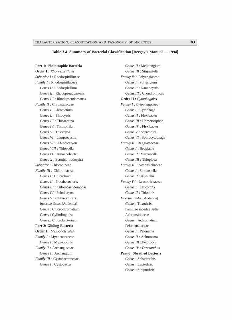
CHARACTERIZATION, CLASSIFICATION AND TAXONOMY OF MICROBES 83
Table 3.4. Summary of Bacterial Classification [Bergey’s Manual — 1994]
Part-1: Phototrophic Bacteria
Order I : Rhodospirillales
Suborder I : Rhodospirillineae
Family I : Rhodospirillaceae
Genus I : Rhodospirillum
Genus II : Rhodopseudomonas
Genus III : Rhodopseudomonas
Family II : Chromatiaceae
Genus I : Chromatium
Genus II : Thiocystis
Genus III : Thiosarcina
Genus IV : Thiospirilum
Genus V : Thiocapsa
Genus VI : Lamprocystis
Genus VII : Thiodicatyon
Genus VIII : Thiopedia
Genus IX : Amoebobacter
Genus X : Ectothiorhodospira
Suborder : Chlorobineae
Family III : Chlorobiaceae
Genus I : Chlorobium
Genus II : Prosthecocloris
Genus III : Chloropseudomonas
Genus IV : Pelodictyon
Genus V : Clathrochloris
Incertae Sedis [Addenda]
Genus : Chlorochromatium
Genus : Cylindrogloea
Genus : Chlorobacterium
Part-2: Gliding Bacteria
Order I : Myxobacterales
Family I : Myxococcaceae
Genus I : Myxococcus
Family II : Archangiaceae
Genus I : Archangium
Family III : Cystobacteraceae
Genus I : Cystobacter
Genus II : Melittangium
Genus III : Stigmatella
Family IV : Polyangiaceae
Genus I : Polyangium
Genus II : Nannocystis
Genus III : Chondromyces
Order II : Cytophagales
Family I : Cytophagaceae
Genus I : Cytophaga
Genus II : Flexibacter
Genus III : Herpetosiphon
Genus IV : Flexibacter
Genus V : Saprospira
Genus VI : Sporocytophaga
Family II : Beggiatoaceae
Genus I : Beggiatoa
Genus II : Vitreoscilla
Genus III : Thioploea
Family III : Simonsiellaceae
Genus I : Simonsiella
Genus II : Alysiella
Family IV : Leucotrichaceae
Genus I : Leucothrix
Genus II : Thiothrix
Incertae Sedis [Addenda]
Genus : Toxothrix
Familiae incertae sedis
Achromatiaceae
Genus : Achromatium
Pelonemataceae
Genus I : Pelonema
Genus II : Achronema
Genus III : Peloploca
Genus IV : Desmanthos
Part-3: Sheathed Bacteria
Genus : Sphaerotilus
Genus : Leptothrix
Genus : Streptothrix

84 PHARMACEUTICAL MICROBIOLOGY
Genus : Lieskeela
Genus : Phragmidiothrix
Genus : Crenothrix
Genus : Clonothrix
Part-4: Budding And/Or Appendaged Bacteria
Genus : Hyphomicrobium
Genus : Hyphomonas
Genus : Pedomicrobium
Genus : Caulobacter
Genus : Asticeacaulis
Genus : Ancalomicrobium
Genus : Prosthecomicrobium
Genus : Thiodendron
Genus : Pasteuria
Genus : Blastobacter
Genus : Seliberia
Genus : Gallionella
Genus : Nevskia
Genus : Planctomyces
Genus : Metallogenium
Genus : Caulococcus
Genus : Kusnezonia
Part-5: Spirochaetes
Order I : Spirochaetales
Family I : Spirochaetaceae
Genus I : Spirochaeta
Genus II : Cristispira
Genus III : Treponema
Genus IV : Borrelia
Genus V : Leptospira
Part-6: Spiral And Curved Bacteria
Family I : Spirillaceae
Genus I : Spirillum
Genus II : Campylobacter
Incertae Sedis [Addenda]
Genus : Bdellovibrio
Genus : Microcyclus
Genus : Pelosigma
Genus : Brachyarcus
Part-7: Gram-Negative Aerobic Rods And Cocci
Family I : Pseudomonadaceae
Genus I : Pseudomonas
Genus II : Xanthomonas
Genus III : Zoogloea
Genus IV : Gluconobacter
Family II : Azotobacteraceae
Genus I : Azotobacter
Genus II : Azomonas
Genus III : Beijerinckia
Genus IV : Derxia
Family III : Rhizobiaceae
Genus I : Rhizobium
Genus II : Agrobacterium
Family IV : Methylomonadaceae
Genus I : Methylomonas
Genus II : Methylococcus
Family V : Halobacteriaceae
Genus I : Halobacterium
Genus II : Halococcus
Incertae Sedis [Addenda]
Genus : Alcaligenes
Genus : Acetobacter
Genus : Brucella
Genus : Bordetella
Genus : Francisella
Genus : Thermus
Part-8: Gram-Negative Facultatively AnaerobicRods
Family I : Enterobacteriaceae
Genus I : Escherichia
Genus II : Edwardsiella
Genus III : Citrobacter
Genus IV : Salmonella
Genus V : Shigella
Genus VI : Klebsiella
Genus VII : Enterobacter
Genus VIII : Hafnia
Genus IX : Serratia
CHARACTERIZATION, CLASSIFICATION AND TAXONOMY OF MICROBES 85
Genus X : Proteus
Genus XI : Yersinia
Genus XII : Erwinia
Family II : Vibrionaceae
Genus I : Vibrio
Genus II : Acromonas
Genus III : Plesiomonas
Genus IV : Photobacterium
Genus V : Lucibacterium
Incertae Sedis [Addenda]
Genus : Chromobacterium
Genus : Zymomonas
Genus : Flavobacterium
Genus : Haemophilus
Genus : Pasteurella
Genus : Actinobacillus
Genus : Cardiobacterium
Genus : Streptobacillus
Genus : Calymmatobacterium
Part-9: Gram-Negative Anaerobic Bacteria
Family I : Bacteriodaceae
Genus I : Bacteroides
Genus II : Fusobacterium
Genus III : Leptotrichia
Incertae Sedis [Addenda]
Genus : Desulfovibrio
Genus : Butyrivibrio
Genus : Succinivibrio
Genus : Succinimonas
Genus : Lachnospira
Genus : Selenomonas
Part-10: Gram-Negative Cocci And Coccobacilli
Family I : Neisseriaceae
Genus I : Neisseria
Genus II : Branhamella
Genus III : Moraxella
Genus IV : Acinetobacter
Incertae Sedis [Addenda]
Genus : Paracoccus
Genus : Lampropedia
Part-11: Gram-Negative Anaerobic Cocci
Family I : Veillonellaceae
Genus I : Acidaminococcus
Genus II : Veillonella
Genus III : Megasphaera
Part-12: Gram-Negative ChemolithotrophicBacteria
Family 1 : Nitrobacteraceae
Genus I : Nitrobacter
Genus II : Nitrospina
Genus III : Nitrococcus
Genus IV : Nitrosomonas
Genus V : Nitrospira
Genus VI : Nitrosococcus
Genus VII : Nitrosolobus
Organisms Metabolizing Sulphur
Genus I : Thiobacillus
Genus II : Sulfolobus
Genus III : Thiobacterium
Genus IV : Macromonas
Genus V : Thiovulum
Genus VI : Thiospira
Family II : Siderocapsaceae
Genus I : Siderocapsa
Genus II : Naumanniella
Genus III : Ochrobium
Genus IV : Siderococcus
Part-13: Methane Producing Bacteria
Family I : Methanobacteriaceae
Genus I : Methanobacterium
Genus II : Methanosarcina
Genus III : Methanococcus
Part-14: Gram Positive Cocci
Family I : Micrococcaceae
Genus I : Micrococcus
Genus II : Staphylococcus
Genus III : Planococcus
Family II : Streptococcaceae
Genus I : Streptococcus
Genus II : Leuconostoc
86 PHARMACEUTICAL MICROBIOLOGY
Genus III : Pediococcus
Genus IV : Acrococcus
Genus V : Gemella
Family III : Peptococcaceae
Genus I : Peptococcus
Genus II : Peptostreptococcus
Genus III : Ruminococcus
Genus IV : Sarcina
Part-15: Endospore Forming Rods and Cocci
Family I : Bacillaceae
Genus I : Bacillus
Genus II : Sporolactobacillus
Genus III : Clostridium
Genus IV : Desulfotomaculum
Genus V : Sporosarcina
Incertae Sedis [Addenda]
Genus : Oscillospira
Part-16: Gram-Positive Asporogenous Rod-Shaped Bacteria
Family I : Lactobacillaceae
Genus I : Lactobacillus
Incertae Sedis [Addenda]
Genus : Listeria
Genus : Erysipelothrix
Genus : Caryophanon
Part-17: Actinomycetes And Related Organisms
Coryneform Group of Bacteria
Genus I : Corynebacterium
Genus II : Arthrobacter
Incertae Sedis [Addenda]
Genus A : Brevibacterium
Genus B : Microbacterium
Genus III : Cellulomonas
Genus IV : Kurthia
Family I : Propionibacteriaceae
Genus I : Propionibacterium
Genus II : Eubacterium
Order I : Actinomycetales
Family I : Actinomycetaceae
Genus I : Actinomyces
Genus II : Arachnia
Genus III : Bifidobacterium
Genus IV : Bacterionema
Genus V : Rothia
Family II : Mycobacteriaceae
Genus I : Mycobacterium
Family III : Frankiaceae
Genus I : Frankia
Family IV : Actinoplanaceae
Genus I : Actinoplanes
Genus II : Spirillospora
Genus III : Streptosporangium
Genus IV : Amorphosphorangium
Genus V : Ampullariella
Genus VI : Pilimelia
Genus VII : Planomonospora
Genus VIII : Planobispora
Genus IX : Dactylosporangium
Genus X : Kitastoa
Family V : Dermatophillaceae
Genus I : Dermatophilus
Genus II : Geodermatophilus
Family VI : Nocardiaceae
Genus I : Nocardia
Genus II : Pseudonocardia
Family VII : Streptomycetaceae
Genus I : Streptomyces
Genus II : Streptoverticilium
Genus III : Sporichthya
Genus IV : Microellobosporia
Family VIII : Micromonosporaceae
Genus I : Micromonospora
Genus II : Thermoactinomyces
Genus III : Actinobifida
Genus IV : Thermonospora
Genus V : Microbispora
Gebus VI : Micropolyspora
Part-18: Rickettsias
Order I : Rickettsiales
Family : Rickettsiaceae
Tribe I : Rickettsieae
Genus I : Rickettsia

CHARACTERIZATION, CLASSIFICATION AND TAXONOMY OF MICROBES 87
Genus II : Rochalimaea
Genus III : Coxiella
Tribe II : Ehrlichieae
Genus IV : Ehrlichia
Genus V : Cowdria
Genus VI : Neorickettsia
Tribe III : Wolbachieae
Genus VII : Wolbachia
Genus VIII : Symbiotes
Genus IX : Blattabacterium
Genus X : Rickettsiella
Family : Bartonellaceae
Genus I : Bartonella
Genus II : Grahamella
Family : Anaplasmataceae
Genus I : Anaplasma
Genus II : Paranaplasma
Genus III : Aegyptionella
Genus IV : Haemobartonella
Genus V : Eperythrozoon
Order II : Chlamydiales
Family I : Chlamydiaceae
Genus I : Chlamydia
Part-19 : Mycoplasmas
Class Mollicutes
Order I : Mycoplasmatales
Family I : Mycoplasmataceae
Genus I : Mycoplasma
Family II : Acholeplasmataceae
Genus I : Acholeplasma
Incertae Sedis [Addenda]
Genus : Thermoplasma
Incertae Sedis [Addenda]
Genus : Spiroplasma
3.4. TAXONOMY
Taxonomy (Greek : taxis = arrangement or order), and nomos = law, or nemein = to distribute orgovern) refers to the science or discipline that essentially deals with the logical arrangement ofliving things into categories. It may also be defined as ‘the laws and principles of classification of livingorganisms’.
Aristotle—in fact, was the first ever taxonomist in the fourth century BC who painstakingly andmeticulously categorized the so-called ‘living objects’ in the universe into almost 500 well definedspecies of plant and animal kingdoms.
Carolus Linnaeus (1735 – 1759) — a renowned Swedish botanist, virtually named a relativelymuch larger segment of plants and animals and classified them with great skill and wisdom into the twopredominant kingdoms, namely : Plantae and Animalia. In reality, Carolus was instrumental in devis-ing the unique ‘Binomial Scheme of Nomenclature’.
Ernst H Haeckel — in the year 1866 logistically segregated the ‘microorganisms’ from theexisting plant and animal kingdoms. It was Ernst who first and foremost introduced the new terminologyProtist exclusively reserved for the microorganisms. He subsequently coined another term Protista tospecifically and categorically include algae (microscopic), fungi, and protozoa thereby forming a ‘thirdkingdom’.
Comments : (1) There was disapproval with regard to the inclusion of both bacteria and fungitogether in the aforesaid kingdom Protista.
(2) Bearing in mind the recent advances in the domain of ‘Cell Biology’, profuse ob-jections were raised pertaining to the two or three kingdom classification schemes asencountered in Protista.
Robert H Whittaker (1969) — duly put forward a most scientific, plausible, and logical systemof classification of the living organisms which was widely accepted by the modern microbiologists

88 PHARMACEUTICAL MICROBIOLOGY
across the world. However, Robert’s system articulately recognizes the five kingdoms applicable to allliving things, namely: Monera, Protista, Fungi, Animalia, and Plantae.
Monera — predominantly includes bacteria and cyanobacteria.
Protista — essentially comprises of eukaryotes and protozoa.
Fungi — specifically belongs to the organisms attached to the kingdom of fungi.
Animalia and Plantae — particularly include the traditional animals and plants.
It is, however, pertinent to mention here some of the main terminologies, one may frequentlycome across in the proper and elaborated description of the taxonomy of microorganisms, such as:(a) Species – i.e., the fundamental rank in the classification system; (b) genus – i.e., clubbing togetherof two or more species ; (c) family – i.e., the collection of genera; (d) order – i.e., the collection offamilies with identical characteristic features ; (e) class – i.e., the arranging together of order ; (f) phylum(or division) – i.e., grouping together of classes; and (g) kingdom – i.e., collection of two or morephyla.
Taxon, also known as the basic taxonomic group represents the species i.e., a collection ofstrains with almost similar characteristic features. In usual practice, the microbial species invariablycomprise of a specialized typical strain termed as the type strain, along with all other strains which areregarded very much identical to the type strain so as to justify their logical inclusion in the species. Inother words, the type strain is symbolized and designated to be the permanent reference specimen forthe species. However, it may be stressed that it is not necessarily always the particular strain whichhappens to be most characterwise typical of all the strains strategically included in the species, whereasit is essentially the specific strain to which all the rest of the strains should be critically compared toascertain, whether they do have a close resemblance sufficient enough to belong to the same species.The above glaring statement of facts pertaining to the type strains are extremely vital and important;and, therefore, specialized and particular attention need to be given to their genuine and regular mainte-nance as well as preservation. The following are two world famous reference collection centres locatedin USA and UK, namely:
(a) American Type Culture Collection (ATCC), Rockville, Maryland, USA, and
(b) National Collection of Type Cultures (NCTC), UK.
Interestingly, one may critically observe that the various strains strategically present very muchwithin species may differ slightly from one another in three prominent manners, namely:
(a) Biovars: These are variant bacterial strains and are duly characterized by biochemical orphysiological characteristics.
(b) Morphovars: These are variants within a species defined by variation in morphologicalcharacteristics.
(c) Serovars: These are variants within a species defined by variation in serological reactions.
3.5. THE KINGDOM PROKARYOTAE
It was Haeckel, who first and foremost in the year 1866 vehemently suggested that the microor-ganisms present in the particular kingdom, Protista, should essentially be composed of both theprokaryotes as well as the eukaryotes. Almost a century later, Murray in 1968 unequivocally andstrongly proposed the ‘prokaryotae’ as an extremely specific and overwhelmingly typical taxon at thehighest level to include essentially all microorganisms distinctly characterized by the presence of a

CHARACTERIZATION, CLASSIFICATION AND TAXONOMY OF MICROBES 89
* A fine thread made up of long, interwoven or irregularly placed threadlike structures.
** A resistant cell produced by certain bacteria to withstand harsh environments; usually spores are asexual incharacter.
*** Refer to filaments of mold, or parts of mold mycelium.
**** Having a dividing wall.
***** Resembling a nucleus.
definitive nucleoplasm free from both the fundamental proteins as well as the nuclear membrane. Inter-estingly, the ‘eukaryotes’ are invariably designated as a possible taxon occurring almost at the samehighest level so as to include other protists, plants, and animals. Ultimately, Allsopp commanded thatthe aforesaid two taxon variants be christened as kingdoms, Prokaryotae and Eukaryotae.
The following members from the ‘Kingdom Prokaryotae’, namely:
(a) Actinomycetes
(b) Bacteria
(c) Rickettsia and Coxiella, and
(d) Spirochaetes
shall be discussed in an elaborated manner in the sections that follows.
3.5.1. Actinomycetes
The Actinomycetes [s., actinomycete], according to the latest edition of Bergey’s Manual(Volume 4), represent an aerobic, Gram-positive bacteria which predominantly and essentially give riseto specific branching filaments* or asexual spores** or hyphae***. It has been duly observed that theelaborated morphology, arrangement of spores, explicit cell-wall chemistry, and above all the variouskinds of carbohydrates critically present in the cell extracts are specifically vital and equally importantrequirement for the exhaustive taxonomy of the actinomycetes. Consequently, these informations areutilized meticulously to carry out the articulated division of these bacteria into different well-definedcategories with great ease and fervour. It is quite pertinent to state at this juncture, that the actinomycetesdo possess and exert an appreciable practical impact by virtue of the fact that they invariably play anapparent major role in the following two highly specialized and particular aspects, namely:
(a) Mineralization of organic matter in the soil, and
(b) Primary source of most naturally synthesized antibiotics.
3.5.1.1. General Characteristics
The general characteristics of the actinomycetes are as stated under :
(a) The branching network of hyphae usually developed by the actinomycetes, grows criticallyboth on the surface of the solid substratum (e.g., agar) as well as into it to give rise to theformation of substrate mycelium. However, the septate**** mostly divide the hyphae intospecific elongated cells (viz., 20 μm and even longer) essentially consisting of a plethora ofnucleoids*****.
(b) Invariably, the actinomycetes afford the development of thallus. Noticeably, a large cross-
section of the actinomycetes do possess an aerial mycelium that extends above the solid
90 PHARMACEUTICAL MICROBIOLOGY
subtratum, and produces articulately asexual, thin-walled spores known as conidia[s., conidium] or conidiospores at the terminal ends of filaments. In an event, when thespores are located strategically in a sporangium, they are termed as sporangiospores.
(c) The spores present in the actinomycetes not only vary widely in terms of shape and size, butalso develop them (spores) by the help of septal formation at the tips of the filaments, invari-
ably in response to nutrient deprivation. Besides, a larger segment of these spores are
specifically devoid of any thermal resistance; however, they do withstand dessication quite
satisfactorily, and thus exhibit considerable adaptive value.
(d) Generally, most actinomycetes are not found to be motile,* and the motility is particularly
confined to the flagellated spores exclusively.
In the recent past, several taxonomically characteristic features and useful techniques are of
immense value and worth, such as:
• Morphological features and the colour of mycelia and sporangia
• Surface properties and arrangement of conidiospores
• % (G + C) in DNA
• Phospholipid content and composition of cell membranes
• Thermal resistance encountered in spores
• Comparison of 16S rRNA sequences and their values
• Production of relatively larger DNA fragments by means of restriction enzyme digestion,
and
• Ultimate separation and comparison of ‘larger DNA fragments’ by the aid of Pulsed FieldElectrophoresis.
3.5.1.2. Significance of Actinomycetes
There are, in actual practice, three most important practical significances of the actinomycetes,as mentioned below:
(1) Actinomycetes are predominantly the inhabitants of soil and are distributed widely.
(2) They are able to degrade a large variety and an enormous quantum of organic chemical
entities. However, these are of immense significance in the mineralization of organic matter.
(3) They invariably and critically give rise to a large excess of extremely vital ‘natural antibi-otics’ that are used extensively in the therapeutic armamentarium e.g., actinomycetin. Im-
portantly, a plethora of actinomycetes represent free-living microbes, whereas a few arepathogens to human beings, animals, and even certain plants.
Fig. 3.5. illustrates the cross-section of an actinomycete colony with living and dead hyphae.The substrate and aerial mycelium having chains of conidiospores have been depicted evidently.
* Having spontaneous movement.
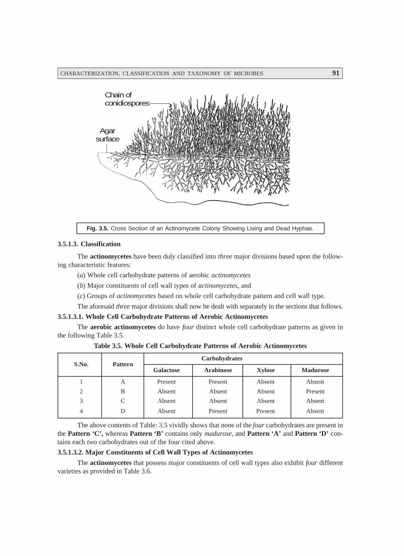
CHARACTERIZATION, CLASSIFICATION AND TAXONOMY OF MICROBES 91
Fig. 3.5. Cross Section of an Actinomycete Colony Showing Living and Dead Hyphae.
3.5.1.3. Classification
The actinomycetes have been duly classified into three major divisions based upon the follow-ing characteristic features:
(a) Whole cell carbohydrate patterns of aerobic actinomycetes
(b) Major constituents of cell wall types of actinomycetes, and
(c) Groups of actinomycetes based on whole cell carbohydrate pattern and cell wall type.
The aforesaid three major divisions shall now be dealt with separately in the sections that follows.
3.5.1.3.1. Whole Cell Carbohydrate Patterns of Aerobic Actinomycetes
The aerobic actinomycetes do have four distinct whole cell carbohydrate patterns as given inthe following Table 3.5.
Table 3.5. Whole Cell Carbohydrate Patterns of Aerobic Actinomycetes
CarbohydratesS.No. Pattern
Galactose Arabinose Xylose Madurose
1 A Present Present Absent Absent
2 B Absent Absent Absent Present
3 C Absent Absent Absent Absent
4 D Absent Present Present Absent
The above contents of Table: 3.5 vividly shows that none of the four carbohydrates are present inthe Pattern ‘C’, whereas Pattern ‘B’ contains only madurose, and Pattern ‘A’ and Pattern ‘D’ con-tains each two carbohydrates out of the four cited above.
3.5.1.3.2. Major Constituents of Cell Wall Types of Actinomycetes
The actinomycetes that possess major constituents of cell wall types also exhibit four differentvarieties as provided in Table 3.6.
Chain ofconidiospores
Agarsurface
92 PHARMACEUTICAL MICROBIOLOGY
Table 3.6. Major Constituents of Cell Wall Types of Actinomycetes
DAP* Carbohydrates/Amino AcidS.No. Cell Wall Type
meso– LL– Arabinose Galactose Glycine
1 I Absent Present Absent Absent Present
2 II Present Absent Absent Absent Present
3 III Present Absent Absent Absent Absent
4 IV Present Absent Present Present Absent
3.5.1.3.3. Groups of Actinomycetes Based on Whole Cell Carbohydrate Pattern and Cell Wall Type
There are in all five different varieties of cell wall types having carbohydrate and genera vari-ants in groups of actinomycetes, as given in Table 3.7 under :
Table 3.7. Actinomycetes Based on Whole Cell Carbohydrate Pattern and Cell Wall Type
S.No. Cell Wall Type Carbohydrate Pattern Genera
1 I Characteristic feature Microellobosporia; Sporichthya; Streptomyces;absent Streptoverticillium;
2 II A Actinoplanes; Amorphosporangium ; Ampulla-riella ; Dactylosporangium; Micromonospora;
3 III B Actinobifida ; Geodermatophilus ; Thermo-actinomyces ;
4 IV C Dermatophilus ; Microbispora ; Nocardiama-durae type (Actinomadura) ; Spirillospora ;Streptosporangium ;
5 V D Mycobacterium ; Nocardia ; Micropolyspora ;Pseudonocardia, Thermomonospora ;
One may observe from Table 3.7 that the cell wall type I is devoid of the characteristic featurepertaining to the specific carbohydrate pattern.
3.5.1.3.4. Actinomycetes with Multiocular** Sporangia***
The latest version of Bergey’s Manual has explicitly described the actinomycetes occurring asthe ‘clusters of spores’ in a specific situation when a hypha undergoes division both transversely andlogitudinally. In reality, all the three genera critically present in this section essentially possess chemotypeIII cell walls, whereas the cell extract carbohydrate patterns differ prominently.
Salient Features: The salient features of the actinomycetes with multiocular sporangia are asfollows :
(1) The mole % (G + C) values varies from 57 to 75.
(2) Chemotype III C Cell Walls****: Geodermatophillus belonging to this category has motilespores and is specifically an aerobic soil organism.
* Diaminopimelic acid.
** Having many cells or compartments.
*** Sacs enclosing spores.
**** It is a soil organism.
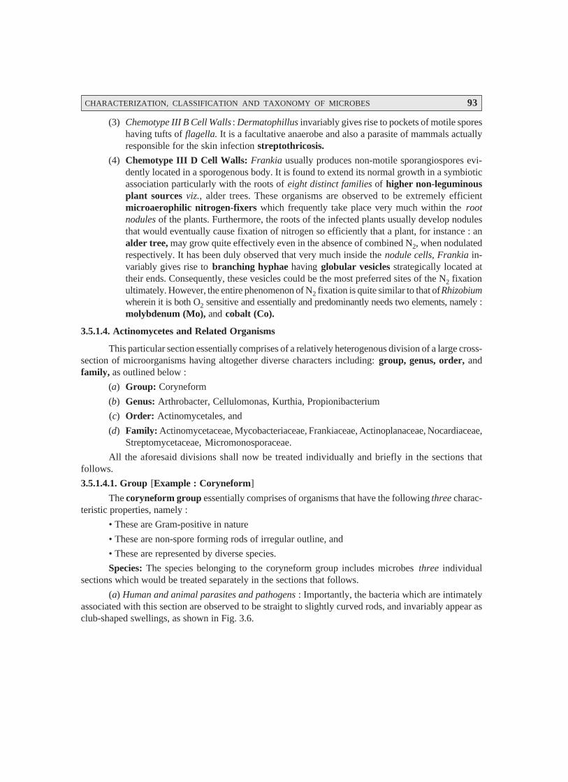
CHARACTERIZATION, CLASSIFICATION AND TAXONOMY OF MICROBES 93
(3) Chemotype III B Cell Walls : Dermatophillus invariably gives rise to pockets of motile sporeshaving tufts of flagella. It is a facultative anaerobe and also a parasite of mammals actuallyresponsible for the skin infection streptothricosis.
(4) Chemotype III D Cell Walls: Frankia usually produces non-motile sporangiospores evi-dently located in a sporogenous body. It is found to extend its normal growth in a symbioticassociation particularly with the roots of eight distinct families of higher non-leguminousplant sources viz., alder trees. These organisms are observed to be extremely efficientmicroaerophilic nitrogen-fixers which frequently take place very much within the rootnodules of the plants. Furthermore, the roots of the infected plants usually develop nodulesthat would eventually cause fixation of nitrogen so efficiently that a plant, for instance : analder tree, may grow quite effectively even in the absence of combined N2, when nodulatedrespectively. It has been duly observed that very much inside the nodule cells, Frankia in-variably gives rise to branching hyphae having globular vesicles strategically located attheir ends. Consequently, these vesicles could be the most preferred sites of the N2 fixationultimately. However, the entire phenomenon of N2 fixation is quite similar to that of Rhizobiumwherein it is both O2 sensitive and essentially and predominantly needs two elements, namely :molybdenum (Mo), and cobalt (Co).
3.5.1.4. Actinomycetes and Related Organisms
This particular section essentially comprises of a relatively heterogenous division of a large cross-section of microorganisms having altogether diverse characters including: group, genus, order, andfamily, as outlined below :
(a) Group: Coryneform
(b) Genus: Arthrobacter, Cellulomonas, Kurthia, Propionibacterium
(c) Order: Actinomycetales, and
(d) Family: Actinomycetaceae, Mycobacteriaceae, Frankiaceae, Actinoplanaceae, Nocardiaceae,Streptomycetaceae, Micromonosporaceae.
All the aforesaid divisions shall now be treated individually and briefly in the sections thatfollows.
3.5.1.4.1. Group [Example : Coryneform]
The coryneform group essentially comprises of organisms that have the following three charac-teristic properties, namely :
• These are Gram-positive in nature
• These are non-spore forming rods of irregular outline, and
• These are represented by diverse species.
Species: The species belonging to the coryneform group includes microbes three individualsections which would be treated separately in the sections that follows.
(a) Human and animal parasites and pathogens : Importantly, the bacteria which are intimatelyassociated with this section are observed to be straight to slightly curved rods, and invariably appear asclub-shaped swellings, as shown in Fig. 3.6.
94 PHARMACEUTICAL MICROBIOLOGY
Fig. 3.6. Coryneform Bacteria in Straight Rods, Slightly Curved Rods,and Club-shaped Swellings.
Salient Features : The salient features of coryneform bacteria are as follows:
(1) They are usually non-motile, Gram-positive, and non-acid fast.
(2) They are mostly chemoorganotrophs, aerobic, and also facultatively anaerobic.
(3) They are widely distributed in nature with % (G + C) values ranging between 52 to 68 molesper cent.
(4) The type species belonging to this class is represented by C. diphtheriae which is particu-larly known to produce a highly lethal exotoxin and causes the dreadly disease in humanscalled diphtheria.
(b) Plant pathogenic corynebacteria: Interestingly, the bacteria belonging to this particular classis closely akin to those present in section (a) above; however, these are essentially characterized by threeprominent features, namely: (i) less pleomorphic, (ii) strictly aerobic in nature, and (iii) possess % (G + C)values ranging between 65–75 moles per cent.
Based on ample scientific evidences, this particular section is further sub-divided into fourcategories, such as: (i) types of polysaccharide antigens, (ii) composition of amino acids present duly incell walls, (iii) minimal nutritional requirements, and (iv) etiology of the disease caused in plants.
(c) Non-pathogenic corynebacteria: This particular section essentially consists of non-pathogeniccorynebacteria quite commonly derived and isolated from soil, water, air, and are invariably described inthe literature very scantily by virtue of their morphological similarities and hence, the virtual scope ofany possible distinct differentiation.
3.5.1.4.2. Genus
The four prominent genus shall be treated individually in the sections that follows:
(a) Arthrobacter: The genus Arthrobacter essentially consists of such organisms that undergo amarked and pronounced change in form particularly in the course of their respective growth on thecomplex media. It has been duly observed that the relatively ‘older cultures’ do comprise of coccoidcells* very much resembling to micrococci in their appearance. In certain specific instances, the cellscould be either spherical to ovoid or slightly elongated. Importantly, when these are carefully trans-ferred to the ‘fresh culture media’, consequently the ultimate growth takes place by two distinct modes,namely : (a) due to swelling, and (b) due to elongation of the coccoid cells, to produce rods thatessentially have a diameter precisely much less in comparison to the corresponding enlarged cells.
* Resembling a micrococcus.

CHARACTERIZATION, CLASSIFICATION AND TAXONOMY OF MICROBES 95
Eventually, there may be predominant ‘outgrowths’ occurring at more than one segment of the cell asdepicted in Fig. 3.7.
Fig. 3.7. Diagramatic Sketch of Arthrobacters
Arthrobacter’s subsequent growth and followed up divisions usually yields irregular rods thatvary appreciably both in size and shape.
Importantly, a small segment of the rods are invariably arranged at an ‘angle’ to each otherthereby causing deformation. However, in richer media, cells may exhibit preliminary (rudimentary)branching, whereas the formation of true mycelia cease to form. Besides, along with the passage of the‘exponential phase’, the rods turn out to be much shorter and get converted to the corresponding coccoidcells. A few other prevalent characteristics are as follows:
• Rods are either non-motile completely or motile by one sub-polar or a few lateral flagella.
• Coccoid cells are Gram-positive in nature, chemoorganotrophic, aerobic soil organisms hav-ing a distinct respiratory metabolism.
• Species present within the genus are invariably categorized and differentiated solely de-pending on the composition of cell wall; hydrolysis of gelatin, starch etc.; and the ultimategrowth-factor requirement.
It is, however, pertinent to state here the two other genera although whose actual and preciseaffiliation is still ‘uncertain’, yet they are quite related to the Arthrobacter, namely: Brevibacterium andMicrobacterium.
(b) Cellulomonas: The genus Cellulomonas essentially comprises of bacteria that have the com-petence and ability to hydrolyse the cellulose particularly.
Salient Features : The various vital and important salient features are as stated below:
(1) The cells usually observed in young cultures are irregular rods having a diameter nearly 0.5 μmand a length ranging either between 0.7 to 2 μm or even slightly in excess.
(2) The appearance of the cells could be straight, slightly curved, or angular or beaded or occa-sionally club-shaped.
(3) Importantly, certain cells may be arranged strategically at an angle to each other as could beobserved in the case of Arthrobacter [see section 5.1.4.2(a)]; besides, they (cells) may infre-quently exhibit rudimentary branching as well.
(4) Older cultures are invariably devoid of ‘true mycelia’ but the ‘coccoid cells’ do predomi-nate in number.
(5) The cells may be Gram-positive to Gram-negative variable, motile to non-motile variable,non acid-fast, aerobic chemo-organotrophos, having an optimum growth temperature at 30°C.
(6) The % (G + C) values ranges between 71.7 to 72.7 moles.
96 PHARMACEUTICAL MICROBIOLOGY
Interestingly, there exists only one species, Cellulomonas flavigenum, which is exclusively knownand recognized; and found commonly in the soil.
(c) Kurthia: The genus Kurthia is specifically characterized by organisms that are prominentlyand rigidly aerobic in nature; besides, they happen to be chemoorganotrophs. Young cultures essen-tially comprise of cells that are mostly unbranched rods having round ends, and occurring as distinctparallel chains. Older cultures normally comprise of coccoid cells that are critically obtained by thefragmentation of rods.
Salient Features: The salient features of the organisms belonging to the genus Kurthia are asgiven under:
(1) The rods are rendered motile by the presence of peritrichous flagella*.
(2) The cells predominantly grow in abundance, particularly in the presence of sodium chloride(NaCl) solution [4 to 6% (w/v)] prepared in sterilized distilled water.
(3) The optimum temperature required for the healthy growth of the cells usually varies between25 to 30°C.
Interestingly, there prevails only one species, Kurthia zoefi, that has been duly recognized anddescribed in the literature.
There are certain characteristic features of the genera Corynebacterium, Arthrobacter,Cellulomonas, and Kurthia that have been duly summarized in Table 3.8.
Table 3.8. Certain Characteristic Features of Coryneform Group of Microorganisms
S.No. Genus % (G+ C) Relationship Motility CelluloseMoles with Oxygen Utilization
1 Arthrobacter 60–72 Strictly aerobic + or – –
2 Cellulomonas 71.7–72.7 Aerobic to facultatively anaerobic + or – +
3 Corynebacterium 57–60 —do— – –
4 Kurthia — Strictly aerobic + –
(d) Propionibacterium: The family Propionibacteriaceae invariably consists microbes that havethe following characteristic features :
(i) They are all Gram-positive, non-spore forming, anaerobic to aerotolerant, pleomorphic,branching or filamentous or regular rods.
(ii) On being subjected to ‘fermentative procedures’ it has been duly observed that the majorend-products ultimately generated are, namely : propionic acid, acetic acid, carbon diox-ide, or a mixture of butyric, formic, lactic together with other monocarboxylic acids.
(iii) Growth: Their normal growth is usually enhanced by the very presence of carbon dioxide,and
(iv) Habitat: These microbes are normally inhabitants of skin, respiratory, and the intestinaltracts of a large cross-section of animals.
A survey of literature would reveal the description of two genera, namely : Propionibacteriumand Eubacterium. These two genera shall now be dealt with briefly and separately in the sections thatfollows:
* Indicating microorganisms that have flagella covering the entire surface of a bacterial cell.
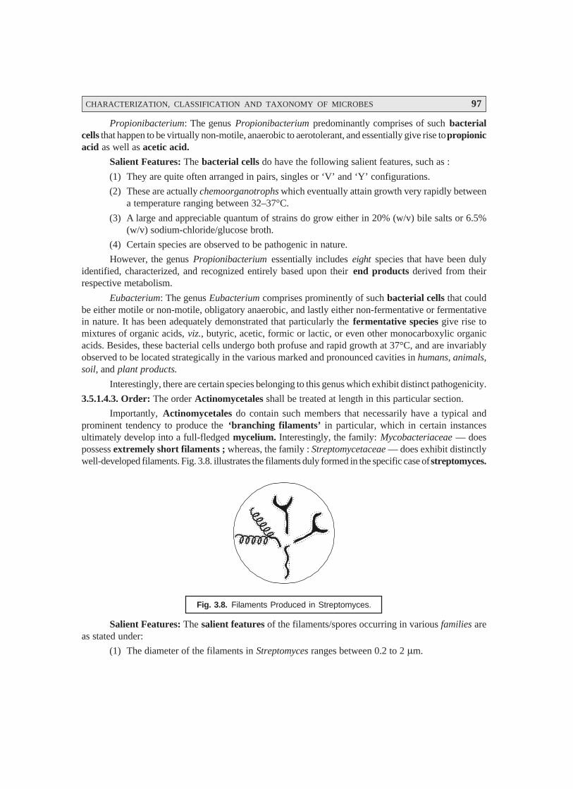
CHARACTERIZATION, CLASSIFICATION AND TAXONOMY OF MICROBES 97
Propionibacterium: The genus Propionibacterium predominantly comprises of such bacterialcells that happen to be virtually non-motile, anaerobic to aerotolerant, and essentially give rise to propionicacid as well as acetic acid.
Salient Features: The bacterial cells do have the following salient features, such as :
(1) They are quite often arranged in pairs, singles or ‘V’ and ‘Y’ configurations.
(2) These are actually chemoorganotrophs which eventually attain growth very rapidly betweena temperature ranging between 32–37°C.
(3) A large and appreciable quantum of strains do grow either in 20% (w/v) bile salts or 6.5%(w/v) sodium-chloride/glucose broth.
(4) Certain species are observed to be pathogenic in nature.
However, the genus Propionibacterium essentially includes eight species that have been dulyidentified, characterized, and recognized entirely based upon their end products derived from theirrespective metabolism.
Eubacterium: The genus Eubacterium comprises prominently of such bacterial cells that couldbe either motile or non-motile, obligatory anaerobic, and lastly either non-fermentative or fermentativein nature. It has been adequately demonstrated that particularly the fermentative species give rise tomixtures of organic acids, viz., butyric, acetic, formic or lactic, or even other monocarboxylic organicacids. Besides, these bacterial cells undergo both profuse and rapid growth at 37°C, and are invariablyobserved to be located strategically in the various marked and pronounced cavities in humans, animals,soil, and plant products.
Interestingly, there are certain species belonging to this genus which exhibit distinct pathogenicity.
3.5.1.4.3. Order: The order Actinomycetales shall be treated at length in this particular section.
Importantly, Actinomycetales do contain such members that necessarily have a typical andprominent tendency to produce the ‘branching filaments’ in particular, which in certain instancesultimately develop into a full-fledged mycelium. Interestingly, the family: Mycobacteriaceae — doespossess extremely short filaments ; whereas, the family : Streptomycetaceae — does exhibit distinctlywell-developed filaments. Fig. 3.8. illustrates the filaments duly formed in the specific case of streptomyces.
Fig. 3.8. Filaments Produced in Streptomyces.
Salient Features: The salient features of the filaments/spores occurring in various families areas stated under:
(1) The diameter of the filaments in Streptomyces ranges between 0.2 to 2 μm.

98 PHARMACEUTICAL MICROBIOLOGY
* Arranged like the spokes of a wheel or a whorl.
(2) A few families do possess such filaments that usually tend to fragment; and subsequently theensuing fragmentation gives rise to coccoid, elongate, or diploid bacterial cells.
(3) In certain families, one may observe the formation of ‘true spores’ occurring specificallyeither on aerial or substrate hyphae.
(4) Invariably, spores may be produced either singly or in chains that could be straight, looped,or spiral in appearance; and such chains usually come into being either singly or in averticillate* manner.
(5) It may be seen that the spores are duly borne in sporangia as in the particular instance of thefamily : Actinoplanaceae, which could be either motile or non-motile. Importantly, the or-ganism though is Gram-positive in character, but the aforesaid reaction might change withaging.
(6) The characteristic features of certain other family members of the order Actinomycetalesare as given under:
(a) Mycobacteriaceae: are acid fast in character
(b) Nocardiaceae: are found to be weakly acid-fast in nature
3.5.1.4.4. Family : There are in fact, seven prominent families belonging to the category ofActinomycetes and Related Organisms, which shall be treated individually in the sections that fol-lows:
(a) Actinomycetaceae : The cardinal characteristic features of the family Actinomycetaceae areas follows:
(1) Bacteria are predominantly ‘diploid’ in shape that have been observed to exhibit a cleartendency to give rise to the formation of branched filaments during certain stages of their‘cultural development’.
(2) Evidently, the fragmentation of filaments invariably takes place quite rapidly to producediploid as well as coccoid cells.
(3) The formation of ‘aerial mycelium’ and ‘spores’ do not take place at all.
(4) The bacterial cells are non-motile that invariably extend their growth as anaerobicfacultatively, whereas quite a few may turn out to be either absolutely anaerobic or aerobicin nature.
It has been duly observed that the family Actinomycetaceae has five distinct genera exclusivelybased upon their intimate (direct) relationship to oxygen.
(b) Mycobacteriaceae: The salient features of a large segment of the members belonging to thefamily Mycobacteriaceae are as stated under :
(1) Invariably most of its members are pathogenic in nature.
(2) The bacterial cells are slightly curved or straight rods which are occasionally exhibited in a‘branching mode’.

CHARACTERIZATION, CLASSIFICATION AND TAXONOMY OF MICROBES 99
(3) Importantly, both mycelium and filamentous type growths are generally found ; and even-tually they get fragmented into the corresponding rods or coccoid cells.
(4) The bacterial cells are usually found to be acid-fast, non-motile, and failed to give rise to theformation of endospores,* conidia,** and capsules.***
(5) The bacterial cells are usually characterized specifically by a relatively much higher lipidcontent and are also comprised of long, branched chains of mycolic acids.****
Importantly, the genus Mycobacterium includes prominently the host of such critical and vitalcomponents as: obligate parasites, saprophytes, and intermediate forms which do vary in their nutri-tional requirements appreciably. Besides, all microbes are usually aerobic in nature, and a possiblegrowth may take place very much in depths of the ensuing medium. Generally, they are found in warm/cold blooded animals, soil and water; whereas, the % (G + C) values range between 62–70 moles percent.
(c) Frankiaceae: The family Frankiaceae predominantly comprises of such organisms that aresymbiotic, mycelial, and filamentous in nature. Besides, they are capable of inducing and residingparticularly in the root modules of a large cross-section of non-leguminous dicotyledonous plants assummarized in Table 3.9.
Table 3.9. Non-leguminous Nodule-bearing Dicotyledonous Plants with Frankia Species asEndophyte
S.No. Frankia Species Plant Genus
1 Frankia alini Alnus; Elaeagnus;
2 Frankia brunchorstii Myrica; Gale; Comptomia;
3 Frankia casurinae Casuarina
4 Frankia ceanothi Ceanothus
5 Frankia cercocarpi Cereocarpus
6 Frankia coriarlae Coriaria
7 Frankia discariae Discardia
8 Frankia dryadis Dryas
9 Frankia elaeagni Hippophae; Shepherdia;
10 Frankia purshiae Purshia
(d) Actinoplanaceae: The family Actinoplanaceae consists of microorganisms which do pos-sess the following characteristic features, such as :
(1) They give rise to distinct mycelia that may be either intramatrical or occasionally aerial innature.
(2) The filaments have a diameter ranging between 0.2 to 2.6 μm mostly.
* A thick-walled spore produced by a bacterium to enable it to survive unfavourable environmental conditions.
** Asexual spores of fungi.
*** A sheath or continuous enclosure around an organ or structure.
**** They have the basic structure R2CH(OH)CHR1 COOH where R1 is a C20 to C24 linear alkane and R2 is a morecomplex structure of 30 to 60 C-atoms that may contain various numbers of carbon-carbon double bondsand/or cyclopropane rings, methyl branches or oxygen functions, such as : C = O ; H3COCH = ; COOH.
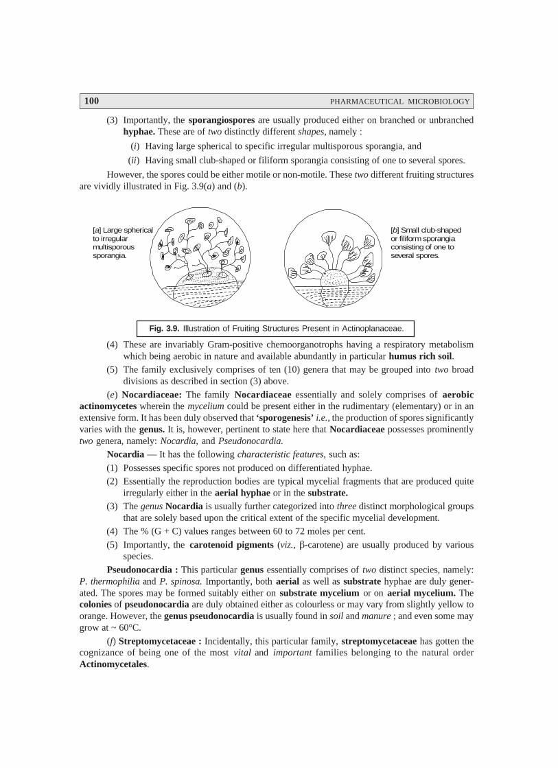
100 PHARMACEUTICAL MICROBIOLOGY
(3) Importantly, the sporangiospores are usually produced either on branched or unbranchedhyphae. These are of two distinctly different shapes, namely :
(i) Having large spherical to specific irregular multisporous sporangia, and
(ii) Having small club-shaped or filiform sporangia consisting of one to several spores.
However, the spores could be either motile or non-motile. These two different fruiting structuresare vividly illustrated in Fig. 3.9(a) and (b).
[ ] Large sphericalto irregularmultisporoussporangia.
a [ ] Small club-shapedor filiform sporangiaconsisting of one toseveral spores.
b
Fig. 3.9. Illustration of Fruiting Structures Present in Actinoplanaceae.
(4) These are invariably Gram-positive chemoorganotrophs having a respiratory metabolismwhich being aerobic in nature and available abundantly in particular humus rich soil.
(5) The family exclusively comprises of ten (10) genera that may be grouped into two broaddivisions as described in section (3) above.
(e) Nocardiaceae: The family Nocardiaceae essentially and solely comprises of aerobicactinomycetes wherein the mycelium could be present either in the rudimentary (elementary) or in anextensive form. It has been duly observed that ‘sporogenesis’ i.e., the production of spores significantlyvaries with the genus. It is, however, pertinent to state here that Nocardiaceae possesses prominentlytwo genera, namely: Nocardia, and Pseudonocardia.
Nocardia — It has the following characteristic features, such as:
(1) Possesses specific spores not produced on differentiated hyphae.(2) Essentially the reproduction bodies are typical mycelial fragments that are produced quite
irregularly either in the aerial hyphae or in the substrate.(3) The genus Nocardia is usually further categorized into three distinct morphological groups
that are solely based upon the critical extent of the specific mycelial development.(4) The % (G + C) values ranges between 60 to 72 moles per cent.
(5) Importantly, the carotenoid pigments (viz., β-carotene) are usually produced by variousspecies.
Pseudonocardia : This particular genus essentially comprises of two distinct species, namely:P. thermophilia and P. spinosa. Importantly, both aerial as well as substrate hyphae are duly gener-ated. The spores may be formed suitably either on substrate mycelium or on aerial mycelium. Thecolonies of pseudonocardia are duly obtained either as colourless or may vary from slightly yellow toorange. However, the genus pseudonocardia is usually found in soil and manure ; and even some maygrow at ~ 60°C.
(f) Streptomycetaceae : Incidentally, this particular family, streptomycetaceae has gotten thecognizance of being one of the most vital and important families belonging to the natural orderActinomycetales.
CHARACTERIZATION, CLASSIFICATION AND TAXONOMY OF MICROBES 101
* A natural or synthetic substance that destroys microorganisms or inhibits their growth.
** Any organic colouring matter found in the body.
Salient Features: The various salient features of the family streptomycetaceae are as enumer-ated under:
(1) The vegetative hyphae ranges between 0.5 to 2 μm in diameter.
(2) One of the most common apparent features being the presence of a well-branched myceliumwhich fails to undergo rapid fragmentation.
(3) The phenomenon of reproduction predominantly takes place either due to spores or occa-sionally by the aid of simultaneous growth of mycelial fragments.
(4) They invariably behave as Gram-positive microbes, and also are aerobic having Type-I CellWalls.
(5) The % (G + C) values of the DNA in the specific genera so far examined ranges between 69to 73 moles per cent.
The Streptomycetaceae family has essentially four distinctly well-recognized genera that areobviously segregated based entirely upon the typical sporulation characteristic features, as givenbelow:
(i) Streptomyces — Importantly, the genus streptomyces received a well-deserved world widerecognition by virtue of its critical role in the production of antibiotic. In fact, there areseveral strains identified and examined, which precisely gave rise to either one specific or aplethora of antibiotics.
• The bacterial cells are found to be heterotrophic, aerobic, and also extremely oxidative.
• Various other members of this family, Streptomyces, do give rise to a broad spectrum ofpigments.
• Bergey’s Manual include at least 463 species of the specific genus, and surprisingly agood number of them do possess even ‘uncertain taxonomic status’.
(ii) Streptoverticillium : Interestingly, the genus Streptoverticillium vividly consists of fortyspecies. The characteristic features of this particular genus are as follows:
• Aerial mycelium and substrate are both present.
• The branching ‘aerial mycelium’ more or less looks very much similar to the ‘barbed-wire’.
• Reproduction is accomplished by means of either spores or by fragmentation of the corre-sponding mycelium.
• The specific genus, Streptoverticillium, critically responsible for the production of a largecross-section of vital and important ‘antibiotics’* and ‘pigments’.**
(iii) Sporichthya : The genus, Sporichthya, possesses such vital members that essentially giverise to the formation of hyphae which are found to be not only branched, but also reason-ably short in structure. Its characteristic features are as stated below:
• The aerial mycelium is found to be strategically attached to the solid medium criticallywith the help of hold-fasts that actually originate from the very wall of the hyphae base.
• The aerial hyphae are observed to be articulately split up into smooth walled spores thatessentially possess a collar-like structure which in turn gives rise to the origination of aflagellum.

102 PHARMACEUTICAL MICROBIOLOGY
* A bacterial spore formed by segmentation.
** The liquid product of inflammation composed of albuminous susbtances, a thin fluid, and leukocytes ; gen-erally yellow in colour.
• The spores are motile in water.
• The genus forms Gram-positive/Gram-negative strains, heterotrophic in nature, grows onrich media, and lastly bacteria-like growth is observed explicitely.
(iv) Microellobospora: The genus, Microellobospora, critically comprises of such organismshaving slender hyphae with a diameter of 1 μm. It has been duly observed that the substratemycelium usually grows into a compact layer. Besides, the aerial mycelia and the substratemycelia predominantly form sporangia, strategically located on short sporangiophores.
Sporangia do contain a single longitudinal row consisting of non-motile sporangiospores.
Spores are observed to come into being by virtue of the simultaneous division of the specificintrasporangia hyphae.
Antibiotics : Certain typical strains belonging to this genus produce useful antibiotics as well.
Cell-wall is found to be typically of type-I and also aerobic and heterotrophic in character ;whereas, the formation of arthrospores* is not observed.
(g) Micromonosporaceae: The family Micromonosporaceae necessarily comprises of suchmembers that cause the production of aerial mycelium as well as substrate mycelium except in thegenus micromonospora.
The various characteristic features of the family Micromonosporaceae are as stated under :
(i) They are devoid of the sporophores or are sometimes quite short in structure ; and also incertain specific instances do exhibit dichotomous branching.
(ii) In a broader perspective, these are aerobic in nature, largely mesophilic; and certain speciesare thermophilic, besides being primarily saprophytic in the environment of the soil.
In fact, the family Micromonosporaceae comprises of six distinct genera that are exclusivelyclassified based upon either the presence or the absence of aerial mycelia together with other corre-sponding typical sporulation characteristics.
3.5.2. Bacteria [Plural of ‘Bacterium’]
Exhaustive historical evidences based on the survey of literatures, amply stress and reveal thefact that ‘bacteria’ predominantly share with the ‘blue-green algae’ a unique status and place, in the‘world of living organisms’.
A bacterium may be regarded as a one-celled organism without true nucleus or functionallyspecific components of metabolism that essentially belongs to the kingdom Prokaryotae (Monera), aname which means primitive nucleus. However, all other living organisms are termed as Eukaryotes, aname that precisely implies a true or proper nucleus.
It has been duly observed and established that ‘bacteria’ are exclusively responsible for thecausation of several painful ailments in humans, namely : tonsillitis, pneumonia, cystitis, school sores,and conjunctivitis.
Alternatively, one may define bacteria as microscopic single-celled organisms that can pen-etrate into healthy tissues and start multiplying into vast numbers. Interestingly, when they do this, theyinvariably damage the tissue that they are infecting, causing it to break down into the formation of

CHARACTERIZATION, CLASSIFICATION AND TAXONOMY OF MICROBES 103
pus.** Due to the damage they (bacteria) cause, the affected and involved area becomes red, swollen,hot and painful. In this manner, the waste products of the damaged tissue, together with the bacteria,rapidly spread into the blood stream, and this virtually stimulates the brain to elevate the body tempera-ture so as to fight off the contracted infection; and this ultimately gives rise to the development of‘fever’ (normal body temperature being 37°C or 98.4°F).
3.5.2.1. Salient Features
The various salient features of ‘bacteria’ are as stated under:
(1) The body is invariably invaded by millions of organisms every day, but very few surprisinglymay ever succeed in causing serious problems by virtue of the fact that body’s defencemechanisms usually destroy the majority of the invading microbes.
In fact, the white-blood cells (WBCs) are the main line of defence against the prevailinginfections. Evidently, the WBCs rapidly migrate to the zone of ‘unwanted bacteria’ and dohelp in engulfing them and destroying them ultimately. Importantly, when these defencemechanisms get overwhelmed, that a specific infection develops and noticed subsequently.
(2) Nomenclature: Each species of organisms or bacteria (and fungi but not viruses) has twonames: first — a family name (e.g., Staphylococcus) that essentially makes use of a capitalinitial letter and comes first always; and secondly — a specific species name (e.g., aureus)which uses a lower case initial letter and comes second.
Example: The golden staph bacteria that gives rise to several serious throat infections istherefore termed as Staphylococcus aureus, but should be normally abbreviated to S. aureus.
(3) As different types of bacteria invariably favour different segments of the body and therebylead to various glaring symptoms; therefore, it is absolutely necessary to choose and pick-upan appropriate educated guess about the antibiotic(s) to be administered by a ‘physician’. Inthe event of any possible doubt it is always advisable to take either a ‘sample’ or a ‘swab’being sent to a ‘microbiological laboratory’ for an expert analysis, so that the precise organ-ism may be identified, together with the most suitable antibiotic to destroy it completely.
(4) Obviously, there are a plethora of organisms (bacteria), specifically those present in the‘gut’, observed to be quite useful with respect to the normal functioning of the body. Theseorganisms usually help in the digestive process, and prevent infections either caused byfungi (e.g., thrush) or sometimes by viruses. Importantly, antibiotics are capable of killingthese so called ‘good bacteria’ also, which may ultimately give rise to certain apparent side-effects due to the prolonged usage of antibiotics, such as : diarrhoea, fungal infections ofthe mouth and vagina.
The most commonly observed ‘bacteria’ that invariably attack the humans and the respectivediseases they cause, or organs they attack, are as listed under :
S.No. Bacteria Diseases or Place of Infections
1 Bacteroides Pelvic organs2 Bordetella pertussis Whooping cough3 Brucella abortus Brucellosis4 Chlamydia trachomatis Vineral disease, pelvic organs, eye
5 Clostridium perfringens Gas gangrene, pseudomembranous colitis.6 Clostridium tetani Tetanus7 Corynebacterium diphtheriae Diphtheria
104 PHARMACEUTICAL MICROBIOLOGY
8 Escherichia coli Urine, gut, fallopian tubes, peritonitis
9 Haemophilus influenzae Ear, meningitis
10 Helicobacter pylori Peptic ulcers
11 Klebsiella pneumoniae Lungs, urine
12 Legionella pneumophilia Lungs
13 Mycobacterium leprae Leprosy
14 Mycobacterium tuberculosis Tuberculosis
15 Mycoplasma pneumoniae Lungs
16 Neisseria gonorrhoea Gonorrhoea, pelvic organs
17 Proteus Urine, ear
18 Pseudomonas aeruginosa Urine, ear, lungs, heart
19 Salmonella typhi Typhoid
20 Shigella dysenteriae Gut infections
21 Staphylococcus aureus Lungs, throat, sinusitis, ear, skin, eye, gut,
meningitis, heart, bone, joints
22 Streptococcus pyrogens Sinuses, ear, throat, skin
23 Streptococcus viridans Heart
24 Treponema pallidum Syphillis
25 Yersinia pestis Plague
3.5.2.2. Structure and Form of the Bacterial Cell: These characteristic form of the bacterial cell maybe sub-divided into two heads, namely:
(i) Size and shape, and
(ii) Structure
These two categories shall now be dealt with separately in the sections that follows :
3.5.2.2.1. Size and Shape: The size and shape of bacteria largely vary between the dimensions of 0.75– 4.0 μm. They are invariably obtained as definite unicellular structures that may be essentially foundeither as spherical forms (i.e., coccoid forms) or as cylindrical forms (i.e., rod-shaped forms). How-ever, the latter forms, in one or two genera, may be further modified into two sub-divisions, namely:
(a) With a single twist (or vibrios), and
(b) With several twists very much akin to ‘cork screw’ (or spirochaetes).
In actual practice, there prevails another predominant characteristic feature of the bacterial formi.e., the inherent tendency of the coccoid cells to exhibit growth in aggregates. It has been duly observedthat these ‘assemblies’ do exist in four distinct manners, such as:
(i) As ‘pairs’ (or diplococci),
(ii) As ‘groups of four systematically arranged in a cube’ (or sarcinae),
(iii) As ‘unorganized array like a bunch of grapes’ (or staphylococci), and
(iv) As ‘chains like a string of beads’ (or streptococci).
In general, these ‘aggregates’ are so specific and also characteristic that they usually assign aparticular generic nomenclature to the group, for instance :
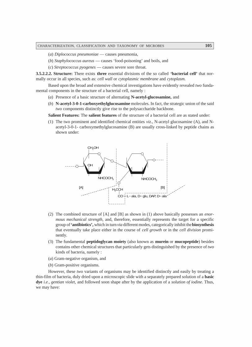
CHARACTERIZATION, CLASSIFICATION AND TAXONOMY OF MICROBES 105
(a) Diplococcus pneumoniae — causes pneumonia,
(b) Staphylococcus aureus — causes ‘food-poisoning’ and boils, and
(c) Streptococcus pyogenes — causes severe sore throat.
3.5.2.2.2. Structure: There exists three essential divisions of the so called ‘bacterial cell’ that nor-mally occur in all species, such as: cell wall or cytoplasmic membrane and cytoplasm.
Based upon the broad and extensive chemical investigations have evidently revealed two funda-mental components in the structure of a bacterial cell, namely :
(a) Presence of a basic structure of alternating N-acetyl-glucosamine, and
(b) N-acetyl-3-0-1-carboxyethylglucosamine molecules. In fact, the strategic union of the saidtwo components distinctly give rise to the polysaccharide backbone.
Salient Features: The salient features of the structure of a bacterial cell are as stated under:
(1) The two prominent and identified chemical entities viz., N-acetyl glucosamine (A), and N-acetyl-3-0-1- carboxymethylglucosamine (B) are usually cross-linked by peptide chains asshown under:
H CCH3
CO
NHCOCH3
CH OH2
L ala, D glu, DAP, D ala
[B][A]
OH
NHCOCH3
(2) The combined structure of [A] and [B] as shown in (1) above basically possesses an enor-mous mechanical strength, and, therefore, essentially represents the target for a specificgroup of ‘antibiotics’, which in turn via different modes, categorically inhibit the biosynthesisthat eventually take place either in the course of cell growth or in the cell division promi-nently.
(3) The fundamental peptidoglycan moiety (also known as murein or mucopeptide) besidescontains other chemical structures that particularly gets distinguished by the presence of twokinds of bacteria, namely :
(a) Gram-negative organism, and
(b) Gram-positive organisms.
However, these two variants of organisms may be identified distinctly and easily by treating athin-film of bacteria, duly dried upon a microscopic slide with a separately prepared solution of a basicdye i.e., gentian violet, and followed soon shape after by the application of a solution of iodine. Thus,we may have:
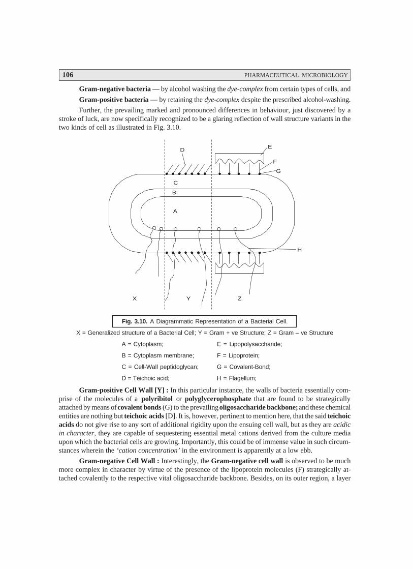
106 PHARMACEUTICAL MICROBIOLOGY
Gram-negative bacteria — by alcohol washing the dye-complex from certain types of cells, and
Gram-positive bacteria — by retaining the dye-complex despite the prescribed alcohol-washing.
Further, the prevailing marked and pronounced differences in behaviour, just discovered by astroke of luck, are now specifically recognized to be a glaring reflection of wall structure variants in thetwo kinds of cell as illustrated in Fig. 3.10.
D E
F
G
H
X Y Z
C
B
A
Fig. 3.10. A Diagrammatic Representation of a Bacterial Cell.
X = Generalized structure of a Bacterial Cell; Y = Gram + ve Structure; Z = Gram – ve Structure
A = Cytoplasm; E = Lipopolysaccharide;
B = Cytoplasm membrane; F = Lipoprotein;
C = Cell-Wall peptidoglycan; G = Covalent-Bond;
D = Teichoic acid; H = Flagellum;
Gram-positive Cell Wall [Y] : In this particular instance, the walls of bacteria essentially com-prise of the molecules of a polyribitol or polyglycerophosphate that are found to be strategicallyattached by means of covalent bonds (G) to the prevailing oligosaccharide backbone; and these chemicalentities are nothing but teichoic acids [D]. It is, however, pertinent to mention here, that the said teichoicacids do not give rise to any sort of additional rigidity upon the ensuing cell wall, but as they are acidicin character, they are capable of sequestering essential metal cations derived from the culture mediaupon which the bacterial cells are growing. Importantly, this could be of immense value in such circum-stances wherein the ‘cation concentration’ in the environment is apparently at a low ebb.
Gram-negative Cell Wall : Interestingly, the Gram-negative cell wall is observed to be muchmore complex in character by virtue of the presence of the lipoprotein molecules (F) strategically at-tached covalently to the respective vital oligosaccharide backbone. Besides, on its outer region, a layer
CHARACTERIZATION, CLASSIFICATION AND TAXONOMY OF MICROBES 107
* 2nd Edition.
of lipopolysaccharide (E) along with the presence of protein critically attached by hydrophobic interac-tions and divalent metal cations e.g., Ca2+, Fe2+, Mg2+, Cu2+, whereas, in its inner side is a layer ofphospholipid.
3.5.3. Rickettsia and Coxiella
Bergey’s Manual* describes the genus Rickettsia, which is duly placed in the order Rickettsialesand family Rickettsiaceae of the ααααα-proteobacteria ; whereas, Coxiella is shown in the orderLegionellales and family Coxiellaceae of the γγγγγ-proteobacteria. Based upon their close and intimatesimilarity in the ‘life-style’, despite their apparent ‘phylogenetic distance’, these two genera shall bediscussed together.
Salient Features : The salient-features of Rickettsia and Coxiella are as enumerated under:
(1) The bacteria belonging to these two genera are found to be rod-shaped, coccoid, or pleomorphichaving typical Gram-negative walls and devoid of any flagella; however, their actual sizeusually varies but they tend to be relatively very small.
Examples:
Rickettsia — 0.3 to 0.5 μm (diameter); and 0.8 to 2.0 μm (length);
Coxiella — 0.2 to 0.4 μm (diameter); and 0.4 to 1.0 μm (length);
(2) It has been duly observed that all species happen to be either parasitic or mutualistic innature. Interestingly, the former species (i.e., parasitic ones) invariably grow in vertebrateerythrocytes, macrophages, and vascular endothelial cells; and they usually reside in blood-sucking arthropods viz., ticks, lice, mites, fleas, tse-tse flies that essentially serve either asvectors or as primary hosts.
(3) By virtue of the fact that these two genera predominantly comprise of vital and important‘human-active pathogens’, both their metabolism as well as reproduction have been inves-tigated intensively and extensively.
Rickettsias: are found to gain entry into the host-cell by the induction of the phenomenon of‘phagocytosis’. Thus the bonafide members belonging to the genus Rickettsia immediatelyget free from the ensuing ‘phagosome’ and get reproduced due to the ‘binary fission’ in thecytoplasm.
Coxiella: In contract, it remains within the phagosome after it has undergone fusion strategi-cally with a ‘lysosome’, and virtually undergo reproduction very much within the‘phagolysosome’. Thus, the host-cell ultimately bursts, thereby providing the release of anabundant quantum of newer organisms specifically.
(4) Physiology and Metabolism: Importantly, the rickettsias are prominently quite different incomparison to most other bacteria with respect to physiology and metabolism. Some of thehighlights observed are as stated below:
(a) Rickettsias: normally lack the glycolytic path way and do not make use of ‘glucose’ as asource of energy, but categorically oxidize both ‘glutamate’ and ‘tricarboxylic acidcycle (TCA-Cycle) intermediates, e.g., succinic acid.
(b) Rickettsial plasma membrane critically possesses the specific carrier-mediated trans-port systems; and thereupon, the host cell nutrients as well as the ensuing coenzymesget absorbed and consumed almost directly.
108 PHARMACEUTICAL MICROBIOLOGY
Examples: (i) Rickettsias are observed to make use of both NAD* and uridine phosphate glu-cose.
(ii) The membrane of rickettsias also possesses particularly an adenylate exchange carrier whichmeticulously exchanges ADP for the corresponding external ATP, whereby the latter (i.e., the host ATP)may be able to cater for a good deal of ‘energy’ essentially required for the ultimate growth.**
Rickettsial Pathogenic Organisms — are duly identified and recognized as given below :
Rickettsia prowazekii — associated with typhus fever
Rickettsia typhi — associated with typhus fever
Rickettsia rickettsii — associated with Rocky Mountain spotted fever.
3.5.4. Spirochaetes
The phylum Spirochaetes [Greek: spira = a coil ; and chaete = hair] essentially and distinguish-ably comprises of Gram-negative, chemoheterotrophic bacteria characterized by their specific structureand mechanism of motility.
Salient Features : The various vital and important salient features of the spirochaetes are asenumerated below :
(1) They are slender long bacteria having diameter 0.1 to 3.0 μm, and length 5 to 250 μm ; andpredominantly with a flexible and helical shape that may sometimes also occur in the formof chains.
(2) Multiplication of the spirochaetes invariably takes place by transverse fission.
(3) The bacterial cells consist of protoplasmic cylinder interwined with either one or moreaxial fibrils, that originate in nearly equal number from the subterminal attachment discstrategically located at either ends of the aforesaid proto-plasmic cylinder. Importantly,both the protoplasmic cylinder as well as the axial fibrils are duly enclosed in the outerenvelope meticulously. However, the unattached ends of the axial fibrils may invariably getextended beyond the terminals of the protoplasmic cylinder that finally be observed as ‘po-lar flagella’.
(4) The motility existing in the spirochaetes are usually found to be of three types, namely :
(i) Obtained by the rapid rotation about the long axis of the helix
(ii) Derived by the flexion of the bacterial cells, and
(iii) Brought about by the locomotion invariably observed along a helical or a serpentinepath
(5) It has been observed that many species of spirochaetes are so slim that they may exclusivelyand vividly visible in a light-microscope either by the help of a phase-contrast microscopeor a dark-field optics.
(6) The spectacular and distinctive features of the spirochaete morphology are quite evident bymeans of an ‘electron micrograph’ which explicitely reveals the following characteristicfeatures, such as :
* Nicotinamide adenine dinucleotide.
** The prevailing metabolic dependence vividly clarifies and explains why many of these organisms should bespecifically cultivated in the yolk-sacs of chick embryos or in tissue culture cells.
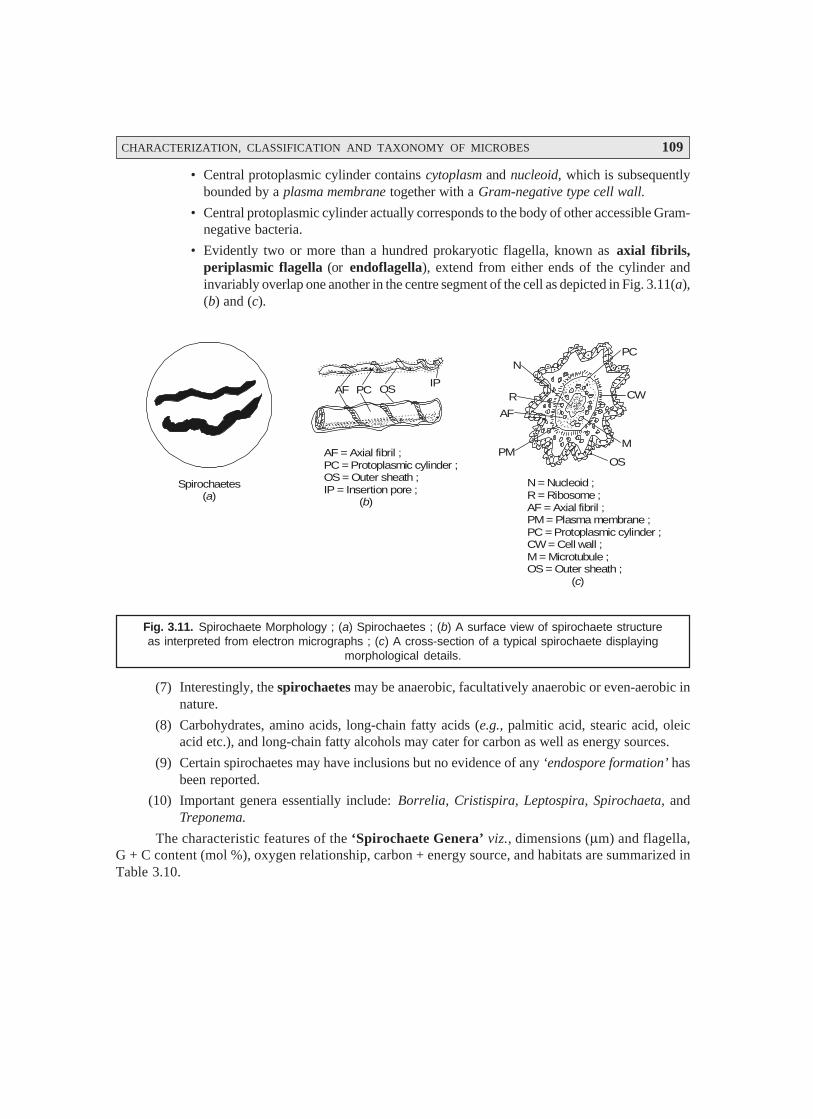
CHARACTERIZATION, CLASSIFICATION AND TAXONOMY OF MICROBES 109
• Central protoplasmic cylinder contains cytoplasm and nucleoid, which is subsequentlybounded by a plasma membrane together with a Gram-negative type cell wall.
• Central protoplasmic cylinder actually corresponds to the body of other accessible Gram-negative bacteria.
• Evidently two or more than a hundred prokaryotic flagella, known as axial fibrils,periplasmic flagella (or endoflagella), extend from either ends of the cylinder andinvariably overlap one another in the centre segment of the cell as depicted in Fig. 3.11(a),(b) and (c).
Spirochaetes( )a
AF PC OSIP
AF = Axial fibril ;PC = Protoplasmic cylinder ;OS = Outer sheath ;IP = Insertion pore ; ( )b
N
R
AF
PMOS
M
CW
PC
N = Nucleoid ;R = Ribosome ;AF = Axial fibril ;PM = Plasma membrane ;PC = Protoplasmic cylinder ;CW = Cell wall ;M = Microtubule ;OS = Outer sheath ; ( )c
Fig. 3.11. Spirochaete Morphology ; (a) Spirochaetes ; (b) A surface view of spirochaete structureas interpreted from electron micrographs ; (c) A cross-section of a typical spirochaete displaying
morphological details.
(7) Interestingly, the spirochaetes may be anaerobic, facultatively anaerobic or even-aerobic innature.
(8) Carbohydrates, amino acids, long-chain fatty acids (e.g., palmitic acid, stearic acid, oleicacid etc.), and long-chain fatty alcohols may cater for carbon as well as energy sources.
(9) Certain spirochaetes may have inclusions but no evidence of any ‘endospore formation’ hasbeen reported.
(10) Important genera essentially include: Borrelia, Cristispira, Leptospira, Spirochaeta, andTreponema.
The characteristic features of the ‘Spirochaete Genera’ viz., dimensions (μm) and flagella,G + C content (mol %), oxygen relationship, carbon + energy source, and habitats are summarized inTable 3.10.
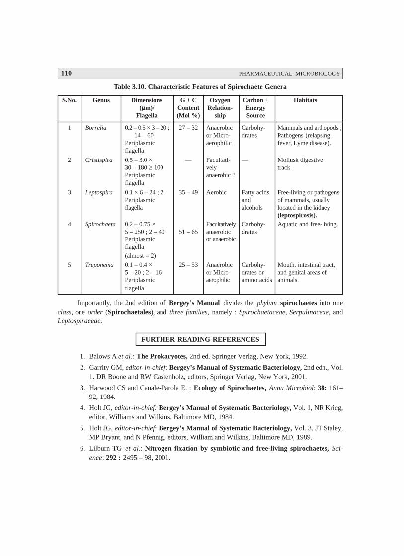
110 PHARMACEUTICAL MICROBIOLOGY
Table 3.10. Characteristic Features of Spirochaete Genera
S.No. Genus Dimensions G + C Oxygen Carbon + Habitats(μμμμμm)/ Content Relation- Energy
Flagella (Mol %) ship Source
1 Borrelia 0.2 – 0.5 × 3 – 20 ; 27 – 32 Anaerobic Carbohy- Mammals and arthopods ;14 – 60 or Micro- drates Pathogens (relapsing
Periplasmic aerophilic fever, Lyme disease).flagella
2 Cristispira 0.5 – 3.0 × — Facultati- — Mollusk digestive30 – 180 ≥ 100 vely track.Periplasmic anaerobic ?flagella
3 Leptospira 0.1 × 6 – 24 ; 2 35 – 49 Aerobic Fatty acids Free-living or pathogensPeriplasmic and of mammals, usuallyflagella alcohols located in the kidney
(leptospirosis).4 Spirochaeta 0.2 – 0.75 × Facultatively Carbohy- Aquatic and free-living.
5 – 250 ; 2 – 40 51 – 65 anaerobic dratesPeriplasmic or anaerobicflagella
(almost = 2)
5 Treponema 0.1 – 0.4 × 25 – 53 Anaerobic Carbohy- Mouth, intestinal tract,5 – 20 ; 2 – 16 or Micro- drates or and genital areas ofPeriplasmic aerophilic amino acids animals.flagella
Importantly, the 2nd edition of Bergey’s Manual divides the phylum spirochaetes into oneclass, one order (Spirochaetales), and three families, namely : Spirochaetaceae, Serpulinaceae, andLeptospiraceae.
FURTHER READING REFERENCES
1. Balows A et al.: The Prokaryotes, 2nd ed. Springer Verlag, New York, 1992.
2. Garrity GM, editor-in-chief: Bergey’s Manual of Systematic Bacteriology, 2nd edn., Vol.1. DR Boone and RW Castenholz, editors, Springer Verlag, New York, 2001.
3. Harwood CS and Canale-Parola E. : Ecology of Spirochaetes, Annu Microbiol: 38: 161–92, 1984.
4. Holt JG, editor-in-chief: Bergey’s Manual of Systematic Bacteriology, Vol. 1, NR Krieg,editor, Williams and Wilkins, Baltimore MD, 1984.
5. Holt JG, editor-in-chief: Bergey’s Manual of Systematic Bacteriology, Vol. 3. JT Staley,MP Bryant, and N Pfennig, editors, William and Wilkins, Baltimore MD, 1989.
6. Lilburn TG et al.: Nitrogen fixation by symbiotic and free-living spirochaetes, Sci-ence: 292 : 2495 – 98, 2001.
CHARACTERIZATION, CLASSIFICATION AND TAXONOMY OF MICROBES 111
7. McBride MJ: Bacterial gliding motility: Multiple mechanisms for cell movement orsurfaces, Annu. Rev. Microbiol, 55: 49–75, 2001.
8. Margulis L., Spirochaetes. In Encyclopedia of Microbiology, 2nd edn. Vol. 4. J. Lederberg,editor-in-chief, Academic Press, San Diego, 353–63, 2000.
9. Radolf JD : Role of outer membrane architecture in immune evasion by Treponemapallidum and Borrelia burgdorferi, Trends Microbiol 2(9) : 307–11, 1994.
10. Radolf JD et. al.: Treponema pallidum. Doing a remarkable job with what it’s got., TrendsMicrobiol. 7(1); 7–9, 1999.
11. Raymond J et. al.: Whole-genome analysis of photosynthetic prokaryotes, Science, 298 :1616–20, 2002.
12. Saint Girons et al.: Molecular biology of the Borrelia bacteria with linear replicons,Microbiology, 140 : 1803–16, 1994.
13. Ting CS et al.: Cyanobacterial photosynthesis in the oceans. The origins and signifi-cance of divergent light-harvesting strategies., Trends Microbiol. 10(3) : 134–42, 2002.
112
IDENTIFICATION OFMICROORGANISMS
4.1. INTRODUCTION
It has been recognized as an universal practice that when a ‘bacterium’ duly isolated and obtainedin a ‘pure culture medium’ it remains to be identified meticulously via certain well-defined broadlyaccepted systematical laid down procedures. The following characteristic features may be studied in anelaborated intensive and extensive manner in the course of their precise and judicious identification,such as :
• Morphology,
• Selective and diagnostic media,
• Cultural characteristics,
• Resistance,
• Metabolism,
• Additional recognized biochemical tests,
• Profile of microbial stains, and
• Rapid identification methods.
Importantly, the basic clinical microbiological evaluations may provide preliminary or definitiveidentification of the microorganisms exclusively dependent upon the following five cardinal aspects,namely :
(1) Microscopic examination of specimens,
(2) Critical investigation with regard to the growth and biochemical characteristic features ofthe isolated microorganisms (pure cultures),
(3) Specific immunologic tests that solely detect either the antibodies or the microbial anti-gens,
(4) Bacteriophage typing (restricted to the research settings and the CDC*), and
(5) Molecular techniques.
* Centres for disease control and prevention.
4• Introduction• Morphology• Selective and Diagnostic Media• Cultural Characteristics• Biochemical Tests (or Properties)• Profile of Microbial Stains
IDENTIFICATION OF MICROORGANISMS 113
The various vital and important aspects of the accurate and precise identification of microor-ganisms shall be treated in an elaborated manner in the sections that follows :
4.2. MORPHOLOGY
Morphology relates to the ‘science dealing with the structures and forms of organisms’. Inreality, the ‘morphology of the bacterium’ exclusively trusts confidently upon a number of factors,namely :
the strain under investigation,
nature of the culture medium,
temperature and time of incubation,
age of the culture, and
the number of ‘subculture’ it has been subjected to.
Importantly, the various characteristic features that may be observed from such meticulousinvestigational studies are : shape, size, arrangement, motility, flagella, spores, and capsules.
The variants observed in the above cited physical characteristic features may be stated as under :
Shape : spherical, rod shaped, comma shaped, spiral shaped, filamentous.
Axis of organism : straight, curved.
Length and breadth : mostly variable.
Sides of organism : convex, concave, parallel, irregular.
Ends of organism : tapering, rounded, straight.
Shape : club shaped, giant forms, navicular, swollen, shadow shaped.
Arrangement : in pairs, in tetrads, packs of eight, in chains (short or long) e.g., cocci ; in shortand long chains at random e.g., bacilli ; in single or in ‘S’ or in spiral forms .e.g., vibrios.
Motility : non-motile, sluggishly motile, actively motile or may show darting motility.
Forms : atrichate (i.e., without flagella) ; monotrichate ; lophotrichate ; amphitrichate ; peritrichate ;
Spores : oval, spherical, ellipsoidal, having same width or wider than the prevailing bacillarybody ; equatorial, subterminal or terminal.
Capsules : may or may not be present.
Techniques used : Electron microscopy ; phase-contrast microscopy ; background illumination ;and hanging drop preparations.
4.3. SELECTIVE AND DIAGNOSTIC MEDIA
A survey of literature has adequately established that there exists varying degree of abilities tocarry out the proper fermentation of ‘carbohydrates’, glaring differences in the ‘pyruvic acid metabo-lism’ utilized to have a clear-cut distinguished features of A. aerogenes and E. coli, varying responses ofbacteria to different inhibitors etc. ; and the ensuing exploitation of these critical differences may beexpatiated by the judicious usage of selective and diagnostic media.
In actual practice, the selective media specifically favour the growth of particular microorgan-isms. MacConkey’s agar medium was introduced first and foremost in the year 1905, so as to isolateEnterobacteriaceae from such sources as : urine, faeces, foods, and water.

114 PHARMACEUTICAL MICROBIOLOGY
MacConkey Agar : Composition
S. No. Ingredients Amount [g.L–1]
1 Pancreatic digest of gelatin 17.0
2 Pancreatic digest of casein 1.5
3 Peptic digest of animal tissue 1.5
4 Lactose 10.0
5 Bile salts 1.5
6 Sodium chloride 5.0
7 Natural red 0.03
8 Crystal violet 0.001
9 Agar 13.5
Importantly, the various ingredients incorporated in the above MacConkey’s medium play adefinitive role as stated below :
Bile Salts : invariably function as a ‘natural surface-active agent which does not inhibit thegrowth of the Enterobacteriaceae, but certainly prevents the growth of Gram-positive bacteria that aregenerally available in the material under investigation.
Lactose : Production of acid from lactose by the help of two organisms, namely : A. aerogenesand E. coli exert their action on this medium thereby changing the original colouration of the indicator,besides adsorbing the said indicator to a certain extent around the growing bacterial cells.
Microorganisms : These may also be selected by incubation in the presence of nutrients whichthey may consume specifically.
Example : Isolation of cellulose-digesting microorganisms may be accomplished by using amedium containing only ‘cellulose’ as a particular source of carbon and energy.
Salient Features : The salient features of selective media are as stated under :
(1) The microorganisms that specifically cause typhoid and paratyphoid fever*, and bacil-lary dysentry** fail to ferment ‘lactose’ ; and, therefore, the resulting colonies of thesemicrobes distinctly appear to be transparent absolutely.
(2) Besides, MacConkey agar there are also two other highly selective media viz., eosin-meth-ylene blue agar, and endo agar that are employed widely and exclusively for the detectionof E. coli (most dreadful faecal organism) and allied bacteria present in water supplies, foodproducts etc., which essentially contains dyes that would critically suppress the growth ofGram-positive organism e.g., staphylococci.
(3) Several accepted modified variants of MacConkey’s medium do exist viz., bile salts areduly replaced with pure synthetic surface-active agent(s).
(4) Selectivity of MacConkey’s medium could be further enhanced by the addition of certainspecific inhibitory dyes e.g., neutral red and crystal violet.
* Salmonella typhi ;** Shigella dysenteriae ;
.
IDENTIFICATION OF MICROORGANISMS 115
* Differential media refer to such media that distinguish between different groups of bacteria and even allowtentative identification of microbes based on their biological characteristics.
** S.typhi may produce hydrogen sulphide (H2, S) specifically from the sulphur-containing amino acids presentin the medium which in turn would react with ferrous sulphate [FeSO4] to produce a black-deposit offerrous sulphide [FeS].
*** Also supposedly present in material to be examined.
(5) Importantly, the MacConkey agar medium serves both differential* and selective, becauseit predominently contains lactose and neutral red dye whereby the particular lactose-fer-menting colonies distinctly appear pink to red in colour, and are distinguished from the‘colonies of nonfermentors’ quite easily.
There are certain other selective media that are invariably prepared by the addition of quite a fewhighly specific components to the corresponding culture medium which may allow the growth of one‘group of microbes’ while suppressing growth of some other groups. A few typical examples of such‘selective media’ are as given under :
(a) Salmonella-Shigella Agar [SS-Agar]. It is exclusively used to isolate both Salmonella andShigella species. In fact, its ‘bile salt mixture’ inhibits the growth of several cardinal groupsof coliforms in particular. Importantly, the Salmonella and Shigella species give rise to al-most colourless colonies by virtue of the fact that they are not capable of fermenting lactose.In fact, lactose-fermenting microorganisms shall produce pink-colonies mostly.
(b) Mannitol-Salt Agar [MS-Agar]. It is solely employed in the isolation of staphylococci.The relatively high-level of selectivity is usually accomplised by the high salt concentration(~ 7.5%) which specifically retards and checks the growth of several groups of microbes. Itis, however, pertinent to state here that the presence of mannitol in the MS-Agar mediumdistinctly aids in the clear-cut differentiation of the ‘pathogenic staphylococci’ from the‘nonpathogenic staphylococci’ due to the fact that the former augments fermentation ofmannitol to yield ‘acid’ whereas the latter fails to do so.
(c) Bismuth-Sulphite Agar [BS-Agar]. BS-Agar medium was duly developed in the 1920ssolely for the identification of Salmonella typhi, especially from the stool and food speci-mens. It has been duly proved that S. typhi reduces the ‘sulphite’ anion to the corresponding‘sulphide’ anion, thereby giving rise to distinct apparently visible black colonies** havinga specific metallic sheen (lustre). On a rather broader perspective BS-Agar medium mayalso be extended to identify the presence of S.typhi in urine, foods, faeces, water, and phar-maceutical products. Generally, the BS-Agar comprises of a buffered nutrient agar contain-ing bismuth sulphite, ferrous sulphate, and brilliant green.
Observations. Following are some of the cardinal observations, such as :
(1) E. coli*** gets usually inhibited by the presence of brilliant green at a concentration of0.0025% (w/v) ; whereas, S. typhi shall attain growth luxuriantly.
(2) Bismuth sulphite may also exert an inhibitory effect to a certain extent upon the E. coli.
In addition to the ‘selective and diagnostic media’ one may also come across such types ofmedia as :
(i) Differential media,
(ii) Enrichment media, and
(iii) Characteristic media.

116 PHARMACEUTICAL MICROBIOLOGY
The above mentioned three types of media shall now be discussed individually in the sectionsthat follows :
4.3.1. Differential Media
The differential media usually refers to the incorporation of certain specifc chemicals into amedium that may eventually give rise to diagnostically useful growth or apparent change in the mediumafter the proper incubation.
A few typical examples are as discussed under :
4.3.1.1. Eosin Methylene Blue Agar [EMB-Agar]
The EMB-Agar media is employed exclusively to differentiate between the ‘lactose fermenters’and the ‘non-lactose-fermenters’. In-fact, the EMB-Agar media essentially comprises of : lactose,salts, and two dyes viz. eosin and methylene blue. From the observations the following inferences maybe drawn :
(a) E. coli (a ‘lactose fermenter’) : will produce either a dark colony or one that has a metallicsheen, and
(b) S. typhi (a ‘nonlactose fermenter’) : shall appear as an absolute colourless colony.
4.3.1.2. MacConkey Agar
It has already been discussed under Section 3.
4.3.1.3. Hektoen Enteric Agar [HE-Agar]
It is invariably used to enhance the overall yield of Salmonella and Shigella species in comparisonto other microbiota. It has been observed that the presence of relatively high bile salt concentrationinhibits the general growth of Gram-positive microorganisms specifically. Besides, HE-Agar also retards(or slows down) the growth of several coliform strains.
4.3.2. Enrichment Media
It has been amply demonstrated and established the critical and judicious incorporation of serum,blood, or extracts to the particular ‘tryptic soy agar’ or broth shall enormously augment the muchdesired growth of a large number of most ‘fastidious microbes’. In actual practice, however, thesemedia are largely employed to isolate primarily the microorganisms from a host of ‘biological fluids’such as : cerebrospinal fluid, pleural fluid, wound abscesses, and sputum. A few typical examplesare as stated under :
4.3.2.1. Blood Agar*
The critical addition of ‘citrated blood’ to the prevailing ‘tryptic soy agar’ renders it to affordvariable haemolysis, that in turn allows the precise differentiation of certain species of microorganisms.It is, however, pertinent to state here that one may observe these distinct haemolytic patterns on bloodagar. A few such typical variations are as stated under :
(a) ααααα-Haemolysis. It may be observed due to the formation of greenish to brownish halo**around the colony e.g., streptococcus gardonii and streptococcus pneumoniae.
* It may also be regarded as a ‘differential medium’.
** A circle of light surrounding a shining body.
IDENTIFICATION OF MICROORGANISMS 117
(b) βββββ-Haemolysis. It represents the virtual complete haemolysis of blood cells thereby givingrise to a distinct clearing effect around growth in the colony e.g., Staphylococcus aureus and Streptococcuspyogenes.
(c) Nonhaemolytic Pattern. In this particular instance practically no change occurs in the me-dium e.g., Staphylococcus epidermidis and Staphylococcus saprophyticus.
4.3.2.2. Chocolate Agar
Interestingly, the ‘chocolate agar’ is specifically made from ‘pre-heated blood’ that essentiallycaters for the requisite and necessary growth factors desired urgently to support the bacterial growthe.g., Haemophilus influenzae and Neisseria gonorrhoeae.
4.3.3. Characteristic Media
The very purpose and extensive utility of the so-called ‘characteristic media’ are to test mi-crobes for ascertaining a few highly specific metabolic activities, products, or their ensuing require-ments.
Following are some of the typical examples, namely :
4.3.3.1. Triple Sugar Iron Agar [TSI Agar]
The TSI-Agar usually comprise of : lactose, sucrose, glucose, ferrous ammonium sulphate[(NH4)2Fe(SO4)2], and sodium thiosulphate [Na2S2O3]. In actual practice TSI-Agar is solely used forthe critical identification of enteric organisms* ; and are broadly based upon their inherent ability toattack the chemical entities viz., glucose, lactose, or sucrose and thus are responsible for liberating‘sulphides’ from ferrous ammonium sulphate or sodium thiosulphate.
The various typical examples of TSI-Agar are as stated under :
(a) Citrate Agar. It contains sodium citrate [C6H5Na3O7], which serves as the exclusive sourceof carbon ; whereas, the ammonium phosphate [(NH4)3PO4] as the sole source of nitrogen. The citrateagar finds its usage to differentate the ‘enteric bacteria’ on the basis of ‘citrate utilization’.
(b) Lysine Iron Agar [LIA]. Importantly, LIA is solely employed to differentate microorgan-isms which may either cause deamination or decarboxylation the amino acid lysine. Because, LIAcomprises of lysine that predominantly and exclusively allows enzyme detection ; whereas the pres-ence of ferric ammonium citrate helps in the detection of H2S production.
(c) Sulphide-Indole-Motility Medium [SIM-Medium]. In fact, the SIM-medium is employedexclusively for the following three different tests, namely :
(i) production of sulphides,
(ii) formation of indole NH
i.e., a metabolite product duly obtained from the subsequent
utilization of tryptophan, and
* Intestinal organisms e.g., Gram-negative non-spore-forming facultatively anaerobic bacilli viz.,Escherichia, Shigella, Salmonella, Klebsiella, and Yersinia.
118 PHARMACEUTICAL MICROBIOLOGY
(iii) causation of ‘motility’.
Precisely, the SIM-Medium is used for making out the differentiation of the various entericorganisms.
A. Selective Media for StaphylococciIn a broader perspective it is invariably necessary to screen and examine a host of pathological
specimens, food, and pharmaceutical products (including dosage forms) to ascertain the presence ofstaphylococci ; besides, specific organisms that are solely responsible for causing serious food contamina-tion/poisoning as well as systemic infections. There are two media that are used extensively, such as :
(a) Selective Media for Enterobacteria [or Enteric Bacteria]. [Greek. enterikos means per-taining to intestine]. In general, all help in the degradation of sugars by means of the Embden-MeyerhofPathway (or EMP-Cycle] which ultimately cause cleavage of ensuing pyruvic acid to yield formicacid in the formic-acid fermentations. It has been established beyond any reasonable doubt that in theselective media for enterobacteria a surface-active agent serves as the ‘main selector’, whereas in thespecific staphylococcal medium the various selectors happen to be : sodium chloride [NaCl] and lithiumchloride [LiCl].
Staphylococci are found to be tolerant against a ‘salt’ concentration extending ~ 7.5% (w/v)e.g., Mannitol salt, Baired-Parker (BP), and Vogel-Johnson (VI) media.
Salient Features. The other vital and important ‘salient features’ with respect to the variousother ‘principles’ concerning the selective media for staphylococci are as enumerated under :
(1) Use of a selective C-source viz., mannitol or sodium pyruvate (soluble salt) along with asuitable ‘buffer’.
(2) Use of an appropriate acid-base indicator e.g., methyl-red phenolphthalein, for distinctlyvisualizing the ensuing metabolic activity.
(3) By observing the ‘inference growth’.
(4) Lecithin (a phospholipid) present in the egg yolk forms a vital ingredient of Baird-Parkermedium seems to undergo hydrolysis strategically by the ensuing staphylococcal (i.e., esterase) activ-ity* in order that the prevailing microorganisms are adequately encircled by a cleared (i.e., transpar-ent) zone in the rest of the opaque medium.
B. Selective Media for PseudomonadsBased on advanced, meticulous researches carried out on the ‘molecular analysis’,
pseudomonads have been duly reclassified, and consequently several former Pseudomonas speciesreallocated to new genera, for instance : Burkholderia, Stenotrophomonas and others.
Importantly, these media solely depend upon the relative resistance of pseudomonas to the par-ticular quaternary ammonium disinfectant cetrimide ; whereas, in certain recipes the incorporation ofnalidixic acid i.e., an antibiotic, affords a reasonable resistance to the pseudomonads.
Laboratory Diagnosis. The bacterium, Pseudomonads, usually grows rapidly on a plethora ofmedia thereby rendering the identification of the pigmented strains of the organism from the clinicalsamples rather easy. However, it has been duly observed that almost 1/10th of the isolates may benonpigmented.
* USP (1980) essentially includes a well-defined test for the presence of staphylococci in the pharmaceuticalproducts, whereas the BP (1980) does not.
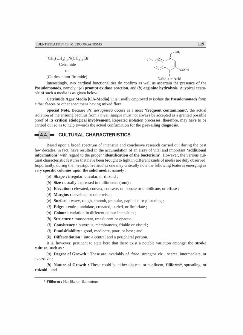
IDENTIFICATION OF MICROORGANISMS 119
[CH3(CH2)15N(CH3)3]Br
Cetrimide
or
[Cetrimonium Bromide]
Interestingly, two cardinal functionalities do confirm as well as ascertain the presence of thePseudomonads, namely : (a) prompt oxidase reaction, and (b) arginine hydrolysis. A typical exam-ple of such a media is as given below :
Cetrimide Agar Media [CA-Media]. It is usually employed to isolate the Pseudomonads fromeither faeces or other specimens having mixed flora.
Special Note. Because Ps. aeruginosa occurs as a most ‘frequent contaminant’, the actualisolation of the ensuing bacillus from a given sample must not always be accepted as a granted possibleproof of its critical etiological involvement. Repeated isolation processes, therefore, may have to becarried out so as to help towards the actual confirmation for the prevailing diagnosis.
4.4. CULTURAL CHARACTERISTICS
Based upon a broad spectrum of intensive and conclusive research carried out during the pastfew decades, in fact, have resulted in the accumulation of an array of vital and important ‘additionalinformations’ with regard to the proper ‘identification of the bacterium’. However, the various cul-tural characteristic features that have been brought to light in different kinds of media are duly observed.Importantly, during the investigative studies one may critically note the following features emerging asvery specific colonies upon the solid media, namely :
(a) Shape : irregular, circular, or rhizoid ;
(b) Size : usually expressed in millimeters (mm) ;
(c) Elevation : elevated, convex, concave, umbonate or umbilicate, or effuse ;
(d) Margins : bevelled, or otherwise ;
(e) Surface : wavy, rough, smooth, granular, papillate, or glistening ;
(f) Edges : entire, undulate, crenated, curled, or fimbriate ;
(g) Colour : variation in different colour intensities ;
(h) Structure : transparent, translucent or opaque ;
(i) Consistency : butyrous, membranous, friable or viscid ;
(j) Emulsifiability : good, mediocre, poor, or best ; and
(k) Differentiation : into a central and a peripheral portion.
It is, however, pertinent to state here that there exist a notable variation amongst the strokeculture, such as :
(a) Degree of Growth : These are invariably of three strengths viz., scarce, intermediate, orexcessive ;
(b) Nature of Growth : These could be either discrete or confluent, filiform*, spreading, orrhizoid ; and
* Filiform : Hairlike or filamentous.
N NH C3
CH3
O
COOH
Nalidixic Acid

120 PHARMACEUTICAL MICROBIOLOGY
(c) Physical Characteristics : These essentially include a wide variety of such physical char-acteristic features as : surface, elevation, edges, colour, structure, odour, emulsifiability, consistency,and the overall critical changes observed in the ensuing medium.
It is pertinent to mention at this point in time that in a particular fluid (or liquid) medium onemay obviously take cognizance of the following characteristic features, namely :
• extent of growth,
• presence of turbidity plus its nature,
• presence of deposit and its character,
• nature of surface growth e.g., pellicle* and its observed quality, and
• ease of disintegration, and colouration.
4.5. BIOCHEMICAL TESTS (OR PROPERTIES)
Extensive and meticulous in depth investigations carried out on a host of fermentative proce-dures using different types of substrates exclusively dependent upon a broad-spectrum of biochemicaltests ultimately lead to the production of ethanol by yeast ; acetylmethylcarbinol ; lactic acid ; aceticacid ; ethanol by E. coli ; acetone plus CO2 ; citric acid (Krebs Cycle) ; and CO2 plus H2.
The most vital and important and abundantly employed biochemical tests are as described be-low with appropriate explanations whenever required in the course of the prevalent discussion :
4.5.1. Carbohydrate (Sugar) Fermentation
The carbohydrate fermentation is normally tested in a ‘sugar media’. Thus, the generation of‘acid’ is indicated by an apparent change in the colouration of the ensuing medium either to pink or red,and the resulting gaseous products produced gets duly collected in a strategically placed Durham’sTube.
4.5.2. Litmus Milk
In this particular instance there may not be any change in the medium, or acid or alkali could begenerated thereby giving rise to clotting of milk, and peptonizaiton or saponification may take placeappreciably. The resulting ‘clot’ i.e., coagulation of the milk protein (viz., casein) could face a disrup-tion by virtue of the gas evolved (usually termed as ‘stormy fermentation’).
4.5.3. Indole Production
In actual practice the ‘indole production’ is normally tested in a peptone-water culture after aninterval of 48 or 96 hours incubation at 37°C ; whereby the generation of indole from the amino acidtryptophan is duly ascertained as given below :
* A thin film or surface on a liquid.
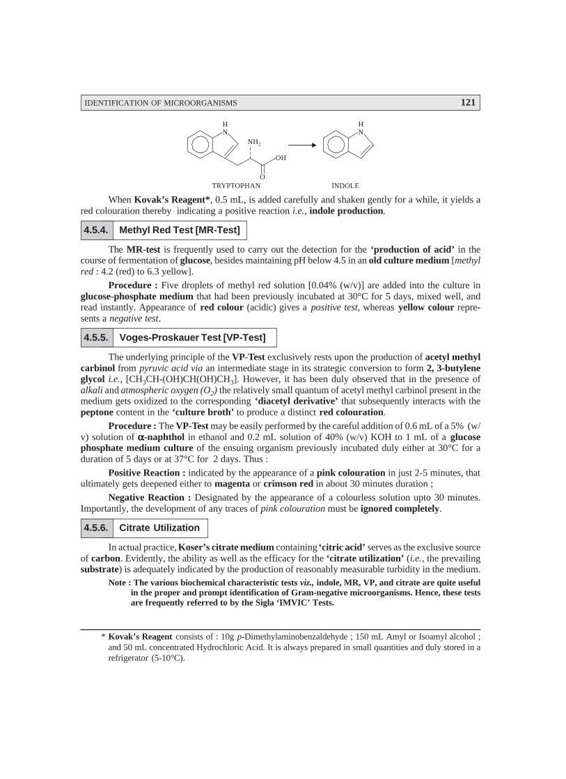
IDENTIFICATION OF MICROORGANISMS 121
HN
NH2
O
OH
HN
TRYPTOPHAN INDOLE
When Kovak’s Reagent*, 0.5 mL, is added carefully and shaken gently for a while, it yields ared colouration thereby indicating a positive reaction i.e., indole production.
4.5.4. Methyl Red Test [MR-Test]
The MR-test is frequently used to carry out the detection for the ‘production of acid’ in thecourse of fermentation of glucose, besides maintaining pH below 4.5 in an old culture medium [methylred : 4.2 (red) to 6.3 yellow].
Procedure : Five droplets of methyl red solution [0.04% (w/v)] are added into the culture inglucose-phosphate medium that had been previously incubated at 30°C for 5 days, mixed well, andread instantly. Appearance of red colour (acidic) gives a positive test, whereas yellow colour repre-sents a negative test.
4.5.5. Voges-Proskauer Test [VP-Test]
The underlying principle of the VP-Test exclusively rests upon the production of acetyl methylcarbinol from pyruvic acid via an intermediate stage in its strategic conversion to form 2, 3-butyleneglycol i.e., [CH3CH-(OH)CH(OH)CH3]. However, it has been duly observed that in the presence ofalkali and atmospheric oxygen (O2) the relatively small quantum of acetyl methyl carbinol present in themedium gets oxidized to the corresponding ‘diacetyl derivative’ that subsequently interacts with thepeptone content in the ‘culture broth’ to produce a distinct red colouration.
Procedure : The VP-Test may be easily performed by the careful addition of 0.6 mL of a 5% (w/v) solution of ααααα-naphthol in ethanol and 0.2 mL solution of 40% (w/v) KOH to 1 mL of a glucosephosphate medium culture of the ensuing organism previously incubated duly either at 30°C for aduration of 5 days or at 37°C for 2 days. Thus :
Positive Reaction : indicated by the appearance of a pink colouration in just 2-5 minutes, thatultimately gets deepened either to magenta or crimson red in about 30 minutes duration ;
Negative Reaction : Designated by the appearance of a colourless solution upto 30 minutes.Importantly, the development of any traces of pink colouration must be ignored completely.
4.5.6. Citrate Utilization
In actual practice, Koser’s citrate medium containing ‘citric acid’ serves as the exclusive sourceof carbon. Evidently, the ability as well as the efficacy for the ‘citrate utilization’ (i.e., the prevailingsubstrate) is adequately indicated by the production of reasonably measurable turbidity in the medium.
Note : The various biochemical characteristic tests viz., indole, MR, VP, and citrate are quite usefulin the proper and prompt identification of Gram-negative microorganisms. Hence, these testsare frequently referred to by the Sigla ‘IMVIC’ Tests.
* Kovak’s Reagent consists of : 10g p-Dimethylaminobenzaldehyde ; 150 mL Amyl or Isoamyl alcohol ;and 50 mL concentrated Hydrochloric Acid. It is always prepared in small quantities and duly stored in arefrigerator (5-10°C).
122 PHARMACEUTICAL MICROBIOLOGY
* Nessler’s Reagent : Alkaline solution of potassium tetraiodomercurate (II).
Alternatively, another cardinal physiological difference that may be exploited specifically per-tains to the ensuing ‘growth temperature’. It has been duly demonstrated that at 44°C only A. aerogenesshall display growth particularly, whereas E. coli will not. Therefore, the specific incubation at 44°Cshall be able to make a clear cut distinction between these two microorganisms which is invariablyknown as the Eijkman (E) test. The menomic i.e., aiding the memory is IMVEC, wherein E stands forEijkman. Conclusively, the four cardinal tests are normally distinguished by mnemonic IMVIC or whenthe Eijkman test is also included, IMVEC, and several texts predominantly refer to the IMVIC or IMVECcharacteristic features of these and other, related organisms.
Summararily, therefore, the apparent behaviour of the said two microorganisms may be stated asbelow, whereby a sort of comparison between E. coli and A. aerogenes has been recorded :
Various tests PerformedS.No. Organisms
Indole MR VP Citrate At 44°C
1 E. coli + + – – –
2 A. aerogenes – – + + +
4.5.7. Nitrate Reduction
The ‘nitrate reduction’ test is carried out after allowing the specific bacterium to grow for 5days at 37° C in a culture broth containing potassium nitrate [1% (w/v)]. The test reagent comprises ofa mixture of equal volumes of the solutions of sulphanilic acid and ααααα-naphthylamine in acetic acidcarefully mixed just before use. Now, 0.1 mL of the test reagent is duly incorporated to the culturemedium. The results of the test may be inferred as given under :
Positive Reaction : Development of a red colouration within a short span of a few minutesconfirms a positive reaction.
Negative Reaction : The critical absence of the above mentioned red colouration signifies anegative reaction.
Importance : The ‘nitrate reduction’ test indicates particularly the presence of the enzymenitrate reductase that helps to reduce nitrate to nitrite.
4.5.8. Ammonia Production
The ‘ammonia production’ test is usually performed by incorporating very carefully the Nessler’sReagent* into a peptone-water culture grown meticulously for 5 days at 37°C. The inferences of thistest may be drawn as stated under :
Positive Test : Appearance of a Brown colour ;Negative Test : Appearance of faint Yellow colour.
4.5.9. Urease Test
The ‘test’ is usually carried out in Christensen’s Urea-Agar medium or Christensen’s ureasemedium.
Procedure. The slope is inoculated profusely and incubated at 37°C. The slope is duly examinedat intervals of 4 hours and 24 hours incubation. The test must not be taken as negative till after a durationof 4 days after incubation.
IDENTIFICATION OF MICROORGANISMS 123
Result : The urease positive cultures give rise to a distinct purple-pink colouration*. The exactmechanism may be explained by virtue of the fact that urease producing microorganisms largely help inthe conversion of urea to ammonia** (gas) which is particularly responsible for the desired colouration.
4.5.10. Production of Hydrogen Sulphide (H2S)
Importantly, there are several S-containing amino acids e.g., cystine, cysteine, methionine thatmay decompose certain organisms to yield H2S (gas) amongst the products of microbial degradation. Inthis particular instance lead acetate [Pb(CH3CO)2]*** is duly incorporated into the culture media whicheventually gets turned into either black or brown due to the formation of PbS as given below :
Pb(CH3CO)2 + H2S ⎯→ PbS↓ + 2CH3COOH
Lead Acetate Hydrogen Lead Acetic Acidsulphide sulphide
(Black)
Procedure : The organisms are grown in culture tubes. In actual practice a filter-paper stripsoaked in a lead acetate solution [10% (w/v) freshly prepared] is strategically inserted between thecotton plug and the empty-space in the culture tube.
Result : The gradual browning of the filter paper strip rightly confirms the H2S-production.
4.5.11. Reduction of Methylene Blue ****
The reduction of 1 drop of the aqueous methylene blue reagent [1% (w/v) added into the brothculture and incubated at 37°C. The results are as indicated below :
Strongly positive : exhibited by complete decolourization
Weakly positive : displayed by green colouration.
4.5.12. Production of Catalase [Tube catalase Test]
In this specific test a loopful (either a wooden applicator stick or a nichrome wire loop) H2O2i.e., hydrogen peroxide (3%) is placed meticulously right upon the colonies grown on the nutrient agarmedium. The catalase production is indicated by the prompt effervescence of oxygen (O2) due to thefact that the enzyme catalase aids in the conversion of H2O2 into water and oxygen bubbles (in the formof effervescence).
Importance : It has the unique means of differentiation between Streptococcus (catalase nega-tive) from Staphylococcus (catalase positive).
Caution : Such ‘culture media’ that specifically contain blood as an integral component aredefinitely not suitable for the ‘tube catalase test’ because the blood itself contains the enzyme catalase.
4.5.13. Oxidase Reaction
The underlying principle of the ‘oxidase reaction’ is exclusively by virtue of an enzyme knownas cytochrome oxidase that particularly catalyzes oxidation of reduced cytochrome by oxygen.
* Turning the phenol red indicator to red-violet.
** H2N—CO—NH2 + H2O Alk. Medium⎯⎯⎯⎯⎯→ 2NH3↑ + CO2↑*** Instead of lead acetate one may also use either ferric ammonium citrate or ferrous acetate.
**** It is a basic dye.
124 PHARMACEUTICAL MICROBIOLOGY
Procedure : A solution of tetramethyl p-phenylene diamine dihydrochloride [concentration 1.0to 1.5% (w/v)] is poured gently as well as carefully over the colonies. The result is duly indicated by theoxidase positive colonies turning into maroon-purple-black in a span of 10 to 30 minutes.
Kovac’s Method : Alternatively, the ‘oxidase reaction’ may also be performed by Kovac’smethod. In this method a strip of filter paper is adequately moistened with a few drops of 1% (w/v)solution of tetramethyl p-phenylene diamine dihydrochloride. By the help of a sterilized wooden appli-cator the actual growth from an agar medium is carefully smeared onto the exposed surface of the saidstrip of filter paper. Thus, a positive test is invariably indicated by the distinct development of a purplecolouration almost promptly (within 10 seconds).
Importance : The obvious importance of the ‘oxidase reaction’ is judiciously employed toobtain a clear cut differentiation/separation of the enterics from the pseudomonads.
Example : Pseudomonads aeruginosa : Positive Test.
Escherichia coli : Negative Test.
4.5.14. Egg-Yolk Reaction
It has been duly demonstrated and proved that all such organisms which essentially and specifi-cally produce the enzyme lecithinase e.g., Clostridium perfringens, on being carefully grown on a solidegg-yolk medium, gives rise to well-defined colonies usually surrounded by a zone of clearing.
4.5.15. Growth in Presence of Potassium Cyanide (KCN)
Occasionally, buffered liquid-culture medium containing KCN in a final concentration of ap-proximately 1/13,000 (i.e., 7.69 × 10⁻⁵) is employed critically to identify certain KCN-tolerant entericbacilli.
4.5.16. Composite Media
In the domain of ‘Biochemical Tests’ the pivotal role of composite media is gaining legitimaterecognition for the particular identification of biological isolates.
Advantages : The various cardinal advantages of the so called composite media are as enumer-ated under :
it serves as an economical and convenient culture media ; anda ‘single composite medium’ strategically indicates different characteristic properties of
the bacterium (under investigation) that otherwise necessarily might have required the essential usageof several individual cultural media.
Examples : The two most commonly employed ‘composite media’ are as described under :(a) Triple Sugar Iron Medium (TSI-Medium) : It represents a rather popular ‘composite
medium’ that specifically indicates whether a bacterium under investigation :• ferments glucose exclusively,• ferments either, lactose or sucrose,
• gas formation occurs or not, and• indicates production of H2S gas.
In actual practice, TSI-medium is distributed in various tubes along with a butt and a slant.After having subjected them to proper innoculation under perfect asceptic conditions one may draw thefollowing inferences :
Red slant + Yellow butt. indicates that all sugars viz., glucose, lactose, and sucrose areduly fermented.

IDENTIFICATION OF MICROORGANISMS 125
• Appearance of bubbles in the butt—shows production of gas, and
• Blackening of the medium—displays evolution of H2S gas in the TSI-Agar Reaction.
Importance : The most spectacular and major advantages of the TSI-medium is to predomi-nantly facilitate the preliminary identification of the Gram-negative Bacilli.
(b) Test for Amino Acid Decarboxylation : The specific biochemical test essentially involvesthe ‘decarboxylases’ (viz., arginine, lysine, ornithine) ; and the phenomenon of decarboxylation ofthe amino acids invariably gives rise to the corresponding release of amine and CO2. In reality, thisparticular test is solely employed for the identification of enteric bacteria.
In conclusion, one may add that there are certain other tests as well, namely ; fermentation oforganic acids, hydrolysis of sodium hippurate, and oxidation of gluconate which are used some-times to carry out the identification of certain critical organism(s). Now, with the advent of ever-increas-ing wisdom and knowledge pertaining to the plethora of metabolic processes in the growth of variousmicroorganisms, the number of reliable tests also is increasing progressively.
Note : One may consult the ‘special referred manuals’ to have an access to the detaileddescriptions as well as actual utilities of these tests.
Biochemical Tests for Identification of Bacterial Isolates : After having accomplished themicroscopic and the critical growth characteristic features of a pure culture of organisms are duly exam-ined ; highly precise and specific ‘biochemical tests’ may be carried out to identify them exactly. Basedon the survey of literature and genuine evidences from various researches carried out one may comeacross certain ‘biochemical tests’ usually employed by most clinical microbiologists in the proper andmethodical diagnosis of organism from the patients specimen.
A few such typical examples are summarised duly in the following Table : 4.1.
Table : 4.1. Specific Biochemical Tests Carried out by Clinical Microbiologists for the
Critical Diagnosis of Microorganisms Derived from the Patient’s Specimen Directly
S.No. Specific Biochemical
Test
Articulated Inference (s) Application inLaboratory Procedure (s)
1 Fermentation ofCarbohydrate
Gas (CO2) and/or acid are generatedin the course of fermentation (i.e.,fermentative growth) along with sug-ars or sugar alcohols.
Distinctly utilized to cultivate andalso differentiate the aerobicactinomycetes entirely based uponthe casein utilization, such as :Streptomyces uses casein whereasNocardia fails to do so.
Specific sugars upon fermentationinvariably ascertain to differentiateclearly not only the enteric bacteriabut also other species or genera.
2 Hydrolysis of casein(i.e., milk protein)
Aids in the detection for the presenceof caseinase, an enzyme capable ofhydrolyzing exclusively the milk pro-tein casein. Microorganisms whichmake use of ‘casein’ mostly appear ascolonies surrounded by a clear zone.
3 Catalase activity The very presence of ‘catalase’ is de-tected that solely helps in the conver-sion of hydrogen peroxide to waterand oxygen as under :
2H2O2 ⎯→ 2H2O + O2
Clearly differentiates betweenStreptococcus (–) fromStaphylococcus (+) ; and also Ba-cillus (+) from Clostridium (–).

126 PHARMACEUTICAL MICROBIOLOGY
4 Utilization of Citrate When ‘citrate’ gets consumed as anexclusive ‘source of carbon’, it givesrise to an ultimate alkalinization ofthe medium.
Employed solely in the due classifi-cation of ‘enteric microorganisms,for instance : Klebsiella (+),Enterobacter (+), Salmonella (+),Escherichia (–), and Edward siella(–).
5 Coagulase activity Critically detects the presence of theenzyme ‘coagulase’ that causesplasma to clot.
Categorically differentiatesStaphylococcus aureus (+) from S.epidermidis (–).
6 Decarboxylases (e.g.,arginine, lysine, andornithine)
Decarboxylation of the cited aminoacids.
Classification of enteric microor-ganisms is accomplished aptly.
7 Hydrolysis of Esculin Helps in the detection of cleavage ofa glycosidic linkage.
Solely employed for the differentia-tion of S. aureus, Streptococcusmitis, and others (–) from S.bovis,S. mutans, and enterococci (+).
8 Liquefaction of Gelatin Essentially detects whether or not amicroorganism can give rise toproteases which in turn either carryout the hydrolysis of gelatin or helpin the liquefaction of solid gelatinmedium (culture).
Identification of clostridiumFlavobacterium, Pseudomonas, andSerratia are ascertained.
9 Liberation of HydrogenSulphide (H2S)
Detects the production of H2S fromthe S-containing amino acid cysteinedue to cysteine desulphurase en-zyme.
10 Indole ; Methyl Red ;Voges-Proskauer ;Citrate [IMViC]
Detects the generation of ‘indole’from the amino acid tryptophan. Me-thyl red serves as a pH indicator soas to confirm the presence of acidproduced by the bacterium. TheVoges-Proskauer Test* (VP-Test)helps in the detection of the produc-tion of acetoin. The citrate Test**establishes whether or not the bacte-rium is capable of utilizing sodiumcitrate as an ‘exclusive’ source ofcarbon.
Extensively employed to separateEscherichia (showing MR+ ; VP– ;indole + ;) from Enterobacter (hav-ing MR– ; VP+ ; indole– ;) andKlebsiella pneumoniae (havingMR– ; VP– ; and indole– ;) ; besidesbeing to characterize the variousmembers belonging to the genus Ba-cillus.
* VP-Test : It is a colorimetric procedure that detects the acetoin precursor of butanediol and is +ve withbutanediol fermenters but not with mixed acid fermenters. It ascertains the presence of acetyl methylcarbinol to assist in distinguishing between species of the coliform group.
** A solution of sodium citrate being added to the culture medium as a sole carbon source, which ultimatelygives rise to the ‘alkalinization’ of the medium.
Distinctly vital and important inthe precise identification ofEdward siella, Proteus, and
Salmonella.
IDENTIFICATION OF MICROORGANISMS 127
11 Hydrolysis of Lipid Helps in the detection, for the pres-ence of ‘lipase’ that eventually causescleavage of lipids into the correspond-ing simple fatty acids and glycerol.
Used in the distinct separation ofclostridia.
12 Nitrate Reduction Helps to detect whether a bacteriumis capable of using nitrate as an ‘elec-tron acceptor’.
Employed in the identification ofenteric microorganisms specificallythat are found to be invariably (+).
13 Oxidase Activity Aids in the detection of cytochromeoxidase which is capable of reducingoxygen (O2), besides the artificialelectron acceptors.
Extremely vital and important tocarry out the distinction of Neisseriaand Moraxella spp.(+) fromAcinetobacter (–) and enteries (all–) from pseudomonads (+).
14 Hydrolysis of Starch Detects the presence of the enzymeamylase, that particularly hydrolyzesstarch.
Employed solely to identify typicalstarch hydrolyzers, e.g., Bacillusspp.
15 Urease Activity Helps to detect the enzyme, Urease,that cleaves urea into ammonia(NH3), and carbon dioxide (CO2).
Largely used to distinguish the or-ganisms Proteus, Providenciarettgere, and Klebsiella pneumoniae(+) from Salmonella, Shigella andEscherichia (–).
4.6. PROFILE OF MICROBIAL STAINS
Obviously the microorganisms are extremely too small in size and shape that these cannot beseen with an unaided eye. Therefore, it is almost necesary to visualize them (microbes) with the help ofa specially and specifically designed device known as microscope.
Interestingly, quite a few microorganisms are easily visible more willingly in comparison to oth-ers by virtue of either their larger inherent dimension (size) or more rapidly observable characteristicfeatures. In actual practice, it has been duly observed that there are substantial number of microorgan-isms which need to undergo systematic and methodical several staining techniques whereby their cellwalls, membranes, and other relevant structural features critically happen to lose their opacity(opaqueness) or colourless natural status.
Metric Units of Length : Both the microorganisms along with their integral component partsdo possess very small physical features ; therefore, they are usually measured in units which are evi-dently not-so-common to most of us in daily routine. The microorganisms are measured in the metricunits of length (i.e., the ‘Metric System’). Importantly, the standard unit of length in the domain ofthe ‘metric system’ is the meter (m), which remarkably has the major advantage of having the ‘units’that are invariably related to one another by factors of 10, such as : 1 m ≡ 10 decimeters (dm) ≡ 100centimeters (cm) ≡ 1000 millimeters (mm) : as shown below :

128 PHARMACEUTICAL MICROBIOLOGY
S.no. Metric Unit Meaning of Prefix Metric Equivalent
1 1 kilometer (km) kilo = 1000 1000 m = 103 m
2 1 meter (m) — Standard unit of Length
3 1 decimeter (dm) deci = 1/10 0.1 m = 10–1 m
4 1 centimeter (cm) centi = 1/100 0.01 m = 10–2 m
5 1 millimeter (mm) milli = 1/1000 0.001 m = 10–3 m
6 1 micrometer (μm) micro = 1/1,000,000 0.000001 m = 10–6 m
7 1 nanometer (nm) nano = 1/1,000,000,000 0.000000001 m = 10–9 m
However, an angstrom (Å)* is equal to 0.0000000001 m (10–10m). Stain usually refers to apigment or dye used in colouring specifically the microscopic objects (e.g., microorganisms) andtissues.
4.6.1. Preparation of Bacterial Specimens for Light Microscopy
As a large segment of living microorganisms invariably appear almost colourless when seenthrough a standard light microscope, one should always subject them to a highly specific treatment forpossible vivid observation. Staining (or colouring) is regarded to be one of the widely accepted phe-nomenon to accomplish the aforesaid objective.
The various aspects of ‘staining’ shall be duly elaborated in the following sequential manner,namely :
• Stained preparations
• Preparation of smears for staining
• Gram staining
• Differential staining
• Miscellaneous staining e.g.,
Capsule staining ; Endospore staining ; Flagellar staining.
4.6.1.1. Stained Preparations
In usual practice a large number of investigative studies related to the specific shapes and cellulararrangements of various microbes are effectively carried out with the help of stained preparations. Inother words, different means and ways to colour the microorganisms with a particular and appropriatedye (i.e., staining) is performed meticulously so as to emphasize certain structures vividly and explicitely.It may be worthwhile to state here that before one commences the ‘staining’ of the microbes they shouldbe duly fixed (or attached) onto the surface of the microscopic slide ; naturally without proper fixing,the requisite stain could wash them off the slide instantly.
4.6.1.2. Preparation of Smears for Staining
The ‘fixing’ of specific specimen may be accomplished by first spreading a resonably thin filmof the material onto the surface of the microscopic slide. In fact, this ‘thin film’, is termed as smear,
* The angstrom (Å) is no more an official unit of measure, but due to its widespread presence in scientificliterature one must be familiar with it.

IDENTIFICATION OF MICROORGANISMS 129
which is subsequently air dried. The air dried slide is now carefully exposed to a low flame of a Bünsenburner a number of times, taking special care that the smear side is always up. The aforesaid mostcommon ‘staining methodology’ comprising of air-drying followed by flame-heating allows the fixingof the microorganisms onto the surface of the slide, and invariably kill them completely. After this, the‘suitable stain’ is adequately applied, and subsequently washed off with ample slow-running water.The wet slide is now gently blotted with absorbent paper. The resulting slide having the stainedmicroorganisms are actually ready for detailed microscopic examinations, whatsoever.
4.6.1.3. Gram Staining
Hans Christian Gram (1884) – a Danish bacteriologist first and foremost developed the wellknown staining procedure called as Gram staining. Since, its inception earned a well-deserved recognitionacross the globe by virtue of the fact that it categorically divides microorganisms into two major categories,namely : (a) Gram-positive*, and (b) Gram-negative**.
Methodology : The various steps involved are as follows :
(1) A heat-fixed bacterial smear is duly covered with the following staining reagents in asequential manner, namely : (a) crystal violet (i.e., a basic purple dye) which eventually imparts itscolour to all cells ; and hence usually referred to as a primary strain ; (b) iodine solution i.e., clearlywashing off the purple dye after a short while, the smear is covered with iodine solution that serves as amordant*** ; (c) alcohol**** i.e., the iodine is washed off thereby causing a ‘decolourizing effect’ ;and (d) safranin – a basic red due (or other appropriate agent) i.e., to act as a counterstrain.
(2) The resulting ‘smear’ is washed again, blotted dry, and carefully examined microscopically.
(3) In this manner, the purple dye (crystal violet) and the iodine combine with each bacteriumthereby imparting to it a distinct purple or dark violet colouration.
Gram-positive Bacteria : The bacteria which ultimately retain the purple or dark violet colourationeven after the alcohol treatment to decolourize them are grouped together as Gram-positive bacteria.Besides, it has been duly observed that as these specific class of microorganisms do retain the originalpurple stain, they are significantly not affected by the safranin counterstain at all.
Gram-negative Bacteria : The bacteria that eventually lose the crystal violet, are dulycounterstained by the safranin ; and, therefore appear red in colour.
The characteristic features enumerated below for Gram +ve and Gram –ve bacteria vividlyjustifies why the Gram-staining technique renders some microorganisms purple-violet and others redin appearance.
* Staphylococcus aureus, Bacillus subtilis etc. are bacteria that stain violet in the Gram’s staining technique.** Escherichia coli, Pseudomonas etc., are bacteria that do not retain the crystal violet-iodine complex on
being subjected to the Gram’s staining technique.*** An agent invariably used to make dyeing more permanent.
**** Ethanol or ethanol-acetone solution.

130 PHARMACEUTICAL MICROBIOLOGY
S. No. Characteristic Gram-positive Bacteria Gram-negative Bacteria
Features
1. Thickness : Cell-walls Thicker Thinner
2. Lipid : Content (%) Lower Higher
3. Scanty lipid content-proneto dehydration-decreasedpore size-lowered porosityCV-I complex* not ex-tractable – Gram +ve bac-terium retain purple-violetcolour.
Extract excess lipid-in-creased permeability ofcell wall – CV-I complexgets extracted faster –Gram –ve bacteriumdecolourized.
Alcohol Treatment
*CV-I complex : Crystal violet-iodine complex.
Figure : 4.1 illustrates the four stages involved in the Gram-staining procedure.
Application ofcrystal violet(purple dye)
Application ofiodine (mordant)
Alcohol wash(decolorization)
Application ofsafranin (counterstain)
Crystal violet
Iodine
Alcohol
Safranin
Fig. 4.1. Gram-Staining Procedure
(1) A heat-fixed bacterial smear of cocci and rods is first duly covered with a basic purple dye (primarystain) e.g., crystal violet, and the dye is washed off subsequently.
(2) Resulting smear is covered with iodine (a mordant), and washed off. At this particular stage bothGram +ve and Gram –ve bacteria are purple in appearance.
(3) The treated slide is washed with ethanol or an alcohol-acetone solution (a decolourizer), and washedwith water subsequently. At this stage Gram +ve cells are purple, and Gram –ve cells are colourless.
(4) Final step, safranin, is added as a counterstain, and the slide is washed, dried, and examined micro-scopically. Gram +ve bacteria retain the purple dye, whereas the Gram –ve bateria appear as pink.
[Adapted from : Tortora et. al. Microbiology an Introduction, The Benjamin/Cummings PublishingCo. Inc., New York, 5th edn, 1995].

IDENTIFICATION OF MICROORGANISMS 131
4.6.1.4. Differential Staining
In bacteriology, a stain for instance Gram’s stain which evidently enables one to differentiatedistinctly amongst the various kinds of bacteria. It may be emphasized at this material time that unlikesimple stains, the differential stains very much interact altogether in a different manner with specificallydifferent types of microorganisms ; and, therefore, this criterion may be exploited to afford a clear cutdistinction amongst them. In actual practice, however, the differential stains largely employed formicroorganisms are (a) the Gram’s stain ; and (b) the Acid-Fast Stain.
4.6.1.4.1. Gram’s Stain
It has already been discussed at length in the Section 4.6.1.3.
4.6.1.4.2. Acid-Fast Stain
Acid-fast stain is used invariably in bacteriology, especially for staining Mycobacteriumtuberculosis, and Mycobacterium leprae. This acid-fast stain possesses an inherent ability to get boundintimately only to such microbes that have a waxy material in their cells (e.g., all bacteria in the genusMycobacterium). Besides, this particular stain is also employed to identify precisely the disease-producingstains belonging to the genus Nocardia.
Methodology : The various steps involved in the acid-fast stain are as enumerated under :
(1) A specially prepared solution of the red dye carbolfuschin is generously applied onto theexposed surface of a heat-fixed bacterial smear ; and the treated slide is warmed* gently for severalminutes.
(2) The slide is brought to the room temperature (cooled) and washed duly with water.(3) The, resulting smear is now treated with acidic-alcohol (i.e., a decolourizer) that removes
critically the red stain from microorganisms which are not acid-fast.(4) Thus, the acid-fast microbes do retain the red colour (due to carbolfuschin) by virtue of the
fact that the red dye shows far greater solubility in the waxes present in the cell wall rather than the acid-alcohol.
(5) In non-acid-fast microorganisms, whose cell walls are devoid of specific waxy compo-nents, the dye carbolfuschin gets readily removed in the course of decolourization thereby renderingthe cells almost colourless.
(6) Finally, the resulting smear is duly stained with methylene blue counterstain whereby thenon-acid-fast cells appear blue distinctly and the acid-fast cells as red.
Ziehl-Neelsen Method (for staining M. tuberculosis) : This method was developed by two notedscientists, namely : (a) Franz Ziehl – a German Bacteriologist (1857-1926), and (b) Fried rich KarlAdolf Neelsen – a German Pathologist (1854-1894), whereby the causative organism M. tuberculosiscould be stained effectively. A solution of carbolfuschin is applied duly, which the organism retainsafter usual rinsing with acid-alcohol admixture.
4.6.1.5. Miscellaneous Staining
There are centain equally important staining procedures which do not fall within the techniquesdiscussed under Sections 4.6.1.1. through 4.6.1.4. Hence, these special staining procedures shall betreated individually in the sections that follows :
* Heating enhances the phenomenon of both penetration and retention of carbolfuschin significantly.

132 PHARMACEUTICAL MICROBIOLOGY
4.6.1.5.1. Capsule Staining (or Negative Staining for Capsules)
Capsule : The bacterial capsule refers to the membrane that particularly surrounds certainbacterial cells, thereby offering adequate protection against the phagocytosis* and allowing evasion ofhost-defense mechanisms**.
It has been duly observed that a host of microorganisms essentially comprise of a gelatinouscovering (i.e., capsule). However, in the domain of medical microbiology the very presence of a capsulespecifically establishes the virulence*** of the said organism, the extent to which a pathogen may beable to cause disease.
In general, the capsule staining is rather more complicated and difficult in comparison to otherkinds of staining techniques due to the fact that the particular capsular materials are not only watersoluble but also removable during the thorough washing procedure.
Methodology : The various steps involved during the capsule staining are as stated under :
(1) First of all the microorganisms are carefully mixed in a solution comprising of a fine colloi-dal suspension of some distinct coloured particles (one may invariably make use of either nigrosin orIndia ink) to afford a dark background.
(2) The bacteria may now be stained duly with a simple stain, for instance : safranin.(3) By virtue of the fact that capsules do have a highly peculiar chemical composition fail to
accept a plethora of ‘biological dyes’ e.g., safranin ; and, therefore, they mostly appear as haloes****just surrounding every stained microbial cell.
(4) Importantly, the application of India ink duly demonstrates a negative-staining procedureso as to give rise to a distinct contrast between the capsule and the adjoining dark medium.
4.6.1.5.2. Endospore (Spore) Staining
Endospore refers to a thick-walled spore produced by a bacterium to enable it to surviveunfavourable environmental conditions. In actual practice, the occurrence of endospores arecomparatively not-so-common in the microbial cells ; however, they may be adequately generated byseveral genera of microorganisms. It is pertinent to mention here that the endospores cannot be stainedby such ordinary techniques as : (a) simple staining ; and (b) Gram staining, due to the fact thebiological dyes are incapable of penetration through the wall of the endospore.
Schaeffer-Fulton Endospore Stain (or Schaeffer-Fulton Procedure) : In the Schaeffer-Fultonprocedure the endospores are first and foremost stained by heating together the respectivemicroorganisms with malachite green, that happens to be a very strong stain which is capable ofpenetrating the endospores. Once the malachite green treatment is duly carried out, the rest of the cellis washed rigorously free of dye with water, and finally counterstained with safranin. Interestingly, thisspecific technique gives rise to a green endospore clearly resting in a pink to red cell as depicted inFig. 4.2.
* Ingestion and digestion of bacteria and particles by phagocytes.** A complex interacting system that protects the host from endogenous and exogenous microorganisms. It
includes physical and chemical barriers, inflammatory response reticuloendothelial system, and immuneresponses.
*** The relative power and degree of pathogenicity possessed by organisms.**** A circle of light, especially one round the head of a sacred figure (i.e., a stained bacterial cell).
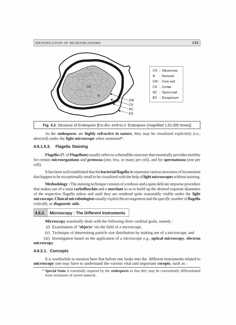
IDENTIFICATION OF MICROORGANISMS 133
Fig. 4.2. Structure of Endospore [Bacillus anthracis Endospore (magnified 1,51,000 times)]
As the endospores are highly refractive in nature, they may be visualized explicitely (i.e.,detected) under the light microscope when unstained*.
4.6.1.5.3. Flagella Staining
Flagella (Pl. of Flagellum) usually refers to a threadlike structure that essentially provides motilityfor certain microorganisms and protozoa (one, few, or many per cell), and for spermatozoa (one percell).
It has been well established that the bacterial flagella do represent various structures of locomotionthat happen to be exceptionally small to be visualized with the help of light microscopes without staining.
Methodology : The staining technique consists of a tedious and a quite delicate stepwise procedurethat makes use of a stain carbolfuschin and a mordant so as to build up the desired requisite diametersof the respective flagella unless and until they are rendered quite reasonably visible under the lightmicroscope. Clinical microbiologists usually exploit the arrangement and the specific number of flagellacritically as diagnostic aids.
4.6.2. Microscopy : The Different Instruments
Microscopy essentially deals with the following three cardinal goals, namely :(i) Examination of ‘objects’ via the field of a microscope,
(ii) Technique of determining particle size distribution by making use of a microscope, and(iii) Investigation based on the application of a microscope e.g., optical microscopy, electron
microscopy.
4.6.2.1. Concepts
It is worthwhile to mention here that before one looks into the different instruments related tomicroscopy one may have to understand the various vital and important cocepts, such as :
* Special Stain is essentially required by the endospores so that they may be conveniently differentiatedfrom inclusions of stored material.
134 PHARMACEUTICAL MICROBIOLOGY
Light microscopes usually make use of glass lenses so as to either bend or focus theemerging light rays thereby producing distinct enlarged images of tiny objects. A light micro-scope affords resolution which is precisly determined by two guiding factors, namely :
(a) Numerical aperture of the lens-system, and(b) Wavelength of the light it uses :
However, the maximum acheivable resolution is approximately. 0.2 μμμμμm.Light microscopes that are commonly employed are : the Bright field, Darkfield, Phase-
contrast and Fluorescence microscopes. Interestingly, each different kind of these variants giverise to a distinctive image ; and, hence may be specially used to visualize altogether differentprevailing aspects of the so called microbial morphology.
As a rather good segment of the microorganisms are found to be almost virtually colour-less ; and, therefore, they are not so easily visible in the Bright field Microscope directly whichmay be duly fixed and stained before any observation.
One may selectively make use of either simple or differential staining (see Section 4.6.1.1.)to spot and visualize such particular bacterial structures as : capsules, endospores and flagella.
The Transmission Electron Microscope accomplishes real fabulous resolution (approx.0.5 nm) by employing direct electron beams having very short wave length in comparison to thevisible light.
The Scanning Electron Microscope may used to observe the specific external featuresquite explicitely, that produces an image by meticulously scanning a fine electron beam onto thesurface of specimens directly in comparison to the projection of electrons through them.
Advent of recent advances in research has introduced two altogether newer versions ofmicroscopy thereby making a quantam jump in the improvement and ability to study the microor-ganisms and molecules in greater depth, such as : (a) Scanning Probe Microscope ; and (b) Scan-ning Laser Microscope.
4.6.2.2. Microscope Variants
Microbiology invariably deals with a host of microorganisms which are practically invisiblewith the unaided eye. This particular discipline essentially justifies the evolution of a variety of micro-scopes with crucial importance so that the scientists could carry out an elaborated and meaningfulresearch.
The variants in microscopes are as stated under :(a) Bright-field Microscope,(b) Dark-field Microscope,(c) Phase-contrast Microscope,(d) Differential Interference Contrast (DIC) Microscope,(e) Fluorescence Microscope, and(f) Electron Microscope.The aforesaid microscope variants shall now be treated individually and briefly in the sections
that follows.
4.6.2.2.1. Bright-Field Microscope
In actual, practice, the ‘ordinary microscope’ is usually refereed to as a bright-field micro-scope by virtue of the fact that it gives rise to a distinct dark image against a brighter background.
Description : Bright-field microscope essentially comprises of a strong metalic body with abase and an arm to which the various other components are duly attached as shown in Fig. 4.3 (a). It isprovided with a ‘light source’ either an electric bulb (illuminator) or a plano-concave mirror strategi-
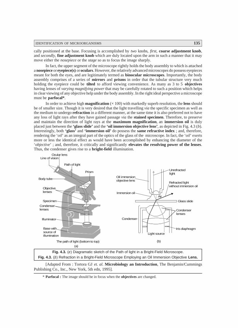
IDENTIFICATION OF MICROORGANISMS 135
cally positioned at the base. Focusing is accomplished by two knobs, first, coarse adjustment knob,and secondly, fine adjustment knob which are duly located upon the arm in such a manner that it maymove either the nosepiece or the stage so as to focus the image sharply.
In fact, the upper segment of the microscope rightly holds the body assembly to which is attacheda nosepiece or eyepiece(s) or oculars. However, the relatively advanced microscopes do possess eyepiecesmeant for both the eyes, and are legitimately termed as binocular microscopes. Importantly, the bodyassembly comprises of a series of mirrors and prisms in order that the tubular structure very muchholding the eyepiece could be tilted to afford viewing convenience. As many as 3 to 5 objectiveshaving lenses of varying magnifying power that may be carefully rotated to such a position which helpsin clear viewing of any objective help under the body assembly. In the right ideal perspective a microscopemust be parfocal*.
In order to achieve high magnification (× 100) with markedly superb resolution, the lens shouldbe of smaller size. Though it is very desired that the light travelling via the specific specimen as well asthe medium to undergo refraction in a different manner, at the same time it is also preferred not to haveany loss of light rays after they have gained passage via the stained specimen. Therefore, to preserveand maintain the direction of light rays at the maximum magnification, an immersion oil is dulyplaced just between the ‘glass slide’ and the ‘oil immersion objective lens’, as depicted in Fig. 4.3 (b).Interestingly, both ‘glass’ and ‘immersion oil’ do possess the same refractive index ; and, therefore,rendering the ‘oil’ as an integral part of the optics of the glass of the microscope. In fact, the ‘oil’ exertsmore or less the identical effect as would have been accomplished by enhancing the diameter of the‘objective’ ; and, therefore, it critically and significantly elevates the resolving power of the lenses.Thus, the condenser gives rise to a bright-field illumination.
Line of visionOcular lens
Path of light
Prism
Body tube
Objectivelenses
Specimen
Condenserlenses
Illuminator
Base withsource ofillumination
Light source
Iris diaphragm
Condenserlenses
Condenser
Immersion oil
Oil immersionobjective lens
Unrefractedlight
Refracted lightwithout immersion oil
(a)
(b)The path of light (bottom to top)
Air
Glass slide
Fig. 4.3. (a) Diagramatic sketch of the Path of light in a Bright-Field Microscope.Fig. 4.3. (b) Refraction in a Bright-Field Microscope Employing an Oil Immersion Objective Lens.
[Adapted From : Tortora GJ et. al. Microbiology an Introduction, The Benjamin/CummingsPublishing Co., Inc., New York, 5th edn, 1995].
* Parfocal : The image should be in focus when the objectives are changed.
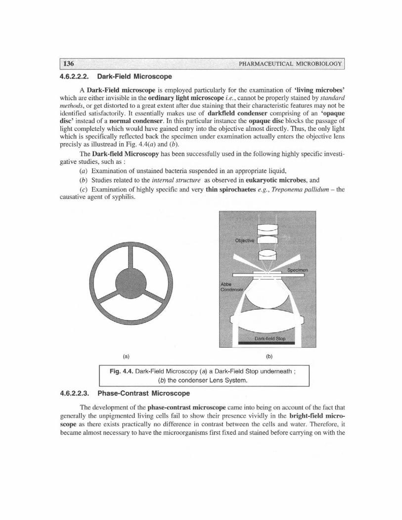
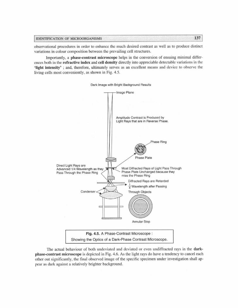
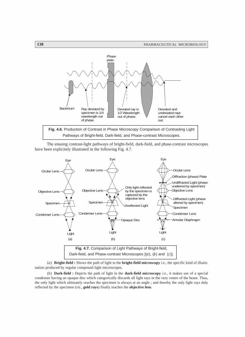
138 PHARMACEUTICAL MICROBIOLOGY
Phaseplate
Bacterium Ray deviated byspecimen is 1/4wavelength outof phase.
Deviated ray is1/2 Wavelengthout of phase.
Deviated andundeviated rayscancel each otherout.
Fig. 4.6. Production of Contrast in Phase Microscopy Comparison of Contrasting Light
Pathways of Bright-field, Dark-field, and Phase-contrast Microscopes.
The ensuing contrast-light pathways of bright-field, dark-field, and phase-contrast microscopeshave been explicitely illustrated in the following Fig. 4.7.
Ocular Lens
Objective Lens
Specimen
Condenser Lens
Light
Eye Eye
Ocular Lens
Objective Lens
Specimen
Condenser Lens
Light
Only light reflectedby the specimen iscaptured by theobjective lens
Unreflected Light
Opaque Disc
Ocular Lens
Objective Lens
Specimen
Condenser Lens
Annular Diaphragm
Diffracted Light (phasealtered by specimen)
Undiffracted Light (phaseunaltered by specimen)
Diffraction (phase) Plate
Eye
Light
(a) (b) (c)
Fig. 4.7. Comparison of Light Pathways of Bright-field,
Dark-field, and Phase-contrast Microscopes [(a), (b) and (c)].
(a) Bright-field : Shows the path of light in the bright-field microscopy i.e., the specific kind of illumi-nation produced by regular compound light microscopes.
(b) Dark-field : Depicts the path of light in the dark-field microscopy i.e., it makes use of a specialcondenser having an opaque disc which categorically discards all light rays in the very centre of the beam. Thus,the only light which ultimately reaches the specimen is always at an angle ; and thereby the only light rays dulyreflected by the specimen (viz., gold rays) finally reaches the objective lens.
IDENTIFICATION OF MICROORGANISMS 139
(c) Phase-contrast : Illustrates the path of light in the phase-contrast microscopy i.e., the light rays aremostly difracted altogether in a different manner ; and, therefore, do travel various pathways to reach the eye of theviewer. Thus, the diffracted light rays are duly indicated in gold ; whereas, the undiffracted light rays are dulyshown in red.
4.6.2.2.4. Differential Interference Contrast (DIC) Microscope
The differential interference contrast (DIC microscope bears a close resemblance to the phase-contrast microscope (Section 4.6.2.2.3.) wherein it specifically produces an image based upon theensuing differences in two fundamental physical parameters, namely : (a) refractive indices ; and (b)thickness. In acutal practice, two distinct and prominent beams of plane-polarized light strategicallyheld at right angles to each other are duly produced by means of prisms. Thus, in one of the particularset-ups, first the object beam happens to pass via the specimen ; and secondly the reference beam ismade to pass via a clear zone in the slide. Ultimately, after having passed via the particular specimen,the two emerging beams are combined meticulously thereby causing actual interference with eachother to give rise to the formation of an ‘image’.
Applications : DIC-microscope helps to determine :
(1) Live, unstained specimen appears usually as 3D highly coloured images.
(2) Clear and distinct visibility of such structures : cell walls, granules, vacuoles, eukaryoticnuclei, and endospores.
Note : The resolution of a DIC-Microscope is significantly higher in comparison to a standard phase-contrast microscope, due to addage of contrasting colours to the specific specimen.
4.6.2.2.5. Fluorescence Microscope
Interestingly, the various types of microscopes discussed so far pertinently give rise to an ‘im-age’ from light which happens to pass via a specimen. It is, however, important to state here that thefluorescence microscopy exclusively based upon the inherent fluorescence characteristic feature ofa ‘substance’ i.e., the ability of an object (substance) to emit light distinctly. One may put forward aplausible explanation for such an unique physical phenomenon due to the fact that ‘certain molecules’do absorb radiant* energy thereby rendering them highly excited ; however, at a later convenientstage strategically release a reasonable proportion of their acquired trapped energy in the form of ‘light’(an energy). It has been duly proved and established that any light given out by an excited moleculeshall possess definitely a longer wavelength (i.e., having lower energy) in comparison to the raditionabsorbed initially’.
Salient Features : There are certain ‘salient features’ with regard to the fluorescence microscopyas stated under :
(1) Quite a few microbes do fluoresce naturally on being subjected to ‘special lighting’.
(2) Fluorochromes : In such an instance when the ‘specimen under investigation’ fails tofluoresce normally, it may be stained adequately with one of a group of fluorescent dyes termed as‘fluorochromes’.
(3) Microorganisms upon staining with a fluorochrome when examined with the help of afluorescence microscope in an UV or near-UV light source, they may be observed as luminescencebright objects against a distinct dark background.
* Radiant : Emitting beams of light.

140 PHARMACEUTICAL MICROBIOLOGY
Examples : A few typical examples are as follows :
(a) Mycobacterium tuberculosis : Auramine O (i.e., a fluorochrome) that usually glows yel-low on being exposed to UV-light, gets strongly absorbed by M. tuberculosis (a pathogenic ‘tuberculo-sis’ causing organism). Therefore, the dye when applied to a specific sample being investigated for thisbacterium, its presence may be detected by the distinct visualization of bright yellow microbes against adark background,
(b) Bacillus anthracis : Fluorescein isothiocyanate (FITC) (i.e., a fluorochrome) stains B.anthracis particularly and appears as ‘apple green’ distinctly. This organism is a causative agent ofanthrax.
(a) Mercury Vapor Arc Lamp
(b) Heat filterExciter FilterAllows only short wavelengthlight (about 400 nm) through
Mirror
(d) Dark-Field CondenserProvides dark Backgroundfor Fluorescence
(e) Specimen Stained with FluorochromeEmits Fluorescence when activatedby exciting wavelength of light
(f) Barrier FilterRemoves any remaining exciterwavelength (up to about 500 nm)without absorbing longer wavelengthsof fluorescing objects
Eyepiece
ObjectiveLens (g)
(c)
Fig. 4.8. Vital Components and Underlying Principles ofOperation of a Fluorescence Microscope.
Fig. 4.8. illustrates the diagramatic sketch of the vital components and the underlying principlesof operation of a Fluorescence Microscope.
IDENTIFICATION OF MICROORGANISMS 141
Methodology : The various steps involved in the operational procedures of a fluorescence mi-croscope are as under :
(1) A particular specimen is exposed to UV-light or blue light or violet light thereby giving riseto the formation of an image of the ‘specified object’ along with the resulting fluorescence light.
(2) A highly intense beam is duly generated either by a Mercury Vapour Lamp (a) or anyother appropriate source ; and the ensuing heat transfer is duly limited by a specially designed Infra-red Filter (b).
(3) Subsequently, the emerged light is made to pass through an Exciter Filter (c) which allowsthe specifc transmission of exclusively the desired wavelength.
(4) A Darkfield Condenser (d) critically affords a black background against which the fluo-rescent objects usually glow.
(5) Invariably the particular specimen is stained with fluorochrome (dye molecule) (e), whichultimately fluoresces brightly on being exposed to light of a particular wavelength ; whereas, there arecertain organisms that are autofluorescing in nature.
(6) A Barrier Filter (f) is strategically positioned after the objective Lenses (g) helps to re-move any residual UV-light thereby causing two important functional advantages, namely :
(i) To protect the viewer’s eyes from getting damaged, and
(ii) To suitably eliminate the blue and violet light thereby minimising the image’s actualcontrast.
Applications : The various useful applications of a Fluorescence Microscope are as enumerated under :
(1) It serves as an essential tool in both ‘microbial ecology’ and ‘medical microbiology’.(2) Important microbial pathogens like : M. tuberculosis may be distinctly identified in two
different modalities, for instance :
(i) particularly labeling the microorganisms with fluorescent antibodies employing highlyspecialized immunofluorescence techniques, and
(ii) specifically staining them (microbes) with fluorochromes.
(3) Ecological investigative studies is usually done by critically examining the specific micro-organisms duly stained with either fluorochromes e.g., acridine orange, and diamidino-2-phenylindole(DAPI)-a DNA specific stain or fluorochrome-labeled probes.
4.6.2.2.6. Electron Microscope
An electron microscope refers to a microscope that makes use of streams of electrons dulydeflected from their course either by an electrostatic or by an electromagnetic field for the magnifica-tion of objects. The final image is adequately viewed on a fluorescent screen or recorded on a photo-graphic plate. By virtue of the fact that an electron microscope exhibits greater resolution, the ensuingimages may be magnified conveniently even upto the extent of 4,00,000 diameters.
It is, however, pertinent to mention here that objects that are smaller than 0.2 μμμμμm, for instance :internal structures of cells, and viruses should be examined, characterized and identified by the aid ofan electron microscope.
Importantly, the electron microscope utilizes only a beam of electrons rather than a ray oflight. The most acceptable, logical, and plausible explanation that an electron microscope affordsmuch prominent and better resolution is solely on account of the fact that the ‘electrons’ do possessshorter wavelengths significantly. Besides, the wavelengths of electrons are approximately 1,00,000times smaller in comparison to the wavelengths of visible light.
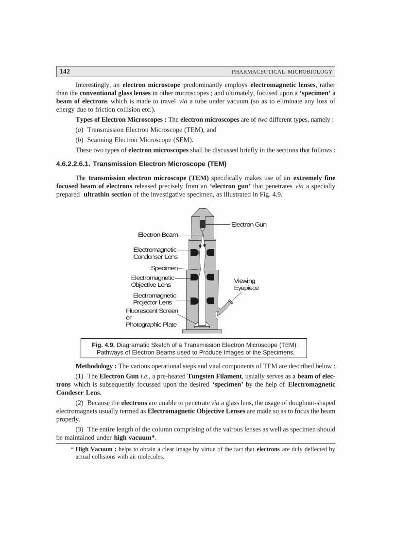
142 PHARMACEUTICAL MICROBIOLOGY
Interestingly, an electron microscope predominantly employs electromagnetic lenses, ratherthan the conventional glass lenses in other microscopes ; and ultimately, focused upon a ‘specimen’ abeam of electrons which is made to travel via a tube under vacuum (so as to eliminate any loss ofenergy due to friction collision etc.).
Types of Electron Microscopes : The electron microscopes are of two different types, namely :
(a) Transmission Electron Microscope (TEM), and
(b) Scanning Electron Microscope (SEM).
These two types of electron microscopes shall be discussed briefly in the sections that follows :
4.6.2.2.6.1. Transmission Electron Microscope (TEM)
The transmission electron microscope (TEM) specifically makes use of an extremely finefocused beam of electrons released precisely from an ‘electron gun’ that penetrates via a speciallyprepared ultrathin section of the investigative specimen, as illustrated in Fig. 4.9.
Electron Gun
Electron Beam
ElectromagneticCondenser Lens
ElectromagneticObjective Lens
Specimen
ElectromagneticProjector Lens
Fluorescent ScreenorPhotographic Plate
ViewingEyepiece
Fig. 4.9. Diagramatic Sketch of a Transmission Electron Microscope (TEM) :Pathways of Electron Beams used to Produce Images of the Specimens.
Methodology : The various operational steps and vital components of TEM are described below :
(1) The Electron Gun i.e., a pre-heated Tungsten Filament, usually serves as a beam of elec-trons which is subsequently focussed upon the desired ‘specimen’ by the help of ElectromagneticCondeser Lens.
(2) Because the electrons are unable to penetrate via a glass lens, the usage of doughnut-shapedelectromagnets usually termed as Electromagnetic Objective Lenses are made so as to focus the beamproperly.
(3) The entire length of the column comprising of the vairous lenses as well as specimen shouldbe maintained under high vacuum*.
* High Vacuum : helps to obtain a clear image by virtue of the fact that electrons are duly deflected byactual collisions with air molecules.
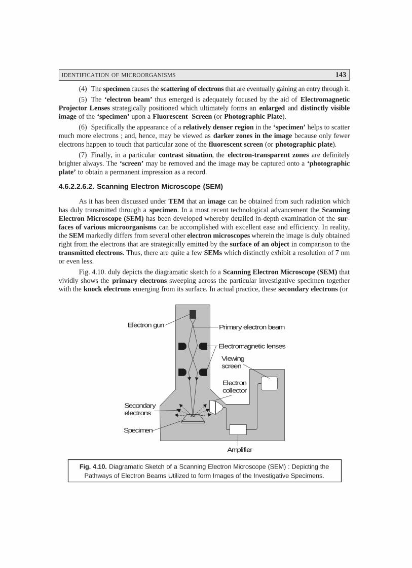
IDENTIFICATION OF MICROORGANISMS 143
(4) The specimen causes the scattering of electrons that are eventually gaining an entry through it.
(5) The ‘electron beam’ thus emerged is adequately focused by the aid of ElectromagneticProjector Lenses strategically positioned which ultimately forms an enlarged and distinctly visibleimage of the ‘specimen’ upon a Fluorescent Screen (or Photographic Plate).
(6) Specifically the appearance of a relatively denser region in the ‘specimen’ helps to scattermuch more electrons ; and, hence, may be viewed as darker zones in the image because only fewerelectrons happen to touch that particular zone of the fluorescent screen (or photographic plate).
(7) Finally, in a particular contrast situation, the electron-transparent zones are definitelybrighter always. The ‘screen’ may be removed and the image may be captured onto a ‘photographicplate’ to obtain a permanent impression as a record.
4.6.2.2.6.2. Scanning Electron Microscope (SEM)
As it has been discussed under TEM that an image can be obtained from such radiation whichhas duly transmitted through a specimen. In a most recent technological advancement the ScanningElectron Microscope (SEM) has been developed whereby detailed in-depth examination of the sur-faces of various microorganisms can be accomplished with excellent ease and efficiency. In reality,the SEM markedly differs from several other electron microscopes wherein the image is duly obtainedright from the electrons that are strategically emitted by the surface of an object in comparison to thetransmitted electrons. Thus, there are quite a few SEMs which distinctly exhibit a resolution of 7 nmor even less.
Fig. 4.10. duly depicts the diagramatic sketch fo a Scanning Electron Microscope (SEM) thatvividly shows the primary electrons sweeping across the particular investigative specimen togetherwith the knock electrons emerging from its surface. In actual practice, these secondary electrons (or
Electron gun Primary electron beam
Electromagnetic lenses
Viewingscreen
Electroncollector
Amplifier
Secondaryelectrons
Specimen
Fig. 4.10. Diagramatic Sketch of a Scanning Electron Microscope (SEM) : Depicting thePathways of Electron Beams Utilized to form Images of the Investigative Specimens.

144 PHARMACEUTICAL MICROBIOLOGY
knock electrons) are meticulously picked up by a strategically positioned collector, duly amplified, andtransmitted onto a viewing screen or a photographic plate (to have a permanent record/impression of theinvestigative specimen).
Methodology : The various steps involved in the operative sequential steps are as stated under :
(1) Specimen preparation : It is quite simple and not so cumbersome ; and even in certaincases one may use air-dried specimen for routine examination directly. In general, largely the microor-ganisms should first be fixed, dehydrated, and dried meticulously so as to preserve not only the so called‘surface-structure’ of the specimen but also to prevent the ‘possible collapse of the cells’ when theseare directly exposed to the SEM’s high vacuum. Before, carrying out the usual viewing activities, thedried samples are duly mounted and carefully coated with a very thin layer of metal sheet in order tocheck and prevent the buildup of an accumulated electrical charge onto the surface of the specimen andto provide a distinct better image.
(2) SEM helps in the scanning of a relatively narrow and tapered electron beam both back andforth onto the surface of the specimen. Thus, when the beam of electron happens to strike a specific zoneof the specimen, the surface atoms critically discharge a small shower of electrons usually termed as‘secondary electrons’, which are subsequently trapped and duly registered by a specially designeddetector.
(3) The ‘secondary electrons’ after gaining entry into the ‘detector’ precisely strike a scintillatorthereby enabling it to emit light flashes which is adequately converted into a stream of elctrical currentby the aid of a photomultiplier tube. Finally, the emerging feeble electrical current is duly amplified.
(4) The resulting signal is carefully sent across to a strategically located cathode-ray tube, andforms a sharp image just like a television picture, that may be either viewed or photographed accord-ingly for record.
Notes :
(i) The actual and exact number of secondary electrons that ultimately reach the ‘detector’ exclu-sively depends upon the specific nature of the surface of the investigative specimen.
(ii) The ensuing ‘electron beam’ when strikes a raised surface area, a sizable large number ‘sec-ondary electrons’ gain due entry into the ‘detector’ ; whereas, fewer electrons do escape adepression in the surface of the specimen and then reach the detector. Therefore, the raisedzones appear comparatively lighter on the screen, and the depressions are darker in appear-ance. Thus, one may obtain a realistic 3D image of the surface of the microorganism having avisible intensive depth of focus.
FURTHER READING REFERENCES
1. Atlas RM : Handbook of Microbiological Media, CRC Press, Boca Raton, Fla. 2nd., 1997.
2. Balows A et al. (eds) : Manual of Clinical Microbiology, American Society for Microbiology,Washington DC, 5th edn, 1991.
3. Clark GL (ed.) : Staining Procedures used by the Biological Stain Commission, Williamsand Wilkins, Baltimore, 3rd edn, 1973.
4. Difco Laboratories : Difco Manual of Dehydrated Culture Media and Reagents forMicrobiology, BD Bioscience, sparks Md. 11th edn, 1998.
5. Kotra LP et al. : Visualizing Bacteria at High Resolution, ASM News, 66 (11) : 675-81,2000.
IDENTIFICATION OF MICROORGANISMS 145
6. Lichtman JW : Confocal Microscopy, Scientific American, 271 (2) : 40-45, August 1994.
7. Lillie RD : HJ Conn’s Biological Stains, Williams and Wilkins, Baltimore, 8th edn, 1969.
8. Lippincott-Schwartz J and Patterson GH : Development and Use of Fluorescent ProteinMarkers in Living cells, Science, 300 : 87-91, 2003.
9. Perkins GA and Frey TG : Microscopy, Optical : In Encyclopedia of Microbiology,Ledenberg J (ed), Academic Press, San Diego, Vol. 3, 2nd edn, pp 288-306, 2000.
10. Perkins GA and Frey TG : Microscopy Confocal : In Encyclopedia of Microbiology,Ledenberg J (ed), Academic Press, San Diego, Vol. 3, 2nd edn., 2000.
11. Postek MT et al. : Scanning Electron Microscopy : A student’s Handbook, Ladd ResearchIndustries, Burlington Vt. 1980.
12. Power DA (ed) : Manual of BBL Products and Laboratory Procedures, Becton, Dickinson,and Co., Cockeysville, Md. 6th edn, 1988.
13. Scherrer Rene : Gram’s Staining Reaction, Gram Types and Cell Walls of Bacteria,Trends Biochem. Sci., 9 : 242-45, 1984.
14. Stephens DJ and Alian VJ : Light Microscopy Techniques for Live Cell Imaging, Science,300 : 82-86, 2003.
15. Taylor DI et al. : The New Vision of Light Microscope, American Scientist, 90 (4) : 322-335, July-August, 1992.
16. Wischnitzer, S : Introduction to Electron Microscopy, Pergamon Press, New York, 3rdEdn., 1981.
146
5.1. INTRODUCTION
In a rather broader perspective the ‘bacteria’ are markedly distinguished by their inherent extrememetabolic diversity ; whereas, a few of them may conveniently sustain themselves exclusively on‘inorganic substances’ by strategically making use of such specific pathways which are practicallyabsent amongst the plant as well as animal kingdoms.
Based upon the aforesaid statement of facts one may individually explore and exploit the variouscardinal factor(s) that essentially govern the nutrition, cultivation (growth), and isolation of bacteria,actinomycetes, fungi and viruses as enumerated under :
5.2. BACTERIA
The nutrition, cultivation (growth), and isolation of bacteria shall be dealt with in the sectionsthat follows :
5.2.1. Nutrition of Microorganisms (Bacteria)
Interestingly, the microbial cell represents an extremely complex entity, which is essentiallycomprised of approximately 70% of by its weight as water, and the remaining 30% by its weight as thesolid components. Besides, the two major gaseous constituents viz., oxygen (O2) and hydrogen (H2) themicrobial cell predominantly consists of four other major elements, namely : Carbon (C), nitrogen(N), sulphur (S), and phosphorus (P). In fact, the six aforesaid constituents almost account for 95% ofthe ensuing cellular dry weight. The various other elements that also present but in relatively muchlesser quantum are : Na+, K+, Ca2+, Mg2+, Mn2+, Co2+, Zn2+, Cu2+, Fe3+ and Mo4+. Based on thesecritical observations and findings one may infer that the microorganisms significantly require an excep-tionally large number of elements for its adequate survival as well as growth (i.e., cultivation).
The following Table 5.1 displays the various chemical composition of an Escherichia coli cell.
NUTRITION, CULTIVATION ANDISOLATION : BACTERIA-ACTINOMYCETES-FUNGI-VIRUSES5
• Introduction• Bacteria• Actinomycetes• Fungi• Viruses

NUTRITION, CULTIVATION AND ISOLATION 147
Table 5.1 : Chemical Composition of an E. coli Cell
S.No. Component
Percentage of Number of Number of
Total Cell Weight Molecules Per Various Types
(%) Cell (Approx.) actually present
1 Water 70 4 × 10¹⁰ 1
2 Inorganic Ions 1 2.5 × 10⁸ 20
3 Carbohydrates and 3 2 × 10⁸ 200
Their Precursors
4 Amino Acids and 0.4 3 × 10⁷ 100
Their Precursors
5 Proteins 15 1 × 10⁶ 2000 – 3000
6 Nucleotides and 0.4 1.2 × 10⁷ 200
Their Precursors
7 Lipids and Their 2 2.5 × 10⁷ 50
Precursors
8 Other Relatively 0.2 1.5 × 10⁷ 250
Small Molecules
9 Nucleic Acids
• DNA 1 1 – 4 1
• RNA 6 5 × 10⁵ 1000
[Adapted From : Tauro P et al. An Introduction to Microbiology, New Age International, NewDelhi, 2004]
It has been amply proved and established that carbon represents an integral component of almostall organic cell material ; and, hence, constitutes practically half of the ensuing dry cell weight.Nitrogen is more or less largely confined to the proteins, coenzymes, and the nucleic acids (DNA,RNA). Sulphur is a vital component of proteins and coenzymes ; whereas, phosphorus designates asthe major component of the nucleic acids.
It is, however, pertinent to mention here that as to date it is not possible to ascertain the preciserequirement of various elements viz. C, N, S and O, by virtue of the fact that most bacteria predomi-nantly differ with regard to the actual chemical form wherein these elements are invariably consumed asnutrients.
5.2.2. Cultivation (Growth) of Bacteria
The cultivation (growth) of bacteria may be defined, as — ‘a systematic progressive increasein the cellular components’. Nevertheless, an appreciable enhancement in ‘mass’ exclusively may notalways reflect the element of growth because bacteria at certain specific instances may accumulateenough mass without a corresponding increment in the actual cell number. In the latest scenario theterms ‘balanced growth’ has been introduced which essentially draws a line between the so called‘orderly growth’ and the ‘disorderly growth’.
148 PHARMACEUTICAL MICROBIOLOGY
Campbell defined ‘balanced growth’ as — ‘the two-fold increase of each biochemical unit ofthe cells very much within the prevailing time period by a single division without having aslightest change in the rate of growth’. However, one may accomplish theoretically cultures with a‘balanced growth’ having a more or less stable and constant chemical composition, but it is rather nextto impossible to achieve this.
Following are some of the cardinal aspects of cultivation of bacteria, such as :
5.2.2.1. Binary Fission
It has been established beyond any reasonable doubt that the most abundantly available means ofbacterial cultivation (reproduction) is binary fission, that is, one specific cell undergoes division togive rise to the formation of two cells.
Now, if one may start the process with a single bacterium, the corresponding enhancement inpopulation is given by the following geometric progression :
1 —→ 2 —→ 22 —→ 23 —→ 2′ —→ 25 —→ 26 —→ 2n
where, n = Number of generations.
Assuming that there is no cell death at all, each succeding generation shall give rise to double itspopulation. Thus, the total population ‘N’ at the end of a specific given time period may be expressedas follows :
N = 1 × 2n ...(a)
Furthermore, under normal experimental parameters, the actual number of organisms N0 inocu-lated at time ‘zero’ is not ‘1’ but most probably may range between several thousands. In such a situa-tion, the aforesaid ‘formula’ may now be given as follows :
N = N0 × 2n ...(b)
Now, solving Eqn. (b) for the value of ‘n’, we may have :
log10 N = log10 N0 + n log10 2
or n = 10 10 0
10
log N log N
log 2
−...(c)
Substituting the value of log10 2 (i.e., 0.301) in Eqn. (c) above, we may ultimately simplify theequation to :
n = 10 10 0log N log N
0.301
−
or n = 3.3 (log10 N – log10 N0) ...(d)
Application of Eqn. (d), one may calculate quite easily and conveniently the actual ‘number ofgenerations’ which have virtually occurred, based on the precise data with respect to the following twoexperimental stages, namely :
(i) Initial population of bacteria, and
(ii) Population after growth affected.
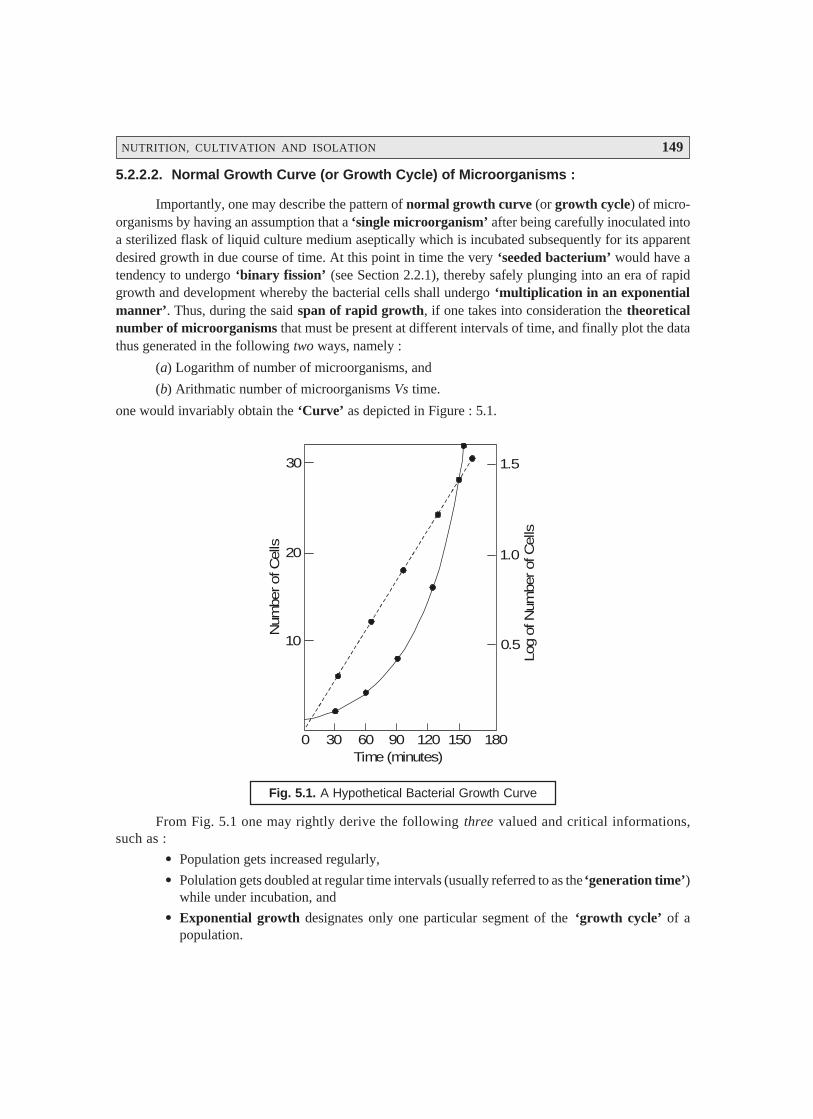
NUTRITION, CULTIVATION AND ISOLATION 149
5.2.2.2. Normal Growth Curve (or Growth Cycle) of Microorganisms :
Importantly, one may describe the pattern of normal growth curve (or growth cycle) of micro-organisms by having an assumption that a ‘single microorganism’ after being carefully inoculated intoa sterilized flask of liquid culture medium aseptically which is incubated subsequently for its apparentdesired growth in due course of time. At this point in time the very ‘seeded bacterium’ would have atendency to undergo ‘binary fission’ (see Section 2.2.1), thereby safely plunging into an era of rapidgrowth and development whereby the bacterial cells shall undergo ‘multiplication in an exponentialmanner’. Thus, during the said span of rapid growth, if one takes into consideration the theoreticalnumber of microorganisms that must be present at different intervals of time, and finally plot the datathus generated in the following two ways, namely :
(a) Logarithm of number of microorganisms, and
(b) Arithmatic number of microorganisms Vs time.
one would invariably obtain the ‘Curve’ as depicted in Figure : 5.1.
30
20
10
0 30 60 90 120 150 180
0.5
1.5
1.0
Num
ber of
Cel
ls
Log
of N
umbe
r of
Cel
ls
Time (minutes)
Fig. 5.1. A Hypothetical Bacterial Growth Curve
From Fig. 5.1 one may rightly derive the following three valued and critical informations,such as :
• Population gets increased regularly,
• Polulation gets doubled at regular time intervals (usually referred to as the ‘generation time’)while under incubation, and
• Exponential growth designates only one particular segment of the ‘growth cycle’ of apopulation.

150 PHARMACEUTICAL MICROBIOLOGY
5.2.2.3. The Lag Phase of Microbial Growth
In actual practice, however, when one carefully inoculates a fresh-sterilized culture medium witha stipulated number of cells, subsequently finds out the ensuing bacterial population intermittentlyunder the following two experimental parameters :
(a) during an incubation period of 24 hours, and
(b) plot the curve between logarithms of the number of available microbial cells Vs time (inminutes),
thereby obtaining a typical curve as illustrated in Fig. 5.2.
Time (Hours)
Log
of N
umbe
rs o
f Via
ble
Bac
teria
A
B
C
D
Fig. 5.2. Bacterial Growth Curve Showing Four Phases
Curve A : Lag Phase ; Curve B : Exponential Phase or Log (Logarithmic) Phase ;
Curve C : Stationary Phase ; and Curve D : Death (or Decline) Phase.
From Fig. 5.2. one may distinctly observe the following salient features :
• Lag Phase — i.e., at initial stages there exist almost little growth of bacteria,
• Exponential (or Log) Phase — i.e., showing a rather rapid growth,
• Stationary Phase — i.e., depicting clearly a levelling off growth of microbes, and
• Death (or Decline) Phase — i.e., showing a clear cut decline in the viable population ofmicroorganisms.
5.2.2.4. Translational Periods Between Various Growth Phases
A close look at Fig. 5. 2 would reveal that a culture invariably proceeds rather slowly from oneparticular phase of growth to the next phase. Therefore, it categorically ascertains the fact that all thebacterial cells are definitely not exposed to an identical physiological condition specifically as theyapproach toward the end of a given phase of growth. Importantly, it involves critically the ‘time factor’essentially needed for certain bacteria to enable them catch up with the others in a crowd of microbes.

NUTRITION, CULTIVATION AND ISOLATION 151
5.2.2.5. Synchronous Growth
It has been duly observed that there are quite a few vital aspects with regard to the internsivemicrobiological research wherein it might be possible to decepher and hence relate the various aspectsof the bacterial growth, organization, and above all the precise differentiation to a specific stage of thecell-division cycle. However, it may not be practically feasible to carry out the analysis of a singlebacterium due to its extremely small size. At this stage if one may assume that all the available cells ina culture medium were supposed to be having almost the same stage of the specific division cycle, theultimate result from the ensuing analysis of the cell crop might be logically interpreted equivalent to asingle cell. With the advent of several well elaborated and practised laboratory methodologies onecould conveniently manipulate the on going growth of cultures whereby all the available cells shallessentially be in the same status of their ensuing growth cycle. i.e., having a synchronus growth.
Salient Features : The various salient features pertaining to the aforesaid synchronous growthare as stated under :
(1) Synchrony invariably lasts for a few generations, because even the daughters of a singlecell usually get out of phase with one another very much within a short span.
(2) The prevailing population may be synchronized judiciously by carrying out the manipula-tion either of the chemical composition of the culture medium or by altering the physicalenvironment of the culture medium.
Example : The above hypothesis may be expatiated by subjecting the bacterial cells to a carefulinoculation into a culture medium duly maintained at a suboptimal temperature. Interestingly, underthese prevailing circumstances after a certian lapse of time the bacterial cells shall metabolize gradu-ally, but certainly may not undergo cell division. However, when the temperature is enhanced from thesuboptimal level to the elevated stage, the bacterial cells shall undergo a synchronized division.
Time
Log
of N
umbe
r of
Cel
ls
Random Cell Divisions
Synchronous Cell Divisions
Fig. 5.3. The Synchronous Growth of Microorganism
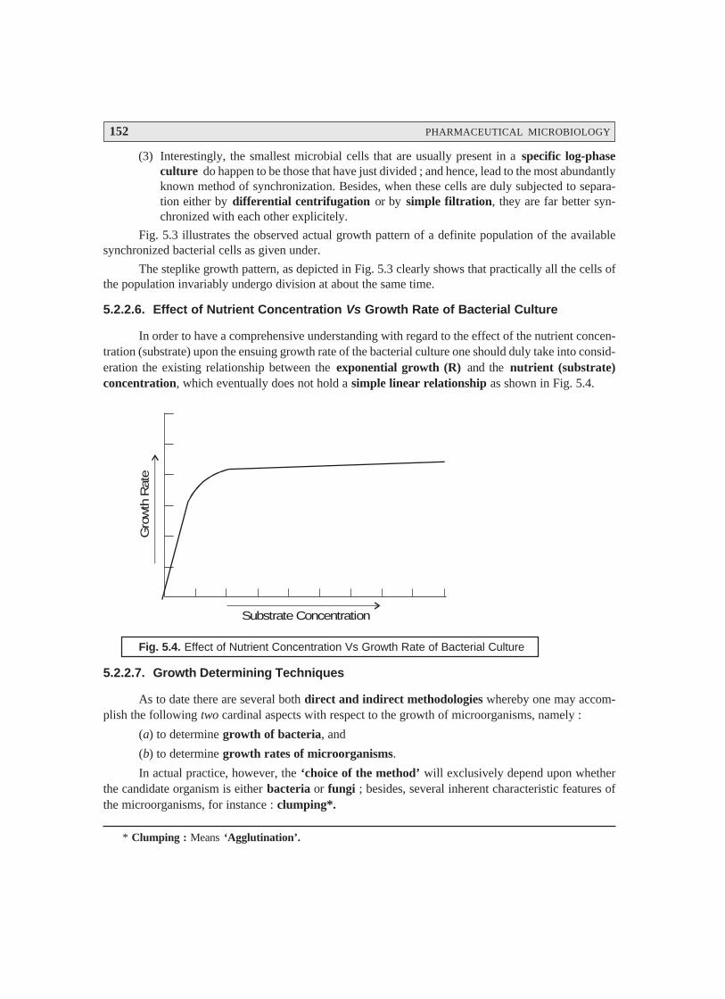
152 PHARMACEUTICAL MICROBIOLOGY
(3) Interestingly, the smallest microbial cells that are usually present in a specific log-phaseculture do happen to be those that have just divided ; and hence, lead to the most abundantlyknown method of synchronization. Besides, when these cells are duly subjected to separa-tion either by differential centrifugation or by simple filtration, they are far better syn-chronized with each other explicitely.
Fig. 5.3 illustrates the observed actual growth pattern of a definite population of the availablesynchronized bacterial cells as given under.
The steplike growth pattern, as depicted in Fig. 5.3 clearly shows that practically all the cells ofthe population invariably undergo division at about the same time.
5.2.2.6. Effect of Nutrient Concentration Vs Growth Rate of Bacterial Culture
In order to have a comprehensive understanding with regard to the effect of the nutrient concen-tration (substrate) upon the ensuing growth rate of the bacterial culture one should duly take into consid-eration the existing relationship between the exponential growth (R) and the nutrient (substrate)concentration, which eventually does not hold a simple linear relationship as shown in Fig. 5.4.
Substrate Concentration
Gro
wth
Rat
e
Fig. 5.4. Effect of Nutrient Concentration Vs Growth Rate of Bacterial Culture
5.2.2.7. Growth Determining Techniques
As to date there are several both direct and indirect methodologies whereby one may accom-plish the following two cardinal aspects with respect to the growth of microorganisms, namely :
(a) to determine growth of bacteria, and
(b) to determine growth rates of microorganisms.
In actual practice, however, the ‘choice of the method’ will exclusively depend upon whetherthe candidate organism is either bacteria or fungi ; besides, several inherent characteristic features ofthe microorganisms, for instance : clumping*.
* Clumping : Means ‘Agglutination’.
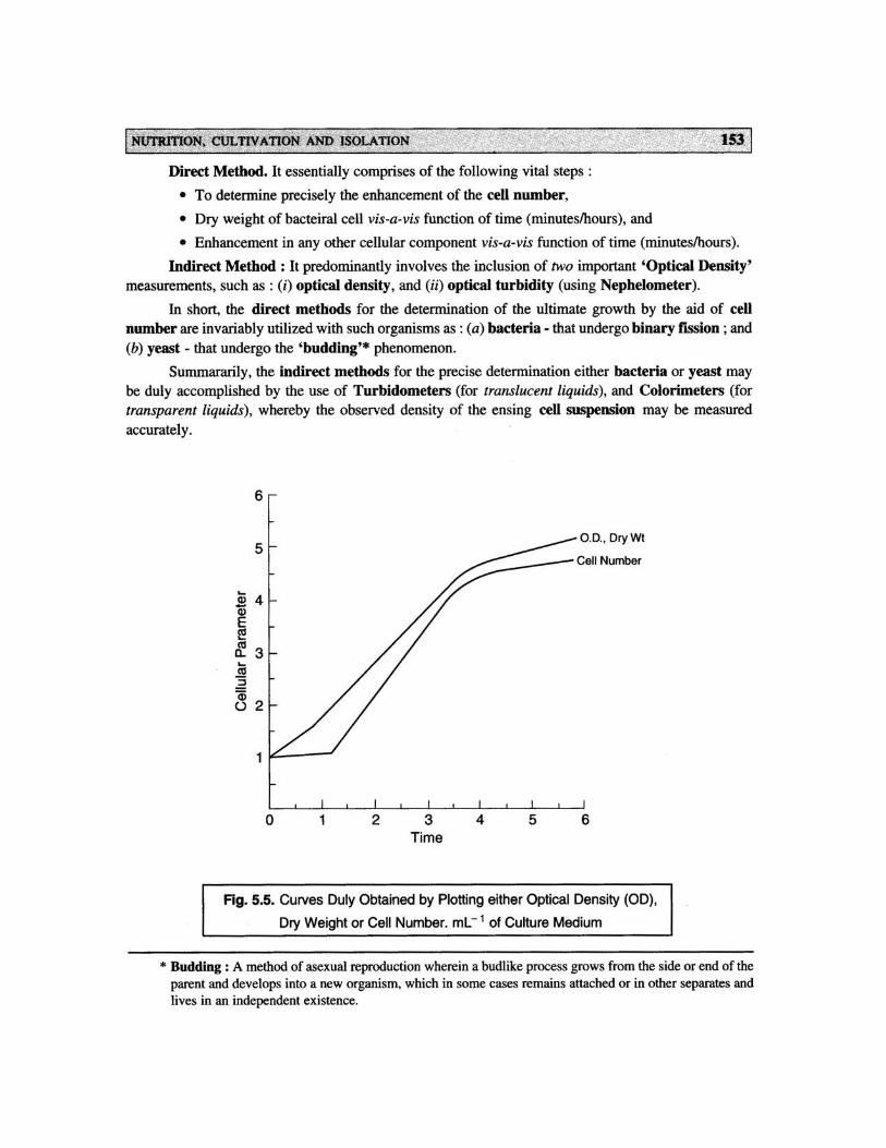
154 PHARMACEUTICAL MICROBIOLOGY
Fig. 5.5 illustrates the kind of curves which one obtains when the ensuing growth is invariablymeasured in a liquid medium by various methods. It has been amply proved and established that theactual changes which take place in the cell population strategically after the inoculation into the freshgrowth medium are represented more accurately and precisely by the dry weight or optical densitymeasurements.
5.2.3. Isolation of Bacteria
The isolation of ‘Bacteria’ may be accomplished in several recognized and well-establishedmethods, such as :
(a) Selective and diagnostic media,
(b) Bismuth sulphite agar, and
(c) Selective media for staphylococci.
The aforesaid three methodologies invariably used for the isolation of bacteria shall be treatedindividually in the sections that follows :
5.2.3.1. Selective and Diagnostic Media
McConkey’s medium was first and foremost introduced in 1995 so as to isolateEnterobacteriaceae from faeces, urine, foods, water etc. The medium essentially comprises of severalnutrients viz., bile salts, lactose, and an appropriate indicator.
Bile salts categorically serve as an important natural surface-active agent which, fails toinhibit the growth of the Enterobacteriaceae, but distinctly inhibits the growth of the specific Gram-positive microorganisms that are probably present in the material to be examined.
Lactose aids in the production of ‘acid’ from E. coli and A. aerogenes upon this culture mediumthereby changing the colour of the suitable indicator added ; besides, the said two microorganisms mayalso adsorb a certain amount of the indicator that may eventually get duly precipitated around thegrowing cells. Importantly, the bacteria responsible for causing typhoid and paratyphoid fever, andbacillary dysentery fail to ferment lactose ; and, therefore, the resulting colonies produced duly bythese organisms appear to be absolutely transparent.
Modifications of McConkey’s Medium — are as stated under :
(1) Synthetic surface-active agent may replace the ‘Bile Salts’,
(2) Selectivity of McConkey’s medium may be enhanced significantly by the incorporation ofinhibitory dyes e.g. crystal violet, neutral red. In fact, these dyes particularly suppress the growth ofGram-positive microorganisms viz., Staphylococci.
5.2.3.2. Bismuth Sulphite Agar
The discovery of the bismuth sulphite agar medium dates back to 1920s for the identificationof Salmonella typhi in pharmaceutical preparations, foods, faeces, urine, and water. It essentiallycomprises of a ‘buffered nutrient agar’ consisting of bismuth sulphate, ferrous sulphate, and anindicator brilliant green.
NUTRITION, CULTIVATION AND ISOLATION 155
E. coli gets inhibited at a concentration 0.0025% of brilliant green employed, whereas anotherorganism Salmonella typhi shall grow predominantly. It has been observed that bismuth sulphite doesexert certain degree of inhibitory effect upon E. coli.
S. typhi, in the presence of glucose, causes reduction of bismuth sulphite to the correspondingbismuth sulphide (i.e., a black compound), thereby ascertaining the fact that the investigative organismmay generate H2S from the S-containing amino acids in the medium, which in turn shall interact withFeSO4 to produce a distinct black precipitate of FeS (ferrous sulphide).
5.2.3.3. Selective Media for Staphylococci
The presence of the Staphylococci organisms in various specimens viz., pharmaceutical prod-ucts, food items, and pathological specimens, may ultimately cause food poisoning as well as serioussystemic infections.
A few typical examples of selective media for various organisms are as follows :
(a) Enterobacteria — a surface active agent serves as the main-selector.
(b) Staphylococci — NaCl and LiCl serve as the main selectors. Besides, Staphylococci ingeneral are found to be sufficiently tolerant of NaCl concentrations upto an extent of 7.5%.
5.3. ACTINOMYCETES
Actinomycetes refers to any bacterium of the order Actinomycetales, which essentially includesthe families : Mycobacteriaceae, Actinomycetaceae, Actinoplanaceae, Dermatophilaceae,Micromonosporaceae, Nocardiaceae, and Streptomycetaceae.
In fact, Actinomyces represents a genus of bacteria belonging to the family Actinomycetaceaewhich contain Gram-positive staining filaments. In general, these organisms cause various diseasesboth in humans and animals.
Another school of thought describes actinomycetes as the filamentous microorganisms. It hasbeen duly observed that superficially their morphology very much looks alike that of the filamentousfungi. Nevertheless, the filaments of actinomycetes invariably comprise of the prokaryotic cells hav-ing diameters relatively much smaller in size in comparison to those of the molds. However, there existcertain typical actinomycetes which resemble the molds by making use of externally carried asexualspores for accomplishing the desired reproduction.
Interestingly, actinomycetes are very common inhabitants of soil, whereas filamentous habit ofgrowth has definitely the added advantages. In this manner, the organism can conveniently bridge thewater-free gaps existing between the soil particles to allow them to migrate to a new nutritional site.It is pertinent to state here that this ensuing particular morphology very much provides the organisms anappreciably higher surface-area-to-volume ratio, thereby the nutritional efficiency gets improvedsignificantly in the highly competitive soil environment.
Importantly, the best-known genus of actinomycetes is Streptomyces, which is one of thebacteria most abundantly isolated from soil.
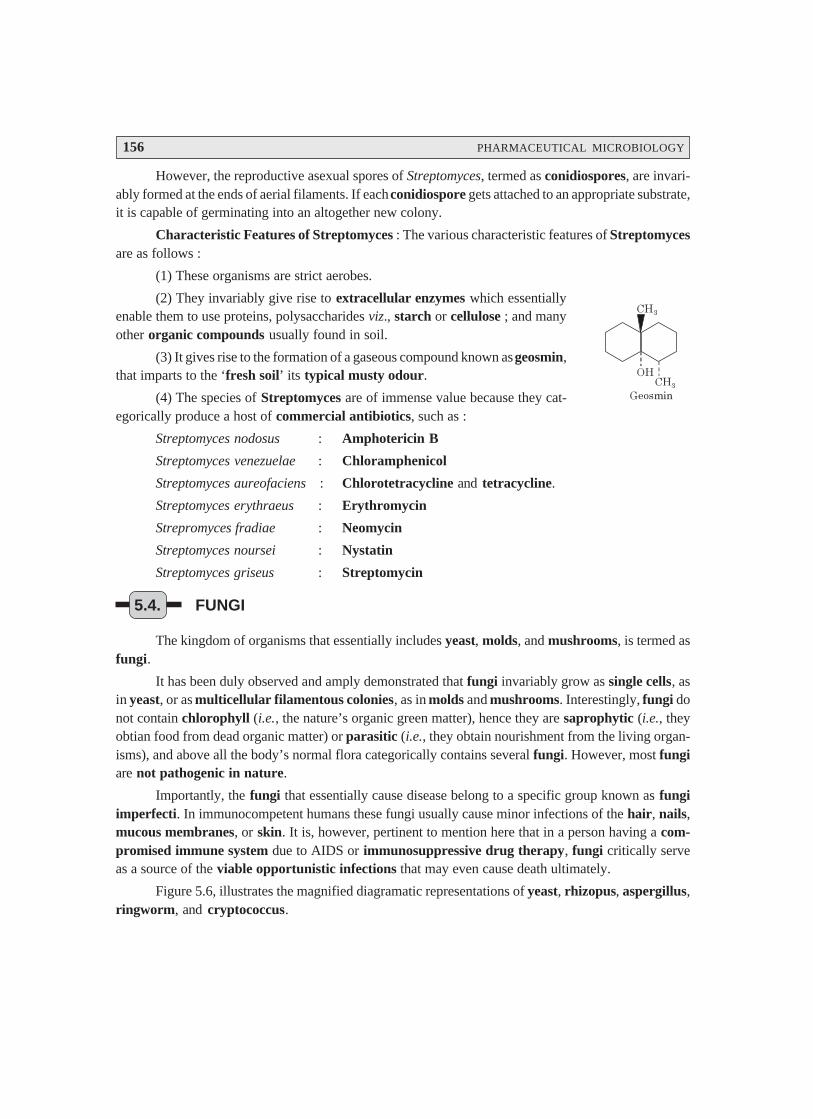
156 PHARMACEUTICAL MICROBIOLOGY
However, the reproductive asexual spores of Streptomyces, termed as conidiospores, are invari-ably formed at the ends of aerial filaments. If each conidiospore gets attached to an appropriate substrate,it is capable of germinating into an altogether new colony.
Characteristic Features of Streptomyces : The various characteristic features of Streptomycesare as follows :
(1) These organisms are strict aerobes.
(2) They invariably give rise to extracellular enzymes which essentiallyenable them to use proteins, polysaccharides viz., starch or cellulose ; and manyother organic compounds usually found in soil.
(3) It gives rise to the formation of a gaseous compound known as geosmin,that imparts to the ‘fresh soil’ its typical musty odour.
(4) The species of Streptomyces are of immense value because they cat-egorically produce a host of commercial antibiotics, such as :
Streptomyces nodosus : Amphotericin B
Streptomyces venezuelae : Chloramphenicol
Streptomyces aureofaciens : Chlorotetracycline and tetracycline.
Streptomyces erythraeus : Erythromycin
Strepromyces fradiae : Neomycin
Streptomyces noursei : Nystatin
Streptomyces griseus : Streptomycin
5.4. FUNGI
The kingdom of organisms that essentially includes yeast, molds, and mushrooms, is termed asfungi.
It has been duly observed and amply demonstrated that fungi invariably grow as single cells, asin yeast, or as multicellular filamentous colonies, as in molds and mushrooms. Interestingly, fungi donot contain chlorophyll (i.e., the nature’s organic green matter), hence they are saprophytic (i.e., theyobtian food from dead organic matter) or parasitic (i.e., they obtain nourishment from the living organ-isms), and above all the body’s normal flora categorically contains several fungi. However, most fungiare not pathogenic in nature.
Importantly, the fungi that essentially cause disease belong to a specific group known as fungiimperfecti. In immunocompetent humans these fungi usually cause minor infections of the hair, nails,mucous membranes, or skin. It is, however, pertinent to mention here that in a person having a com-promised immune system due to AIDS or immunosuppressive drug therapy, fungi critically serveas a source of the viable opportunistic infections that may even cause death ultimately.
Figure 5.6, illustrates the magnified diagramatic representations of yeast, rhizopus, aspergillus,ringworm, and cryptococcus.
����
�
���
�����
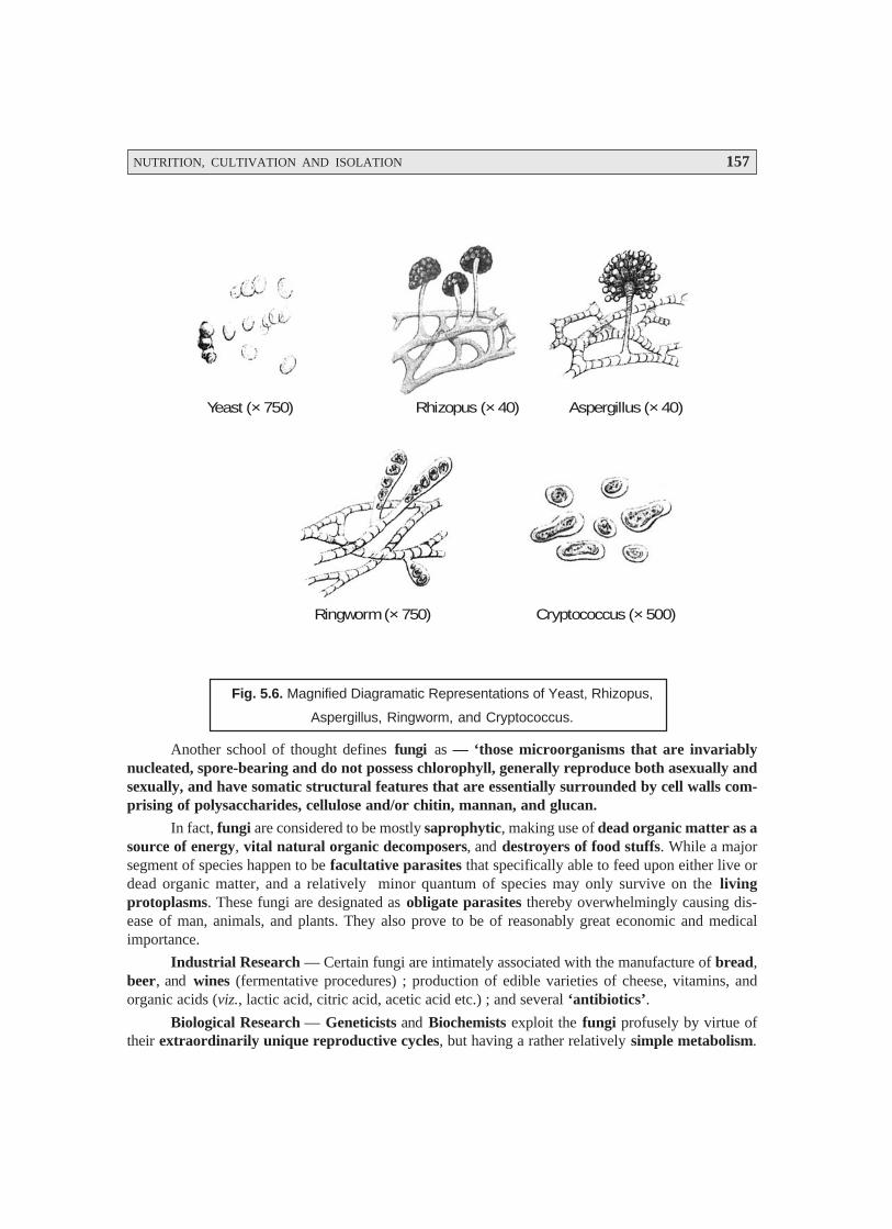
NUTRITION, CULTIVATION AND ISOLATION 157
Yeast (× 750) Rhizopus (× 40) Aspergillus (× 40)
Ringworm (× 750) Cryptococcus (× 500)
Fig. 5.6. Magnified Diagramatic Representations of Yeast, Rhizopus,
Aspergillus, Ringworm, and Cryptococcus.
Another school of thought defines fungi as — ‘those microorganisms that are invariablynucleated, spore-bearing and do not possess chlorophyll, generally reproduce both asexually andsexually, and have somatic structural features that are essentially surrounded by cell walls com-prising of polysaccharides, cellulose and/or chitin, mannan, and glucan.
In fact, fungi are considered to be mostly saprophytic, making use of dead organic matter as asource of energy, vital natural organic decomposers, and destroyers of food stuffs. While a majorsegment of species happen to be facultative parasites that specifically able to feed upon either live ordead organic matter, and a relatively minor quantum of species may only survive on the livingprotoplasms. These fungi are designated as obligate parasites thereby overwhelmingly causing dis-ease of man, animals, and plants. They also prove to be of reasonably great economic and medicalimportance.
Industrial Research — Certain fungi are intimately associated with the manufacture of bread,beer, and wines (fermentative procedures) ; production of edible varieties of cheese, vitamins, andorganic acids (viz., lactic acid, citric acid, acetic acid etc.) ; and several ‘antibiotics’.
Biological Research — Geneticists and Biochemists exploit the fungi profusely by virtue oftheir extraordinarily unique reproductive cycles, but having a rather relatively simple metabolism.
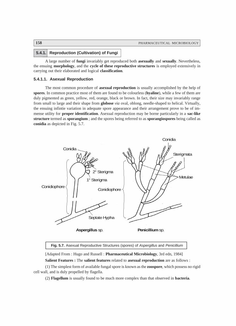
158 PHARMACEUTICAL MICROBIOLOGY
5.4.1. Reproduction (Cultivation) of Fungi
A large number of fungi invariably get reproduced both asexually and sexually. Nevertheless,the ensuing morphology, and the cycle of these reproductive structures is employed extensively incarrying out their elaborated and logical classification.
5.4.1.1. Asexual Reproduction
The most common procedure of asexual reproduction is usually accomplished by the help ofspores. In common practice most of them are found to be colourless (hyaline), while a few of them areduly pigmented as green, yellow, red, orange, black or brown. In fact, their size may invariably rangefrom small to large and their shape from globose via oval, oblong, needle-shaped to helical. Virtually,the ensuing infintie variation in adequate spore appearance and their arrangement prove to be of im-mense utility for proper identification. Asexual reproduction may be borne particularly in a sac-likestructure termed as sporangium ; and the spores being referred to as sporangiospores being called asconidia as depicted in Fig. 5.7.
Conidiophore
Conidia
1° Sterigma
2° Sterigma
Septate Hypha
Conidiophore
Metulae
Sterigmata
Conidia
Aspergillus sp. Penicillium sp.
Fig. 5.7. Asexual Reproductive Structures (spores) of Aspergillus and Penicillium
[Adapted From : Hugo and Russell : Pharmaceutical Microbiology, 3rd edn, 1984]
Salient Features : The salient features related to asexual reproduction are as follows :
(1) The simplest form of available fungal spore is known as the zoospore, which possess no rigidcell wall, and is duly propelled by flagella.
(2) Flagellum is usually found to be much more complex than that observed in bacteria.
NUTRITION, CULTIVATION AND ISOLATION 159
(3) Flagellum is made up of 11 parallel fibrils, of which 9 forming a cylinder and 2 placedcentrally.
(4) Base of flagellum enters the cell and gets attached to the nucleus by a structure termed asrhizoplast.
(5) Flagellum structure (9 + 2 fibrils) is usually found to be fairly consistent with that shownfor other flagellated organisms.
(6) Sporangium designates the asexual reproductive structure pertaining to these aquaticfungi. In its early stages it is found to be loaded with nuclei and protoplasm.
(7) Cleavage takes place subsequently whereby the numerous sections invariably get developedinto the corresponding uninucleate zoospores.
(8) Finally, following a motile phase, the resulting zoospore encysts, losing its flagellum, andrests quietly just prior to germination.
5.4.1.2. Sexual Reproduction
Importantly, the sexual reproduction is characterized by the strategical union of two compat-ible nuclei ; and the entire phenomenon may be distinctly divided into three phases, namely :
Phase I : The union of the gametangia (i.e., sex-organs) brings the nuclei into close proximitywithin the same protoplast. It is also referred to as plasmogamy.
Phase II : It is known as karyogamy, which takes place with the fusion of two nuclei. It has beenduly observed that in the lower fungi the said two processes may take place in immediate sequence ;whereas, in the higher fungi they do occur at two altogether different time periods in the course of theirlife-cycle.
Phase III : It is known as meiosis that essentially takes care of the nuclear fusion whereby theactual number of the chromosomes is distinctly and significantly reduced to its original haploid state.
5.4.2. Industrial Importance of Fungi
There are several vital and important industrial importance of fungi, which shall be enumer-ated briefly as under :
5.4.2.1. Production of Wines and Beer
Natural yeasts have been employed over the centuries in Italy and France, to ferment fruitjuices (wines) or cereal products viz., malt (silent alcohol) in the commercial production of varioustypes of world-class whiskies, rums, vodkas, brandies, gins, and the like. The high-tech industrial manu-facturers of today largely make use of the critical and effective pasteurization of the yeast Saccharomycescerevisiae.
In the production of wine and beer, the lower temperature favours the fermentation of yeast.Under these circumstances the organisms (bacteria) are usually discouraged due to two major reasons,namely :
(a) acidity of the fermentation medium, and
(b) addition of hops that exert a mild inhibitory action to the microorganisms.
160 PHARMACEUTICAL MICROBIOLOGY
Thus, the fermentation invariably takes place under the anaerobic conditions thereby givingrise to the production of alcohol (i.e., ethanol).
Examples : Following are certain typical examples of alcohols commonly used in the manufac-ture of ‘alcoholic beverages’, such as :
(i) Silent Spirits — Spirits obtained by the fractional distilation of alcohol produced by fruit orcereal fermentation.
(ii) Brandy — obtained from wine.
(iii) Whisky — obtained from malted cereals (Barley).
(iv) Rum — obtained from fermented molasses (i.e., a by product from sugar-industrycontaining unrecoverable sugar upto 8–10%).
5.4.2.2. Production of Bakery Products
The baker, strain of Saccharomyces cerevisiae are meticulously selected for their specific highproduction of CO2 under the aerobic parameters. In actual practice, the Baker’s Yeast is particularlymanufactured for bread-making, and is available commonly as ‘dried yeast’ or ‘compressed yeast’.These also find their abundant use as a food supplement by virtue of the fact that are fairly rich inVitamin B variants.
5.4.2.3. Production of Cheeses
There are certain typical fungi which are specifically important in the manufacture of cheeses.
Example : The mould Penicillium roqueforti is usually employed in the production of the blue-veined cheeses. In actual practice, the spores of the fungus are normally used to inoculate the cheese,that is subsequently ‘ripened’ at 9°C in order to discourage the very growth of organisms other than thePenicillium. Because, the moulds happen to be of aerobic nature, adequate perforations are carefullymade in the main bulk of the cheese so as to allow the passage of air to gain entry. However, thedecomposition of fat takes place to impart these cheeses a characteristic flavour.
Interestingly, the mould Penicillium comemberti grows very much on the surface of the cheese,and develops inwards producing the characteristic liquefaction and softening of the surface, i.e., incontrast to the aforesaid P. roqueforti that grows within the body of the cheese.
5.5. VIRUSES
The world has broadly witnessed by 1900 and accepted generally that severlal of the recognizeddreadful human ailments were duly caused by various microorganisms. However, the first and foremostevidence of viruses responsible for causing human disease came into notice in 1892 when Iwanowskirightly demonstrated that the cell-free extracts of the diseased tobacco leaves passed through thebacteria-proof filters may ultimately cause disease in the ‘healthy plants’. Furthermore, such cell-freefiltrates when cultured upon the bacterial growth media they eventually exhibited practically littlegrowth thereby suggesting that the said filtrates contained the actual disease causing agents that areother than microorganisms. Martinus Beijernick, another scientist reconfirmed the excellent epoch makingfindings of Iwanowski.
NUTRITION, CULTIVATION AND ISOLATION 161
Twort and d’Herrelle (1915) individually showed the ‘glassy phenomenon’ present very muchin the microorganisms when it was observed clearly and distinctly that the bacterial cells might beadequately infected with and duly destroyed by the filterable agents, which in turn caused variousserious diseases both affecting the animal kingdom and the plant kingdom. Later on, these diseaseproducing filterable agents are known as bacteriophages (i.e., the bacteria-eaters).
Wendell M Stanley (1935), an American Chemist, first and foremost isolated the tobacco mo-saic virus (genus Tobamovirus) thereby making it possible to perform the chemical as well as struc-tural studies on a purified virus. Interestingly, almost within the same time, the invention of the elec-tron microscope took place which eventually made it quite possible to visualize the said viruses for thefirst time.
The galloping advancement and progress in the in-depth studies on the viruses across the globebased duly upon the latest molecular biology techniques in the 1980s and 1990s have remarkably ledto the discovery of the new dreadful human viruses. In the year 1989, the world has duly acknowledged thediscovery of Hepatitis C virus, and Pestivirus, which specifically causes acute pediatric diarrhoea. Theyear 1993, critically observed the outbreak of a Hantavirus infection occurring exclusively in the South-western USA, which essentially possesses the potential for new infections to emerge at any time.Hantavirus disease refers to the acute ailment related to respirator disease and may even prove fatal.
5.5.1. Bacteriophages
Bacteriophages designated the ‘last group of viruses’ which were duly recognized and bestcharacterized. As to date one may have the evidence for the presence of such disease producing agentsthat are found to be even smaller in size than the viruses, and termed as viriods. They usually consist ofthe nucleic acids (i.e., DNA and RNA) exclusively.
Example : The spindle tuber disease of potatoes is a glaring example of a specific diseaseinvariably caused by the viriods.
A good number of bacteriophages infecting various microorganisms have now been duly iso-lated, characterized, and recognized. The following Table 5.2. records the variuos bacteriophages,host(s), particle dimensions (viz., head and tail in nm), structure, and composition adequately.
Table 5.2 : Characteristic Features of Certain Bacteriophages
Particle Dimensions (nm) Nucleic Acid
S.No. Bacteriophage Host Head Tail Structure(s) (Mol. Wt.)× 106 Daltons
1 T1 E. coli 50 10 × 150 Hexagonal head, DNA (DS*) 27Simple tail
2 T2, T4, T6 E. coli 80 × 110 25 × 110 Prolate, Icosa, Hedral DNA (DS)Head, Complex, tail 105–120with Fibres
3 T3, T7 E. coli 60 10 × 15 Hexagonal Head, DNA (DS) 25Short tail
4 T5 E. coli 65 10 × 170 Hexagonal Head, DNA (DS) 66Simple tail
(Contd.)
162 PHARMACEUTICAL MICROBIOLOGY
1 Single-s trandedDNA, nonenve-loped
Parvoviridae 18–25Dependovirus Depend on coinfection withadenoviruses ; invariablycause faetal death, and gas-troenteritis.
2 Double-strandedDNA, nonenveloped
Adenoviridae Mastadenovirus(adenovirus)
70–90 Medium-sized viruses thatcause various respiratory in-fections in humans ; a few ofthem even produce neo-plasms (tumours) in animals.
5 λλλλλ Phase E. coli 54 10 × 140 —do— DNA (DS) 31
6 SPO1 B. subtilis 90 30 × 120 Hexagonal Head DNA (DS) 105
Complex Tail
7 PM2 Pseudomonas 60 None Hexagonal Head DNA (DS) 6
8 φφφφφ X174, S13 E. coli 27 —do— Icosahedral DNA (SS)**1.7
9 f1, fd, M13 E. coli 5-10 × 800 —do— Filamentous DNA (SS) 1.3
10 M16 Pseudomonas 65 —do— Polyhedral Head RNA (DS) 9.5
11 MS2, f2, Qβββββ E. coli 24 —do— Icosahedral RNA (SS) 1.2
*DS = Double Stranded ; **SS = Single Stranded ;[Adapted From : Tauro P et al., An Introduction to Microbiology, New Age International, New Delhi,2004].
Viral Species : A viral species may be defined as ‘a group of viruses essentially sharing thesame genetic information and ecological niche.
It is, however, pertinent to state here that the particular epithets for viruses have not yet beenestablished completely, thereby logically and emphatically the viral species are duly designated by suchcommon descriptive nomenclatures as : human immunodeficiency virus (HIV), with subspeciesduly indicated by a number (HIV-1).
Standardization of the ‘viral nomenclature’ is now in an active and progressive stage ; and assuch the following specific criteria are being adopted in the latest textbooks and literature alike,namely :
• New viral family• Genus names• Common species names
• Common names are expressed in regular type viz., herpes simplex virus• Genus names are now usually capitalized and italicized viz., Simplexvirus.
Table 5.3. records a comprehensive summary of the latest classification of viruses that invari-ably infect the human beings.
Table 5.3. Latest Classification of Human Viruses
S. No. Characteristic Viral Family
Viral Genus Dimension
Special FeaturesFeatures
(with Represen- of Viriontative species) [Dia. in nm]and UnclassifiedMembers*
(Contd.)
NUTRITION, CULTIVATION AND ISOLATION 163
3 Double-strandedDNA, enveloped
Poxviridae Orthopoxvirus(vaccinia andsmallpox viruses)Molluscipoxvirus
200-350 Very large, complex, brickshaped viruses that causediseases e.g. smallpox (variola),molluscum contagiosum(wartlike skin lesion),cowpox and vaccinia.Vaccinia virus providesspecific immunity tosmallpox.
4 Single-stranded
RNA, nonenveloped+ strand
Picornviridae 28-30 Upto 70 human entero-virusesare known, including the po-lio-, coxsackie-, and echovi-ruses ; more than 100 rhinovi-ruses exist and prove to be themost common cause of colds.
5 Single-strandedRNA, enveloped +strand
Togaviridae A l p h a v i r u s ,Rubivirus, (ru-bella virus)
60-70 Essentially include severalviruses transmitted by ar-thropods (Alphavirus) ; dis-eases include EasternEquine Encephalitis (EEE),Rubella virus is transmittedby the respiratory route.
6 – strand, one strandof RNA
Rhabdoviridae Vesiculovirus (ve-sicular stomatitisvirus), Lyssavirus(rabies virus)
70-180 Bullet-shaped viruses havinga spiked envelope ; invari-ably cause rabies and severalanimal diseases.
7 – strand, multiplestrands of RNA
Orthomyxo-viridae
Inf luenzavirus(Influenza virusesA and B), Influ-enza C virus.
80-200 Envelope spikes can agglu-tinate red blood cells(RBCs).
8 Produce DNA Retroviridae O n c o v i r u s e sLentivirus (HIV)
100-120 Includes all RNA neoplasmviruses and double-strandedRNA viruses. Theoncoviruses invariably causeleukemia and neoplasms inanimals, and the lentivirusHIV causes AIDS.
9 Double-strandedRNA nonenveloped
Reoviridae Reovirus Colo-rado tick fevervirus
60-80 Involved in mild respiratoryinfections ; an unclassifiedspecies causes Colorado tickfever.
E n t e r o v i r u sRhinovirus (Com-mon cold virus),Hepatitis A virus

164 PHARMACEUTICAL MICROBIOLOGY
5.5.2. Growth of Bacteriophages in the Laboratory
It is practically possible to grow the bacteriophages in two different manners, namely :
(a) In suspensions of organisms in liquid media, and
(b) In bacterial cultures on solid media.
Advantages of using Solid Media : In actual practice, the use of solid media makes it feasibleand possible the plaque method for the easy detection and rapid counting of the viruses.
Methodology (Plaque Method) : The various steps that are involved in the ‘plaque method’are as enumerated under :
(1) Sample of bacteriophage is duly mixed with the host bacteria and molten agar.
(2) The resulting agar countaining the various bacteriophages as well as the host bacteria isthen poured carefully into a Petri-plate adequately containing a hardened layer of the agar growthmedium.
(3) In this manner, the ensuing mixture of virus-bacteria gets solidified into a thin top-layerthat invariably comprises of a layer of organisms nearly one-cell thick. This specific step allows eachvirus to infect a bacterium, multiplies subsequently, and helps to release several hundred altogether newviruses.
(4) Nevertheless, these newly generated viruses in turn duly infect other organisms that are presentin the immediate close vicinity ; and hence, more new crop of viruses are produced ultimately.
(5) Thus, several accomplished virus multiplication cycles, all the organisms duly present in thearea surrounding the original virus are destroyed finally. In this way, a good number of ‘clearings’ orplaques are produced, which may be seen against a “lawn” of bacterial growth upon the surface of theagar ; whereas, the plaques are observed to form uninfected microorganisms elsewhere in the Petridish (or Petri plate) undergoing rapid multiplication and giving rise to a turbid background finally.
Note : Each plaque correspond theoretically to a single virus in the initial suspension. Hence, the concentra-tions of viral suspensions measured by the actual number of plaques are invariably expressed interms of plaque-forming units (pfu).
5.5.3. Bacteriophage Lambda : The Lysogenic Cycle
In a broader and precise perspective the bacteriophage may conveniently exist in three phages,namely :
(a) As a free particle virion,
(b) In a lysogenic state as a prophage, and
(c) In the vegetative state i.e., lytic cycle.
One may, however, observe that virion is inert in nature ; and hence, cannot reproduce.
Salient Features : The various salient features of the bacteriophage lambda are as statedunder :
(1) In the critical ‘lysogenic state’, the DNA of the phage is duly integrated very much within thebacterial DNA. It usually exists in a non-infectious form known as the prophage, and adequatelyreplicates in synchrony with the bacterial DNA.
(2) In the corresponding ‘lytic cycle’, the phage particle infects the susceptible host, undergoesmultiplication, and ultimately causes the lysis of the bacterial cell with the concomitant release of theprogeny virus particles.
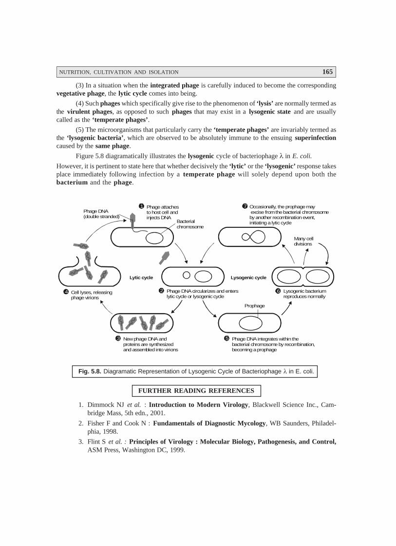
NUTRITION, CULTIVATION AND ISOLATION 165
(3) In a situation when the integrated phage is carefully induced to become the correspondingvegetative phage, the lytic cycle comes into being.
(4) Such phages which specifically give rise to the phenomenon of ‘lysis’ are normally termed asthe virulent phages, as opposed to such phages that may exist in a lysogenic state and are usuallycalled as the ‘temperate phages’.
(5) The microorganisms that particularly carry the ‘temperate phages’ are invariably termed asthe ‘lysogenic bacteria’, which are observed to be absolutely immune to the ensuing superinfectioncaused by the same phage.
Figure 5.8 diagramatically illustrates the lysogenic cycle of bacteriophage λ in E. coli.
However, it is pertinent to state here that whether decisively the ‘lytic’ or the ‘lysogenic’ response takesplace immediately following infection by a temperate phage will solely depend upon both thebacterium and the phage.
Phage DNA(double stranded)
1
4
5
2 6
Phage attachesto host cell andinjects DNA
7 Occasionally, the prophage may excise from the bacterial chromosomeby another recombination event,initiating a lytic cycle
Cell lyses, releasingphage virions
Phage DNA circularizes and enterslytic cycle or lysogenic cycle
Lysogenic bacteriumreproduces normally
Phage DNA integrates within the bacterial chromosome by recombination,becoming a prophage
3 New phage DNA andproteins are synthesizedand assembled into virions
Lytic cycle Lysogenic cycle
Many celldivisions
Bacterialchromosome
Prophage
Fig. 5.8. Diagramatic Representation of Lysogenic Cycle of Bacteriophage λ in E. coli.
FURTHER READING REFERENCES
1. Dimmock NJ et al. : Introduction to Modern Virology, Blackwell Science Inc., Cam-bridge Mass, 5th edn., 2001.
2. Fisher F and Cook N : Fundamentals of Diagnostic Mycology, WB Saunders, Philadel-phia, 1998.
3. Flint S et al. : Principles of Virology : Molecular Biology, Pathogenesis, and Control,ASM Press, Washington DC, 1999.
166 PHARMACEUTICAL MICROBIOLOGY
4. Gulbins E and Lang F : American Scientist, 89 :406-13, 2001.
5. Jotlik WK et al., Zinnser Microbiology, Appleton and Lange, E. Norwalk, Conn., 20th edn,1992.
6. Mandell GL et al. : Principles and Practice of Infectious Diseases, Churchill Livingstone,New York, 2000.
7. Murray PR : Manual of Clinical Microbiology, ASM Press, Washington DC., 8th edn.,2003.
8. Rhen M et al., Trends Microbiol., 11 (2) : 80-86, 2003.
9. Richman D et al. : Clinical Virology, ASM Press, Washington DC, 2002.
10. Roberts LR and Jonovy J : Fundamentals of Parasitology, McGraw Hill, Dubuque, Iowa,6th edn., 2000.
11. Salyers AA and Whitt DD, Bacterial Pathogenesis - A Molecular Approach, ASM Press,Washington DC, 2nd edn, 2001.
12. Sundstrom P : Fungal Pathogens and Host Response, ASM News, 69 (3) : 127-31, 2003.

167
6.1. INTRODUCTION
The microbial genetics as well as the molecular biology specifically and predominantly focusupon the very nature of ‘genetic information’. Besides, it invariably modulates the precise developmentand function of various cells and organisms. In fact, the application of microorganisms has beenenormously useful in mustering a definitely better and exceptionally vivid in-depth understanding of theactual mechanism of ‘gene function’.
Importantly, it has been adequately observed that practically most of the ‘microbial traits’ areeither strategically controlled or logically influenced due to heredity. In true sense, the inherited traitsof microorganisms essentially comprise of the following cardinal aspects :
• shape and structural features i.e., morphology,
• biochemical reactions i.e., metabolism,
• ability to move or behave in different manners, and
• ability to interact with other microorganisms — thereby causing human ailment.
In a rather broader perspective one may consider that the individual organisms prominently dotransmit these characteristic features directly to their offspring via genes, that are nothing but the hereditarymaterials (DNA) which essentially possess relevant information(s) that precisely determines these typicalcharacteristic features.
It has been amply proved and duly established that almost all ‘living organisms’ prominentlyfind it rather advantageous to share the hereditary materials derived from a ‘genetic pool’. However,under the influence of an effective environmental change, the microorganisms that critically possesssuch ‘genes’ which are proved to be advantageous under these new conditions* shall definitely exhibita better chance (scope) of reproduction thereby enhancing their actual numbers in the overall population.
Both eukaryotic and prokaryotic organisms usually exhibit different types of reproductive means,such as :
MICROBIAL GENETICSAND VARIATIONS
* Presumably acquired by random mutations.
6• Introduction• Microbial Genetics• Microbial Variations [Genetic Manipulation in Microorganisms]
168 PHARMACEUTICAL MICROBIOLOGY
Eukaryotic Organisms — invariably make use of ‘sexual reproduction’ having its distinctlyimproved survival value vis-a-vis its sharing capacity with respect to this general ‘gene pool’.
Prokaryotic Organisms — usually do not have the capacity for sexual reproduction as such.Thus, they essentially acquire other mechanisms so as to avoid the ‘genetic uniformity’, which couldprove even ‘fatal’ in the microbial species when certain experimental parameters, namely : developmentof an antibiotic.
Importantly, in the recent past the ‘microbial geneticists’ do play a vital and an important role inthe ever developing field of ‘applied microbiology’ which gives rise to the production of altogether‘newer microbial strains’ that predominantly possess remarkably higher efficiency in the syntheses ofmedicinally and commercially useful end products.
Salient Features : The salient features of the ‘microbial genetics’ are as enumerated under :
(1) Genetic Techniques are largely employed to test such substances that have the ability tocause neoplasm (cancer).
(2) Genetic Engineering is the most recent outcome of the ‘microbial genetics’ and ‘molecularbiology’ that has enormously contributed to various dynamic scientific studies viz., microbiology, biology,and medicine.
(3) Meticulously ‘engineered microorganisms’ are invariably utilized to produce a plethora ofextremely useful ‘life-saving drugs’, for instance : hormones, antibiotics, vaccines, and a host ofother drugs.
(4) ‘New Genes’ may be strategically inserted into the animal and plant species :
Example : Development of wheat and corn nitrogen-fixation genes so that they may not absolutelyrequire nitrogen fertilizers viz., urea.
The earlier investigative studies by Beadle and Tatum, to make use of microorganisms e.g., breadmold Neurospora, provided sufficient vital clues with respect to the ‘genetic control and influence ofthe cellular functions’. Subsequently, bacteria and viruses have actually played an important andmajor role to substantiate and elucidate further the various intricating mechansims of the genetic control.
Advantages : The various cardinal advantages in employing the ‘microorganisms’ for anelaborative genetic studies are as detailed below :
(1) Fast rate of growth e.g., E. coli may duplicate in 20 minutes at 37° C,
(2) Greater ease with which relatively large populations of microbes may be handled in a laboratorye.g., a single sterile petri-dish may hold upto 200–300 colonies,
(3) Relatively much simpler ‘growth media’ for the microbes are needed, and
(4) Much simpler features of the ‘genetic material’ required.
The present subject matter in this particular chapter shall be treated under the following twomajor heads :
(a) Microbial Genetics, and
(b) Microbial Variations.
MICROBIAL GENETICS AND VARIATIONS 169
6.2. MICROBIAL GENETICS
It has been observed that the ‘chromosomes’ are the cellular structures which physically bearuseful hereditary information(s), and the chromosomes actually contain the genes. Besides, genetics isthe science of heredity. It essentially includes the study of genes, ability to carry information, replica-tion mode, passage to subsequent generation of cells or between organisms, and also the critical expres-sion of relevant information available within an organism which ultimately determines the specific en-suing characteristic features of that microorganism.
6.2.1. Structure and Function of Genetic Material
DNA* constitute an integral component of genes**, whereas, nucleotides designate a macro-molecule made up of repeatable units present in DNA. In fact, each nucleotide essentially comprises ofa ‘nitrogenous base’ viz., adenine (A), thymine (T), cytosine (C), and guanine(G) — besides, apentose sugar deoxyribose, together with a ‘phosphate’ moiety. It has been duly established that withina cell the DNA is invariably present as ‘long strands of nucleotides that are duly twisted together inpairs to form a double-helix’.***
Each strand of DNA prominently bears two structural features, namely :
(a) a string of alternating sugar and phosphate moieties, and
(b) a nitrogenous base attached duly to each sugar moiety in its backbone.
Thus, the ‘pair of strands’ are intimately held together strategically by means of H-bonds be-tween their respective nitrogenous bases. However, the ‘pairing of nitrogenous bases’ are found to bein a specific manner i.e., adenine pairs with thymine ; and cytosine pairs with guanine [either AT orCG pairs]. Due to this specific base pairing mode, the base sequence of one DNA strand categoricallydetermines the base sequence of the other strand. Therefore, one may observe that the strands of DNAare actually complementary.**** It has already been proved and established that the aforesaid ‘com-plementary structure of DNA’ goes a long way to expatiate as well as explain the manner by whichthe DNA actually stores and critically transmits the so called ‘genetic information’.
The functional product is invariably a messenger RNA (designated as mRNA) molecule, thateventually results in the formation of a protein. Interestingly, it may also be a ribosomal RNA (i.e.,rRNA). It is, however, pertinent to state here that both these types of RNA (viz., mRNA and rRNA) areprominently involved in the process of protein synthesis.
Genetic Code : It has been duly ascertained that relevant ‘genetic information’ is meticulouslyencoded by the sequence of bases along a specific strand of DNA, which is almost similar to the usageof ‘linear sequence of alphabets’ to first construct ‘words’, and secondly, the ‘sentences’. Impor-tantly, the so called ‘genetic language’ largely makes use of only four letters viz., A, T, C and G
* DNA : Deoxyribonucleic acid ;
** Gene : A gene may be defined as a ‘segment of DNA’ (i.e., a sequence of nucleotides in DNA) whichessentially codes for a functional product.
*** James Watson and Francis Crick double-helix model of DNA.
**** Complementary : Very much similar to the positive and negative of a photograph.

170 PHARMACEUTICAL MICROBIOLOGY
(representing 4-amino acids). However, 1000 of the aforesaid four bases, the number usually containedin an average gene, may be conveniently arranged upto 41000 different variants. Therefore, the usual‘genetic manipulation’ can be accomplished successfully to provide all the ‘necessary vital informa-tions’ a cell essentially requires for its growth and deliver its effective functions based on the astronomi-cal huge number gene variants. In short, the ‘genetic code’ overwhelmingly determines the intricaciesof a nucleotide sequence for its conversion into the corresponding amino acid sequence of a particularprotein structure.
6.2.2. Genotype and Phenotype
The genotype of an organism refers to the entire genetic constitution ; besides, the alleles presentat one or more specific loci. In other words, the genotype of an organism represents its genetic make up,the information which invariably codes for all the specific characteristic features of the organism. Im-portantly, the genotype critically designates the potential properties, but certainly not the propertiesthemselves.
The phenotype of an organism refers to the entire physical, biochemical, and physiologicalmake up as determined both genetically and environmentally. In other words, phenotype specificallyrefers to carry out a particular chemical reaction. Precisely, phenotype is nothing but the manifestationof genotype.
Within the broader perspective of ‘molecular biology’ — an organism’s genotype very muchrepresents its collection of genes i.e., its entire DNA. Likewise, in molecular terms, an organism’sphenotype designates its collection of proteins. It has been duly observed that a major segment of acell’s inherent characteristic features normally derive from the structures and functions of its proteins.
Interestingly, in the microbial kingdom the proteins are largely available in two distinct types,such as :
(a) Enzymatic Type (i.e., catalyze specific reactive processes in vivo (largely available), and
(b) Structural Type i.e., participate actively in relatively large functional complexes viz.,ribosomes, membranes.
One may even observe that such phenotypes which solely depend upon the structural macro-molecules different from proteins viz., polysaccharides (starches) or lipids, very much rely indirectlyupon the available proteins.
Example : Structure of a ‘complex polysaccharide or lipid molecule’ : It usually results fromthe catalytic profiles of enzymes which not only synthesize, but also initiate the process, and causenoticeable degradation of such structures.
6.2.3. Adaptation and Mutation
Adaptation refers to the adjustment of an organism, to change in internal or external conditionsor circumstances.
Mutation means a permanent variation in genetic structure with offspring differing from parentsin its characteristics and is, thus, differentiated from gradual variation through several generations.
In fact, the above two absolutely distinct and remarkable observations adequately gained cogni-zance even much before the emergence of ‘genetics’ as a highly prominent discipline in ‘molecularbiology’, which evidently explained ‘adaptation’ as — ‘such environmental factors that may affectthe bacterial behaviour’ ; and ‘mutation’ as — ‘such organism which give rise to bacteria’.
MICROBIAL GENETICS AND VARIATIONS 171
Examples : Various examples are as given under :
(a) For Adaptation. A particular bacterium which first failed to grow on a certain culturemedium would do so after a certain lapse of time. This kind of adaptation was usually observed to takeplace with any slightest alteration in the genetic material.
(b) For Mutation. A particular ‘bacterial strain’ that initially had the ability to grow on lactose,but finally lost this ability altogether.
Eventually, it has been widely accepted that such mutations are invariably caused due to adefinite change in either of the two following means, namely :
(i) alteration in the nucleotide sequence, and
(ii) loss of nucleotides in the DNA.
The above two modalities with regard to the nucleotides quite often lead to either absolute non-occurrence of synthesis, or synthesis of exclusively non-functional peptides. Interestingly, this ulti-mately gives rise to an observable change in the ‘phenotype’ of the organism.
One may define the ‘rate of mutation’ as — ‘the probability which a specific gene shallmutate each time a cell undergoes the phenomenon of ‘division’, and is usually, expressed as thenegative exponent per cell division’.
Thus, mutation takes place almost spontaneously but the rate of spontaneous mutation is alwaysfound to be extremely small viz., 10– 6 to 10– 9 per cell division.
Example : In case, there exists only ‘one possible change’ in a million that a gene shall un-dergo mutation when a cell divides then the determined ‘rate of mutation’ will be 10– 6 per celldivision.
6.2.4. DNA and Chromosomes
Bacteria do possess a typical single circular chromosome comprising of a single circular mol-ecule of DNA having associated proteins. It has been duly observed that the chromosome is duly looped,folded, and linked at one or several points to the respective plasma membrane. In reality, the DNA ofE. coli*, has approximately four million base pairs, nearly 1 mm in length, and almost 1000 timeslarger than the entire cell. It may be observed that the DNA is rather quite thin, and is closely packedinside the cell, thereby this apparently twisted and adequately coiled macromolecule convenientlytakes up merely 10% of the entire cell’s volume.
Eukaryotic chromosome’s DNA is generally found to be more tightly condensed (i.e., coiled)in comparison to the prokaryotic DNA.
The following Table 6.1 records the comparison between the eukaryotic and the prokaryoticchromosomes :
* The extensively studied bacterial species.
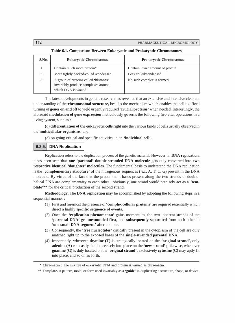
172 PHARMACEUTICAL MICROBIOLOGY
Table 6.1. Comparison Between Eukaryotic and Prokaryotic Chromosomes
S.No. Eukaryotic Chromosomes Prokaryotic Chromosomes
1 Contain much more protein*. Contain lesser amount of protein.
2. More tightly packed/coiled /condensed. Less coiled/condensed.
3. A group of proteins called ‘histones’ No such complex is formed.invariably produce complexes around
which DNA is wound.
The latest developments in genetic research has revealed that an extensive and intensive clear cutunderstanding of the chromosomal structure, besides the mechanism which enables the cell to affordturning of genes on and off to yield urgently required ‘crucial proteins’ when needed. Interestingly, theaforesaid modulation of gene expression meticulously governs the following two vital operations in aliving system, such as :
(a) differentiation of the eukaryotic cells right into the various kinds of cells usually observed inthe multicellular organisms, and
(b) on going critical and specific activities in an ‘individual cell’.
6.2.5. DNA Replication
Replication refers to the duplication process of the genetic material. However, in DNA replication,it has been seen that one ‘parental’ double-stranded DNA molecule gets duly converted into tworespective identical ‘daughter’ molecules. The fundamental basis to understand the DNA replicationis the ‘complementary structure’ of the nitrogenous sequences (viz., A, T, C, G) present in the DNAmolecule. By virtue of the fact that the predominant bases present along the two strands of double-helical DNA are complementary to each other ; obviously, one strand would precisely act as a ‘tem-plate’** for the critical production of the second strand.
Methodology. The DNA replication may be accomplished by adopting the following steps in asequential manner :
(1) First and foremost the presence of ‘complex cellular proteins’ are required essentially whichdirect a highly specific sequence of events.
(2) Once the ‘replication phenomenon’ gains momentum, the two inherent strands of the‘parental DNA’ get unwounded first, and subsequently separated from each other in‘one small DNA segment’ after another.
(3) Consequently, the ‘free nucleotides’ critically present in the cytoplasm of the cell are dulymatched right up to the exposed bases of the single-stranded parental DNA.
(4) Importantly, wherever thymine (T) is strategically located on the ‘original strand’, onlyadenine (A) can easily slot in precisely into place on the ‘new strand’ ; likewise, wheneverguanine (G) is duly located on the ‘original strand’, exclusively cytosine (C) may aptly fitinto place, and so on so forth.
* Chromatin : The mixture of eukaryotic DNA and protein is termed as chromatin.
** Template. A pattern, mold, or form used invariably as a ‘guide’ in duplicating a structure, shape, or device.
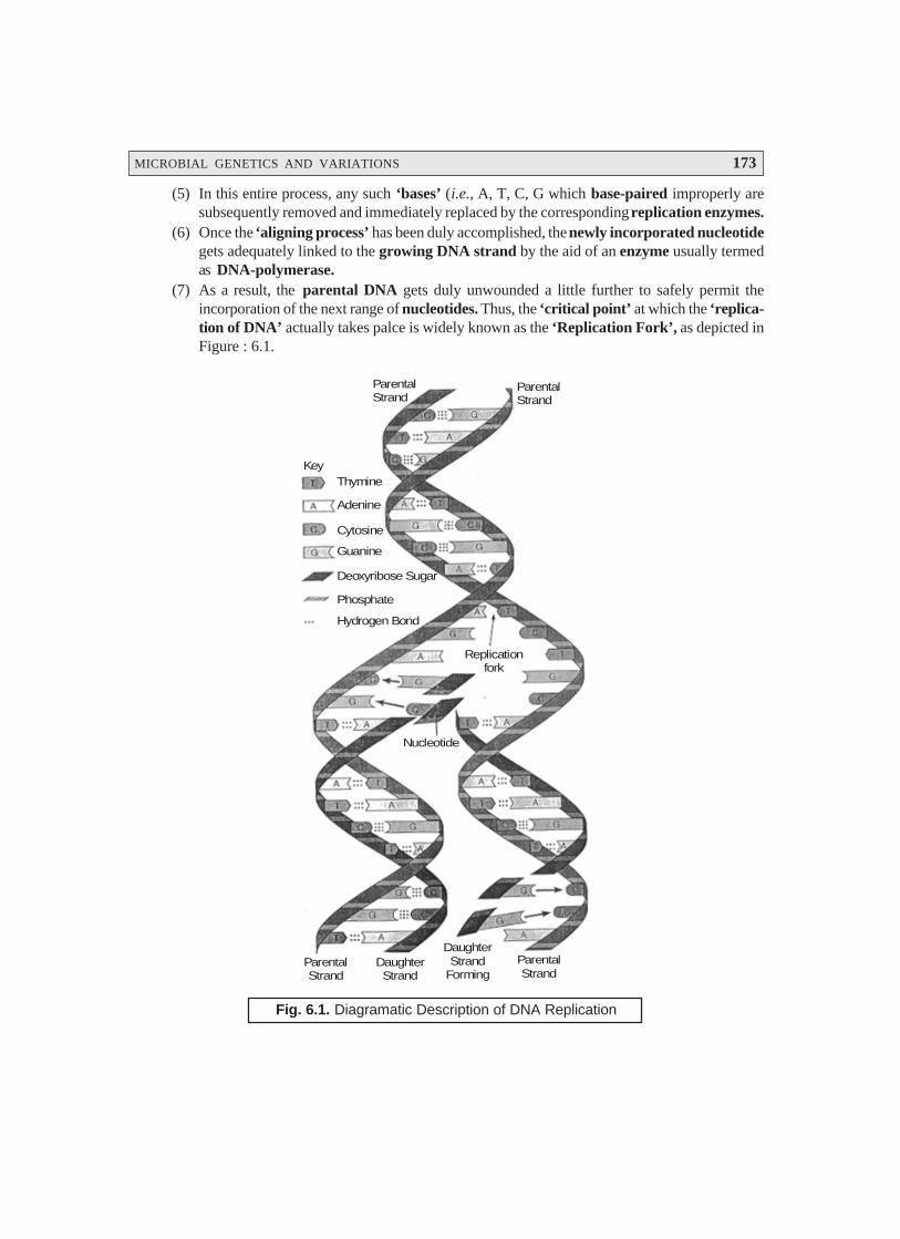
MICROBIAL GENETICS AND VARIATIONS 173
(5) In this entire process, any such ‘bases’ (i.e., A, T, C, G which base-paired improperly aresubsequently removed and immediately replaced by the corresponding replication enzymes.
(6) Once the ‘aligning process’ has been duly accomplished, the newly incorporated nucleotidegets adequately linked to the growing DNA strand by the aid of an enzyme usually termedas DNA-polymerase.
(7) As a result, the parental DNA gets duly unwounded a little further to safely permit theincorporation of the next range of nucleotides. Thus, the ‘critical point’ at which the ‘replica-tion of DNA’ actually takes palce is widely known as the ‘Replication Fork’, as depicted inFigure : 6.1.
KeyThymine
Adenine
Cytosine
Guanine
Deoxyribose Sugar
Phosphate
Hydrogen Bond
ParentalStrand
ParentalStrand
Replicationfork
Nucleotide
ParentalStrand
DaughterStrand
DaughterStrand
FormingParentalStrand
Fig. 6.1. Diagramatic Description of DNA Replication
174 PHARMACEUTICAL MICROBIOLOGY
Explanation of Figure 6.1. The various cardinal points that explain the DNA replication in
Fig. 6.1. are as follows :
(1) Double helix of the parental DNA gets separated as weak H-bonds between the nucleotides
strategically located on opposite strands usually break in response to the action of replica-
tion enzymes.
(2) H-bonds that critically come into being between new complementary nucleotides and each
strand of the parental template to give rise to the formation of the newer base pairs.
(3) Enzymes catalyze the formation of sugar-phosphate bonds existing between the sequen-
tial nucleotides critically positioned on each resulting daugther strand.
It has been duly observed that the ‘replication fork’ mostly moves very much along the
parental DNA, whereas each of the unwound single strands strategically combines with
new nucleotides. In this way, both these strands, namely : (a) Original strand, and (b)
Daugther strand (newly synthesized), get rewound, intimately. As we critically notice that
each of the newly formed double-stranded DNA molecule essentially comprise of one
original conserved strand and one altogether new strand, the phenomenon of replication
under these conditions is invariably termed as semiconservative replication.
6.2.6. Rate of DNA Replication
Beyond any stretch of imagination the DNA synthesis happens to be an extremely rapid phenom-
enal process, which stands at approximately 1000 nucleotides. sec– 1 in E. coli at 37° C. Of course,
initially it mostly appears that the prevailing ‘speed of DNA synthesis’ is not likely to happen, in view
of the fact that ‘nucleotide substrates’ should be first synthesized adequately, and subsequently must
undergo diffusion to the ‘replication fork’. Besides, there could be quite a few genuine attempts whereby
the so called ‘wrong nucleotides’ to pair at each strategic position well before the correct bases do pair
up actually. However, it is pertinent to state here that the ultimate speed, as well as specificity of DNA
replication are virtually monitored and governed by almost the same fundamental principles which
actually guide all chemical reactions.
Figure 6.2. illustrates the overall summary of various events that usually take place at the ‘repli-
cation fork’.

MICROBIAL GENETICS AND VARIATIONS 175
1 23
4 5 6
Enzymes unwind theparental double helix.
Proteins stabilize the unwound parental DNA.
The leading strand issynthesized continuously in the 5 — 3 direction by DNApolymerase.′ → ′
The lagging strand issynthesized discontinuously.RNA polymerase synthesizes ashort RNA primer, which is thenextended by DNA polymerase.
DNA polymerasedigests RNA primerand replaces it with DNA.
DNA ligase joinsthe discontinuousfragments of thelagging strand.
DNA Polymerase
ReplicationFork
RNA Primer
DNA PolymeraseDNA Ligase
RNAPolymerase
ParentalStrand
DNAPolymerase
3′
5′
3′
5′
3′
5′
Overall direction of replication
Fig. 6.2. Summarized Sequel of Events at the Replication Fork
Explanation : The sequel of events at the ‘replication fork’ may be explained as under :
(1) Log-Phase Growth : Under certain experimental parameters viz., log-phase growth in arelatively rich nutrient culture medium the E. coli cells have been seen to grow exceedinglyfaster in comparison to the two ensuing ‘replication forks’ are able to complete the circularchromosomes.
(2) Furthermore, under such conditions, the E. coli cell eventually initiates the distinct ‘multiplereplication forks’ strategically located at the very origin on the chromosome. Thus, an alto-gether ‘new pair of forks’ critically comes into being before the ‘last pair of forks’ has justfinished.
(3) Follow up of above (2) evidently suggests that the overall ‘rate of DNA synthesis’, in fact,closely matches the ‘rate’ at which the E. coli cell undergoes division.
(4) Likewise, when the actual growth of the cell slows down noticeably, one may apparentlyobserve a ‘delayed initiation of DNA synthesis’ occurring at the origin of replication.
(5) In a broader perspective, one may observe that the rate at which each and every replicationfork moves, mostly remains constant. However, the careful modulation of the initiativeprocedure in replication enables the ‘cell’ to largely monitor and control its overall predomi-nant rate of DNA synthesis to closely match not only its rate of growth but also its cell-division.
6.2.7. Flow of Genetic Information
Genetic information refers to such informations that are pertaining to or determined bygenes.

176 PHARMACEUTICAL MICROBIOLOGY
It has been well established that DNA replication makes it quite possible to maintain and sustainthe flow of genetic information right from one generation to the next one.
Figure : 6.3 clearly shows the two different ways whereby the genetic information can flowconveniently.
DNA
Parent cell
BETWEEN GENERATIONS OF CELLS WITHIN A CELL
Replication
Cell divides
Daughter cellsCell metabolizes
and grows
DNA
mRNA
Protein
Transcription
Translation
Fig. 6.3. Diagramatic Sketch of Two Different Ways Genetic Information Can Flow
[Adapted From : Tortora GJ et al. ‘Microbiology : An Introduction’, The Benjamin/CummingsPublishing Co., Inc., New York, 5th end., 1995]
From Fig. 6.3, it is quite evident that the DNA of a cell undergoes replication before cell division ;and, therefore, each ‘daughter cell’ speficially receives a chromosome which is found to be verymuch identical to that of the ‘parent cell’. Thus, inside each metabolizing cell, the ensuing geneticinformation intimately associated in DNA also affords definite flow in two different modes, namely :
(a) Transcription i.e., genetic information is duly transcribed into messenger RNA (mRNA),and
(b) Translation i.e., subsequently, the transcribed mRNA is duly translated into respectivedesired proteins. These two aspects shall again be treated individually in Sections 2.8 and 2.9.
Salient Features : The salient features of Fig. 6.3 are as stated under :
(i) Genetic information may be transferred between generations of cells via replication ofDNA,
(ii) Genetic information can also be exploited very much within a cell to produce the proteinswhich the cell requires to function. In fact, such a vital and important information is dulytransferred via the processes of transcription and translation, and
MICROBIAL GENETICS AND VARIATIONS 177
(iii) The diagramatic representation of the cell is a bacterium which essentially bears a single
circular chromosome.
6.2.8. Bacterial Transformation
Bacterial transformation usually refers to a specific type of mutation taking place in bacteria.
In fact, it results from DNA of a bacterial cell penetrating to the host cell and becoming incorporated
right into the genotype of the host.
The era stretching over 1940s witnessed and recognized that the prevailing inheritence in micro-
organisms (bacteria) was adequately monitored and regulated basically by the same mechanisms as
could be seen in higher eukaryotic organisms.
Interestingly, it was duly realized that bacteria to designate a ‘useful tool’ to decepher the intri-
cate mechanism of heredity as well as genetic transfer ; and, therefore, employed extensively in the
overall genetic investigative studies.
Griffith’s Experimental Observations* : Griffith (1928), a British Health Officer, carefully
injected mice with a mixture comprising of two different kinds of cells, namely :
(a) A few rough (i.e., noncapsulated and nonpathogenic) pneumococcal cells, and
(b) A large number of heat-killed smooth (i.e., capsulated and pathogenic cells.)
Living smooth pneumococci cells — usually causes pneumonia is human beings and a host of
animals.
‘Rough’ and ‘Smooth’ — invariably refer to the ensuing surface texture of the colonies of the
respective cells.
Consequently, the mice ultimately died of pneumonia, and ‘live smooth cells’ were meticu-
lously isolated from their blood. Thus, one may observe critically that there could be certain cardinal
factor exclusively responsible for the inherent pathogenicity of the smooth bacteria ; and it had eventu-
ally transformed these organisms into pathogenic smooth ones.
Griffith also ascertained that the transforming factor might have been sailed from the trans-
formed cells right into their progeny (i.e., offspring), and hence the inheritence of the characteristic
features of a gene. Fig. 6.4 depicts the experiment of Griffith.
* From the Office of Technology Assessment.
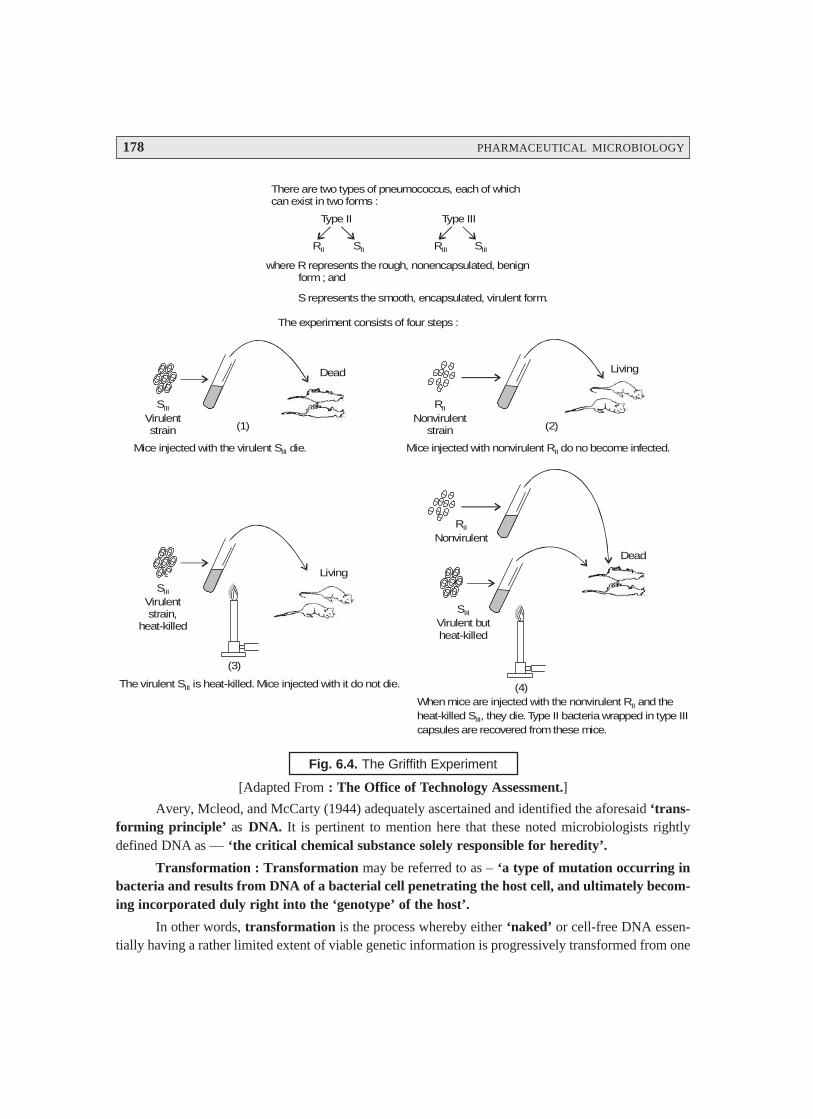
178 PHARMACEUTICAL MICROBIOLOGY
There are two types of pneumococcus, each of whichcan exist in two forms :
Type II Type III
RII SII RIII SIII
where R represents the rough, nonencapsulated, benign form ; and
S represents the smooth, encapsulated, virulent form.
The experiment consists of four steps :
SVirulentstrain
III
Dead
RNonvirulent
strain
II
Living
(1) (2)
Mice injected with the virulent S die.III Mice injected with nonvirulent R do no become infected.II
SVirulentstrain,
heat-killed
III
(3)
The virulent is heat-killed. Mice injected with it do not die.SIII
Living
RNonvirulent
II
SVirulent butheat-killed
III
Dead
(4)When mice are injected with the nonvirulent R and theheat-killed , they die. Type II bacteria wrapped in type IIIcapsules are recovered from these mice.
II
IIIS
Fig. 6.4. The Griffith Experiment
[Adapted From : The Office of Technology Assessment.]
Avery, Mcleod, and McCarty (1944) adequately ascertained and identified the aforesaid ‘trans-forming principle’ as DNA. It is pertinent to mention here that these noted microbiologists rightlydefined DNA as — ‘the critical chemical substance solely responsible for heredity’.
Transformation : Transformation may be referred to as – ‘a type of mutation occurring inbacteria and results from DNA of a bacterial cell penetrating the host cell, and ultimately becom-ing incorporated duly right into the ‘genotype’ of the host’.
In other words, transformation is the process whereby either ‘naked’ or cell-free DNA essen-tially having a rather limited extent of viable genetic information is progressively transformed from one
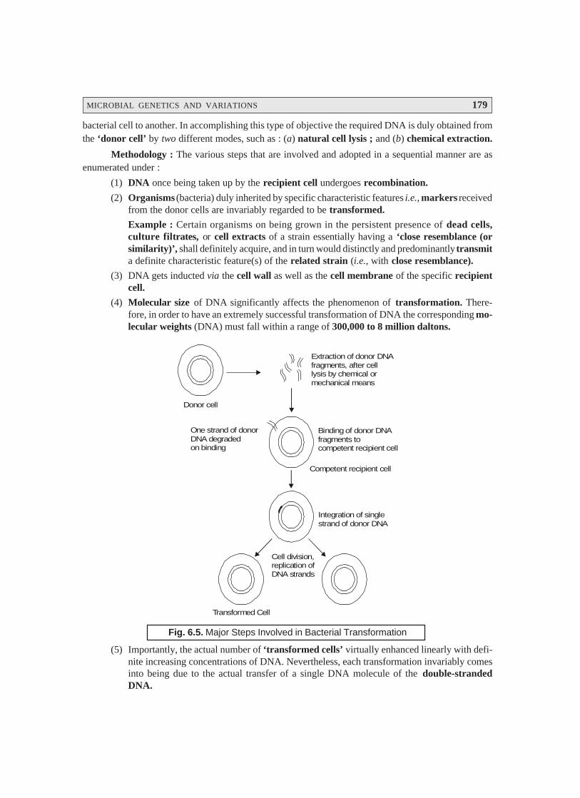
MICROBIAL GENETICS AND VARIATIONS 179
bacterial cell to another. In accomplishing this type of objective the required DNA is duly obtained fromthe ‘donor cell’ by two different modes, such as : (a) natural cell lysis ; and (b) chemical extraction.
Methodology : The various steps that are involved and adopted in a sequential manner are asenumerated under :
(1) DNA once being taken up by the recipient cell undergoes recombination.
(2) Organisms (bacteria) duly inherited by specific characteristic features i.e., markers receivedfrom the donor cells are invariably regarded to be transformed.
Example : Certain organisms on being grown in the persistent presence of dead cells,culture filtrates, or cell extracts of a strain essentially having a ‘close resemblance (orsimilarity)’, shall definitely acquire, and in turn would distinctly and predominantly transmita definite characteristic feature(s) of the related strain (i.e., with close resemblance).
(3) DNA gets inducted via the cell wall as well as the cell membrane of the specific recipientcell.
(4) Molecular size of DNA significantly affects the phenomenon of transformation. There-fore, in order to have an extremely successful transformation of DNA the corresponding mo-lecular weights (DNA) must fall within a range of 300,000 to 8 million daltons.
Extraction of donor DNAfragments, after celllysis by chemical ormechanical means
One strand of donorDNA degradedon binding
Binding of donor DNAfragments tocompetent recipient cell
Competent recipient cell
Integration of singlestrand of donor DNA
Cell division,replication ofDNA strands
Transformed Cell
Donor cell
Fig. 6.5. Major Steps Involved in Bacterial Transformation
(5) Importantly, the actual number of ‘transformed cells’ virtually enhanced linearly with defi-nite increasing concentrations of DNA. Nevertheless, each transformation invariably comesinto being due to the actual transfer of a single DNA molecule of the double-strandedDNA.

180 PHARMACEUTICAL MICROBIOLOGY
(6) Once the DNA gains its entry into a cell, one of the two strands gets degraded almost in-stantly by means of the available enzymes deoxyribonucleases ; whereas, the second strandparticularly subject to base pairing with a homologous segment of the correspondingrecipient cell chromosome. Consequently, the latter gets meticulously integrated into therecipient DNA, as illustrated beautifully in Figure 6.5.
(7) Transformation of Closely Related Strains of Bacteria : In reality, the transformation ofclosely related strains of bacterial could be accomplished by virtue of the fact that comple-mentary base pairing predominantly occurs particularly between one strand of the donorDNA fragment and a highly specific segment of the recipient chromosome.
However, the major steps involved in the bacterial transformation have been clearly shown inFig. 6.5.
Examples : The bacterial species which have been adequately transformed essentially include :
Bacterial species : Streptococcus pneumoniae (Pneumococcus)
Genera : Bacillus ; Haemophilus ; Neisseria ; and Rhizobium
6.2.9. Bacterial Transcription
Bacterial transcription refers to the – ‘synthesis of a complementary strand of RNAparticularly from a DNA template’.
In fact, there exists three different types of RNA in the bacterial cells, namely : (a) messengerRNA ; (b) ribosomal RNA, and (c) transfer RNA.
Messenger RNA (mRNA) : It predominantly carries the ‘coded information’ for the produc-tion of particular proteins from DNA to ribosomes, where usually proteins get synthesized.
Ribosomal RNA : It invariably forms an ‘integral segment’ of the ribosomes, that strategicallyexpatiates the cellular mechanism with regard to protein synthesis.
Transfer RNA : It is also intimately and specifically involved in the protein synthesis.
Process of Bacterial Transcription :
Importantly, during the process of bacterial transcription, a strand of messenger RNA (mRNA)gets duly synthesized by the critical usage of a ‘specific gene’ i.e., a vital segment of the cell’s DNA–asa template, as illustrated beautifully in Figure : 6.6. Thus, one may visualize the vital and important‘genetic information’ adequately stored in the sequence of nitrogenous bases (viz., A, T, C and G) ofDNA, that may be rewritten so that the same valuable ‘genetic information’ appears predominantly inthe base sequence of mRNA.
Examples :
(1) In the DNA replication phenomenon, it has been duly observed that a G in DNA templateusually dictates a C in the mRNA ; and a T in DNA template invariably dictates an A in themRNA.
(2) An A in DNA template normally dictates a uracil (U) in the mRNA by virtue of the fact thatRNA strategically contains U instead of T*.
* U essentially possesses a chemical structure which is slightly different from T ; however, it base-pairs moreor less in the same manner.
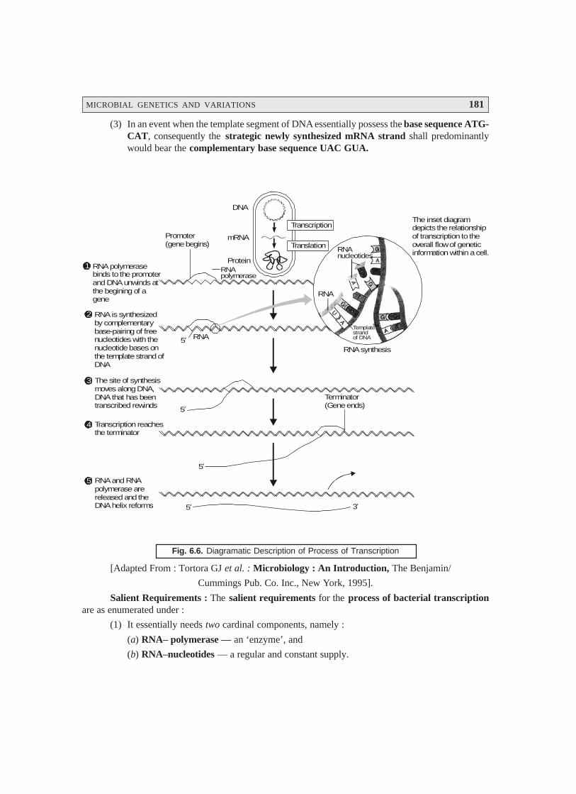
MICROBIAL GENETICS AND VARIATIONS 181
(3) In an event when the template segment of DNA essentially possess the base sequence ATG-CAT, consequently the strategic newly synthesized mRNA strand shall predominantlywould bear the complementary base sequence UAC GUA.
RNA and RNApolymerase arereleased and theDNA helix reforms
5
Transcription reachesthe terminator
4
The site of synthesismoves along DNA,DNA that has beentranscribed rewinds
3
RNA is synthesizedby complementarybase-pairing of freenucleotides with the nucleotide bases onthe template strand ofDNA
2
RNA polymerasebinds to the promoterand DNA unwinds atthe begining of a gene
1
The inset diagramdepicts the relationof transcription overall flow information within a cell.
shipto the
of genetic
Transcription
Translation
DNA
mRNA
ProteinRNApolymerase
Promoter(gene begins)
RNAnucleotides
RNA
Templatestrandof DNA
RNA synthesis
Terminator(Gene ends)
RNA5′
5′
5′
5′ 3′
Fig. 6.6. Diagramatic Description of Process of Transcription
[Adapted From : Tortora GJ et al. : Microbiology : An Introduction, The Benjamin/
Cummings Pub. Co. Inc., New York, 1995].
Salient Requirements : The salient requirements for the process of bacterial transcriptionare as enumerated under :
(1) It essentially needs two cardinal components, namely :
(a) RNA– polymerase — an ‘enzyme’, and
(b) RNA–nucleotides — a regular and constant supply.

182 PHARMACEUTICAL MICROBIOLOGY
* Includes, all ‘exons’ and ‘introns’.
(2) Promoter — Usually transcription commences once the RNA polymerase gets strategi-cally bound to the DNA at a specific site termed as ‘promoter’.
(3) Precisely, only one of the two DNA strands invariably caters as the particularly required‘template for the synthesis for a given ‘gene’.
Examples : There are several typical examples that explains the above intricate phenomenonvividly :
(a) Just as DNA, the RNA gets synthesized duly and specifically in the 5′′′′′ ⎯→⎯→⎯→⎯→⎯→ 3′′′′′ direction.Nevertheless, the ‘equivalence point’ (i.e., endpoint) for transcription of the gene issignaled suitably by a terminator segment present strategically in the DNA. Interestingly,at this particular zone, one may observe the release from the DNA of these two entitiesprominently : (i) RNA polymerase ; and (ii) newly generated single-stranded mRNA.
(b) ‘Regions of genes’ present critically in ‘eukaryotic cells’ which essentially afford ‘coding’for the respective proteins are usually interrupted by the so-called ‘noncoding DNA’. There-fore, the ‘eukaryotic genes’ are made up of ‘exons’ i.e., the specific segments of DNAexpressed appropriately ; besides, ‘introns’ i.e., the designated intervening segments ofDNA which fail to code for the corresponding protein. Besides, in the eukaryotic cell thenucleus predominantly synthesizes RNA polymerase from the entire gene–a fairly longand continuous RNA product* usually termed as the RNA transcript.
Mechanism : The ‘elongated RNA’ is subsequently processed by a host of other enzymesthat particularly help in the removal of the intron-derived RNA and also splice together theexon derived RNA thereby producing an mRNA which is exclusively capable of ‘directlythe on-going protein synthesis’. Consequently, the RNA gracefully walks out of the nu-cleus, and ultimately turns into a mRNA of the ensuing cytoplasm.
Ribozymes — are non protein enzymes (i.e., a RNA enzyme) duly obtained as a result ofcertain enzymes which are actually cut by the RNA itself.
(c) Importantly, in eukaryotic organisms, the ensuing transcription usually occurs in thenucleus. It has been observed that mRNA should be completely synthesized and duly movedacross the nuclear membrane right into the cytoplasm before the actual commencement ofthe phenomenon of translation. Besides, mRNA is duly subjected to further processingmode before it virtually gets out of the nucleus.
Summararily, the valuable genetic information, derived from prokaryotes and eukaryotes,pertaining to protein synthesis is stored meticulously in DNA, and subsequently passedon to mRNA during the phenomenon of ‘transcription’. Ultimately, mRNA prominentlyserves as the source of information for the required protein synthesis.
6.2.10. Bacterial Translation
Bacterial translation may be defined as — ‘the specific process via which the criticalnitrogenous-base sequence of mRNA affords determination of the amino acid sequence of protein’.
One may precisely observe that in an organism that is particularly devoid of a membrane-enclosed nucleus, both ‘transcription’ and ‘translation’ invariably occur in the cytoplasm. Thus, inan eukaryotic organisms, the process of ‘translation’ actually comes into being in a situation whenmRNA gains its entry into the cytoplasm.

MICROBIAL GENETICS AND VARIATIONS 183
Figure : 6.7 illustrates the eight major sequential stages that are intimately involved in the proc-ess of translation, namely :
Stage-1 : Various components that are essentially required to commence the ‘phenomenon oftranslation’ first come together.
Stage-2 : On the assembled ribosome a transfer RNA (tRNA) carrying the ‘first amino acid’is duly paired with the start codon on the mRNA ; and a ‘second amino acid’ beingcarried by tRNA approaches steadily.
State-3 : Critical place on the chromsome at which the very first tRNA sites is known as the Psite. Thus, in the corresponding A site next to it, the second codon of the mRNA pairswith a tRNA carrying the second amino acid.
Stage-4 : First amino acid gets hooked on to the second amino acid by a peptide linkage
, and the first tRNA gets released.
Note : Nucleotide bases are duly labeled only for the first two codons.
Stage-5 : Ribosome gradually moves along the mRNA until the second tRNA is in the P site,and thus the process continues.
Stage-6 : Ribosome very much continues to move along the mRNA, and thus, newer aminoacids are progressively added on to the ‘polypeptide chain’ strategically.
Stage-7 : Ribosome when ultimately gets upto the ‘stop codon’, the duly formed polypeptide isreleased.
Stage-8 : Last tRNA gets released finally, and thus the ribosome falls apart. Finally, the re-leased polypeptide gives rise to an altogether new protein.
Process of Bacterial Translation : The various steps encountered in the elaborated process of‘bacterial translation’ are :
(1) Proteins are usually synthesized strategically in the 5′′′′′ →→→→→ 3′′′′′ direction, as present in DNAand RNA (i.e., nucleic acids).
(2) First and foremost, the 5′′′′′ end of the specific mRNA molecule becomes associated with aribosome, which being the major cellular machinery that predominantly helps to catalyzethe ‘protein synthesis’.
(3) Ribosomal RNA [rRNA] : Ribosomes usually comprise of two subunits ; of which, onebeing a special type of RNA termed as ribosomal RNA (rRNA) and the other proteins. Atthe very outset of the process of bacterial translation, the two ribosomal subunits happento get closer vis-a-vis the mRNA plus many other components engaged in this phenomenon.
(4) Even before the suitable amino acids may be joined together to yield a ‘protein’, they shouldbe adequately ‘activated’ by strategic attachment to transfer RNA (tRNA).
Figure : 6.8(a) represents the various diagramatic sketch of structures and articulatedfunction of transfer RNA (tRNA).
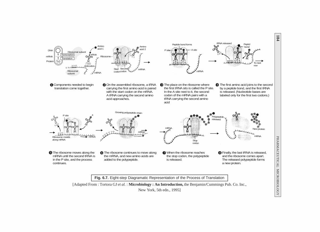
184P
HA
RM
AC
EU
TIC
AL
MIC
RO
BIO
LO
GY
Fig. 6.7. Eight-step Diagramatic Representation of the Process of Translation
[Adapted From : Tortora GJ et al. : Microbiology : An Introduction, the Benjamin/Cummings Pub. Co. Inc.,New York, 5th edn., 1995]
7 When the ribosome reachesthe stop codon, the polypeptideis released.
8 Finally, the last tRNA is released,and the ribosome comes apart.The released polypeptide formsa new protein.
mRNA
Stopcodon
Polypeptidereleased
mRNA
New protein
3 The place on the ribosome wherethe first tRNA sits is called the P site.In the A site next to it, the secondcodon of the mRNA pairs with atRNA carrying the second aminoacid
4 The first amino acid joins to the secondby a peptide bond, and the first tRNAis released. (Nucleotide bases arelabeled only for the first two codons.)
P site
Peptide bond forms
A site
mRNA
tRNA released
AU
C
To nextrow
Peptid bond
Ribosomal subunitDNA
mRNA
Protein
Ribosomalsubunit mRNA
tRNA
Aminoacid 1
A U G
UAC
Ribosome
1 Components needed to begin translation come together.
2 On the assembled ribosome, a tRNAcarrying the first amino acid is pairedwith the start codon on the mRNA.A tRNA carrying the second aminoacid approaches.
Startcodon
Secondcodon
mRNA
Aminoacid 2
Anticodon
5 The ribosome moves along themRNA until the second tRNA isin the P site, and the processcontinues.
6 The ribosome continues to move alongthe mRNA, and new amino acids areadded to the polypeptide.
Ribosome movesalong mRNA
mRNA
P site
A U G
UAC
AU
GU U A mRNA
Growing polypeptide chain4
Transcription
Translation
5′
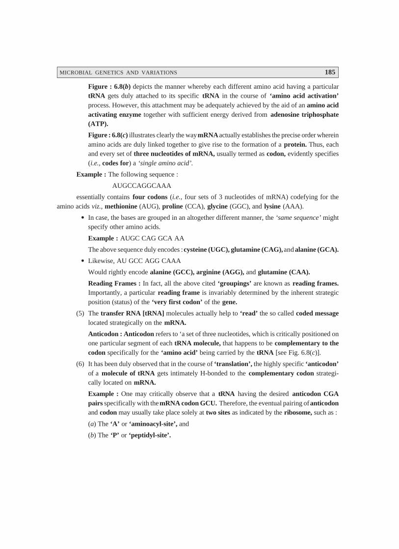
MICROBIAL GENETICS AND VARIATIONS 185
Figure : 6.8(b) depicts the manner whereby each different amino acid having a particulartRNA gets duly attached to its specific tRNA in the course of ‘amino acid activation’process. However, this attachment may be adequately achieved by the aid of an amino acidactivating enzyme together with sufficient energy derived from adenosine triphosphate(ATP).
Figure : 6.8(c) illustrates clearly the way mRNA actually establishes the precise order whereinamino acids are duly linked together to give rise to the formation of a protein. Thus, eachand every set of three nucleotides of mRNA, usually termed as codon, evidently specifies(i.e., codes for) a ‘single amino acid’.
Example : The following sequence :
AUGCCAGGCAAA
essentially contains four codons (i.e., four sets of 3 nucleotides of mRNA) codefying for theamino acids viz., methionine (AUG), proline (CCA), glycine (GGC), and lysine (AAA).
• In case, the bases are grouped in an altogether different manner, the ‘same sequence’ mightspecify other amino acids.
Example : AUGC CAG GCA AA
The above sequence duly encodes : cysteine (UGC), glutamine (CAG), and alanine (GCA).
• Likewise, AU GCC AGG CAAA
Would rightly encode alanine (GCC), arginine (AGG), and glutamine (CAA).
Reading Frames : In fact, all the above cited ‘groupings’ are known as reading frames.Importantly, a particular reading frame is invariably determined by the inherent strategicposition (status) of the ‘very first codon’ of the gene.
(5) The transfer RNA [tRNA] molecules actually help to ‘read’ the so called coded messagelocated strategically on the mRNA.
Anticodon : Anticodon refers to ‘a set of three nucleotides, which is critically positioned onone particular segment of each tRNA molecule, that happens to be complementary to thecodon specifically for the ‘amino acid’ being carried by the tRNA [see Fig. 6.8(c)].
(6) It has been duly observed that in the course of ‘translation’, the highly specific ‘anticodon’of a molecule of tRNA gets intimately H-bonded to the complementary codon strategi-cally located on mRNA.
Example : One may critically observe that a tRNA having the desired anticodon CGApairs specifically with the mRNA codon GCU. Therefore, the eventual pairing of anticodonand codon may usually take place solely at two sites as indicated by the ribosome, such as :
(a) The ‘A’ or ‘aminoacyl-site’, and
(b) The ‘P’ or ‘peptidyl-site’.
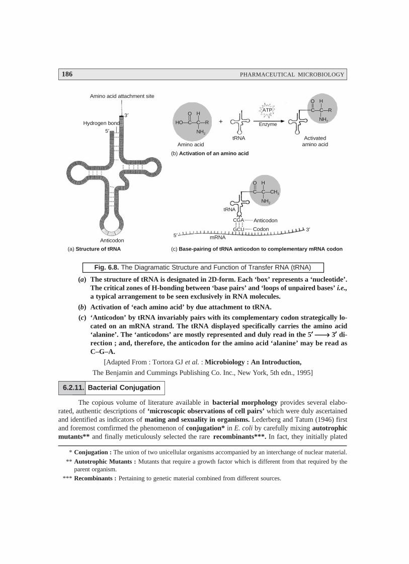
186 PHARMACEUTICAL MICROBIOLOGY
ATP
Enzyme
Activatedamino acid
tRNA
HO—C—C—R
O H
NH2
C—C—CH3
O H
NH2
+
Amino acid
C—C—R
O H
NH2
tRNA
Anticodon
Codon
CGA
GCU
mRNA5′3′
Anticodon
Hydrogen bond
Amino acid attachment site
5′
3′
(a) Structure of tRNA (c) Base-pairing of tRNA anticodon to complementary mRNA codon
(b) Activation of an amino acid
Fig. 6.8. The Diagramatic Structure and Function of Transfer RNA (tRNA)
(a) The structure of tRNA is designated in 2D-form. Each ‘box’ represents a ‘nucleotide’.The critical zones of H-bonding between ‘base pairs’ and ‘loops of unpaired bases’ i.e.,a typical arrangement to be seen exclusively in RNA molecules.
(b) Activation of ‘each amino acid’ by due attachment to tRNA.
(c) ‘Anticodon’ by tRNA invariably pairs with its complementary codon strategically lo-cated on an mRNA strand. The tRNA displayed specifically carries the amino acid‘alanine’. The ‘anticodons’ are mostly represented and duly read in the 5′′′′′ ⎯→⎯→⎯→⎯→⎯→ 3′′′′′ di-rection ; and, therefore, the anticodon for the amino acid ‘alanine’ may be read asC–G–A.
[Adapted From : Tortora GJ et al. : Microbiology : An Introduction,
The Benjamin and Cummings Publishing Co. Inc., New York, 5th edn., 1995]
6.2.11. Bacterial Conjugation
The copious volume of literature available in bacterial morphology provides several elabo-rated, authentic descriptions of ‘microscopic observations of cell pairs’ which were duly ascertainedand identified as indicators of mating and sexuality in organisms. Lederberg and Tatum (1946) firstand foremost comfirmed the phenomenon of conjugation* in E. coli by carefully mixing autotrophicmutants** and finally meticulously selected the rare recombinants***. In fact, they initially plated
* Conjugation : The union of two unicellular organisms accompanied by an interchange of nuclear material.
** Autotrophic Mutants : Mutants that require a growth factor which is different from that required by theparent organism.
*** Recombinants : Pertaining to genetic material combined from different sources.
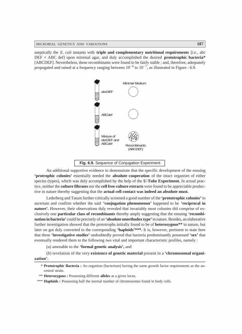
MICROBIAL GENETICS AND VARIATIONS 187
* Prototrophic Bacteria : An organism (bacterium) having the same growth factor requirements as the an-cestral strain.
** Heterozygous : Possessing different alleles at a given locus.
*** Haploids : Possessing half the normal number of chromosomes found in body cells.
aseptically the E. coli mutants with triple and complementary nutritional requirements [i.e., abcDEF × ABC def] upon minimal agar, and duly accomplished the desired prototrophic bacteria*[ABCDEF]. Nevertheless, these recombinants were found to be fairly stable ; and, therefore, adequatelypropogated and raised at a frequency ranging between 10– 6 to 10– 7, as illustrated in Figure : 6.9.
abcDEF
ABCdef
Mixture ofabcDEF andABCdef Recombinants
(ABCDEF)
Minimal Medium
Fig. 6.9. Sequence of Conjugation Experiment
An additional supportive evidence to demonstrate that the specific development of the ensuing‘protrophic colonies’ essentially needed the absolute cooperation of the intact organism of eitherspecies (types), which was duly accomplished by the help of the U-Tube Experiment. In actual prac-tice, neither the culture filtrates nor the cell free culture extracts were found to be appreciable produc-tive in nature thereby suggesting that the actual cell contact was indeed an absolute must.
Lederberg and Tatum further critically screened a good number of the ‘prototrophic colonies’ toascertain and confirm whether the said ‘conjugation phenomenon’ happened to be ‘reciprocal innature’. However, their observations duly revealed that invariably most colonies did comprise of ex-clusively one particular class of recombinants thereby amply suggesting that the ensuing ‘recombi-nation in bacteria’ could be precisely of an ‘absolute unorthodox type’ in nature. Besides, an elaborativefurther investigation showed that the prototrophs initially found to be of heterozygous** in nature, butlater on got duly converted to the corresponding ‘haploids’***. It is, however, pertinent to state herethat these ‘investigative studies’ undoubtedly proved that bacteria predominantly possessed ‘sex’ thateventually rendered them to the following two vital and important characteristic profiles, namely :
(a) amenable to the ‘formal genetic analysis’, and
(b) revelation of the very existence of genetic material present in a ‘chromosomal organi-zation’.

188 PHARMACEUTICAL MICROBIOLOGY
SinteredGlass
STRAIN[I]
STRAIN[II]
Fig. 6.10. The U-Tube Experiment
The process of conjugation essentially suggests that :
(1) large fragments of DNA were adequately transferred from one bacterium to another in anon-reciprocal manner, and
(2) such transfer invariably took place from a given point.One may also critically take cognizance of the fact that the exact size (dimension) of DNA
transferred from one cell to another was found to be much larger in comparison to the correspondingtransformation. Certainly, the process of conjugation proved to be much more an absolutely com-mendable and useful technique for the so called ‘gene mapping’ in organism.
Donor Bacteria i.e., such organisms that are responsible for transferring DNA, andRecipient Bacteria i.e., such organisms that are responsible for receiving DNA.
6.2.12. Bacterial Transduction
Bacterial transduction may be defined as — ‘a phenomenon causing genetic recombinationin bacteria wherein DNA is carried from one specific bacterium to another by a bacteriophage’.
It has been duly observed that a major quantum of bacteriophages, particularly the ‘virulent’ones, predominantly undergo a rather quick lytic growth cycle in their respective host cells. During thisphenomenon they invariably inject their nucleic acid, normally DNA, right into the bacterium, where ittakes up the following two cardinal steps :
(a) undergoes ‘replication’ very fast, and(b) directs the critical synthesis of new phage proteins.Another school of thought may put forward another definition of bacterial transduction as —
‘the actual and legitimate transfer by a bacteriophage, serving as a vector, of a segment of DNAfrom one bacterium (a donor) to another (a recipient)’.
Zinder and Ledenberg (1952) first and foremost discovered the wonderful phenomenon duringan intensive search for ‘sexual conjugations’ specifically amongst the Salmonella species.
Methodology : The various steps that are involved in the bacterial transduction phenomenonare as stated under :
(1) Auxotrophic mutants were carefully mixed together ; and subsequently, isolated theprototrophic recombinant colonies from the ensuing selective nutritional media.
(2) U-Tube Experiment : The U-tube experiment was dulyperformed with a parental auxotrophic strain in eacharm (viz., I and II), and adequately separated by amicroporous fritted glass (MFG) filter, whereby theresulting ‘prototrophs’ distinctly appeard in one armof the tube, as shown in Fig. 6.10.
As the MFG-filter particularly checked and prevented cell-to-cell contact, but at the same time duly permits the ‘free pas-sage’ of fluid between the said two cultures [i.e., strains I and II], itmay be safely inferred that there must be certain ‘phenomenon’other than the ‘conjugation’ was involved.
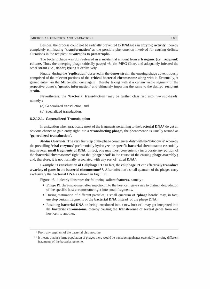
MICROBIAL GENETICS AND VARIATIONS 189
Besides, the process could not be radically prevented to DNAase (an enzyme) activity, therebycompletely eliminating ‘transformation’ as the possible phenomenon involved for causing definitealterations in the recipient auxotrophs to prototrophs.
The bacteriophage was duly released in a substantial amount from a lysogenic (i.e., recipient)culture. Thus, the emerging phage critically passed via the MFG-filter, and adequately infected theother strain (i.e., donor) lyzing it exclusively.
Finally, during the ‘replication’ observed in the donor strain, the ensuing phage adventitiouslycomprised of the relevant portions of the critical bacterial chromosome along with it. Eventually, itgained entry via the MFG-filter once again ; thereby taking with it a certain viable segment of therespective donor’s ‘genetic information’ and ultimately imparting the same to the desired recipientstrain.
Nevertheless, the ‘bacterial transduction’ may be further classified into two sub-heads,namely :
(a) Generalized transduction, and
(b) Specialized transduction.
6.2.12.1. Generalized Transduction
In a situation when practically most of the fragments pertaining to the bacterial DNA* do get anobvious chance to gain entry right into a ‘transducting phage’, the phenomenon is usually termed as‘generalized transduction’.
Modus Operandi : The very first step of the phage commences duly with the ‘lytic cycle’ wherebythe prevailing ‘viral enzymes’ preferentially hydrolyze the specific bacterial chromosome essentiallyinto several small fragments of DNA. In fact, one may most conveniently incorporate any portion ofthe ‘bacterial chromosome’ right into the ‘phage head’ in the course of the ensuing phage assembly ;and, therefore, it is not normally associated with any sort of ‘viral DNA’.
Example : Transduction of Coliphage P1 : In fact, the coliphage P1 can effectively transducea variety of genes in the bacterial chromosome**. After infection a small quantum of the phages carryexclusively the bacterial DNA as shown in Fig. 6.11.
Figure : 6.11 clearly illustrates the following salient features, namely :
• Phage P1 chromosomes, after injection into the host cell, gives rise to distinct degradationof the specific host chromosome right into small fragments.
• During maturation of different particles, a small quantum of ‘phage heads’ may, in fact,envelop certain fragments of the bacterial DNA instead of the phage DNA.
• Resulting bacterial DNA on being introduced into a new host cell may get integrated intothe bacterial chromosome, thereby causing the transference of several genes from onehost cell to another.
* From any segment of the bacterial chromosome.
** It means that in a large population of phages there would be transducing phages essentially carrying differentfragments of the bacterial genome.
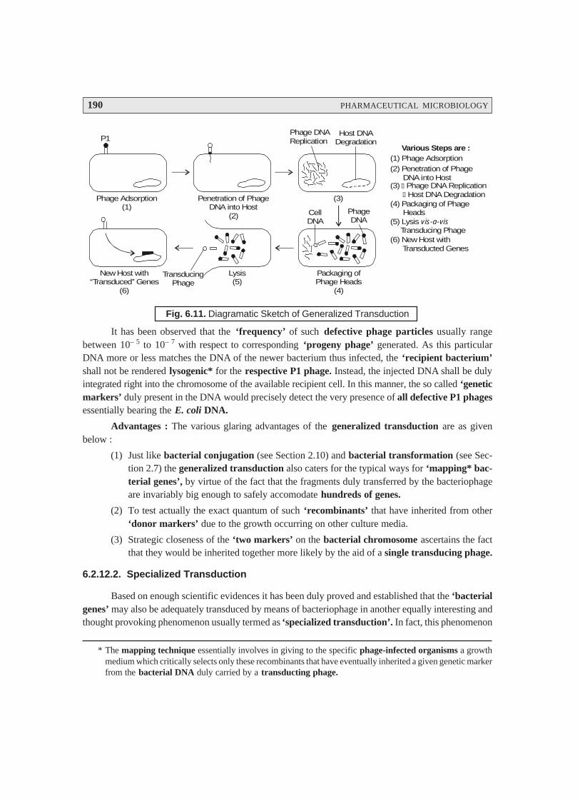
190 PHARMACEUTICAL MICROBIOLOGY
Phage Adsorption(1)
Penetration of PhageDNA into Host
(2)
(3)
Phage DNAReplication
Host DNADegradation
CellDNA
PhageDNA
Packaging ofPhage Heads
(4)
Lysis(5)
New Host with‘‘Transduced’’ Genes
(6)
TransducingPhage
Various Steps are :(1) Phage Adsorption(2) Penetration of Phage DNA into Host(3) � Phage DNA Replication � Host DNA Degradation(4) Packaging of Phage Heads(5) Lysis Transducing Phage
vis-a-vis
(6) New Host with Transducted Genes
P1
Fig. 6.11. Diagramatic Sketch of Generalized Transduction
It has been observed that the ‘frequency’ of such defective phage particles usually rangebetween 10– 5 to 10– 7 with respect to corresponding ‘progeny phage’ generated. As this particularDNA more or less matches the DNA of the newer bacterium thus infected, the ‘recipient bacterium’shall not be rendered lysogenic* for the respective P1 phage. Instead, the injected DNA shall be dulyintegrated right into the chromosome of the available recipient cell. In this manner, the so called ‘geneticmarkers’ duly present in the DNA would precisely detect the very presence of all defective P1 phagesessentially bearing the E. coli DNA.
Advantages : The various glaring advantages of the generalized transduction are as givenbelow :
(1) Just like bacterial conjugation (see Section 2.10) and bacterial transformation (see Sec-tion 2.7) the generalized transduction also caters for the typical ways for ‘mapping* bac-terial genes’, by virtue of the fact that the fragments duly transferred by the bacteriophageare invariably big enough to safely accomodate hundreds of genes.
(2) To test actually the exact quantum of such ‘recombinants’ that have inherited from other‘donor markers’ due to the growth occurring on other culture media.
(3) Strategic closeness of the ‘two markers’ on the bacterial chromosome ascertains the factthat they would be inherited together more likely by the aid of a single transducing phage.
6.2.12.2. Specialized Transduction
Based on enough scientific evidences it has been duly proved and established that the ‘bacterialgenes’ may also be adequately transduced by means of bacteriophage in another equally interesting andthought provoking phenomenon usually termed as ‘specialized transduction’. In fact, this phenomenon
* The mapping technique essentially involves in giving to the specific phage-infected organisms a growthmedium which critically selects only these recombinants that have eventually inherited a given genetic markerfrom the bacterial DNA duly carried by a transducting phage.

MICROBIAL GENETICS AND VARIATIONS 191
* Perhaps they are missing a piece of phage-genetic information duly adopted by the respective bacterialgenes.
** This could possibly provide adequate code for the missing functions of the resulting defective phages.
confirms duly that certain template phage strains may be capable of transferring merely a handful of‘restricted genes’ belonging categorically to the ‘bacterial chromosomes’.
In other words, the ensuing phages particularly transduce exclusively such bacterial genes thatare strategically positioned quite adjacent to the prophage in the bacterial chromosome. Therefore,this particular process is sometimes also referred to as ‘restricted transduction’. Interestingly, in anevent when such a phage duly infects a cell, it invariably carries along with it the specified group ofbacterial genes which ultimately turns out to be an integral part of it. Consequently, such genes mayrecombine meticulously with the homologous DNA of the prevailing infected cell.
Phage Lambda (λλλλλ) of E. coli. : In a broader perspective, the most elaboratedly researched spe-cialized transducting phage is duly represented by the phage lambda (λλλλλ) of E. coli. The exact locationof the ensuing λλλλλ prophage present in the bacterial chromosome invariably lies between the bacterialgenes gal and bio. It may be observed that whenever phages duly carrying either a gal or bio genes doinfect an altogether ‘new host’, then the desired recombination either with the gal or bio genes of therespective may take place articulately. Fig. 6.12 depicts vividly the phenomenon of specializedtransduction.
Salient Features : The various salient features highlighting the process of specializedtransduction in Figure 6.12 are stated as under :
(1) Practically ‘all phages’ which essentially carry certain bacterial genes solely on account of‘‘incorrect’’ excision are obviously found to the ‘defective’ with respect to certain highlyimportant functions.*
(2) Thorough passage via the entire ‘replication cycle’ cannot be accomplsihed ; whereas, theensuing cell may suitably give rise to certain phages, provided it is also duly infected witha rather ‘complete phage’**.
Gal
λ
λ
λ dg
Fig. 6.12. Diagramatic Sketch for Specialized Transduction
Explanations : The various stages illustrated in Fig. 6.12 are as follows :(1) When a cell gets duly infected by phage λλλλλ, its DNA is precisely inserted right into the
bacterial genome next to the genes meant for galactose metabolism (i.e, gal genes).(2) Invariably when such a cell is being induced, the λ DNA emerges out promptly, get repli-
cated, and subsequently turned into a normal phage.
192 PHARMACEUTICAL MICROBIOLOGY
(3) Sometimes, the respective λ DNA is excised imperfectly thereby taking along with it the galgenes ; and hence leaving behind certain quantum of itself that may finally lead to λλλλλ dg (i.e.,defective-galactose transducing phage.)
6.2.13. Bacterial Transfection
Bacterial transfection refers to — ‘the infection of bacteria by purified phage DNA afterpretreatment with Ca2+ ions or conversion to spheroplasts.*
Nevertheless, the wonderful discovery of transformation critically revealed that ‘large mo-lecular weight DNA’ may also penetrate the cell walls of a plethora of so called competent bacteria.In fact, Fraenkel-Courat et al., (1957) amply demonstrated that the purified RNA meticulously derivedfrom the well known tobacco mosaic virus was also found to be equally ‘infective’ in nature.
Since then, quite a few classical examples of the ‘infective characteristic features’ of the nu-cleic acids viz., DNA, RNA, have been adequately brought to light to the knowledge of the variousresearchers. Foldes and Trautner (1964) proved to be the pioneers to exhibit and demonstrate explicitelythat the ‘protoplasts’ of organisms may also be duly infected even with the ‘purified nucleic acid’,which phenomenon was baptized by them as ‘Transfection’. However, it was duly ascertained that the‘competent cells’ exclusively were found to be sensitive and the infection was equally sensitive to theenzyme DNAase.
Consequently, further extensive and intensive studies do ascertain that ‘transfection’ is dulyextended to a plethora of other organisms as well.
6.2.14. Phage Conversion
Freeman (1951) critically took cognizance of the fact that in a specific condition ‘certain non-toxic strains’ of the bacterial sp. Corynebacterium diphtheriae (causing the dreadful disease ‘diphthe-ria’ amongst children), are duly subjected to adequate treatment with a ‘phage suspension’ that hasbeen carefully obtained from a highly ‘virulent toxigenic strain’ of the same species, then a certainproportion of survivors acquired the substantial capability of synthesizing the toxin and maintaining theadequate desired ‘immunity’ particularly to the ‘lytic infection’ by the respective phage.
However, further follow up investigational studies have duly revealed that this specific sort oftypical conversion from a nontoxigenic to a toxigenic strain was primarily caused on account of theadequate establishment of the phenomenon of ‘lysogeny’**, and subsequently the inherent ability tocause production of the toxin was lost virtually along with the complete loss of the phage.
Conclusively, based upon the marked and pronounced presence of the correlation between‘lysonization’ and ‘toxin generation’ the said phenomenon was approximately termed as — ‘lysogenicconversion’. Nevertheless, further elaborative studies distinctly helped to discover the fact that particu-lar virulent mutants of the converting phages may also reasonably initiate the toxin synthesis, whichis known as ‘phage conversion’.
* Spheroplast : In biotechnology the cell wall remaining after Gram negative organisms have been dulylysed. Spheroplasts may be formed when the synthesis of the cell wall is prevented by the action of certainchemicals while cells are growing.
** Lysogeny : A special kind of virus-bacterial cell interaction maintained by a complex cellular regulatorymechanism. Bacterial strains duly isolated from their natural environment may invariably contain a lowconcentration of bacteriophage. This phage in turn will lyse other related bacteria. Cultures that essentiallycontain these substances are said to be ‘lysogenic’.
MICROBIAL GENETICS AND VARIATIONS 193
Example : Phage conversion seems to be extraordinarily abundant and most frequent amongstorganisms.
The glaring production of the somatic antigens in the Salmonella sp. by the help of variousrecognized strains of the ‘Group E’ has been duly observed to be intimately related to the presence ofsome very specific bacteriophage genomes.
6.3. MICROBIAL VARIATIONS [GENETIC MANIPULATION INMICROORGANISMS]
Importantly, the very fundamental unit of biological relatedness prevailing predominantly invarious species as well as in bacteria that reproduce sexually are invariably defined by the prevelentability of its members to copulate with one another. In this manner, the species do retain their ‘basicidentity’ articulately by virtue of the fact that there exist certain natural barriers that particularlycheck and prevent the ensuing genetic material existing between the unrelated-organisms. Ulti-mately, this critical identity is retained overwhelmingly via one generation to another (i.e., sustainingthe so called ‘heredity’).
It has been well established that such organisms which reproduce asexually the basic concept ofa species solely rests upon the nature’s capability to check and prevent the exchange of the ‘geneticmaterial’ occurring amongst the ‘unrelated members’. One may, however, come across the abovephenomenon quite abundantly amongst the microorganisms even though they occupy the same kind ofhabitat.
Example : E. coli and Clostridium spp. : In fact, these two altogether divergent organismsusually found in the ‘animal gut’, but these are quite unrelated. Furthermore, they fail to exchangethe ensuing ‘genetic information’, and thus enables the proper maintenance of these species verymuch in an absolutely common environment. In fact, the entero-bacteria predominently exhibit suchvital restrictions that could be seen amongst these types of closely related organisms.
Biologically Functional DNA Molecules : The meticuolously designed tailor-madebiologically functional DNA molecules in the test-tube (i.e., in vitro) could be plausible and feasiblebased upon the enough concrete evidences pieced together with regard to the knowledge of the ‘natureof genetic material present in the living systems’. In other words, one would safely conclude that theconstruction of DNA might not only replicate faithfully, but also maintain its originality gracefully.
Chang et al. (1973) made an epoch making discovery of constructing a miraculous biologicallyfunctional DNA molecule in a test tube which explicitely combined genetic information from twodifferent sources.
Methodology : The design and construction of such hybrid molecules were duly accom-plished by carefully splicing together the ‘segments’ of two altogether different plasmids, and subse-quently, inserting this composite DNA plasmid strategically right into the pervailing E. coli cells. Atthis location, it replicated duly and thereby succeeded in expressing the information of both parentalplasmids.
By adopting the identical procedural details the ribosomal genes of the toad Xenopus werestrategically introduced into the E. coli wherein these organisms not only replicated effectively but alsoexpressed genuinely. Nevertheless, the RNA-DNA hybridization technique duly detected the expressionof the inducted genes. Thus, the newly formed ‘DNA composite molecules’ were termed as DNAchimeras. These may be regarded as the molecular counterparts of the ‘hybrid plant chimeras’ that
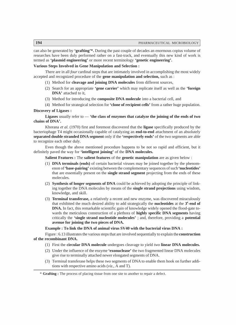
194 PHARMACEUTICAL MICROBIOLOGY
can also be generated by ‘grafting’*. During the past couple of decades an enormous copius volume ofresearches have been duly performed rather on a fast-track, and eventually this new kind of work istermed as ‘plasmid engineering’ or more recent terminology ‘genetic engineering’.Various Steps Involved in Gene Manipulation and Selection :
There are in all four cardinal steps that are intimately involved in accomplishing the most widelyaccepted and recognized procedure of the gene manipulation and selection, such as :
(1) Method for cleavage and joining DNA molecules from different sources,
(2) Search for an appropriate ‘gene carrier’ which may replicate itself as well as the ‘foreignDNA’ attached to it,
(3) Method for introducing the composite DNA molecule into a bacterial cell, and
(4) Method for strategical selection for ‘clone of recipient cells’ from a rather huge population.
Discovery of Ligases :Ligases usually refer to — ‘the class of enzymes that catalyze the joining of the ends of two
chains of DNA’.Khorana et al. (1970) first and foremost discovered that the ligase specifically produced by the
bacteriophage T4 might occasionally capable of catalyzing an end-to-end attachment of an absolutelyseparated double stranded DNA segment only if the ‘respectively ends’ of the two segments are ableto recognize each other duly.
Even though the above mentioned procedure happens to be not so rapid and efficient, but itdefinitely paved the way for ‘intelligent joining’ of the DNA molecules.
Salient Features : The salient features of the genetic manipulation are as given below :
(1) DNA terminals (ends) of certain bacterial viruses may be joined together by the phenom-enon of ‘base-pairing’ existing between the complementary sequences of such ‘nucleotides’that are essentially present on the single strand segment projecting from the ends of thesemolecules.
(2) Synthesis of longer segments of DNA could be achieved by adopting the principle of link-ing together the DNA molecules by means of the single strand projections using wisdom,knowledge, and skill.
(3) Terminal transferase, a relatively a recent and new enzyme, was discovered miraculouslythat exhibited the much desired ability to add strategically the nucleotides at the 3′′′′′-end ofDNA. In fact, this remarkable scientific gain of knowledge widely opened the flood-gate to-wards the meticulous construction of a plethora of highly specific DNA segments havingcritically the ‘single strand nucleotide molecules’ ; and, therefore, providing a potentialavenue for joining the two pieces of DNA.
Example : To link the DNA of animal virus SV40 with the bacterial virus DNA :Figure : 6.13 illustrates the various steps that are involved sequentially to explain the construction
of the recombinant DNA.(1) First the circular DNA molecule undergoes cleavage to yield two linear DNA molecules.
(2) Under the influence of the enzyme ‘exonuclease’ the two fragmented linear DNA moleculesgive rise to terminally attached newer elongated segments of DNA.
(3) Terminal transferase helps these two segments of DNA to enable them hook on further addi-tions with respective amino acids (viz., A and T).
* Grafting : The process of placing tissue from one site to another to repair a defect.
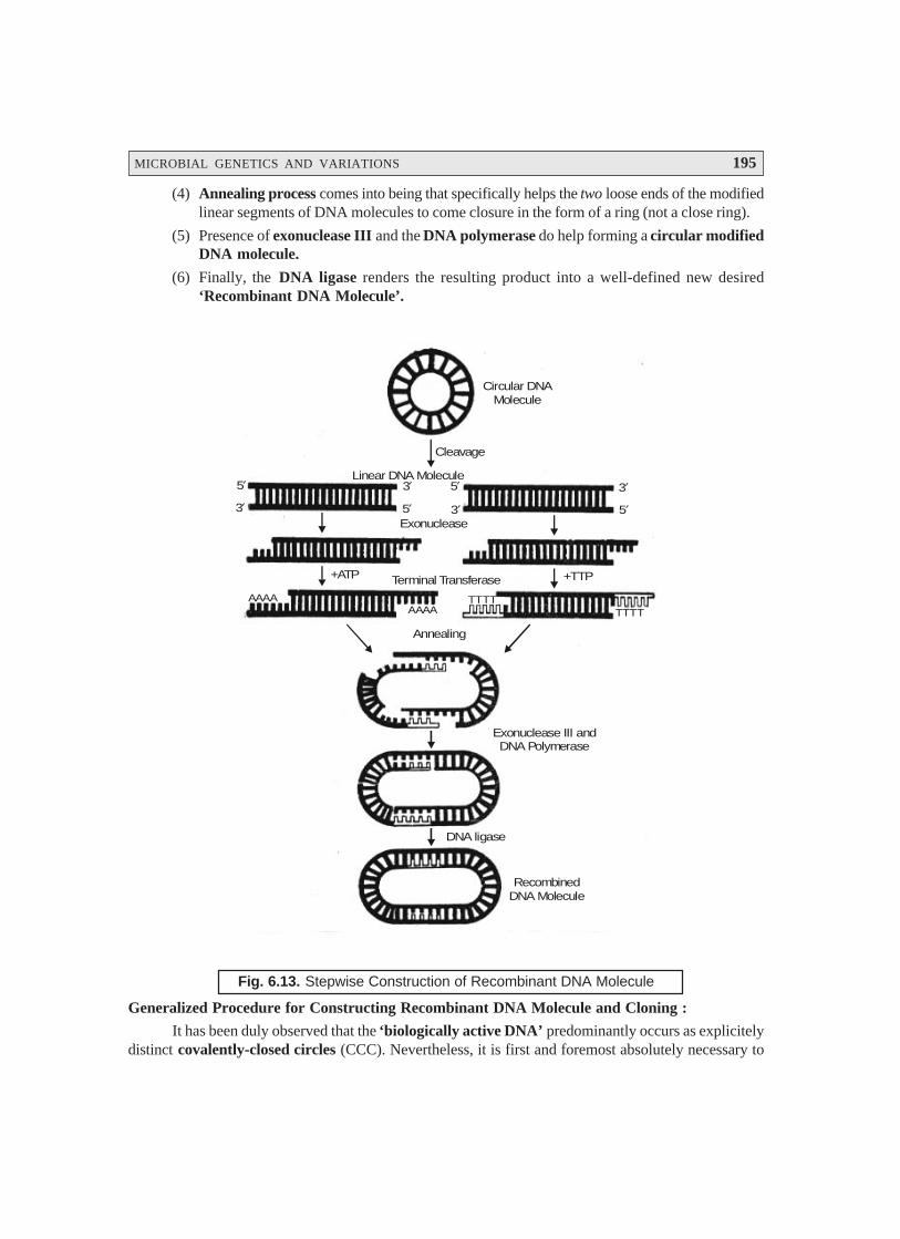
MICROBIAL GENETICS AND VARIATIONS 195
(4) Annealing process comes into being that specifically helps the two loose ends of the modifiedlinear segments of DNA molecules to come closure in the form of a ring (not a close ring).
(5) Presence of exonuclease III and the DNA polymerase do help forming a circular modifiedDNA molecule.
(6) Finally, the DNA ligase renders the resulting product into a well-defined new desired‘Recombinant DNA Molecule’.
AAAATTTTAAAA
TTTT
Circular DNAMolecule
Cleavage
Linear DNA Molecule
Exonuclease
Terminal Transferase
Annealing
Exonuclease III andDNA Polymerase
DNA ligase
RecombinedDNA Molecule
3′ 5′
3′5′
5′
3′ 5′
3′
+TTP+ATP
Fig. 6.13. Stepwise Construction of Recombinant DNA Molecule
Generalized Procedure for Constructing Recombinant DNA Molecule and Cloning :
It has been duly observed that the ‘biologically active DNA’ predominantly occurs as explicitelydistinct covalently-closed circles (CCC). Nevertheless, it is first and foremost absolutely necessary to
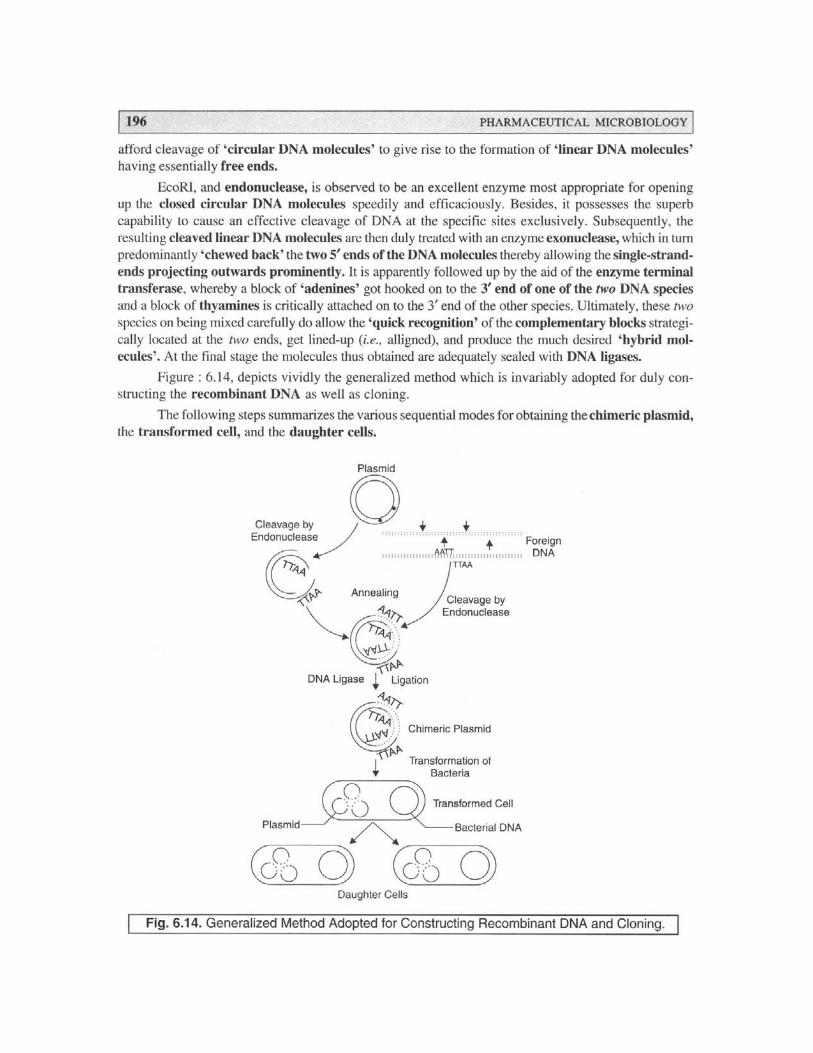
MICROBIAL GENETICS AND VARIATIONS 197
(a) Plasmid gets cleaved to corresponding linear molecules by endonuclease ; also accom-plished by ‘foreign DNA’.
(b) Annealing process commences to obtain the desired closed circular DNA.
(c) ‘Chimeric plasmid’ is duly accomplished via ‘ligation’ with DNA ligase.
(d) Transformed cell is obtained subsequently due to the transformation of organisms.
(e) Daughter cells are ultimately obtained from the respective transformed cell.
FURTHER READING REFERENCES
1. Hartwell L H et al. : Genetics : From Genes to Genomes, McGraw-Hill, New York, 2nd.edn, 2004.
2. Jagus R, and Joshi B : Protein Biosynthesis : In : Encyclopedia of Microbiology, LederbergJ, Editor-in-Chief, Academic Press, San Diego, 3 Vol., 2nd edn., 2000.
3. Leonard AC, and Grimwade JE : Chromosome Replication and Segregation. In : Ency-clopedia of Microbiology, Lederberg IJ, Editor-in-Chief, Academic Press, San Diego, 2000.
4. Lewin B : Genes, Oxford University Press, New York, 7th edn, 2000.
5. Murray A, and Hunt T : The Cell Cycle : An Introduction, WH Freeman, New York,1993.
6. Ptashne M : A Genetic Switch, Blackwell Scientific Publications, Cambridge Mass, 2nd.edn, 1992.
7. Snyder L, and Champness W : Molecular Genetic of Bacteria, DC : ASM Press, Washing-ton, 1997.
8. Voet D, and Voet JG : Biochemistry, John Wiley and Sons, New York, 2nd, edn., 1995.
9. Watson JD et al. : Molecular Biology of the Gene, Benjamin/Cummings, Redwod City,Calif., 1988.
10. Weaver RF : Molecular Biology, McGraw Hill, Dubaque, Iowa, 2nd edn., 2002.

198
7.1. INTRODUCTION
The wonderful universally accepted and recognized concept and idea of ‘microbial control’was predominantly introduced in the domain of microbiology by two altogether entirely different dedicatedresearchers, namely :
Ignatz Semmelweis (1816 – 1865)—Hungarian Physician ;
Joseph Lister (1827–1912)—British Physician.
Interestingly, Semmelweis was pioneer in the introduction of strict mandatory procedures towash the hands of all personnels with chlorinated lime water (i.e., bleaching powder containing approx.38% available chlorine), which procedure significantly reduced the exhorbitant rate of infection. Likewise,Lister accomplished enormous success by treating the surgical wounds with solutions of ‘phenol’ (i.e.,carbolic acid) ; ever the surgical procedures were carried out in an adequate carbolic acid aerosolenvironment, which in turn drastically minimized the incidence of any probable wound infection.
Since a long stretch of nearly 150 years from Semmelweis ; and almost 100 good years fromLister there have been really a sea-change with respect to the highly specific and precise manipulative,logical, and scientific control of the ‘microbial growth’ both by physical methods and chemicalmethods.
Now, each of these two aforesaid methodologies will be treated individually in the sections thatfollows with appropriate typical examples wherever necessary.
7.2. PHYSICAL METHODS
The physical methods related to microbial control (or growth) are as enumerated under :
(a) Heat,
(b) Moist Heat,
MICROBIAL CONTROL BY PHYSICALAND CHEMICAL METHODS7
• Introduction• Physical Methods• Chemical Methods• Experimental Parameters Influencing the Antimicrobial Agent Activity
MICROBIAL CONTROL BY PHYSICAL AND CHEMICAL METHODS 199
(c) Pasteurization,
(d) Dry-Heat Sterilization,
(e) Filtration,
(f) Cold,
(g) Desiccation,
(h) Osmotic Pressure, and
(i) Radiation.
All these individual methods shall now be treated separately in the sections that follows :
7.2.1. Heat
Heat represents probably the most common effective, and productive means whereby organismsare almost killed. In fact, it is a usual practice to have the laboratory media, laboratory glasswares, andhospital surgical instruments adequately sterilized by heat i.e., moist heat in an electric autoclave.
Salient Features. Following are the salient features of heat controlled microbes, namely :
(1) Most economical and easily controlable means of microbial growth.
(2) Usually kill microbes by causing denaturation of their respective enzymes.
(3) Heat resistance capacity of the organism must be studied carefully and taken into considera-
tion.
(4) Thermal Death Time (TDT). TDT is referred to as the minimal length of time whereby all
microbes present in a liquid culture medium will be killed at a given temperature.
(5) Thermal Death Point (TDP). TDP designates the lowest temperature at which all of the
microorganisms present in a liquid suspension will be killed in just 10 minutes. In fact, heat resistancepredominantly varies amongst the different range of organisms ; besides, these glaring differences may
be duly expressed via the concept of thermal death point (TDP).
However, it is pertinent to state here that both TDP and TDT are equally vital, important, and
useful guidelines which essentially indicate the actual prevailing severity of treatment needed to kill agiven population of organisms.
(6) Decimal Reduction Time [DRT or D-Value]. DRT or D-Value represents a 3rd conceptwhich is directly associated with the organism’s extent of heat resistance. In fact, it is very much
equivalent to the time (minutes), whereby almost 90% of the population of prevailing microbes at an
exact specified temperature shall be killed as illustrated in Fig. 7.1, having DRT of 1 minute. It is,however, pertinent to mention here that DRT is of an extreme importance and usefulness in the
‘canning industry’ dealing with fruit concentrates, fruit pulps, fruit slices, baked beans, corned-beef,
fish products, fish chuncks, baby corns, lentils, and the like.
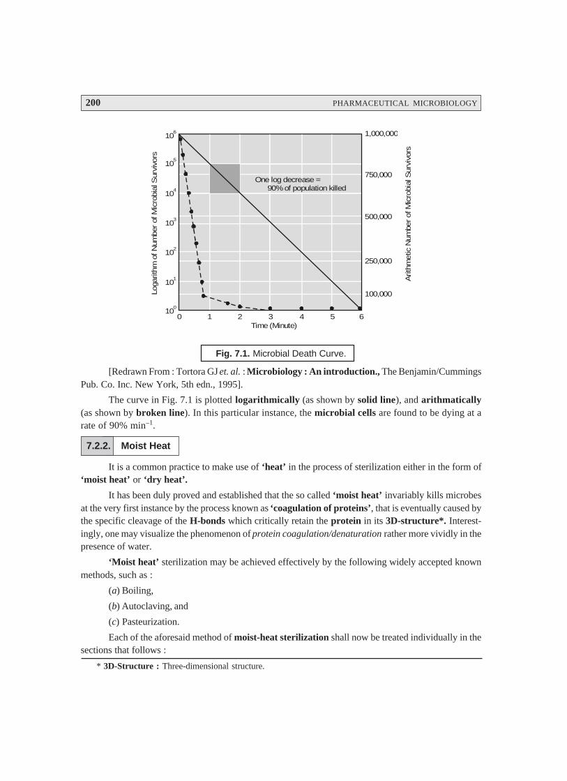
200 PHARMACEUTICAL MICROBIOLOGY
0 1 2 3 4 5 610
0
101
102
103
104
105
106
Time (Minute)
Loga
rithm
of N
umbe
r of
Mic
robi
al S
urvi
vors
1,000,000
750,000
500,000
250,000
100,000
Arit
hmet
ic N
umbe
r of
Mic
robi
al S
urvi
vors
One log decrease = 90% of population killed
Fig. 7.1. Microbial Death Curve.
[Redrawn From : Tortora GJ et. al. : Microbiology : An introduction., The Benjamin/CummingsPub. Co. Inc. New York, 5th edn., 1995].
The curve in Fig. 7.1 is plotted logarithmically (as shown by solid line), and arithmatically(as shown by broken line). In this particular instance, the microbial cells are found to be dying at arate of 90% min–1.
7.2.2. Moist Heat
It is a common practice to make use of ‘heat’ in the process of sterilization either in the form of‘moist heat’ or ‘dry heat’.
It has been duly proved and established that the so called ‘moist heat’ invariably kills microbesat the very first instance by the process known as ‘coagulation of proteins’, that is eventually caused bythe specific cleavage of the H-bonds which critically retain the protein in its 3D-structure*. Interest-ingly, one may visualize the phenomenon of protein coagulation/denaturation rather more vividly in thepresence of water.
‘Moist heat’ sterilization may be achieved effectively by the following widely accepted knownmethods, such as :
(a) Boiling,
(b) Autoclaving, and
(c) Pasteurization.
Each of the aforesaid method of moist-heat sterilization shall now be treated individually in thesections that follows :
* 3D-Structure : Three-dimensional structure.
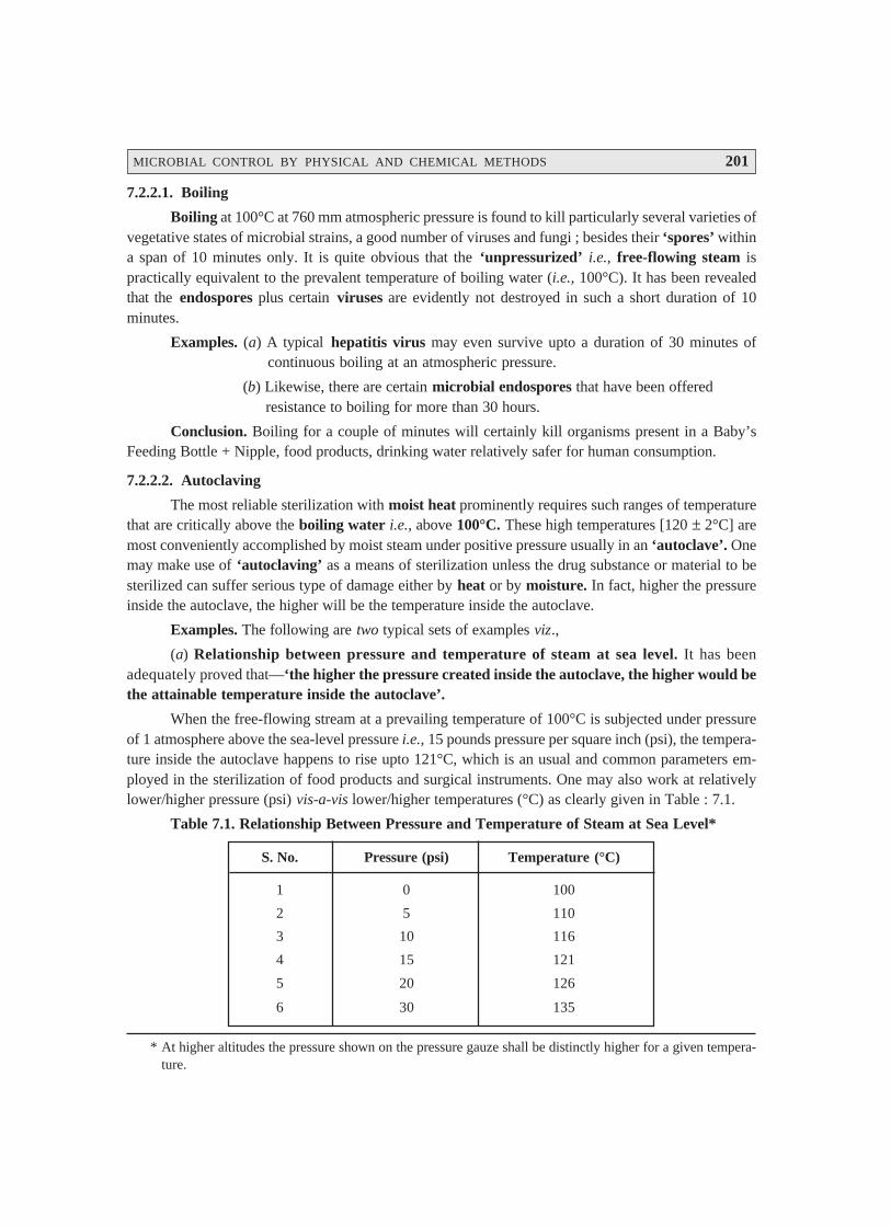
MICROBIAL CONTROL BY PHYSICAL AND CHEMICAL METHODS 201
7.2.2.1. Boiling
Boiling at 100°C at 760 mm atmospheric pressure is found to kill particularly several varieties ofvegetative states of microbial strains, a good number of viruses and fungi ; besides their ‘spores’ withina span of 10 minutes only. It is quite obvious that the ‘unpressurized’ i.e., free-flowing steam ispractically equivalent to the prevalent temperature of boiling water (i.e., 100°C). It has been revealedthat the endospores plus certain viruses are evidently not destroyed in such a short duration of 10minutes.
Examples. (a) A typical hepatitis virus may even survive upto a duration of 30 minutes ofcontinuous boiling at an atmospheric pressure.
(b) Likewise, there are certain microbial endospores that have been offeredresistance to boiling for more than 30 hours.
Conclusion. Boiling for a couple of minutes will certainly kill organisms present in a Baby’sFeeding Bottle + Nipple, food products, drinking water relatively safer for human consumption.
7.2.2.2. Autoclaving
The most reliable sterilization with moist heat prominently requires such ranges of temperaturethat are critically above the boiling water i.e., above 100°C. These high temperatures [120 ± 2°C] aremost conveniently accomplished by moist steam under positive pressure usually in an ‘autoclave’. Onemay make use of ‘autoclaving’ as a means of sterilization unless the drug substance or material to besterilized can suffer serious type of damage either by heat or by moisture. In fact, higher the pressureinside the autoclave, the higher will be the temperature inside the autoclave.
Examples. The following are two typical sets of examples viz.,
(a) Relationship between pressure and temperature of steam at sea level. It has beenadequately proved that—‘the higher the pressure created inside the autoclave, the higher would bethe attainable temperature inside the autoclave’.
When the free-flowing stream at a prevailing temperature of 100°C is subjected under pressureof 1 atmosphere above the sea-level pressure i.e., 15 pounds pressure per square inch (psi), the tempera-ture inside the autoclave happens to rise upto 121°C, which is an usual and common parameters em-ployed in the sterilization of food products and surgical instruments. One may also work at relativelylower/higher pressure (psi) vis-a-vis lower/higher temperatures (°C) as clearly given in Table : 7.1.
Table 7.1. Relationship Between Pressure and Temperature of Steam at Sea Level*
S. No. Pressure (psi) Temperature (°C)
1 0 100
2 5 110
3 10 116
4 15 121
5 20 126
6 30 135
* At higher altitudes the pressure shown on the pressure gauze shall be distinctly higher for a given tempera-ture.
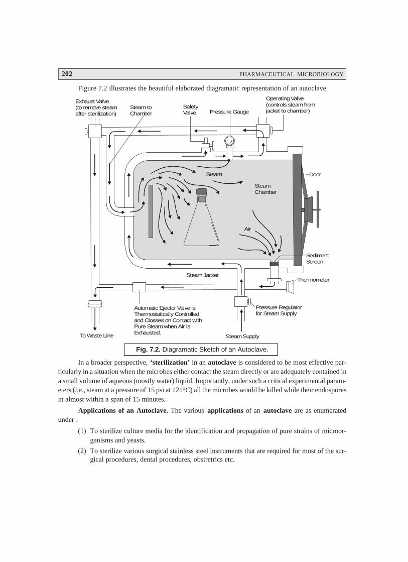
202 PHARMACEUTICAL MICROBIOLOGY
Figure 7.2 illustrates the beautiful elaborated diagramatic representation of an autoclave.
Steam
SteamChamber
Air
Door
SedimentScreen
Thermometer
Pressure Regulatorfor Steam Supply
Steam Supply
Automatic Ejector Valve isThermostatically Controlledand Closses on Contact withPure Steam when Air isExhausted.To Waste Line
Steam toChamber
SafetyValve Pressure Gauge
Operating Valve(controls steam fromjacket to chamber)
Exhaust Valve(to remove steamafter sterilization)
Steam Jacket
Fig. 7.2. Diagramatic Sketch of an Autoclave.
In a broader perspective, ‘sterilization’ in an autoclave is considered to be most effective par-ticularly in a situation when the microbes either contact the steam directly or are adequately contained ina small volume of aqueous (mostly water) liquid. Importantly, under such a critical experimental param-eters (i.e., steam at a pressure of 15 psi at 121°C) all the microbes would be killed while their endosporesin almost within a span of 15 minutes.
Applications of an Autoclave. The various applications of an autoclave are as enumeratedunder :
(1) To sterilize culture media for the identification and propagation of pure strains of microor-ganisms and yeasts.
(2) To sterilize various surgical stainless steel instruments that are required for most of the sur-gical procedures, dental procedures, obstretrics etc.

MICROBIAL CONTROL BY PHYSICAL AND CHEMICAL METHODS 203
(3) To sterilize various types of surgical dressings, gauzes, sutures etc.
(4) To sterilize a host of IV applicators, equipments, solutions, and syringes as well.
(5) To sterilize transfusion equipment(s) and a large number of other alied items that can con-veniently withstand high pressures and temperatures.
(6) When the ‘large industrial premises’ make use of the autoclaves, these are knwon as re-torts, whereas, the small domestic applications invariably employ pressure cookers (bothbased on exactly the same principles) for preparation of food* and canning of processedfood products.
Important Aspects. In a situation, when we essentially look for extended heat requirement soas to specifically reach the exact centre of the solid materials viz., canned meats, fish (tuna), due to thefact that such materials fail miserably to develop the most desired efficient convection currents whichinvariably take place in the body of liquids.
Therefore, the particular heating of large containers/vessels does essentially require extra timeperiod (in minutes) as given in Table 7.2.
Table 7.2. Overall Effect of Container Size upon Autoclave Sterilization Times(Minutes) for Liquid Solutions**.
S. No. Size of Container Volume of Autoclave Sterilization
Liquid (mL) Time (Minutes)
1 Fermentation Bottle (9L) 6750 70
2 Erlenmeyer Flask (2L) 1500 30
3 Erlenmeyer Flask (125 mL) 95 15
4 Test tube [Size : 18 × 150 mm] 10 15
** (i) The autoclave sterilization times in the autoclave very much include the time required for the contents ofthe containers to perfectly reach the sterilization temperatures.
(ii) Obviously, for a very small container this is only 5 minutes or even less, whereas for a 9 L capacityfermentation bottle it might be as high as ~ 70 minutes.
(iii) All containers that are supposed to be sterilized by ‘autoclave’ are invariably filled only upto 3/4th thetotal volume i.e., their actual capacity.
Salient Features. The salient features of ‘autoclave sterilization’ are briefly stipulated asunder :
(1) In order to sterilize duly the surface of a solid, one must allow the ‘steam’ to actually contactthe same. Nevertheless, particular care must be taken to allow the perfect sterilization of bandages, dry-glasswares, and the like so as to ascertain that steam gets into contact with all the exposed surfaces.
Example. Aluminium foil does not allow the passage of steam to pass across (i.e., impervious),and hence must be avoided to wrap such materials meant to be sterilized ; instead, one may freely makeuse of brown wrapping paper (cellulose).
(2) Trapped Air. All necessary precautions and requisite care must be taken to get rid of anytrapped air strategically located at the bottom of a ‘dry container’, due to the fact that the ‘trapped air’
* Food is cooked usually under most hygenic conditions within a short span of time thereby saving a lot ondomestic cooking gas/electricity.
204 PHARMACEUTICAL MICROBIOLOGY
shall not be replaced by ‘steam’ at any cost, which being lighter than air. However, one may just visual-ize imaginatively the so called ‘trapped air’ as a mini-hot air oven, that would eventually require notonly a higher temperature but also a much longer duration to sterilize materials.
Based on the actual experience one may specifically tackle such containers which have a ten-dency to trap air must be positioned in a ‘tipped state’ in order that all the steam shall ultimately helpto force out the air.
Note. Importantly, such products which obstruct penetration by moisture viz., petroleumjelly, mineral oil (furnace oil) are not usually sterilized by the same methods as adoptedto sterilize aqueous solutions.
7.2.2.3. Pasteurization
Pasteurization refers to ‘the process of heating of a fluid at a moderate temperature for adefinite period of time to destroy undesirable microorganisms without changing to any extent thechemical composition.’
Example. In pasteurization of milk, pathogenic organisms are invariably destroyed by heating at62° C for a duration of 30 minutes, or by ‘flash’ heating to higher temperatures for less than 1 minute,which is otherwise known as high-temperature short time (HTST) pasteurization.
In a broader perspective the pasteurization of milk, effectively lowers the total bacterial count ofthe milk by almost 97 to 99%, due to the fact that the most prevalent milk-borne pathogens viz., Tuberclebacillus*, and Samonella, Streptococcus, and Brucella organisms, fail to form ‘spores’, and are quitesensitive to heat.
It may, however, be observed that several relatively heat-resistant (thermoluric) microorganismsdo survive pasteurization, and these may ultimately fail to :
• Cause refrigerated milk to turn sour (spoil) in a short span of time, and
• Cause any sort of disease in humans.
Ultra-High-Temperature (UHT) Treatments. Sterilization of milk is absolutely different frompasteurization. It may be duly accomplished by UHT treatments in order that it can be most easily andconveniently stored even without any sort of refrigeration. So as to maintain the first order ‘organolepticcharacteristic features’** of fresh milk and to avoid attributing to the milk a prevalent cooked taste,the UHT system gained reasonable qualified success and hence due recognition across the globe, wherebythe liquid milk never touches a surface hotter than the milk itself during the course of heating by steam.
Methodology. The various steps involved are as follows :
(1) Milk is allowed to fall in a thin-film vertically down through a stainless-steel (SS) chamberof ‘superheated steam’, and attains 140°C in less than 1 second.
(2) Resulting milk is adequately held for a duration of only 3 seconds duly in a ‘holding tube’.
(3) Ultimately, the pre-heated milk is cooled in a ‘vacuum chamber’, wherein the steam simplyflashes off.
(4) The above stated process [(in (3)] distinctly enables the milk to raise its temperature from74—140°C in just 5 seconds, and suddenly drops back to 74°C again.
* Tuberculosis bacterium.
** Organoleptic characteristic features. These refer to the specific taste, flavour, colour, and overall physicalappearance of the product.
MICROBIAL CONTROL BY PHYSICAL AND CHEMICAL METHODS 205
Summararily, the very concept of equivalent treatments* clearly expatiates the particular rea-sons of the various methods of killing microbes, such as :
Pasteurization : At 63°C for 30 minutes ;
HTST-Treatment : At 72°C for 15 seconds ;
UHT-Treatment : At 140°C for < 1 second ;
7.2.2.4. Dry-Heat SterilizationIt is a well known fact that microorgansims get killed by dry heat due to the oxidation effects.
Direct Flaming. Direct flaming designates one of the most simple method of dry-heat sterili-zation. In reality, the dry-heat sterilization is mostly used in a ‘microbiology laboratory’ for the steri-lization of the ‘inoculating loops’, which is duly accomplished by heating the loop wire to a ‘red-glow’, and this is 100% effective in actual practice. Likewise, the same principle is even extended to theprocess of ‘inceneration’ to sterilize as well as dispose of heavily contaminated paper bags, cups, andused dressings.
Hot-Air Sterilization. It may be regarded as another kind of dry-heat sterilization. In thisparticular process, the various items need to be sterilized are duly kept in an electric oven, preferablywith a stainless-steel chamber inside, and duly maintained at 170°C for a duration of approximately 2hours (to ensure complete sterilization).
It has been adequately observed that the longer the period plus higher temperature are neededprofusely due to the fact that the heat in water is more rapidly passed onto a ‘cool body’ in comparisonto the heat in air.
Example. The experience of exposing the ‘finger’ in a boiling water at 100°C (212°F) vis-a-visexposing the same ‘finger’ in a hot-air oven at the same tempearture for the same duration.
7.2.2.5. FiltrationFiltration may be defined as ‘the process of removing particles from a solution by allowing
the liquid position to pass through a membrane or other particle barrier’. In reality, it essentiallycontains tiny spaces or holes which exclusively allow the liquid to pass but are too small to permit thepassage of the small particles.
In other words, one may also explain ‘filtration’ as the process of a liquid or gaseous substancevia a screen-like material having suitable pores small enough to retain the microorganisms (bacteria). Avacuum which is formed in the ‘receiver flask’ actually aids by means of gravity to suck the liquid viathe filter medium engaged. However, in actual practice the phenomenon of filtration is invariably em-ployed to sterilize the specific heat sensitive substances, namely : culture media ; vaccines ; enzymes ;and several antibiotic solutions.
High-Efficiency Particulate Air (HEPA) Filters. HEPA-Filters are mostly used to get rid ofpractically all microbes that happen to be larger than 0.3 μμμμμm in diameter.
Examples. HEPA-Filters are largely used in :
(a) Intensive-Care Units [ICUs] in specialized hospitals treating severe Burn cases.
(b) In Sterile Zones of High-Value Antibiotic Preparations, Packaging, IV-injections, and othersuch sensitive sterile preparations.
Membrane Filters. In the recent past, technologically advanced membrane filters made up ofeither Cellulose Esters or Plastic Polymers have been employed profusely for the laboratory andindustrial applications as shown in Fig. 7.3 and 7.4.
* The ‘heat treatments’ viz., pasteurization, HTST, and UHT treatment.
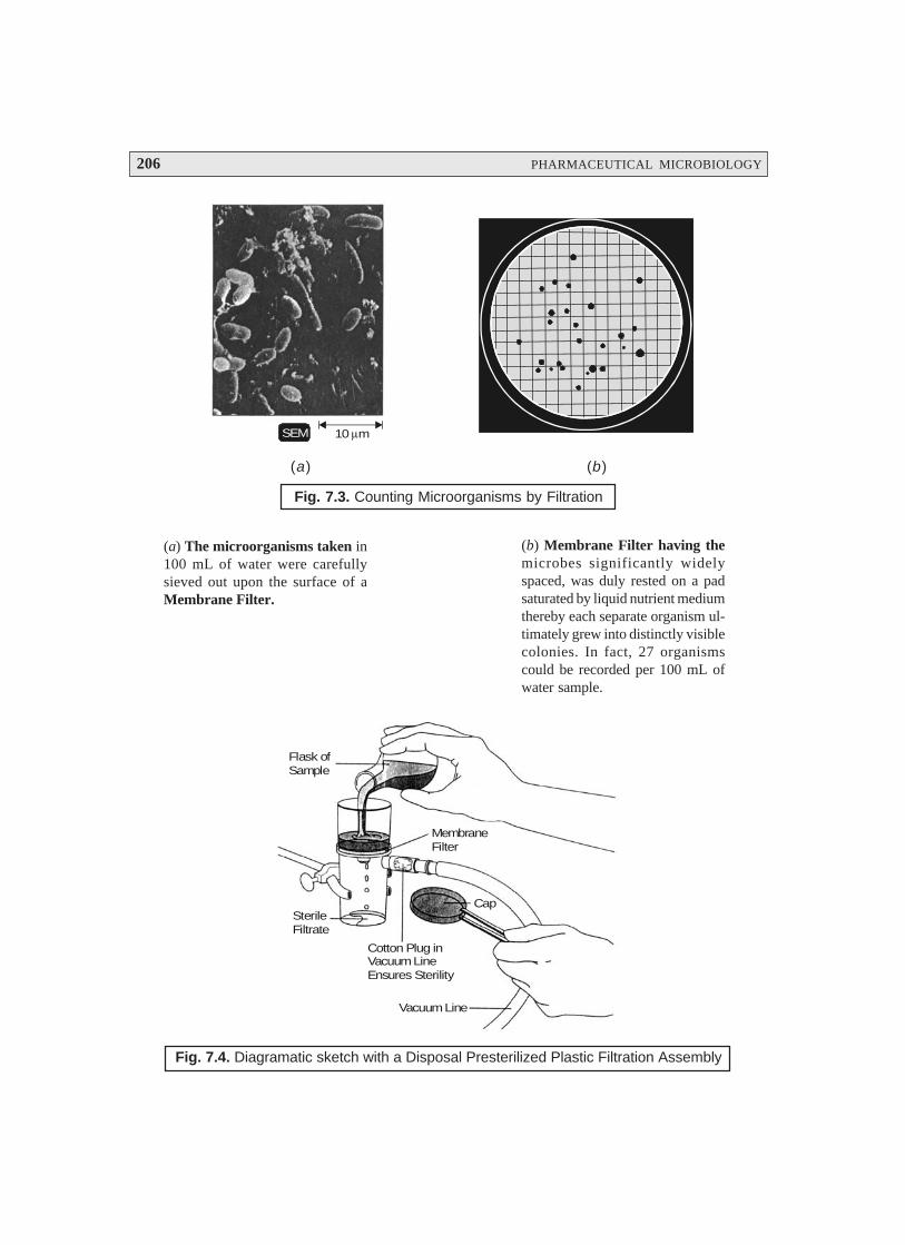
206 PHARMACEUTICAL MICROBIOLOGY
SEM 10 mμ
(a) (b)
Fig. 7.3. Counting Microorganisms by Filtration
MembraneFilter
CapSterileFiltrate
Cotton Plug inVacuum LineEnsures Sterility
Vacuum Line
Flask ofSample
Fig. 7.4. Diagramatic sketch with a Disposal Presterilized Plastic Filtration Assembly
(a) The microorganisms taken in100 mL of water were carefullysieved out upon the surface of aMembrane Filter.
(b) Membrane Filter having themicrobes significantly widelyspaced, was duly rested on a padsaturated by liquid nutrient mediumthereby each separate organism ul-timately grew into distinctly visiblecolonies. In fact, 27 organismscould be recorded per 100 mL ofwater sample.

MICROBIAL CONTROL BY PHYSICAL AND CHEMICAL METHODS 207
Explanation for Fig. 7.4 :
1. The sample to be filtered is duly loaded into the ‘upper chamber’, and consequently forcedthrough the strategically placed membrane filter.
2. The pores present in the membrane filter are definitely much smaller in comparison to themicroorganisms ; and, therefore, the microorganisms present are obviously retained upon thesurface of the filter.
3. Sterilized sample (free from microbes) may now be decanted conveniently from the ‘lowerchamber’.
Specifications of Membrane Filters. Membrane filters usually have a thickness of 0.1 μm,and having almost uniform pores. However, in certain commercially available brands, the film is dulyirradiated so as to generate extremely uniform holes, where the radiation particles have made itspassage, are critically etched in the plastic. The pores of membrane filters usually range between 0.22 to0.45 μm, intended for microorganisms.
Note. (1) Certain highly flexible microbes viz. spirochaetes, and the wall-less bacteria viz.,mycoplasma, may sometimes pass through such membrane filters.
(2) To retain certain viruses and large-sized protein molecules are duly retained bysuch filters with pore size as small as 0.01 μμμμμm.
7.2.2.6. Cold
It has been critically observed that the overall effect of ‘low temperature’ upon the microorgan-isms exclusively depends on the specific organism and the intensity of the application.
Example. At temperatures ranging between 0–7°C (i.e., the ordinary refrigerator), the actualrate of metabolism of majority of microorganisms gets reduced substantially to such an extent that theyare rendered incapable of either synthesizing toxins* or causing reproduction.**
Thus, one may conclude that ‘ordinary refrigeration’ exerts a distinct bacteriostatic effecti.e., stops the multiplication vis-a-vis growth of microbes.
Psychotrophs***, however, are found to grow appreciably but slowly particularly at therefrigerator temperature conditions ; and may change the very appearance and taste of food productsafter a certain lapse of time.
Salient Features. The various salient features of microbes in a ‘cold’ environment are as follows :
(1) A few microbes may even grow at sub-freezing temperatures (i.e., below the freezingtemperature).
(2) Sudden exposure to sub-freezing temperatures invariably render bacteria into the ‘dormant-state’; however, they do not kill them (bactericidal effect) ultimately.
(3) Gradual Freezing is observed to be quite harmful and detrimental to microorganisms, per-haps due to the fact that the ice-crystals which eventually form and grow do disrupt the cellu-lar as well as the molecular structure of the microorganisms.
* Toxin : A poisonous substance of animal or plant origin.
** Reproduction : The process by which animals, plants, and microbes produce offspring.
*** Psychotrophs : Such microorganisms that are responsible for low temperature food spoilage.

208 PHARMACEUTICAL MICROBIOLOGY
(4) Life-Span of Frozen Vegetative Microbes—Usually remain active for a year upto 33% ofthe entire initial population, whereas other microbial species may afford relatively veryscanty survival rates.
7.2.2.7. Desiccation
In order to have both normal growth and adequate multiplication the microorganisms do re-quire water. Desiccation represents a typical state of microbes in the absence of water ; however, theirgrowth and reproduction remain restricted but could sustain viability for several years. Interestingly, assoon as ‘water’ is duly made available to them the said organisms resume their usual growth and divi-sion as well. This highly specific ability has been adequately employed in the laboratory manipulationswhereby the microbes are carefully preserved by lyophilization.*
It has been duly observed that the ensuing resistance of the vegetative cells to undergo thephenomenon of desiccation changes with the specific species as well as the microorganism’s environment.
Example : Gonorrhea** organism, Neisseria gonorrhoeae (Gonococcus), possess an ability towithstand dryness only upto a duration 60 minutes hardly ; whereas, Tuberculosis*** bacterium,Mycobacterium tuberculosis (Bacillus) may even remain completely viable for months together at astretch.
Important Points : Following are certain important points which should always be borne inmind :
(a) An invariably susceptible microbe is found to be appreciably resistant when it gets dulyembedded in pus cells, mucous secretions, and in faeces.
(b) In contract to microbes the viruses are usually found to be quite resistant to the phenomenonof ‘desiccation; however, they do not exhibit resistance comparable to the bacterialendospores.
(c) Importantly, in a typical hospital environment (setting) the presence and subsequent abilityof some particular dried bacteria and endospores do remain absolutely viable, such as :beddings, clothings, dust particulate matters, and above all the disposable (used) dressingsfrom patients may contain infectious organisms strategically located in dried pus, faecalmatter, mucous secretions, and urine.
7.2.2.8. Osmotic Pressure
Osmotic pressure refers to–‘the pressure which develops when two solutions of differentconcentrations are duly separated by a semipermeable membrane’.
In actual age-old practice, the preservation of food products viz., pickles, fruits, are duly accom-plished by the use of high-concentrations of salts and sugars which eventually exert their effects onaccount of the osmotic pressure. The most logical and probable underlying mechanism being the creation
* Lyophilization (Freeze-Drying] : The process of rapidly freezing a substance at an extremely low tempera-ture, and then dehydrating the substance in a high vacuum.
** A specific, contagious, catarrhal inflammation of the genital mucous membrane of either sex.
*** An infectious disease caused by the tubercle bacillus, M. tuberculosis, that causes formation of tubercles,necrosis, absecesses, fibrosis, and clacification.
MICROBIAL CONTROL BY PHYSICAL AND CHEMICAL METHODS 209
of an extremely hypertonic environment due to the presence of these substances (salts and sugars) athigh concentrations that enables water to leave the microbial cell precisely. In fact, the preservationafforded by the osmotic pressure very much resembles to that caused by desiccation (see Section7.2.2.7), besides, the glaring fact that both processes evidently deny the microbial cell of the requisitequantum of moisture essentially required for its normal growth. Dehydration of the microbial cell actu-ally renders the plasma membrane to shrink away from the respective cell-wall (i.e.,plasmolysis),whereby the consequent cell stops growth (and hence reproduction), and it may not cause an instantdeath. In a broader perspective, the fundamental principle of osmotic pressure is largely exploited inthe prolonged preservation of food products.
Examples : (a) Concentrated Salt Solutions (Brine Solution) may be used profusely in thepreservation and cure of meats, fish, vegetables, pickles etc.
(b) Concentrated Sugar Solutions (Sugar Syrup) may be employed, extensively in the preser-vation of lime juice, fruits etc.
7.2.2.9. Radiation
Radiation refers to — ‘any form of radiant energy emission or divergence, as of energy inall directions from luminous bodies, radiographical tubes, particle accelerators, radioactive ele-ments, and fluorescent substances’.
It has been established beyond any reasonable doubt that radiation exerts its various effects onthe cells, depending upon its wavelength, intensity, and duration as well. Generally, one may comeacross two kinds of radiation which would cause a bactericidal effects on microbes, or usually referredto as the ‘sterilizing radiation’, namely :
(a) Ionizing Radiation, and
(b) Nonionizing Radiation.
Each of the aforesaid types of radiation shall be treated individually in the sections that follows :
7.2.2.9.1. Ionizing Radiation
The ionizing radiation normally possess a wavelength distinctly shorter in comparison to thenonionizing radiation (size < 1 nm) e.g., γγγγγ-rays, X-rays, or high-energy electron beams.
Figure 7.5 vividly depicts that the said ionization radiation invariably carries a significant quan-tum of energy ranging between 10–5 nm (γγγγγ-rays) to 10–3 nm (X-rays).
γγγγγ-Rays : These are emitted by radioactive cobalt (Co),
X-Rays : These are produced by X-ray machines, and
Electron Beams : These are generated by accelerating electrons to high energies in specialmachines.
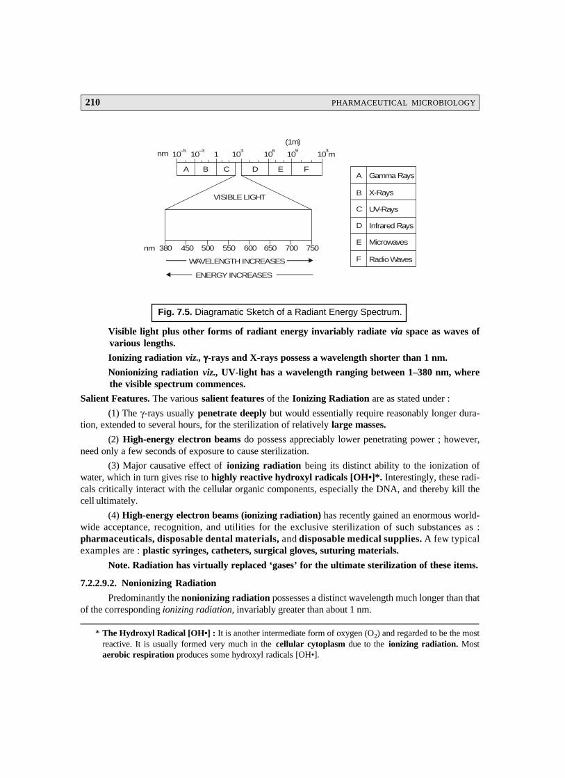
210 PHARMACEUTICAL MICROBIOLOGY
Gamma Rays
X-Rays
UV-Rays
Infrared Rays
Microwaves
Radio Waves
A
B
C
D
E
F
10–5
10–3
1 103
106
109
10 m3
(1m)
nm
WAVELENGTH INCREASES
ENERGY INCREASES
nm 380 450 500 550 600 650 700 750
VISIBLE LIGHT
A B C D E F
Fig. 7.5. Diagramatic Sketch of a Radiant Energy Spectrum.
Visible light plus other forms of radiant energy invariably radiate via space as waves ofvarious lengths.
Ionizing radiation viz., γγγγγ-rays and X-rays possess a wavelength shorter than 1 nm.
Nonionizing radiation viz., UV-light has a wavelength ranging between 1–380 nm, wherethe visible spectrum commences.
Salient Features. The various salient features of the Ionizing Radiation are as stated under :
(1) The γ-rays usually penetrate deeply but would essentially require reasonably longer dura-tion, extended to several hours, for the sterilization of relatively large masses.
(2) High-energy electron beams do possess appreciably lower penetrating power ; however,need only a few seconds of exposure to cause sterilization.
(3) Major causative effect of ionizing radiation being its distinct ability to the ionization ofwater, which in turn gives rise to highly reactive hydroxyl radicals [OH•]*. Interestingly, these radi-cals critically interact with the cellular organic components, especially the DNA, and thereby kill thecell ultimately.
(4) High-energy electron beams (ionizing radiation) has recently gained an enormous world-wide acceptance, recognition, and utilities for the exclusive sterilization of such substances as :pharmaceuticals, disposable dental materials, and disposable medical supplies. A few typicalexamples are : plastic syringes, catheters, surgical gloves, suturing materials.
Note. Radiation has virtually replaced ‘gases’ for the ultimate sterilization of these items.
7.2.2.9.2. Nonionizing Radiation
Predominantly the nonionizing radiation possesses a distinct wavelength much longer than thatof the corresponding ionizing radiation, invariably greater than about 1 nm.
* The Hydroxyl Radical [OH•] : It is another intermediate form of oxygen (O2) and regarded to be the mostreactive. It is usually formed very much in the cellular cytoplasm due to the ionizing radiation. Mostaerobic respiration produces some hydroxyl radicals [OH•].
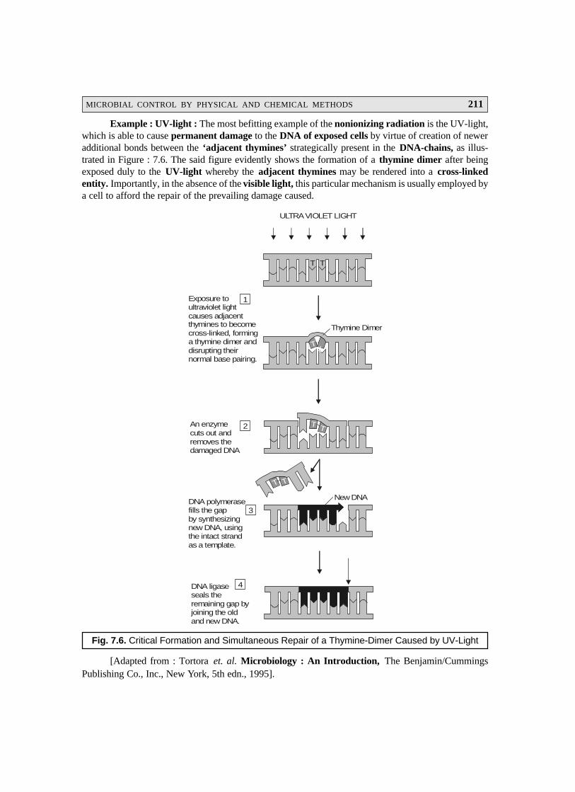
MICROBIAL CONTROL BY PHYSICAL AND CHEMICAL METHODS 211
Example : UV-light : The most befitting example of the nonionizing radiation is the UV-light,which is able to cause permanent damage to the DNA of exposed cells by virtue of creation of neweradditional bonds between the ‘adjacent thymines’ strategically present in the DNA-chains, as illus-trated in Figure : 7.6. The said figure evidently shows the formation of a thymine dimer after beingexposed duly to the UV-light whereby the adjacent thymines may be rendered into a cross-linkedentity. Importantly, in the absence of the visible light, this particular mechanism is usually employed bya cell to afford the repair of the prevailing damage caused.
T T
T T
T T
ULTRA VIOLET LIGHT
Thymine Dimer
New DNA
T T
Exposure toultraviolet lightcauses adjacentthymines to becomecross-linked, forminga thymine dimer anddisrupting theirnormal base pairing.
An enzymecuts out andremoves thedamaged DNA
DNA polymerasefills the gapby synthesizingnew DNA, usingthe intact strandas a template.
DNA ligaseseals the remaining gap byjoining the oldand new DNA.
1
2
3
4
Fig. 7.6. Critical Formation and Simultaneous Repair of a Thymine-Dimer Caused by UV-Light
[Adapted from : Tortora et. al. Microbiology : An Introduction, The Benjamin/CummingsPublishing Co., Inc., New York, 5th edn., 1995].
212 PHARMACEUTICAL MICROBIOLOGY
In reality, these ‘thymine dimers’ are found to cause effective inhibition in correcting replica-tion of the DNA in the course of division (reproduction) of the cell. It has been duly established that theUV-wavelengths at nearly 260 nm are most effective and useful for killing microbes due to the factthat these are exhaustively absorbed by the cellular DNA.
Advantages of UV Light : are as given under :
(1) It controls and maintains the miroorganisms in the air.
(2) A ‘UV-Radiation Lamp’ or a ‘Germicidal Lamp’ is abundantly and profusely employedin a variety of such sensitive areas as : operation theaters, hospital rooms, nurseries, andcafeterias.
(3) UV Light or Radiation is invariably employed to sterilize a plethora of highly sensitivebiological products commonly used in the therapeutic armamentarium, such as : serum,toxins, and a variety of vaccines.
(4) UV Light is also employed to sterilize the drinking water in homes, hospitals, and publicplaces.
(5) UV Radiation is also used for the sterilization of the ultimate treated ‘municipal-wastewaters’ for agriculture and horticulture purposes.
Disadvantages of UV Light : These are as stated under :
(1) UV Radiation is found to be not very penetrating in nature ; and, therefore, the microorgan-isms intended to be killed should be exposed almost directly to the UV-rays.
(2) Besides, such microbes that are adequately shielded (protected) by means of textiles, col-oured, glass, and paper (i.e., textured cellulose materials) are observed to be least affectedby the UV radiation.
(3) Serious Problem. In fact, UV light poses a serious problem in causing permanent damage tohuman eyes on direct exposure, besides, prolonged exposure may even cause sun burns aswell as skin cancers.
Note : (1) Antimicrobial effect of UV sunlight is on account of the exclusive formation of the‘singlet oxygen in the cytoplasm’.
(2) Microwaves (in the microwave oven) do not exhibit any direct effect on the microbes,but kill them indirectly by heating the food stuff.
A comprehensive summary of the various physical methods invariably utilized for the effectivecontrol of the microbial growth has been duly recorded in Table : 7.3.
Table : 7.3. Comprehensive Summary of Various Physical Methods Utilized for theEffective Control of Microbial Growth
S.No. Method Specification(s) Mode of Action Remarks Applications
1. Heat 1. Moist Heat
(a) Boiling or Denaturation Most effective to kill Equipments, basins, pipe-Running Steam bacterial and fungal lines, SS-joints, SS-valoes,
pathogens (vegetative dishes, SS-pumps etc.origin) plus severalviruses within 10 mts.Less effective upon theendospores.
MICROBIAL CONTROL BY PHYSICAL AND CHEMICAL METHODS 213
(b) Autoclaving Denaturation Extremely effective Microbiological culturemethod at 15 psi of media, solutions, dressings,pressure (121°C). Kills utensils, dressings, linensalmost all vegetative which are capable of with-cells and their corres- standing both pressure andponding endospores in elevated temperature.just 15 mts.
2. Pasteurization Denaturation Heat treatment of fresh Milk (whole and skimmed),milk (at 72°C for 15 secs.) cream, along with somewhich kills all pathogens fermented alcoholic beve-plus certain non patho- rages e.g., beer and wine.gens as well.
3. Dry Heat(a) Direct Flaming Contaminants Most effective method Inoculating ‘loops’ for
are burnt to ashes. of sterilization. transfer of pure culturesaseptically.
(b) Inceneration Burning to ashes. Very effective means Contaminated dressings,of sterilization. wipes, bags, and paper
cups.
(c) Hot-air sterili- Oxidation Extremely effective Surgical instruments, dentalzation means of sterilization– instruments, needles, empty
requires heating at 170°C glassware, and glassfor 2 hours. syringes.
2. Filtration Segregation of Liquid/Gas is made to Invariably useful for themicrobes from the pass via a screenlike effective sterilization ofsuspending liquid material which traps liquid samples, such as :medium. bacteria ; common filters vaccines, toxins, and
in use consist of either enzymes.nitrocellulose or cellu-lose acetate.
3. Cold 1. Refrigeration Reduced chemical Exerts a bacteriostatic Drug substances, food(2–10°C) reactions, and pro- effect. products, and preservation
bable changes in of ‘pure cultures’.proteins.
2. Deep-freezing —do— A very effective means —do—(–50° to –95°C) for preserving micro-
bial cultures, whereinthe cultures are quick-frozen between –50° to
–95°C.
3. Lyophilization Decreased chemi- Long-term preservation —do—cal reactions, and of bacterial cultures.possible changes in Water removed by high-proteins. vacuum at low tempe-
rature.
4. Desiccation — Absolute disruption Removal of moisture Preservation of foodof metabolism. (water) from microbes, products.
causes bacteriostaticaction primarily.
214 PHARMACEUTICAL MICROBIOLOGY
* Inanimate : Non-living or lifeless.
** Pathogens : The disease producing causative microorganisms in humans.
*** Disinfectant : A substance that prevents infection by killing microbes.
5. Osmotic — Plasmolysis [i.e., Affords loss of water —do—Pressure shrinking of cyto- from the microbial
plasm in a living cells.cell caused by lossof water by osmosis.
6. Radiation 1. Ionizing Destruction of Not so common in Extensively used for theDNA by γγγγγ-Rays, routine sterilization. sterilization of pharmaceu-and High-Energy tical products, plus medicalElectron Beams. and dental supplies.
2. Nonionizing Cause permanent Over all radiation is Control of microbes in adamage to DNA by not very penetrating. closed environment usingUV Light or UV a UV Lamp (produces a
Radiation. germicidal effect).
7.3. CHEMICAL METHODS
A survey of literature would reveal that there exists quite a few well recognized ‘chemical enti-ties’ which are being used in the management and control for the usual growth of microorganismsspecifically on both living tissue and inanimate* objects. However, a relatively much smaller segment ofchemical agents can actually accomplish complete sterility effectively. Interestingly, a large segment ofsuch substances only succeed either in lowering the so called ‘microbial populations’ to a much saferlevels or getting rid of the vegetative forms of the pathogens** from the infected objects.
As we have observed under the ‘physical methods’ that there exists not even a single appropri-ate method for the effective and meaningful microbial control which may be successfully used inevery situation. Exactly, on the same lines there occurs no one typical disinfectant which would beperfectly suitable for most of the prevailing circumstances.
In order to have a better understanding of the various aspects of the ‘chemical methods ofmicrobial control’, we may extensively categorize them under the following three heads :
(a) Effective Disinfection — Fundamentals,
(b) Disinfectant — Critical Evaluation, and
(c) Variants — In Disinfectants.
The aforesaid three classes shall now be discussed explicity in the sections that follows :
7.3.1. Effective Disinfection—Fundamentals
In order to critically select a disinfectant*** which must serve as an effective agent for completesterilization one should bear in mind the following cardinal factors, namely :
(1) The concentration of a distinfectant actually determines its action (which is usually stated onthe ‘label’ clearly).

MICROBIAL CONTROL BY PHYSICAL AND CHEMICAL METHODS 215
* AOAC : American Official Analytical Chemists.
(2) Disinfectant should be diluted strictly according to the directives given on the ‘label’ by itsmanufacturer.
(3) Diluted solutions (very weak) may serve as a bacteriostatic rather than a bactericidal.
(4) Nature of the material to be disinfected must be taken into account.
Examples : A few typical examples are :
(a) Organic Substances — may directly or indirectly interfere with the specific character-istic action of the disinfectant.
(b) pH — of the medium frequently exerts a considerable effect upon the disinfectant’sinherent activity profile.
(5) Accessibility to Microbes. The ease and convenience with which the disinfectant is capa-ble of gaining an access to the prevailing microbes poses a vital consideration. Thus, an areato be treated may require to be scrubbed, and rinsed subsequently just prior to the actualapplication of the disinfectant. If need be, the disinfectant must be left in contact with the‘affected surface’ for many hours.
(6) Temperature. Higher the temperature used for the actual application of the ‘disinfectant’,the higher would be its effectiveness or versatility.
7.3.2. Disinfectant—Critical Evaluation
The critical evaluation of the disinfectants may be accomplished adopting any one of the fol-lowing two techniques, namely :
(a) Use-Dilution Tests, and
(b) Filter-Paper Method.
7.3.2.1. Use-Dilution Tests
It is, however, pertinent to state here that there is an absolute necessity to cause an effectiveevaluation of the various disinfectants and antiseptics commonly used.
Phenol-Coefficient Test : It has been duly employed as the ‘standard test’, that particularlycompared the activity of a ‘given disinfectant’ with that of ‘phenol’ (as a standard).
AOAC* Method : The AOAC dilution method is the standard currently being employed forthe evaluation of disinfectants. Methodology — Three strains of microorganisms are usually employedin the AOAC-method, such as : Salmonella choleraesuis, Staphylococcus aureus, and Pseudomonasaeruginosa. The various steps involved are as follows :
(1) To carry out a use-dilution test, the metal-carrier rings are duly dipped into the standardcultures of the test organism adequately grown in a liquid media—removed carefully–dried at 37°C for a short duration.
(2) Resulting ‘dried cultures’ are subsequently placed in contact with a solution of the disin-fectant at a concentration specified by its manufacturer, and left there for a duration of 10minutes at 20°C.
(3) Consequently, the carrier rings are duly transferred to a medium which would allow thegrowth of any surviving microorganisms.
216 PHARMACEUTICAL MICROBIOLOGY
* Including the lipid component of the enveloped viruses.
(4) Result — The actual effectiveness of the disinfectant may be estimated by the residualnumber of cultures.
7.3.2.2. Filter Paper Method
The filter paper method is commonly used in the efficacious evaluation of a ‘chemical agent’as a disinfectant in teaching practice in laboratories. A small disk of filter paper (preferably ‘Whatman’Grade) is duly soaked in a solution of the ‘chemical agent’, and placed aseptically on the surface of anagar-plate which has been previously inoculated and incubated duly with a pure test organism. Theeffectiveness of the ‘chemical agent’ under investigation will be exhibited by a clear zone (known asthe zone of inhibition) designating precisely the inhibition of growth just around the disk.
7.3.3. Disinfectant Variants
A good number of the disinfectant variants are being used extensively based on their individualmerits and superb characteristic features, such as :
(i) Alcohols (ii) Aldehydes,
(iii) Chlorohexidine, (iv) Gaseous chemosterilizers,
(v) Heavy Metals and Derivatives, (vi) Halogens,
(vii) Organic Acid and Derivatives, (viii) Oxidizing Agents,
(ix) Phenol and Phenolics (x) Quaternary Ammonium Compounds (QUATS), and
(xi) Surface-Active Agents.
The aforesaid disinfectant variants shall now be treated individually with appropriate typicalexamples in the sections that follows :
7.3.3.1. Alcohols
It has been duly observed and established that alcohols specifically exert a bactricidal and fun-gicidal action quite effectively. However, they fail to cause any noticeable action upon the endosporesand the nonenveloped viruses.
Mechanisms of action : Alcohols invariably display their activity as a disinfectant due to theprotein denaturation of the bacteria. Besides, they may also cause disinfectant action based on thefollowing two mechanisms, namely :
(a) disruption of tissue membranes, and
(b) dissolution of several lipids* (fats).
Advantages : There are as stated under :
(i) They usually exert their action upon the microbes due to protein denaturation—evaporatingreadily—and leaving virtually no residue at all.
(ii) Degermination (or swabbing) of the skin-surface before an injection (IM or IV), the majorcomponent of the microbial control activity is simply provided by wiping out the micro-organisms along with the possible presence of the dirt.

MICROBIAL CONTROL BY PHYSICAL AND CHEMICAL METHODS 217
Demerit : The main demerit of alcohols as ‘antiseptics’ when applied to the exposed woundsbeing their ability to cause immediate coagulation of a layer of protein beneath which theorganisms do have a tendency to grow and multiply.
Examples : The Two most frequently employed alcohols are, namely :
(1) Ethanol [H5C2–OH]. The usual recommended optimal strength (concentration) of ethanolis 70% (v/v) ; however, varying concentrations between 60–95% (v/v) appear to cause bac-tericidal/fungicidal effect quite rapidly. Interestingly, pure ethanol [> 98% (v/v)] is foundto be amazingly less effective in comparison to the corresponding aqueous ethanolic solu-tions by virtue of the fact that the phenomenon of denaturation essentially requires water.
(2) Isopropanol [(H3C)2CHOH] [Syn. : Rubbing Alcohol] — is observed to be definitelysuperior to ethanol as an antiseptic as well as disinfectant. Besides, it is available moreconveniently, less volatile in nature (than ethanol), and less expensive.
Common Feature : Both ethanol and isopropanol are remarkably and distinctly employed toaugment (or potentiate) the overall effectiveness of certain other chemical substances.
Examples : Following are two typical examples, namely :
(a) Aqueous Solution of ZephiranTM — is found to kill almost 40% of the prevailing popula-tion of a ‘test microbe’ in less than two minutes.
N
CH3
H3C⊕ R
. Cl R = C H to C H378 17 18
Benzalkonium Chloride[Zephiran ; Callusolve ; Pharmatex ; Sagrotan ;]
TM
(b) Tincture of ZephiranTM — is observed to kill nearly 85% of the test organism in just twominutes.
7.3.3.2. Aldehydes
In general, the aldehydes are found to be the most effective antimicrobial agents (disinfect-ants).
There are two most glaring examples, such as :
(a) Formaldehyde — It invariably causes inactivation of the proteins by forming
the most critical covalent cross-linkages together with a plethora of ‘organic functional moieties’ onthe proteins viz., —NH2, —OH, —COOH, and —SH.
Important Points — Formaldehyde gas is found :
(i) to exert an excellent disinfectant action.
(ii) Formalin (i.e., a 37% aqueous solution of ‘formaldehyde gas’) was previously employed toembalm dead bodies, to preserve biological specimens, and also to cause inactivation of microbes andviruses in vaccines.
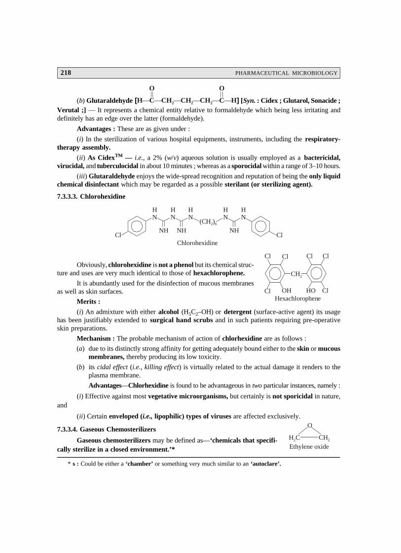
218 PHARMACEUTICAL MICROBIOLOGY
(b) Glutaraldehyde [ ]H C CH CH CH⎯ ⎯ ⎯ ⎯ ⎯ ⎯2 2 2 C H
O O
[Syn. : Cidex ; Glutarol, Sonacide ;Verutal ;] — It represents a chemical entity relative to formaldehyde which being less irritating anddefinitely has an edge over the latter (formaldehyde).
Advantages : These are as given under :
(i) In the sterilization of various hospital equipments, instruments, including the respiratory-therapy assembly.
(ii) As CidexTM — i.e., a 2% (w/v) aqueous solution is usually employed as a bactericidal,virucidal, and tuberculocidal in about 10 minutes ; whereas as a sporocidal within a range of 3–10 hours.
(iii) Glutaraldehyde enjoys the wide-spread recognition and reputation of being the only liquidchemical disinfectant which may be regarded as a possible sterilant (or sterilizing agent).
7.3.3.3. Chlorohexidine
Cl
HN
HN
HN
NH NH(CH )2 6
HN
HN
NHCl
Chlorohexidine
Obviously, chlorohexidine is not a phenol but its chemical struc-ture and uses are very much identical to those of hexachlorophene.
It is abundantly used for the disinfection of mucous membranesas well as skin surfaces.
Merits :
(i) An admixture with either alcohol (H5C2–OH) or detergent (surface-active agent) its usagehas been justifiably extended to surgical hand scrubs and in such patients requiring pre-operativeskin preparations.
Mechanism : The probable mechanism of action of chlorhexidine are as follows :
(a) due to its distinctly strong affinity for getting adequately bound either to the skin or mucousmembranes, thereby producing its low toxicity.
(b) its cidal effect (i.e., killing effect) is virtually related to the actual damage it renders to theplasma membrane.
Advantages—Chlorhexidine is found to be advantageous in two particular instances, namely :
(i) Effective against most vegetative microorganisms, but certainly is not sporicidal in nature,and
(ii) Certain enveloped (i.e., lipophilic) types of viruses are affected exclusively.
7.3.3.4. Gaseous Chemosterilizers
Gaseous chemosterilizers may be defined as—‘chemicals that specifi-cally sterilize in a closed environment.’*
* s : Could be either a ‘chamber’ or something very much similar to an ‘autoclare’.
Cl Cl
Cl OH HO Cl
CH2
Cl Cl
Hexachlorophene
H C2
O
CH2
Ethylene oxide
MICROBIAL CONTROL BY PHYSICAL AND CHEMICAL METHODS 219
* It is found to be highly toxic and explosive in its purest state ; hence, it is invariably mixed with a non-inflammable gas e.g., CO2 or N2.
** A disease (infection) that the infants normally could have contracted during their passage via the birth canal.However, nowadays it has been largely replaced by more effective ‘antibiotics’.
Example : The typical example being Ethylene oxide.
Mechanism : The most probable mechanism of action of ethylene oxide solely depends upon itsinherent ability to cause ‘denaturation of proteins’. In fact, the labile H-atoms strategically attachedto the proteins viz., —OH, —SH, or —COOH are critically replaced by the available alkyl moieties(alkylation), for instance : —H2C—CH2—OH.
Advantages—These are as stated below :
(1) Ethylene oxide practically kills all microorganisms besides the endospores ; however, itmay require a perceptionally lengthy exposure ranging between 4–18 hours.*
(2) It has an extremely high degree of penetrating power to such an extent that it was specificallyselected for the complete sterilization of spacecraft despached to land on the Moon pluscertain other planets.
7.3.3.5. Heavy Metals and Derivatives
A plethora of heavy metals and their corresponding derivatives viz., Hg, HgCl2, Cu, CuSO4,Ag, AgNO3, Zn, ZnCl2 find extensive usages as germicidal and antiseptic agents.
Mechanism — Oligodynamic action refers to the precise ability of relatively smaller quantumof heavy metals viz., Ag and Cu, to predominantly exert antimicrobial activity. In reality, the respectivemetal ions (e.g., Ag+ and Cu2+) categorically combine with the specific—SH moieties critically locatedon the ‘cellular proteins’ thereby causing denaturation ultimately.
Examples : A few typical examples are cited below :
(a) Ag in AgNO3 1% (w/v) Solution : It was a mandatory practice earlier to treat the eyes of thenewborns with a few drops of silver nitrate solution to prevent and protect against any possi-ble infection of the eyes usually termed as gonorrheal ophthalmia neonatorum.**
(b) HgCl2 : It perhaps enjoy the longest historical usage as an effective disinfectant. It indeedpossessed a rather broad-spectrum of activity together with its prime bacteriostatic activ-ity. The usage of the ‘mercurochrome antiseptic’ (i.e., an organic mercury compound) isstill prevalent in the domain of domestic chests.
(c) CuSO4 : It finds its abundant utility for the critical destruction of green algae (an algicide)which grow profusely in fish-aquariums, swimming pools, and reservoirs.
(d) ZnCl2 : It is mostly an essential ingredient in mouth washes like ‘Listerine’ etc. It alsoserves as a potential antifungal agent in acrylic-based paints.
7.3.3.6. Halogens
The two most important halogens that are effectively employed as the antimicrobial agents areiodine (I2) and chlorine (Cl2) frequently in solution ; besides, being used as the integral constituents ofboth organic or inorganic compounds.
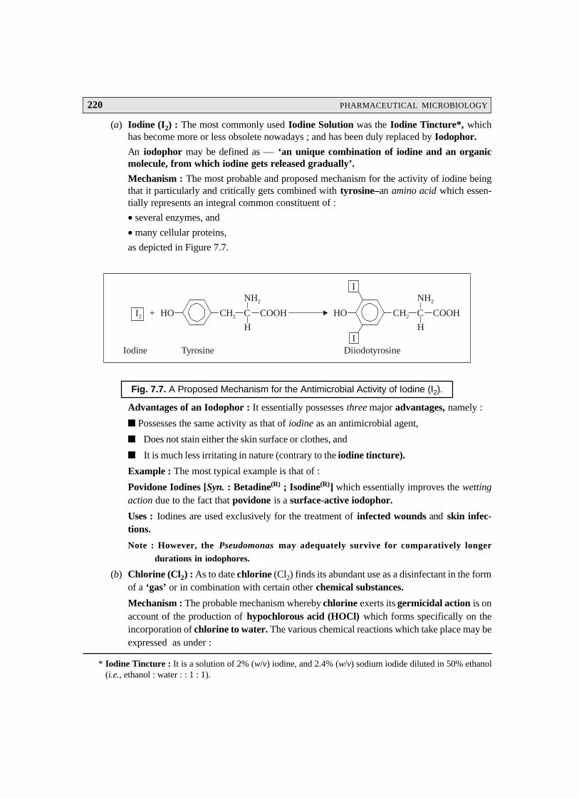
220 PHARMACEUTICAL MICROBIOLOGY
* Iodine Tincture : It is a solution of 2% (w/v) iodine, and 2.4% (w/v) sodium iodide diluted in 50% ethanol(i.e., ethanol : water : : 1 : 1).
(a) Iodine (I2) : The most commonly used Iodine Solution was the Iodine Tincture*, whichhas become more or less obsolete nowadays ; and has been duly replaced by Iodophor.
An iodophor may be defined as — ‘an unique combination of iodine and an organicmolecule, from which iodine gets released gradually’.
Mechanism : The most probable and proposed mechanism for the activity of iodine beingthat it particularly and critically gets combined with tyrosine–an amino acid which essen-tially represents an integral common constituent of :
• several enzymes, and
• many cellular proteins,
as depicted in Figure 7.7.
CH2 C
NH2
H
COOHHO CH2 C
NH2
H
COOHHO
I
I
I2 +
Iodine Tyrosine Diiodotyrosine
Fig. 7.7. A Proposed Mechanism for the Antimicrobial Activity of Iodine (I2).
Advantages of an Iodophor : It essentially possesses three major advantages, namely :
Possesses the same activity as that of iodine as an antimicrobial agent,
Does not stain either the skin surface or clothes, and
It is much less irritating in nature (contrary to the iodine tincture).
Example : The most typical example is that of :
Povidone Iodines [Syn. : Betadine(R) ; Isodine(R)] which essentially improves the wettingaction due to the fact that povidone is a surface-active iodophor.
Uses : Iodines are used exclusively for the treatment of infected wounds and skin infec-tions.
Note : However, the Pseudomonas may adequately survive for comparatively longer
durations in iodophores.
(b) Chlorine (Cl2) : As to date chlorine (Cl2) finds its abundant use as a disinfectant in the formof a ‘gas’ or in combination with certain other chemical substances.
Mechanism : The probable mechanism whereby chlorine exerts its germicidal action is onaccount of the production of hypochlorous acid (HOCl) which forms specifically on theincorporation of chlorine to water. The various chemical reactions which take place may beexpressed as under :
MICROBIAL CONTROL BY PHYSICAL AND CHEMICAL METHODS 221
COOH
Benzoic Acid
(i) Cl2 + H2O ⎯⎯→ H+ + Cl– + HOCl
Chlorine Water Hydrogen Chlorine Hypochlorous
ion ion acid
(ii) HOCl ⎯⎯→ H+ + OCl–
Hypochlorous Hydrogen Hypochlorite
acid ion ion
Hypochlorous Acid. The precise and exact mechanism whereby hypochlorous acid causes the‘cidal effect’ (i.e., killing power) is not yet known fully. Neverthless, it is indeed a strong oxidizingagent which eventually blocks and prevents a major segment of the vital cellular enzyme system tofunction in a normal manner.
Advantages : There are two main advantageous functionalities of hypochlorous acid, namely :
(a) It represents the most effective form of chlorine (Cl2) by virtue of it being absolutely neutralwith respect to its electrical charge ; and, therefore, undergoes diffusion as quickly as possiblevia the cell wall.
(b) The hypochlorite ion [OCl–] [see Eqn. (ii)] bears a distinct negative charge which criticallyrenders its free entry and access into the body of the infected cell.
Liquid Chlorine Gas : The usage of pure liquid form of compressed chlorine (Cl2) gas is invari-ably done for carrying out the effective disinfection of municipal supply of potable (drinking) water,swimming-pool water, and sometimes even the municipal sewage-drain outlets.
Compounds of Chlorine : A good number of compounds of chlorine viz., calcium hypochlorite[Ca(OCl)2], and sodium hypochlorite [NaOCl] are largely employed as effective disinfectants.
Ca(OCl)2 is used to disinfect both the ‘dairy-equipments’ and ‘cooking/eating utencils’ ineateries (restaurants).
Clorox(R). It is a frequently used household disinfectant and a bleach that finds its extensiveapplications in various industrial and hospital environments, such as :
Dairy-Processing Organisations — industry ;
Food-Processing Establishments — industry ; and
Haemodialysis Systems — hospital.
7.3.3.7. Organic Acids and Derivatives
A large number of organic acids are employed both extensively and profusely as potential pre-servatives to control the growth of mold.
Examples : There are several typical examples, such as :
(a) Benzoic Acid [or salt derivative Sodium Benzoate] is duly recog-nized as a vital antifungal agent which is observed to be extremelyeffective at relatively lower pH values.
Uses : Benzoic acid/Sodium benzoate are employed extensively ina broad range of acidic food products viz., pickles, lime juices ; bev-erages viz., soft drinks, lime cordials, fruit squashes, canned fruit-juices ; and processedfood products viz., fruit jams, cheese, neat products, vegetables/fruits (canned), tomatopastes,tomato-sauces, and the like.
222 PHARMACEUTICAL MICROBIOLOGY
(b) Sorbic Acid [or salt derivative Potassium Sorbate] isinvariably employed to prevent and inhibit the mold growthin acidic foods particularly viz., cheese.
(c) Parabens — e.g., methylparaben and propylparaben find their abundant applicationsto control and inhibit mold growth in galenicals, liquid cosmetics, foods, shampoos,and beverages.
HO
OCH3
O
MethylparabenHO
OC H73
O
Propylparaben
[Note : Parabens are nothing but derivatives of ‘benzoic acid’ that essentially work ata neutral pH (viz., 7).]
(d) Calcium Propionate— is an inhibitor of moulds and other microor-ganisms invariably found in a wide-spectrum of products, such as :foods, tobacco, pharmaceuticals, butyl-rubber to improve theprocessability, and scorching resistance.
Mechanism : The precise mechanism of activity of these aforesaid organic acid and theirrespective derivatives is not exclusively associated to their inherent acidity but realisticallyto the following two cardinal aspects, namely :
(i) inhibition of enzymatic activity, and
(ii) inhibition of metabolic activity.
In a rather broader perspective the human body is capable of metabolizing these organic acidsquite rapidly thereby rendering their usage in vivo quite safe in all respects.
7.3.3.8. Oxidizing Agents
It has been observed that the oxidizing agents usually display and exert their ‘antimicrobialactivity’ by specifically oxidizing the cellular components of the treated microorganisms.
A few typical examples are discussed briefly as under :
(a) Ozone [O3]. It is an extremely reactive state of oxygen (O2) that may be generated by pass-ing oxygen via a high-voltage electrical discharge system. In fact, one may critically observethe presence of ozone in the following particular instances :
• presence of air’s fresh odour immediately after a lightning storm,
• nearest place to a reasonably large electric spark, and
• in the vicinity of an UV light (or lamp).
Important Points : There are two vital points to note :
(i) Though ozone [O3] exerts a more effective, marked and pronounced cidal effect(or killing effect), yet its overall residual activity is practically difficult to main-tain in water, and
(ii) Ozone is definitely more expensive than chlorine as an antimicrobial agent.
H CCH = CHCH = CH.COOH3
Sorbic Acid
OOCCH CH2 3
OOCCH CH2 3
Ca
Calcium propionate

MICROBIAL CONTROL BY PHYSICAL AND CHEMICAL METHODS 223
(b) Hydrogen Peroxide [H2O2] : Hydrogen peroxide finds a pivotal place in several hospitalsupply facilities as well as household medicine cabinets.
Mechanism : Ozone gets rapidly cleaved into water and nescent gaseous oxygen due to thecritical action of the enzyme catalase usually found in human cells, as illustrated under :
CatalaseH2O2 ⎯⎯⎯⎯→ H2O + (O)
Hydrogen Water Nescent
peroxide oxygen
Perhaps it could be the valid supportive evidence and proof that ozone fails to serve as a‘good antiseptic’ particularly for the open wounds.
Uses : These are as follows :
(1) It effectively disinfects the inanimate (i.e., showning no sign of being alive) objects.
(2) It proves to be sporocidal in nature, specifically at elevated temperature(s).
(3) Presence of usual protective enzymes belonging to the aerobic microorganims, andthe facultative anaerobes in the non-living surface zones, are found to be largelyoverwhelmed by the critical high concentrations of hydrogen peroxide actually em-ployed.
Based on these stark realities and superb functionalities the hydrogen peroxide is frequentlyused in :
food industry for ‘aspectic packaging’,* and
users of ‘contact lenses’ (i.e., a pharmaceutical aid) usually disinfect them (lenses) withH2O2. After carrying out the said disinfection procedure, a Pt-catalyst invariably present inthe lens-disinfecting kit helps to cause destruction of the residual H2O2 ; and, therefore, itno more persists on the contact lens, where it could serve as an irritant.
(c) Benzoyl Peroxide [Syns. : Debroxide ; Lucidol ; Nericur ;Sanoxit ; Theraderm ; Xerac BP ;] — Benzoyl peroxide is anuseful oxidizing agent for treating such wounds that are usuallyinfected by the anaerobic pathogens. However, it is found to bethe major component in most of the over-the-counter (OTC) medi-caments meant for curing acne** that is generally caused by aspecific kind of anaerobic bacterium infecting the hair-follicles.
7.3.3.9. Phenol and Phenolics
Phenol [Syn. : Carbolic acid ; Phenic acid ;] happens to be the first and foremost chemicalsubstance that was duly used by the famous British Physician Joseph Lister for sterilization of his ‘op-eration theater’. However, it has become quite obsolete as an antiseptic or disinfectant due to twomajor drawbacks, namely :
irritating action on skin, and
highly inherent sharp disagreable odour.
* The packaging material is made to pass through a hot solution of H2O2 before being assembled into a con-tainer.
** Acne : An inflammatory disease of the sebaceous glands and hair follicles of the skin characterized bycomedones, papules, and pustules.
C
O
C
O
O
Benzoyl peroxide
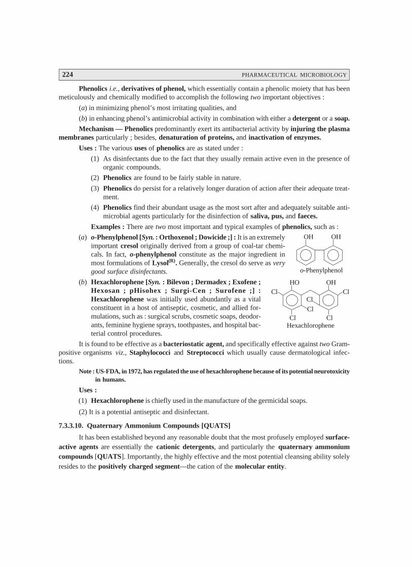
224 PHARMACEUTICAL MICROBIOLOGY
Phenolics i.e., derivatives of phenol, which essentially contain a phenolic moiety that has beenmeticulously and chemically modified to accomplish the following two important objectives :
(a) in minimizing phenol’s most irritating qualities, and
(b) in enhancing phenol’s antimicrobial activity in combination with either a detergent or a soap.
Mechanism — Phenolics predominantly exert its antibacterial activity by injuring the plasmamembranes particularly ; besides, denaturation of proteins, and inactivation of enzymes.
Uses : The various uses of phenolics are as stated under :
(1) As disinfectants due to the fact that they usually remain active even in the presence oforganic compounds.
(2) Phenolics are found to be fairly stable in nature.
(3) Phenolics do persist for a relatively longer duration of action after their adequate treat-ment.
(4) Phenolics find their abundant usage as the most sort after and adequately suitable anti-microbial agents particularly for the disinfection of saliva, pus, and faeces.
Examples : There are two most important and typical examples of phenolics, such as :
(a) o-Phenylphenol [Syn. : Orthoxenol ; Dowicide ;] : It is an extremelyimportant cresol originally derived from a group of coal-tar chemi-cals. In fact, o-phenylphenol constitute as the major ingredient inmost formulations of Lysol(R). Generally, the cresol do serve as verygood surface disinfectants.
(b) Hexachlorophene [Syn. : Bilevon ; Dermadex ; Exofene ;Hexosan ; pHisohex ; Surgi-Cen ; Surofene ;] :Hexachlorophene was initially used abundantly as a vitalconstituent in a host of antiseptic, cosmetic, and allied for-mulations, such as : surgical scrubs, cosmetic soaps, deodor-ants, feminine hygiene sprays, toothpastes, and hospital bac-terial control procedures.
It is found to be effective as a bacteriostatic agent, and specifically effective against two Gram-positive organisms viz., Staphylococci and Streptococci which usually cause dermatological infec-tions.
Note : US-FDA, in 1972, has regulated the use of hexachlorophene because of its potential neurotoxicityin humans.
Uses :
(1) Hexachlorophene is chiefly used in the manufacture of the germicidal soaps.
(2) It is a potential antiseptic and disinfectant.
7.3.3.10. Quaternary Ammonium Compounds [QUATS]
It has been established beyond any reasonable doubt that the most profusely employed surface-active agents are essentially the cationic detergents, and particularly the quaternary ammoniumcompounds [QUATS]. Importantly, the highly effective and the most potential cleansing ability solelyresides to the positively charged segment—the cation of the molecular entity.
Cl ClHO OH
Cl ClCl
Cl
Hexachlorophene
OH OH
o-Phenylphenol
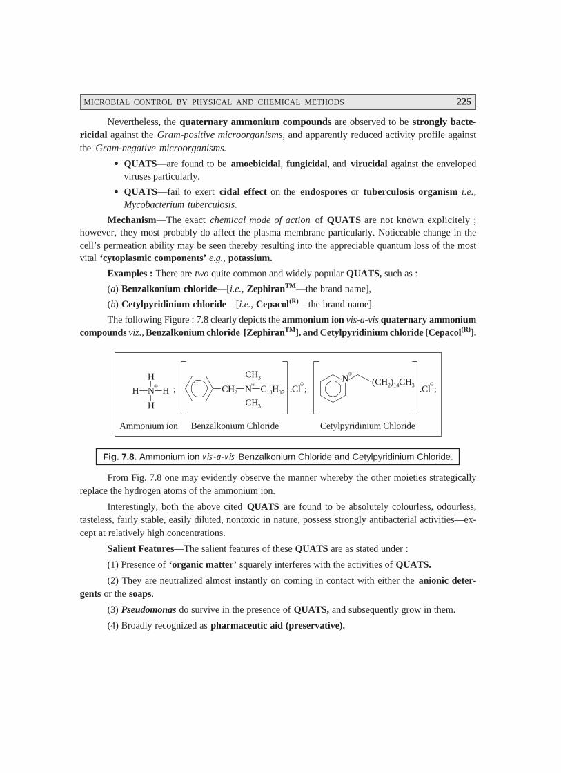
MICROBIAL CONTROL BY PHYSICAL AND CHEMICAL METHODS 225
Nevertheless, the quaternary ammonium compounds are observed to be strongly bacte-ricidal against the Gram-positive microorganisms, and apparently reduced activity profile againstthe Gram-negative microorganisms.
• QUATS—are found to be amoebicidal, fungicidal, and virucidal against the envelopedviruses particularly.
• QUATS—fail to exert cidal effect on the endospores or tuberculosis organism i.e.,Mycobacterium tuberculosis.
Mechanism—The exact chemical mode of action of QUATS are not known explicitely ;however, they most probably do affect the plasma membrane particularly. Noticeable change in thecell’s permeation ability may be seen thereby resulting into the appreciable quantum loss of the mostvital ‘cytoplasmic components’ e.g., potassium.
Examples : There are two quite common and widely popular QUATS, such as :
(a) Benzalkonium chloride—[i.e., ZephiranTM—the brand name],
(b) Cetylpyridinium chloride—[i.e., Cepacol(R)—the brand name].
The following Figure : 7.8 clearly depicts the ammonium ion vis-a-vis quaternary ammoniumcompounds viz., Benzalkonium chloride [ZephiranTM], and Cetylpyridinium chloride [Cepacol(R)].
CH2 N
CH3
CH3
C18H37
⊕.Cl ;
N⊕
(CH ) CH2 14 3 .Cl ;H N H
H
H
;
Ammonium ion Benzalkonium Chloride Cetylpyridinium Chloride
⊕
Fig. 7.8. Ammonium ion vis-a-vis Benzalkonium Chloride and Cetylpyridinium Chloride.
From Fig. 7.8 one may evidently observe the manner whereby the other moieties strategicallyreplace the hydrogen atoms of the ammonium ion.
Interestingly, both the above cited QUATS are found to be absolutely colourless, odourless,tasteless, fairly stable, easily diluted, nontoxic in nature, possess strongly antibacterial activities—ex-cept at relatively high concentrations.
Salient Features—The salient features of these QUATS are as stated under :
(1) Presence of ‘organic matter’ squarely interferes with the activities of QUATS.
(2) They are neutralized almost instantly on coming in contact with either the anionic deter-gents or the soaps.
(3) Pseudomonas do survive in the presence of QUATS, and subsequently grow in them.
(4) Broadly recognized as pharmaceutic aid (preservative).

226 PHARMACEUTICAL MICROBIOLOGY
7.3.3.11. Surface-Active Agents [or Surfactants]
Surface-active agents may be defined as—‘substances that specifically lower, the surfacetension prevailing amongst the molecules of a liquid. Such agents essentially include oil, soaps, andvarious types of detergents.
Soap—The soap is made by the saponification of vegetable oils with the removal of glycerine asa by-product. Though it possesses rather little value as an antiseptic/disinfectant as such, but it doesexert an extremely important function in the mechanical removal of microorganisms by means of gentlescrubbing*.
In actual practice, the soap actually aids in the careful cleavage of the thin-oily film (present onthe skin-surface) via a superb phenomenon invariably termed as emulsification, whereby the mixture ofwater/soap meticulously abstracts the emulsified oil together with the debris of dead cells, dirt particulatematters, and microorganisms, and float them away swiftly when the latter thus produced is flushed outwith water.
Uses :
(1) In general, soaps do serve as reasonably good and efficacious degerming agents.
(2) Deodorant soap essentially containing typical chemical entities e.g., triclocarban, predomi-nantly inhibit the Gram-positive microorganisms.
Triclocarban [Syn : Cutisan ; Nobacter ; Solubacter ;] :
Cl
HN
HN
Cl
Cl
O
Triclocarban
Triclocarban finds its abundant usage as a bacteriostat and antiseptic in soaps (medicated) andother cleansing compositions.
Acid-Anionic Surface-Active Sanitizers : They usually designate an extremely vital and im-portant group of chemical substances that are being used extensively in the cleaning of dairy utensilsand equipments. It has been duly observed that their ‘sanitizing ability’ is duly confined to the strategicnegatively charged segment (anion) of the molecule, that eventually interacts critically with the re-spective plasma membrane. Besides, such type of sanitizers invariably exert their action upon a broadspectrum of the microorganisms, even including certain most fussy and troublesome thermoduric mi-crobes. In reality, these sanitizers are found to be absolutely nontoxic, fast-acting, and above allnoncorrosive in nature.
Table : 7.4 records a summarized details of the various chemical agents, as described from Sec-tions 7.3.3.1 to 7.3.3.11, that efficiently controls the microbial growth in general.
* Normal skin surface usually contains dead cells, dried sweat, dust particulate matter, oily secretions, andmicroorganisms.
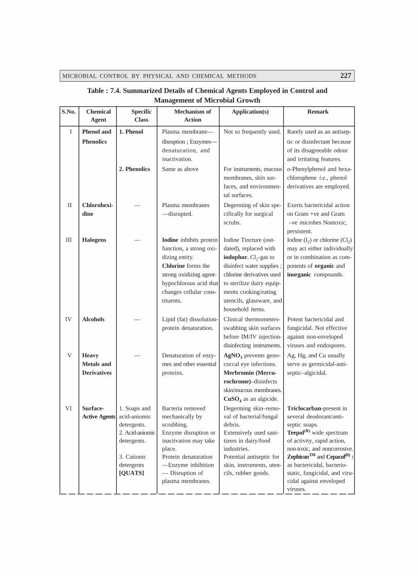
MICROBIAL CONTROL BY PHYSICAL AND CHEMICAL METHODS 227
Table : 7.4. Summarized Details of Chemical Agents Employed in Control andManagement of Microbial Growth
S.No. Chemical Specific Mechanism of Application(s) RemarkAgent Class Action
I Phenol and 1. Phenol Plasma membrane— Not so frequently used. Rarely used as an antisep-
Phenolics disruption ; Enzymes— tic or disinfectant because
denaturation, and of its disagreeable odour
inactivation. and irritating features.
2. Phenolics Same as above For instruments, mucous o-Phenylphenol and hexa-
membranes, skin sur- chlorophene i.e., phenol
faces, and environmen- derivatives are employed.
tal surfaces.
II Chlorohexi- — Plasma membranes Degerming of skin spe- Exerts bactericidal action
dine —disrupted. cifically for surgical on Gram +ve and Gram
scrubs. –ve microbes Nontoxic,
persistent.
III Halogens — Iodine inhibits protein Iodine Tincture (out- Iodine (I2) or chlorine (Cl2)function, a strong oxi- dated), replaced with may act either individually
dizing entity. iodophor. Cl2-gas to or in combination as com-
Chlorine forms the disinfect water supplies ; ponents of organic and
strong oxidizing agent- chlorine derivatives used inorganic compounds.
hypochlorous acid that to sterilize dairy equip-
changes cellular cons- ments cooking/eating
tituents. utencils, glassware, and
household items.
IV Alcohols — Lipid (fat) dissolution- Clinical thermometers- Potent bactericidal and
protein denaturation. swabbing skin surfaces fungicidal. Not effective
before IM/IV injection- against non-enveloped
disinfecting instruments. viruses and endospores.
V Heavy — Denaturation of enzy- AgNO3 prevents geno- Ag, Hg, and Cu usually
Metals and mes and other essential coccal eye infections. serve as germicidal-anti-
Derivatives proteins. Merbromin (Mercu- septic–algicidal.
rochrome)–disinfects
skin/mucous membranes.
CuSO4 as an algicide.
VI Surface- 1. Soaps and Bacteria removed Degerming skin–remo- Triclocarban-present inActive Agents acid-aniomic mechanically by val of bacterial/fungal several deodorant/anti-
detergents. scrubbing. debris. septic soaps.2. Acid-aniomic Enzyme disruption or Extensively used sani- Teepol(R) wide spectrumdetergents. inactivation may take tizers in dairy/food of activity, rapid action,
place. industries. non-toxic, and noncorrosive.3. Cationic Protein denaturation Potential antiseptic for ZephiranTM and Cepacol(R) :detergents —Enzyme inhibition skin, instruments, uten- as bactericidal, bacterio-[QUATS] — Disruption of cils, rubber goods. static, fungicidal, and viru-
plasma membranes. cidal against envelopedviruses.

228 PHARMACEUTICAL MICROBIOLOGY
VII Organic — Metabolic inhibition— Benzoic acid/Sorbic Mostly employed in food
Acids largely affecting molds. acid–effective at low products and cosmetic
pH viz., cosmetics, sha- variants to control moldsmpoos. Calcium pro- and certain microbes.pionate–for bakeryproducts. All are anti-fungal.
VIII Aldehydes — Affords protein CidexTM [Glutaralde- Glutaraldehyde–regulatedinactivation. hyde]–is less irritating as a ‘Liquid Sterilant’.
than formaldehyde ;used for medical equip-ment sterilization.
IX Gaseous — Denaturation Superb and excellant Ethylene oxide–is emplo-Chemosteri- sterilizing agent ; speci- yed most abundantly.lizers fically for such objects
which may be damagedseriously due to heat.
X Oxidizing — Oxidation Effective against oxy- Ozone [O3]–gaining recog-Agents gen-sensitive anaerobes nition (in place of Cl2) ;
viz., deep wounds, H2O2–good disinfectanthighly contaminated but a poor antiseptic.surfaces.
7.4. EXPERIMENTAL PARAMETERS INFLUENCING THEANTIMICROBIAL AGENT ACTIVITY
It has been amply demonstrated, proved, and well documented that the actual prevalent destructionof various pathogenic/nonpathogenic microorganisms and their subsequent inhibition of the resulting‘microbial growth’ are not simple matters at all, as the underlying efficacy of an antimicrobial agent*is invariably and predominantly affected by the following six cardinal factors, namely :
7.4.1. Population Size
It may be observed that usually an equal fraction of a microbial population gets killed duringeach stipulated period (interval); and, therefore, a larger population certainly needs a relatively longerduration to die than a smaller one. Importantly, the same principle holds good for the chemical antimi-crobial agents.
7.4.2. Population Composition
Importantly, the overall effectiveness of an antimicrobial agent exclusively changes with theprevailing nature of the microorganisms under investigation due to the fact that they differ distinctly intheir susceptibility.
* Antimicrobial Agent— An agent that kills microorganisms or inhibits their actual growth.
MICROBIAL CONTROL BY PHYSICAL AND CHEMICAL METHODS 229
Salient Features : These are as follows :
(a) Microbial endospores are found to be much more resistant to a large segment of the antimi-crobial agents in comparison to the vegetative forms.
(b) Younger cells are invariably more prone to rapid destruction than the corresponding matureorganisms.
(c) Certain specific species may withstand adverse experimental parameters better thanothers.
Example : Mycobacterium tuberculosis (causative organism for tuberculosis is found to be muchmore resistant to antimicrobial agents vis-a-vis other microorganisms.
7.4.3. Concentration of Antimicrobial Agent
One may observe quite often that the more concentrated a ‘chemical agent’ or ‘intense a physi-cal agent’—the more quickly the microorganisms get destroyed. Nevertheless, the ‘agent effective-ness’ is not normally associated with either concentration or intensity directly.
Salient Features—are as given under :
(1) Spread over a short-range a rather small increase in the concentration of antimicrobial agentultimately leads to a definite exponential rise in its effectiveness ; however, beyond a certain criticalpoint one may not observe any more increase in the rate of killing.
(2) Occasionally, an antimicrobial agent is found to be more effective even at much lower con-centrations.
Example : Ethanol 70% (v/v) is more effective in comparison to 95% (v/v), by virtue of the factthat its (EtOH) activity gets markedly enhanced by the presence of water.
7.4.4. Duration of Exposure
The longer a particular population of microbes is duly exposed to a microcidal agent, the morenumber of microorganisms would be killed. In order to accomplish perfect sterilization, an exposureduration just sufficient to reduce the ensuing survival probability to either 10–6 or less must be em-ployed effectively.
7.4.5. Temperature
It has been noticed that an increase in the temperature at which a particular chemical agentinvariably exerts its action often increases its activity. Quite often a lower concentration of either asterilizing agent or disinfectant may be suitably employed at a higher temperature effectively.
7.4.6. Local Environment
It is, however, pertinent to state here that the population to be controlled is not isolated by sur-rounded by several environmental factors which may cause :
• offer due protection, and
• afford destruction.
230 PHARMACEUTICAL MICROBIOLOGY
Examples :
(a) As heat kills more rapidly at an acidic pH, hence the acidic beverages and food products viz.,tomatoes and fruits are much convenient and easy to get pasteurized in comparison to such foodshaving higher pHs e.g., milk.
(b) Organic matter present in a surface-biofilm would eventually afford due protection of thebiofilm’s microorganisms ; besides, the biofilm together with its associated microorganisms often shallbe difficult to remove efficaciously.
FURTHER READING REFERENCES
1. Block SS (ed.) : Disinfection, Sterilization and Preservation, Lea and Febiger, Philadel-phia, 4th edn., 1991.
2. Block TD : Membrane Filtration : An User’s Guide and Reference Manual, ScienceTech. Publishers, Madison, Wis., 1983.
3. Collins CH et al. (eds) : Microbiological Methods, Butterworths, Stoneham, Mass, 6th edn.,1989.
4. Gilbert P and McBain AJ : Potential Impact of Increased use of Biocides in ConsumerProducts on Prevalence of Antibiotic Resistance, Clin. Microbial. Rev. 16(2) : 189–208,2003.
5. McDonnell G and Russell AD : Antiseptics and Disinfectants : Activity, Action and Re-sistance, Clin. Microbial. Rev., 12(1) : 147–79, 1999.
6. Richardson JH (ed.) : Laboratory Safety : Principles and Practices, American Society forMicrobiology, Washington, DC., 2nd edn., 1994.
7. Russell AD : Bacterial Spores and Chemical Sporicidal Agents, Clin. Microbial. Rev.,3(2) : 99–119, 1990.
8. Rutala WA and Weber DJ : Uses of Inorganic Hypochlorite (Bleach) in Healthcare Fa-cilities, Clin. Microbial Rev., 10(4) : 597–610, 1997.
9. Sorhaug T : Temperature Control, In :Encyclopedia of Microbiology, Lederberg J (Ed-in-Chief), Academic Press, San Diego, Vol. 4, 1st edn., 1992.
10. Tortora GJ et al. : Microbiology : An Introduction, The Benjamin/Cummings, PublishingCo., Inc., New York, 5th edn., 1995.
231
8.1. INTRODUCTION
A sterility test may be defined as — ‘a test that critically assesses whether a sterilizedpharmaceutical product is free from contaminating microorganisms’.
According to Indian Pharmacopoea (1996) the sterility testings are intended for detecting thepresence of viable forms of microorganisms in or on the pharmacopoeal preparations.
In actual practice, one invariably comes across certain absolutely important guidelines and vitalprecautionary measures that must be adhered to strictly so as to accomplish the utmost accuracy andprecision of the entire concept of sterility testing for life-saving secondary pharmaceutical products(drugs). A few such cardinal factors, guidelines, and necessary details are as enumerated under :
(a) Sterility testing, due to its inherent nature, is intimately associated with a statistical processwherein the portion of a batch is sampled almost randomly* ; and, therefore, the chance of the particularbatch (lot) duly passed for actual usage (consumption) solely depends upon the ‘sample’ having passedthe stringent sterility test.
(b) Sterility tests should be performed under conditions designed to avoid accidental contaminationof the product (under investigation) during the test. Nevertheless, such particular precautions preciselytaken for this purpose must not, in any case, adversely affect any microbes that should be revealed in thetest ultimately.
(c) Working environment wherein the sterility tests are meticulously carried out must be adequatelymonitored at regular intervals by sampling the air and the surface of the working area by performingnecessary control tests.
* From a thorough investigative study, it has been duly proposed that the random sampling must be judiciouslyapplied to : (a) such products that have been processed and filled aseptically ; and (b) with products heat-sterilized in their final containers must be drawn carefully from the potentially coolest zone of the load.
STERILITY TESTING :PHARMACEUTICAL PRODUCTS8
• Introduction• Test for Sterility : Pharmaceutical Products• Sampling : Probability Profile• Overall conclusions
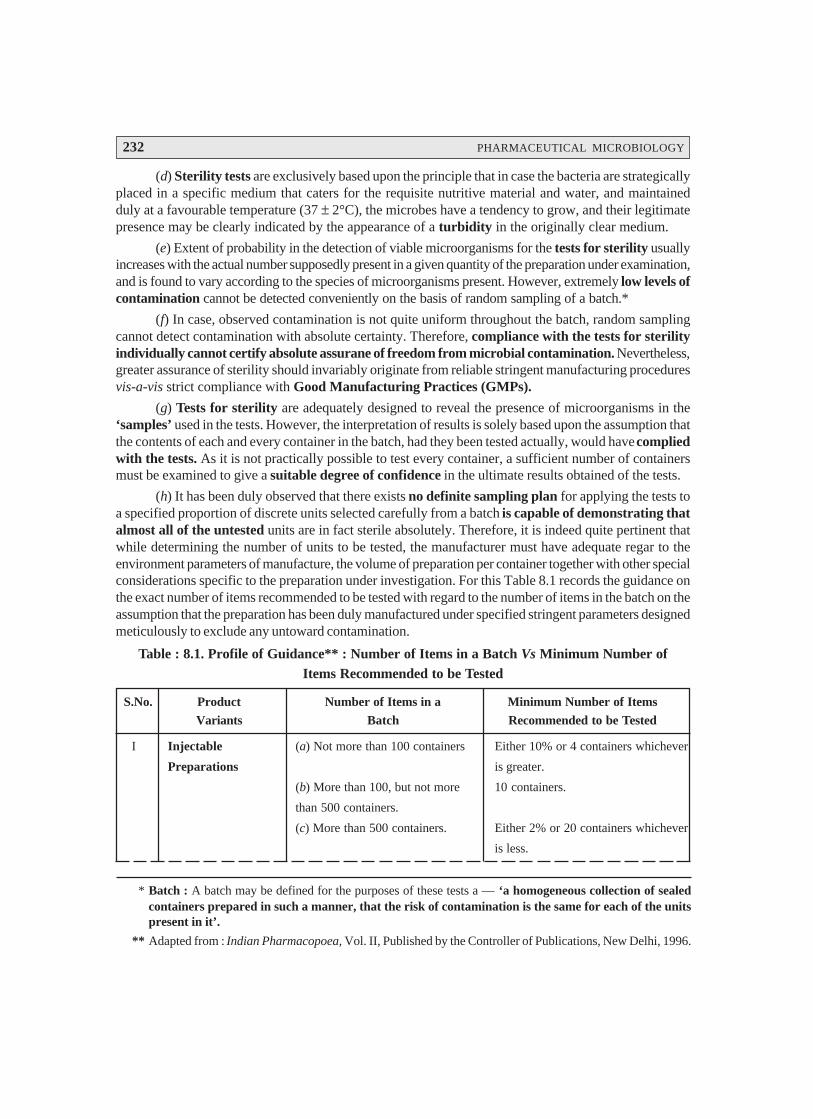
232 PHARMACEUTICAL MICROBIOLOGY
(d) Sterility tests are exclusively based upon the principle that in case the bacteria are strategicallyplaced in a specific medium that caters for the requisite nutritive material and water, and maintainedduly at a favourable temperature (37 ± 2°C), the microbes have a tendency to grow, and their legitimatepresence may be clearly indicated by the appearance of a turbidity in the originally clear medium.
(e) Extent of probability in the detection of viable microorganisms for the tests for sterility usuallyincreases with the actual number supposedly present in a given quantity of the preparation under examination,and is found to vary according to the species of microorganisms present. However, extremely low levels ofcontamination cannot be detected conveniently on the basis of random sampling of a batch.*
(f) In case, observed contamination is not quite uniform throughout the batch, random samplingcannot detect contamination with absolute certainty. Therefore, compliance with the tests for sterilityindividually cannot certify absolute assurane of freedom from microbial contamination. Nevertheless,greater assurance of sterility should invariably originate from reliable stringent manufacturing proceduresvis-a-vis strict compliance with Good Manufacturing Practices (GMPs).
(g) Tests for sterility are adequately designed to reveal the presence of microorganisms in the‘samples’ used in the tests. However, the interpretation of results is solely based upon the assumption thatthe contents of each and every container in the batch, had they been tested actually, would have compliedwith the tests. As it is not practically possible to test every container, a sufficient number of containersmust be examined to give a suitable degree of confidence in the ultimate results obtained of the tests.
(h) It has been duly observed that there exists no definite sampling plan for applying the tests toa specified proportion of discrete units selected carefully from a batch is capable of demonstrating thatalmost all of the untested units are in fact sterile absolutely. Therefore, it is indeed quite pertinent thatwhile determining the number of units to be tested, the manufacturer must have adequate regar to theenvironment parameters of manufacture, the volume of preparation per container together with other specialconsiderations specific to the preparation under investigation. For this Table 8.1 records the guidance onthe exact number of items recommended to be tested with regard to the number of items in the batch on theassumption that the preparation has been duly manufactured under specified stringent parameters designedmeticulously to exclude any untoward contamination.
Table : 8.1. Profile of Guidance** : Number of Items in a Batch Vs Minimum Number ofItems Recommended to be Tested
S.No. Product Number of Items in a Minimum Number of Items
Variants Batch Recommended to be Tested
I Injectable (a) Not more than 100 containers Either 10% or 4 containers whichever
Preparations is greater.
(b) More than 100, but not more 10 containers.
than 500 containers.
(c) More than 500 containers. Either 2% or 20 containers whichever
is less.
* Batch : A batch may be defined for the purposes of these tests a — ‘a homogeneous collection of sealedcontainers prepared in such a manner, that the risk of contamination is the same for each of the unitspresent in it’.
** Adapted from : Indian Pharmacopoea, Vol. II, Published by the Controller of Publications, New Delhi, 1996.
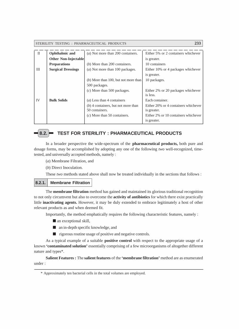
STERILITY TESTING : PHARMACEUTICAL PRODUCTS 233
II Ophthalmic and (a) Not more than 200 containers. Either 5% or 2 containers whichever
Other Non-Injectable is greater.
Preparations (b) More than 200 containers. 10 containers
III Surgical Dressings (a) Not more than 100 packages. Either 10% or 4 packages whichever
is greater.
(b) More than 100, but not more than 10 packages.
500 packages.
(c) More than 500 packages. Either 2% or 20 packages whicheveris less.
IV Bulk Solids (a) Less than 4 containers Each container.
(b) 4 containers, but not more than Either 20% or 4 containers whichever50 containers. is greater.
(c) More than 50 containers. Either 2% or 10 containers whicheveris greater.
8.2. TEST FOR STERILITY : PHARMACEUTICAL PRODUCTS
In a broader perspective the wide-spectrum of the pharmaceutical products, both pure anddosage forms, may be accomplished by adopting any one of the following two well-recognized, time-tested, and universally accepted methods, namely :
(a) Membrane Filtration, and
(b) Direct Inoculation.
These two methods stated above shall now be treated individually in the sections that follows :
8.2.1. Membrane Filtration
The membrane filtration method has gained and maintained its glorious traditional recognitionto not only circumvent but also to overcome the activity of antibiotics for which there exist practicallylittle inactivating agents. However, it may be duly extended to embrace legitimately a host of otherrelevant products as and when deemed fit.
Importantly, the method emphatically requires the following characteristic features, namely :
an exceptional skill,
an in-depth specific knowledge, and
rigorous routine usage of positive and negative controls.
As a typical example of a suitable positive control with respect to the appropriate usage of aknown ‘contaminated solution’ essentially comprising of a few microorganisms of altogether differentnature and types*.
Salient Features : The salient features of the ‘membrane filtration’ method are as enumeratedunder :
* Approximately ten bacterial cells in the total volumes are employed.

234 PHARMACEUTICAL MICROBIOLOGY
(1) The solution of the product under investigation is carefully filtered via a hydrophobic-edgedmembrane filter that would precisely retain any possible contaminating microorganisms.
(2) The resulting membrane is duly washed in situ to get rid of any possible ‘traces of antibiotic’that would have been sticking to the surface of the membrane intimately.
(3) Finally, the segregated microorganisms are meticulously transferred to the suitable culturemedia under perfect aseptic environment.
Microorganisms for Positive Control Tests : There are, infact, four typical microorganismsthat are being used exclusively for the positive control tests along with their respective type of specificenzymatic activity mentioned in parentheses :
(a) Bacillus cerreus : [Broad spectrum] ;
(b) Staphylococcus aureus : [Penicillinase] ;
(c) Klebsiella aerogenes : [Penicillinase + Cephalosporinase] ; and
(d) Enterobacter species : [Cephalosporinase].
Interestingly, the microorganisms invariably employed for the positive control tests togetherwith a particular product containing essentially an ‘antimicrobial agent’ must be, as far as possible,explicitely sensitive to that agent, in order that the ultimate growth of the microbe solely indicatesthree vital and important informations, namely :
satisfactory inactivation,
satisfactory dilution, and
satisfactory removal of the agent.
Specific Instances of Pharmaceutical Products : Virtually all the ‘Official Compendia’ viz.,Indian Pharmacopoea (IP) ; British Pharmacopoea (BP), United States Pharmacopoea (USP) ;European Pharmacopoea (Eur. P), and International Pharmacopoea (Int. P.) have duly providedcomprehensive and specific details with regard to the ‘tests for sterility’ of parenteral products (e.g.., IVand IM injectables), ophthalmic preparations (e.g., eye-drops, eye-ointments, eye-lotions etc.) ; besidesa plethora of non-injectable preparations, such as : catgut, dusting powder, and surgical dressings.
Test Procedures : In a broader perspective, the membrane filtration is to be preferred exclu-sively in such instances where the substance under investigation could any one of the following fourclasses of pharmaceutical preparations :
(i) an oil or oil-based product,
(ii) an ointment that may be put into solution,
(iii) a non-bacteriostatic solid that does not become soluble in the culture medium rapidly, and
(iv) a soluble powder or a liquid that essentially possesses either inherent bacteriostatic orinherent fungistatic characteristic features.
The membrane filtration must be used for such products where the volume in a container iseither 100 mL or more. One may, however, select the exact number of samples to be testedfrom Table 8.1 ; and subsequently use them for the respective culture medium suitably selected formicroorganisms and the culture medium appropriately selected for fungi.

STERILITY TESTING : PHARMACEUTICAL PRODUCTS 235
* Such as : Bacteriological Peptone.
Precautionary Measures : In actual practice, however, the tests for sterility must always becarried out under highly specific experimental parameters so as to avoid any least possible accidentalcontamination of the product being examined, such as :
(a) a sophisticated laminar sterile airflow cabinet (provided with effective hepa-filters),
(b) necessary precautionary measures taken to be such so as to avoid contamination that they donot affect any microbes which must be revealed duly in the test.
(c) ensuing environment (i.e., working conditions) of the laboratory where the ‘tests for sterility’is performed must always be monitored at a definite periodical interval by :
sampling the air of the working area,
sampling the surface of the working area, and
perforing the stipulated control tests.
Methodology : In usual practice, it is absolutely urgent and necessary to first clean meticulouslythe exterior surface of ampoules, and closures of vials and bottles with an appropriate antimicrobialagent ; and thereafter, the actual access to the contents should be gained carefully in a perfect asepticmanner. However, in a situation where the contents are duly packed in a particular containerunder vacuum, introduction of ‘sterile air’ must be done by the help of a suitable sterile device, forinstance : a needle duly attached to a syringe barrel with a non-absorbent cotton.
Apparatus : The most suitable unit comprises of a closed reservoir and a receptacle betweenwhich a properly supported membrane of appropriate porosity is placed strategically.
A membrane usually found to be quite suitable for sterility testing essentially bears anominal pore size not more than 0.45 μμμμμm, and diameter of nearly 47 mm, the effectivenessof which in the retention of microbes has been established adequately.
The entire unit is most preferably assembled and sterilised with the membrane in place priorto use.
In case, the sample happens to be an oil, sterilize the membrane separately and, after thoroughdrying, assemble the unit, adopting appropriate aseptic precautionary measures.
Diluting of Fluids : In the ‘test for sterility’ one invariably comes across with two differenttypes of fluids which will be treated individually in the sections that follows :
(a) Fluid A—Digest 1 g of peptic digest of animal tissue* or its equivalent in water to make upthe volume upto 1L, filter or centrifuge to clarify, adjust to pH 7.1 ± 0.2, dispense into flasks in 100 mLquantities, and finally sterilize at 121° C for 20 minutes (in an ‘Autoclave’).
Note : In a specific instance, where Fluid A is to be used in carrying out the tests for sterility on aspecimen of the penicillin or cephalosporin class of anibiotics, aseptically incorporate an amountof sterile penicillinase to the Fluid A to be employed to rinse the membrane(s) sufficient toinactivate any residual antibiotic activity on the membrane(s) after the solution of the specimen
has been duly filtered.
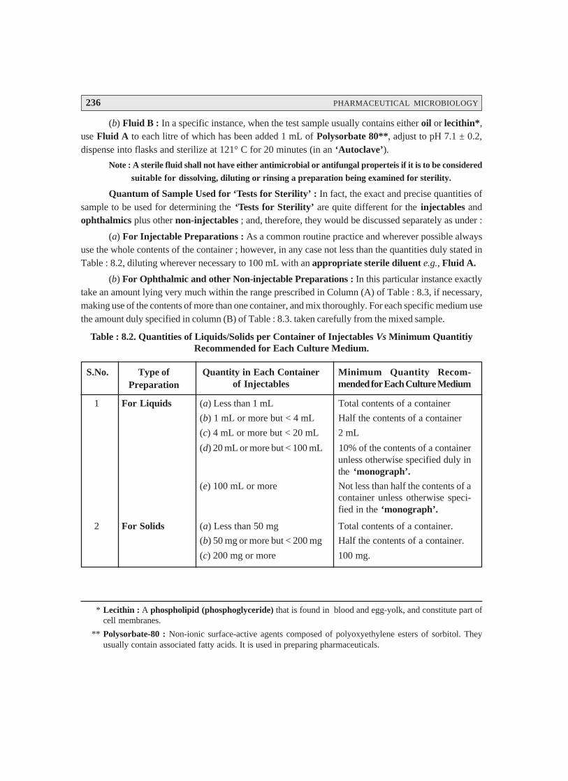
236 PHARMACEUTICAL MICROBIOLOGY
(b) Fluid B : In a specific instance, when the test sample usually contains either oil or lecithin*,use Fluid A to each litre of which has been added 1 mL of Polysorbate 80**, adjust to pH 7.1 ± 0.2,dispense into flasks and sterilize at 121° C for 20 minutes (in an ‘Autoclave’).
Note : A sterile fluid shall not have either antimicrobial or antifungal properteis if it is to be considered
suitable for dissolving, diluting or rinsing a preparation being examined for sterility.
Quantum of Sample Used for ‘Tests for Sterility’ : In fact, the exact and precise quantities ofsample to be used for determining the ‘Tests for Sterility’ are quite different for the injectables andophthalmics plus other non-injectables ; and, therefore, they would be discussed separately as under :
(a) For Injectable Preparations : As a common routine practice and wherever possible alwaysuse the whole contents of the container ; however, in any case not less than the quantities duly stated inTable : 8.2, diluting wherever necessary to 100 mL with an appropriate sterile diluent e.g., Fluid A.
(b) For Ophthalmic and other Non-injectable Preparations : In this particular instance exactlytake an amount lying very much within the range prescribed in Column (A) of Table : 8.3, if necessary,making use of the contents of more than one container, and mix thoroughly. For each specific medium usethe amount duly specified in column (B) of Table : 8.3. taken carefully from the mixed sample.
Table : 8.2. Quantities of Liquids/Solids per Container of Injectables Vs Minimum QuantitiyRecommended for Each Culture Medium.
S.No. Type ofPreparation
Quantity in Each Containerof Injectables
Minimum Quantity Recom-mended for Each Culture Medium
1 For Liquids (a) Less than 1 mL Total contents of a container
(b) 1 mL or more but < 4 mL Half the contents of a container
(c) 4 mL or more but < 20 mL 2 mL
(d) 20 mL or more but < 100 mL 10% of the contents of a containerunless otherwise specified duly inthe ‘monograph’.
(e) 100 mL or more Not less than half the contents of acontainer unless otherwise speci-fied in the ‘monograph’.
2 For Solids (a) Less than 50 mg Total contents of a container.
(b) 50 mg or more but < 200 mg Half the contents of a container.
(c) 200 mg or more 100 mg.
* Lecithin : A phospholipid (phosphoglyceride) that is found in blood and egg-yolk, and constitute part ofcell membranes.
** Polysorbate-80 : Non-ionic surface-active agents composed of polyoxyethylene esters of sorbitol. Theyusually contain associated fatty acids. It is used in preparing pharmaceuticals.
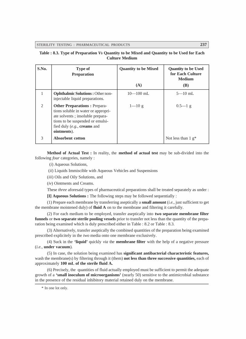
STERILITY TESTING : PHARMACEUTICAL PRODUCTS 237
Table : 8.3. Type of Preparation Vs Quantity to be Mixed and Quantity to be Used for EachCulture Medium
* In one lot only.
S.No. Type ofPreparation
Quantity to be Mixed
(A)
Quantity to be Usedfor Each Culture
Medium
(B)
1 Ophthalmic Solutions : Other non-injectable liquid preparations.
10—100 mL 5—10 mL
2 Other Preparations : Prepara-tions soluble in water or appropri-ate solvents ; insoluble prepara-tions to be suspended or emulsi-fied duly (e.g., creams andointments).
1—10 g 0.5—1 g
3 Absorbent cotton Not less than 1 g*
Method of Actual Test : In reality, the method of actual test may be sub-divided into thefollowing four categories, namely :
(i) Aqueous Solutions,
(ii) Liquids Immiscible with Aqueous Vehicles and Suspensions
(iii) Oils and Oily Solutions, and
(iv) Ointments and Creams.
These three aforesaid types of pharmaceutical preparations shall be treated separately as under :
[I] Aqueous Solutions : The following steps may be followed sequentially :
(1) Prepare each membrane by transferring aseptically a small amount (i.e., just sufficient to getthe membrane moistened duly) of fluid A on to the membrane and filtering it carefully.
(2) For each medium to be employed, transfer aseptically into two separate membrane filterfunnels or two separate sterile pooling vessels prior to transfer not less than the quantity of the prepa-ration being examined which is duly prescribed either in Table : 8.2 or Table : 8.3.
(3) Alternatively, transfer aseptically the combined quantities of the preparation being examinedprescribed explicitely in the two media onto one membrane exclusively.
(4) Suck in the ‘liquid’ quickly via the membrane filter with the help of a negative pressure(i.e., under vacuum).
(5) In case, the solution being examined has significant antibacterial characteristic features,wash the membrane(s) by filtering through it (them) not less than three successive quantities, each ofapproximately 100 mL of the sterile fluid A.
(6) Precisely, the quantities of fluid actually employed must be sufficient to permit the adequategrowth of a ‘small inoculum of microorganisms’ (nearly 50) sensitive to the antimicrobial substancein the presence of the residual inhibitory material retained duly on the membrane.

238 PHARMACEUTICAL MICROBIOLOGY
(7) Once the filtration is completed, aseptically remove the membrane(s) from the holder, cut themembrane in half, if only one is used, immerse the membrane or 1/2 of the membrane, in 100 mL of the‘Fluid Soyabean-Casein Digest Medium’*, and incubate at 20–25°C for a duration of seven days.
(8) Likewise, carefully immerse the other membrane, or other half of the membrane, in 100 mLof ‘Fluid Thioglycollate Medium’,** and incubate duly at 30–35° C for not less than seven days.
[II] Liquids Immiscible with Aqueous Vehicles and Suspensions : For this one may carry outthe ‘test’ as stipulated under [I] Aqueous Solutions, but add a sufficient amount of fluid A to the pooledsample to accomplish fast and rapid rate of filtration.
Special Features : These are as stated under :
(1) Sterile enzyme preparations, for instance :
Penicillinase
Cellulase
can be incorporated to fluid A to help in the dissolution of insoluble substances.
(2) In a situation when the substance under test usually contains lecithin, alway make use offluid B for dilution.]
[III] Oils and Oily Solutions : The various steps that are essentially involved in treating oils andoily solutions for carrying out the ‘test for sterility’ are as enumerated under :
(1) Filter oils or oily solutions of sufficiently low vicosity as such i.e., without any dilution via adry membrane.
(2) It is absolutely necessary to dilute viscous oils as necessary with an appropriate sterilediluent e.g., isopropyl myristate which has been proved beyond any reasonable doubt not to exhibitany antimicrobial activities under the prevailing parameters of the test.
Fluid Soyabean-Casein Digest Medium*
S.No. Ingredients Quantity(g)
1 Pancreatic digest of casein 17.0
2 Papaic digest of soyabean meal 3.0
3 Sodium chloride 5.0
4 Diabasic potassium phosphate 2.5
[K2HPO4]
5 Dextrose monohydrate 2.5
[C6H12O6 . H2O]
6 Distilled water to 1000 mL
Dissolve the solids in distilled water, warmingslightly to effect solution. Cool to room tempera-ture and add, if necessary sufficient 0.1 M NaOHto give a final pH of 7.1 ± 0.2 after sterilization.Distribute into suitable containers and sterilize inan autoclave at 121°C for 20 minutes.
Fluid Thioglycollate Medium**
S.No. Ingredients Quantity(g)
1 L-Cystine 0.5
2 Sodium chloride 2.5
3 Dextrose [C6H12O6 . H2O] 5.5
4 Granular agar [moisture < 15% 0.75
w/w]
5 Yeast-extract (water-soluble) 5.0
6 Pancreatic digest of casein 15.0
7 Sodium thioglycollate or 0.5
8 Thioglycollic acid 0.3 (mL)
9 Resazurin [0.1% fresh solution] 1.0 (mL)
10 Distilled water to 1000 mL
For procedure : Please refer to Appendix 9.5.
Indian Pharmacopea Vol. II, 1996 (p-A : 118)
STERILITY TESTING : PHARMACEUTICAL PRODUCTS 239
(3) Permit the ‘oil’ to penetrate the membrane, and carry out the filtration by the application ofgradual suction (with a vaccum pump).
(4) Wash the membrane by filtering through it at least 3/4 successive quantities, each of nearly100 mL of sterile fluid B or any other appropriate sterile diluent.
(5) Complete the test as described under [I] Aqueous Solutions from step (7) onwards.
[IV] Ointments and Creams : The various steps involved are as stated under :
(1) Dilute ointments carefully either in a ‘fatty base’ or ‘emulsions’ of the water-in-oil (i.e.,w/o) type to yield a fluid concentration of approx. 1% w/v, by applying gentle heat, if necessary, to notmore than 40°C with the aid of an appropriate sterile diluent e.g., isopropyl myristate previouslyadequately sterilized by filtration via a 0.22 μμμμμm membrane filter which has been shown not to possessantimicrobial activities under the prevailing conditions of the test.
(2) Carry out the filtration as rapidly as possible as per details given under ‘Oils and Oily Solu-tions’ [Section III] from step (4) onwards.
(3) However, in certain exceptional instances, it would be absolutely necessary to heat thesubstance to not more than 45°C, and to make use of ‘warm solutions’ for washing the membraneeffectively.
Note : For ointments and oils that are almost insoluble in isopropyl myristate one may employ thesecond method viz., ‘Direct Inoculation’ [Section 2.2].
[V] Soluble Soids : For each individual cultrue medium, dissolve not less the quantity of thesubstance being examined, as recommended in Tables : 8.2 and 8.3, in an appropriate sterile solvente.g., fluid A, and perform the test described under Section (I) i.e., Aqueous Solutions, by employing amembrane suitable for the selected solvents.
[VI] Sterile Devices : Pass carefully and aseptically a sufficient volume of fluid B via each ofnot less than 20 devices so that not less than 100 mL is recovered ultimately from each device. Collectthe fluids in sterile containers, and filter the entire volume collected via membrane filter funnel(s) asdescribed under Section (I), Aqueous Solutions.
8.2.2. Direct Inoculation [or Direct Inoculation of Culture Media]
The three usual methods being used for performing the ‘tests for sterility’ are as enumeratedunder :
(a) Nutrient Broth,
(b) Cooked Meat Medium and Thioglycollate Medium, and
(c) Sabouraud Medium.
These methods shall now be treated individually in the sections that follows :
8.2.2.1. Nutrient Broth
Importantly, it is exclusively suitable for the ‘aerobic microorganisms’.
Oxidation-reduction potential (Eh) value of this medium happens to be quite high to enablethe growth of the anaerobes specifically.

240 PHARMACEUTICAL MICROBIOLOGY
Importantly, such culture media that particularly allow the growth of festidious microor-ganisms, such as : soyabean casein digest broth, Hartley’s digest broth.*
8.2.2.2. Cooked Meat Medium and Thioglycollate Medium
These two different types of media are discussed briefly as under :
(a) Cooked Meat Medium : It is specifically suited for the cultivation (growth) of clostridia**.
(b) Thioglycollate Medium : It is particularly suited for the growth of anaerobic microbes. Itessentially comprises of the following ingredients, namely :
Glucose and Sodium thioglycollate— that invariably serve as :
an inactivator of mercury compounds,
to augment and promote reducing parameters, and
an oxidation-reduction indicator.
Agar—to cause reduction of the ensuing ‘convection currents’.
8.2.2.3. Sabouraud Medium
It is a medium specifically meant for fungal species. It essentially bears two vital and importantcharacteristic features, such as :
an acidic medium, and
contains a rapidly fermentable carbohydrate e.g., glucose or maltose.
Note : (1) All the three aforesaid media must be previously assessed adequately for their nutritivecharacteristic features i.e., in fertility tests to ascertain the growth of specified microorganisms.
(2) Duly incubated at the stipulated temperature(s).
The direct inoculation method shall now be dealt with in a sequential manner under the followingthree categories, such as :
Quantities of sample to be employed,
Method of test, and
Observation and Interpretation of Results.
Quantities of Sample to be used : In actual practice, the precise quantum of the substance orpharmaceutical preparation under investigation, that is required to be used for inoculation in therespective culture media usually varies justifiably as per the amount present in each particular con-tainer, and is stated clearly in Table : 8.2 together with the exact volume of the culture medium to beemployed.
* Hartley’s Digest Broth : It is prepared by the tryptic digestion of defatted ox heart.
** Clostridium : A genus of bacteria belonging to the family Bacillaceae. They are found commonly in the soiland in the intestinal tract of humans and animals, and are frequently found in wound infections. However, inhumans several species are pathogenic in nature, being the primary causative agents of gas gangrene.
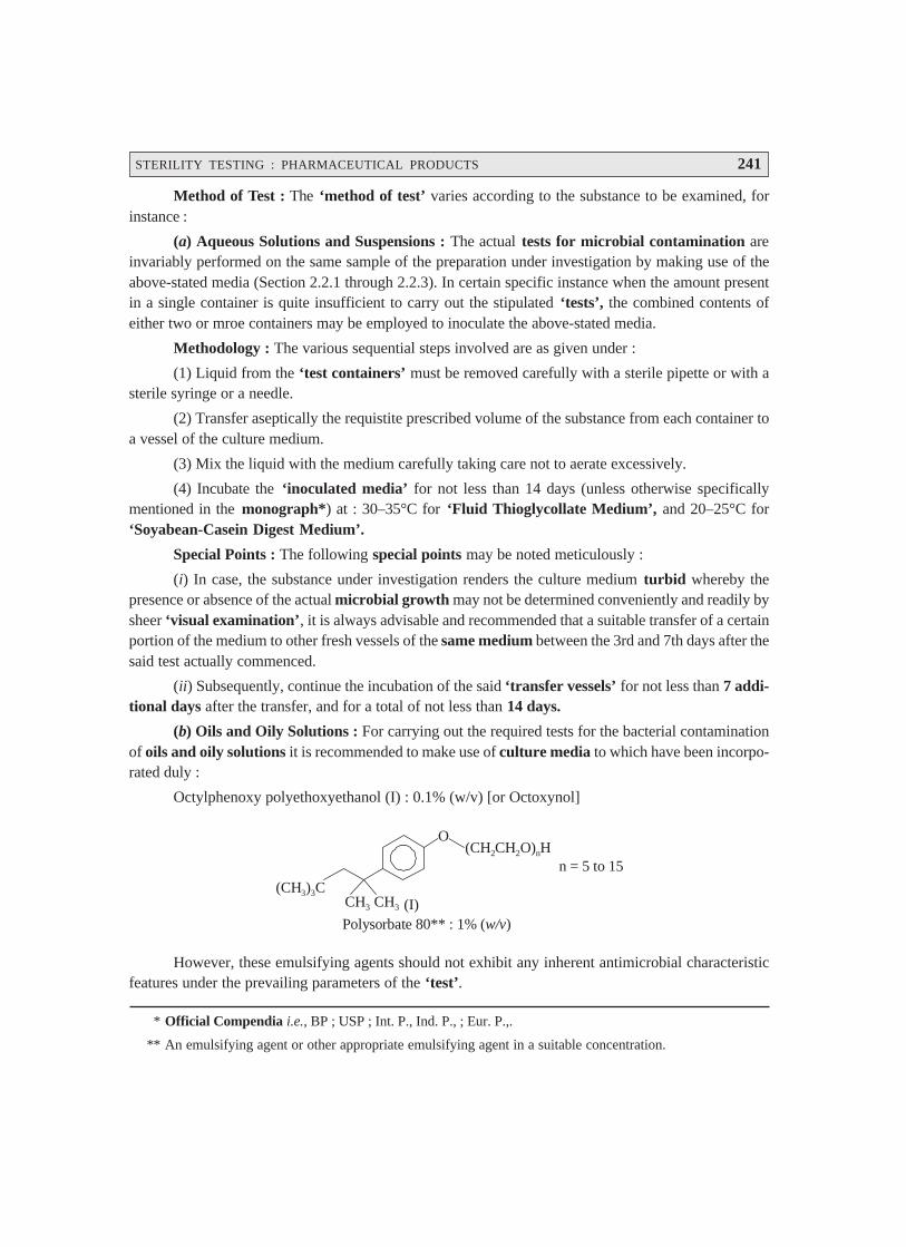
STERILITY TESTING : PHARMACEUTICAL PRODUCTS 241
* Official Compendia i.e., BP ; USP ; Int. P., Ind. P., ; Eur. P.,.
** An emulsifying agent or other appropriate emulsifying agent in a suitable concentration.
Method of Test : The ‘method of test’ varies according to the substance to be examined, forinstance :
(a) Aqueous Solutions and Suspensions : The actual tests for microbial contamination areinvariably performed on the same sample of the preparation under investigation by making use of theabove-stated media (Section 2.2.1 through 2.2.3). In certain specific instance when the amount presentin a single container is quite insufficient to carry out the stipulated ‘tests’, the combined contents ofeither two or mroe containers may be employed to inoculate the above-stated media.
Methodology : The various sequential steps involved are as given under :
(1) Liquid from the ‘test containers’ must be removed carefully with a sterile pipette or with asterile syringe or a needle.
(2) Transfer aseptically the requistite prescribed volume of the substance from each container toa vessel of the culture medium.
(3) Mix the liquid with the medium carefully taking care not to aerate excessively.
(4) Incubate the ‘inoculated media’ for not less than 14 days (unless otherwise specificallymentioned in the monograph*) at : 30–35°C for ‘Fluid Thioglycollate Medium’, and 20–25°C for‘Soyabean-Casein Digest Medium’.
Special Points : The following special points may be noted meticulously :
(i) In case, the substance under investigation renders the culture medium turbid whereby thepresence or absence of the actual microbial growth may not be determined conveniently and readily bysheer ‘visual examination’, it is always advisable and recommended that a suitable transfer of a certainportion of the medium to other fresh vessels of the same medium between the 3rd and 7th days after thesaid test actually commenced.
(ii) Subsequently, continue the incubation of the said ‘transfer vessels’ for not less than 7 addi-tional days after the transfer, and for a total of not less than 14 days.
(b) Oils and Oily Solutions : For carrying out the required tests for the bacterial contaminationof oils and oily solutions it is recommended to make use of culture media to which have been incorpo-rated duly :
Octylphenoxy polyethoxyethanol (I) : 0.1% (w/v) [or Octoxynol]
(CH ) C33CH3 CH3
O(CH CH O) Hn2 2
n = 5 to 15
Polysorbate 80** : 1% ( )w/v(I)
However, these emulsifying agents should not exhibit any inherent antimicrobial characteristicfeatures under the prevailing parameters of the ‘test’.
242 PHARMACEUTICAL MICROBIOLOGY
The required test must be carried out as already described under Section (a) above i.e., AqueousSolutions and Suspensions.
Precautionary Measures : The following two precautionary measures should be takenadequately :
(i) Cultures essentially comprising of ‘oily preparations’ should be shaken gently every day.
(ii) Importantly, when one employs the fluid thioglycollate medium for the ultimate detection ofthe anaerobic microorganisms, shaking or mixing must be restricted to a bear minimum level so as tomaintain perfect anaerobic experimental parameters.
(c) Ointments : The following steps may be adopted in a sequential manner :
(1) Carefully prepare the ‘test sample’ by diluting ten times in a sterile diluent, for instance :Fluid B or any other suitable aqueous vehicle which is capable of dispersing the test material homogene-ously throughout the ‘fluid mixture’.*
(2) Mix 10 mL of the fluid mixture thus obtained with 80 mL of the medium, and subsequentlyproceed as per the method given under Section (a) i.e., Aqueous Solutions and Suspensions.
(d) Solids : The various steps involved are as stated under :
(1) Transfer carefully the requisite amount of the preparation under examination to the quantityof culture medium as specified in Table : 8.3, and mix thoroughly.
(2) Incubate the inoculated media for not less than 14 days, unless otherwise mentioned in themonograph at 30–35°C in the particular instance of fluid thioglycollate medium, and at 20–25°C in thespecific case of soyabean-casein digest medium.
(e) Sterile Devices : For articles of such size and shape as allow the complete immersion in notmore than 1 L of the culture medium test the intact article, using the suitable media ; and incubating asstated under Section (a) i.e., Aqueous Solutions and Suspensions.
(f) Transfusion or Infusion Assemblies : For transfusion or infusion assemblies or where thesize of an item almost renders immersion impracticable, and exclusively the ‘liquid pathway’ should besterile by all means, flush carefully the lumen of each of twenty units with a sufficient quantum of fluidthioglycollate medium and the lumen of each of 20 units with a sufficient quantum of soyabean-casein digest medium to give an ultimate recovery of not less than 15 mL of each medium. Finally,incubate with not less than 100 mL of each of the two media as prescribed under Section (a) i.e., Aque-ous Solutions and Suspensions.
Exception : Such ‘medical devices’ wherein the lumen is so small such that fluid thioglycollatemedium will not pass through easily, appropriately substitute alternative thioglycollate mediuminstead of the usual fluid thioglycollate medium and incubate that duly inoculated medium anaerobically.
Note : In such situations where the presence of the specimen under examination, in the culturemedium critically interferes with the test by virtue of the ensuing bacteriostatic or fungistaticaction, rinse the article thoroughly with the bare minimum quantum of fluid A. Finally recoverthe rinsed fluid and carry out the ‘test’ as stated under ‘Membrane Filtration’ for SterileDevices.
* Before use, test the dispersing agent to ascertain that in the concentration employed it clearly exerts abso-lutely no significant antimicrobial activities during the time interval for all transfers.
STERILITY TESTING : PHARMACEUTICAL PRODUCTS 243
Observation and Interpretation of Results : In the case of ‘direct inoculation’ the variousobservation and interpretation of results may be accomplished by taking into consideration the fol-lowing cardinal factors, such as :
(1) Both at intervals during the incubation period, and at its completion, the media may beexamined thoroughly for the critical macroscopic evidence of the bacterial growth.
(2) In the event of a negative evidence, the ‘sample’ under examination passes the ‘tests forsterility’.
(3) If positive evidence of microbial growth is found, reserve the containers exhibiting this, andunless it is amply proved and adequately demonstrated by any other means that their (microorganisms)presence is on account such causes unrelated to the ‘sample’ being examined ; and, therefore, the testsfor sterility are pronounced invalid. In such cases, it may be recommended to carry out a ‘retest’employing an identical number of samples and volumes to be tested, and the media as in the originaltest.
(4) Even then, if no evidence of microbial growth is duly observed, the ‘sample’ under inves-tigation precisely passes the ‘tests for sterility’.
(5) In case, reasonable evidence of bacterial growth is observed, one may go ahead with theisolation and subsequent identification of the organisms.
(6) If they are found to be not readily distinguishable from those (microbes) growing in thecontainers reserved in the very First Test, the ‘sample’ under investigation fails the ‘tests for sterility’.
(7) In case, the microorganisms are readily distinguishable from the ones actually growing in thecontainers reserved in the ‘First Test’, it is very much advisable to carry out a ‘Second Retest’ byemploying virtually twice the number of samples.
(8) Importantly, if no evidence of bacterial growth is observed in the ‘Second Retest’, the sampleunder examination legitimately passes the ‘tests for sterility’.
(9) Contrarily, if evidence of growth of any microorganisms is duly observed in the ‘secondretest’, the sample under investigation obviously fails the ‘tests for sterility’.
8.3. SAMPLING : PROBABILITY PROFILE
Sampling refers to—‘the process of selecting a portion or part to represent the whole’.
In usual practice, a ‘sterility test’ attempts to infer and ascertain the state (sterile or non-sterile)of a particular batch ; and, therefore, it designates predominantly a ‘statistical operation’.
Let us consider that ‘p’ duly refers to the proportion of infected containers in a batch, and ‘q’
the proportion of corresponding non-infected containers. Then, we may have :
p + q = 1
or q = 1 – p
Further, we may assume that a specific ‘sample’ comprising of two items is duly withdrawnfrom a relatively large batch containing 10% infected containers. Thus, the probability of a single
item taken at random contracting infection is usually given by the following expression :

244 PHARMACEUTICAL MICROBIOLOGY
p = 0.1 [i.e., 10% = 0.1]
whereas, the probability of such an item being non-infected is invariably represented by the followingexpression :
q = 1 – p = 1 – 0.1 = 0.9
Probability Status—The probability status of the said two items may be obtained virtually inthree different forms, such as :
(a) When both items get infected : p2 = 0.01
(b) When both items being non-infected : q2 = (1 – p)2 = (0.9)2 = 0.81, and
(c) When one item gets infected and the other one non-infected : 1 – (p2 + q2)
or = 1 – (0.01 + 0.81) = 1 – (0.82)
or = 0.18
i.e., = 2pq
Assumption : In a particular ‘sterility test’ having a ‘sample’ size of ‘n’ containers, the ensuingprobability p of duly accomplishing ‘n’ consecutive ‘steriles’ is represented by the following expres-sion :
qn = (1 – p)n
Consequently, the ensuing values for various levels of ‘p’* having essentially a constant samplesize are as provided in the following. Table 8 : 4A, that evidently illustrates that the ‘sterility test’ failsto detect rather low levels of contamination contracted/present in the ‘sample’.
Likewise, in a situation whereby different sample sizes were actually used**, it may be em-phatically demonstrated that as the sample size enhances, the probability component of the batchbeing passed as sterile also gets decreased accordingly.
Table 8.4 : Sampling in Sterility Testing
Percentage of Infected Items in Batch
S. No. 0.1 1 5 10 20 50
1 p 0.001 0.01 0.05 0.1 0.2 0.5
2 q 0.999 0.99 0.95 0.9 0.8 0.5
3 Probability p of A1 0.98 0.82 0.36 0.12 0.012 < 0.00001
drawing 20 consecutive B2 > 0.99 0.99 0.84 0.58 0.11 0.002
sterile items
1. A : First Sterility Test : Calculated from P = (1 – p)20 = q20
2. B : First Re-Test : Calculated from P = (1 – p)20 [2 – (1 – p)20]
[Adapted From : Hugo and Russell : Pharmaceutical Microbiology, PG Publishing Pvt. Ltd.,New Delhi, 3rd edn., 1984]
* i.e., the proportion of infected containers present duly in a Batch.
** It is also based upon (1 – p)n factor.
STERILITY TESTING : PHARMACEUTICAL PRODUCTS 245
In actual practice, however, the additional tests, recommended by BP (1980), enhances substan-tially the very chances of passing a specific batch essentially comprising of a proportion or part of theinfected items (see Table : 8.4B). Nevertheless, it may be safely deduced by making use of the followingmathematical formula :
(1 – p)n [2 – (1 – p)n]
that provides adequate chance in the ‘First Re-Test’ of passing a batch comprising of a proportion orpart ‘p’ of the infected containers.
8.4. OVERALL CONCLUSIONS
The various techniques described in this chapter essentially make a sincere and benevolent attemptto accomplish to a reasonably large extent, the stringent control and continuous monitoring of a specificsterilization process. However, it is pertinent to state here that the ‘sterility test’ on its own fails toprovide any guarantee with respect to the specific sterility of a batch. Nevertheless, it categoricallyacounts for an ‘additional check’, besides a continued compliance and offer sufficient cognizableconfidence pertaining to the degree of an aseptic process or a sterilization technique being adopted.
Interestingly, an absolute non-execution of a prescribed (as per the ‘Official Compendia’) sterilitytest of a particular batch, despite the equivocal major criticism and objection of its gross inability andlimitations to detect other than the gross contamination, could tantamount to both moral consequencesand important legal requirements.
US-FDA promulgates and strongly advocates the adherence of USP-prescribed requirements forthe ‘sterility test’ for parenterals as the most authentic, reliable, and trustworthy ‘guide for testing theofficial sterile products’.
On a broader perspective, it may be observed that the ‘sterility test’ is not exclusively intendedas a thoroughly evaluative test for a product duly subjected to a known sterilization method of unknowneffectiveness. Nevertheless, it is solely meant primarily as an intensive ‘check test’ on the ensuingprobability that :
a previously validated sterilization process has been repeated duly, and
to provide adequate assurance vis-a-vis its continued effictiveness legitimately.
FURTHER READING REFERENCES
1. Brown MRW and Gilbert P : Increasing the probability of sterility of medicinal products, J.of Pharmacy and Pharmacology, 27 : 484–491, 1977.
2. Denyer SP et al. : Filtration Sterilization : In Principles and Practice of Disinfection,Preservation and Sterilization (ed. Russell AD et al.) Blackwell Scientific Publications,Oxford (UK), 1982.
3. Hugo WB and Russell AD : Pharmaceutical Microbiology, PG Publishing Pvt. Ltd., Sin-gapore, 3rd edn, 1984.
4. Indian Pharmacopoea : Published by the Controller of Publications, Delhi, Vol. II, 1996.
5. Remington : The Science and Practice of Pharmacy, Lippincott Williams & Wilkins,New York, Vol.–1, 21st. edn, 2006.

246
9.1. INTRODUCTION
Immunity may be defined as — ‘the state of being immune to or protected from a diseaseespecially an infectious disease’.
Importantly, this particular state is invariably induced by having been exposed to the antigenicmarker on an microorganism that critically invades the body or by having been duly immunized with avaccine capable of stimulating the production of specific antibodies.
Immunology, the generation of an immune response solely depends upon the prevailinginteraction of three cardinal components of the immune mechanism, such as :
immunogen stimulation,
humoral immune system, and
cellular immune system.
Since 1901 and as to date the epoch making discovery and spectacular evolution of‘immunobiotechnology’ i.e., conglomeration of immune system variants, across the world hasrevolutionized not only the safer quality of life of human beings but also provided a broad spectrum ofnewer avenues in combating the complicated dreadful not-so-easy diseases of the present day.
Immune Response : In reality, the immune responses do refer to such processes whereby animals(including humans) give rise to certain specifically reactive proteins (known as ‘antibodies’) andadequate cells in response to a great number of foreign organic molecule and macromolecule variants.Based on the scientifically demonstrated proofs and evidences the generalized immune responseessentially possesses four major primary characteristic features, such as :
(a) discrimination,
(b) specificity,
(c) anamnesis, and
(d) transferability by living cells.
IMMUNE SYSTEMS9• Introduction• Types of Specific Immunity• Duality of Immune Systems• Immunological Memory• Natural Resistance and Nonspecific Defence Mechanisms
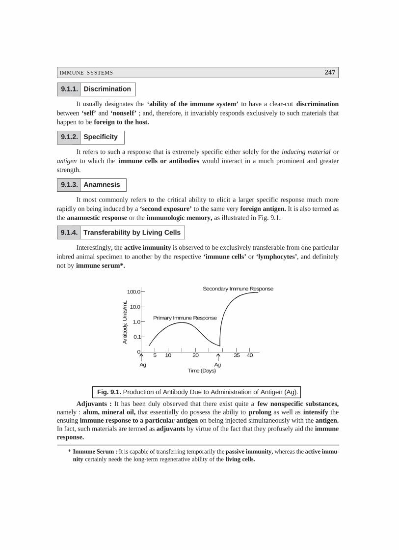
IMMUNE SYSTEMS 247
* Immune Serum : It is capable of transferring temporarily the passive immunity, whereas the active immu-nity certainly needs the long-term regenerative ability of the living cells.
9.1.1. Discrimination
It usually designates the ‘ability of the immune system’ to have a clear-cut discriminationbetween ‘self’ and ‘nonself’ ; and, therefore, it invariably responds exclusively to such materials thathappen to be foreign to the host.
9.1.2. Specificity
It refers to such a response that is extremely specific either solely for the inducing material orantigen to which the immune cells or antibodies would interact in a much prominent and greaterstrength.
9.1.3. Anamnesis
It most commonly refers to the critical ability to elicit a larger specific response much morerapidly on being induced by a ‘second exposure’ to the same very foreign antigen. It is also termed asthe anamnestic response or the immunologic memory, as illustrated in Fig. 9.1.
9.1.4. Transferability by Living Cells
Interestingly, the active immunity is observed to be exclusively transferable from one particularinbred animal specimen to another by the respective ‘immune cells’ or ‘lymphocytes’, and definitelynot by immune serum*.
Ag Ag
5 10 20 35 40
Time (Days)
Primary Immune Response
Secondary Immune Response
0
0.1
1.0
10.0
100.0
Ant
ibod
y, U
nits
/mL
Fig. 9.1. Production of Antibody Due to Administration of Antigen (Ag).
Adjuvants : It has been duly observed that there exist quite a few nonspecific substances,namely : alum, mineral oil, that essentially do possess the abiliy to prolong as well as intensify theensuing immune response to a particular antigen on being injected simultaneously with the antigen.In fact, such materials are termed as adjuvants by virtue of the fact that they profusely aid the immuneresponse.
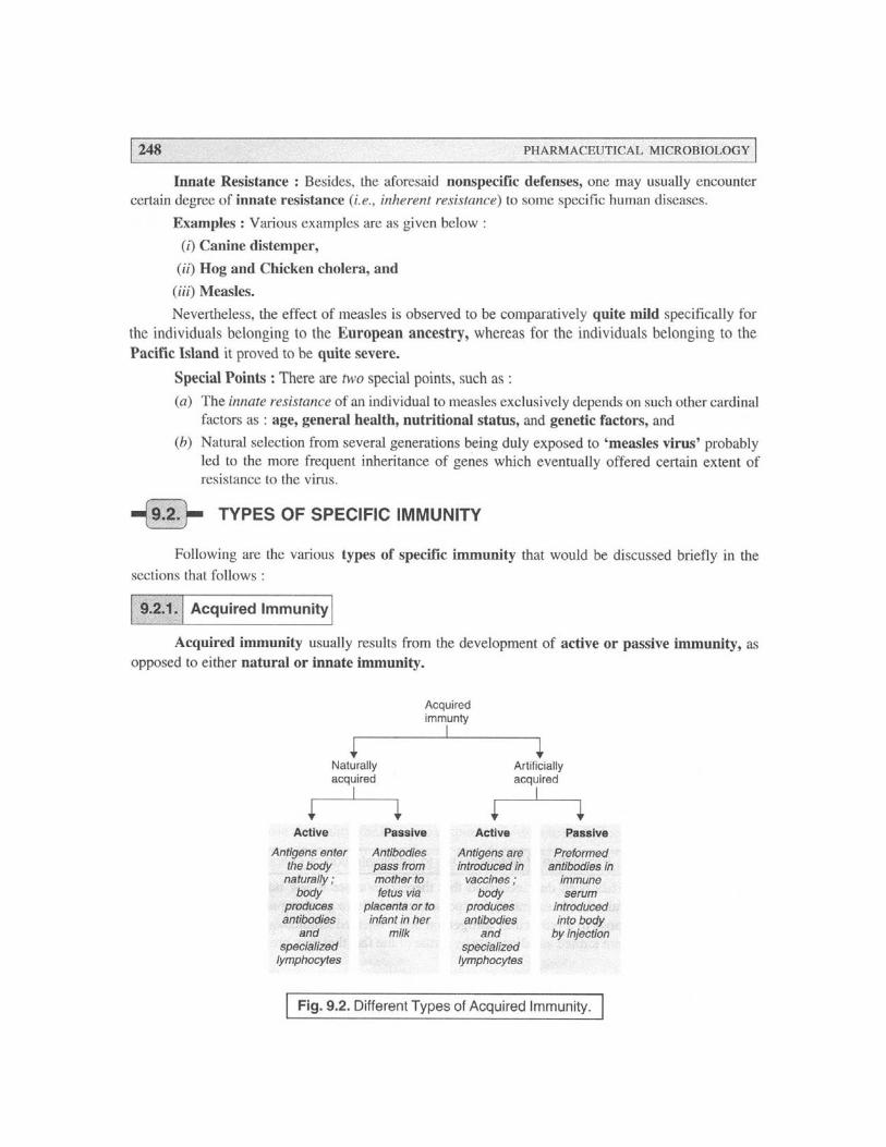
IMMUNE SYSTEMS 249
In other words, acquired immunity invariably refers to the ‘protection’ an animal inherentlydevelops against certain types of microorganisms or foreign substances. In reality, the acquired immu-nity gets developed in the course of an individual’s lifespan. Fig. 9.2 depicts the different types ofacquired immunity in a summarized form.
9.2.2. Active Immunity
Active immunity refers to the specific immunity obtained from the development within the bodyof antibodies or sensitized T lymphocytes (T Cells) which critically neutralize or destroy the infectiveagent. It may eventually result from the immune response to an invading organism or from inoculationwith a vaccine essentially containing a foreign antigen.
9.2.3. Cell-Mediated Immunity [or T-cell Mediated Immunity]
It has been duly observed that the regulatory and cytotoxic actions of T cells during the specificimmune response is known as the cell-mediated immunity. However, the entire process essentiallyneeds almost 36 hr to accomplish its full effect. It is also called as T cell mediated immunity.
Physiological Actions : Interestingly, unlike B cells, T cells invariably fail to recognize the socalled foreign antigens on their own. A foreign antigen is duly recognized by a macrophage whichengulfs it and displays part of the antigen on its surface next to a histocompatibility or ‘self’ antigen(macrophage processing). Finally, the presence of these two markers together with the secretion of acytokine, interleukin-1 (IL-1) by macrophages and other antigen-presenting cells duly activates CD4+/CD8 T cells (i.e., helper T cells), that categorically modulate the activities of other cells adequatelyinvolved in the immune response.
Thus, the CD4+T cells secrete interleukin-2 (IL-2), that stimulates the activity of natural killercells (NK cells), cytotoxic T cells, and B cells ; and ultimately promotes the proliferation of CD+T cellsin order that the invading pathogen may be destroyed or neutralized effectively. Besides, GammaInterferon secreted by CD+T cells increases distinctly the macrophage cytotoxicity and antigenprocessing. However, the T-cell mediated immunity plays a significant and pivotal role in the rejec-tion of transplanted tissues and in ‘tests for allergens’ i.e., the delayed hypersensitivity reaction.
9.2.4. Congenital Immunity
The congenital immunity refers to the immunity critically present at birth. It may be eithernatural or acquired, the latter predominantly depends upon the antibodies solely received from the mother’sblood.
9.2.5. Herd Immunity
The herd immunity represents the immune protection duly accomplished via vaccination of aportion of a population, that may eventually minimise the spread of a disease by restricting the numberof potential hosts for the respective pathogen.
9.2.6. Humoral Immunity [or B-cell Mediated Immunity]
Humoral immunity respresents the immunity duly mediated by antibodies in body fluids e.g.,plasma or lymph. As these antibodies are adequately synthesized and subsequently secreted by B cells,
250 PHARMACEUTICAL MICROBIOLOGY
that protect the body against the infection or the reinfection by common organisms, such as : streptococciand staphylococci, it is also known as B-cell mediated immunity. In reality, the B cells are stimulatedby direct contact with a foreign antigen and differentiate into the plasma cells that yield antibodiesagainst the antigen ; and the corresponding memory cells which enable the body to rapidly producethese antibodies if the same antigen appears at a later time.
It is, however, pertinent to state here that B cell differentiation is also stimulated duly by interleukin-2(IL-2), secreted by the T4 cells, and by foreign antigens processed by macrophages.
9.2.7. Local Immunity
Local immunity is usually limited to a given area or tissue of the body.
9.2.8. Natural Immunity
Natural immunity refers to the immunity programmed in the DNA, and is also known as thegenetic immunity. It has been observed that there are certain pathogens that fail to infect some speciesdue to the fact that the cells are not exposed to appropriate environments, for instance : the ‘measlesvirus’ cannot reproduce in the canine cells ; and, therefore, dogs do have natural immunity to measles.
9.2.9. Passive Immunity
Passive immunity specifically refers to the immunity acquired by the introduction of preformedantibodies into an unprotected individual. It may take place either through injection or in utero fromantibodies that usually pass from the mother to the foetus via the placenta. It can also be acquired by thenewborn by ingesting the mother’s milk.
9.3. DUALITY OF IMMUNE SYSTEM
Evidences from an exhaustive survey of literature has revealed that during the early stagespertaining to the historical development of immunological experimentation, the, biologists duly learnedthat ‘certain specific types of immunity’ may be meticulously transferred between animals (belongingto the same species) by actually transferring serum from immunized to nonimmunized animals.Importantly, other kinds of immunity could not be transferred effectively via blood serum. Obviously, at amuch later stage it was duly understood that these special types of immunity may be easily and convenientlytransferable only when certain specific lymphocytes were transferred actually.
Based on the further extensive and intensive researches carried out by the ‘immunologists’ acrossthe globe ultimately accumulated copious volumes of valuable informations and results that are now well-known to reflect the two major segments of the so called vertebrate immune system, namely :
(a) immunity associated with serum-transfer reflecting the activities of the humoral (anti-body-mediated) immune system*, and
* For Humoral Immunity — see Section 2.6 Chapter 9.
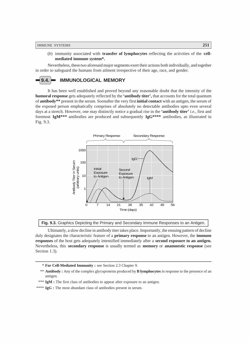
IMMUNE SYSTEMS 251
* For Cell-Mediated Immunity : see Section 2.3 Chapter 9.
** Antibody : Any of the complex glycoproteins produced by B lymphocytes in response to the presence of anantigen.
*** IgM : The first class of antibodies to appear after exposure to an antigen.
**** IgG : The most abundant class of antibodies present in serum.
(b) immunity associated with transfer of lymphocytes reflecting the activities of the cell-mediated immune system*.
Nevertheless, these two aforesaid major segments exert their actions both individually, and togetherin order to safeguard the humans from ailment irrespective of their age, race, and gender.
9.4. IMMUNOLOGICAL MEMORY
It has been well established and proved beyond any reasonable doubt that the intensity of thehumoral response gets adequately reflected by the ‘antibody titer’, that accounts for the total quantumof antibody** present in the serum. Soonafter the very first initial contact with an antigen, the serum ofthe exposed person emphatically comprises of absolutely no detectable antibodies upto even severaldays at a stretch. However, one may distinctly notice a gradual rise in the ‘antibody titer’ i.e., first andforemost IgM*** antibodies are produced and subsequently IgG**** antibodies, as illustrated inFig. 9.3.
InitialExposureto Antigen
SecondExposureto Antigen
IgG
IgM
0 7 14 21 28 35 42 49 56
1
10
100
1000
Time (days)
Ant
ibod
y Tite
r in
Ser
um(a
rbitr
ary
units
)
Primary Response Secondary Response
Fig. 9.3. Graphics Depicting the Primary and Secondary Immune Responses to an Antigen.
Ultimately, a slow decline in antibody titer takes place. Importantly, the ensuing pattern of declineduly designates the characteristic feature of a primary response to an antigen. However, the immuneresponses of the host gets adequately intensified immediately after a second exposure to an antigen.Nevertheless, this secondary response is usually termed as memory or anamnestic response (seeSection 1.3).
252 PHARMACEUTICAL MICROBIOLOGY
It has been observed that there exists certain activated B lymphocytes that fail to turn into the socalled antibody-producing plasma cells, but do persist and sustain as the long-lived memory cells.After a long span even stretching over to several decades, when such ‘cells’ are duly stimulated by the‘same antigen’, they invariably tend to differentiate rapidly into the much desired antibody-producingplasma cells. Actually, this ultimately affords the fundamental basis of the secondary immune responseas depicted in Fig. 9.3.
9.5. NATURAL RESISTANCE AND NONSPECIFIC DEFENSEMECHANISMS [OR DEFENSIVE MECHANISMS OF BODY]
In a broader sense the ensuing interaction existing between a host (human body) and amicroorganism designates an excellent unique dynamic phenomenon whereby each and every protagonistcritically serves to maximize its overall survival. It has been duly observed that in certain typical instances,after a specific microbe gains its entry or comes in contact with a host, a distinct positive mutuallybeneficial relationship takes place which ultimately becomes integral to the final health of the host. Inthis manner, the microorganisms turn out to be the normal microbiota*. However, in other such cases,the particular microorganism causes, induces or produces apparent devastating and deleterious overalleffects upon the host ; and, therefore, may finally even cause death of the host via a dreadful ailment.
Interestingly, the prevailing environment of a ‘host’ is heavily surrounded with microorganisms,and there lies an ample scope and opportunity to come in their contact every moment of the day.Nevertheless, quite a few of these microbes are pathogenic in nature (i.e., cause disease). Surprisingly,these pathogens are at times duly guarded and prevented from producing a disease due to the inherentcompetition offered by the normal microbiota. In reality, the invading pathogens are squarely keptaway from the host by the ‘normal microbiota’ by using nutrients, resources, space, and may even yieldsuch chemical substances which would repel them ultimately.
In addition to the above stated glaring scientific fact and evidences these ‘normal microbiota’grossly prevent colonization of pathogens to a great extent ; and, thereby, most probably checking thedisease (to the host) via ‘bacterial interference’.
Example : An excellent typical example is stated as under :
Lactobacilli – present strategically in the female genital tract (FGT) usually maintain a low pH(acidic), and thereby exclusively afford the colonization by the pathogenic microbes. Besides, thecorynebacteria located critically upon the skin surface give rise to the formation of ‘fatty acids’ whichultimately inhibit the phenomenon of colonization by the pathogenic organisms.
Note : It is an excellent example of ‘amensalism’. (i.e., symbiosis wherein one population (or individual)gets affected adversely and the other is unaffected).
Interestingly, the ‘normal microbiota’ usually give rise to protection confined to a certain degreefrom the invading pathogens ; however, they may themselves turn into pathogenic in character andcause disease under certain particular circumstances. Thus, these ‘converted pathogens’ are invariablyknown as ‘opportunistic microorganisms’** or pathogens.
Based on the above statement of facts and critical observations one may conclude that on onehand pathogen makes use of all the opportune moments available at its disposal to cause and induct
* Microbiota : The microscopic organisms of an area.
** Opportunistic Microorganisms : are found to be adapted to the specific non invasive mode of life dulydefined by the limitations of the environment wherein they are living.
IMMUNE SYSTEMS 253
infection, the host’s body possesses a plethora of ‘defense mechanisms’ to encounter the infection. Infact, the observed intricacies prevailed upon by the host-pathogen relationship are not only numerousbut also quite divergent in nature, which may be classified under the following three heads, such as :
(a) Natural Resistance,
(b) Internal Defense Mechanisms, and
(c) Nonspecific Defense Mechanisms.
The aforesaid three categories shall now be discussed separately in the sections that follows :
9.5.1. Natural Resistance
It has been observed that the two cardinal aspects, namely : (a) physiological needs, and (b) meta-bolic requirements, of a pathogen are an absolute necessity in establishing precisely the extent vis-a-visthe range of potentially susceptible hosts. However, the naturally resistant hosts exert their action intwo variant modes, such as :
miserably fail to cater for certain urgently required environmental factors by the microbesfor their usual growth, and
essentially possess defense mechanisms to resist infection considerably.
Besides, there are some other factors pertaining to the host’s general health, socioeconomic sta-tus, level of nutrition potentiality, and certain intangible conditions viz., stress, mental agony, depres-sion etc.
Natural resistance essentially comprises of the following four vital and important aspects :
9.5.1.1. Species Resistance
In general, the fundamental physiologic characteristics of humans, namely : normal body tem-perature may give a positive clue whether or not a specific bacterium can be pathogenic in nature.Likewise, in host-specific e.g., human and bovine species, the tubercle bacillus is found to cross-infect both humans and cattle having almost an identifical body temperature.
Salient Features : The salient features of species resistance are as given under :
(1) inability of a bacterium to induct disease in the resistant species under the natural environ-ments,
(2) critical production in the specific resistant species of either a localized or a short-periodinfection caused solely due to an experimental inoculation vis-a-vis a progressive or gener-alized ailment in naturally susceptible species, and
(3) introduction of experimental disease particularly in the resistant species exclusively causedby massive doses of the microbes, usually in two different ways :
(a) under unnatural parameters, and
(b) by an unnatural route.
9.5.1.2. Racial Resistance
Exhaustive and intensive studies have amply proved that the very presence of a pathogen in theisolated races give rise to a gradual selection for resistant members, because the susceptible mem-bers die of progressive infection ultimately. It may be further expatiated by the following three glaringexamples :
254 PHARMACEUTICAL MICROBIOLOGY
Examples :
(i) Incorporation of altogether ‘new pathogens’ e.g., tubercle bacillus, by the relatively resist-ant Europeans into an isolated American Indians population*, finally caused epidemicsthat almost destroyed a major proportion of the ensuing population.
(ii) African Blacks (Negros) invariably demonstrate a relatively high resistance to the tropicaldiseases, namely : malaria, yellow fever, and
(iii) Orientals do exhibit a much reduced susceptibility to syphilis.
9.5.1.3. Individual Resistance
It may be critically observed that there are certain individuals who apparently experience feweror less severe infections in comparison to other subjects, irrespective of the fact that :
both of them essentially possess the same racial background, and
do have the same opportunity for ultimate exposure.
Causation : Individual resistance of this nature and kind is perhaps on account of :
natural in-built resistance factor, and
adaptive resistance factor.
Age Factor – is equally important, for instance :
aged people are more prone to such ailments as : Pneumonia – most probably due to apossible decline of the ‘immune functions’ with advancement in growing age.
children i.e., very young individuals are apparently more susceptible to such ‘children’sdisease’ as : Chicken-pox, measles–just prior to their having acquired enough in-builtresistance/immunity that essentially follows both inapparent and overt contracted infections.
Genetic Factor – Immunodeficiencies** found in some, individuals are caused solely due to‘genetic defects’, that largely enhance the probability and susceptibility to disease.
Other Factors – include malnutrition, personal hygiene, and an individual’s attitude to sex pro-file ; hazards and nature of work-environment ; incidence of contacts with infected individuals, and anindividual’s hormonal vis-a-vis endocrine balance – they all do affect the overall frequency as well asselectivity of some critical ailments.
9.5.1.4. External Defense Mechanisms
In fact, the external defense mechanisms do represent another cardinal and prominent factor innatural resistance ; however, they essentially involve the chemical barriers as well. Besides, two otherpredominant factors viz., (a) mechanical barriers, and (b) host secretions, essentially make up thebody’s First-Line of Defense Mechanism against the invading microorganisms.
Mechanical Barriers – actually comprise of such materials as : intact (unbroken) skin andmucous membranes that are practically incapable of getting across to the infectious agents. However,the said two mechanical barriers viz., intact skin and mucous membranes do afford a substantial ‘effec-tive barrier’, whereas hair follicles, dilatation of sweat glands, or abrasions do allow the gainfulentry for the microbes into the human body.
* Who did not earlier developed a usual resistance to the organism.
** Immunodeficiencies : An inability to develop perfect immunity to pathogens.
IMMUNE SYSTEMS 255
* Polymorphonuclear Leukocytes : Leukocytes possessing a nucleus consisting of several parts or lobesconnected by fine strands.
Examples : Various typical examples are as given under :
(1) Large segment of microbes are duly inhibited by such agents as :
low pH (acidity),
lactic acid present in sweat, and
fatty acids present in sweat.
(2) Mucous secretions caused by respiratory tract (RT), digestive tract (DT), urogenitaltract (UT) plus other such tissues do form an integral protective covering of the respectivemucous membranes thereby withholding and collecting several microorganisms until theymay be either disposed of effectively or lose their infectivity adequately.
(3) Chemical Substances – Besides, the ensuing mechanical action caused by mucous, saliva,and tears in the critical removal of microorganisms, quite a few of these secretions do con-tain a number of chemical substances which critically cause inhibition or destruction ofmicroorganisms.
Examples : A few typical examples are as stated under :
(a) Lysozyme – an enzyme invariably observed in several body fluids and secretions viz.,blood, plasma, urine, saliva, cerebrospinal fluid, sweat, tears etc., that predominantly doexert an effective antimicrobial action on account of its inherent ability to lyse someparticular Gram positive microbs by specifically affording the hydrolysis ofpeptidoglycan,
(b) Several other hormones and enzymes are capable of producing distinct chemical, physi-ological, and mechanical effects that may ultimately cause minimization of susceptibil-ity to reduction, and
(c) The prevailing inherent acidity or alkalinity of certain ‘body fluids’ possess an apparentdeleterious effect upon several microbes, and helps to check and prevent the potentialpathogens for gaining an easy access to the deeper tissues present in the body.
(d) Lactoferrin-Lactoferrin is an iron-containing red-coloured protein found in milk (viz.,human and bovine) that essentially possesses known antibacterial characteristic features.It is also found in a plethora of body-secretions that specifically and profusely bathe thehuman mucosal surfaces, namely :
• bronchial mucous ; • seminal fluids ;
• hepatic bile ; • saliva ;
• nasal discharges ; • tears ; and
• pancreatic juice ; • urine.
Lactoferrin forms a vital and important constituent of the highly particular granules of the‘polymorphonuclear leukocytes’*.
(5) Transferrin : It represents the serum counterpart of lactoferrin. In fact, both these typicalproteins essentially possess high molecular weights ~ 78,000 daltons, besides having severalmetal-binding critical sites.
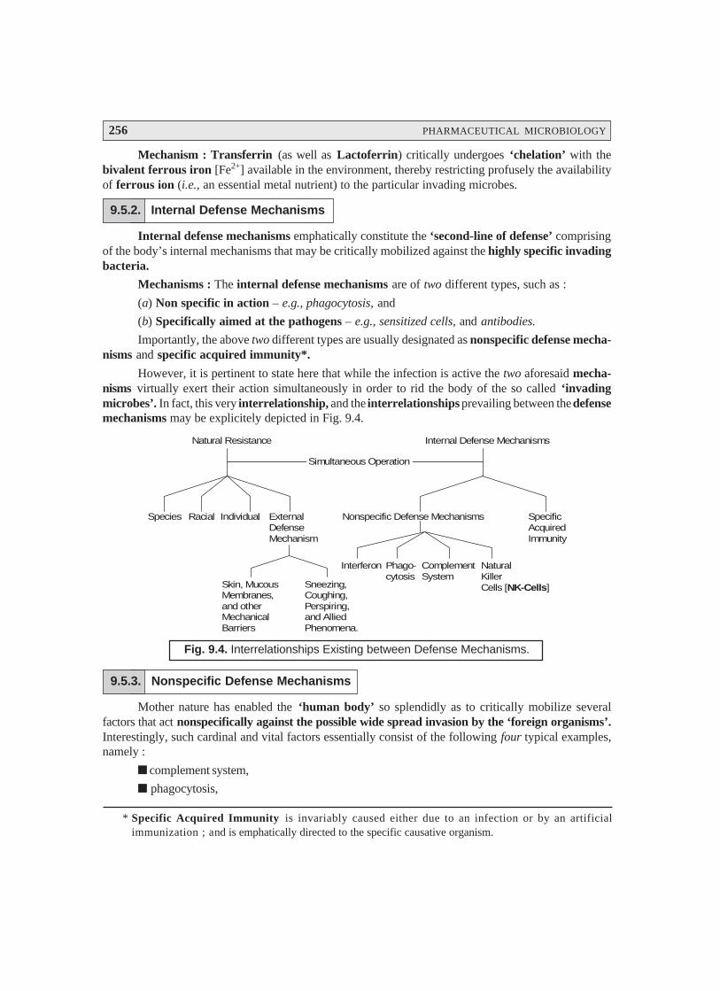
256 PHARMACEUTICAL MICROBIOLOGY
Mechanism : Transferrin (as well as Lactoferrin) critically undergoes ‘chelation’ with thebivalent ferrous iron [Fe2+] available in the environment, thereby restricting profusely the availabilityof ferrous ion (i.e., an essential metal nutrient) to the particular invading microbes.
9.5.2. Internal Defense Mechanisms
Internal defense mechanisms emphatically constitute the ‘second-line of defense’ comprisingof the body’s internal mechanisms that may be critically mobilized against the highly specific invadingbacteria.
Mechanisms : The internal defense mechanisms are of two different types, such as :
(a) Non specific in action – e.g., phagocytosis, and
(b) Specifically aimed at the pathogens – e.g., sensitized cells, and antibodies.
Importantly, the above two different types are usually designated as nonspecific defense mecha-nisms and specific acquired immunity*.
However, it is pertinent to state here that while the infection is active the two aforesaid mecha-nisms virtually exert their action simultaneously in order to rid the body of the so called ‘invadingmicrobes’. In fact, this very interrelationship, and the interrelationships prevailing between the defensemechanisms may be explicitely depicted in Fig. 9.4.
Natural Resistance
Simultaneous Operation
Species Racial Individual ExternalDefenseMechanism
Nonspecific Defense Mechanisms SpecificAcquiredImmunity
Skin, MucousMembranes,and otherMechanicalBarriers
Sneezing,Coughing,Perspiring,and AlliedPhenomena.
Interferon Phago-cytosis
ComplementSystem
NaturalKillerCells [ ]NK-Cells
Internal Defense Mechanisms
Fig. 9.4. Interrelationships Existing between Defense Mechanisms.
9.5.3. Nonspecific Defense Mechanisms
Mother nature has enabled the ‘human body’ so splendidly as to critically mobilize severalfactors that act nonspecifically against the possible wide spread invasion by the ‘foreign organisms’.Interestingly, such cardinal and vital factors essentially consist of the following four typical examples,namely :
complement system,
phagocytosis,
* Specific Acquired Immunity is invariably caused either due to an infection or by an artificialimmunization ; and is emphatically directed to the specific causative organism.

IMMUNE SYSTEMS 257
* Precursors : In biological processes, a substance from which another, usually more active or mature, sub-stance is formed duly.
** Capillary dilatation
*** The action of opsonins (i.e., a substance that coats ‘foreign antigens’, to facilitate phagocytosis.
**** Chemotaxis : The movement of additional white blood cells (WBCs) to an area of inflamation in responseto the release of chemical mediators by neutrophils, monocytes, and injured tissue.
naturally occurring cytotoxic lymphocytes, and
interferon.
Each of the aforesaid factors shall now be treated individually in the sections that follows :
9.5.3.1. Complement System
Higher animal’s serum usually made up of a particular group of ‘eleven proteins’, which arehighly specific in nature, and are widely referred to collectively as the so called complement system byvirtue of the fact that its action complements predominantly to that of some prominent antibody-medi-ated reactions. In other words, the complement system critically enacts a pivotal role with respect tothe overall generalized resistance against the infection caused by the ‘pathogens’ ; and, therefore,accounts for as the ‘principal mediator’ of the ensuing specific inflammatory response.
Mode of Action (Modus Operandi) : The various steps involved are as follows :
(1) When the very ‘First Protein’, belonging to cluster of elevan proteins, gets duly activatedthere exist distinctly a prominent ‘sequential cascade’ whereby the ‘active molecules’ dulycome into being via the inactive precursors*.
(2) Some of the protein variants do get activated very much along the ‘sequential cascade’ thatmay function as mediators of a specific response, and eventually serves as activators of thenext step.
Table 9.1 : Records certain of the functional activities of the Host Complement System presentduly in the Host Defense against the infection.
Table 9.1 : Functional Activities of Host Complement System in Host Defense Vs. Infection
S.No. Observed Activity Complement Compoundsor Fragments
1 Lysis of tumour cells, viruses, virus-infected cells, protozoa, C1 to C9mycoplasma, and microorganisms.
2 Virus neutralization C1, C4, C2, and C3
3 Endotoxin inactivation C1 to C5
4 Anaphylatoxin** release C1a, C4a, C5a
5 Enhancement of cell-mediated cytotoxicity, stimulation of C3bproduction of B-cell lymphokines, and Opsonization***.
6 Increased induction of antibody formation. C3b, C3d
7 Chemotaxis**** of eosinophils, monocytes, and neutrophils. C5a
8 Stimulation of macrophage adherence and spreading. Bb
Complement Fixation (or Attachment) : In a broader perspective, the complement system isquite capable of attacking and killing the invading cells exclusively after the antibody gets bound to the
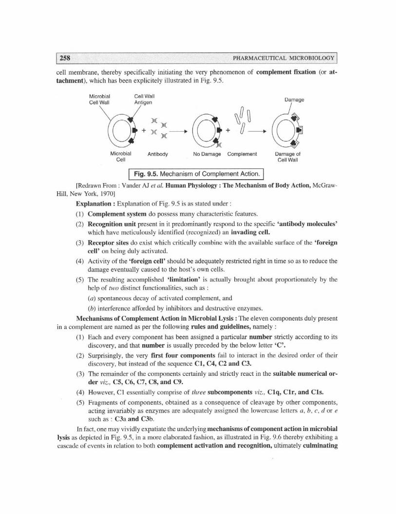
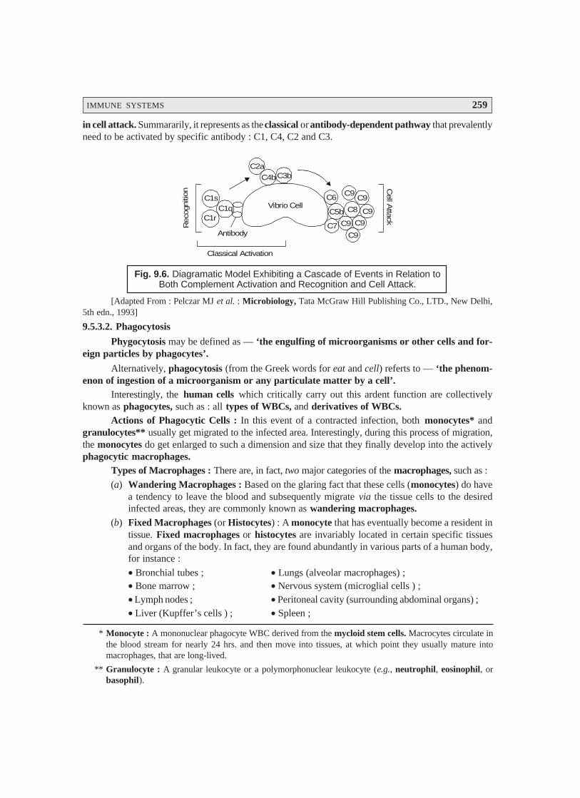
IMMUNE SYSTEMS 259
in cell attack. Summararily, it represents as the classical or antibody-dependent pathway that prevalentlyneed to be activated by specific antibody : C1, C4, C2 and C3.
C3bC4b
C2a
C5b
C6
C7
C8
C9
C9
C9
C9
C9C1q
C1s
C1r
Vibrio Cell
Antibody
Cell A
ttack
Classical Activation
Reco
gniti
on
C9
Fig. 9.6. Diagramatic Model Exhibiting a Cascade of Events in Relation toBoth Complement Activation and Recognition and Cell Attack.
[Adapted From : Pelczar MJ et al. : Microbiology, Tata McGraw Hill Publishing Co., LTD., New Delhi,5th edn., 1993]
9.5.3.2. Phagocytosis
Phygocytosis may be defined as — ‘the engulfing of microorganisms or other cells and for-eign particles by phagocytes’.
Alternatively, phagocytosis (from the Greek words for eat and cell) referts to — ‘the phenom-enon of ingestion of a microorganism or any particulate matter by a cell’.
Interestingly, the human cells which critically carry out this ardent function are collectivelyknown as phagocytes, such as : all types of WBCs, and derivatives of WBCs.
Actions of Phagocytic Cells : In this event of a contracted infection, both monocytes* andgranulocytes** usually get migrated to the infected area. Interestingly, during this process of migration,the monocytes do get enlarged to such a dimension and size that they finally develop into the activelyphagocytic macrophages.
Types of Macrophages : There are, in fact, two major categories of the macrophages, such as :
(a) Wandering Macrophages : Based on the glaring fact that these cells (monocytes) do havea tendency to leave the blood and subsequently migrate via the tissue cells to the desiredinfected areas, they are commonly known as wandering macrophages.
(b) Fixed Macrophages (or Histocytes) : A monocyte that has eventually become a resident intissue. Fixed macrophages or histocytes are invariably located in certain specific tissuesand organs of the body. In fact, they are found abundantly in various parts of a human body,for instance :
• Bronchial tubes ; • Lungs (alveolar macrophages) ;• Bone marrow ; • Nervous system (microglial cells ) ;• Lymph nodes ; • Peritoneal cavity (surrounding abdominal organs) ;• Liver (Kupffer’s cells ) ; • Spleen ;
* Monocyte : A mononuclear phagocyte WBC derived from the mycloid stem cells. Macrocytes circulate inthe blood stream for nearly 24 hrs. and then move into tissues, at which point they usually mature intomacrophages, that are long-lived.
** Granulocyte : A granular leukocyte or a polymorphonuclear leukocyte (e.g., neutrophil, eosinophil, orbasophil).
260 PHARMACEUTICAL MICROBIOLOGY
Importantly, the macrophage variants critically present in the body strategically constitute themononuclear phagocytic (reticuloendothelial) system.
9.5.3.2.1. Functions of Phagocytes (or Phagocytic Cells) : It has been duly observed that whenan infection gets contracted one may apparently observe a distinct shift taking place predominantly inthe particular types of WBC which runs across the blood stream. Thus, the following cardinal pointsmay be noted, carefully :
Granulocytes – particularly the ‘neutrophils’ occur overwhelmingly in the initial phase ofinfection, at this point in time they are found to be extremely phagocytic in nature.
Distinct aforesaid dominance is evidently shown by the presence of their actual number in adifferential WBC count.
With the progress of contracted infection, the macrophages also predominate – scavenge –phagocytize remaining live/dead/dying microorganisms.
Enhanced number of monocytes, that eventually develop into the corresponding macrophages,is adequately reflected in the WBC-differential count explicitely.
Blood and lymph containing bacteria when made to pass via various organs in the bodyhaving fixed macrophages, cells of the mononuclear phagocytic system ultimately get ridof the bacteria by phagocytosis.
Mononuclear phagocytic system also helps in the critical disposal of the worn-out bloodcells.
Table 9.2 records the classification as well as a summary of phagocytic cells and their functions.
Table : 9.2 : Classification and Functions of Phagocytes
S.No. Phagocyte Variant Functional Cells Functions
1. Monocytes Mononuclear phagocytic Phagocytic against microbes with thesystem : wandering macro- progress of infection, and against worn-phages and fixed macro- out blood cells as the infection getsphages (histocytes) reduced. Besides, duly involved in cell-
mediated immunity.
2. Granulocytes Neutrophils and eosinophils. Phagocytic against microbes, usuallyencountered during the initial phaseof infection.
9.5.3.2.2. Mechanism of Phagocytosis : In order to understand the exact and precise mechanismof phagocytosis, we may have to divide the phenomenon of phagocytosis, as illustrated in Fig. 9.6,into four cardinal phases, such as : chemotaxis, adherence, ingestion, and digestion. These four dis-tinct phases shall now be treated briefly in the sections that follows from [A] through [D] :[A] Chemotaxis [Syn : Chemotropism] :
Chemotaxis may be defined as — ‘the movement of additional white blood cells to an area ofinflammation in response to the release of chemical mediators by neutrophils, monocytes, andinjured tissue’.
In other words, chemotaxis refers to the chemical attraction of the phagocytes to microbes.Importantly, the various ‘chemotactic chemical susbtances’ which specifically attract the
phagocytes happen to be such microbial products as components of :
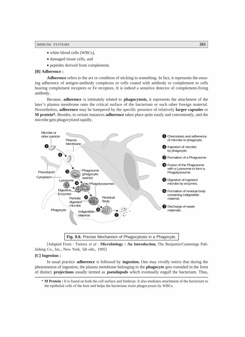
IMMUNE SYSTEMS 261
* M Protein : It is found on both the cell surface and fimbriae. It also mediates attachment of the bacterium tothe epithelial cells of the host and helps the bacterium resist phagocytosis by WBCs.
• white blood cells (WBCs),
• damaged tissue cells, and
• peptides derived from complement.
[B] Adherence :
Adherence refers to the act or condition of sticking to something. In fact, it represents the ensu-ing adherence of antigen-antibody complexes or cells coated with antibody or complement to cellsbearing complement receptors or Fe receptors. It is indeed a sensitive detector of complement-fixingantibody.
Because, adherence is intimately related to phagocytosis, it represents the attachment of thelater’s plasma membrane onto the critical surface of the bacterium or such other foreign material.Nevertheless, adherence may be hampered by the specific presence of relatively larger capsules orM protein*. Besides, in certain instances adherence takes place quite easily and conveniently, and themicrobe gets phagocytized rapidly.
1
2
3
4
5
6
7
1Microbe orother particle
PlasmaMembrane
Pseudopod
Cytoplasm
Phagosome(phagocyticvesicle)
Phagolysosomes
ResidualBody
Partiallydigestedmicrobe
IndigestibleMaterial
Phagocyte
Lysosome
DigestiveEnzymes
Chemotaxis and adherenceof microbe to phagocyte.
2 Ingestion of microbeby phagocyte.
3 Formation of a Phagosome
4 Fusion of the Phagosomewith a Lysosome to form a Phagolysosome.
5 Digestion of ingestedmicrobe by enzymes.
6 Formation of residual bodycontaining indigestiblematerial.
7 Discharge of wastematerials.
Fig. 9.6. Precise Mechanism of Phagocytosis in a Phagocyte.
[Adapted From : Tortora et al : Microbiology : An Introduction, The Benjamin/Cummings Pub-lishing Co., Inc., New York, 5th edn., 1995]
[C] Ingestion :
In usual practice adherence is followed by ingestion. One may vividly notice that during thephenomenon of ingestion, the plasma membrane belonging to the phagocyte gets extended in the formof distinct projections usually termed as pseudopods which eventually engulf the bacterium. Thus,
262 PHARMACEUTICAL MICROBIOLOGY
once the bacterium gets duly surrounded, the pseudopods meet and fuse ultimately, thereby surround-ing the bacterium with a particular ‘Sac’ known as phagocytic vesicle or phagosome.
[D] Digestion :
Digestion refers to the particular phase of phagocytosis, wherein the respective phagosome*gets detached from the plasma membrane and duly enters the cytoplasm. Later on, within the cytoplasmthe phagosome meticulously gets in touch with the lysosomes** which essentially comprise of twoimportant components, namely :
• digestive enzymes, and
• bactericidal substances.
Modus Operandi [or Mode of Action] : The various steps involved are as given below :
(1) Both phagosome and lysosome membranes upon contacting each other invariably gets fusedto result into the formation of a ‘single larger structure’ termed as ‘phagolysosome’.
(2) Interestingly, the integral contents of the phagolysosome usually ‘kills’ most types ofmicroorganisms within a span of 10–30 minutes. The most plausible and possible reason forsuch a marked and pronounced bactericidal effect is perhaps due to the specific contents ofthe lysosomes.
(3) Residual body : After completion of the process of digestion the actual contents of thephagolysosome are duly brought into the cell by ‘ingestion’ ; and, therefore thephagolysosome essentially and exclusively comprises of the indigestible material, which isusually known as the ‘residual body’.
(4) Residual body subsequently takes a step forward toward the cell boundary and criticallydischarges its ‘waste products’ very much outside the cell.
A Few Exceptions : These exceptions are as stated below :
(a) Toxins of certain microorganisms viz., toxin-producing Staphylococci plus the bacteriumActinobacillus (present in dental plaque, may actually exert a cidal effect upon the phagocytes.
(b) Some other microbes, for instance : Chlamydia, Leishmania, Mycobacterium, and Shigellatogether with the ‘malarial parasites’ may possibly dodge and evade the various compo-nents of the immune system by gaining an access into the phagocytes.
(c) Besides, the said microorganisms may virtually block the ultimate fusion between phagosomeand lysosome, as well as the adequate process of acidification (with HCl) of the digestiveenzymes.
9.5.3.3. Natural Killer Cells [NK Cells]
It has been amply proved and widely accepted that the body’s cell-mediated defense systemusually makes use of such cells that are not essentially the T cells***. Further, certain lymphocytes thatare known as natural killer (NK) cells, are quite capable of causing destruction to other cells, particu-larly (a) tumour cells, and (b) virus-infected cells. However, the NK cells fail to be immunologically
* Phagosome : A membrane-bound vacuole inside a phagocyte which contains material waiting to bedigested.
** Lysosomes : A cell organelle that is part of the intracellular digestive system. Inside its limiting membrane,contains a plethora of hydrolytic enzymes capable of breaking down proteins and certain carbohydrates.
*** T cells or Thymus-derived T lymphocytes : Play an important role in the immune-response mechanismspecifically in the cell-mediated immunity (CMI). [Sambamurthy K and Kar A : PharmaceuticalBiotechnology, New Age International, New Delhi, 2006.]
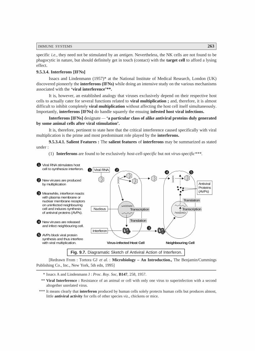
IMMUNE SYSTEMS 263
specific i.e., they need not be stimulated by an antigen. Nevertheless, the NK cells are not found to bephagocytic in nature, but should definitely get in touch (contact) with the target cell to afford a lysingeffect.
9.5.3.4. Interferons [IFNs]
Issacs and Lindenmann (1957)* at the National Institute of Medical Research, London (UK)discovered pioneerly the interferons (IFNs) while doing an intensive study on the various mechanismsassociated with the ‘viral interference’**.
It is, however, an established analogy that viruses exclusively depend on their respective hostcells to actually cater for several functions related to viral multiplication ; and, therefore, it is almostdifficult to inhibit completely viral multiplication without affecting the host cell itself simultaneously.Importantly, interferons [IFNs] do handle squarely the ensuing infested host viral infections.
Interferons [IFNs] designate — ‘a particular class of alike antiviral proteins duly generatedby some animal cells after viral stimulation’.
It is, therefore, pertinent to state here that the critical interference caused specifically with viralmultiplication is the prime and most predominant role played by the interferons.
9.5.3.4.1. Salient Features : The salient features of interferons may be summarized as statedunder :
(1) Interferons are found to be exclusively host-cell-specific but not virus-specific***.
Transcription
Translation
Transcription
Translation
AntiviralProteins(AVPs)
Nucleus
Interferon
Viral RNA
Virus-infected Host Cell Neighbouring Cell
1 2
3
4 5
1 Viral RNA stimulates hostcell to synthesize interferon.
2 New viruses are producedby multiplication
3 Meanwhile, interferon reactswith plasma membrane ornuclear membrane receptorson uninfected neighbouringcell and induces synthesisof antiviral proteins (AVPs).
4 New viruses are releasedand infect neighbouring cell.
5 AVPs block viral proteinsynthesis and thus interferewith viral multiplication.
Fig. 9.7. Diagramatic Sketch of Antiviral Action of Interferon.
[Redrawn From : Tortora GJ et al. : Microbiology – An Introduction., The Benjamin/CummingsPublishing Co., Inc., New York, 5th edn, 1995]
* Issacs A and Lindenmann J : Proc. Roy. Soc, B147, 258, 1957.
** Viral Interference : Resistance of an animal or cell with only one virus to superinfection with a secondaltogether unrelated virus.
*** It means clearly that interferon produced by human cells solely protects human cells but produces almost,little antiviral activity for cells of other species viz., chickens or mice.
264 PHARMACEUTICAL MICROBIOLOGY
(2) Interferon of a particular species is active against a plethora of different viruses.
(3) Not only do various animal species generate interferon variants, but also altogether variouskinds of cells in an animal give rise to interferon variants.
(4) All interferons [IFNs] are invariably small proteins having their molecular weights rangingbetween 15,000 to 30,000. They are observed to be fairly stable at low pH range (acidic),and are quite resistant to heat (thermostable).
(5) Interferons are usually produced by virus-infected host cells exclusively in very smallquantum.
(6) Interferon gets diffused into the uninfected neighbouring cells as illustrated in Fig. 9.7.
Explanation : The various steps involved are as follows :
(1) Interferon happens to interact with plasma or nuclear membrane receptors, including theuninfected cells to produce largely mRNA essentially required for the critical synthesis ofantiviral proteins (AVPs).
(2) In fact, AVPs are enzymes which causes specific disruption in the different stages of viralmultiplication.
Examples : These are as given under :
(a) One particular AVP inducts the inhibition of ‘translation’ of viral mRNA by affordingcomplete blockade in the initiation of the ensuing protein synthesis,
(b) Another AVP causes the inhibition of the phenomenon of ‘polypeptide elongation’,and
(c) Still another AVP takes care of the process of destruction with regard to mRNA before‘translation’.
9.5.3.4.2. Interferon : An Ideal Antiviral Substance : Various cardinal points are as statedbelow :
• Prevailing ‘low concentrations’ at which interferon affords inhibition of viral multiplica-tion are found to be absolutely nontoxic to the uninfected cells.
• Interferon possesses essentially a good number of beneficial characteristic properties.
• Interferon is distinguishably effective for only short span.
• Interferon plays a pivotal and vital role in such critical infections which happen to be quiteacute and transient in nature, for instance : influenza and common colds.
Drawback : Interferon has a serious drawback, as it has practically little effect upon the viralmultiplication in cells that are already infected.
9.5.3.4.3. Interferon Based on Recombinant DNA Technology : In the recent past ‘interferon’has acquired an enormous recognition and importance by virtue of its potential as an antineoplasticagent, and, therefore, enabled its production in a commercial scale globally on a top public-healthpriority. Obviously, the interferons specifically produced by means of the recombinant DNA technologyare usually termed as recombinant interferons [rINFs]. The rINFs have gained an overwhelmingglobal acceptability, popularity, and utility due to two extremely important reasons, namely : (a) highpurity, and (b) abundant availability.
IMMUNE SYSTEMS 265
Usefulness of rINFs : Since 1981, several usefulness of rINFs have been duly demonstratedand observed, such as :
Antineoplastic activity – Large dosage regimens of rINFs may exhibit not so appreciable overalleffects against certain typical neoplasms (tumours), whereas absolute negative effect on others.
However, the scanty results based on the exhaustive clinical trials with regard to the usage ofrINFs towards anticancer profile may be justifiably attributed to the following factual observations,such as :
several variants of interferons vis-a-vis definitive antineoplastic properties,
rINFs in cojunction with other known chemotherapeutic agents might possibly enhancethe overall antineoplastic activity,
quite significant and encouraging results are duly achievable by making use of a combina-tion of :
rINFs + doxorubicin*
or rINFs + cimetidine**
subjects who actually failed to respond reasonably well earlier to either particular chemo-therapy or follow up treatment with interferon distinctly showed remarkable improve-ment when again resorted to the ‘original chemotherapy’.
9.5.3.4.4. Classical Recombinant Interferons [rIFNs] : There are quite a few classicalrecombinant interferons [rIFNs] have been meticulously designed and screened pharmacologically toestablish their enormous usefulness in the therapeutic armamentarium. A few such rIFNs shall now betreated briefly in the sections that follows :
[A] Interferon-ααααα [Syn : Alfa-interferon ; Leukocyte interferon ; Lymphoblastoid interferon ;]
Interferon-ααααα is a glycopeptide produced by a genetic engineering techniques based on the humansequence. It does affect several stages of viral infections, but primarily inhibits the viral-protein trans-lation.
It is invariably employed to prevent and combat the hepatitis B and C infections. In usualpractice the drug is administered either via subcutaneous (SC) route or intramuscular (IM) route.However, it gets rapidly inactivated but generally the overall effects outlast the ensuing plasmaconcentration.
Toxicities – include neurotoxicity, flu-like syndrome, and bone-marrow suppression.
Drug interactions – may ultimately result from its ability to minimize the specific hepatic syn-drome P450-mediated metabolism.
[B] Interferon Alfa-2A, Recombinant [Syn : IFA-ααααα A ; R0-22-8181 ; Canferon ; Laroferon ; Roferon-A ;]
Interferon alfa-2A refers to the recombinant HuIFN-ααααα produced in E. coli, and made up of165 amino acids.
* This particular combination usually employed to treat a wide-spectrum of solid neoplasms or blood cancers.
** In fact, this specific combination invariably used for the treatment of ulcers.
266 PHARMACEUTICAL MICROBIOLOGY
Characteristic Pharmacologic Activities : These are as follows :
(1) Enhances class I histocompatibility molecules strategically located on lymphocytes.
(2) Increases the production of ILs-1 and -2 that critically mediates most of the therapeutic andtoxic effects.
(3) Regulates precisely the antibody responses.
(4) Increases NK cell activities.
(5) Particularly inhibits the neoplasm-cell growth via its distinct ability to inhibit appreciablythe protein synthesis.
(6) Being antiproliferative in nature it may exert its immunosuppressive activity.
(7) Action on the NK cells happens to be the most vital for its antineoplastic action.
(8) Approved for use in hairy-cell leukemia and AIDS-related Kaposi’s sarcoma.
(9) Drug of first choice for the treatment of renal-cell carcinoma.
(10) Preliminary clinical trials ascertained virtually its promising efficacy against quite a fewtypical disease conditions as : ovarian carcinoma, non-Hodgkin’s lymphoma, and meta-static carcinoid tumour.
(11) Besides, it exhibits marked and pronouned antiviral activity against the RNA viruses.
(12) Effective in the treatment of varicella in immunocompromised children, non-A and non-B hepatitis, genital warts, rhinoviral colds, possible opportunistic bacterial infectionsin renal and transplant recipients.
(13) Increases the targetting process associated with monoclonal antibody (MAB)-tetheredcytotoxic drugs to the neoplasm cells.
[C] Interferon Alfa-2B, Recombinant [Syn : IFNααααα2 ; Introna; Intron A ; Viraferon ; Seh-30500 ;YM-14090 ;] ;
The recombinant HuIFNααααα is produced in E. coli.
Therapeutic Applications : are as stated under :
(1) Approved for use in several disease conditions as : hairy-cell leukemia, AIDS-relatedKaposi’s sarcoma, myclogenous leukemia, melanoma, chronic hepatitis, andcondylomata acuminata.
(2) Most of its actions are very much similar to those of rIFN-αααααA.
FURTHER READING REFERENCES
1. Abbas AK et al. : Cellular and Molecular Immunology, WB Saunders, Philadelphia (USA),3rd edn, 1997.
2. Chattergoon M et al. : Genetic Immunization : A New Era in Vaccines and ImmuneTherapies, FASEB J., 11 : 754–60, 1997.
3. Collier RJ and Koplan DA : Immunotoxins, Sci. Am., 251 (2) : 56-64, 1984.
4. Goldsby RA et al. : Kuby Immunology, WH Freeman, New York, 4th edn., 2000.
IMMUNE SYSTEMS 267
5. Johnson HM et al., : ‘How Interferons Fight Disease’, Scientific American, 270 (5) : 68-75, May 1994.
6. Kaufman SH, Sher A, and Fafi A : Immunology of Infectious Diseases, ASM Press,Washington DC, 2001.
7. Old LJ : ‘Tumour Necrosis Factor’, Scientific American, 258 (5) : 59-75, May 1988.
8. Roitt IM et al. : Immunology, CV Mosby, St. Louis, 5th edn, 1998.
9. Roitt IM : Essential Immunology, Blackwell Scientific Publications, Boston (USA), 9thedn, 1997.
10. Rose NR and Afanasyeva M : From Infection to Autoimmunity : The Adjuvant Effect,ASM News, 69 (3) : 132-37, 2003.
11. Ross GD : Immunology of the Complement System : An Introduction for Research andClinical Medicine, Academic Press, New York, 1986.
12. Science (Special Issue) : Elements of Immunity, Science, 272 : (5258) : 50–79, 1996.
268
MICROBIOLOGICAL (MICROBIAL)ASSAYS : ANTIBIOTICS–VITAMINS–AMINO ACIDS
10.1. INTRODUCTION
There are, in fact, three most critical and highly explicite situations, wherein the absolute neces-sity to assay the ‘antimicrobial agents’ arise, namely :
(a) Production i.e., in the course of commercial large-scale production for estimating the ‘po-tency’ and stringent ‘quality control’,
(b) Pharmacokinetics i.e., in determining the pharmacokinetics* of a ‘drug substance’ inhumans or animals, and
(c) Antimicrobial chemotherapy i.e., for strictly managing, controlling, and monitoring theensuing antimicrobial chemotherapy**.
Summararily, the very ‘first’ situation i.e., (a) above, essentially involves the assay of relativelyhigh concentration of ‘pure drug substance’ in a more or less an uncomplicated solution, forinstance : buffer solution and water. In addition to the ‘second’ and ‘third’ i.e., (b) and (c) above,critically involve the precise and accurate measurement at relatively low concentration of the ‘drugsubstance’ present in biological fluids, namely : serum, sputum, urine, cerebrospinal fluid (CSF), gas-tric juice, nasal secretions, vaginal discharges etc. Nevertheless, these biological fluids by virtue of theirinherent nature invariably comprise of a plethora of ‘extranaceous materials’ which may overtly andcovertly interfere with the assay of antibiotics.
* Pharmacokinetics : It refers to the study of the ‘quantitative relationships’ of the rates of drug adsorption,distribution, metabolism, and elimination (ADME) processes ; data used to establish dosage regimen andfrequency for desired therapeutic response.
** Chemotherapy : The therapeutic concept developed by Paul Ehrlich (1854–1915) whereby a specific drugor chemical is invariably employed to treat an ensuing infectious disease or cancer ; ideally, the chemicalmust destroy the pathogens completely without harming the host.
10• Introduction• Variants in Assay Profile• Types of Microbiological (Microbial) Assays• Radioenzymatic [Transferase] Assays• Analytical Methods for Microbial Assays• Examples of Pharmaceutical Microbial Assays
• Assay of Antibiotics by Turbidimetric (or Nephelometric) Methods
MICROBIOLOGICAL (MICROBIAL) ASSAYS : ANTIBIOTICS–VITAMINS–AMINO ACIDS 269
10.1.1. Importance and Usefulness
The actual inhibition of the observed microbial growth under stringent standardized experimen-tal parameters may be judiciously utilized and adequately exploited for demonstrating as well as estab-lishing the therapeutic efficacy of antibiotics.
It is, however, pertinent to state here that even the slightest and subtle change duly incorporatedin the design of the antibiotic molecule may not be explicitely detected by the host of usual ‘chemicalmethods’, but will be revealed by a vivid and clear-cut change in the observed ‘antimicrobial activity’.Therefore, the so called microbiological assays do play a great useful role for ascertaining and resolv-ing the least possible doubt(s) with respect to the change in potency of antibiotics and their respectiveformulations i.e., secondary pharmaceutical products.
10.1.2. Principle
The underlying principle of microbiological assay is an elaborated comparison of the ‘inhibi-tion of growth’ of the microbes by a measured concentration of the antibiotics under investigationagainst that produced by the known concentrations of a ‘standard preparation of antibiotic’ with aknown activity.
10.1.3. Methodologies
In usual practice, two ‘general methods’ are employed extensively, such as :(a) Cylinder-plate (or Cup-plate) Method, and(b) Turbidimetric (or Tube-assay) Method.
Each of the two aforesaid methods shall now be discussed briefly in the sections that follows :
10.1.3.1. Cylinder-Plate Method (Method-A)
The cylinder-plate method solely depends upon the diffusion of the antibiotic from a verticalcylinder via a solidified agar layer in a Petri-dish or plate to an extent such that the observed growth ofthe incorporated microorganism is prevented totally in a zone just around the cylinder containing asolution of the ‘antibiotic’.
10.1.3.2. Turbidimetric (or Tube-Assay) Method (Method-B)
The turbidimetric method exclusively depends upon the inhibition of growth of a ‘microbialculture’ in a particular uniform solution of the antibiotic in a fluid medium which is quite favourableand congenial to its rather rapid growth in the absence of the ‘antibiotic’.
Conditionalities : The various conditionalities required for the genuine assay may be designedin such a manner that the ‘mathematical model’ upon which the potency equation is entirely basedcan be established to be valid in all respects.
Examples : The various typical examples are as stated under :
(a) Parallel-Line Model — If one happens to choose the parallel-line model, the two log-dose-response lines of the preparation under investigation and the standard preparationmust be parallel, i.e., they should be rectilinear over the range of doses employed in thecalculation. However, these experimental parameters need to be critically verified by thevalidity tests referred to a given probability.
(b) Slope-Ratio Method : It is also feasible to make use of other mathematical models, forinstance : the ‘slope-ratio method’ provided that proof of validity is adequately demonstrated.

270 PHARMACEUTICAL MICROBIOLOGY
10.1.4. Present Status of Assay Methods
Based on the copious volume of evidences cited in the literatures it may be observed that the‘traditional antimicrobial agents’ have been duly determined by microbiological assay procedures.Importantly, in the recent past significant greater awareness of the various problems of poor assayresults specificity associated with such typical examples as :
partially metabolized drugs,
presence of other antibiotics, and
urgent need for more rapid/reproducible/reliable analytical techniques ;
has appreciably gained ground and equally encouraged the judicious investigation of a host of otherfairly accurate and precise methodologies, namely :
Enzymatic assays,
Immunological assays,
Chromatographic assays, including :
—High Performance Liquid Chromatography (HPLC)
—Reverse-Phase Chromatography (RPC)
—Ion-Pair Chromatography (IPC)
This chapter will cover briefly the underlying principles of these aforesaid techniques.
10.2. VARIANTS IN ASSAY PROFILE
There are several well-recognized variants in assay profile for antibiotics, vitamins, and aminoacids, namely :
(a) Calibration of assay,
(b) Precision of assay,
(c) Accuracy of assay, and
(d) Evaluation of assay performance.
The various aspects of assay profile stated above shall now be treated briefly in the sections thatfollows :
10.2.1. Calibration of Assay
Irrespective of the method adopted for the microbial assay it is absolutely necessary to work outa proper calibration in case the ultimate result is necessarily expected in terms of the absolute units viz.,mg.L– 1.
Calibrator Solutions — The calibrator solutions are essentially prepared either from a puresample of the drug to be assayed or a sample of known potency.
Importantly, there are certain drug substances that are hygroscopic in nature ; and, therefore,their inherent potency may be expressed as :
MICROBIOLOGICAL (MICROBIAL) ASSAYS : ANTIBIOTICS–VITAMINS–AMINO ACIDS 271
(a) ‘as-is’ potency — which refers to — ‘the potency of the powder without drying’,* and
(b) ‘dried potency’ — which refers to — ‘the potency after drying to constant weight underspecified/defined experimental parameters’.
Importantly, in as-is potency, the drug should be stored in such a manner that it may not lose orabsorb water ; whereas, in dried potency the drug should always be dried first before weighing.
Thus, once an appropriate ‘standard materials’ is actually accomplished, the calibrator solu-tions** usually covering a suitable range of concentrations should be prepared accordingly. However,the actual number and concentration range of the collaborators shall solely depend on the specifictype of assay being carried out. Likewise, the matrix*** wherein the calibrators are dissolved duly isalso quite vital and important, unless it may be shown otherwise, must be very much akin to the respec-tive matrix of the samples.
Note : (1) It should be absolutely important when carying out the assay of drugs present in ‘serum’, dueto the fact that protein-binding may invariably influence the ultimate results of microbio-logical assay predominantly.
(2) No assay can give rise to fairly accurate results unless and until the suitable ‘calibrator solu-tions’ (i.e., calibrators) precisely prepared in an appropriate matrix.
10.2.2. Precision of Assay
Precision refers to – ‘agreement amongst the repeated measurements’.
Alternatively, precision is an exact measure of reproducibility, and is duly estimated by replicat-ing a single sample a number of times thereby determining :
mean result ( )X ,
standard deviation (SD), and
coefficient of variation (SD/ X × 100).
Intra-Assay Precision—usually refers to the precision within a single-run exclusively.
Inter-Assay Precision—normally refers to the precision between two or more runs.
Degree of Precision—required in a specific instance essentially will determine two cardinal factors,namely :
number replicates actually needed for each calibrator, and
number plus concentration range of calibrators.
Note : Importantly, the overall precision of several assays usually changes with concentration ; andtherefore, must be assayed with low, medium, and high concentration samples.
* Assuming it has been stored properly so that moisture (water) is neither gained nor lost on storage.
** i.e., calibrators.
*** Matrix—The fluid wherein the ‘drug to be assayed’ is dissolved is invariably termed as the matrix.
272 PHARMACEUTICAL MICROBIOLOGY
10.2.3. Accuracy of Assay
Accuracy may be defined as — ‘a measure of the correctness of data as these correspond tothe true value’.
Considering that the calibrator solutions were prepared correctly from the suitable ‘drug’, theresulting accuracy of a specific result shall exclusively depend upon two important aspects, namely :
precision of assay, and
specificity of assay.
Poor Specificity is encountered usually in the following three instances, such as :
samples comprising of endogenous interfering materials,
presence of other antibacterial agents, and
active metabolites of the ‘drug’ being assayed.
Positive Bias i.e., if the other drugs or drug metabolites are present simultaneously, accuracyof assay shall be expressed predominantly as a positive bias.*
Negative Bias i.e., if there are antagonists present in an appreciable quantum, accuracy of assaywill be expressed mostly as a negative bias.
Note : In fact, inaccuracy caused due to apparent poor precision will invariably exhibit absolutely‘no bias’, and that caused on account of either under–or over-potent calibrators will exhibitpositive and negative bias respectively.
10.2.4. Evaluation of Assay Performance
It has been duly proved and established that while assessing the performance characteristics of analtogether newly developed assay, both intra–and inter–assay precision duly spread over the entirerange of expected concentrations must be estimated precisely.
Important Points : These are as stated under :
(1) It is extremely important to check the accuracy with the help of the ‘spiked samples’** verymuch spread over the entire range of concentrations used in the assay.
(2) Assaying ‘drug substances’ in biological fluids e.g., urine, blood, serum, sputum, cerebrospinalfluid (CSF) etc.
(3) Samples withdrawn from individual subjects who have been duly administered with the drugeither enterally*** or parenterally**** by virtue of the fact that in vitro metabolites may only beapparent in these instances.
* i.e., the ultimate results are higher than expected.
** Spiked Samples : Samples with known concentrations.
*** Enterally i.e., adminstered within the intestinal tract (between mouth and rectum).
**** Parenterally i.e., administered via IV and IM routes.
MICROBIOLOGICAL (MICROBIAL) ASSAYS : ANTIBIOTICS–VITAMINS–AMINO ACIDS 273
(4) Such substances that might have an inherent tendency to interfere in the assay should bethoroughly checked for there possible interference either alone or in the presence of the ‘drug sub-stance’ being assayed.
(5) In an ideal situation, preferentially a relatively large number of samples must be assayedboth by the ‘new method’ and the ‘reference method’ individually, and the subsequent results ob-tained may be meticulously by linear regression ; and thus the ensuing correlation coefficient of thesaid two methods determined.
(6) Routinely employed methods may be tackled with ‘internal controls’* almost in every run ;and, therefore, the laboratories that are actively engaged in the assay of clinical specimens must takepart in an external quality control programme religiously.
10.3. TYPES OF MICROBIOLOGICAL (MICROBIAL) ASSAYS
There are mainly two different types of microbiological assays usually encountered bearing inmind the response of an ever-growing population of microbes vis-a-vis ascertaining the profile ofantimicrobial agent measurements, such as :
(a) Agar Plate diffusion assays, and
(b) Rapid-reliable-reproducible microbial assay methods.
Each of the two aforesaid types of microbiological assays will now be discussed individually inthe sections that follows :
10.3.1. Agar Plate Diffusion Assays (Method-A)
In the agar-plate diffusion assays the ‘drug substance’ gets slowly diffused into agar seededduly with a susceptible microbial population. Subsequently, it gives rise to a ‘specific zone of growthinhibition’. However, the agar-plate diffusion assay may be one-, two- or three-dimensional (i.e.,1D, 2D or 3D).
All these three different types shall now be discussed briefly in the sections that follows :
10.3.1.1. One-Dimensional Assay
In this particular assay the capillary tubes consisting of agar adequately seeded with ‘indicatororganism’ are carefully overlaid with the ‘drug substance’. The drug substance e.g., an antibioticnormally gets diffused downwards into the agar thereby giving rise to the formation of a ‘zone of inhi-bition’. However, this specific technique is more or less obsolete now-a-days.
Merits : There are three points of merits, such as :
perfectly applicable for the assay of antibiotics anaerobically,
may efficiently take care of very small samples, and
exhibits an appreciable precision,
Demerit : It essentially has a critical demerit with regard to the difficulty in setting up andsubsequent standardization.
* Internal Controls i.e., samples having known value.
274 PHARMACEUTICAL MICROBIOLOGY
10.3.1.2. 2D- or 3D-Assay
As to date, the 2D- or 3D-assay methods represent the commonest and widely accepted form ofthe microbiological assay. Nevertheless, in this particular instance the samples need to be assayed areadequately applied in a certain specific type of reservoir viz., cup, filter-paper disc, or well, to a thin-layer of agar previously seeded with an indicator microorganism aseptically in a Laminar Air FlowBench. In this way, the ‘drug substance’ gets gradually diffused into the medium, and after suitableincubation at 37°C for 48–72 hrs. in an ‘incubation chamber’, a clear cut distinctly visible zone ofgrowth inhibition comes into being*. However, the diameter of the zone of inhibition very muchremains within limits, provided that all other factors being constant, and the same is associated with theconcentration of the antibiotic present in the reservoir.**
10.3.1.3. Dynamics of Zone Formation
It has been duly observed that during the process of incubation the antibiotic gets diffused fromthe reservoir. Besides, a proportion of the bacterial population is moved away emphatically from theinfluence of the antibiotic due to cell-division.
Important Observations : Following are some of the important observations, namely :
(1) Edge of a zone is usually obtained in a situation when the minimum concentration of theantibiotic that will effectively cause the inhibition in the actual growth of the organism on the agar-plate(i.e., critical concentration accomplished) attains, for the very first time, a specific population densitywhich happens to be excessively too big in dimension and quantum for it to inhibit effectively.
(2) The precise and exact strategic position of the zone-edge is subsequently determined bymeans of the following three vital factors, such as :
initial population density,
rate of diffusion of ‘antibiotic’, and
rate of growth of ‘organism’.
(3) Critical Concentration (C′′′′′) : The critical concentration (C′′′′′) strategically located at theedge of a ‘zone of inhibition’ and formed duly may be calculated by the following expression :
In C′ = 2In
4DCd
To
where, C = Concentration of drug in Reservoir,
d = Distance between Reservoir and zone-edge,
D = Diffusion coefficient***, and
To = Critical time at which the position of zone-edge was determined critically.
Graphical Representation : It is feasible and possible to have a ‘graphical representation’ toobtain a zone of inhibition in different ways, for instance :
* In this instance – a circle around the reservoir can be observed.
** Reservoir : Cup, filter-paper disc (soaked with ‘antibiotic’ soln., or well.
*** Constant for a given ‘antibiotic’ in a given ‘matrix’ being diffused into a given medium at a given tem-perature.

MICROBIOLOGICAL (MICROBIAL) ASSAYS : ANTIBIOTICS–VITAMINS–AMINO ACIDS 275
(1) An assay wherein the value of To and D happen to be constant, an usual plot of In C Vs d2 fora definite range of concentrations shall, within certain limits, produce a ‘straight line’ that may beconveniently extrapolated to estimate C′′′′′ i.e., critical concentration.
(2) In fact, C′′′′′ duly designates the obvious minimum value of C that would yield a specific zoneof inhibition. Evidently, it is absolutely independent of D and To.
(3) However, the resulting values of D and To may be manipulated judiciously to lower or en-hance the dimensions of zone based on the fact that the concentrations of C is always greater than C′′′′′.i.e., the concentration of ‘drug’ in reservoir > critical concentration of the ‘drug’.
(4) Pre-incubation would certainly enhance the prevailing number and quantum of microbespresent actually on the agar-plate ; and, therefore, the critical population density shall be duly accom-plished rather more rapidly (i.e., To gets reduced accordingly) thereby reducing the observed zones ofinhibition.
(5) Minimizing the particular microbial growth rate suitably shall ultimately give rise to rela-tively ‘larger zones of inhibition’.
(6) Carefully enhancing either the sample size or lowering the thickness of agar-layer willcritically increase the zone size and vice-versa.
(7) Pre-requistes of an Assay—While designing an assay, the following experimental param-eters may be strictly adhered to in order to obtain an optimized appropriately significant fairly largerange of zone dimensions spread over duly the desired range of four antibiotic concentrations, suchas :
proper choice of ‘indicator organism’,
suitable culture medium,
appropriate sample size, and
exact incubation temperature.
10.3.1.4. Management and Control of Reproducibility
As the observed dimensions of the zone of inhibition depend exclusively upon a plethora ofvariables*, as discussed above, one should meticulously take great and adequate precautionary meas-ures not only to standardise time, but also to accomplish reasonably desired good precision.
Methodologies : The various steps involved in the management and control of reproducibil-ity are as stated under :
(1) A large-size flat-bottomed plate [either 30 × 30 cm or 25 × 25 cm] must be employed, andshould be meticulously levelled before the agar is actually poured.
(2) Explicite effects of variations in the ‘composition of agar’ are adequately reduced by pre-paring, and making use of aliquots of large batches.
(3) Inoculum dimension variants with respect to the ‘indicator organisms’ may be minimizedproportionately by duly growing a reasonably large volume of the organism by the following two waysand means, such as :
* Variables e.g., sample size, agar-thickness, indicator organism, population density, organism growth rate,and drug-diffusion rate.

276 PHARMACEUTICAL MICROBIOLOGY
dispensing it accordingly into the aliquots just enough for a single agar plate, and
storing them under liquid N2 so as to preserve its viability effectively.
(4) In the specific instance when one makes use of the ‘spore inocula’, the same may be ad-equately stored for even longer durations under the following two experimental parameters, forinstance :
absolute inhibition of germination, and
effective preservation of viability.
(5) It is a common practice to ensure the ‘simultaneous dosing’ of both calibrators and sam-ples onto a single-agar plate. In this manner, it is possible and feasible to achieve the following threecardinal objectives :
thickness of the agar-plate variants,
critical edge-effects, and
incubation temperature variants caused on account of irregular warming inside the ‘incuba-tor’ must be reduced to bare minimum by employing some sort of ‘predetermined random layout’.
(6) ‘Random Patterns’ for Application in Microbiological Plate Assay : In usual practice, wefrequently come across two prevalent types ‘random patterns’ for application in the microbiologicalplate assay, namely :
(a) Latin-Square Arrangement – in this particular case the number of replicates almost equalsthe number of specimens (samples) ; and the ultimate result ensures the maximum preci-sion, as shown in Fig. 10.1(a).
(b) Less Acceptable (Demanding) Methods – employing rather fewer replicates are invariablyacceptable for two vital and important purposes, such as :
clinical assays, and
pharmacokinetic studies,
as illustrated in Figs. 10.1(b) and (c).
3 4 8 1 7 6 5 2
2 7 3 8 5 1 4 6
8 1 5 2 4 3 6 7
4 2 1 6 3 5 7 8
7 8 2 5 6 4 3 1
6 5 7 3 8 2 1 4
1 3 6 4 2 7 8 5
5 6 4 7 1 8 2 3
5 1 3 7 14 10 12 16
8 4 2 6 9 13 15 11
14 7 5 16 12 1 3 10
2 11 9 4 15 6 8 13
3 10 12 6 5 2 14 7
15 6 8 13 2 11 9 4
12 16 14 10 3 7 5 1
9 13 15 11 8 4 2 6
(a) Latin Square (b) 16 Doses [Calibrators/Samples
in Quadruplicate]
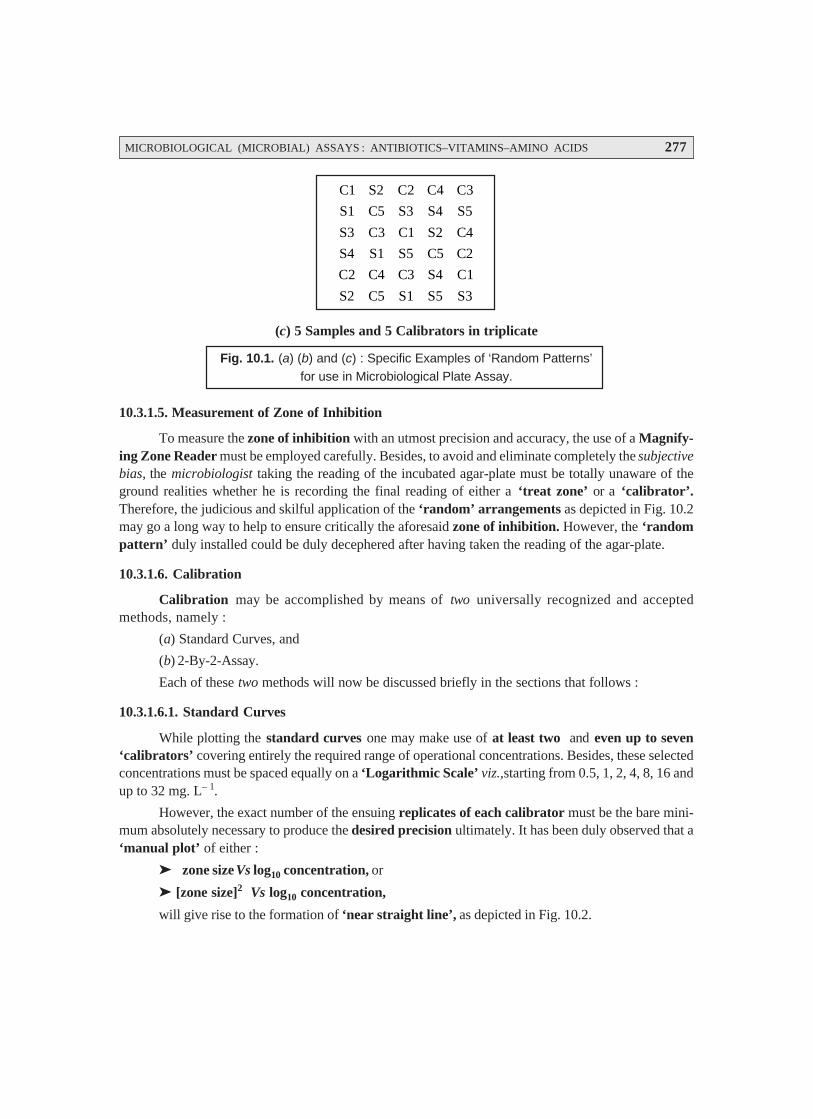
MICROBIOLOGICAL (MICROBIAL) ASSAYS : ANTIBIOTICS–VITAMINS–AMINO ACIDS 277
C1 S2 C2 C4 C3
S1 C5 S3 S4 S5
S3 C3 C1 S2 C4
S4 S1 S5 C5 C2
C2 C4 C3 S4 C1
S2 C5 S1 S5 S3
(c) 5 Samples and 5 Calibrators in triplicate
Fig. 10.1. (a) (b) and (c) : Specific Examples of ‘Random Patterns’for use in Microbiological Plate Assay.
10.3.1.5. Measurement of Zone of Inhibition
To measure the zone of inhibition with an utmost precision and accuracy, the use of a Magnify-ing Zone Reader must be employed carefully. Besides, to avoid and eliminate completely the subjectivebias, the microbiologist taking the reading of the incubated agar-plate must be totally unaware of theground realities whether he is recording the final reading of either a ‘treat zone’ or a ‘calibrator’.Therefore, the judicious and skilful application of the ‘random’ arrangements as depicted in Fig. 10.2may go a long way to help to ensure critically the aforesaid zone of inhibition. However, the ‘randompattern’ duly installed could be duly decephered after having taken the reading of the agar-plate.
10.3.1.6. Calibration
Calibration may be accomplished by means of two universally recognized and acceptedmethods, namely :
(a) Standard Curves, and
(b) 2-By-2-Assay.
Each of these two methods will now be discussed briefly in the sections that follows :
10.3.1.6.1. Standard Curves
While plotting the standard curves one may make use of at least two and even up to seven‘calibrators’ covering entirely the required range of operational concentrations. Besides, these selectedconcentrations must be spaced equally on a ‘Logarithmic Scale’ viz.,starting from 0.5, 1, 2, 4, 8, 16 andup to 32 mg. L– 1.
However, the exact number of the ensuing replicates of each calibrator must be the bare mini-mum absolutely necessary to produce the desired precision ultimately. It has been duly observed that a‘manual plot’ of either :
zone size Vs log10 concentration, or
[zone size]2 Vs log10 concentration,
will give rise to the formation of ‘near straight line’, as depicted in Fig. 10.2.

278 PHARMACEUTICAL MICROBIOLOGY
Log Antibiotic Concentration10O
Zon
e D
iam
eter
(m
m)
Fig. 10.2. Standard Curve for an Agar-Plate Diffusion Assay
Note : A microcomputer may by readily installed and programmed to derandomise the realistic andactual zone pattern by adopting three steps in a sequetial manner viz., (a) consider the mean ofthe ‘zone sizes’ ; (b) compute the standard curve ; and (c) calcuate the ultimate results for thetests ; and thereby enabling the ‘zone sizes’ to be read almost directly from the incubatedagar-plate right into the computer.
10.3.1.6.2. 2-By-2-Assay
The 2-by-2-assay is particularly suitable for estimating the exact and precise potency of a plethoraof ‘Pharmaceutical Formulations’. In this method a relatively high degree of precision is very muchrequired, followed by another two critical aspects may be duly taken into consideration, such as :
Latin square design with tests, and
Calibrators at 2/3 levels of concentration.
Example : An 8 × 8 Latin square may be employed gainfully in two different ways :
First— to assay 3 samples + 1 calibrator, and
Second— to assay 2 samples + 2 calibrators,
invariably at two distinct levels of concentrations* each, and having a ‘coefficient of variation’ atabout 3%.
Evidently, based on this technique, one may obtain easily and conveniently the ‘parallel dose–response lines’ strategically required for the calibrators vis-a-vis the tests performed at two distinctdilutions, as depicted in Fig. 10.3. Importantly, it is quite feasible and possible to establish the exact andprecise potency of samples may be computed effectively or estimated from meticulously derivednomograms.**
* Usually spread over a two- or four-fold range.
** Nomogram : Graphic representation comprising of several lines marked off to a scale, arranged in such away that by using a straight edge to connect known values on two lines an unknown value may be read at thepoint of intersection with a third line ; used to determine drug doses for specific persons.
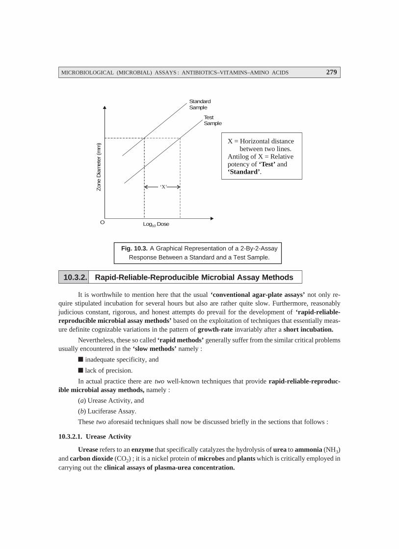
MICROBIOLOGICAL (MICROBIAL) ASSAYS : ANTIBIOTICS–VITAMINS–AMINO ACIDS 279
‘X’
O Log Dose10
StandardSample
TestSample
Zon
e D
iam
eter
(m
m) X = Horizontal distance
between two lines.Antilog of X = Relativepotency of and
.‘Test’
‘Standard’
Fig. 10.3. A Graphical Representation of a 2-By-2-AssayResponse Between a Standard and a Test Sample.
10.3.2. Rapid-Reliable-Reproducible Microbial Assay Methods
It is worthwhile to mention here that the usual ‘conventional agar-plate assays’ not only re-quire stipulated incubation for several hours but also are rather quite slow. Furthermore, reasonablyjudicious constant, rigorous, and honest attempts do prevail for the development of ‘rapid-reliable-reproducible microbial assay methods’ based on the exploitation of techniques that essentially meas-ure definite cognizable variations in the pattern of growth-rate invariably after a short incubation.
Nevertheless, these so called ‘rapid methods’ generally suffer from the similar critical problemsusually encountered in the ‘slow methods’ namely :
inadequate specificity, and
lack of precision.
In actual practice there are two well-known techniques that provide rapid-reliable-reproduc-ible microbial assay methods, namely :
(a) Urease Activity, and
(b) Luciferase Assay.
These two aforesaid techniques shall now be discussed briefly in the sections that follows :
10.3.2.1. Urease Activity
Urease refers to an enzyme that specifically catalyzes the hydrolysis of urea to ammonia (NH3)and carbon dioxide (CO2) ; it is a nickel protein of microbes and plants which is critically employed incarrying out the clinical assays of plasma-urea concentration.

280 PHARMACEUTICAL MICROBIOLOGY
Importanlty, the microorganism Proteius mirabilis grows significanlty in a urea-containingculture medium, whereupon it particularly causes the hydrolysis of urea to ammonia, and thereby helpsto raise the pH of the medium. However, the actual production of urease is reasonably inhibited by theso called ‘aminoglycoside antibiotics’,* such as : amikacin, gentamicin, kanamycin, neomycin,netilmicin, tobramycin, doxorubicin, cephalosporins, cephamycius, thienamycin, lincomycin,clindamycin, erythromycin, clarithromycin, azithromycin, oleandomycin, spramycins, and the like.
Methodology : The various steps involved are as follows :
(1) Assay is performed with two series of tubes of urea-containing culture medium that havebeen duly incorporated with a range of calibrator solutions.
(2) First series of tubes in duly added a certain volume of the sample which is essentiallyequivalent to the volume of the calibrator.
(3) Second series of tubes is duly added exactly half the volume of the sample.
(4) Both ‘set of tubes’ are subsequently inoculated with P. mirabilis, and duly incubated for aduration of 60–70 minutes.
(5) pH of the resulting solution is measured accurately upto 0.01 pH units.
(6) In fact, it is possible to obtain two distinct ‘calibration curves’ by plotting pH Vs log10 i.e.,the ensuing calibrator concentration for each of the two series.
(7) The ‘vertical distance’ existing between the two curves is found to be almost equal to thelegarithm of 1/2 the concentration of ‘drug substance’ present in the sample.
Note : (1) In usual practice, it is rather difficult to obtain ‘reliable’ results by adopting the ‘UreaseActivity’ method.
(2) A standardized, senstitive, and reliable pH Meter is an absolute must for this particularassay.
10.3.2.2. Luciferase Assay
In the specific ‘Luciferase Assay’, the firefly luciferase** is made use of for the actual meas-urement of small quantum of ATP*** duly present in a microbial culture, whereby the levels of ATP getproportionately reduced by the ensuing action of the aminoglycoside antibiotics (see Section 10.3.2.1).
Methodology : The various steps involved in the ‘Luciferase Assay’ are as enumerated undersequentially :
(1) Both test solutions (i.e., after preliminary heating provided the matrix is serum) along withcalibrators are carefully added into the various tubes of the culture medium specifically containing agrowing microbial culture (i.e., organism).
* Kar, A : ‘Pharmacognosy and Pharmacobiotechnology’, New Age International Pvt. Ltd. Publishers,New Delhi, 2nd edn., 2007.
** Luciferase : An ‘enzyme’ that critically acts on luciferine (i.e., substances present in some organisms,which become luminescent on being acted upon by luciferase) to oxidize them and eventually causebioluminescence. It is present in certain organisms (e.g., fireflies, other insects) that emit light either inter-mittently or continuously.
*** ATP : Adenosine Triphosphate (an enzyme).
MICROBIOLOGICAL (MICROBIAL) ASSAYS : ANTIBIOTICS–VITAMINS–AMINO ACIDS 281
(2) After adequate incubation for a 90 minute duration the cultures are duly treated with ‘apyrase’so as to ensure the complete destruction of the extracellular ATP.
(3) The resulting solution is duly extracted with EDTA/sulphuric acid, and thus the intracellularATP critically assayed with the firefly enzyme using a ‘Luminometer’.
(4) Finally, a ‘calibration curve’ is constructed meticulously by plotting the two vital compo-nents, namely : (a) intracellular ATP content, and (b) log10 i.e., the calibrator concentration.
Note : As to date, the ‘Luciferase Assay’ has not yet accomplished a wide application ; however, itmay find its enormous usage in the near future with the advent of such ‘luciferase formula-tions’ that would turn out to be even much more active, reliable, and dependable.
10.4. RADIOENZYMATIC [TRANSFERASE] ASSAYS
The ‘radioenzymatic assays’ have gained their abundant acceptance and recognition for theassay of aminoglycoside antibiotics e.g., amikacin, gentamicin, kanamycin, neomycin, netilmicin,tobramycin, doxorubicin, cephalosporins, cephamycins, thienamycin, lincomycin, clindamycin, eryth-romycin, clarithromycin, azithromycin, oleandomycin, spramycins etc ; and chloramphenicol (orChloromycetine). Importantly, the radioenzymatic assays are exclusively based upon the fact that theprevailing inherent microbial resistance to the said aminoglycoside antibiotics and chloramphenicolis predominantly associated with the specific as well as the critical presence of certain highly specializedenzymes* that particularly render the ‘antibiotics’ absolutely inactive via such biochemical means as :acetylation, adenylation, and phosphorylation.
It has been duly proved and established that :
aminoglycoside antibiotics—are susceptible to prominent attack by these critical and
specific enzymes as :
Aminoglycoside acetyltransferases (AAC) ;Aminoglycoside adenylyltransferases (AAD) ;Aminoglycoside phosphotransferases (APH).
Chloramphenicol—is prone to predominant attack by the enzyme :
Chloramphenicol acetyl transferases (CAT).
Mechanism of Action : The mechanism of action of these enzymes viz, AAC, AAD, and APHare not the same :
Acetyltransferases [i.e., AAC]—invariably attack the most susceptible amino moieties (–NH2),and to accomplish this critical function may require acetyl coenzyme A (AcCoA).
Adenylyltransferases [i.e., AAD] and Phosphotransferases [i.e., APH]—these enzymes usu-ally attack the most susceptible hydroxyl moieties (–OH), and specifically requires adenosinetriphosphate [ATP] i.e., another nucleotide triphosphate.
Applications : As to date quite a few AAC and AAD enzymes have been judiciously employedfor the radioenzymatic assays.
Example : Both the enzyme and the suitable radiolabelled cofactor [1 – 14C]** acetyl coenzymeA, or [2 – 3H]*** ATP are used frequently in order to specifically radiolabel the ‘drug substance’ underinvestigation.
* Quite often coded for by the transmissible plasmids.
** 14C : Radiolabelled carbon atom i.e., an isotope of carbon.
*** 3H : Radiolabelled hydrogen atom, also known as ‘Tritium’.
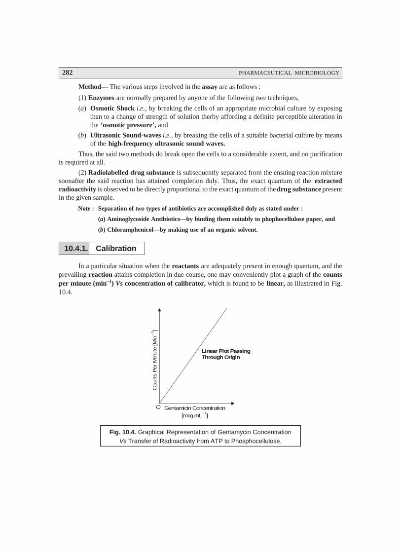
282 PHARMACEUTICAL MICROBIOLOGY
Method— The various steps involved in the assay are as follows :
(1) Enzymes are normally prepared by anyone of the following two techniques,
(a) Osmotic Shock i.e., by breaking the cells of an appropriate microbial culture by exposingthan to a change of strength of solution therby affording a definite perceptible alteration inthe ‘osmotic pressure’, and
(b) Ultrasonic Sound-waves i.e., by breaking the cells of a suitable bacterial culture by meansof the high-frequency ultrasonic sound waves.
Thus, the said two methods do break open the cells to a considerable extent, and no purificationis required at all.
(2) Radiolabelled drug substance is subsequently separated from the ensuing reaction mixturesoonafter the said reaction has attained completion duly. Thus, the exact quantum of the extractedradioactivity is observed to be directly proportional to the exact quantum of the drug substance presentin the given sample.
Note : Separation of two types of antibiotics are accomplished duly as stated under :
(a) Aminoglycoside Antibiotics—by binding them suitably to phophocellulose paper, and
(b) Chloramphenicol—by making use of an organic solvent.
10.4.1. Calibration
In a particular situation when the reactants are adequately present in enough quantum, and theprevailing reaction attains completion in due course, one may conveniently plot a graph of the countsper minute (min–1) Vs concentration of calibrator, which is found to be linear, as illustrated in Fig.10.4.
Gentamicin Concentration
[mcg.mL ]– 1
O
Cou
nts
Per
Min
ute
[Min
]–
1
Linear Plot PassingThrough Origin
Fig. 10.4. Graphical Representation of Gentamycin ConcentrationVs Transfer of Radioactivity from ATP to Phosphocellulose.

MICROBIOLOGICAL (MICROBIAL) ASSAYS : ANTIBIOTICS–VITAMINS–AMINO ACIDS 283
10.4.2. Non-Isotopic Modification
The calibration accomplished by using the radiolabelled drug essentially needs either a Gei-ger Müller Counter or a Scintillation Counter, formeasuring the ensuing radio activity (in mC) of the ra-dioactive chemicals, which being an enormously ex-pensive equipment, and a skilled technician. There-fore, in order to circumvent these glaring untoward se-rious problems one may adopt a photometric varia-tion of the aminoglycoside acetyltransferases [AAC] assay meticulously. For this the sulphydry rea-gent viz., 5, 5′′′′′-dithiobis (2-nitrobenzoic acid) is incorporated carefully into the on-going assay-sys-tem. Thus, the said reagent specifically interacts with the corresponding coenzyme A (reduced form)duly generated thereby producing a distinct yellow-coloured product that may be quantitatively as-sayed by using a previously standardized UV-Visible Spectrophotometer.
(a) Reactions : The two reactions are as follows :
(b) Aminoglycoside + Acetyl CoA —→ Acetyl – Aminoglycoside + CoASH
CoASM + DTNB —→ Yellow Product
10.5. ANALYTICAL METHODS FOR MICROBIAL ASSAYS
There are several sophisticated analytical methods that are used most abundantly for the precisequantitative methods microbial assays, such as :
(a) High Performance Liquid Chromatography (HPLC),
(b) Reverse-Phase Chromatography (RPC), and
(c) Ion–Pair (or Paired-Ion) Chromatography,
These three chromatographic techniques shall now be discussed briefly in the sections that fol-lows :
10.5.1. High Performance Liquid Chromatography [HPLC]
Preamble : Giddings* (1964) rightly predicted that the careful and meticulous application of
relatively ‘small particulate matter’ under the influence of excessively enhanced flow pressure could
definitely improve upon the performance of ‘Liquid Chromatography’ significantly ; and ultimatelyone could easily, accomplish an appreciably high number of ‘theoretical plate numbers’. Towards the
later half of 1960s world’s two eminent scientists, Horvath and Lipsky at Yale University (USA), came
forward with the first ever HPLC, and named it as ‘high pressure liquid chromatography’. Neverthe-less, the early 1970s the world witnessed the ever glorious technological supremacy by producing and
* Giddings JC : Anal. Chem., 36 : 1890, 1964.
O N2
HOOC S—S6
3
42
1 5COOH
6′
3′
4′ 2′
1′5′
5, 5 –Dithiobis(2-nitrobenzoic acid) [DTNB]
′
NO2
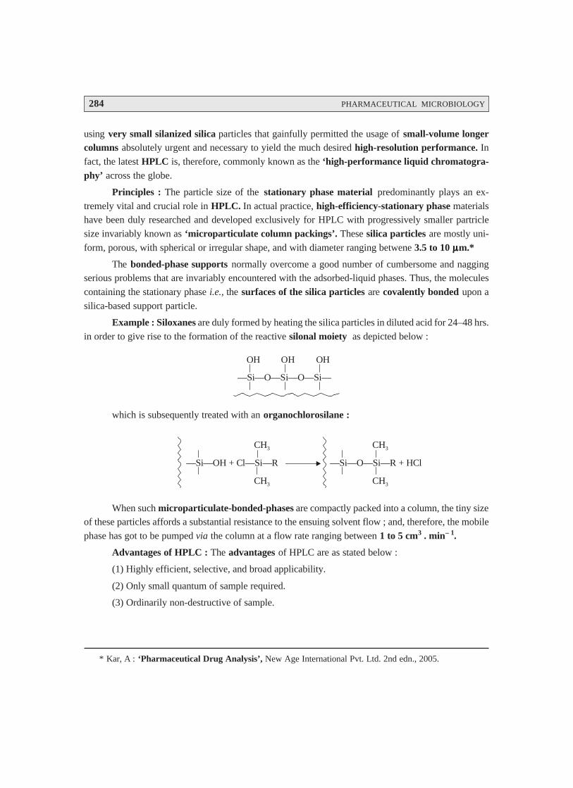
284 PHARMACEUTICAL MICROBIOLOGY
using very small silanized silica particles that gainfully permitted the usage of small-volume longercolumns absolutely urgent and necessary to yield the much desired high-resolution performance. Infact, the latest HPLC is, therefore, commonly known as the ‘high-performance liquid chromatogra-phy’ across the globe.
Principles : The particle size of the stationary phase material predominantly plays an ex-
tremely vital and crucial role in HPLC. In actual practice, high-efficiency-stationary phase materialshave been duly researched and developed exclusively for HPLC with progressively smaller partricle
size invariably known as ‘microparticulate column packings’. These silica particles are mostly uni-
form, porous, with spherical or irregular shape, and with diameter ranging betwene 3.5 to 10 μμμμμm.*
The bonded-phase supports normally overcome a good number of cumbersome and naggingserious problems that are invariably encountered with the adsorbed-liquid phases. Thus, the molecules
containing the stationary phase i.e., the surfaces of the silica particles are covalently bonded upon a
silica-based support particle.
Example : Siloxanes are duly formed by heating the silica particles in diluted acid for 24–48 hrs.
in order to give rise to the formation of the reactive silonal moiety as depicted below :
—Si—O—Si—O—Si—
OH OH OH
which is subsequently treated with an organochlorosilane :
—Si—OH + Cl—Si—R
CH3
CH3
—Si—O—Si—R + HCl
CH3
CH3
When such microparticulate-bonded-phases are compactly packed into a column, the tiny sizeof these particles affords a substantial resistance to the ensuing solvent flow ; and, therefore, the mobile
phase has got to be pumped via the column at a flow rate ranging between 1 to 5 cm3 . min– 1.
Advantages of HPLC : The advantages of HPLC are as stated below :
(1) Highly efficient, selective, and broad applicability.
(2) Only small quantum of sample required.
(3) Ordinarily non-destructive of sample.
* Kar, A : ‘Pharmaceutical Drug Analysis’, New Age International Pvt. Ltd. 2nd edn., 2005.
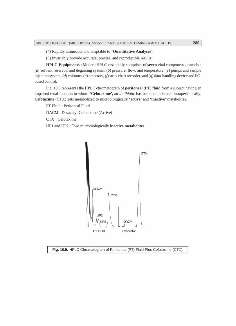
MICROBIOLOGICAL (MICROBIAL) ASSAYS : ANTIBIOTICS–VITAMINS–AMINO ACIDS 285
(4) Rapidly amineable and adaptable to ‘Quantitative Analyses’.
(5) Invariably provide accurate, precise, and reproducible results.
HPLC-Equipments : Modern HPLC essentially comprises of seven vital components, namely :(a) solvent reservoir and degassing system, (b) pressure, flow, and temperature, (c) pumps and sampleinjection system, (d) columns, (e) detectors, (f) strip-chart recorder, and (g) data-handling device and PC-based control.
Fig. 10.5 represents the HPLC chromatogram of peritoneal (PT) fluid from a subject having animpaired renal function to whom ‘Cefotaxime’, an antibiotic has been administered intraperitoneally.Cefotaxime (CTX) gets metabolized to microbioligically ‘active’ and ‘inactive’ metabolites.
PT Fluid : Peritoneal Fluid
DACM : Desacetyl Cefotaxime (Active)
CTX : Cefotaxime
UP1 and UP2 : Two microbiologically inactive metabolites
DACM
CTX
CTX
DACM
CalibratorPT Fluid
UP2
UP2
Fig. 10.5. HPLC Chromatogram of Peritoneal (PT) Fluid Plus Cefotaxime (CTX).

286 PHARMACEUTICAL MICROBIOLOGY
10.5.2. Reverse-Phase Chromatography [RPC]
The Reverse-Phase Chromatography (RPC) or Reversed-Phase HPLC (RP-HPLC) isinvariably employed for the separation of organic compounds.
In RPC, specifically a relatively nonpolar stationary phase is employed along with such polarmobile phase as :
methanol, acetonitrile, tetrahydrofuran, water, or
mixture of organic solvents and water.
Organic Solvent—the organic solvent is usally termed as the ‘modifier’ e.g., acetonitrile.
Water—Water content is mostly varied according to the required polarity.
Methanol—It is used for acidic compounds.
Acetonitrile—It is employed for basic compounds.
Tetrahydrofuran (THF)—It is usually used for those compounds having large dipoles comparatively.
In fact, most of these solvents do have low viscosity and are UV-transparent.
Bonded Phases—The abundantly used bonded phases are :
n-Octyldecyl (i.e., C-18 chain),
n-Decyl (i.e., C-8 chain), and
Phenyl Moieties
Polar-Reversed Phase Columns— The polar-reversed phase columns essentially are
polyethylene glycol (PEG) which contain either moieties that interact with polar analytes e.g., phe-nolic compounds, multiaromatic ring systems, and hydroxyl-containing compounds.
10.5.3. Ion-Pair (or Paired-Ion) Chromatography
Importantly, perhaps the most valuable of the secondary equilibria variants usually encoun-
tered in the ‘pharmaceutical analysis’ being the ion-pair formation, that may be adequately expressedfor a reversed-phase LLC-System as :
AM+ + BM
– ABS
where, A+ = Might be a ‘drug cation’,
B– = An ‘ion-pairing anion’ added to the mobile phase
AB = Ion-pair generated.

MICROBIOLOGICAL (MICROBIAL) ASSAYS : ANTIBIOTICS–VITAMINS–AMINO ACIDS 287
It has been duly observed that the ion-pair AB thus formed is capable of partitioning very muchinto the ensuing stationary phase. However, in many instances the ions A+ and B– fail to do so by virtueof the fact that their ultimate polarity gain entry into the stationary-phase gradually thereby the evolvedchromatographic resolution is controlled exclusively by the so called ion-pairing phenomenon.
It is, however, pertinent to state here that one may invariably come across a host of ‘drug sub-stances’ that are either acidic or basic in character ; and, therefore, they may be duly rendered into ionicby carefully regulating the pH of the ensuing mobile phase. In short, ion-pair chromatography pos-sesses an enormous applicability in the separation of drug substances.
Examples : A few-typical examples pertaining to the ion-pair chromatography are as describedunder :
(1) Separation of Niacin, Niacinamide, Pyridoxine, Thiamine and Riboflamin. The admix-ture of five vitamins can be separated effectively by making use of thesodium hexanesulphonate as the ion-pairing agent, on a C—18 col-umn i.e., ODS-column
(2) Antihistamines and decongestants may be separated efficaciously on a phenyl column.
10.6. EXAMPLES OF PHARMACEUTICAL MICROBIAL ASSAYS
The microbial assays have been effectively extended to a plethora of pharmaceuticalpreparations i.e., the secondary pharmaceutical products. However, this particular section will dealwith only the following three types of such products, namely :
(a) Antibiotics, (b) Vitamins, and (c) Amino Acids.
10.6.1. Antibiotics Assays
The microbial assays of ‘antibiotics’ are usually carried out by two standard methods as perIndian Pharmacopoea* (1996), namely :
Method A i.e., the ‘Cylinder-Plate Method’ as discussed in Section 10.1.3.1 and Section 10.3.1.
Method B i.e., the ‘Turbidimetric Method’ as described in Section 10.1.3.2.
A comprehensive account of the ‘Antibiotic Assays’ shall now be dealt with under the followingsub-heads :
10.6.1.1. Standard Preparation and Units of Activity
Standard preparation may be defined as— ‘the authentic sample of the appropriate antibi-otic for which the potency has been precisely determined with reference to the appropriate inter-national standard’
However, the potency of the standard preparation may be duly expressed either in Interna-tional Units (IU) or in μμμμμg.mg–1 with respect to the ‘pure antibiotic’.
* Indian Pharmacopoea, Published by the Controller of Publications, New Delhi, Vol. II, 1996.
H C—(CH ) —S—ONa3 2 5
O
O
Sodium hexane sulphonate
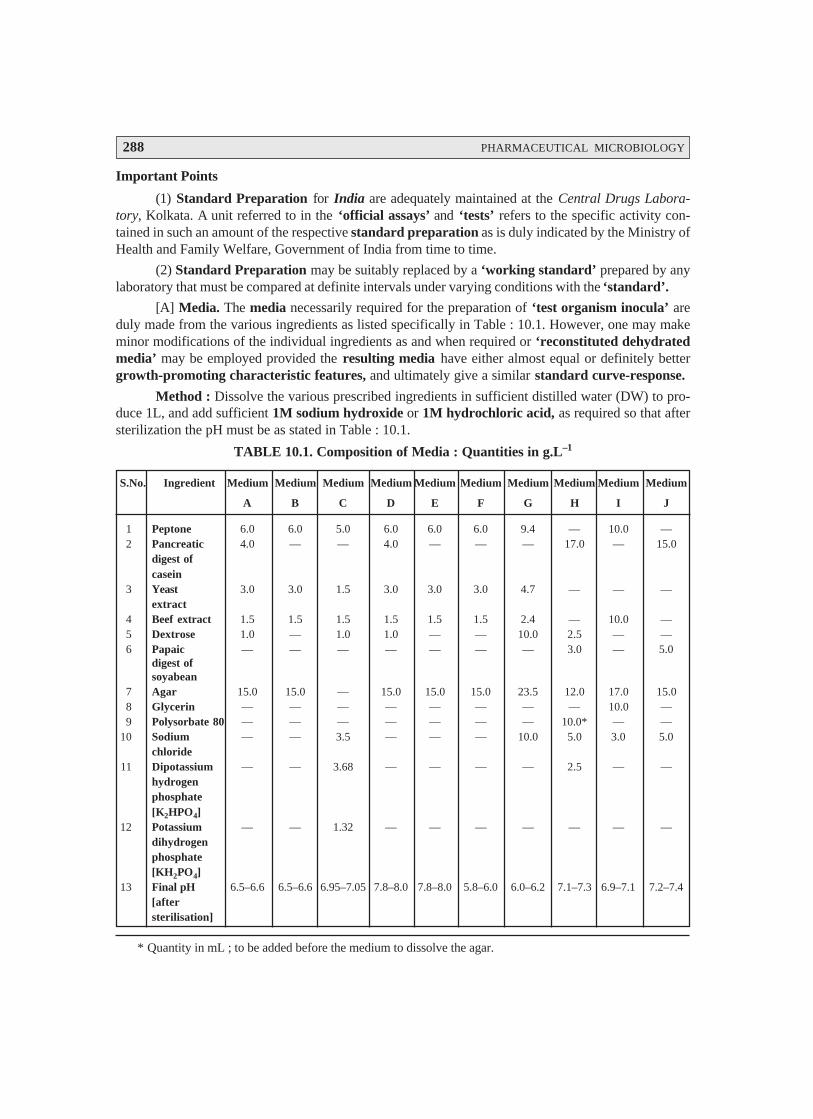
288 PHARMACEUTICAL MICROBIOLOGY
Important Points
(1) Standard Preparation for India are adequately maintained at the Central Drugs Labora-tory, Kolkata. A unit referred to in the ‘official assays’ and ‘tests’ refers to the specific activity con-tained in such an amount of the respective standard preparation as is duly indicated by the Ministry ofHealth and Family Welfare, Government of India from time to time.
(2) Standard Preparation may be suitably replaced by a ‘working standard’ prepared by anylaboratory that must be compared at definite intervals under varying conditions with the ‘standard’.
[A] Media. The media necessarily required for the preparation of ‘test organism inocula’ areduly made from the various ingredients as listed specifically in Table : 10.1. However, one may makeminor modifications of the individual ingredients as and when required or ‘reconstituted dehydratedmedia’ may be employed provided the resulting media have either almost equal or definitely bettergrowth-promoting characteristic features, and ultimately give a similar standard curve-response.
Method : Dissolve the various prescribed ingredients in sufficient distilled water (DW) to pro-duce 1L, and add sufficient 1M sodium hydroxide or 1M hydrochloric acid, as required so that aftersterilization the pH must be as stated in Table : 10.1.
TABLE 10.1. Composition of Media : Quantities in g.L–1
S.No. Ingredient Medium Medium Medium Medium Medium Medium Medium Medium Medium Medium
A B C D E F G H I J
1 Peptone 6.0 6.0 5.0 6.0 6.0 6.0 9.4 — 10.0 —2 Pancreatic 4.0 — — 4.0 — — — 17.0 — 15.0
digest ofcasein
3 Yeast 3.0 3.0 1.5 3.0 3.0 3.0 4.7 — — —extract
4 Beef extract 1.5 1.5 1.5 1.5 1.5 1.5 2.4 — 10.0 —5 Dextrose 1.0 — 1.0 1.0 — — 10.0 2.5 — —6 Papaic — — — — — — — 3.0 — 5.0
digest ofsoyabean
7 Agar 15.0 15.0 — 15.0 15.0 15.0 23.5 12.0 17.0 15.08 Glycerin — — — — — — — — 10.0 —9 Polysorbate 80 — — — — — — — 10.0* — —
10 Sodium — — 3.5 — — — 10.0 5.0 3.0 5.0chloride
11 Dipotassium — — 3.68 — — — — 2.5 — —hydrogenphosphate[K2HPO4]
12 Potassium — — 1.32 — — — — — — —dihydrogenphosphate[KH2PO4]
13 Final pH 6.5–6.6 6.5–6.6 6.95–7.05 7.8–8.0 7.8–8.0 5.8–6.0 6.0–6.2 7.1–7.3 6.9–7.1 7.2–7.4[aftersterilisation]
* Quantity in mL ; to be added before the medium to dissolve the agar.

MICROBIOLOGICAL (MICROBIAL) ASSAYS : ANTIBIOTICS–VITAMINS–AMINO ACIDS 289
[B] Buffer Solutions : Prepare the buffer solutions by dissolving the quantities (see Table 10.2)of K2HPO4 and KH2PO4 in sufficient distilled water to produce 1L after adjusting the pH with 8 M .H3PO4 or 10 M.KOH. The buffer solutions are duly sterilized after prepares and the final pH specified ineach case must be the one that is obtained after sterilization.
Table 10.2 : Buffer Solutions
Buffer Number Dipotassium Hydrogen Potassium Dihydrogen After Sterilization
Phosphate [K2HPO4] Phosphate [KH2PO4] pH
(g) (g) Adjusted To
1 2.0 8.0 6.0 ± 0.1
2 16.73 0.523 8.0 ± 0.1
3 — 13.61 4.5 ± 0.1
4 20.0 80.00 6.0 ± 0.1
5 35.0 — 10.5 ± 0.1*
6 13.6 4.0 7.0 ± 0.2
*After addition of 2 mL of 10 M potassium hydroxide.
10.6.1.2. Preparation of Standard Solution
In order to prepare a ‘Stock Solution’, dissolve a quantity of the Standard Preparation of agiven antibiotic, weighed accurately and precisely, and dried previously as duly indicated in Table 10.3in the solvent specified in the said Table ; and subsequently dilute to the required concentration asindicated specifically. It is advisable to store the ‘Stock Solution’ duly in a refrigerator (+ 1–5°C), anduse within the stipulated period indicated.
On the particular day intended for carrying out the assay, prepare from the ‘Stock Solution’ atleast five or even more test dilutions whereby the successive solutions increase stepwise in concentra-tion, invariably in the ratio 1 : 1.25 for method A or smaller for method B. Use the final diluentspecified and a sequence in a such a manner that the middle or median should have the concentration asspecified duly in Table 10.3.
10.6.1.3. Preparation of Sample Solution
Based on the available information for the ‘drug substance’ under investigation (i.e., the ‘un-known’) assign to it an assumed potency per unit weight or volume, and on this assumption prepare onthe day of the assay a ‘Stock Solution’ and test dilution(s) as duly specified for each individual antibi-otic in Table 10.3, taking particular care to use the same final diluent as employed for the StandardPreparation. The assay with 5 levels of the Standard necessarily requires only one level of the ‘un-known’ at a concentration assumed very much equal to the ‘median level’ of the ‘Standard’.

290 PHARMACEUTICAL MICROBIOLOGY
TABLE 10.3 : Stock Solutions and Test Dilutions of Standard Preparations
S. No. Standard Stock Solution Test Dilution
Antibiotic Assay Prior Initial solvent Final stock Use before Final Median Incubation
method drying (further diluent, concentra- (number of diluent dose μμμμμg temp. (°C)
if different) tion per ml days) or units
per ml
(1) (2) (3) (4) (5) (6) (7) (8) (9)
1 Amikacin B No Water 1 mg 14 Water 10 μg 32–35
2 Amphotericin B A Yes DMF7 1 mg same day B5 1.0 μg 29–31
3 Bacitracin A Yes 0.01 M HCl 100 Units same day B1 1.0 Unit 32–35
4 Bleomycin A Yes B68 2 Units 14 B6 0.04 Unit 32–35
5 Carbenicillin A No B1 1 mg 14 B6 20 μg 36–37.5
6 Doxycycline B No 0.1 M HCl 1 mg 5 Water 0.1 μg 35–37
7 Erythromycin A Yes Methanol 1 mg 14 B2 1.0 μg 35–37
(10 mg/ml)8, (B2)
8 Framycetin A Yes B2 1 mg 14 B2 1.0 μg 30–35
9 Gentamicin A Yes B2 1 mg 30 B2 0.1 μg 36–37.5
10 Kanamycin A1 No B2 800 Units 30 B2 0.8 Unit 37–39
sulphate B2 No Water 1000 Units 30 Water 10 Units 32–35
11 Kanamycin B A No B2 1000 Units 30 B2 1.0 Unit 32–35
12 Neomycin A Yes B2 1 mg 14 B2 1.0 μg 36–37.5
13 Novobiocin A Yes Ethanol 1 mg 5 B4 0.5 μg 32–35
(10 mg/ml)9, (B2)
14 Nystatin A Yes DMF7 1000 Units same day B4 20 Units 29–31
15 Oxytetra- A3 No 0.1 M HCl 1 mg 4 B3 2.5 μg 32–35
cycline B2 No 0.1 M HCl 1 mg 4 Water 0.24 μg 35–39
16 Polymyxin B A Yes Water, (B4) 10,000 Units 14 B4 10 Units 35–39
17 Rifampicin A No Methanol 1 mg 1 B1 5.0 μg 29–31
18 Streptomycin A4 Yes Water 1 mg 30 Water 1.0 μg 32–35
B5 Yes Water 1 mg 30 Water 30 μg 32–37
19 Tetracycline A3 No 0.1 M HCl 1 mg 1 Water 2.5 μg 32–35
B6 No 0.1 M HCl 1 mg 4 Water 0.24 μg 35–37
1. With Bacillus pumilus ATCC 14884 (NCTC 8241) as test organism; 2. With Staphylococcus aureus ATCC29737 as test organism; 3. With Bacillus cereus or mycoides ATCC 11778 (NCTC 10320) as test organism; 4. WithBacillus subtilis ATCC 6633 (NCTC 8236) as test organism; 5. With Klebsiella peumoniae ATCC 10031 (NTCC10320) as test organism; 6. With Staphylococcus aureus ATCC 29737 as test organism; 7. DMF =Dimethylformamide; 8. In columns 4 and 7, B denotes buffer soultion and the number following refers to the buffernumber in Table 2; 9. Initial concentration of stock solution.
Notes—For Amphotericin B and Nystatin, prepare the standard solutions and the sample test solutionsimultaneously.
MICROBIOLOGICAL (MICROBIAL) ASSAYS : ANTIBIOTICS–VITAMINS–AMINO ACIDS 291
For Amphotericin B, further dilute the stock solution with dimethylformamide to give concentrations of12.8, 16, 20, 25 and 31.2 μμμμμg per ml prior to making the test solutions. The test dilution of the sampleprepared from the solution of the substance being examined should contain the same amount ofdimethylformamide as the test dilutions of the Standard Preparation.
For Bacitracin, each of the standard test dilutions should contain the same amount of hydrochloric acid as thetest dilution of the sample.
For Nystatin, further dilute the stock solution with dimenthylformamide to give concentrations of 64.0, 80.0,100.0, 125.0 and 156.0 μμμμμg per ml prior to making the test dilutions. Prepare the standard response linesolutions simultaneously with dilutions of the sample being examined. The test dilution of the sample pre-pared from the solution of the substance being examined should contain the same amount of dimethylformamideas the test dilutions of the Standard Preparation. Protect the soultions from light.
When making the stock solution of Polymyxin B, add 2 ml of water for each 5 mg of the weighed StandardPreparation material.
Where indicated, dry about 100 mg of the Standard Preparation before use in an oven at a pressure notexceeding 0.7 kPa at 60° for 3 hours, except in the fine of Bleomycin (dry at 25° for 4 hours), Novobiocin (dryat 100° for 4 hours), Gentamicin (dry at 110° for 3 hours) and Nystatin (dry at 40° for 2 hours).
Where two-level factorial assays are performed use the following test doses per ml; Amphotericin B, 1.0 to 4.0μμμμμg; Bacteracin, 1.0 to 4.0 Units; Kanamycin Sulphate, 5.0 to 20.0 units; Streptomycin, 5.0 to 20.0 μμμμμg.
[Adapted From : Indian Pharmacopoea. Vol. II, 1996]
10.6.1.4. Test Organisms
The various test organisms for each antibiotic is duly listed in Table 10.4, along with its prop-erly documented identification number in the following recognized and approved compendia as :
• American Type Culture Collection (ATCC)
• National Collection of Type Cultures (NCTC)
• National Collection of Industrial Bacteria (NCIB).
Usually maintain a ‘culture’ on the slants of the medium, and under the specified incubationconditions as mentioned duly in Table 10.5, and transfer weekly to fresh slants.
TABLE 10.4 : Test Organisms for Microbiological Assays of Antibiotics
S. No. Antibiotic Test Organisms ATCC1 No. NCTC2 No.
(NCIB3 No.)
1 Amikacin Staphylococcus aureus 29737 7447
2 Amphotericin B Saccharomyces cerevisiae 9763 10716
3 Bacitracin Micrococcus luteus 10240 7743
4 Bleomycin Mycobacterium smegmatis 607 —
5 Carbenicillin Pseudomonas aeruginosa 25619 —
6 Doxycycline Staphylococcus aureus 29737 7447
7 Erythromycin Micrococcus luteus 9341 (8553)
8 Framycetin Bacillus pumilus 14884 8241
Bacillus subtilis 6633 8236, 10400

292 PHARMACEUTICAL MICROBIOLOGY
9 Gentamicin Staphylococcus epidermidis 12228 (8853)
10 Kanamycin Sulphate Bacillus pumilus 14884 8241
Staphylococcus aureus 29737 7447
11 Kanamycin B Bacillus subtilis 6633 8236
12 Neomycin Staphylococcus epidermidis 12228 (8853)
13 Novobiocin Staphylococcus epidermidis 12228 (8853)
14 Nystatin Saccharomyces cerevisiae 2601 10716
15 Oxytetracycline Bacillus cereus var. mycoides 11778 10320
Staphylococcus aureus 29737 7447
16 Polymyxin B Bordetella bronchiseptica 4617 8344
17 Rifampicin Bacillus subtilis 6633 8236
18 Streptomycin Bacillus subtilis 6633 8236
Klebsiella penumoniae 10031 9111
19 Tetracycline Bacillus cereus 11778 10320
Staphylococcus aureus 29737 7447
1. American Type Culture Collection, 21301 Park Lawn Drive, Rockville, MD 20852, USA.
2. National Collection of Type Cultures, Central Public Health Laboratory, Colindale Avenue, London NW95HT, England.
3. National Collection of Industrial Bacteria, Torry Research Station, P.O. Box 31, 135 Abbey Road, Aberdeen
98 DC, Scotland.
[Adapted From : Indian Pharmacopoea, Vol. II, 1996]
TABLE 10.5 : Preparation of Inoculum
Incubation conditions Suggested Suggested inoculum composition
S. No. Test organism Medium/ Temp. Time dilution Medium Amount Antibiotics
Method of (°C) factor (ml per assayed
preparation 100 ml)
1 Bacillus cereus A1/2 32–35 5 days — F As required Oxytetra-
var. mycoides cycline
Tetracycline
2 Bacillus pumilus A1/2 32–3 5 days — D As required Framycetin
Kanamycin
Sulphate
3 Bacillus subtilis A1/2 32–35 5 days — E As required Framycetin
E As required Kanamy-
cin B
B As required Rifampicin

MICROBIOLOGICAL (MICROBIAL) ASSAYS : ANTIBIOTICS–VITAMINS–AMINO ACIDS 293
4 Bordetella A/1 32–35 24 hr 1:20 H 0.1 Polymyxin B
bronchiseptica
5 Klebsiella A/1 36–37 24 hr 1:25 C 0.1 Streptomycin
pneumoniae
6 Micrococcus A/1 32–35 24 hr 1:40 D 1.5 Erythromycin
luteus (9341)
7 Micrococcus A/1 32–35 24 hr 1:35 A 0.3 Bacitracin
luteus (10240)
8 Mycobacterium J/4 36–37.5 48 hr As I 1.0 Bleomycin
smegmatis determined
9 Pseudomonas A/1 36–37.5 24 hr 1:25 H 0.5 Carbenicillin
aeruginosa2
10 Saccharomyces G/3 29–31 48 hr As G 1.0 Amphotericin B
cerevisiae (9763) determined
11 Saccharomyces G/3 29/31 48 hr As G 1.0 Nystatin
cerevisiae (2601) determined
12 Staphylococcus A/1 32–35 24 hr 1:20 C 0.1 Amikacinaureus Doxycycline
OxytetracyclineTetracycline
C 0.2 KanamycinSulphate
13 Staphylococcus A/1 32–35 24 hr 1:40 D 0.03 Gentamicinepidermidis D 0.4 Neomycin
A 4.0 Novobiocin
1. Use Medium A containing 300 mg of manganese sulphate per litre.
2. For Pseudomonas aeruginosa in the assay of Carbenicillin, use the dilution yielding 25% light transmission,rather than the stock suspension, for preparing the inoculum suspension.
Methods of preparation of test organism suspension
1. Maintain the test organism on slants of Medium A and transfer to a fresh slant once a week. Incubate theslants at the temperature indicated above for 24 hours. Using 3 ml of saline solution, wash the organism fromthe agar slant onto a large agar surface of Medium A such as a Roux bottle containing 250 ml of agar.Incubate for 24 hours at the appropriate temperature. Wash the growth from the nutrient surface using 50 mlof saline solution. Store the test organism under refrigeration. Determine the dilution factor which will give25% light transmission at about 530 nm. Determine the amount of suspensions to be added to each 100 ml ofagar of nutrient broth by use of test plates or test broth. Store the suspension under refrigeration.
2. Proceed as described in Method 1 but incubate the Roux bottle for 5 days. Centrifuge and decant thesupernatant liquid. Resuspend the sediment with 50 to 70 ml of saline solution and heat the suspension for

294 PHARMACEUTICAL MICROBIOLOGY
30 minutes at 70°. Wash the spore suspension three times with 50 to 70 ml of saline solution. Resuspend in50 to 70 ml of saline solution and heat-shock again for 30 minutes. Use test plates to determine the amountof the suspension required for 100 ml of agar. Store the suspension under refrigeration.
3. Maintain the test organism on 10 ml agar slants of Medium G. Incubate at 32° to 35° for 24 hours. Inoculate100 ml of nutrient broth. Incubate for 16 to 18 hours at 37° and proceed as described in Method 1.
4. Proceed as described in Method 1 but wash the growth from the nutrient surface using 50 ml of Medium 1(prepared without agar) in place of saline solution.
[Adapted From : Indian Pharmacopoea, Vol. II, 1996]
10.6.1.5. Preparation of Inoculum
The method of preparation of the microbial suspensions for preparing the inoculum for theassay of various antibiotics is clearly stated in Table 10.5. In an event when the suspensions are duly
prepared by these methods, one may accomplish and observe that the growth characteristic features are
fairly uniform in order that the inoculum could be determined by carrying out the following trials.
10.6.1.5.1. For Method A. After the suspension is prepared, as given under Table 10.5, adddifferent volumes of it to each of several different flasks containing 100 ml of the medium specified in
Table 10.4 (the volume of suspension suggested in Table 10.4 may be used as a guide). Using these
inocula, prepare inoculated plates as described for the specific antibiotic assay. While conducting cylin-der-plate assays, double layer plates may be prepared by pouring a seed layer (inoculated with the
desired micro-organism) over a solidified uninoculated base layer. For each Petri dish, 21 ml of the base
layer and 4 ml of the seed layer may be generally suitable. Fill each cylinder with the median concentra-tion of the antibiotic (Table 10.4) and then incubate the plates. After incubation, examine and measure
the zones of inhibition. The volume of suspension that produces the optimum zones of inhibition with
respect to both clarity and diameter determines the inoculum to be used for the assay.
10.6.1.5.2. For Method B. Proceed as descirbed for Method A and, using the several inocula,
carry out the procedure as described for the specific antibiotic assay running only the high and low con-
centrations of the standard response curve. After incubation, read the absorbances of the appropriatetubes. Determine which inoculum produces the best response between the low and high antibiotic con-
centrations and use this inoculum for the assay.
Apparatus. All equipment is to be thoroughly cleaned before and after each use. Glassware for
holding and transferring test organisms is sterilised by dry heat or by steam.
10.6.1.6. Temperature Control
Thermostatic control is required in several stages of a microbial assay, when culturing a micro-organisms and preparing its inoculum and during incubation in a plate assay. Closer control of the
temperature is imperative during incubation in a tube assay which may be achieved by either circulated
air or water, the greater heat capacity of water lending it some advantage over circulating air.
MICROBIOLOGICAL (MICROBIAL) ASSAYS : ANTIBIOTICS–VITAMINS–AMINO ACIDS 295
10.6.1.7. Spectrophotometer
Measuring transmittance within a fairly narrow frequency band requiers a suitablespectrophotometer in which the wavelength of the light source can be varied or restricted by the use ofa 580 nm filter for preparing inocula of the required density, or with a 530 nm filter for reading theabsorbance in a tube assay. For the latter purpose, the instrument may be arranged to accept the tube inwhich incubation takes place, to accept a modified cell fitted with a drain that facilitates rapid change ofcontents, or preferably fixed with a flow-through cell for a continuous flow-through analysis. Set theinstrument at zero absorbance with clear, uninoculated broth prepared as specified for the particularantibiotic, including the same amount of test solution and formaldehyde as found in each sample.
10.6.1.8. Cylinder-Plate Assay Receptacles
Use rectangular glass trays or glass or plastic Petri dishes (approximately 20 × 100 mm) havingcovers of suitable material and assay cylinders made of glass, porcelain, aluminium or stainless steelwith outside diameter 8 mm ± 0.1 mm, inside diameter 6 mm ± 0.1 mm and length 10 mm ± 0.1 mm.Instead of cylinders, holes 5 to 8 mm in diameter may be bored in the medium with a sterile borer, orpaper discs of suitable quality paper may be used. Carefully clean the cylinders to remove all residues.An occasional acid-bath, e.g., with about 2M nitric acid or with chromic acid solution is needed.
10.6.1.9. Turbidimetric Assay Receptacles
For assay tubes, use glass or plastic test-tubes, e.g., 16 mm × 125 mm or 18 mm × 150 mm thatare relatively uniform in length, diameter, and thickness and substantially free form surface blemishesand scratches. Cleanse thoroughly to remove all antibiotic residues and traces of cleaning solution andsterilise tubes that have been used previously before subsequent use.
10.6.1.10. Assay Designs
Microbial assays gain markedly in precision by the segregation of relatively large sources ofpotential error and bias through suitable experimental designs. In a cylinder-plate assay, the essentialcomparisons are restricted to relationships between zone diameter measurements within plates, exclu-sive of the variation between plates in their preparation and subsequent handling. To conduct aturbidimetric assay so that the difference in observed turbidity will reflect the differences in the antibi-otic concentration requires both greater uniformity in the environment created for the tubes throughcloser thermostatic control of the incubator and the avoidance of systematic bias by a random placementof replicate tubes in separate tube racks, each rack containing one complete set of treatments. Theessential comparisons are then restricted to relationships between the observed turbidities within racks.
Within these restrictions, two alternative designs are recommended; i.e., a 3-level (or 2-level)factorial assay, or a 1-level assay with a standard curve. For a factorial assay, prepare solutions of 3 or 2corresponding test dilutions for both the standard and the unknowns on the day of the assay, as describedunder Preparation of the Standard and Preparation of the Sample. For a 1-level assay with a standardcurve, prepare instead solutions of five test dilutions of the standard and a solution of a single mediantest level of the unknown as described in the same sections. Consider an assay as preliminary if itscomputed potency with either design is less than 60% or more than 150% of that assumed in preparingthe stock solution of the unknown. In such a case, adjust its assumed potency accordingly and repeat theassay.

296 PHARMACEUTICAL MICROBIOLOGY
Microbial determinations of potency are subject to inter-assay variables as well as intra-assayvariables, so that two or more independent assays are required for a reliable estimate of the potency of agiven assay preparation or unknown. Starting with separately prepared stock solutions and test dilutionsof both the standard and the unknown, repeat the assay of a given unknown on a different day. If theestimated potency of the second assay differs significantly, as indicated by the calculated standard error,from that of the first, conduct one or more additional assays. The combined result of a series of smaller,independent assays spread over a number of days is a more reliable estimate of potency than that from asingle large assay with the same total number of plates or tubes.
10.6.1.10.1. Methods. The microbiological assay of antibiotics may be carried out by MethodA or Method B.
[A] Cylinder-Plate or Cup-Plate Method
Inoculate a previously liquefied medium appropriate to the assay (Tables 10.1 and 10.3) with therequisite quantity of suspension of the micro-organisms, add the suspension to the medium at a tempera-
ture between 40° and 50° and immediately pour the inoculated medium into Petri dishes or large rectan-
gular plates to give a depth of 3 to 4 mm (1 to 2 mm for nystatin). Ensure that the layers of medium areuniform in thickness, by placing the dishes or plates on a level surface.
The prepared dishes or plates must be stored in a manner so as to ensure that no significant
growth or death of the test organism occurs before the dishes or plates are used and that the surface of
the agar layer is dry at the time of use.
Using the appropriate buffer solutions indicated in Tables 10.2 and 10.3, prepare solutions of
known concentration of the Standard Preparation and solutions of the corresponding assumed concen-trations of the antibiotic to be examined. Where directions have been given in the individual monograph
for preparing the solutions, these should be followed and further dilutions made with buffer solution as
indicated in Table 10.3. Apply the solutions to the surface of the solid medium in sterile cylinders or incavities prepared in the agar. The volume of soluiton added to each cylinder or cavity must be uniform
and sufficient almost to fill the holes when these are used. When paper discs are used these should be
sterilised by exposure of both sides under a sterilising lamp and then impregnated with the standardsolutions or the test solutions and placed on the surface of the medium. When Petri dishes are used,
arrange the solutions of the Standard Preparation and the antibiotic to be examined on each dish so that
they alternate around the dish and so that the highest concentrations of standard and test preparations arenot adjacent. When plates are used, place the solutions in a Latin square design, if the plate is a square,
or if it is not, in a randomised block design. The same random design should not be used repeatedly.
Leave the dishes or plates standing for 1 to 4 hours at room temperature or at 4°, as appropriate,
as a period of pre-incubation diffusion to minimise the effects of variation in time between the applica-tion of the different solutions. Incubate them for about 18 hours at the temperature indicated in Table
10.3. Accurately measure the diameters or areas of the circular inhibition zones and calculate the results.
Selection of the assay design should be based on the requirements stated in the individual mono-
graph. Some of the usual assay designs are as follows.

MICROBIOLOGICAL (MICROBIAL) ASSAYS : ANTIBIOTICS–VITAMINS–AMINO ACIDS 297
[A.1] One-Level Assay with Standard Curve
Standard solution. Dissolve an accurately weighted quantity of the Standard Preparation of theantibiotic, previously dried where necessary, in the solvent specified in Table 10.3, and then dilute to therequired concentration, as indicated, to give the stock solution. Store in a refrigerator and use within theperiod indicated. On the day of the assay, prepare from the stock solutions, 5 dilutions (solutions S1 toS5) representing five test levels of the standard and increasing stepwise in the ratio of 4 : 5. Use thedilution specified in Table 10.3 and a sequence such that the middle or median has the concentrationgiven in the table.
Sample solution. From the information available for the antibiotic preparation which is beingexamined (the “unknown”) assign to it an assumed potency per unit weight or volume and on thisassumption prepare on the day of the assay a stock solution with the same solvent as used for thestandard. Prepare from this stock solution a dilution to a concentration equal to the median level of thestandard to give the sample solution.
Method. For preparing the standard curve, use a total of 12 Petri dishes or plates to accommo-date 72 cylinders or cavities. A set of three plates (18 cylinders or cavities) is used for each dilution. Oneach of the three plates of a set fill alternate cylinders or cavities with solution S3 (representing themedian concentration of the standard solution) and each of the remaining 9 cylinders or cavities withone of the other 4 dilutions of the standard solution. Repeat the process for the other 3 dilutions of thestandard solutions. For each unknown preparation use a set of three plates (18 cylinders or cavities) andfill alternate cylinders or cavities with the sample solution and each of the remaining 9 cylinders ofcavities with solution S3.
Incubate the plates for about 18 hours at the specified temperature and measure the diameters orthe zones of inhibition.
Estimation of potency. Average the readings of solution S3 and the readings of the concentra-tion tested on each set of three plates, and average also all 36 readings of solution S3. The average of the36 readings of soluiton S3 is the correction point for the curve. Correct the average value obtained foreach concentration (S1, S2, S4 and S5) to the figure it would be if the readings for solution S3 for that setof three plates were the same as the correction point. Thus, in correcting the value obtained with anyconcentration, say S1, if the average of 36 readings of S3 is, for example, 18.0 mm and the average of theS3 concentrations on one set of three plates is 17.8 mm, the correction is + 0.2 mm. If the averagereading of S1 is 16.0 mm, the corrected reading of S1 is 16.2 mm. Plot these corrected values includingthe average of the 36 readings for solutions S3 on two-cycle semilog paper, using the concentrations inUnits or μg per ml (as the ordinate logarithmic scale) and the diameter of the zones of inhibition as theabscissa. Draw the straight response line either through these points by inspection or through the pointsplotted for highest and lowest zone diameters obtained by means of the following expressions :
L = 3 2
5+ + −a b c e
; H = 3 2
5+ + −e d c a
where L = the calculated zone diameter for the lowest concentration of the standard curve response line.
H = the calculated zone diameter for the highest concentration of the standard curve responseline.
c = average zone diameter of 36 readings of the reference point standard solution.

298 PHARMACEUTICAL MICROBIOLOGY
a, b, d, e = corrected average values for the other standard solutions, lowest to highestconcentrations, respectively.
Average the zone diameters for the sample solution and for solutions S3 on the plates used for thesample soluiton. If the sample gives a large average zone size than the average of the standard (solutionS3), add the difference between them to the zone size of solution S3 of the standard response line. If theaverage sample zone size is smaller than the standard values, subtract the difference between them fromthe zone size of solution S3 of the standard response line. From the response line read the concentrationcorresponding to these corrected values of zone sizes. From the dilution factors the potency of thesample may be calculated.
[A.2] Two-Level Factorial Assay
Prepare parallel dilutions containing 2 levels of both the standard (S1 and S2) and the unkown(U1 and U2). On each of four or more plates, fill each of its four cylinders or cavities with a different testdilution, alternating standard and unknown. Keep the plates at room temperature and measure the diam-eters of the zones of inhibition.
Estimation of potency. Sum the diameters of the zones of each dilution and calculate the %potency of the sample (in terms of the standard) from the following equation :
% potency = Antilog (2.0 + a log I)
wherein a may have a positive or negative value and should be used algebracially and
where a = 1 2 1 2
1 2 1 2
(U + U ) – (S + S )(U + U ) + (S – S )
U1 and U2 are the sums of the zone diameters with solutions of the unknown of high and lowlevels.
S1 and S2 are the sums of the zone diameters with solutions of the standard of high and lowlevels.
I = ratio of dilutions.
If the potency of the sample is lower than 60% or greater than 150% of the standard, the assay isinvalid and should be repeated using higher or lower dilutions of the same solutions. The potency of thesample may be calculated from the expression.
% potency × assumed potency of the sample100
.
[A.3] Other Designs
(1) Factorial assay containing parallel dilution of three test levels of standard and the unknown.
(2) Factorial assay using two test levels of standard and two test levels of two different un-knowns.
[B] Turbidimetric or Tube Assay Method
The method has the advantage of a shorter incubation period for the growth of the test organism(usually 3 to 4 hours) but the presence of solvent residues or other inhibitory substances affects this
MICROBIOLOGICAL (MICROBIAL) ASSAYS : ANTIBIOTICS–VITAMINS–AMINO ACIDS 299
assay more than the cylinder-plate assay and care should be taken to ensure freedom from such sub-stances in the final test solutions. This method is not recommended for cloudy or turbid preparations.
Prepare five different concentrations of the standard solution for preparing the standard curve bydiluting the stock solution of the Standard Preparation of the antibiotic (Table 10.3) and increasingstepwise in the ratio 4 : 5. Select the median concentration (Table 10.3) and dilute the solution of thesubstance being examined (unknown) to obtain approximately this concentration. Place 1 mL of eachconcentration of the standard solution and of the sample solution in each of the tubes in duplicate. Toeach tube add 9 ml of nutrient medium (Table 10.3) previously seeded with the appropriate test organ-ism (Table 10.3).
At the same time prepare three control tubes, one containing the inoculated culture medium (cul-ture control), another identical with it but treated immediately with 0.5 mL of dilute formaldehyde solu-tion (blank) and a third containing uninoculated culture medium.
Place all the tubes, randomly distributed or in a randomized block arrangement, in an incubator ora water-bath and maintain them at the specified temperature (Table 10.3) for 3 to 4 hours. After incubationadd 0.5 mL of dilute formaldehyde solution to each tube. Measure the growth of the test organism bydetermining the absorbance at about 530 nm of each of the solutions in the tubes against the blank.
Estimation of potency. Plot the average absorbances for each concentration of the standard onsemi-logarithmic paper with the absorbances on the arithmetic scale and concentrations on the logarith-mic scale. Construct the best straight response line through the points either by inspection or by meansof the following expressions :
L = 3 2
5+ + −a b c e
; H = 3 2
5+ + −e d c a
where L = the calculated absorbance for the lowest concentration of the standard response line.
H = the calculated absorbance for the highest concentration of the standard response line.
a, b, c, d, e = average absorbance values for each concentration of the standard response line
lowest to highest respectively.
Plot the values obtained for L and H and connect the points. Average the absorbances for thesample and read the antibiotic concentration from the standard response line. Multiply the concentrationby the appropriate dilution factors to obtain the antibiotic content of the sample.
Precision of Microbiological Assays
The fiducial limits of error of the estimated potency should be not less than 95% and not morethan 105% of the estimated potency unless otherwise stated in the individual monograph. This degree ofprecision is the minimum acceptable for determining that the final product complies with the officialrequirements and may be inadequate for those deciding, for example, the potency which should bestated on the label or used as the basis for calculating the quantity of an antibiotic to be incorporated ina preparation. In such circumstances, assays of greater precision may be desirable with, for instance,fiducial limits of error of the order of 98% to 102%. With this degree of precision, the lower fiduciallimit lies close to the estimated potency. By using this limit, instead of the estimated potency, to assign apotency to the antibiotic either for labelling or for calculating the quantity to be included in a prepara-
300 PHARMACEUTICAL MICROBIOLOGY
tion, there is less likelihood of the final preparation subsequently failing to comply with the officialrequirements for potency.*
10.7. ASSAY OF ANTIBIOTICS BY TURBIDIMETRIC(OR NEPHELOMETRIC) METHOD
A large number of antibioics, namely : chlortetracycline, doxycyline, gentamicin, neomycin,
streptomycin, tobramycin and the like may be assayed tubidimetrically with fairly good accuracy.
10.7.1. Assay of Chlorotetracycline
Theory. Inoculate a medium consisting of : peptone : 6 g, beef extract : 1.5 g, yeast extract : 3 g,sodium chloride : 3.5 g, D-glucose monohydrate : 1.0 g, dipotassium hydrogen orthophosphate : 3.68 g,potassium hydrogen orthophosphate : 1.32 g and dissolve in sufficient water to produce 1 L with aknown quantity of a suspension of Staphylococcus aureus (NCTC 6571**) so as to obtain a readilymeasured opacity after an incubation of about 4 hours. The micro-organisms must exhibit a sensitivity tothe antibiotic under investigation to such an extent that a sufficiently large inhibition of growth takesplace in the prevailing conditions of the test.
In actual practice, it is always advisable that the inoculated medium should be used immediatelyafter its preparation. Using a phosphate buffer of pH 4.5 (dissolve 13.61 g of KH2 PO4 in about 750 mlof water, adjusting the pH to 4.5 with 0.1 M NaOH and diluting to 1 L with water), prepare solutions ofthe Standard Preparation and the substance under investigation at concentrations presumed to be equal.
To enable the validity of the assay to be examined, it is desirable to use at least three doses of theStandard Preparation and of the substance being examined. It is also advisable to use doses in logarith-mic progression in a parallel line assay.
Materials Required : Standard chlortertracyline ; sterilized media (as described above) : 1 L ;authentic and pure strain of microorganism Staphylococcus aureus (NCTC 6571) ; formaldehyde solu-tion (34–37% w/v) 10 mL ; matched identical test tubes : 20 ;
Procedure : Distribute into identical test-tubes an equal volume of standard tetracycline solutionand the sample to be examined (having presumed equal concentrations) and add to each tube an equalvolume of inoculated nutrient medium (for instance 1 mL of the solution and 9 ml of the medium). Prepareat the same time two control tubes without the chlorotetracycline, one containing the inoculated mediumand the other identical with it but treated immediately with 0.5 mL of formaldehyde solution. These tubesare used to set the optical apparatus employed to measure the growth.
Place all the tubes, randomly distributed, in a water-bath or other suitable means of bringing all thetubes rapidly to 35–37°C i.e., the incubation temperature and maintain them at that temperature for 3 to 4hours, taking due precautions to ensure uniformity of temperatures and identical incubation times. Afterincubation, stop the growth of the microorganisms by adding 0.5 mL of formaldehyde solution, each tubeand subsequently measures the opacity to at least three significant figures using a suitable
* The Section 10.6. related to ‘Examples of Pharmaceutical Microbial Assays’ has been mostly and largelybased on the factual details as given in the ‘Indian Pharmaeopoea’ Vol. II, 1996, so that the researchars,students, and teachers get fully acquainted and familiarized with Standard Operating Methodologies.
** NCTC = National Collection of Type Culture.
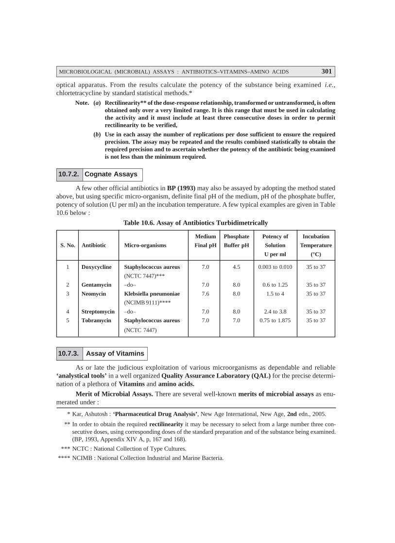
MICROBIOLOGICAL (MICROBIAL) ASSAYS : ANTIBIOTICS–VITAMINS–AMINO ACIDS 301
optical apparatus. From the results calculate the potency of the substance being examined i.e.,chlortetracycline by standard statistical methods.*
Note. (a) Rectilinearity** of the dose-response relationship, transformed or untransformed, is oftenobtained only over a very limited range. It is this range that must be used in calculatingthe activity and it must include at least three consecutive doses in order to permitrectilinearity to be verified,
(b) Use in each assay the number of replications per dose sufficient to ensure the requiredprecision. The assay may be repeated and the results combined statistically to obtain therequired precision and to ascertain whether the potency of the antibiotic being examinedis not less than the minimum required.
10.7.2. Cognate Assays
A few other official antibiotics in BP (1993) may also be assayed by adopting the method statedabove, but using specific micro-organism, definite final pH of the medium, pH of the phosphate buffer,potency of solution (U per ml) an the incubation temperature. A few typical examples are given in Table10.6 below :
Table 10.6. Assay of Antibiotics Turbidimetrically
Medium Phosphate Potency of Incubation
S. No. Antibiotic Micro-organisms Final pH Buffer pH Solution Temperature
U per ml (°C)
1 Doxycycline Staphylococcus aureus 7.0 4.5 0.003 to 0.010 35 to 37
(NCTC 7447)***
2 Gentamycin –do– 7.0 8.0 0.6 to 1.25 35 to 37
3 Neomycin Klebsiella pneumoniae 7.6 8.0 1.5 to 4 35 to 37
(NCIMB 9111)****
4 Streptomycin –do– 7.0 8.0 2.4 to 3.8 35 to 37
5 Tobramycin Staphylococcus aureus 7.0 7.0 0.75 to 1.875 35 to 37
(NCTC 7447)
10.7.3. Assay of Vitamins
As or late the judicious exploitation of various microorganisms as dependable and reliable‘analystical tools’ in a well organized Quality Assurance Laboratory (QAL) for the precise determi-nation of a plethora of Vitamins and amino acids.
Merit of Microbial Assays. There are several well-known merits of microbial assays as enu-merated under :
* Kar, Ashutosh : ‘Pharmaceutical Drug Analysis’, New Age International, New Age, 2nd edn., 2005.
** In order to obtain the required rectilinearity it may be necessary to select from a large number three con-secutive doses, using corresponding doses of the standard preparation and of the substance being examined.(BP, 1993, Appendix XIV A, p, 167 and 168).
*** NCTC : National Collection of Type Cultures.
**** NCIMB : National Collection Industrial and Marine Bacteria.
302 PHARMACEUTICAL MICROBIOLOGY
(1) These are as precise and accurate as the ‘chemical methods’.
(2) These are invariably quite simple, convenient, not-so-cumbersome, and above all definitelyinexpensive.
(3) A very small quantum of the ‘sample’ is required for the recommended microbial assay.
(4) They hardly need any elaborated instrumentation.
(5) These microbial assays do require the following essential criteria, such as :
• ascertains continuous checks for consistency of results,
• ensures specificity, and
• prevents any possible interferences.
(6) Automation of microbial assays may essentially overcome any possible limitations, accu-racy of observations, and the sample-handling capacity to a significant extent.
Example : In an ‘automatic photometric assay’ the following activities do take place in asequential manner, namely :
measures the exact quantum of antibiotic present in a given solution,
incorporates requisite quantum of inoculum and nutrient medium,
incubates the resulting mixture for 100 minutes,
transfers the incubated mixture to photometer cell, and
results are adequately read and recorded.Principle. It has been amply proved and established that there are some specific microbes which
predominantly require vitamin (factor) for their usual normal growth phenomenon ; and, therefore, arequite sensitive to the extremely small quantities of the much desired ‘factor’. Nevertheless, it is pre-cisely the critical inherent ability of these particular microorganisms (i.e., the ‘test organisms’) to carryout the synthesis of the ‘factor’ being determined. This ultimately gives rise to the fundamental basis ofthe microbial assay of vitamins. To accomplish the ultimate objective, the ‘test organism’ is dulyinoculated in the highly specialized culture media that are essentially complete in every possible re-spects except the presence of the ‘factor’ under investigative study. In reality, it evidently caters for the‘control’ wherein either little or almost minimal growth of microbes is duly exhibited. Importantly, inanother set of parallel/identical experiments, one may incorporate meticulously the ‘graded quantitiesof factor’ the thus the ultimate growth of the test organism (i.e., response) is observed adequately.However, one may observe invariably that the ‘response’ (i.e., growth of the ‘test organism’) is directlyproportional to the ‘factor’ (i.e.,quantum of the dose) actually incorporated to the culture medium.
Microbial assays of the following three water-soluble vitamins would be discussed individuallyin the sections that follows :
(a) Calcium Pantothenate,(b) Niacin (or Niacinamide), and(c) Vitamin B12 (or Cyanocobalamin).
10.7.3.1. Calcium Pantothenate
It refers to one of the B complex vitamins (or vitamin B complex). The various steps involved
for the assay are enumerated under sequentially :
(1) Reagents. The various reagents essentially required for the assay of ‘calcium pantothenate’are :

MICROBIOLOGICAL (MICROBIAL) ASSAYS : ANTIBIOTICS–VITAMINS–AMINO ACIDS 303
(a) Standardized Stock Solution. Each mL of this stock solution consists of 50 mcg of cal-cium panthothenate. It may be prepared by carefully dissolving 50 mg of BPCRS* calcium pantothenatein 500 mL of double-distilled water ; 10 mL of 0.2 M acetic acid, 100 mL of 1.6% (w/v) sodium acetate ;
and volume made upto 1 L with DW.
Note : The resulting solution must be stored under a layer of ‘toluene’ in a refrigerator.
(b) Standard Solution. The standard solution should contain approximately 0.04 mcg of cal-
cium pantothenate in 1 mL, and is duly prepared by diluting the Standard Stock Solution (a).
(c) Test Solution. The test solution essentially contains nearly the same equivalent amount of
calcium pantothenate as present in the Standard Solution (a) above i.e., 0.4 mcg.mL–1 prepared in
double-distilled water.
(d) Culture Medium. The culture medium is composed of the following solutions and ingredi-ents :
(i) Casein hydrolysate solution** : 25 mL
(ii) Cysteine-tryptophane solution : 25 mL
(iii) Polysorbate-80 solution*** : 0.25 mL
(iv) Dextrose (anhydrous) : 10 g
(v) Sodium acetate (anhydrous) : 5 g
(vi) Adenine-guanine-uracil solution : 5 mL
(vii) Riboflavin-Thiamine hydrochloride-Biotin Solution : 5 mL
(viii) PABA****-Niacin-Pyridoxine hydrochloride solution : 5 mL
(ix) Calcium pantothenate solution A : 5 mL
(x) Calcium pantothenate solution B : 5 mL
The culture medium is usually prepared by dissolving both anhydrous dextrose and sodiumacetate in previously mixed solutions and the pH is carefully adjusted to 6.8 with 1 M.NaOH solution.The final volume is duly made upto 250 mL with distilled water and mixed thoroughly.
(2) Stock Culture of Organism : The stock culture of organism may be prepared dissolving 2g water-soluble yeast extract in 100 mL DW, 500 mg anhydrous dextrose, 500 mg anhydrous sodiumacetate, and 1.5 g agar. The resulting mixture is heated gently so as to dissolve the agar. Now, 10 mL ofhot solution is transferred to test tubes and sterilized at 121°C by keeping in an upright position. The‘stab culture’***** is now prepared duly in three tubes employing Lactobacillus plantarum, incubatedat 30 to 37°C for 16 to 24 hours, and stored in a refrigerator ultimately.
* BPCRS : British Pharmacopeal Chemical Reference Standard.
** Casein-hydrolysate solution. Dissolve 100 g of acid-digested casein hydrolysate in sufficient distilled wa-ter to make 500 mL, adjust the pH to 7.2 with 10 M.NaOH, and sterilize by heating at 121°C for 20 minutes.
*** Polysorbate-80 solution. Mix together polysorbate-80 (10 mL) and phosphate buffer solution (90 mL), andsterilize by heating at 121°C for 20 minutes. Store in a cold place.
**** PABA—para-Amino Benzoic Acid.
*****Stab Culture : A bacterial culture in which the organism is introduced into a solid gelatin medium eitherwith a needle or platinum wire.
304 PHARMACEUTICAL MICROBIOLOGY
(3) Preparation of Inoculum. The cells consequently obtained from the stock culture, (a) above,organism are duly transferred to a sterile tube containing 10 mL of the culture emdium (d). Finally, it isincubated at 30 to 37°C for a duration of 16–24 hours.
(4) Methodology. The various steps involved are as stated below :
(i) Standard Solution (b) is added to five test tubes in varying amounts viz., 1, 2, 3, 4 and 5mL in duplicate.
(ii) To each of the five above test tubes plus another four similar tubes without any standardsolution is added 5 mL of culture medium, and the final volume made upto 10 mL withDW.
(iii) Now, volumes of test solution (c) corresponding to either three or more of the levels astaken above, are incorporated carefully to similar test tubes, in duplicate.
(iv) To each test tube 5 mL of the medium solution, and volume is made upto 10 mL withDW. Thus, we may have two separate racks :
First Rack : Having complete set of standard plus assay tubes ; and
Second Rack : Having duplicate set only.
(v) Tubes of both the series are duly heated in an autoclave at 121°C for 5 minutes only ;cooled to ambient temperature, added 1 drop of inoculum (3) to each tube except two ofthe four tubes that specifically has no ‘standard solution’ (i.e., the uninoculated tubes),and mixed thoroughly. The tubes are adequately incubated at 121°C at 30–37°C for 16–24 hours.
(vi) Transmittance of the various tubes is measured with a spectrophotometer at wave-length ranging between 540–660 nm.
(5) Calculation. First of all, a standard concentration response curve is plotted between thetransmittance Vs log mL (volume) of the standard solution in each tube. In this way, the response isduly calculated by summing up the two transmittances for each level of the test solution.
Finally, the exact concentration of the calcium pantothenate in the ‘test sample’ is determinedaccurately with the aid of the standard concentration-response curve obtained.
10.7.3.2. Niacin (or Niacinamide)
Preamble. In this particular assay the most appropriate organism should be such that must beable to fully use up there five vital and important components, namely : niacin, nicotinuric acid,miacinamide, niacinamide, nucleoside, and coenzymase (an enzyme). This organism that may criti-cally satisfy the aforesaid requirements happens to be Lactobacillus plantarum. Interestingly, this acidforming organism is found to be quite incapable to afford the synthesis of niacin for its on-going meta-bolic processes. A few other equally important criteria of this organism are as given under :
• Non-pathogenic in nature
• Easy to culture
• Least affected by various stimulatory or inhibitory constituents usually present in ‘pharma-ceutical formulations’ containing niacin.
• Conveniently grown upon a rather simple stab culture comprising of gelatin, yeast extract,and glucose.

MICROBIOLOGICAL (MICROBIAL) ASSAYS : ANTIBIOTICS–VITAMINS–AMINO ACIDS 305
Note. (1) For the assay of niacin, it is cultured in the assay tubes by actually transferring to theensuing liquid culture medium comprising of the basic medium having an optimized quan-tum of added niacin.
(2) To obtain a measurable response the amount of niacin present in each tube may rangebetween 0.05 to 0.5 mcg.
(1) Reagents. The various reagents used for the microbial assay of niacin are as enumeratedunder :
(a) Standard Stock Solution of Niacin (I). It essentially contains 100 mcg.mL–1 of niacinUSPCRS*.
(b) Standard Stock Solution of Niacin (II). It consists of 10 mcg.mL–1 of niacin USPCRS;and is prepared by dilution of solution (I) in the ratio 1 : 10, i.e., 1 mL of solution (I) is madeup to 10 mL in DW.
(c) Standard Niacin Solution. It critically contains niacin ranging between 10-40 ng(i.e.,nanogram). mL–1, and may be prepared from Solution II by an appropriate dilutionwith DW.
(d) Basal Culture Medium Stock Solution. The basal culture medium stock solution may beprepared by the following requisite proportion of various ingredients and solutions as enu-merated under :
(i) Casein hydrolysate solution** : 25 mL(ii) Cystine-tryptophane solution : 25 mL
(iii) Anhydrous dextrose : 10 g(iv) Anhydrous sodium acetate : 5 g
(v) Adenine-guanine-uracil solution : 5 mL(vi) Riboflavin-Thiamine hydrochloride-Biotin Solution : 5 mL
(vii) PABA-Calcium patothenate-Pyridoxinehydrochloride solution : 5 mL
(viii) Niacin solution A : 5 mL(ix) Niacin solution B : 5 mL
The culture medium is duly perpared by carefully dissolving anhydrous dextrose and anhydroussodium acetate into the previously mixed solutions, and adjusting the pH precisely to 6.8 by the dropwiseaddition of 1 M.NaOH. The final volume was made up to 250 mL with DW.
(e) Culture Medium. Into a series of labeled ‘test tubes’ containing 5 mL of the Basal CultureMedium Stock Solution [(d) above] 5 mL of water containing exactly 1 mcg of niacin areincorporated carefully. The sterilization of all these ‘test tube’ are carried out by first plug-ging each of them with cotton, and subsequently autoclaving them at 121°C for 15 minutes.
(2) Preparation of Inoculum. Transfer from the stock culture of Lactobacillus plantarumcells aseptically into a sterilie test tube containing 10 mL of culture medium [(e) above]. The resultingculture is duly incubated at a temperature ranging between 30–37°C for a duration of 16–24 hours. Thecell suspension of the said organism is termed as the inoculum.
* USPCRS : United State Pharmacopeal Chemical Reference Standard.
100 mcg.mL–1 solution may be prepared by dissolving 10 mg of niacin in 1000 mL of DW
** Refer to ‘assay of Calcium Pantothenate’ in Section 10.6.2.1.
306 PHARMACEUTICAL MICROBIOLOGY
(3) Methodology. The various steps that are involved in the microbial assay of niacin are de-scribed as under in a sequential manner :
(i) First and foremost the ‘spectrophotometer’ is duly calibrated according to the proce-dural details mentioned in the ‘official compendia’*.
(ii) Standard Niacin Solution is added in duplicate into various Standard Niacin Tubesin varying quantities viz., 0, 0.5, 1.0, 1.5, 2.0, 2.5 ...... 5.0 mL respectively. To each ofthese tubes add 5.0 mL of the Basal Culture Medium Stock Solution [(d) above] plussufficient distilled water to make 10 mL.
(iii) Test Solution Tubes containing varying amounts of niacin are carefully prepared bymaking in duplicate 1, 2, 3, 4, and 5 mL respectively of the ‘test solution’. To thesetubes are added 5 mL of the Basal Culture Medium Stock Solution [(d) above], andfollowed by water to make upto 10 mL.
(iv) All the tubes obtained in (iii) above are duly plugged with cotton, and adequately steri-lized in an ‘autoclave’ (for 15 minutes at 121°C).
(v) After having brought down the hot tubes to the ambient temperature, they are carefullyinoculated asepticlly with one drop of inoculum [(2) above], and subsequently be-tween 30–37°C for a duration of 16 to 24 hours.
(vi) Having set the percentage transmittance at 1 for the ‘uninoculated blank’, the varioustransmittance of the inoculated tubes is duly noted, and recorded.
(4) Calculation : First of all a ‘Standard Curve’ is plotted for niacin between :
• standard transmittances for each level of Standard Niacin Solution, and
• exact quantum of niacin (in mcg) present duly in the respective tubes.
Thus, from the ‘Standard Curve’, one may easily obtain the niacin precisely present in the ‘testsolution’ of each tube by interpolation.
Finally, the exact niacin content of the ‘test material’ may be calculated from the ‘averagevalues’ duly obtained from at least six tubes which should not vary by more than ± 10% with respect tothe average values.
10.7.3.3. Vitamin B12 [or Cynocobalamin]
It is pertinent to state here that the ‘basic culture medium’ employed for the assay of vitaminB12 is found to be extremely complex in nature, and essentially comprises of a large number of varying
constituents in the form of a mixture in solution.
Various steps are as follows :
(1) First set of tubes contains solely the measured quantum of a Standard CyanocobalaminSolution.
(2) Second set of tubes essentially comprise of the graded volumes of the ‘test sample’ (i.e.,unknown).
(3) All the ‘tubes’ (i.e., first set + second set) are carefully inoculated with a small quantity ofthe culture of Lactobacillus leichmanni, and subsequently incubated duly.
* ‘Official Compendia’ : Such as BP, USP, Eur. P., Int. P., I.P.;
MICROBIOLOGICAL (MICROBIAL) ASSAYS : ANTIBIOTICS–VITAMINS–AMINO ACIDS 307
(4) The precise extent of growth is assayed by measuring the percentage transmittance by thehelp of a standardized (calibrated) spectrophotometer.
(5) The concentration-response curve is now prepared mediculously by plotting the followingtwo observed parameters :
• Transmittance values (i.e., response), and
• Different concentrations (i.e.,dose) of Standard cyanocobalamin solution.
(6) Ultimately, the exact quantum of vitamin B12 duly present in the given ‘test sample’ (i.e.,unknown) is calculated based on the ‘Standard Curve’ by the interpolation.
10.7.4. Assay of Amino Acids
As discussed earlier the critical and specific requirements of a microorganism for an ‘aminoacid’ may be employed categorically to assay the exact quantum of the amino acid duly present in aplethora of pharmaceutical formulations or even food products by allowing the particular organismto grow optimally in a medium containing all the ‘essential requirements’, and thus the measureddoses of the ‘substance’ called be assayed accurately.
FURTHER READING REFERENCES
1. Dassa E : ABC Transport : In : Encyclopedia of Microbiology, 2nd edn., Vol. 1, LederbergJ, Ed-in-Chief, 1–12, Academic Press, San Diego, 2000.
2. Hohmann S et al. : Microbial MIP Channels, Trends. Microbiol, 8(1) : 33–38, 2000.
3. Hugo WB and Russell AD : Pharmaceutical Microbiology, PG Publishing Pte. Ltd., NewDelhi, 3rd. edn., 1984.
4. Indian Pharmacopoea, Controller of Publications, New Delhi, Vol. II, 1996.
5. Prescott LM et. al. : Microbiology, McGraw Hill-Higher Education, New Delhi, 6th edn.,2005.
GLOSSARY
1. AB Toxins. The structure and activity of many exotoxins are based on the AB model. In thismodel, the B portion of the toxin is responsible for toxin binding to a cell but does not directlyharm it. The A portion enters the cell and disrupts its function.
2. Accessory Pigments. Photosynthetic pigments such as carotenoids and phycobiliproteins thataid chlorophyll in trapping light energy.
3. Acid Fast. Refers to bacteria like the mycobacteria that cannot be easily decolorized with acidalcohol after being stained with dyes such as basic fuchsin.
4. Acid-Fast Staining. A staining procedure that differentiates between bacteria based on theirability to retain a dye when washed with an acid alcohol solution.
5. Acidophile. A microorganism that has its growth optimum between about pH 0 and 5.5
6. Acquired Immune Deficiency Syndrome (AIDS). An infectious disease syndrome caused bythe human immunodeficiency virus and is characterized by the loss of a normal immuneresponse, followed by increased susceptibility to opportunistic infections and an increased riskof some cancers.
7. Acquired Immune Tolerance. The ability to produce antibodies against nonself antigens while“tolerating” (not producing antibodies against) self-antigens.
8. Acquired Immunity. Refers to the type of specific (adaptive) immunity that develops after ex-posure to a suitable antigen or is produced after antibodies are transferred from one individual toanother.
9. Actinobacteria. A group of Gram-positive bacteria containing the actinomycetes and theirhigh G + C relatives.
10. Actinomycete. An aerobic, Gram-positive bacterium that forms branching filaments (hyphae)and asexual spores.
11. Actinorhizae. Associations between actinomycetes and plant roots.
12. Activated Sludge. Solid matter or sediment composed of actively growing microorganisms thatparticipate in the aerobic portion of a biological sewage treatment process. The microbes readilyuse dissolved organic substrates and transform them into additional microbial cells and carbondioxide.
13. Active Immunization. The induction of active immunity by natural exposure to a pathogen orby vaccination.
14. Acute Infections. Virus infections with a fairly rapid onset that last for a relatively short time.
15. Acute Viral Gastroenteritis. An inflammation of the stomach and intestines, normally causedby Norwalk and Norwalklike viruses, other caliciviruses, rotaviruses, and astroviruses.
16. Adenine. A purine derivative, 6-aminopurine, found in nucleosides, nucleotides, coenzymes,and nucleic acids.
17. Adenosine Diphosphate (ADP). The nucleoside diphosphate usually formed upon the break-down of ATP when it provides enregy for work.
18. Adenosine 5′′′′′-triphosphate (ATP). The triphosphate of the nucleoside adenosine, which is ahigh energy molecule or has high phosphate group transfer potential and serves as the cell’smajor form of energy currency.
308

GLOSSARY 309
19. Adhesin. A molecular component on the surface of a microorganism that is involved in adhesion toa substratum or cell. Adhesion to a specific host issue usually is a preliminary stage in pathogenesis,and adhesins are important virulence factors.
20. Adjuvant. Material added to an antigen to increase its immunogenicity. Common examples arealum, killed Bordetella pertussis, and an oil emulsion of the antigen, either alone (Freund’sincomplete adjuvant) or with killed mycobacteria (Freund’s complete adjuvant).
21. Aerobe. An organism that grows in the presence of atmospheric oxygen.
22. Aerobic Anoxygenic Photosynthesis. Photosynthetic process in which electron donors such asorganic matter or sulfide, which do not result in oxygen evolution, are used under aerobic condi-tions.
23. Aerobic Respiration. A metabolic process in which molecules, often organic, are oxidized withoxygen as the final electron acceptor.
24. Aerotolerant Anaerobes. Microbes that grow equally well whether or not oxygen is present.
25. Aflatoxin. A polyketide secondary fungal metabolite that can cause cancer.
26. Agar. A complex sulfated polysaccharide, usually from red algae, that is used as a solidifyingagent in the preparation of culture media.
27. Agglutinates. The visible aggregates or clumps formed by an agglutination reaction.
28. Agglutination Reaction. The formation of an insoluble immune complex by the cross-linking ofcells or particles.
29. Airborne Transmission. The type of infectious organism transmission in which the pathogen istruly suspended in the air and travels over a meter or more from the source to the host.
30. Alkinetes. Specialized, nonmotile, dormant, thick-walled resting cells formed by somecyanobacteria.
31. Alga. A common term for a series of unrelated groups of photosynthetic eucaryotic micro-organisms lacking multicellular sex organs (except for the charophytes) and conducting vessels.
32. Algicide. An agent that kills algae.
33. Alkalophile. A microorganism that grows best at pHs from about 8.5 to 11.5.
34. Allergen. A substance capable of inducing allergy or specific susceptibility.
35. Alpha Hemolysis. A greenish zone of partial clearing around a bacteria colony growing on bloodagar.
36. Alpha-proteobacteria. One of the five subgroups of proteobacteria, each with distinctive 16SrRNA sequences. This group contains most of the oligotrophic proteobacteria ; some haveunusual metabolic modes such as methylotrophy, chemolithotrophy, and nitrogen fixing ability.Many have distinctive morphological features.
37. Alveolar Macrophage. A vigorously phagocytic macrophage located on the epithelial surface ofthe lung alveoli where it ingests inhaled particulate matter and microorganisms.
38. Amensalism. A relationship in which the product of one organism has a negative effect on anotherorganism.
39. Ames Test. A test that uses a special Salmonella strain to test chemicals for mutagenicity andpotential carcinogenicity.

310 PHARMACEUTICAL MICROBIOLOGY
40. Amino Acid Activation. The initial stage of protein synthesis in which amino acids are attachedto transfer RNA molecules.
41. Aminoglycoside Antibiotics. A group of antibiotics synthesized by Streptomyces andMicromonospora, which contain a cyclohexane ring and amino sugars; all aminoglycoside antibi-otics bind to the small ribosomal subunit and inhibit protein synthesis.
42. Amphibolic Pathways. Metabolic pathways that function both catabolically and anabolically.
43. Amphitrichous. A cell with a single flagellum at each end.
44. Amphotericin B. An antibiotic from a strain of Streptomyces nodosus that is used to treat sys-temic fungal infections; it also is used topically to treat candidiasis.
45. Anaerobe. An organism that grows in the absence of free oxygen.
46. Anaerobic Digestion. The microbiological treatment of sewage wastes under anaerobic condi-tions to produce methane.
47. Anaerobic Respiration. An erergy-yielding process in which the electron transport chain ac-ceptor is an inorganic molecule other than oxygen.
48. Anammox Process. The coupled use of nitrite as an electron acceptor and ammonium ion as adonor under anaerobic conditions to yield nitrogen gas.
49. Anaphylaxis. An immediate (type I) hypersensitivity reaction following exposure of a sensi-tized individual to the appropriate antigen. Mediated by reagin antibodies, chiefly IgE.
50. Anthrax. An infectious disease of animals caused by ingesting Bacillus anthracis spores. Canalso occur in humans and is sometimes called woolsorter’s disease.
51. Antibiotic. A microbial product or its derivative that kills susceptible microorganisms or inhib-its their growth.
52. Antimetabolite. A compound that blocks metabolic pathways function by competitively inhibit-ing a key enzyme’s use of a metabolite because it closely resembles the normal enzyme substrate.
53. Antimicrobial Agent. An agent that kills microorganisms or inhibits their growth.
54. Antisepsis. The prevention of infection or sepsis.
55. Antiseptic. Chemical agents applied to tissue to prevent infection by killing or inhibiting patho-gens.
56. Antitoxin. An antibody to a microbial toxin, usually a bacterial exotoxin, that combines specifi-cally with the toxin, in vivo and in vitro, neutralizing the toxin.
57. Apoptosis. Programmed cell death. The fragmentation of a cell into membrane-bound particlesthat are eliminated by phagocytosis. Apoptosis is a physiological suicide mechanism that pre-serves homeostasis and occurs during normal tissue turnover. It causes cell death in pathologicalcircumstances, such as exposure to low concentrations of xenobiotics and infections by HIV andvarious other viruses.
58. Artificially Acquired Active Immunity. The type of immunity that results from immunizing ananimal with a vaccine. The immunized animal now produces its own antibodies and activatedlymphocytes.
59. Artificially Acquired Passive Immunity. The type of immunity that results from introducing intoan animal antibodies that have been produced either in another animal or by in vitro methods.Immunity is only temporary.

GLOSSARY 311
60. Ascocarp. A multicellular structure in ascomycetes lined with specialized cells called asci inwhich nuclear fusion and meiosis produce ascospores. An ascocarp can be open or closed and maybe referred to as a fruiting body.
61. Ascogenous Hypha. A specialized hypha that gives rise to one or more asci.
62. Ascomycetes. A division of fungi that form ascospores.
63. Ascus. A specialized cell, characteristic of the ascomycetes, in which two haploid nuclei fuse toproduce a zygote, which immediately divides by meiosis ; at maturity an ascus will containascospores.
64. Aspergillosis. A fungal disease caused by species of Aspergillus.
65. Atomic Force Microscope. A type of scanning probe microscope that images a surface by mov-ing a sharp probe over the surface at a constant distance : a very small amount of force is exerted onthe tip and probe movement is followed with a laser.
66. Attenuation. (1) A mechanism for the regulation of transcription of some bacterial operons byaminoacyl-tRNAs. (2) A procedure that reduces or abolishes the virulence of a pathogen withoutaltering its immunogenicity.
67. Attenuator. A rho-independent termination site in the leader sequence that is involved in attenua-tion.
68. Autoclave. An apparatus for sterilizing objects by the use of steam under pressure. Its develop-ment tremendously stimulated the growth of microbiology.
69. Autogenous Infection. An infection that results from a patient’s own microbiota, regardless ofwhether the infecting organism became part of the patient’s microbiota subsequent to admission toa clinical care facility.
70. Autoimmune Disease. A disease produced by the immune system attacking self-antigens.Autoimmune disease results from the activation of self-reactive T and B cells that damage tissuesafter stimulation by genetic or environmental triggers.
71. Autoimmunity. Autoimmunity is a condition characterized by the presence of serum autoantibodiesand self-reactive lymphocytes. It may be benign or pathogenic. Autoimmunity is a normal conse-quence of aging ; is readily inducible by infectious agents, organisms, or drugs ; and is potentiallyreversible in that it disappears when the offending “agent” is removed or eradicated.
72. Autotroph. An organism that uses CO2 as its sole or principal source of carbon.
73. Auxotroph. A mutated prototroph that lacks the ability to synthesize an essential nutrient ; and,therefore, must obtain it or a precursor from its surroundings.
74. Axenic. Not contaminated by any foreign organisms ; the term is used in reference to pure micro-bial cultures or to germfree animals.
75. Bacillus. A rod-shaped bacterium.
76. Bacteremia. The presence of viable bacteria in the blood.
77. Bacteria. The domain that contains procaryotic cells with primarily diacyl glycerol diesters intheir membranes and with bacterial rRNA. Bacteria also is a general term for organisms that arecomposed of procaryotic cells and are not multicellular.
78. Bacterial Artificial Chromosome (BAC). A cloning vector constructed from the E. coli F-factorplasmid that is used to clone foreign DNA fragments in E. coli.
312 PHARMACEUTICAL MICROBIOLOGY
79. Bacterial Vaginosis. Bacterial vaginosis is a sexually trasmitted disease caused by Gardnerellavaginalis, Mobiluncus spp., Mycoplasma hominis, and various anaerobic bacteria. Although amild disease it is a risk factor for obstetric infections and pelvic inflammatory disease.
80. Bactericide. An agent that kills bacteria.
81. Bacteriochlorophyll. A modified chlorophyll that serves as the primary light-trapping pigmentin purple and green photosynthetic bacteria.
82. Bacteriocin. A protein produced by a bacterial strain that kills other closely related strains.
83. Bacteriophage. A virus that uses bacteria as its host ; often called a phage.
84. Bacteriophage (phage) Typing. A technique in which strains of bacteria are identified based ontheir susceptibility to bacteriophages.
85. Bacteriostatic. Inhibiting the growth and reproduction of bacteria.
86. Bacteroid. A modified, often pleomorphic, bacterial cell within the root nodule cells of legumes;after transformation into a symbiosome it carries out nitrogen fixation.
87. Baeocytes. Small, spherical, reproductive cells produced by pleurocapsalean cyanobacteriathrough multiple fission.
88. Balanced Growth. Microbial growth in which all cellular constituents are synthesized at con-stant rates relative to eath other.
89. Balanitis. Inflammation of the glans penis usually associated with Candida fungi ; a sexuallytransmitted disease.
90. Barophilic or Barophile. Organisms that prefer or require high pressures for growth and repro-duction.
91. Barotolerant. Organisms that can grow and reproduce at high pressures but do not requirethem.
92. Basal Body. The cylindrical structure at the base of procaryotic and eucaryotic flagella thatattaches them to the cell.
93. Batch Culture. A culture of microorganisms produced by inoculating a closed culture vesselcontaining a single batch of medium.
94. B-cell Antigen Receptor (BCR). A transmembrane immunoglobulin complex on the surface ofa B cell that binds an antigen and stimulates the B cell. It is composed of a membrane-boundimmunoglobulin, usually IgD or a modified IgM, complexed with another membrane protein(the Ig-α/Ig-β heterodimer).
95. Beta Hemolysis. A zone of complete clearing around a bacterial colony growing on blood agar.The zone does not change significantly in color.
96. βββββ-Oxidation Pathway. The major pathway of fatty acid oxidation to produce NADH, FADH2,and acetyl coenzyme A.
97. Beta-proteobacteria. One of the five subgroups of proteobacteria, each with distinctive 16SrRNA sequences. Members of this subgroup are similar to the alpha-proteobacteria metabolically,but tend to use substances that diffuse from organic matter decomposition in anaerobic zones.

GLOSSARY 313
98. Binal Symmetry. The symmetry of some virus capsids (e.g., those of complex phages) that is acombination of icosahedral and helical symmetry.
99. Binary Fission. Asexual reproduction in which a cell or an organism separates into two cells.
100. Bioaugmentation. Addition of pregrown microbial cultures to an environment to perform aspecific task.
101. Biochemical Oxygen Demand (BOD). The amount of oxygen used by organisms in water un-der certain standard conditions ; it provides an index of the amount of microbially oxidizableorganic matter present.
102. Biodegradation. The breakdown of a complex chemical through biological processes that canresult in minor loss of functional groups, fragmentation into larger constituents, or completebreakdown to carbon dioxide and minerals. Often the term refers to the undesired microbial-mediated destruction of materials such as paper, paint, and textiles.
103. Biofilms. Organized microbial systems consisting of layers of microbial cells associated withsurfaces, often with complex structural and functional characteristics. Biofilms have physical/chemical gradients that influence microbial metabolic processes. They can form on inanimatedevices (catheters, medical prosthetic devices) and also cause fouling (e.g., of ships’ hulls, waterpipes, cooling towers).
104. Biogeochemical Cycling. The oxidation and reduction of substances carried out by livingorganisms and/or abiotic processes that results in the cycling of elements within and betweendifferent parts of the ecosystem (the soil, aquatic environment, and atomshpere).
105. Bioinsecticide. A pathogen that is used to kill or disable unwanted insect pests. Bacteria, fungi,or viruses are used, either directly or after manipulation, to control insect populations.
106. Biologic Transmission. A type of vector-borne transmission in which a pathogen goes throughsome morphological or physiological change within the vector.
107. Bioluminescence. The production of light by living cells, often through the oxidation of mol-ecules by the enzyme luciferase.
108. Biopesticide. The use of a microorganism or another biological agent to control a specific pest.
109. Bioremediation. The use of biologically mediated processes to remove or degrade pollutantsfrom specific environments. Bioremediation can be carried out by modification of the environ-ment to accelerate biological processes, either with or without the addition of specific microor-ganisms.
110. Biosensor. The coupling of a biological process with production of an electrical signal or light todetect the presence of particular substances.
111. Bioterrorism. The intentional or threatened use of viruses, bacteria, fungi, or toxins from livingorganisms to produce death or disease in humans, animals, and plants.
112. Biotransformation or Microbial Transformation. The use of living organisms to modify sub-stances that are not normally used for growth.
113. Black Peidra. A fungal infection caused by Piedraia hortae that forms hard black nodules on thehairs of the scalp.
114. Blastomycosis. A systemic fungal infection caused by Blastomyces dermatitidis and marked bysuppurating tumors in the skin or by lesions in the lungs.
314 PHARMACEUTICAL MICROBIOLOGY
115. Botulism. A form of food poisoning caused by a neurotoxin (botulin) produced by Clostridiumbotulinum serotypes A-G; sometimes found in improperly canned or preserved food.
116. Bright-field Microscope. A microscope that illuminates the specimen directly with bright lightand forms a dark image on a brighter background.
117. Broad-spectrum Drugs. Chemotherapeutic agents that are effective against many different kindsof pathogens.
118. Budding. A vegetative outgrowth of yeast and some bacteria as a means of asexual reproduction;the daughter cell is smaller than the parent.
119. Bulking Sludge. Sludges produced in sewage treatment that do not settle properly, usually dueto the development of filamentous microorganisms.
120. Butanediol Fermentation. A type of fermentation most often found in the familyEnterobacteriaceae in which 2, 3-butanediol is a major product; acetoin is an intermediate in thepathway and may be detected by the Voges-Proskauer test.
121. Candidiasis. An infection caused by Candida species of dimorphic fungi, commonly involvingthe skin.
122. Capsule. A layer of well-organized material, not easily washed off, lying outside the bacterialcell wall.
123. Carboxysomes. Polyhedral inclusion bodies that contain the CO2 fixation enzyme ribulose 1, 5-bisphosphate carboxylase; found in cyanobacteria, nitrifying bacteria, and thiobacilli.
124. Carrier. An infected individual who is a potential source of infection for others and plays animportant role in the epidemiology of a disease.
125. Caseous Lesion. A lesion resembling cheese or curd; cheesy. Most caseous lesions are caused byM. tuberculosis.
126. Casual Carrier. An individual who harbors an infectious organism for only a short period.
127. Cathelicidins. Antimicrobial peptides that are produced by skin cells and kill bacterial patho-gens. They destroy invaders by either punching holes in their membranes or solubilizing mem-branes through detergent-like action.
128. Cellulitis. A diffuse spreading infection of subcutaneous skin tissue caused by streptococci,staphylococci, or other organisms. The tissue is inflamed with edema, redness, pain, and inter-ference with function.
129. Cell Wall. The strong layer or structure that lies outside the plasma membrane; it supports andprotects the membrane and gives the cell shape.
130. Cephalosporin. A group of βββββ-lactam antibiotics derived from the fungus Cephalosporium,which share the 7-aminocephalosporanic acid nucleus.
131. Chancroid. A sexually transmitted disease caused by the Gram-negative bacterium Haemophilusducreyi. Worldwide, chancroid is an important cofactor in the transmission of the AIDS virus.Also known as genital ulcer disease due to the painful circumscribed ulcers that form on thepenis or entrance to the vagina.
132. Chemical Oxygen Demand (COD). The amount of chemical oxidation required to convertorganic matter in water and waste water to CO2.
133. Chemolithotropic Autotrophs. Microorganisms that oxidize reduced inorganic compounds toderive both energy and electrons; CO2 is their carbon source. Also called chemolithoautotrophs.
GLOSSARY 315
134. Chemoorganotrophic Heterotrophs. Organisms that use organic compounds as sources ofenergy, hydrogen, electrons, and carbon for biosynthesis.
135. Chemostat. A continuous culture apparatus that feeds medium into the culture vessel at the samerate as medium containing microorganisms is removed; the medium in a chemostat contains oneessential nutrient in a limiting quantity.
136. Chemotaxis. The pattern of microbial behaviour in which the microorganism moves towardchemical attractants and/or away from repellents.
137. Chemotherapeutic Agents. Compounds used in the treatment of disease that destroy pathogensor inhibit their growth at concentrations low enough to avoid doing undesirable damage to thehost.
138. Chemotrophs. Organisms that obtain energy from the oxidation of chemical compounds.
139. Chickenpox (varicella). A highly contagious skin disease, usually affecting 2- to 7- year-oldchildren; it is caused by the varicella-zoster virus, which is acquired by droplet inhalation into therespiratory system.
140. Chlamydiae. Members of the genus Chlamydia: gram-negative, coccoid cells that reproduceonly within the cytoplasmic vesicles of host cells using a life cycle that alternates between el-ementary bodies and reticulate bodies.
141. Chlamydial Pneumonia. A penumonia caused by Chlamydia pneumoniae. Clinically, infec-tions are mild and 50% of adults have antibodies to the chlamydiae.
142. Cholera. An acute infectious enteritis, endemic and epidemic in Asia, which periodically spreadsto the Middle East, Africa, Southern Europe, and South America; caused by Vibrio cholerae.
143. Choleragen. The cholera toxin; an extremely potent protein molecule elaborated by strains ofVibrio cholerae in the small intestine after ingestion of feces-contaminated water or food. It actson epithelial cells to cause hypersecretion of chloride and bicarbonate and an outpouring of largequantities of fluid from the mucosal surface.
144. Chromoblastomycosis. A chronic fungal skin infection, producing wartlike nodules that mayulcerate. It is caused by the black molds Phialophora verrucosa or Fonsecaea pedrosoi.
145. Cilia. Threadlike appendages extending from the surface of some protozoa that beat rhythmi-cally to propel them; cilia are membrane-bound cylinders with a complex internal array ofmicrotubules, usually in a 9 + 2 pattern.
146. Classical Complement Pathway. The antibody-dependent pathway of complement activation;it leads to the lysis of pathogens and stimulates phagocytosis and other host defenses.
147. Classification. The arrangement of organisms into groups based on mutual similarity or evolu-tionary relatedness.
148. Clone. A group of genetically identical cells or organisms derived by asexual reproduction froma single parent.
149. Coaggregation. The collection of a variety of bacteria on a surface such as a tooth surfacebecause of cell-to-cell recognition of genetically distinct bacterial types. Many of these interac-tions appear to be mediated by a lectin on one bacterium that interacts with a complementarycarbohydrate receptor on another bacterium.
150. Coagulase. An enzyme that induces blood clotting; it is characteristically produced by patho-genic staphylococci.
151. Coccidioidomycosis. A fungal disease caused by Coccidioides immitis that exists in dry, highlyalkaline soils. Also known as valley fever, San Joaquin fever, or desert rheumatism.
316 PHARMACEUTICAL MICROBIOLOGY
152. Coccus. A roughly spherical bacterial cell.
153. Cold Sore. A lesion caused by the herpes simplex virus; usually occurs on the border of the lipsor nares. Also known as a fever blister or herpes labialis.
154. Colicin. A plasmid-encoded protein that is produced by enteric bacteria and binds to specificreceptors on the cell envelope of sensitive target bacteria, where it may cause lysis or attackspecific intracellular sites such as ribosomes.
155. Coliform. A Gram-negative, non-sporing, facultative rod that ferments lactose with gas forma-tion within 48 hours at 35°C.
156. Colonization. The establishment of a site of microbial reproduction on an inanimate surface ororganism without necessarily resulting in tissue invasion or damage.
157. Colony. An assemblage of microorganisms growing on a solid surface such as the surface of anagar culture medium; the assemblage often is directly visible, but also may be seen only micro-scopically.
158. Colony Forming Units (CFU). The number of microorganisms that form colonies when cul-tured using spread plates or pour plates, an indication of the number of viable microorganisms ina sample.
159. Colorless Sulphur Bacteria. A diverse group of non-photosynthetic proteobacteria thatcan oxidize reduced sulfur compounds such as hydrogen sulfide. Many are lithotrophs andderive energy from sulfur oxidation. Some are unicellular, whereas others are filamentousgliding bacteria.
160. Combinatorial Biology. Introduction of genes from one microorganism into another microor-ganism to synthesize a new product or a modified product, especially in relation to antibioticsynthesis.
161. Cometabolism. The modification of a compound not used for growth by a microorganism, whichoccurs in the presence of another organic material that serves as a carbon and energy source.
162. Commensal. Living on or within another organism without injuring or benefiting the otherorganism.
163. Common Vehicle Transmission. The transmission of a pathogen to a host by means of aninanimate medium or vehicle.
164. Communicable Disease. A disease associated with a pathogen that can be transmitted from onehost to another.
165. Competent. A bacterial cell that can take up free DNA fragments and incorporate them into itsgenome during transformation.
166. Competition. An interaction between two organisms attempting to use the same resource (nutri-ents, space, etc.).
167. Competitive Exclusion Principle. Two competing organisms overlap in resource use, whichleads to the exclusion of one of the organisms.
168. Complex Medium. Culture medium that contains some ingredients of unknown chemical com-position.
169. Complex Viruses. Viruses with capsids having a complex symmetry that is neither icosahedralnor helical.
170. Composting. The microbial processing of fresh organic matter under moist, aerobic conditions,resulting in the accumulation of a stable humified product, which is suitable for soil improve-ment and stimulation of plant growth.

GLOSSARY 317
171. Confocal Scanning Laser Microscope (CSLM). A light microscope in which monochromaticlaser-derived light scans across the specimen at a specific level and illuminates one area at a timeto form an image. Stray light from other parts of the specimen is blocked out to give an imagewith excellent contrast and resolution.
172. Congenital (neonatal) Herpes. A infection of a newbown caused by transmission of theherpesvirus during vaginal delivery.
173. Conjugation. The form of gene transfer and recombination in bacteria that requires direct cell-to-cell contact 2. A complex form of sexual reproduction commonly employed by protozoa.
174. Conjugative Plasmid. A plasmid that carries the genes for sex pili and can transfer copies ofitself to other bacteria during conjugation.
175. Conoid. A hollow cone of spirally coiled filaments in the anterior tip of certain apicomplexanprotozoa.
176. Constitutive Mutant. A strain that produces as inducible enzyme continually, regardless ofneed, because of a mutation in either the operator or regulator gene.
177. Constructed Wetlands. Intentional creation of marshland plant communities and their associatedmicroorganisms for environmental restoration or to purify water by the removal of bacteria, or-ganic matter, and chemicals as the water passes through the aquatic plant communities.
178. Consumer. An organism that feeds directly on living or dead animals, by ingestion or byphagocytosis.
179. Contact Transmission. Transmission of the pathogen by contact of the source or reservoir of thepathogen with the host.
180. Continuous Culture System. A culture system with constant environmental conditions main-tained through continual provision of nutrients and removal of wastes.
181. Convalescent Carrier. An individual who has recovered from an infectious disease but contin-ues to harbor large numbers of the pathogen.
182. Cooperation. A positive but not obiligatory interaction between two different organisms. Alsocalled protocooperation.
183. Cortex. The layer of a bacterial endospore that is thought to be particularly important in confer-ring heat resistance on the endospore.
184. Cryptococcosis. An infection caused by the basidiomycete. Cryptococcus neoformans, whichmay involve the skin, lungs, brain, or meninges.
185. Cryptosporidiosis. Infection with protozoa of the genus Cryptosporidium. The most commonsymptoms are prolonged diarrhea, weight loss, fever, and abdominal pain.
186. Cutaneous Diphtheria. A skin disease caused by Corynebacterium diphtheriae that infectswound or skin lesions, causing a slow-healing ulceration.
187. Cyanobacteria. A large group of bacteria that carry out oxygenic photosynthesis using a systemlike that present in photosynthetic eucaryotes.
188. Cyst. A general term used for a specialized microbial cell enclosed in a wall. Cysts are formed byprotozoa and a few bacteria. They may be dormant, resistant structures formed in response toadverse conditions or reproductive cysts that are a normal stage in the life cycle.
189. Cytopathic Effect. The observable change that occurs in cells as a result of viral replication.Examples include ballooning, binding together, clustering, or ever death of the cultured cells.

318 PHARMACEUTICAL MICROBIOLOGY
190. Cytoplasmic Matrix. The protoplasm of a cell that lies within the plasma membrane and outsideany other organelles. In bacteria it is the substance between the cell membrane and the nucleoid.
191. Cytotoxin. A toxin or antibody that has a specific toxic action upto cells; cytotoxins are namedaccording to the cell for which they are specific (e.g., nephrotoxin).
192. Dane Particle. A 42 nm spherical particle that is one of three that are seen in hepatitis B virusinfections. The Dane particle is the complete virion.
193. Dark-Field Microscopy. Microscopy in which the specimen is brightly illuminated while thebackground is dark.
194. Death Phase. The decrease in viable microorganisms that occurs after the completion of growth ina batch culture.
195. Decimal Reduction Time (D or D value). The time required to kill 90% of the microorganismsor spores in a sample at a specified temperature.
196. Decomposer. An organism that breaks down complex materials into simpler ones, including therelease of simple inorganic products. Often a decomposer such as an insect or earthworm physi-cally reduces the size of substrate particles.
197. Defensin. Specific peptides produced by neutrophils that permeabilize the outer and inner mem-branes of certain microorganisms, thus killing them.
198. Defined Medium. Culture medium made with components of known composition.
199. Delta-proteobacteria. One of the five subgroups of proteobacteria. Chemoorganotrophic bac-teria that usually are either predators on other bacteria or anaerobes that generate sulfide fromsulfate and sulfite.
200. Dendrogram. A treelike diagram that is used to graphically summarize mutual similarities andrelationships between organisms.
201. Denitrification. The reduction of nitrate to gaseous products, primarily nitrogen gas, duringanaerobic respiration.
202. Dental Plaque. A thin film on the surface of teeth consisting of bacteria embedded in a matrix ofbacterial polysaccharides, salivary glycoproteins, and other substances.
203. Deoxyribonucleic Acid (DNA). The nucleic acid that constitutes the genetic material of all cellu-lar organisms. It is a polynucleotide composed of deoxyribonucleotides connected by phosphodiesterbonds.
204. Dermatomycosis. A fungal infection of the skin; the term is a general term that comprises thevarious forms of tinea, and it is sometimes used to specifically refer to athelete’s foot (tinea pedis).
205. Desert Crust. A crust formed by microbial binding of sand grains in the surface zone of desertsoil; crust formation primarily involves cyanobacteria.
206. Detergent. An organic molecule, other than a soap, that serves as a wetting agent and emulsifier;it is normally used as cleanser. But some may be used as antimicrobial agents.
207. Deuteromycetes. In some classification systems, the deuteromycetes or Fungi Imperfecti are aclass of fungi. These organisms either lack a sexual stage or it has not yet been discovered.
208. Diauxic Growth. A biphasic growth pattern or response in which a microorganism, when exposedto two nutrients, initially uses one of them for growth and then alters its metabolism to make use ofthe second.

GLOSSARY 319
209. Differential Interference Contrast (DIC) Microscope. A light microscope that employs twobeams of plane polarized light. The beams are combined after passing through the specimen andtheir interference is used to create the image.
210. Differential Media. Culture media that distinguish between groups of microorganisms based ondifferences in their growth and metabolic products.
211. Differential Staining Procedures. Staining procedures that divide bacteria into separate groupsbased on staining properties.
212. Diffusely Adhering E. coli (DAEC). DAEC strains of E. coli adhere over the entire surface ofepithelial cells and usually cause diarrheal disease in immunologically naive and malnourishedchildren.
213. Dikaryotic Stage. In fungi, having pairs of nuclei within cells or compartments. Each cell con-tains two separate haploid nuclei, one from each parent.
214. Dinoflagellate. An algal protist characterized by two flagella used in swimming in a spinningpattern. Many are bioluminescent and an important part of marine phytoplankton, some also areimportant marine pathogens.
215. Diphtheria. An acute, highly contagious childhood disease that generally affects the membranesof the throat and less frequently the nose. It is caused by Corynebacterium diphtheriae.
216. Dipicolinic Acid. A substance present at high concentrations in the bacterial endspore. It isthought to contribute to the endospore’s heat resistance.
217. Diplococcus. A pair of cocci.
218. Directed or Adaptive Mutation. A mutation that seems to be chosen so the organism can betteradapt to its surroundings.
219. Disinfectant. An agent, usually chemical, that disinfects; normally, it is employed only with inani-mate objects.
220. Disinfection. The killing, inhibition, or removal of microorganisms that may cause disease. Itusually refers to the treatment of inanimate objects with chemicals.
221. Disinfection By-products (DBPs). Chlorinated organic compounds such as trihalomethanes formedduring chlorine use for water disinfection. Many are carcinogens.
222. Dissimilatory Nitrate Reduction. The process in which some bacteria use nitrate as the electronacceptor at the end of their electron transport chain to produce ATP. The nitrate is reduced tonitrite or nitrogen gas.
223. Dissimilatory Reduction. The use of a substance as an electron acceptor in energy generation.The acceptor (e.g., sulfate or nitrate) is reduced but not incorporated into organic matter duringbiosynthetic processes.
224. DNA Vaccine. A vaccine that contains DNA which encodes antigenic proteins. It is injected di-rectly into the muscle; the DNA is taken up by the muscle cells and encoded protein antigens aresynthesized. This produces both humoral and cell-mediated responses.
225. Eclipse Period. The initial part of the latent period in which infected host bacteria do not containany complete virions.
226. Effacing Lesion. The type of lesion caused by enteropathogenic strains of E. coli (EPEC) whenthe bacteria destroy the brush border of intestinal epithelial cells. The term AE (attaching-effac-ing) E. coli is now used to designate true EPEC strains that are an important cause of diarrhea inchildren from developing countries and in traveler’s diarrhoea.
320 PHARMACEUTICAL MICROBIOLOGY
227. Ehrlichiosis. A tick-borne (Dermacentor andersoni, Amblyomma americanum) rickettsialdisease caused by Ehrlichia chaffeensis. Once inside leukocytes, a nonspecific illness developsthat resembles Rocky Mountain spotted fever.
228. Endogenous Infection. An infection by a member of an individual’s own normal bodymicrobiota.
229. Endosymbiont. An organism that lives within the body of another organism in a symbiotic asso-ciation.
230. Endosymbiosis. A type of symbiosis in which one organism is found within another organism.
231. Endosymbiotic Theory or Hypothesis. The theory that eucaryotic organelles such asmitochondria and chloroplasts arose when bacteria established an endosymbiotic relationshipwith the eucaryotic ancestor and then evolved into organelles.
232. Enteric Bacteria (enterobacteria). Members of the family Enterobacteriaceae (Gram-nega-tive, peritrichous or nonmotile, facultatively anaerobic, straight rods with simple nutritional re-quirements); also used for bacteria that live in the intestinal tract.
233. Enterohemorrhagic E. coli (EHEC). EHEC strains of E. coli (O157:H7) produce severalcytotoxins that provoke fluids secretion in traveller’s diarrhea; however, their mode of action isunknown.
234. Enterionvasive E. coli (EIEC). EIEC strains of E. coli cause traveller’s diarrhoea by penetrat-ing and binding to the intestinal epithelial cells, EIEC may also produce a cytotoxin andenterotoxin.
235. Enteropathogenic E. coli (EPEC). EPEC strains of E. coli attach to the brush border of intes-tinal epithelial cells and cause a specific type of cell damage called effacing lesions that lead totraveller’s diarrhea.
236. Enterotoxigenic E. coli (ETEC). ETEC strains of E. coli produce two plasmid-encodedenterotoxins (which are responsible for traveller’s diarrhea) and the distinguished by their heatstability : heat-stable enterotoxin (ST) and heat-labile enterotoxin (LT).
237. Epidemic (louse-borne) Typhus. A disease caused by Rickettsia prowazekii that is transmittedfrom person to person by the body louse.
238. Epsilon-proteobacteria. One of the five subgroups of proteobacteria, each with distinctive 16SrRNA sequences. Slender Gram-negative rods, some of which are medically important(Campylobacter and Helicobacter).
239. Ergot. The dried sclerotium of Claviceps purpurea. Also, an ascomycete that parasitizes rye andother higher plants causing the disease called ergotism.
240. Ergotism. The disease or toxic condition caused by eating grain infected with ergot ; it is oftenaccompanied by gangrene, psychotic delusions, nervous spasms, abortion, and convulsions in hu-mans and in animals.
241. Eucarya. The domain that contains organisms composed of eucaryotic cells with primarilyglycerol fatty acyl diesters in their membranes and eucaryotic rRNA.
242. Excystation. The escape of one or more cells or organisms from a cyst.
243. Exergonic reaction. A reaction that spontaneously goes to completion as written ; the standardfree energy change is negative, and the equilibrium constant is greater than one.
244. Exogenote. The piece of donor DNA that enters a bacterial cell during gene exchange and recom-bination.

GLOSSARY 321
245. Exotoxin. A heat-labile, toxic protein produced by a bacterium as a result of its normal me-tabolism or because of the acquisition of a plasmid or prophage. It is usually released into thebacterium’s surroundings.
246. Exponential Phase. The phase of the growth curve during which the microbial population isgrowing at a constant and maximum rate, dividing and doubling at regular intervals.
247. Extracutaneous Sporotrichosis. An infection by the fungus Sporothrix schenckii that spreadsthroughout the body.
248. Extreme Barophilic Bacteria. Bacteria that require a high-pressure environment to function.249. Extreme Environment. An environment in which physical factors such as temperature, pH, salin-
ity, and pressure are outside of the normal range for growth of most microorganisms ; these condi-tions allow unique organisms to survive and function.
250. Extremophiles. Microorganisms that grow under harsh or extreme environmental conditions suchas very high temperatures or low pHs.
251. Extrinsic Factor. An environmental factor such as temperature that influences microbial growthin food.
252. Facultative Anaerobes. Microorganisms that do not require oxygen for growth, but do growbetter in its presence.
253. Fecal Coliform. Coliforms whose normal habitat is the intestinal tract and that can grow at 44.5°C.254. Fecal Enterococci. Enterococci found in the intestine of humans and other warm-blooded ani-
mals. They are used as indicators of the fecal pollution of water.255. Fimbria (fimbriae). A fine, hairlike protein appendage on some gram-negative bacteria that helps
attach them to surfaces.256. Flagellin. The protein used to construct the filament of a bacterial flagellum.
257. Flagellum (flagella). A thin, threadlike appendage on many prokaryotic and eukaryotic cellsthat is responsible for their motility.
258. Fluorescence Microscope. A microscope that exposes a specimen to light of a specific wave-length and then forms an image from the fluorescent light produced. Usually the specimen is stainedwith a fluorescent dye or fluorochrome.
259. Fomite (fomites). An object that is not in itself harmful but is able to harbor and transmit patho-genic organisms. Also called fomes.
260. Food-borne Infection. Gastrointestinal illness caused by ingestion of microorganisms, fol-lowed by their growth within the host. Symptoms arise from tissue invasion and/or toxin produc-tion.
261. Food Web. A network of many interlinked food chains, encompassing primary producers, con-sumers, decomposers, and detritivores.
262. Gamma-proteobacteria. One of the five sub-groups of proteobacteria, each with distinctive 16SrRNA sequences. This is the largest subgroup and is very diverse physiologically ; many importantgenera are facultatively anaerobic chemoorganotrophs.
263. Gas Gangrene. A type of gangrene that arises from dirty, lacerated wounds infected by anaerobicbacteria, especially species of Clostridium. As the bacteria grow, they release toxins and fer-ment carbohydrates to produce carbon dioxide and hydrogen gas.
322 PHARMACEUTICAL MICROBIOLOGY
264. Gastroenteritis. An acute inflammation of the lining of the stomach and intestines, characterizedby anorexia, nausea, diarrhea, abdominal pain, and weakness. It has various causes including foodpoisoning due to such organisms as E. coli, S. aureus, Campylobacter (campy-lobacteriosis), andSalmonella species ; consumption of irritating food or drink ; or psychological factors such asanger, stress, and fear. Also called enterogastritis.
265. Gas Vacuole. A gas-filled vacuole found in cyanobacteria and some other aquatic bacteria thatprovides flotation. It is composed of gas vesicles, which are made of protein.
266. Generalized Transduction. The transfer of any part of a bacterial genome when the DNA frag-ment is packaged within a phage capsid by mistake.
267. General Recombination. Recombination involving a reciprocal exchange of a pair of homolo-gous DNA sequences ; it can occur any place on the chromosome.
268. Generation Time. The time required for a microbial population to double in number.269. Genetic Engineering. The deliberate modification of an organism’s genetic information by
directly changing its nucleic acid genome.270. Genital Herpes. A sexually transmitted disease caused by the herpes simplex virus type 2.271. Germicide. An agent that kills pathogens and many nonpathogens but not necessarily bacterial
endospores.272. Giardiasis. A common intestinal disease caused by the parasitic protozoan Giardia lamblia.273. Glycocalyx. A network of polysaccharides extending from the surface of bacteria and other cells.274. Gnotobiotic. Animals that are germfree (microorganisms free) or live in association with one or
more known microorganisms.275. Gonococci. Bacteria of the species Neisseria gonorrhoeae—the organism causing gonorrhea.
276. Gonorrhea. An acute infectious sexually transmitted disease of the mucous membranes of thegenitourinary tract, eye, rectum, and throat. It is caused by Neisseria gonorrhoeae.
277. Gram Stain. A differential staining procedure that divides bacteria into Gram-positive and Gram-negative groups based on their ability to retain crystal violet when decolorized with an organicsolvent such as ethanol.
278. Greenhouse Gases. Gases released from the Earth’s surface through chemical and biologicalprocesses that interact with the chemicals in the stratosphere to decrease the release of radiationfrom the Earth. It is believed that this leads to global warming.
279. Guillain-Barré Syndrome. A relatively rare disease affecting the peripheral nervous system,especially the spinal nerves, but also the cranial nerves. The cause is unknown, but it most oftenoccurs after an influenza infection or flu vaccination. Also called French Polio.
280. Halophile. A microorganism that requires high levels of sodium chloride for growth.
281. Harborage Transmission. The mode of transmission in which an infectious organism does notundergo morphological or physiological changes within the vector.
282. Healthy Carrier. An individual who harbors a pathogen, but is not ill.
283. Hemolysis. The disruption of red blood cells and release of their hemoglobin. There are severaltypes of hemolysis when bacteria such as streptococci and staphylococci, grow on blood agar.In ααααα-hemolysis, a narrow greenish zone of incomplete hemolysis forms around the colony. Aclear zone of complete hemolysis without any obvious colour change is formed during βββββ-hemolysis.
284. Hemolytic Uremic Syndrome. A kidney disease characterized by blood in the urine and oftenby kidney failure. It is caused by enterohemorrhagic strains of Escherichia coli O157 : H7 thatproduce a Shiga-like toxin, which attacks the kidneys.
GLOSSARY 323
285. Hepatitis A. (formerly infectious hepatitis). A type of hepatitis that is transmitted by fecal -oral contamination ; it primarily affects children and young adults, especially in environmentswhere there is poor sanitation and overcrowding. It is caused by the hepatitis A virus, a single-stranded RNA virus.
286. Hepatitis B. (formely serum hepatitis). This form of hepatitis is caused by a double-strandedDNA virus (HBV) formerly called the “DNA particle”. The virus is transmitted by body fluids.
287. Hepatitis C. About 90% of all cases of viral hepatitis can be traced to either HAV or HBV. Theremaining 10% is believed to be caused by one and possibly several other types of viruses. At leastone of these is hepatitis C (formerly non-A, non-B).
288. Hepatitis D (formerly delta hepatitis). The liver diseases caused by the hepatitis D virus inthose individuals already infected with the hepatitis B virus.
289. Hepatitis E (formerly enteric-transmitted NANB hepatitis). The liver disease caused by thehepatitis E virus. Usually, a subclinical, acute infection results, however, there is a high mortalityin women in their last trimester of pregnancy.
290. Heterolactic Fermenters. Microorganisms that ferment sugars to form lactate, and also otherproducts such as ethanol and CO2.
291. Heterotroph. An organism that uses reduced, preformed organic molecules as its principal car-bon source.
292. Heterotrophic Nitrification. Nitrification carried out by chemoheterotrophic microorganisms.
293. Hfr strain. A bacterial strain that denotes its genes with high frequency to a recipient cell duringconjugation because the F factor is integrated into the bacterial chromosome.
294. High Oxygen Diffusion Environment. A microbial environment in close contact with air andthrough which oxygen can move at a rapid rate (in comparison wtih the slow diffusion rate ofoxygen through water).
295. Holdfast. A structure produced by some bacteria and algae that attaches them to a solid object.296. Holozoic Nutrition. In this type of nutrition, nutrients (such as bacteria) are acquired by
phagocytosis and the subsequent formation of a food vacuole or phagosome.297. Homolactic Fermenters. Organisms that ferment sugars almost completely to lactic acid.
298. Host. The body of an organism that harbors another organism. It can be viewed as a microenviron-ment that shelters and supports the growth and multiplication of a parasitic organism.
299. Host Restriction. The degradation of foreign genetic material by nucleases after the geneticmaterial enters a host cell.
300. Human Immunodeficiency Virus (HIV). A lentivirus of the family, Retroviridae that is asso-ciated with the onset of AIDS.
301. Hypermutation. A rapid production of multiple mutations in a gene or genes through the activa-tion of special mutator genes. The process may be deliberately used to maximize the possibility ofcreating desirable mutants.
302. Hyperthermophile. A bacterium that has its growth optimum between 80°C and about 113°C.Hyperthermophiles usually do not grow well below 55°C.
303. Hypha (hyphae). The unit of structure of most fungi and some bacteria ; a tubular filament.
304. Identification. The process of determining that a particular, isolate or organism belongs to arecognized taxon.
324 PHARMACEUTICAL MICROBIOLOGY
305. Immobilization. The incorporation of a simple, soluble substance into the body of an organism,making it unavailable for use by other organisms.
306. Inclusion Bodies. Granules of organic or inorganic material lying in the cytoplasmic matrix ofbacteria.
307. Inclusion Conjunctivitis. An infectitious disease that occurs worldwide. It is caused by Chlamydiatrachomatis that infects the eye and causes inflammation and the occurrence of large inclusionbodies.
308. Incubation Period. The period after pathogen entry into a host and before signs and symptomsappear.
309. Incubatory Carrier. An individual who is incubating a pathogen but is not yet ill.310. Indicator Organism. An organism whose presence indicates the condition of a substance or envi-
ronment, for example, the potential presence of pathogens. Coliforms are used as indicators offecal pollution.
311. Infection. The invasion of a host by a microorganism with subsequent establishment and multipli-cation of the agent. An infection may or may not lead to overt disease.
312. Infection Thread. A tubular structure formed during the infection of a root by nitrogen-fixingbacteria. The bacteria enter the root by way of the infection thread and stimulate the formation ofthe root nodule.
313. Infectious Disease Cycle (Chain of Infection). The chain or cycle of events that describes howan infectious organism grows, reproduces, and is disseminated.
314. Infectious Dose 50 (ID50). Refers to the dose or number of organisms that will infect 50% of anexperimental group of hosts within a specified time period.
315. Infectivity. Infectiousness ; the state or quality of being infectious or communicable.
316. Integration. The incorporation of one DNA segment into a second DNA molecule to form a newhybrid DNA. Integration occurs during such processes as genetic recombination, episome incorpo-ration into host DNA, and prophage insertion into the bacterial chromosome.
317. Integrins. A large family of ααααα/βββββ heterodimers. Integrins are cellular adhesion receptors thatmediate cell-cell and cell-substratum interactions. Integrins usually recognize linear amino acidsequences on protein ligands.
318. Integron. A genetic element with an attachment site for site-specific recombination and an integrasegene. It can capture genes and gene cassettes.
319. Intercalating Agents. Molecules that can be inserted between the stacked bases of a DNAdouble helix, thereby distorting the DNA and including insertion and deletion mutations.
320. Interferon (IFN). A glycoprotein that has nonspecific antiviral activity by stimulating cells toproduce antiviral proteins, which inhibit the synthesis of viral RNA and proteins. Interferons alsoregulate the growth, differentiation, and/or function of a variety of immune system cells. Theirproduction may be stimulated by virus infections, intracellular pathogens (chlamydiae andrickettsias), protozoan parasites, endotoxins, and other agents.
321. Interleukin. A glycoprotein produced by macrophages and T cells that regulates growth and dif-ferentiation, particularly of lymphocytes. Interleukins promote cellular and humoral immuneresponses.
322. Intermediate Filaments. Small protein filaments about 8 to 10 nm in diameter, in the cytoplas-mic matrix of eucaryotic cells that are important in cell structure.
GLOSSARY 325
323. Interspecies Hydrogen Transfer. The linkage of hydrogen production from organic matter byanaerobic heterotrophic microorganisms to the use of hydrogen by other anaerobes in the re-duction of carbon dioxide to methane. This avoids possible hydrogen toxicity.
324. Intertriginous Candidiasis. A skin infection caused by Candida species. Involves those areasof the body, usually opposed skin surfaces, that are warm and moist (axillae, groin, skin folds).
325. Intoxication. A disease that results from the entrance of a specific toxin into the body of a host.The toxin can induce the disease in the absence of the toxin producing organisms.
326. Intrinsic Factors. Food-related factors such as moisture, pH, and available nutrients that influ-ence microbial growth.
327. Invasiveness. The ability of a microorganism to enter a host, grow and reproduce within thehost, and spread throughout its body.
328. Kirby-Bauer Method. A disk diffusion test to determine the susceptibility of a microorganismto chemotherapeutic agents.
329. Koch’s Postulates. A set of rules for proving that microorganism causes a particular disease.330. Lactic Acid Fermentation. A fermentation that produces lactic acid as the sole or primary
product.331. Lager. Pertaining to the process of aging beers to allow flavor development.332. Lag Phase. A period following the introduction of microorganisms into fresh culture medium
when there is no increase in cell numbers or mass during batch culture.333. Latent Period. The initial phase in the one-step growth experiment in which no phages are
released.334. Lectin Complement Pathway. The lectin pathway for complement activation is triggered by the
binding of a serum lectin (mannan-binding lectin ; MBL) to mannose-containing proteins or tocarbohydrates on viruses or bacteria.
335. Leishmanias. Zooflagellates, members of the genus Leishmania, that cause the diseaseleishmaniasis.
336. Leishmaniasis. The disease caused by the protozoa called leishmanias.
337. Lepromatous (progressive) Leprosy. A relentless, progressive form of leprosy in which largenumbers of Mycobacterium lepae develop in skin cells, killing the skin cells and resulting in theloss of features. Disfiguring nodules from all over the body.
338. Leprosy or Hansen’s Disease. A severe disfiguring skin disease caused by Mycobacterium leprae.
339. Lethal Dose 50 (LD50). Refers to the dose or number of organisms that will kill 50% of an experi-mental group of hosts within a specified time period.
340. Leukemia. A progressive, malignant disease of blood-forming organs, marked by distortedproliferation and development of leukocytes and their precursors in the blood and bone marrow.Certain leukemias are caused by viruses (HTLV-1, HTLV-2).
341. Leukocidin. A microbial toxin that can damage or kill leukocytes.
342. Lichen. An organism composed of a fungus and either green algae or cyanobacteria in a symbioticassociation.
343. Liebig’s Law of the Minimum. Living organisms and populations will grow until lack of aresource begins to limit further growth.
344. Lipopolysaccharide (LPSs). A molecule containing both lipid and polysaccharide, which isimportant in the outer membrane of the Gram-negative cell wall.
326 PHARMACEUTICAL MICROBIOLOGY
345. Listeriosis. A sporadic disease of animals and humans, particularly those who areimmunocompromised or pregnant, caused by the bacterium Listeria monocytogenes.
346. Lithotroph. An organism that uses reduced inorganic compounds as its electron source.347. Low Oxygen Diffusion Environment. An aquatic environment in which microorganisms are
surrounded by deep water layers that limit oxygen-diffusion to the cell surface. In contrast,microorganisms in thin water films have good oxygen transfer from air to the cell surface.
348. LPS-Binding Protein. A special plasma protein that binds bacterial lipopolysaccharides andthen attaches to receptors on monocytes, macrophages, and other cells. This triggers the releaseof IL-1 and other cytokines that stimulate the development of fever and additional endotoxineffects.
349. Lymphogranuloma Venereum (LGV). A sexually transmitted disease caused by Chalmydiatrachomatis serotypes L1 – L3, which affect the lymph organs in the genital area.
350. Lysogens. Bacteria that are carrying a viral prophage and can produce bacteriophages under theproper conditions.
351. Lysogeny. The state in which a phage genome remains within the bacterial cell after infectionand reproduces along with it rather than taking control of the host and destroying it.
352. Lysosome. A spherical membranous eucaryotic organelle that contains hydrolytic enzymes andis responsible for the intracellular digestion of substances.
353. Macrolide Antibiotic. An antibiotic containing a macrolide ring, a large lactone ring withmultiple keto and hydroxyl groups, linked to one or more sugars.
354. Macromolecule Vaccine. A vaccine made of specific, purified macromolecules derived frompathogenic microorganisms.
355. Macronucleus. The larger of the two nuclei in ciliate protozoa. It is normally popyploid anddirects the routine activities of the cell.
356. Macrophage. The name for a large mononuclear phagocytic cell, present in blood, lymph, andother tissues. Macrophages are derived from monocytes. They phagocytose and destroypathogens ; some macrophages also activate B cells and T cells.
357. Maduromycosis. A subcutaneous fungal infection caused by Madurella mycetoma ; also termedan eumycotic mycetoma.
358. Madurose. The sugar derivative 3-O-methyl-D-galactose, which is characteristic of severalactinomycete genera that are collectively called maduromycetes.
359. Magnetosomes. Magnetite particles in magnetotactic bacteria that are tiny magnets and allowthe bacteria to orient themselves in magnetic fields.
360. Malaria. A serious infectious illness caused by the parasitic protozoan Plasmodium. Malaria ischaracterized by bouts of high chills and fever that occur at regular intervals.
361. Mash. The soluble materials released from germinated grains and prepared as a microbial growthmedium.
362. Mean Growth Rate Constant (k). The rate of microbial population growth expressed in termsof the number of generations per unit time.
363. Meiosis. The sexual process in which a diploid cell divides and forms two haploid cells.
364. Melting Temperature (Tm). The temperature at which double-standard DNA separates intoindividual strands ; it is dependent on the G + C content of the DNA and is used to comparegenetic material in microbial taxonomy.

GLOSSARY 327
365. Membrane Filter Technique. The use of a thin porous filter made from cellulose acetate orsome other polymer to collect microorganisms from water, air and food.
366. Meningitis. A condition that refers to inflammation of the brain or spinal cord meninges (mem-branes). The disease can be divided into bacterial (septic) meningitis and aseptic meningitis syn-drome (caused by nonbacterial sources).
367. Mesophile. A microorganism with a growth optimum around 20 to 45°C, a minimum of 15 to20°C, and a maximum about 45°C or lower.
368. Metachromatic Granules. Granules of polyphosphate in the cytoplasm of some bacteria thatappear a different colour when stained with a blue basic dye. They are storage reservoirs forphosphate. Sometimes called volutin granules.
369. Methanogens. Strictly anaerobic archaeons that derive energy by converting CO2, H2, formate,acetate, and other compounds to either methane or methane and CO2.
370. Methylotroph. A bacterium that uses reduced one-carbon compounds such as methane and metha-nol as its sole source of carbon and energy.
371. Microaerophile. A microorganism that requires low levels of oxygen for growth, around 2 to10%, but is damaged by normal atmospheric oxygen levels.
372. Microbial Ecology. The study of microorganisms in their natural environments, with a majoremphasis on physical conditions, processes, and interactions that occur on the scale of individualmicrobial cells.
373. Microbial Loop. The mineralization of organic matter synthesized by photosyntheticphytoplankton through the activity of microorganisms such as bacteria and protozoa. This process“loops” minerals and carbon dioxide back for reuse by the primary producers and makes theorganic matter unavailable to higher consumers.
374. Microbial Mat. A firm structure of layered microorganisms with complementary physiologicalactivities that can develop on surfaces in aquatic environments.
375. Microbiology. The study of organisms that are usually too small to be seen with the naked eye.Special techniques are required to isolate and grow them.
376. Microbivory. The use of microorganisms as a food source by organisms that can ingest orphagocytose them.
377. Microenvironment. The immediate environment surrounding a microbial cell or other struc-ture, such as a root.
378. Microorganism. An organism that is too small to be seen clearly with the naked eye.
379. Miliary Tuberculosis. An acute form of tuberculosis in which small tubercles are formed in anumber of organs of the body because of disemination of M. tuberculosis throughout the body bythe bloodstream. Also known as reactivation tuberculosis.
380. Mineralization. The release of inorganic nutrients from organic matter during microbial growthand metabolism.
381. Minimal Inhibitory Concentration (MIC). The lowest concentration of a drug that will pre-vent the growth of a particular microorganism.
382. Minimal Lethal Concentration (MLC). The lowest concentration of a drug that will kill aparticular microorganism.
328 PHARMACEUTICAL MICROBIOLOGY
383. Mitochondrion. The eucaryotic organelle that is the site of electron transport, oxidativephosphorylation, and pathways such as the Krebs cycle ; it provides most of a nonphotosyntheticcell’s energy under aerobic conditions. It is constructed of an outer membrane and an inner mem-brane, which contains the electron transport chain.
384. Mitosis. A process that takes place in the nucleus of a eucaryotic cell and results in the formationof two new nuclei, each with the same number of chromosomes as the parent.
385. Mixed Acid Fermentation. A type of fermentation carried out by members of the familyEnterobacteriaceae in which ethanol and a complex mixture of organic acids are produced.
386. Mixotrophic. Refers to microorganisms that combine autotrophic and heterotrophic meta-bolic processes (they use inorganic electron sources and organic carbon sources).
387. Modified Atmosphere Packaging (MAP). Addition of gases such as nitrogen and carbon diox-ide to packaged foods in order to inhibit the growth of spoilage organisms.
388. Mold. Any of a large group of fungi that cause mold or moldiness and that exist as multicellularfilamentous colonies ; also the deposit or growth caused by such fungi. Molds typically do notproduce macroscopic fruiting bodies.
389. Most Probable Number (MPN). The statistical estimation of the probable population in aliquid by diluting and determining end points for microbial growth.
390. Mucociliary Blanket. The layer of cilia and mucus that lines certain portions of the respiratorysystem ; it traps microorganisms up to 10 μm in diameter and then transports them by ciliary actionaway from the lungs.
391. Mucociliary Escalator. The mechanism by which respiratory ciliated cells move material andmicroorganisms, trapped in mucus, out of the pharynx, where it is spit out or swallowed.
392. Multi-drug-resistant Strains of Tuberculosis (MDR-TB). A multi-drug-resistant strain is de-fined as Mycobacterium tuberculosis resistant to isoniazid and rifampin, with or without resist-ance to other drugs.
393. Mutation. A permanent, heritable change in the genetic material.
394. Mutualist. An organism associated with another in an obligatory relationship that is beneficialto both.
395. Mycelium. A mass of branching hyphae found in fungi and some bacteria.
396. Mycolic Acids. Complex 60 to 90 carbon fatty acids with a hydroxyl on the β-carbon and analiphatic chain on the α-carbon, found in the cell walls of mycobacteria.
397. Mycoplasma. Bacteria that are members of the class Mollicutes and order Mycoplasmatales ;they lack cell walls and cannot synthesize peptidoglycan precursors ; most require sterols forgrowth ; they are the smallest organisms capable of independent reproduction.
398. Mycoplasmal Pneumonia. A type of pneumonia caused by Mycoplasma pneumoniae. Spreadinvolves airborne droplets and close contact.
399. Mycorrhizosphere. The region around ectomycorrhizal mantles and hyphae in which nutrientsreleased from the fungus increase the microbial population and its activities.
400. Mycotoxicology. The study of fungal toxins and their effects on various organisms.
401. Myxobacteria. A group of Gram-negative, aerobic soil bacteria characterized by gliding motility,a complex life cycle with the production of fruiting bodies, and the formation of myxospores.
GLOSSARY 329
402. Myxospores. Special dormant spores formed by the myxobacteria.
403. Narrow-spectrum Drugs. Chemotherapeutic agents that are effective only against a limitedvariety of microorganisms.
404. Natural Classification. A classification system that arranges organisms into groups whosemembers share many characteristics and reflect as much as possible the biological nature oforganisms.
405. Necrotizing Fasciitis. A disease that results from a severe invasive group. A streptococcusinfection. Necrotizing fasciitis is an infection of the subcutanious soft tissues, particularly offibrous tissue, and is most common on the extremities. It begins with skin reddening, swelling,pain and cellulitis and proceeds to skin breakdown and gangrene after 3 to 5 days.
406. Negative Staining. A staining procedure in which a dye is used to make the background darkwhile the specimen is unstained.
407. Neurotoxin. A toxin that is poisonous to or destroys nerve tissue ; especially the toxins secretedby C. tetani, Corynebacterium diphtheriae, and Shigella dysrenteriae.
408. Neustonic. The microorganisms that live at the atmospheric interface of a water body.
409. Neutrophile. Microorgansims that grow best at a neutral pH range between pH 5.5 and 8.0.
410. Niche. The function of an organism in a complex system, including place of the organism, theresources used in a given location, and the time of use.
411. Nitrifying Bacteria. Chemolithotrophic, Gram-negative bacteria that are members of thefamily Nitrobacteriaceae and convert ammonia to nitrate and nitrite to nitrate.
412. Nitrogen Fixation. The metabolic process in which atmospheric molecular nitrogen is reducedto ammonia ; carried out by cyanobacteria, Rhizobium, and other nitrogen-fixing procaryotes.
413. Nitrogen Oxygen Demand (NOD). The demand for oxygen is sewage treatment, caused bynitrifying microorganisms.
414. Nocardioforms. Bacteria that resemble members of the genus Nocardia ; they develop a substratemycelium that readily breaks up into rods and coccoid elements (a quality sometimes calledfugacity).
415. Nomenclature. The branch of taxonomy concerned with the assignment of names to taxonomicgroups in agreement with published rules.
416. Nondiscrete Microorganism. A microorganism, best exemplified by a filamentous fungus, thatdoes not have a defined and predictable cell structure or distinct edges and boundaries. Theorganism can be defined in terms of the cell structure and its cytoplasmic contents.
417. Normal Microbiota (also indigenous microbial population, microflora, microbial flora).The microorganisms normally associated with a particular tissue or structure.
418. Nucleoid. An irregularly shaped region in the procaryotic cell that contains its genetic material.
419. Nucleolus. The organelle, located within the eucaryotic nucleus and not bounded by a mem-brane, that is the location of ribosomal RNA synthesis and the assembly of ribosomal subunits.
420. Numerical Aperture. The property of a microscope lens that determines how much light canenter and how great a resolution the lens can provide.
421. Nutrient. A substance that supports growth and reproduction.
422. Nystatin. A polyene antibiotic from Streptomyces noursei that is used in the treatment of Candidainfections of the skin, vagina, and alimentary tract.
330 PHARMACEUTICAL MICROBIOLOGY
423. O Antigen. A polysaccharide antigen extending from the outer membrane of some gram-nega-tive bacterial cell walls ; it is part of the lipopolysaccharide.
424. Obligate Anaerobes. Microorganisms that cannot tolerate the presence of oxygen and die whenexposed to it.
425. One-step Growth Experiment. An experiment used to study the reproduction of lytic phages inwhich one round of phage reproduction occurs and ends with the lysis of the host bacterialpopulation.
426. Open Reading Frame (ORF). A reading frame sequence not interrupted by a stop codon ; it isusually determined by nucleic acid sequencing studies.
427. Opportunistic Microorganism or Pathogen. A microorganism that is usually free-living or apart of the host’s normal microbiota, but which may become pathogenic under certain circum-stances, such as when the immune system is compromised.
428. Opsonization. The action of opsonins in making bacteria and other cells more readilyphagocytosed. Antibodies, complement (especially C3b) and fibronectin are potent opsonins.
429. Optical Tweezer. The use of a focused laser beam to drag and isolate a specific microorganismfrom a complex microbial mixture.
430. Organotrophs. Organisms that use reduced organic compounds as their electron source.431. Osmophilic Microorganisms. Microorgnisms that grow best in or on media of high solute con-
centration.
432. Osmotolerant. Organisms that grow over a fairly wide range of water activity or solute concen-tration.
433. Outer Membrane. A special membrane located outside the peptidoglycan layer in the cellwalls of Gram-negative bacteria.
434. Oxidative Burst. The generation of reactive oxygen species, primarily superoxide anion ( –2O )
and hydrogen peroxide (H2O2) by a plant or an animal, in response to challenge by a potentialbacterial, fungal, or viral pathogen.
435. Oxygenic Photosynthesis. Photosynthesis that oxidizes water to form oxygen ; the form of pho-tosynthesis characteristic of algae and cyanobacteria.
436. Parasite. An organism that lives on or within another organism (the host) and benefits from theassociation while harming its host. Often the parasite obtains nutrients from the host.
437. Parasitism. A type of symbiosis in which one organism benefits from the other and the host isusually harmed.
438. Parfocal. A microscope that retains proper focus when the objectives are changed.
439. Pasteur Effect. The decrease in the rate of sugar catobolism and change to aerobic respirationthat occurs when microorganisms are switched from anaerobic to aerobic conditions.
440. Pasteurization. The process of heating milk and other liquids to destroy microorgnisms that cancause spoilage or disease.
441. Pathogen. Any virus, bacterium, or other agent that causes disease.
442. Pathogen-Associated Molecular Pattern (PAMP). Conserved molecular structures that occurin patterns on microbial surfaces. The structures and their patterns are unique to particular micro-organisms and invariant among members of a given microbial group.
GLOSSARY 331
443. Pathogenicity. The condition or quality of being pathogenic, or the ability to cause disease.
444. Pathogenicity Island. A large segment of DNA in some pathogens that contains the genes re-sponsible for virulence ; often it codes for the type III secretion system that allows the pathogento secrete virulence proteins and damage host cells. A pathogen may have more than onepathogenicity island.
445. Pathogenic Potential. The degree that a pathogen causes morbid signs and symptoms.
446. Ped. A natural soil aggregate, formed partly through bacterial and fungal growth in the soil.
447. Pencillins. A group of antibiotics containing a βββββ-lactam ring, which are active against gram-positive bacteria.
448. Peptic Ulcer Disease. A gastritis caused by Helicobacter pylori.
449. Peptidoglycan. A large polymer composed of long chain of alternating N-acetyl-glucosamineand N-acetylmuramic acid residues. The polysaccharide chains are linked to each other throughconnections between tetrapeptide chains attached to the N-acetylmuramic acids. It provides muchof the strength and rigidity possessed by bacterial cell walls.
450. Peptones. Water-soluble digests or hydrolysates of proteins that are used in the preparation ofculture media.
451. Period of Infectivity. Refers to the time during which the source of an infectious disease isinfectious or is disseminating the pathogen.
452. Periplasmic Space or Periplasm. The space between the plasma membrane and the outermembrane in Gram-negative bacteria, and between the plasma membrane and the cell wall inGram-positive bacteria.
453. Pertussis. An acute, highly contagious infection of the respiratory tract, most frequently affect-ing young children, usually caused by Bordetella pertussis or B. parapertussis. Consists of pecu-liar paroxysms of coughing, ending in a prolonged crowing or whooping respiration ; hencethe name whooping cough.
454. Petri Dish. A shallow dish consisting of two round, overlapping halves that is used to growmicroorganisms on solid culture medium ; the top is larger than the bottom of the dish to preventcontamination of the culture.
455. Phase-contrast Microscope. A microscope that converts slight differences in refractive indexand cell density into easily observed differences in light intensity.
456. Phenetic System. A classification system that groups organisms together based on the similarityof their observable characteristics.
457. Phenol Coefficient Test. A test to measure the effectiveness of disinfectants by comparingtheir activity against test bacteria with that of phenol.
458. Photolithotrophic Autorophs. Organisms that use light energy, an inorganic electron source(e.g., H2O, H2, H2S) and CO2 as a carbon source.
459. Photoorganotrophic Heterotrophs. Microorganisms that use light energy and organic electrondonors, and also employ simple organic molecules rather than CO2 as their carbon source.
460. Phototrophs. Organisms that use light as their energy source.
461. Phycobiliproteins. Photosynthetic pigments that are composed of proteins with attachedtetrapyrroles ; they are often found in cyanobacteria and red algae.
332 PHARMACEUTICAL MICROBIOLOGY
462. Phycobilisomes. Special particles on the membranes of cyanobacteria that contain photosyn-thetic pigments and electron transport chains.
463. Phylogenetic Tree. A graph made of nodes and branches, much like a tree in shape, that showsphylogenetic relationships between groups of organisms and sometimes also indicates the evolu-tionary development of groups.
464. Phytoplankton. A community of floating photosynthetic organisms, largely composed of algaeand cyanobacteria.
465. Phytoremediation. The use of plants and their associated microorganisms to remove, contain,or degrade environmental contaminants.
466. Plankton. Free-floating, mostly microscopic microorganisms that can be found in almost allwaters ; a collective name.
467. Plaque. 1. A clear area in a lawn of bacteria or a localized area of cell destruction in a layer ofanimal cells that results from the lysis of the bacteria by bacteriophages or the destruction of theanimal cells by animal viruses, 2. The term also refers to dental plaque, a film of food debris,polysaccharides, and dead cells that cover the teeth.
468. Plasmid Fingerprinting. A technique used to identify microbial isolates as belonging to thesame strain because they contain the same number of plasmids with the identical molecularweights and similar phenotypes.
469. Plasmodial (acellular) Slime Mold. A member of the devision Myxomycota that exists as a thin,streaming, multinucleate mass of protoplasm which creeps along in an amoeboid fashion.
470. Plasmodium (pl. plasmodia). A stage in the life cycle of myxomycetes (plasmodial slime molds) ;a multinucleate mass of protoplasm surrounded by a membrane. Also, a parasite of the genusPlasmodium.
471. Plastid. A cytoplasmic orgenelle of algae and higher plants that contains pigments such as chlo-rophyll, stores food reserves, and often carries out processes such as photosynthesis.
472. Pleomorphic. Refers to bacteria that are variable in shape and lack a single, characteristic form.
473. Poly-βββββ-hydroxybutyrate (PHB). A linear polymer of β-hydroxybutyrate used as a reserve ofcarbon and energy by many bacteria.
474. Polymerase Chain Reaction (PCR). An in vitro technique used to synthesize large quantities ofspecific nucleotide sequences from small amounts of DNA. It employs oligonucleotide primerscomplementary to specific sequences in the target gene and special heat-stable DNA polymerases.
475. Porin Proteins. Proteins that form channels across the outer membrane of Gram-negative bacte-rial cell walls. Small molecules are transported through these channels.
476. Pour Plate. A petri dish of solid culture medium with isolated microbial colonies growing bothon its surface and within the medium, which has been prepared by mixing microorganisms withcooled, still liquid medium and then allowing the medium to harden.
477. Primary (frank) Pathogen. Any organism that causes a disease in the host by direct interactionwith or infection of the host.
478. Primary Metabolites. Microbial metabolites produced during the growth phase of an organism.
478. Primary Producer. Photoautotrophic and chemoautotrophic organisms that incorporatecarbon dioxide into organic carbon and thus provide new biomass for the ecosystem.
GLOSSARY 333
480. Primary Production. The incorporation of carbon dioxide into organic matter by photosyn-thetic organisms and chemoautotrophic bacteria.
481. Probiotic. A living organism that may provide health benefits beyond its nutritional value whenit is ingested.
482. Procaryotic Cells. Cells that lack a true, membrane-enclosed nucleus ; bacteria are procaryoticand have their genetic material located in a nucleoid.
483. Procaryotic Species. A collection of strains that share many stable properties and differ signifi-cantly from other groups of strains.
484. Propagated Epidemic. An epidemic that is characterized by a relatively slow and prolonged riseand then a gradual decline in the number of individuals infected. It usually results from theintroduction of an infected individual into a susceptible population, and the pathogen is transmit-ted from person to person.
485. Prostheca. An extension of a bacterial cell, including the plasma membrane and cell wall, that isnarrower than the mature cell.
486. Protein Engineering. The rational design of proteins by constructing specific amino acid se-quences through molecular techniques, with the objective of modifying protein characteristics.
487. Proteobacteria. A large group of bacteria, primarily Gram-negative, that 16S rRNA sequencecomparisons show to be phylogenetically related ; proteobacteria contain the purple photosyn-thetic bacteria and their relatives and are composed of the α, β, γ, δ and ε subgroups.
488. Proteome. The complete collection of proteins that an organism produces.
489. Protists. Eucaryotes with unicellular organization, either in the form of solitary cells or coloniesof cells lacking true tissues.
490. Protoplast. A bacterial or fungal cell with its cell wall completely removed. It is spherical inshape and osmotically sensitive.
491. Protoplast Fusion. The joining of cells that have had their walls weakened or completelyremoved.
492. Prototroph. A microorganism that requires the same nutrients as the majority of naturallyoccurring members of its species.
493. Protozoan or Protozoon (pl. Protozoa). A microorganism belonging to the Protozoa subkingdom.A unicellular or acellular eucaryotic protist whose organelles have the functional role of or-gans and tissues in more complex forms. Protozoa vary greatly in size, morphology, nutrition, andlife cycle.
494. Protozoology. The study of protozoa.
495. Pseudopodium or Pseudopod. A nonpermanent cytoplasmic extension of the cell body by whichamoebae and amoeboid organisms move and feed.
496. Psittacosis (ornithosis). A disease due to a strain of Chlamydia psittaci, first seen in parrots andlater found in other birds and domestic fowl (in which it is called ornithosis). It is transmissibleto humans.
497. Psychrophile. A microorganisms that grows well at 0°C and has an optimum growth tempera-ture of 15°C or lower and a temperature maximum around 20°C.

334 PHARMACEUTICAL MICROBIOLOGY
498. Psychrotroph. A microorganism that grows at 0°C, but has a growth optimum between 20 and30°C, and a maximum of about 35°C.
499. Puerperal Fever. An acute, febrile condition following childbirth ; it is characterized by infectionof the uterus and/or adjacent regions and is caused by streptococci.
500. Pulmonary Anthrax. A form of anthrax involving the lungs. Also known as woolsorter’sdisease.
501. Pure Culture. A population of cells that are identical because they arise from a single cell.
502. Putrefaction. The microbial decomposition of organic matter, especially the anaerobic break-down of proteins, with the production of foul-smelling compounds such as hydrogen sulfideand amines.
503. Quellung Reaction. The increase in visibility or the swelling of the capsule of a microorganismin the presence of anitbodies against capsular antigens.
504. Quorum Sensing. The process in which bacteria monitor their own population density by sens-ing the levels of signal molecules that are released by the microorganisms. When these signalmolecules reach a threshold concentration, quorum-dependent genes are expressed.
505. Rabies. An acute infectious disease of the central nervous system, which affects all warmbloodedanimals (including humans). It is caused by an ssRNA virus belonging to the genus Lyssavirus inthe family Rhabdoviridae.
506. Radappertization. The use of gamma rays from a cobalt source for control of microorganismsin foods.
507. Radioimmunoassay (RIA). A very sensitive assay technique that uses a purified radioisotope-labeled antigen or antibody to compete for antibody or antigen with unlabeled standard andsamples to determine the concentration of a substance in the samples.
508. Recombinant DNA Technology. The techniques used in carrying out genetic engineering ; theyinvolve the identification and isolation of a specific gene, the insertion of the gene into a vectorsuch as a plasmid to form a recombinant molecule, and the production of large quantities of thegene and its products.
509. Recombinant-vector Vaccine. The type of vaccine that is produced by the introduction of oneor more of a pathogen’s genes into attenuated viruses or bacteria. The attenuated virus or bacte-rium serves as a vector, replicating within the vertebrate host and expressing the gene(s) of thepathogen. The pathogen’s antigens induce an immune response.
510. Recombination. The process in which a new recombinant chromosome is formed by combin-ing genetic material from two organisms.
511. Red Tides. Red tides occur frequently in coastal areas and often are associated with populationblooms of dinoflagellates. Dinoflagellate pigments are responsible for the red colour of thewater. Under these conditions, the dinoflagellates often produce saxitoxin, which can lead toparalytic shellfish poisoning.
512. Reductive Dehalogenation. The cleavage of carbon-halogen bonds by anaerobic bacteria thatcreates a strong electron-donating environment.
513. Regulatory Mutants. Mutant organisms that have lost the ability to limit synthesis of a product,which normally occurs by regulation of activity of an earlier step in the biosynthetic pathway.
514. Reservoir. A site, alternate host, or carrier that normally harbors pathogenic organisms and servesas a source from which other individuals can be infected.
GLOSSARY 335
515. Reservoir Host. An organism other than a human that is infected with a pathogen that can alsoinfect humans.
516. Residuesphere. The region surrounding organic matter such as a seed or plant part in whichmicrobial growth is stimulated by increased organic matter availability.
517. Resolution. The ability of a microscope to separate or distinguish between small objects that areclose together.
518. Restricted Transduction. A transduction process in which only a specific set of bacterialgenes are carried to another bacterium by a temperate phage ; the bacterial genes are acquiredbecause of a mistake in the excision of a prophage during the lysogenic life cycle.
519. Retroviruses. A group of viruses with RNA genomes that carry the enzyme reverse transcriptaseand form a DNA copy of their genome during their reproductive cycle.
520. Ribotyping. Ribotyping is the use of E.coli rRNA to probe chromosomal DNA in Southernblots for typing bacterial strains. This method is based on the fact that rRNA genes are scatteredthroughout the chromosome of most bacteria and therefore polymorphic restriction endonucleasepatterns result when chromosomes are digested and probed with rRNA.
521. Rocky Mountain Spotted Fever. A disease caused by Rickettsia rickettsii.
522. Root Nodule. Gall-like structures on roots that contain endosymbiotic nitrogen-fixing bacteria(e.g., Rhizobium or Bradyrhizobium is present in legume nodules).
523. Run. The straight line movement of a bacterium.
524. Salmonellosis. An infection with certain species of the genus Salmonella, usually causedby ingestion of food containing salmonellae or their products. Also known as Salmonellagastroenteritis or Salmonella food poisoning.
525. Sanitization. Reduction of the microbial population on an inanimate object to levels judged safeby public health standards ; usually, the object is cleaned.
526. Saprophyte. An organism that takes up nonliving organic nutrients in dissolved form and usu-ally grows on decomposing organic matter.
527. Saprozoic Nutrition. Having the type of nutrition in which organic nutrients are taken up indissolved form ; normally refers to animals or animal-like organisms.
528. Scanning Electron Microscope (SEM). An electron microscope that scans a beam of electronsover the surface of a specimen and forms an image of the surface from the electrons that areemitted by it.
529. Scanning Probe Microscope. A microscope used to study surface features by moving a sharpprobe over the object’s surface (e.g., the Scanning Tunneling Microscope).
530. Secondary Metabolites. Products of metabolism that are synthesized after growth has beencompleted.
531. Secondary Treatment. The biological degradation of dissolved organic matter in the process ofsewage treatment ; the organic material is either mineralized or changed to settleable solids.
533. Selective Media. Culture media that favor the growth of specific microorganisms ; this may beaccomplished by inhibiting the growth of undesired microorganisms.
533. Selective Toxicity. The ability of a chemotherapeutic agent to kill or inhibit a microbial patho-gen while damaging the host as little as possible.
336 PHARMACEUTICAL MICROBIOLOGY
534. Sepsis. Systemic response to infection. The systemic response is manifested by two or more ofthe following conditions as a result of infection : temperature > 38 or < 36 °C ; heart rate > 90beats per min ; respiratory rate > 20 breaths per min, or pCO2 < 32 mm Hg ; leukocyte count >12,000 cells per ml3 or > 10% immature (band) forms. Sepsis also has been defined as the pres-ence of pathogens or their toxins in blood and other tissues.
535. Septicemia. A disease associated with the presence in the blood of pathogens or bacterial toxins.
536. Septic Shock. Sepsis associated with severe hypotension despite adequate fluid resuscitation,along with the presence of perfusion abnormalities that may include, but are not limited to, lacticacidosis, oliguria, or an acute alternation in mental status. Gram-positive bacteria, fungi, andendotoxin-containing Gram-negative bacteria can initiate the pathogenic cascade of sepsis leadingto septic shock.
537. Septum. A partition or crosswall that occurs between two cells in a bacterial (e.g. actinomyceteor fungal filament, or which partitions off fungal structures such as spores. Septa also divideparent cells into two daughter cells during bacterial binary fission.
538. Serotyping. A technique or serological procedure that is used to differentiate between strains(serovars or serotypes) of microorganisms that have differences in the antigenic composition ofa structure or product.
539. Serum (pl. Serums or Sera). The clear, fluid portion of blood lacking both blood cells andfibrinogen. It is the fluid remaining after coagulation of plasma, the noncellular liquid faction ofblood.
540. Serum Resistance. The type of resistance that occurs with bacteria such as Neisseria gonorrhoeaebecause the pathogen interferes with membrane attack complex formation during the comple-ment cascade.
541. Settling Basin. A basin used during water purification to chemically precipitate out fineparticles, microorganisms, and organic material by coagulation or flocculation.
542. Sex Pilus. A thin protein appendage required for bacterial mating or conjugation. The cell withsex pili donates DNA to recipient cells.
543. Sheath. A hollow tubelike structure surroundings a chain of cells and present in several generaof bacteria.
544. Shigellosis. The diarrheal disease that arises from an infection with Shigella spp. Often calledbacillary dysentery.
545. Shine-Dalgarno Sequence. A segment in the leader of procaryotic mRNA that binds to a specialsequence on the 16S rRNA of the small ribosomal subunit. This helps properly orient the mRNAon the ribosome.
546. Shingles (Herpes Zoster). A reactivated form of chickenpox caused by the varicella-zostervirus.
547. Signal Peptide. The special amino-terminal sequence on a peptide destined for transport thatdelays protein folding and is recognized in bacteria by the Sec-dependent pathway machinery.
548. Silent Mutation. A mutation that does not result in a change in the organism’s proteins or pheno-type even though the DNA base sequence has been changed.
549. Simple Matching Coefficient (SSM). An association coefficient used in numerical taxonomy ;the proportion of characters that match regardless of whether or not the attribute is present.
GLOSSARY 337
550. Site-specific Recombination. Recombination of nonhomologous genetic material with achromosome at a specific site.
551. S-layer. A regularly structure layer composed of protein or glycoprotein that lies on the surfaceof many bacteria. It may protect the bacterium and help give it shape and rigidity.
552. Slime. The viscous extracellular glycoproteins or glycolipids produced by staphylococci andPseudomonas aeruginosa bacteria that allows them to adhere to smooth surfaces such as pros-thetic medical devices and catheters. More generally, ther term often refers to an easily removed,diffuse, unorganized layer of extracellular material that surrounds a bacterial cell.
553. Slime Layer. A layer of diffuse, unorganized, easily removed material lying outside the bacte-rial cell wall.
554. Slow Sand Filter. A bed of sand through which water slowly flows ; the gelatinous microbiallayer on the sand grain surface removes waterborne microorganisms, particularly Giardia, byadhesion to the gel. This type of filter is used in some water purification plants.
555. Sorocarp. The fruiting structure of the Acrasiomycetes.
556. Sorus. A type of fruiting structure composed of a mass of spores or sporangia.
557. Source. The location or object from which a pathogen is immediately transmitted to the host,either directly or through an intermediate agent.
558. Species. Species of higher organisms are groups of interbreeding or potentially interbreedingnatural populations that are reproductively isolated. Bacterial species are collections of strainsthat have many stable properties in common and differ significantly from other groups of strains.
559. Spheroplast. A relatively spherical cell formed by the weakening or partial removal of the rigidcell wall component (e.g., by pencillin treatment of Gram-negative bacteria). Spheroplasts areusually osmotically sensitive.
560. Spirillum. A rigid, spiral-shaped bacterium.
561. Spirochete. A flexible, spiral-shaped bacterium with periplasmic flagella.
562. Spore. A differentiated, specialized form that can be used for dissemination, for survival ofadverse conditions because of its heat and dessication resistance, and/or for reproduction. Sporesare usually unicellular and may develop into vegetative organisms or gametes. They may beproduced asexually or sexually and are of many types.
563. Sporulation. The process of spore formation.
564. Spread Plate. A petri dish of solid culture medium with isolated microbial colonies growing onits surface, which has been prepared by spreading a dilute microbial suspension evenly over theagar surface.
565. Stalk. A nonliving bacterial appendage produced by the cell and extending from it.
566. Staphylococcal Food Poisoning. A type of food poisoning caused by ingestion of improperlystored or cooked food in which Staphylococcus aureus has grown. The bacteria produce exotoxinsthat accumulate in the food.
567. Staphylococcal Scalded Skin Syndrome (SSSS). A disease caused by staphylococci that pro-duce an exfoliative toxin. The skin becomes red (erythema) and sheets of epidermic may sepa-rate from the underlying tissue.
568. Starter Culture. An inoculum, consisting of a mixture of carefull selected microorganisms,used to start a commercial fermentation.

338 PHARMACEUTICAL MICROBIOLOGY
569. Stationary Phase. The phase of microbial growth in a batch culture when population growthceases and the growth curve levels off.
570. Stem-nodulating Rhizobia. Rhizobia (members of the genera Rhizobium, Bradyrhizobium, andAzorhizobium) that produce nitrogen-fixing structures above the soil surface on plant stems.These most often are observed in tropical plants and produced by Azorhizobium.
571. Sterilization. The process by which all living cells, viable spores, viruses, and viroids areeither destroyed or removed from an objector habitat.
572. Strain. A population of organisms that descends from a single organism or pure culture iso-late.
573. Streak Plate. A petri dish of solid culture medium with isolated microbial colonies growing onits surface, which has been prepared by spreading a microbial mixture over the agar surface,using an inoculating loop.
574. Streptococcal Pneumonia. A endogenous infection of the lungs caused by Streptococcuspneumoniae that occurs in predisposed individuals.
575. Streptococcal Sore Throat. One of the most common bacterial infections of humans. It is com-monly referred to as “strep throat”. The disease is spread by droplets of saliva or nasal secre-tions and is caused by Streptococcus spp. (particularly group A streptococci).
576. Streptolysin-O (SLO). A specific hemolysin produced by Streptococcus pyogenes that is inacti-vated by oxygen (hence the “O” in its name). SLO casuses beta-hemolysis of blood cells on agarplates incubated anaerobically.
577. Streptolysin-S (SLS). A product of Streptococcus pyogenes that is bound to the bacterial cellbut may sometimes be released. SLS causes beta hemolysis on aerobically incubated blood-agarplates and can act as a leukocidin by killing leukocytes that phogocytose the bacterial cell towhich it is bound.
578. Stromatolite. Dome-like microbial mat communities consisting of filamentous photosyntheticbacteria and occluded sediments (often calcareous or siliceous). They usually have a laminarstructure. Many are fossilized, but some modern forms occur.
579. Superinfection. A new bacterial or fungal infection of a patient that is resistant to the drug(s)being used for treatment.
580. Swab. A wad of absorbent material usually wound around one end of a small stick and used forapplying medication or for removing material from an area ; also, a dacron-tipped polystyreneapplicator.
581. Symbiosis. The living together or close association of two dissimilar organisms, each of theseorganisms being known as a symbiont.
582. Syntrophism. The association in which the growth of one organism either depends on, or isimproved by, the provision of one or more growth factors or nutrients by a neighboring organism.Sometimes both organisms benefit.
583. Systematic Epidemiology. The field of epidemiology that focuses on the ecological and socialfactors that influence the development of emerging and reemerging infectious disease.
584. Systematics. The scientific study of organisms with the ultimate objective being to characterizeand arrange them in an orderly manner ; often considered synonymous with taxonomy.
585. Taxon. A group into which related organisms are classified.

GLOSSARY 339
586. Taxonomy. The science of biological classification; it consists of three parts : classification,nomenclature and identification.
587. T Cell or T Lymphocyte. A type of lymphocyte derived from bone marrow stem cells thatmatures into an immunologically competent cell under the influence of the thymus. T cells areinvolved in a variety of cell-mediated immune reactions.
588. T-Cell Antigen Receptor (TCR). The receptor on the T cell surface consisting of two antigen-binding peptide chains ; it is associated with a large number of other glycoproteins. Binding ofantigen to the TCR, usually in association with MHC, activates the T cell.
589. Teichoic Acids. Polymers of glycerol or ribitol joined by phosphates ; they are found in the cellwalls of Gram-positive bacteria.
590. Temperate Phages. Bacteriophages that can infect bacteria and establish a lysogenic relation-ship rather than immediately lysing their hosts.
591. Tetanolysin. A hemolysin that aids in tissue destruction and is produced by Clostridium tetani.592. Tetrapartite Associations. A symbiotic association of the same plant with three different types
of microorganisms.
593. Theory. A set of principles and concepts that have survived rigorous testing and that provide asystematic account of some aspects of nature.
594. Thermal Death Time (TDT). The shortest period of time needed to kill all the organisms in amicrobial population at a specified temperature and under defined conditions.
595. Thermoacidophiles. A group of bacteria that grow best at acid pHs and high temperatures ; theyare members of the Archaea.
596. Thermophile. A microorganism that can grow at temperatures of 55°C or higher ; the mini-mum is usually around 45°C.
597. Thrush. Infection of the oral mucous membrane by the fungus Candila albicans ; also known asoral candidiasis.
598. Toxigenicity. The capacity of an organism to produce a toxin.
599. Toxin. A microbial product or component that injures another cell or organism. Often the termrefers to a poisonous protein, but toxins may be lipids and other substances.
600. Transformation. A mode of gene transfer in bacteria in which a piece of free DNA is taken upby a bacterial cell and integrated into the recipient genome.
601. Transgenic Animal or Plant. An animal or plant that has gained new genetic information fromthe insertion of foreign DNA. It may be produced by such techniques as injecting DNA intoanimal eggs, electroporation of mammalian cells and plant cell protoplasts, or shooting DNAinto plants cells with a gene gun.
602. Transmission Electron Microscope (TEM). A microscope in which an image is formed bypassing an electron beam through a specimen and focusing the scattered electrons with magneticlenses.
603. Transovarian Passage. The passage of a microorganisms such as a rickettsia from generation togeneration of hosts through tick eggs. (No humans or other mammals are needed as reservoirsfor continued propagation.)
604. Traveller’s Diarrhoea. A type of diarrhoea resulting from ingestion of viruses, bacteria, orprotozoa normally absent from the traveller’s environment. A major pathogen is enterotoxigenicEscherichia coli.
340 PHARMACEUTICAL MICROBIOLOGY
605. Trichomoniasis. A sexually transmitted disease caused by the parasitic protozoan Trichomonasvaginalis.
606. Tripartite Associations. A symbiotic association of the same plant with two types of microor-ganisms.
607. Trophozoite. The active, motile feeding stage of a protozoan organism ; in the malarial parasite,the stage of schizogony between the ring stage and the schizont.
608. Tropism. The movement of living organisms toward or away from a focus of heat, light, or otherstimulus.
609. Tubercle. A small, rounded nodular lesion produced by Mycobacterium tuberculosis.
610. Tuberculoid (neural) Leprosy. A mild, nonprogressive form of leprosy that is associated withdelayed-type hypersensitivity to antigens on the surface of Mycobacterium leprae. It is charac-terized by early nerve damage and regions of the skin that have lost sensation and are surroundedby a border of nodules.
611. Tuberculosis (TB). An infectious disease of humans and other animals resulting from an infec-tion by a species of Mycobacterium and characterized by the formation of tubercles and tissuenecrosis, primarily as a result of host hypersensitivity and inflammation. Infection is usually byinhalation, and the disease commonly affects the lungs (pulmonary tuberculosis), although itmay occur in any part of the body.
612. Tularemia. A plaguelike disease of animals caused by the bacterium Francisella tularensis subsp.tularensis (Jellison type A), which may be transmitted to humans.
613. Tumble. Random turning or tumbling movements made by bacteria when they stop moving in astriaght line.
614. Turbidostat. A continuous culture system equipped with a photocell that adjusts the flow ofmedium through the culture vessel so as to maintain a constant cell density or turbidity.
615. Ultramicrobacteria. Bacteria that can exist normally in a miniaturized form or which are capa-ble of miniaturization under low-nutrient conditions. They may be 0.2 μm or smaller in diameter.
616. Ultraviolet (UV) Radiation. Radiation of fairly short wavelength, about 10 to 400 nm, and highenergy.
617. Vector-borne Transmission. The transmission of an infectious pathogen between hosts by meansof a vector.
618. Vehicle. An inanimate substance or medium that transmits a pathogen.
619. Vibrio. A rod-shaped bacterial cell that is curved to form a comma or an incomplete spiral.
620. Virology. The branch of microbiology that is concerned with viruses and viral diseases.
621. Virulence. The degree or intensity of pathogenicity of an organism as indicated by case fatalityrates and/or ability to invade host tissues and cause disease.
622. Virulence Factor. A bacterial product, usually a protein or carbohydrate, that contributes tovirulence or pathogenicity.
623. Virus. An infectious agent having a simple acellular oganization with a protein coat and a singletype of nucleic acid, lacking independent metabolism and reproducing only within living hostcells.

GLOSSARY 341
624. Vitamin. An organic compound required by organisms in minute quantities for growth and re-production because it cannot be synthesized by the organism ; vitamins often serve as enzymecofactors or parts of cofactors.
625. Whole-genome Shotgun Sequencing. An approach to genome sequencing in which the com-plete genome is broken into random fragments, which are then individually sequenced. Finalllythe fragments are placed in the proper order using sophisticated computer programs.
626. Whole-organism Vaccine. A vaccine made from complete pathogens, which can be of four types :inactivated viruses ; attenuated viruses ; killed microorganisms ; and live, attenuated microbes.
627. Widal Test. A test involving agglutination of typhoid bacilli when they are mixed with serumcontaining typhoid antibodies from an individual having typhoid fever ; used to detect thepresence of Salmonella typhi and S. paratyphi.
628. Winogradsky Column. A glass column with an anaerobic lower zone and an aerobic upperzone, which allows growth of microorganisms under conditions similar to those found in a nutri-ent-rich lake.
629. Xenograft. A tissue graft between animals of different species.
630. Xerophilic Microorganisms. Microorganisms that grow best under low aw conditions, and maynot be able to grow at high aw values.
631. Yellow Fever. An acute infectious disease caused by a flavivirus, which is transmitted to hu-mans by mosquitoes. The liver is affected and the skin turns yellow in this disease.
632. YM Shift. The change in shape by dimorphic fungi when they shift from the yeast (Y) form inthe animal body to the mold or mycelial form (M) in the environment.
Symbols
(HIV-1) 162
α-Inteferon 256α-Haemolysis 116α-Naphthylamine 122
α-Proteobacteria 107β-Carotene 40, 100β-Haemolysis 117
γ-globulin 10γ-rays 209, 210, 21411-γ-Hydroxy-progesterone 15
16 S rRNA sequences 8016S rRNA Molecule 812, 3-butylene glycol 121
2-By-2-Assay 27823S rRNA Molecule 812D- or 3D-Assay 274
2D-electrophoresis 813D-structure 2005, 5′-dithiobis (2-nitrobenzoic
acid) 283
5S rRNA Molecule 81
A
Absorption of Light 66Abundant availability 264
Accuracy of assay 270, 272Acetonitrile 286Acetyl coenzyme A 281
Acetyl methyl carbinol 121Acetylation 281Acetylmethylcarbinol 120
Acetyltransferases 281Achromatic spindle 28Acid-Anionic Surface-Active
Sanitizers 226
Acid-base indicator 118Acid-fast cells 131Acid-fast microbes 131
Acid-Fast Stain 131Acidic compounds 286
Acquired Immunity 248, 249Acridine orange 141Actinomycetales 97
Actinomycetes 89, 90, 91, 92,146, 155
Actinomycetes and RelatedOrganisms 86, 93
Actions of Phagocytic Cells 259
Activated B lymphocytes 252Activation 258Active agents 224
Active immunity 247, 249Active molecules 257Active transport 43
Acute pediatric diarrhoea 161Adansonian Approach 74, 82Adansonian Taxonomy 72Adaptation 170, 171
Adaptive value 90Adenine 77, 169, 172, 196Adenine-thymine 67
Adenosine triphosphate(ATP) 185, 281
Adenosine Triphosphate (ATP)Synthesis 41
Adenylation 281Adenylyltransferases 281Adherence 260, 261
Adhesins 25Adjacent thymines 211Adjuvants 247
Advantages of an Iodophor 220Advantages of HPLC 284Advantages of UV Light 212
Aerial hyphae 100, 101Aerial mycelia 102Aerial mycelium 100, 101, 102
Aero-Microbiology 2
Aerobes 24Aerobic 98Aerobic actinomycetes 91, 100
Aerobic Gram –vearchaeobacteria 41
Aerobic Microbes 65Aerobic microorganims 223, 239Aerobic respiration 210Agar growth medium 164Agar Plate diffusion assays 273Agent effectiveness 229AIDS-related Kaposi’s sarcoma
266Alanine 185Alcohol Fermentations 15Alcoholic beverages 160Alcohols 216, 218Aldehydes 216, 217, 228Algae 4Algicide 219Aligning process 173Alternative thioglycollate me-
dium 242Aluminium foil 203Amensalism 252American Type Culture
Collection (ATCC) 291, 88Amikacin 280, 281, 290, 291,
293Amino acid 185, 271, 287, 301,
307Amino acid activation 185Amino acid sequence 170Aminoacyl-site 185Aminoglycoside
acetyltransferases (AAC)281, 283
Aminoglycosideadenylyltransferases (AAD)281
Index
INDEX 343
Arginine hydrolysis 119
Arthrobacters 95Arthroped 64Arthrospores 102
Asexual Reproduction 158Asexual reproductive structure
159Asexual spores 89
Aspectic packaging 223Aspergillus 156Assay Designs 295
Assay of antibiotics 268Assay of antibiotics by
Turbidimetric(or Nephelometric) Method300
Assay of AntibioticsTurbidimetrically 301
Assay of Chlorotetracycline 300Assay of Vitamins 301, 302
Assembled ribosome 183Association coefficient 81ATP 280
ATP-Synthesis 41Attenuated microorganisms 9Auramine O 140
Autoclave 201, 202Autoclave sterilization 203Autoclaving 201, 213
Automatic photometric assay302
Automation of microbial assays302
Autotrophic 24
Autotrophic Mutants 186Autotrophs 38, 189Azithromycin 280, 281
B
B cell differentiation 250B cells 249B complex vitamins 302
Aminoglycoside antibiotics280, 281, 282
Aminoglycosidephosphotransferases (APH)281
Aminoglycosides 13Amoebicidal 225
Amorphous organic exopolymers44
Amphotericin B 156,290, 291, 293
Ampicillin 2
Anabolism 66Anaerobes 24Anaerobic 98
Anaerobic bacterium 223Anaerobic electron transport
system 38Anaerobic Microbes 65, 240
Anaerobic pathogens 223Analytical methods for microbial
assays 283Anamnesis 247
Anamnestic response 247Anaphase 28Anaphylatoxin 257
Animal kingdom 26, 161Animalia and Plantae 88Annealing 197
Annealing process 195Anopheles mosquito 1Ansamycins 13
Anthrax 3Antibacterial agents 272Antibiotic Assays 287
Antibiotic concentrations 275Antibiotics 2, 10, 12, 24, 36,
52, 58, 69, 101,102, 103,
105, 157, 168, 269,270, 274, 281, 285, 287,289, 291
Antibodies9, 67, 112, 246, 249
Antibody 10, 251
Antibody responses 266Antibody titer 251Antibody-mediated reactions 257
Antibody-producing plasma cells252
Anticodon 185, 186Antigen processing 249
Antigen-antibody reaction 67Antigenic attributes 43Antigenic characterization 67
Antigenic determinants 52Antigenic marker 246Antigens 66
Antihistamines 287Antimicrobial activity 222, 269Antimicrobial Activity of Iodine
220
Antimicrobial agents 268, 217,219, 228, 234
Antimicrobial chemotherapy 268Antimicrobial effect of UV
sunlight 212
Antineoplastic activity 265Antiphagocytic 46Antisepsis 6
Antiseptics 6, 217, 219, 226Antitoxin 9Antiviral activity 266
Antiviral proteins (AVPs) 264AOAC : American Official
Analytical Chemists 215AOAC dilution method 215
AOAC-method 215Apoenzyme 38Applications of an Autoclave
202
Applied microbiology 168Aquatic fungi 159Aqueous Solution of ZephiranTM
217
Archaeobacteria 37, 38, 81Arginine 185

344 PHARMACEUTICAL MICROBIOLOGY
B-cell Mediated Immunity 249,250
Bacillus pumilus 292Bacillus subtilis 292Bacitracin 12, 290, 291, 293
Background illumination 113Bacteria 4, 6, 88, 103Bacteria-eaters 161
Bacterial capsule 132Bacterial cell wall 43Bacterial cells 42, 44, 67, 97,
99, 100, 101, 108, 132Bacterial chromosome 189, 190,
191Bacterial Classification 70,
82, 83
Bacterial Conjugation 186, 190Bacterial cultures 164, 303Bacterial cytoplasm 43, 55
Bacterial DNA 55, 164, 189Bacterial endospores 208Bacterial flagella 48, 133
Bacterial genes 190, 191Bacterial genes gal and bio 191Bacterial genome 191
Bacterial growth media 160Bacterial interference 252Bacterial morphology 186
Bacterial nucleus 56Bacterial pili 48Bacterial strain 171
Bacterial Transcription 180Bacterial Transduction 188, 189Bacterial Transfection 192
Bacterial transformation 177,190
Bacterial Translation 182, 183Bacterial viruses 18
Bactericidal 215, 218Bactericidal effects 209Bacteriocins 56, 69
Bacteriorhodopsin 41Bacteriology 2, 131
Bacteriophage genomes 193
Bacteriophage Lambda : TheLysogenic Cycle 164
Bacteriophage typing 112Bacteriophages 16, 18, 69,
161, 164
Bacteriostat 226Bacteriostatic 215Bacteriostatic agent 224
Bacteriostatic effect 207Bactricidal 216Bad microbial population 6
Baird-Parker medium 118Balanced growth 147, 148Basal culture medium 305
Basal Culture Medium StockSolution 305, 306
Base pairing 180, 194Base pairs 67, 173
Base sequence 68, 169Base sequence ATG-CAT 181Bases in DNA 68
Basic culture medium 306Basic identity 193Basic life phenomena 16
Basic purple dye 129Basophil 259Batch 232
Bdellovibrio 69Beam of electrons 142Benzalkonium chloride 225
Benzoic Acid 221Bergey’s Manual 82Beta-lactam antibiotics 13
Betadine(R) 220Beverage Microbiology 2Beverages 222
Bile salts 52, 114Bilevon 224Binary fission 107, 148,
149, 153
Binocular microscopes 135
Binomial Scheme of Nomencla-ture 87
Biochemical cellular mechanisms12
Biochemical Tests 124, 125Biochemists 20, 157
Biofilm’s microorganisms 230Biogenesis 4Biological dyes 132
Biological fluids 268Biological processes 20Biologically active DNA 195
Biologically functional DNAmolecules 193
Biotechnology 21, 192Bipolar 46
Bismuth sulphite agar medium115, 154
Bismuth-Sulphite Agar 115Bleomycin 290, 291, 293
Blood Agar 116Blue-green bacteria 59, 60Blue-pus organism 12
BOD 3Body’s defence mechanisms 103Boiling 201
Bonded Phases 286Bonded-phase supports 284BPCRS 303
Branched filaments 98Branching filaments 89, 97Branching hyphae 93
Branching mode 98Brevibacterium 95Bright-field 138
Bright-field Microscope 134,136
Bright-field microscopy 138Brilliant green 115
British Pharmacopoea (BP) 234BS-Agar 115BS-Agar medium 115
Buccal cavity (or oral cavity) 69
INDEX 345
Budding 153
Budding and/or AppendagedBacteria 84
Buffer solution 268, 289Buffered nutrient agar 154
Buoyant density 78, 79
C
Calcium hypochlorite [Ca(OCl)2]
221Calcium pantothenate 302, 303
Calcium Propionate 222Calibration 277Calibration curves 280, 281
Calibration of Assay 270Calibrator Solutions 270,
271, 280Calibrators 271, 276, 278, 280
Canferon 265Capsular glucan 46Capsular materials 132
Capsule 44, 45, 46, 99Capsule staining 132Capsules and slimes 42, 44
Carbenicillin 290, 291, 293Carbohydrate fermentation 120Carbolfuschin 131, 133
Carbolic acid 223Carboxy dismutase 60Carboxysomes 60
Carotene 33, 40Carotenoid pigments 100Carrier-mediated transport
systems 107
Casein-hydrolysate solution 303Catabolism 66Catalase 223
Catalase activity 125Cataloguing rRNA 80Catalytic profiles of enzymes
170
Catgut 234Cathode-ray tube 144
Cationic detergents 224
CD+T cells 249CD4+T cells 249CDC 112
Cefotaxime (CTX) 285Cell Biology 87Cell cytoplasm 27
Cell density 137Cell division 20, 175Cell envelope 42, 49
Cell extracts 179Cell free culture extracts 187Cell membrane 43, 54, 55
Cell number 153Cell theory 25Cell viability 34
Cell wall 34Cell-division cycle 151Cell-free filtrates 160
Cell-mediated immune system251
Cell-Mediated Immunity 249,251
Cell-Wall peptidoglycan 106
Cellular cytoplasm 210Cellular enzyme system 221Cellular machinery 183
Cellular proteins 219Cellular reserve materials 43Cellulase 238
Cellulomonas 95Cellulose 114Cellulose Esters 205
Cellulose-digestingmicroorganisms 114
Cepacol(R) 225, 227
Cephalosporins 280, 281Cephamycins 281, 280Cesium chloride 78
Cetrimide 118, 119Cetrimide Agar Media [CA-
Media] 119
Cetrimonium Bromide 119
Cetylpyridinium chloride 225Chains of conidiospores 90, 91Chelation 256
Chemical agent 229Chemical barriers 254Chemical Characteristics 64
Chemical extraction 179Chemical methods 269, 302Chemolithotrophic organisms 60
Chemolithotrophs 42Chemoorganotrophs 42Chemotactic chemical susbtances
260
Chemotaxis 257, 260Chemotherapeutic agents 265Chemotherapy 10
Chemotropism 260Chemotype III cell walls 92Chemotype III D Cell Walls 93
Chick embryo 64Chimeric plasmid 196, 197Chloramphenicol 2,
12, 281, 282, 36, 156
Chlorohexidine 216, 218Chlorinated lime water 198Chlorine 220, 222
Chlorophyll a 33Chlorophyll b 33Chloroplastids 33
Chloroplasts 33Chlorotetracycline 156, 12Chocolate Agar 117
Christensen’s Urea-Agar medium122
Christensen’s urease medium122
Chromatin 172
Chromatographic Method 79Chromatographic resolution 287Chromosomal DNA 68, 77
Chromosomal organization 187
346 PHARMACEUTICAL MICROBIOLOGY
Chromosomal segment 28
Chromosomal structure 172Chromosomes 27, 159, 169,
171, 176Chronic hepatitis 266
Cidal effect 221, 222, 225Cidex 218CidexTM 218, 228
Ciprofloxacin 2Circular chromosomes 175Circular DNA molecule 194, 196
Circular modified DNA molecule195
Citrate Agar 117Citrate utilization 117, 121
Citrated blood 116Citric acid 15Clarithromycin 280, 281
Class I histocompatibility 266Class Mollicutes 87Classical Recombinant
Interferons 265
Classification of Human Viruses162
Cleaved linear DNA molecules196
Clindamycin 280, 281
Clinical assays 276Clinical assays of plasma-urea
concentration 279Clinical microbiologists 125
Clone of recipient cells 194Closed circular DNA 197Closed circular DNA molecules
196
Closed environment 218Clostridium 240Clotting of milk 120
Clumping 152Coagulase activity 126Coagulation of proteins 200
Cocci 80
Coccoid cells94, 96, 98, 99, 104
COD 3Codons 183, 185Coefficient of variation 278
Coenzymase 304Coenzyme A 283Coenzyme F
420 38
Coenzyme M 38Coenzymes 38Cognate Assays 301
Cold 199, 207, 213Colonies 66Colonies of nonfermentors 115
Colony 24Colorimeters 153Commercial antibiotics 156
Competent bacteria 192Competent cells 192Competitive soil environment
155
Complement Fixation 257, 258Complement System 257, 258Complementary 169
Complementary base sequenceUAC GUA 181
Complementary blocks 196Complementary codon 186
Complementary structure of DNA169
Complete phage 191Complex cellular proteins 172
Complex heteropolysaccharide41
Complex natural materials 64Complex polysaccharide 170
Composite DNA molecule 194Composite DNA plasmid 193Composite media 124
Composition of agar 275Composition of Media 288Compounds of Chlorine 221
Compromised immune system156
Computer Aided Classification81
Computer softwares 82Computer technology 81
Computer-aided programmes 82Concentration of Antimicrobial
Agent 229Concentration of calibrator 282
Concentration of the antibiotic274
Concentration-response curve307
Condylomata acuminata 266
Congenital Immunity 249Conidia 90, 99Conidiospores 90, 156
Conjugation 186, 188Conjugation phenomenon 187Contact lenses 223
Contaminated solution 233Control and Management of
Microbial Growth 227Controlled hydrolysis of DNA
79
Conventional agar-plate assays279
Conventional glass lenses 142Cooked Meat Medium 239, 240
Core region 52Coryneform bacteria 94Cotton plugs 5
Counterstain 129Covalent-Bond 106Covalently-closed circles 195
Cowpox vaccine 9Critical concentration 275Critical point 173
Critical population density 275Crucial proteins 172Cryptococcus 156
Crystal violet 114, 129
INDEX 347
Cultivation (Growth) of Bacteria147
Cultural Characteristics 64Culture filtrates 179Culture media 205
Culture medium 275, 305Culture of mammalian tissue cells
65Cutisan 226
CV-I complex 130Cyanobacteria 37, 88, 65Cyanophycine 59
Cyclic polypeptides 13Cycloheximide 36Cylinder-Plate Assay Receptacles
295
Cylinder-Plate Method 287,269, 296
Cylindrical forms 104Cynocobalamin 306
Cysteine 185Cytochrome oxidase 123Cytokines 25, 249
Cytological studies 42Cytoplasm 33, 106, 182Cytoplasm membrane 106
Cytoplasmic components 225Cytoplasmic ribosomes 27Cytosine 67, 77, 169, 172
Cytotoxic drugs 266Cytotoxic T cells 249
D
D-alanine 49, 50
D-glutamic acid 49, 50Dairy-Processing Organisations
221Dandogram 74
Dark-field Microscope 134,136, 138
Dark-field Microscopy 136Dark-field optics 108
Detergent 218, 226
Development of microbiology 3Diagnostic aids 133Diameter of the zone of inhibition
274
Diamidino-2-phenylindole(DAPI) 141
Differential centrifugation 152Differential Interference Contrast
(DIC) Microscope 134, 139
Differential media 116Differential medium 116Differential stains 131
Differentiation of Methanogens38
Diffracted light rays 139Digestion 260, 262
Digestive enzymes 262Diiodotyrosine 220Dimensions of zone 275
Diplococci 80Diploid 98Diploid bacterial cells 98
Direct electron beams 134Direct Flaming 205Direct Inoculation 233, 239,
243
Direct Inoculation of CultureMedia 239
Dirt particulate matters 226Disadvantages of UV Light 212
Discovery of Ligases 194Discrimination 247Disinfectant 214, 215
Disinfectant Variants 216Disorderly growth 147Disposal Presterilized Plastic
Filtration Assembly 206
DNA (deoxyribonucleic acid)19, 171
DNA Base Composition 67DNA Base Composition of
Microorganisms 68
Dark-phase-contrast microscope137
Darkfield condenser 136, 141Daughter cell 176, 196, 197Daughter ribosomes 57
Daugther strand 174Death (or Decline) Phase 150Debris of dead cells 226
Debroxide 223Decarboxylases 126Decimal Reduction Time [DRT
or D-Value] 199
Decolourizing effect 129Decongestants 287Deep-freezing 213
Defective P1 phages 190Defective phage particles 190Defective phages 191
Defective-galactose transducingphage 192
Defense mechanisms 253Degerming agents 226
Degree of Growth 119Degree of Precision 271Delayed initiation of DNA
synthesis 175
Denaturation 212, 213,219, 228
Denaturation of proteins219, 224
Density Gradient Centrifugation78
Density gradients 78Deodorant soap 226Deoxyribonucleases 180
Dermadex 224Desacetyl Cefotaxime 285Descendants of a single cell 24
Description of DNA Replication173
Desiccation 199, 208, 213, 209Destruction of the extracellular
ATP 281

348 PHARMACEUTICAL MICROBIOLOGY
Endospore formation 109
Endospore Forming Rods andCocci 86
Endospores 99, 132, 133,216, 219, 225
Engineered microorganisms 168
Enrichment Media 116Enterally 272Enteric Bacteria 118
Enteric organisms 117Entero-bacteria 193Enveloped 218
Enveloped viruses 216Environmental factors 253Enzymatic activity 234
Enzyme lecithinase 124Enzyme terminal transferase 196Enzymes 25, 69, 205, 282, 304
Eosin-methylene blue agar 114,116
Eosinophil 259Epithets for viruses 162
Equivalent treatments 205Erythromycin 13, 36, 156, 280,
281, 290, 291, 292Erythromycin 280, 281
Essential requirements 307Estimation of potency
297, 298, 299Ethanol 217
Ethylene oxide 217, 219Eubacteria 37Eubacteriales 53, 81
Eubacterium 96Eukaryotae 89Eukaryotes
36, 37, 81, 88, 182, 102
Eukaryotic 26, 167Eukaryotic cells 27, 32, 34, 35Eukaryotic Chromosomes 171,
172
Eukaryotic genes 182Eukaryotic microbes 136
DNA base sequence 77
DNA chimeras 193DNA composite molecules 193DNA ligase 195, 196, 197
DNA of exposed cells 211DNA replication 172, 174, 176DNA replication phenomenon
180
DNA specific stain 141DNA to ribosomes 180DNA-chains 211
DNA-DNA hybridization 71DNA-polymerase 173, 195DNA-Probe 81
DNAase 189, 192Donor Bacteria 188Donor cell 179
Donor DNA fragment 180Donor markers 190Dormant-state 207
Double helical DNA 34Double-helix 20Double-helix model of DNA 169
Double-stranded chromosomalDNA 67
Double-stranded DNA 179Double-stranded structures 71
Dowicide 224Doxorubicin 280, 281Doxycycline 290, 291, 293
Dried bacteria 208Dried cultures 215Drug cation 286
Drug interactions 265Dry heat 200, 213Dry-Heat Sterilization 199, 205
Duality of immune system 250Duration of Exposure 229Dusting powder 234
Dynamics of Zone Formation274
E
E. coli DNA 190Ecological Characteristics 69
Ecological investigative studies141
Edge of a zone 274Effective barrier 254
Effective disinfectants 221Effective Disinfection 214Eijkman (E) test 122
Electromagnetic Condeser Lens142
Electromagnetic lenses 142Electromagnetic Objective
Lenses 142
Electromagnetic Projector Lenses143
Electron beam 143, 209Electron gun 142
Electron micrograph 108Electron microscope
42, 63, 142, 134, 141,143
Electron microscopy21, 33, 133, 56, 113
Electron-transparent zones 143Electron-transport chain 41Elongation of the coccoid cells
94
Embden-Meyerhof Pathway (orEMP-Cycle 118
Emulsification 226Endo agar 114
Endocrine balance 254Endoflagella 109Endogenous interfering materials
272
Endonuclease 196, 197Endophyte 99Endoplasmic reticulum (ER) 27
Endoplasmic reticulum channels27
Endospore (Spore) Staining 132
INDEX 349
Eukaryotic microorganisms 63
Eukaryotic Organisms 168, 182Eukaryotic Protist 30, 32, 36Eukaryotic ribosomes 33, 57
European Pharmacopoea (Eur. P)234
Evaluation of assay performance270
Evenly distributed growth 65
Evidence of growth 243Examples of pharmaceutical
microbial assays 287Exofene 224
Exomicrobiology 2Exon derived RNA 182Exons 182
Exonuclease 194, 196Exonuclease III 195Exponential growth 149, 152
Exponential phase 95, 150Extended heat requirement 203External Defense Mechanisms
254
Extracellular enzymes 156Extrachromosomal DNA 55Extrachromosomal elements 70
Extracted radioactivity 282Extranaceous materials 268Extreme Halophiles 40
Extreme metabolic diversity 146
F
F-factor 48F-pili 48
Facultative Aerobes 65Facultative Anaerobes 65, 223Facultative parasites 157
Faeces 154Fastidious microbes 116Female genital tract (FGT) 252
Fermentation of Carbohydrate125
Fomites 1
Food items 155Food products 307Food-Processing
Establishments 221Foods 154, 222Foreign antigen 247, 249, 257
Foreign organism 256Foreign cell 258Foreign DNA 194, 197
Formaldehyde 217Formaldehyde gas 217Formalin 217
Fossil records 76Framycetin 290, 291, 292Free-flowing steam 201
Free-living nitrogen fixing 19Fried rich Karl Adolf Neelsen
131Functioning of Flagella 47
Functions of Capsules 46Functions of Phagocytes 260Fungi 88, 146, 156
Fungicidal 216, 225
G
Gal genes 191, 192Galactose metabolism 191
Galenicals 222Gametangia 159Gametes 30
Gaseous chemosterilizers 216,218, 228
Geiger-Müller Counter 71, 283Gene function 167
Gene manipulation and selection194
Gene mapping 188Gene pool 168
Gene products 76Genealogical relationship 81General health 248
Fermentation of organic acids125
Fermentative procedures 96Fermented vinegar 14Ferrous sulphate 115
Fibrillar DNA 56Filamentous habit of growth 155Filamentous microorganisms 155
Filaments 46, 99Filaments of actinomycetes 155Filter Paper Method 216
Filterable agents 161Filtration 199, 205, 213Fimbriae 48
Fine colloidal suspension 132First-Line of Defense Mechanism
254Fixed Macrophages 259, 260
Flagella 46, 133Flagella and fimbria 42Flagella Staining 133
Flagellar apparatus 46Flagellated organisms 159Flagellin 46, 47
Flagellum 106, 159Flow of Genetic Information
175, 176Fluid Soyabean-Casein Digest
Medium 238
Fluid Thioglycollate Medium238, 241, 242
Fluorescein isothiocyanate(FITC) 140
Fluorescence Microscope 38,134, 139, 140, 141
Fluorescence microscopy 139Fluorescent Screen 143Fluorescent antibodies 141
Fluorescent screen 143Fluorochrome-labeled
probes 141
Fluorochromes 139, 140, 141Fluoroquinolones 13
350 PHARMACEUTICAL MICROBIOLOGY
Generalized Transduction189, 190
Generation time 149Genes 63, 75, 169, 177Genetic Characteristics 67
Genetic Code 169, 170Genetic combine 21Genetic Engineering 168, 194
Genetic factors 248Genetic immunity 250Genetic information 167, 169,
175, 180, 189, 193
Genetic investigative studies 177Genetic language 169Genetic manipulation 170, 194
Genetic markers 190Genetic material
20, 76, 168, 193Genetic methods 71
Genetic pool 167Genetic position 82Genetic Relatedness 71
Genetic Techniques 168Genetic transfer 177Genetic uniformity 168
Geneticists 20, 157Genetics 169, 170Genome size of DNA 56
Genotype 70, 170Gentamicin 280, 281, 290,
292, 293Gentian violet 105
Geochemical Agents 19Geochemical Microbiology 2Germ theory 6, 9
Germicidal 219Germicidal action 220Germicidal effect 214
Germicidal Lamp 212Germs 5Gibberellic acid 15
Glassy phenomenon 161Glassy transformation 18
Glassy-watery transparentsubstance 18
Gliding Bacteria 83Globose 158Globular vesicles 93
Gluconic acid 15Glucose-phosphate medium 121Glutamine 185
Glutaraldehyde 218, 228Glutarol 218Glycan strands 49
Glycerol phosphate 53Glycine 185Glycogen and Starch 59
Golgi apparatus 33Graded quantities of factor 302Grafting 194
Gram Positive Cocci 85Gram reaction 38Gram-Negative Aerobic Rods
And Cocci 84
Gram-Negative AnaerobicBacteria 85
Gram-Negative Anaerobic Cocci85
Gram-negative Cell Wall 106
Gram-NegativeChemolithotrophic
Bacteria 85Gram-Negative Cocci And
Coccobacilli 85
Gram-Negative FacultativelyAnaerobic Word Rods 84
Gram-positive and gram-negativebacteria 42
Gram-Positive AsporogenousRod-Shaped Bacteria 86
Gram-positive Cell Wall [Y] 106Gram-positive staining filaments
155Gram-staining procedure 130
Gram’s stain 131Granular cytoplasmic inclusions
58
Granulocytes 66, 259, 260
Green algae 219Green endospore 132Griffith Experiment 178
Growing DNA strand 173Growing microbial culture 280Growth cycle 149
Growth Determining Techniques152
Growth factors 117Growth media 168
Growth of bacteria 152Growth of Bacteriophages in the
Laboratory 164Growth rates of microorganisms
152
Growth-promoting characteristicfeatures 288
Growth-rate 279Guanine 67, 77, 169, 172
H
Habitats 55Haemodialysis Systems 221Haemolytic patterns 116
Hairy-cell leukemia 266halobacteria 41Halobacterium cell walls 40
Halococcus cell walls 41
Halogens 216, 219, 227
Haploid 70, 187
Hartley’s digest broth 240
Healthy plants 160
Heat 212
Heat treatments 205
Heat-killed smooth 177
Heavy Metals and Derivatives216, 219, 227
Hektoen Enteric Agar [HE-Agar]116
Helper T cells 249
Hepatitis B and C infections 265
INDEX 351
Herd Immunity 249
Heredity 167, 177, 193Heteropolymers 44Heterotrophic 24
Heterozygous 187Hexachlorophene 224Hexosan 224
Hierarchic Taxonomic Tree 74High Performance Liquid
Chromatography (HPLC)283, 284
High pressure liquid chromatog-raphy 283
High purity 264High-Efficiency Particulate Air
(HEPA) Filters 205High-efficiency-stationary phase
284
High-energy electron beams 209,210, 214
High-frequency ultrasonic soundwaves 282
High-power microscope 63
High-resolution performance 284High-temperature short time
(HTST) pasteurization 204Higher non-leguminous plant
sources 93
Highly reactive hydroxyl radicals[OH•] 210
Highly specific DNA segments194
Histocompatibility 249
Histocytes 259Holoenzyme 38Homologous DNA 191
Homology 71, 77Homopolymers 44Hormones 168
Host Complement System 257Host Defense 257Host secretions 254
Host-defense mechanisms 132Host-pathogen relationship 253
Hot-Air Sterilization 205, 213
Housefly 1HPLC-Equipments 285HTST-Treatment 205
Human immunodeficiency virus(HIV) 162
Human-active pathogens 107Humoral (antibody-mediated)
immune system 250
Humoral Immunity 249, 250Humoral response 251Hybrid molecules 193, 196
Hybrid plant chimeras 193Hybridization 77Hydrogen Peroxide [H
2O
2] 223
Hydrolysis of casein 125Hydrolysis of Esculin 126Hydrolysis of Lipid 127
Hydrolysis of sodium hippurate125
Hydrolysis of Starch 127Hydrophobic-edged membrane
filter 234
Hydroxyl-containing compounds286
Hyphae 89, 90, 100, 101Hyphae base 101
Hypochlorous acid (HOCl) 220,221
Hypochromicity 78
I
Identification number 291
Identification of entericbacteria 125
Identification of microorganisms113
Identification of typhoid organ-ism 67
IFNα2 266IgG 251
IgM 251Immersion oil 135
Immune cells 247
Immune functions 254Immune Response 246, 249Immune serum 247
Immune system variants 246Immunity 8, 10, 192, 246Immunobiotechnology 246
Immunodeficiencies 254Immunofluorescence techniques
141Immunologic memory 247
Immunological memory 251Immunosuppressive activity 266Immunosuppressive drug therapy
156
Inactivating agents 233Inactivation 217Inactivation of enzymes 224
Inactive metabolites 285Inactive precursors 257Inanimate 214, 223
Inceneration 205, 213Incubation chamber 274Incubation temperature 275
Incubator 276Indian Pharmacopoea
287, 291, 292, 294, 231Indicator organism 275
Individual Resistance 254Indole production 120, 121Industrial fermentations 14
Industrial Importance of Fungi159
Industrial Microbiology 2, 14Infected cell 221
Infection 250Infective agents 17Infective characteristic
features 192Inference growth 118Inflammatory response 257
Infolding 55Ingestion 260, 261, 262
352 PHARMACEUTICAL MICROBIOLOGY
Inherited traits 167
Inhibition of germination 276Inhibition of growth 269, 216Injectable Preparations 236
Innate immunity 248Innate Resistance 248Inoculating loops 205
Inoculum 17Inoculum dimension 275Inorganic chemical entities 64
Inorganic phosphates 59Integral segment 180Integrated phage 165
Intelligent joining 194Intensive-Care Units [ICUs] 205Inter-Assay Precision 271
Interbreeding population 70Interferon Alfa-2A 265Interferon Alfa-2B 266
Interferon Based on RecombinantDNA Technology 264
Interferon variants 264Interferon-α 265
Interferons [IFNs] 263, 249,264
Interleukin-1 (IL-1) 249Interleukin-2 (IL-2) 249, 250
Internal architectural design ofthe microbial cell 26
Internal controls 273Internal Defense Mechanisms
256
Internal structures of cells 141International Pharmacopoea (Int.
P.) 234Interphase 28
Interphase-II 30Intra–and inter–assay precision
272Intra-Assay Precision 271
Intracellular ATP content 281Intracellular phosphate
reserve 59
Intrasporangia hyphae 102
Intron A 266Intron-derived RNA 182Introna 266
Introns 182Invading microbes 256Invading pathogens 252
Invisible organisms 6Iodine (I
2) 220
Iodine Solution 220
Iodophor 220Ion–Pair (or Paired-Ion) Chroma-
tography 283, 286, 287Ion-pair formation 286
Ion-pairing anion 286Ion-pairing phenomenon 287Ionizing Radiation 209, 210
Isodine(R) 220Isolation of Bacteria 154Isopropanol 217
Isopropyl myristate 238Itaconic acid 15
K
Kanamycin 280, 281, 292
Kanamycin B 290, 292Kanamycin Sulphate 292, 293Karyogamy 159
KCN-tolerant enteric bacilli 124Killing microbes 212Killing power 221
Kingdom Prokaryotae 89Knock electrons 143, 144Koch’s postulates 7
Koser’s citrate medium 121Kovac’s Method 124Kovak’s Reagent 121
Krebs Cycle 120Kurthia 96
L
L-alanine 49, 50
Labile H-atoms 219
Lactic acid 15, 120
Lactoferrin 255, 256Lactose fermenters 116
Lag Phase 150
Lag Phase of Microbial Growth150
Laminar Air Flow Bench 274
Laminar sterile airflow cabinet235
Large molecular weight DNA192
Larger zones of inhibition 275
Laroferon 265
Last pair of forks 175
Latin square design 278
Latin-Square Arrangement 276
Lecithin 118, 236
Lethal exotoxin 94
Leukocyte interferon 265
Liberation of Hydrogen Sulphide(H
2S) 126
Life-saving drugs 168
Ligases 194
Ligation 197
Light intensity 137
Light Microscopy 128
Light-microscope 108, 133, 134
Limitations of Numerical Tax-onomy 74
Lincomycin 280, 281
Linear DNA molecules 194, 196
Linear regression 273
Linnean Binomial Scheme 76
Lipid molecule 170
Lipid zone 52
Lipopolysaccharide 52, 64, 106
Lipoprotein 106
Liquefaction of Gelatin 126
Liquid Chlorine Gas 221
Liquid Chromatography 283
Liquid cosmetics 222
Liquid Culture Medium 65

INDEX 353
Living and Dead Hyphae 91
Living cells 247Living microbes 136Living organisms 167
Living tissue 214Local Immunity 250Lock and key arrangement 67
Log (Logarithmic) Phase 150Log-dose-response lines 269Log-phase growth 175Log
10 concentration 277
Logarithmic Scale 277Long-lived memory cells 252Lucidol 223Luciferase 280Luciferase Assay 279, 280Lukein 40Luminescent 280Luminometer 281Lycopene 40Lymphoblastoid interferon 265Lymphocytes 66, 247, 266Lyophilization 213, 208Lysates 18Lysine 185Lysine Iron Agar [LIA] 117Lysogenic 165, 190, 192Lysogenic bacteria 165Lysogenic conversion 192Lysogenic state 164, 165Lysogeny 19, 192Lysol(R) 224Lysonization 192Lysosome 107, 262Lysozyme 12, 255Lytic cycle 164Lytic infection 192
M
M protein 261MacConkey agar 114, 115, 116MacConkey’s agar medium 113,
114
Macrolides 13
Macrophage cytotoxicity 249Magnifying Zone Reader 277Malachite green 132
Malachite green treatment 132Malarial parasites 262Management and Control of
Reproducibility 275
Mannitol salt, Baired-Parker (BP)118
Mannitol-Salt Agar [MS-Agar]115
Manual plot 277
Mapping 190Mapping technique 190Marine microbiology 8
Marine Microorganisms 69Markers 179Mathematical model 269
Matrix 271, 274Mature organisms 229McConkey’s medium 154
Measles virus 248Measurement of Zone of Inhibi-
tion 277Mechanical barriers 254
Mechanism of Phagocytosis 260Medical devices 242Medical Microbiology 2, 9,
132, 141
Meiosis 29, 30Melanoma 266Melting Point (T
m) 78
Membrane Filters 205, 206,207, 237
Membrane filtration 233, 234,242
Membrane teichoic acids 53
Mercurochrome antiseptic 219Mercury Vapour Lamp 141Merits of microbial assays 301
Meso-diamino pimelic acid 49,50
Mesophilic 102
Mesosome formation 55Mesosomes 55Messenger RNA 169, 180
Metabolic Characteristics 66Metabolites 272Metabolizing cell 176
Metaphase 28Metaphase-II 30Metchnikoff’s Theory 10
Methane Producing Bacteria 85Methano spirillum 39Methanobacterium 39
Methanobrevibacter 39Methanococcus 39Methanogenic bacteria 38, 39
Methanogenic coenzymes 38Methanogenic organisms 39Methanogenium 39
Methanogens 38Methanol 286Methanomicrobium 39
Methanosarcina 39Methionine 185Methods of preparation of test
organism suspension 293
Methyl Red Test [MR-Test] 121Methyl transfer reactions 38Methyl-red phenolphthalein 118
Methylene blue counterstain 131Methylene blue reagent 123Methylparaben 222
Miacinamide 304Microaerophilic nitrogen-fixers
93Microbacterium 95
Microbes 1, 5, 217Microbes in Buccal Cavity 69Microbial antigens 112
Microbial assay methods 279Microbial assays 287, 302Microbial cell 146, 209

354 PHARMACEUTICAL MICROBIOLOGY
Microbial control 198, 214
Microbial control activity 216Microbial culture 269Microbial Death Curve 200
Microbial ecology 141Microbial endospores 201Microbial geneticists 168
Microbial genetics 167Microbial growth
268, 198, 226, 228, 241Microbial growth rate 275
Microbial Lysis 258Microbial morphology 134Microbial populations 214
Microbial traits 167Microbial world 62Microbiological assay 269,
273, 274
Microbiological assay ofantibiotics 296
Microbiological assay procedures270
Microbiological laboratories 82Microbiological transformations
20Microbiologists 20
Microbiology 1, 2, 8, 19, 20,62, 134
Microbiology of the gastro-intestinal tract (GIT) 8
Microbiota 116, 252
Microcapsule 44Microcidal agent 229Microellobospora 102
Microorganisms 12, 16, 26,149, 168
Microparticulate columnpackings 284
Microparticulate-bonded-phases284
Microporous fritted glass (MFG)filter 188, 189
Microscope Variants 134
Microscopic Examination 79,129
Microscopic organisms 1Microscopy 133Microwaves 212
Mineralization of organic matter89
Mini-hot air oven 204Miniature biochemical units 21
Miscellaneous 13Miscellaneous Staining 131Mitochondria 32, 33
Mitosis 28, 34Mixture of virus-bacteria 164Mobile phase 287
Modifications of McConkey’sMedium 154
Modifier 286Modulation of gene expression
172
Moist Heat 200, 212Moist-heat sterilization 200Mold growth 222
Molds 156, 221Molecular analysis 118Molecular Biology 15,
16, 167, 170
Molecular biology techniques161
Molecular size 179Monera 88
Monoclonal antibody(MAB) 266
Monocytes 66, 259, 260
Mononuclear phagocytic system260
Mordant 129, 133Morphological Characteristics
63
Morphology 38, 63, 113, 158Morphology of the
bacterium 113
Mother ribosomes 57
Motile 90
mRNA 182, 185mRNA codon GCU 185mucopeptide 105
Mucous membranes 218Multiaromatic ring systems 286Multicellular filamentous
colonies 156Multicellular organisms 172Multiple replication forks 175
Multiocular Sporangia 92Murein 105Mushrooms 156
Mutation 45, 170, 171Mycelial fragments 101Myclogenous leukemia 266
Mycloid stem cells 259Mycolic acids 99Mycology 2
Mycoplasm 41Mycoplasmas 87
N
N-acetyl-3-0-1-carboxyethylglucosamine105
N-acetyl-glucosamine 105NAD 108Nalidixic acid 118
National Collection of IndustrialBacteria (NCIB) 291
National Collection of TypeCultures (NCTC) 291
Natural antibiotics 90
Natural barriers 193Natural cell lysis 179Natural Classification 72, 75
Natural immunity 250Natural killer cells
(NK cells) 249, 262, 266
Natural resistance 253, 254Natural surface-active agent 154Naturally resistant hosts 253

INDEX 355
Naturally susceptible species 253
Naturally synthesizedantibiotics 89
Nature of Growth 119
Negative-staining procedure 132Neomycin 12, 290, 292,
293, 156, 280, 281
Neoplasm cells 266Neoplasm-cell growth 266Nephelometer 153
Nericur 223Nessler’s Reagent 122Netilmicin 280, 281
Neutral red 114Neutrophil 259, 260New complementary nucleotides
174
New Genes 168New nucleotides 174New nutritional site 155
New pair of forks 175New phage proteins 188New strand 172
Newer base pairs 174Newer microbial strains 168Newly synthesized mRNA
strand 181
Niacin 304, 306Niacinamide 287, 304Nicotinamide adenine
dinucleotide 108
Nicotinuric acid 304Nigrosin 132Nitrate reductase 122
Nitrate reduction 122, 127Nitrogen fertilizers 168Nitrogenous base 169
NK cell activities 266Nobacter 226Nomogram 278
Non-acid-fast cells 131Non-acid-fast microorganisms
131
Non-Hodgkin’s lymphoma 266
Non-injectable preparations 234Non-Isotopic Modification 283Non-lactose-fermenters 116
Non-leguminous dicotyledonousplants 99
Non-living surface zones 223Non-photosynthetic microorgan-
isms 26
Noncoding DNA 182Nonenveloped viruses 216Nonhaemolytic Pattern 117
Nonionizing radiation 210, 211Nonmethanogenic bacteria 39Nonpathogenic
staphylococci 115Nonpolar stationary phase 286Nonspecific Defense
Mechanisms 256Nonspecific defenses 248Nontoxigenic 192
Normal bacterial colony 18Normal condenser 136Normal flora 23
Normal growth curve 149Normal microbiota 252Normal phage 191
Novobiocin 290, 292, 293Nuclear membrane 182Nuclear membrane receptors 264
Nucleic acid sequencing 21Nucleoids 89Nucleolar organizer 28
Nucleoplasm 33Nucleoside 304Nucleotide bases 183
Nucleotide sequence 170Nucleotide substrates 174Nucleotide triphosphate 281
Nucleotides 79, 169, 173,186, 194
Numerical analysis 75
Numerical Taxonomy 72, 74,82
Nutrient Broth 239Nutrient deprivation 90Nutritional efficiency 155
Nutritional status 248Nutritionally demanding microor-
ganisms 65Nystatin 290, 292, 293, 156
O
o-Phenylphenol 224Objects 223Obligate Aerobes 65
Obligate parasites 99, 157Obligatory Anaerobes 65ODS-column 287
Official assays 288Official compendia 306Oil immersion objective lens 135
Oleandomycin 280, 281Oligodynamic action 219Oligosaccharide backbone 106
On-going protein synthesis 182One-Dimensional Assay 273One-Level Assay with Standard
Curve 297
Opaque medium 118Operational Taxonomic Units
(OTUs) 74Ophthalmic and other Non-
injectable Preparations 236
Ophthalmic preparations 234Opportunistic bacterial infections
266Opportunistic microorganisms
252
Opsonins 257Opsonization 257Optical density 78, 153
Optical density measurements154
Optical microscopy 133
356 PHARMACEUTICAL MICROBIOLOGY
Optical turbidity 153
Oral cavity 69Orderly growth 147Ordinary light microscope 136
Ordinary microscope 134Organic Acid and Derivatives
216, 221Organic Acids 228
Organic chemical entities 64Organic Solvent 286Organochlorosilane 284
Organoleptic characteristicfeatures 204
Original chemotherapy 265Original conserved strand 174
Original strand 172, 174Orthoxenol 224Osmotic barrier 43
Osmotic Pressure199, 208, 209, 214, 282
Osmotic Shock 282Ovarian carcinoma 266
Oxidase Activity 127Oxidase reaction 123, 124Oxidation 66
Oxidation of gluconate 125Oxidation-reduction indicator
240Oxidation-reduction potential
239
Oxidizing Agents 216, 222, 228Oxytetracycline 13, 290, 292Ozone 223, 222, 228
P
PABA 303Pair of strands 169Paired chromosomes 30
Pairing of nitrogenous bases 169Parabens 222Parallel dose–response lines 278
Parallel-Line Model 269Parasites 24
Parasitic 156
Parent cell 176Parental auxotrophic strain 188Parental DNA 172, 173, 174
Parental plasmids 193Parental template 174Parenteral products 234
Parenterally 272Parfocal 135Passive immunity 247, 248, 250
Pasteurization 6,199, 204, 205, 213
Pathogenic 252Pathogenic microorganisms 1
pathogenic staphylococci 115Pathogenicity 69, 177Pathogens 23, 214
Pathological specimens 155Pectinases, 15Penicillinase 238
Penicillins 2, 12, 36Peptic digest 235Peptidoglycan 34, 39, 255
Peptidoglycan moiety 105Peptidoglycan of Bacteria 49Peptidyl-site 185
Peptone-water culture 120Peptonizaiton 120Periplasmic flagella 109
Peritoneal (PT) fluid 285Peritrichous flagella 96Phage 191
Phage conversion 192, 193Phage heads 189Phage Lambda (l) of E. coli. 191
Phage P1 chromosomes 189Phage suspension 192Phage-genetic information 191
Phage-infected organisms 190Phagocytes 10, 52, 262Phagocytic Cells 260
Phagocytic vesicle 262
Phagocytosis 10, 107, 132,
260, 261, 262Phagolysosome 107, 262Phagosome 107, 262
Pharmaceutic aid (preservative)225
Pharmaceutical analysis 286Pharmaceutical Formulations
278, 304, 307
Pharmaceutical Microbiology2, 10, 11
Pharmaceutical preparations 154Pharmaceutical products 155
Pharmacokinetic studies 276Pharmacokinetics 268Phase-contrast 33, 139
Phase-contrast microscope42, 108, 137, 134, 136,138
Phase-contrast microscopy phasematerial 113, 139
Phenic acid 223
Phenol 198, 215, 223Phenol and Phenolics 216, 223Phenol-Coefficient Test 215
Phenolic compounds 286Phenolics 224Phenomenon of phagocytosis
260
Phenomenon of translation 183Phenotype 70, 170, 171Phenotypic Classification 77
Phenyl column 287Phisohex 224Phosphatidylcholine 53, 54
Phosphatidylethanol amine 53,54
Phosphatidylserine 53, 54Phospholipid 118
Phosphorylation 281Phosphotransferases 281Photographic Plate 143
Photographic plate 143

INDEX 357
Photometric variation 283
Photomultiplier tube 144Photosynthetic 26Photosynthetic activity 55
Photosynthetic Apparatus 60Photosynthetic bacteria 55Photosynthetic green bacteria 60
Photosyntheticmicroorganisms 33
Photosynthetic organisms 60
Photosynthetic pigments 60Phototrophic Bacteria 83Phycology 2
Phygocytosis 259Phyletic 75Phyletic classification 75
Phylogenetic 37, 75Phylogenetic Classifiction 72Phylogenetic relationship 81
Phylogeny 80Pigmented strains 118Pigments 101
Pili 48Pilin 48Plant kingdom 26, 161
Plaque 66Plaque method 164Plasma cells 250
Plasma membrane 54, 171Plasmid DNA 56, 57, 68Plasmid engineering 194
Plasmids 34, 55, 68, 193, 197Plasmogamy 159Plasmolysis 209
Plastic Polymers 205Polar 46Polar analytes 286
Polar flagella 48, 108Polar monotrichous 46Polar peritrichous 46
Polar-Reversed Phase Columns286
Poly-β-hydroxybutyric acid 59
Polyene antibiotics 36Polyenes 13Polyglycerophosphate 106
Polymixin 12Polymorphonuclear
leukocytes 255
Polymyxin B 290, 292, 293Polypeptide chain 183Polyribitol 106
Polyribosomes 57Polysomes 58Polysorbate-80 236
Polysorbate-80 solution 303Polyunsaturated fatty acids
(PUFAs) 34Poor Specificity 272
Positive control 233Possible contaminating microor-
ganisms 234Potassium Sorbate 222
Povidone 220Povidone Iodines 220Pre-incubation 275
Pre-requistes of an Assay 275Precision of assay 270, 271,
272Precision of Microbiological
Assays 299
Predetermined random layout276
Predictability 71Preparation of Inoculum
292, 294, 304, 305
Preparation of Sample Solution289
Preparation of Smears forStaining 128
Preparation of Standard
Solution 289Present Status of Assay
Methods 270
Preservation of viability 276
Presumptive diagnostic identifi-cation 79
Primary electrons 143Primary response 251Primary strain 129
Prime bacteriostatic activity 219Principal mediator 257Probability Profile 243
Probability Status 244Process of Bacterial Transcrip-
tion 180, 181Process of Bacterial Translation
183
Process of conjugation 188Process of incubation 274Process of Transcription 181
Process of translation 183Production of antibiotic 101Production of Bakery Products
160
Production of Cheeses 160Production of urease 280Progeny 177
Progeny phage 190Progeny virus particles 164Progesterone 20
Prokaryotae 23, 89, 102Prokaryotes 34, 36, 37,
57, 63, 88, 182
Prokaryotic 34, 35, 43, 167Prokaryotic cells 27, 33, 34Prokaryotic chromosomes 171,
172
Prokaryotic DNA 171Prokaryotic Organisms 168Prokaryotic protists 34
Prokaryotic Ribosome 57Proline 185Promoter 182
Prompt oxidase reaction 119Prophase 28, 164, 191Prophase-I 30
Prophase-II 30
358 PHARMACEUTICAL MICROBIOLOGY
Propionibacterium 96
Propylparaben 222
Proteases 15
Protective enzymes 223
Protein denaturation 216
Protein synthesis58, 169, 180, 183, 264, 266
Proteinaceous toxins 56
Protista 26, 87, 88
Protists 32
Proto-plasmic cylinder 108
Protoplasts 192
Prototrophic bacteria 187
Prototrophs 188, 189
Protozoa 4, 88
Protozoology 2
Protrophic colonies 187
Pseudomonads 118
Pseudomurein 39
Pseudopods 261, 262
Psychotrophs 207
Pt-catalyst 223
Pulsed Field Electrophoresis 90
Pure antibiotic 287
Pure culture medium 112
Pure cultures 8
Pure drug substance 268
Pure synthetic surface-activeagent 114
Pure test organism 216
Purified nucleic acid 192
Purified virus 161
Purple bacteria 60
Pyridoxine 287
Pyruvic acid metabolism 113
Q
Quality Assurance Laboratory(QAL) 301
Quality control 268
Quantitative Analyses 285
Quaternary Ammonium Com-pounds (QUATS) 216,224, 225, 227
Quick lytic growth cycle 188
R
R0-22-8181 265Rabies vaccine 9
Racial Resistance 253Radiant 139Radiant Energy Spectrum 210
Radiation 199, 209, 212, 214Radioenzymatic assays 281Radiolabelled cofactor
[1 – 14C] 281Radiolabelled drug 283Radiolabelled drug substance
282
Random’ arrangements 277Random patterns 276Rapid methods 279
Rapid-reliable-reproduciblemicrobial assay method273, 279
Rapidly fermentable carbohydrate240
Rate of DNA synthesis 175
Rate of growth 175Rate of killing 229Rate of mutation 171
Reactants 282Reactive proteins 246Receiving DNA 188
Receptor sites 258Receptors 52Recipient Bacteria 188
Recipient bacterium 190Recipient cell 179Recipient chromosome 180
Recipient DNA 180Recognition 258Recognition unit 258
Recombinant DNA 194, 196
Recombinant DNA (rDNA)technology 21, 264
Recombinant DNA Molecule195
Recombinant HuIFN-α 265, 266Recombinant interferons [rINFs]
264
Recombinants 186, 265, 266Recombination 179Reconstituted dehydrated media
288
Rectilinearity 301Redistribution of Atoms 66Reference method 273
Refractive index 137Refrigeration 213Regions of genes 182
Reinfection 250Related strain 179Released polypeptide 183
Renal-cell carcinoma 266Replication 172, 188Replication cycle 191
Replication enzymes 173, 174Replication Fork 173, 174, 175Replication of DNA 173
Replication phenomenon 172Reproduction 20, 207Research-role-model 16
Residual number of cultures 216Resistant members 253Resistant species 253
Respective P1 phage 190Respiratory electron transport
system 32Respiratory metabolism 95
Response 307Restricted genes 191Restricted transduction 191
Reverse-Phase Chromatography(RPC) 283, 286
Reversed-Phase HPLC (RP-HPLC) 286

INDEX 359
Reversed-phase
LLC-System 286Rhizopus 156Rhodopsin 41
Ribitol phosphate 53Ribonuclease T
1 81
Ribosomal genes 193
Ribosomal RNA 37, 80,169, 180, 183
Ribosomal subunits 183Ribosomes 43, 57, 58, 183
Ribozymes 182Rickettsial Pathogenic Organisms
108Rickettsial plasma membrane
107
Rickettsias 64, 86Rifampicin 290, 292rIFN-αA 266
Ringworm 156RNA enzyme 182RNA polymerase 182
RNA transcript 182RNA viruses 266RNA– polymerase 181
RNA–nucleotides 181Rod-shaped forms 104Rods 79
Roferon-A 265Role of Enzymes 66Rubbing Alcohol 217
Rumen microbiology 8
S
Sabouraud Medium 239, 240Safranin 132
Safranin counterstain 129Salivary pellicle 66Salmonella-Shigella Agar [SS-
Agar] 115
Salt lakes 40Same phage 165Sample 302
Sensitized T lymphocytes
(T Cells) 249Separation of drug
substances 287
Separation of Niacin 287Septate 89Sequence changes 68
Sequence of Nucleotide Bases inDNA 68
Sequential cascade 257Sequential nucleotides 174
Serpentine path 108Sex factor 48Sex pili 48
Sex-organs 159Sexual conjugations 188Sexual Reproduction 159, 168
Sexually transmitted diseases(STD) 1
Shampoos 222Sheathed Bacteria 83
Short sporangiophores 102Significance of Actinomycetes
90Silanized silica 284
Silonal moiety 284Siloxanes 284Similarity Matrix 73
Simple stab culture 304Simultaneous dosing 276Single bacterium 151
Single circular chromosome171, 177
Single circular molecule 171Single composite medium 124
Single microorganism 149Single strand nucleotide mol-
ecules 194Single strand projections 194
Single strand segment 194Single transducing phage 190Single-stranded DNA 71
Sample size 275
Sample solution 297Sampling 243Sampling in Sterility Testing 244
Sanitizing ability 226Sanoxit 223Saponification 120
Saprophytes 24, 99Saprophytic 102, 156Sarcina 80
Scanning Electron Microscope134, 142, 143
Scanning Laser Microscope 134Scanning Probe Microscope 134
Scattering of electrons 143Schaeffer-Fulton Endospore
Stain 132
Schaeffer-Fulton procedure 132Science of heredity 169Scintillation Counter 283, 71
Scintillator 144Second-line of defense 256Secondary electrons 143, 144
Secondary equilibriavariants 286
Secondary immune response 252
Secondary pharmaceuticalproducts 269, 287
Secondary response 251Sedimented growth 65
Segment of DNA 169Segregated microorganisms 234Seh-30500 266
Selective and Diagnostic Media154
Selective and diagnostic media113, 115
Selective media 113, 114
Selective Media forStaphylococci 155
Selective nutritional media 188Self-producing viruses 18
Semiconservative replication 174
360 PHARMACEUTICAL MICROBIOLOGY
Single-stranded DNA with
RNA 71Single-stranded mRNA 182Single-stranded parental DNA
172
Slime 44, 45Slope-Ratio Method 269Small-volume longer
columns 284Smear 129Smeared bacteria 8
Smooth walled spores 101Soap 226Sodium Benzoate 221
Sodium glycocholate 52Sodium hypochlorite [NaOCl]
221Sodium taurocholate 52
Sodium thioglycollate 240Soil and Agricultural Microbiol-
ogy 3Solid Culture Medium 66
Solid neoplasms 265Solubacter 226Somatic antigenic specificity 52
Sonacide 218Sorbic Acid 222Soyabean casein digest broth 240
Soyabean-Casein Digest Medium241, 242
Span of rapid growth 149Sparse or abundant growth 65
Specialized transducting phage191
Specialized Transduction190, 191
Species 75
Species Resistance 253Specific acquired immunity 256Specific antibodies 246
Specific base pairing mode 169Specific gene 180Specific inhibitory dyes 114
Specific inorganic energy
source 19Specific log-phase culture 152Specific zone of growth inhibi-
tion 273
Specificity 247Specificity of assay 272Specificity of DNA replication
174
Spectrophotometer 295, 306Speed of DNA synthesis 174Spherical forms 104
Spheroplast 192Spiked samples 272Spindle apparatus 30
Spiral and Curved Bacteria 84Spirals 80Spirochaete Genera 109, 110
Spirochaetes 84, 108, 104,109, 110
Splicing 193Spontaneous Generation 4
Sporangia 98Sporangiospores 90, 100, 158Sporangium 158
Spore-forming organisms 1spores 24, 101, 102, 201, 204Sporichthya 101
Sporocidal 218, 223Sporogenesis 100Sporophores 102
Sporulating bacteria 55Sporulation 55Spramycins 280, 281
Stab culture 303Stability of bacterial
suspension 46
Stained microbial cell 132Stained preparations 79, 128Staining 128
Staining methodology 129Staining techniques 127
Standard cultures 215
Standard Curves 277, 306, 307Standard Cyanocobalamin
Solution 306Standard materials 271
Standard Niacin Solution305, 306
Standard preparation 287, 288,289
Standard preparation of antibiotic269
Standard solution 297, 303, 304Standard Stock Solution of
Niacin (I) 305Standard test 215
Standardized Stock Solution 303Staphylococci 80Stationary Phase 150, 287
Stationary phase material 284Sterile Devices 242Sterile enzyme preparations 238
Sterile nutrient broth 5Sterile Zones 205Sterility testing 231
Sterility tests 231, 232, 243,244, 245
Sterilization 202Sterilization of spacecraft 219
Sterilizing agent 218Sterilizing radiation 209Sterols 34
Stock Culture of Organism 303Stock Solution 289, 290, 303Stop codon 183
Stormy fermentation 120Streptococci 80Streptomyces 97
Streptomycin12, 290, 292, 293, 36, 156
Streptothricosis 93Structural macromolecules 170
Structure and Function of Trans-fer RNA (tRNA) 186
INDEX 361
Sub-freezing temperatures 207
Subculture 113
Substrate 100
Substrate hyphae 98
Substrate mycelia 102
Substrate mycelium 89,
100, 102
Sucking arthropods 107
Sugar-phosphate bonds 174
Sugar-uronic acids 46
Sulfolobus 41, 42
Sulphanilic acid 122
Sulphydry reagent 283
Sulphide-Indole-Motility
Medium 117, 118
Sulphur Bacteria 59
Superheated steam 204
Superinfection 165
Surface-active agent 118,216, 226, 224
Surface-active iodophor 220
Surface-area-to-volume ratio 155
Surface-biofilm 230
Surfactants 226
Surgi-Cen 224
Surgical dressings 234
Surgical hand scrubs 218
Surofene 224
Survival probability 229
Susceptible hosts 253
Susceptible microbial
population 273
Svedelberg Unit 35
Symbiosis 12, 69
Symbiotic 69
Synchronous Growth 151
Synthesis of Cell Components 66
Synthesis of longer segments ofDNA 194
Systematized Classification 75
T
T cells 262T-cell mediated immunity 249
Targetting process 266Taxon 75, 88Taxonomic distances 72
Taxonomy 70, 73, 87, 88Teepol(R) 227Teichoic acids 43, 49, 53,
64, 106
Telophase 28Telophase-I 30Temperate phages 165
Temperature Control 294Template 172Template for the synthesis 182
Template phage strains 191Terminal transferase 194Terminator segment 182
Test dilutions 289Test Dilutions of Standard
Preparations 290Test for Amino Acid
Decarboxylation 125
Test for sterility 232, 233, 235,236, 238, 243
Test organism 302, 215, 291Test organism inocula 288
Test sample 306, 307Test solutions 280Tests for allergens 249
Tetracocci 80Tetracyclines 2, 13, 36, 156,
290, 292Tetrahydrofuran (THF) 286
The calibrator concentration 281The Intuitive Method 72Theoretical plate numbers 283
Theraderm 223Therapeutic efficacy of
antibiotics 268
Thermal Death Point (TDP) 199
Thermal Death Time (TDT) 199
Thermoacidophiles 41Thermoduric microbes 226Thermoluric 204
Thermophilic bacteria 65Thermoplasmas 41Thiamine and Riboflamin 287
Thickness of agar-layer 275Thienamycin 280, 281Thin spirochaetes 136
Thin-film growth 65Thiobacilli 60Thioglycollate Medium
239, 240
Thyamines 196Thylakoid 33, 34, 60Thymine 77, 169
Thymine dimer 211, 212Thymine-Dimer Caused by
UV-Light 211
Thymus-derived T lymphocytes262
Time factor 150Tincture of ZephiranTM 217
Tipped state 204Tobacco mosaic virus 161, 192Tobramycin 280, 281
Toxigenic strain 192Toxin generation 192Toxin synthesis 192
Toxins 9, 25, 69, 207Traditional antimicrobial
agents 270Transcription 176, 182Transcription of the gene 182Transducting phage 189, 190Transduction of Coliphage
P1 189Transfection 192
Transfer of lymphocytes 251Transfer RNA 180, 183, 185Transferability by Living
Cells 247
362 PHARMACEUTICAL MICROBIOLOGY
Transferase 196
Transferrin 255, 256Transferring DNA 188Transformation
178, 179, 189, 192
Transformation of organisms 197Transformed cells 177, 179,
196, 197Transforming factor 177
Transforming principle 178Translation 176, 182, 185, 264Translational Periods Between
Various Growth Phase 150
Transmission Electron Micro-scope 134, 142, 143
Transmittance 304Transmitted electrons 143
Trapped Air 203, 204Treat zone 277Tricarboxylic acid cycle (TCA-
Cycle) intermediates 107
Triclocarban 226, 227Triclocarban 226Triple Sugar Iron Agar [TSI
Agar] 117
Triple Sugar Iron Medium (TSI-Medium) 124
tRNA molecule 185True mycelia 95
Tryptic soy agar 116TSI-Agar Reaction 125Tube catalase Test 123
Tuberculocidal 218Tuberculosis organism 225Tumour cells 262
Turbidimetric Assay Receptacles295
Turbidimetric or Tube AssayMethod 269, 287, 298
Turbidity 232
Turbidometers 153Two-Level Factorial Assay 298Type I fimbriae 48
Type-I Cell Walls 101
Types of Electron Microscopes142
Types of Macrophages 259Types of microbiological
assays 273Types of specific immunity 248Types of viruses 218
Typhoid organism 67Typical bacterial cell 42Tyrosine 220
U
U-Tube Experiment 187, 188UHT system 204Ultra-High-Temperature (UHT)
Treatments 204, 205
Ultrasonic Sound-waves 282Uncertain taxonomic status 101Uncommon chemical entities 69
Uninoculated blank 306Uninucleate zoospores 159United States Pharmacopoea
(USP) 234
Unrelated-organisms 193Unstained preparations 79Unwound single strands 174
Urea-containing culture medium280
Urease Activity 279, 127Urease Test 122
Uridine phosphate glucose 108Urkaryotes 27Use-Dilution Tests 215
Usefulness of rINFs 265USPCRS 305Utilization of Citrate 126
UV Radiation 212, 214UV-light 210, 211, 212UV-Radiation Lamp 212
UV-transparent 286UV-Visible Spectrophotometer
283
V
Vaccines 168, 205, 212, 217Vaccinia virus 17
Variable haemolysis 116Variables 275Variants in assay profile 270
Vegetative microorganisms 218Vegetative phage 165Verticillate 98
Verutal 218Viable opportunistic
infections 156
Vibrios 80, 104Viraferon 266Viral Interference 263
Viral mRNA 264Viral Species 162Viriods 161
Virology 2, 17Virucidal 218, 225Virulence 132
Virulent 188Virulent mutants of the
converting phages 192
Virulent organisms 9Virulent phages 165Virulent toxigenic strain 192
Virus-bacterial cellinteraction 192
Virus-infected cells 262
Viruses 146, 217Visible light 211Visual examination 241
Vitamin (factor) 302Vitamin B complex 302Vitamin B
12 306, 307
Vitamins 270, 287, 301Vogel-Johnson (VI) media 118Voges-Proskauer Test [VP-Test]
121, 126
Volutin 59

INDEX 363
Volutin (metachromatic)
Granules 59
W
Wall teichoic acids 53Wandering Macrophages 259
Wasserman Test 11Waste-Treatment Microbiology 3WBC-differential count 260
Widal Test 11Working standard 288
X
X-rays 209, 210Xanthophyll 33
Xerac BP 223
Y
Yeast 4, 156Yeast extract 38
Yeasts 14YM-14090 266
Z
ZephiranTM 225, 227Ziehl-Neelsen Method 131
Zone of inhibition273, 275, 277, 216
Zone size 277Zoospore 158
Zygote 30