Embed Size (px)
Citation preview

Nervous system IThis is your brain
This is your brain on drugs

Functions of the nervous system• Direct immediate response to stimuli• Coordinates activities of other systems
Fig
13.1
Anatomical divisions:
CNS, central nervous system
(located in the cranial & spinal cavities)
PNS, peripheral nervous system-
all other neural tissue, nerves, receptors, neuroglia
Nervous systemCNS PNS
Afferent, sensory
Signal travels from PNS to CNS
Efferent, motor
Signal travels from CNS to PNS
Afferent, sensory
Signal travels from PNS to CNS
Efferent, motor
Signal travels from CNS to PNS
Somatic sensory
Receives signals from receptors in muscles, skin, joints
Visceral sensory
Receives signals from receptors in smooth muscle digestive organs
Somatic motor
Voluntary control
Conscious control
Sends signals to skeletal muscles
Visceral motor
Autonomic nervous system
involuntary controlUnconscious control
Sends signals to smooth, cardiac muscle, glands
S.A.M.E.Sensory/afferent-sends signal towards the brainMotor/efferent-sends signal away from the brain

Autonomic nervous system
• Two functional divisions:• Sympathetic• Parasympathetic

Nervous System Terminology
Gray Matter – mostly nerve cell bodies.
White Matter – mostly myelinated axons.
Nerve fiber – a single axon of a neuron.
Tract – a bundle of axons ins the CNS.
Ganglion – a cluster of nerve cell bodies in PNS.
Nucleus – gray matter in CNS with common function.
Nerve – a bundle of axons in the PNS.

Cells of the nervous system
• Two types:• Neuroglia-supporting cells 5:1 neuron• Neurons-transfer and process information

neuron• Cell that transmit electrical impulses from
the dendrites to the synaptic terminals• Organelles: mitochondria, ribosomes, ER
– Lack centrioles no cell division, can’t be replaced
• Surface covered by glial cells Fig
13.3
Receives signal Sends signal

Fig
13.9
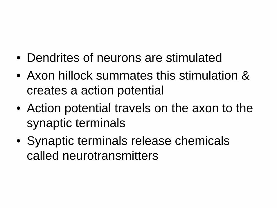
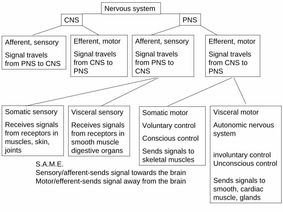
• Dendrites of neurons are stimulated• Axon hillock summates this stimulation &
creates a action potential• Action potential travels on the axon to the
synaptic terminals• Synaptic terminals release chemicals
called neurotransmitters

Fig
13.10
No axonRare, not myelinated
May be myelinated
Sensory neurons
Most common
Myelinated
Motor neurons
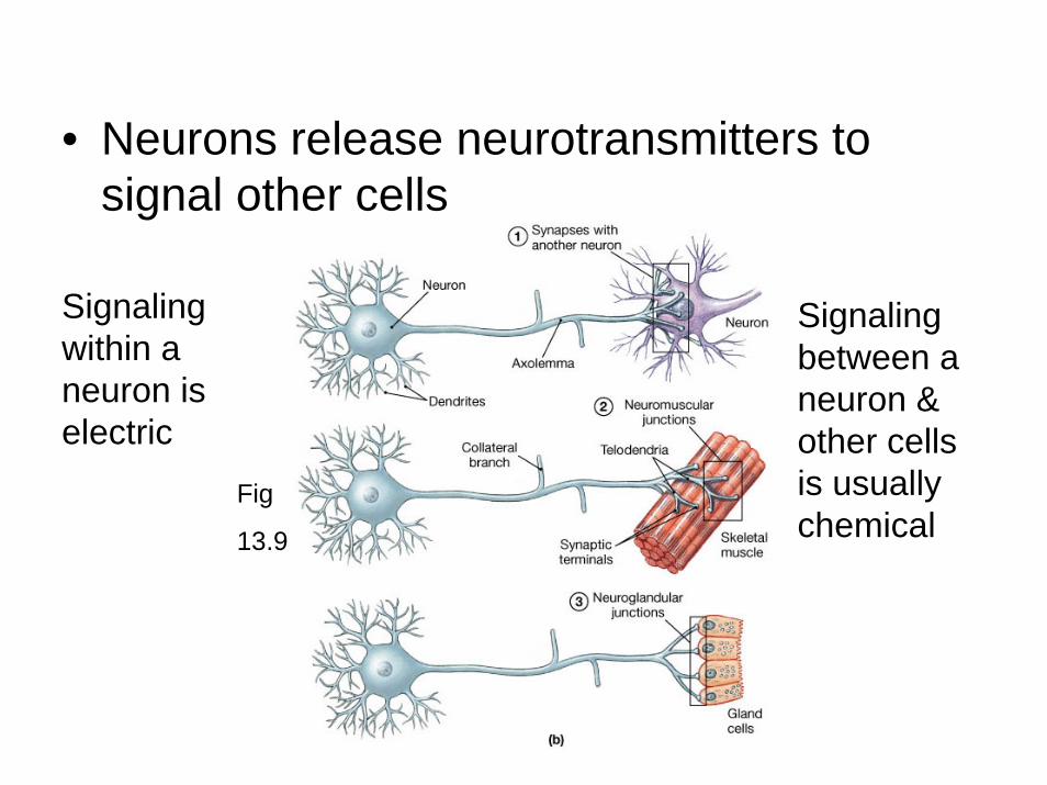
• Neurons release neurotransmitters to signal other cells
Fig
13.9
Signaling within a neuron is electric
Signaling between a neuron & other cells is usually chemical

The neurogliaFig
13.4
CNS neuroglia
• Astrocytes-most numerous– Repair damages neurons– Control interstitial environment– Blood brain barrier
• surround capillaries to isolate the brain from chemicals in the plasma
• Ependymal cells-with capillaries produce cerebral spinal fluid in the brain
• Oligodendrocytes-myelinate axons in the CNS– Works like insulation making actions
potentials travel down axons ~ 6 times faster• Microglia-break down cellular waste and
pathogens in the CNS

Fig
13.5

PNS neuroglia
• Schwann cells-myelinate axons in the PNS
• Satellite cells-exchange waste/nutrients cell body & extracellullar fluid
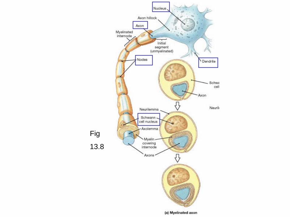
Fig
13.8

Nerve impulse
• A neuron is electrically stimulated to threshold (summation @ axon hillock)
• At the threshold the cell membrane permeability to ions Na+/K+ changes
• This creates an action potential• Large myelinated axon sends signals at
300 mhp!
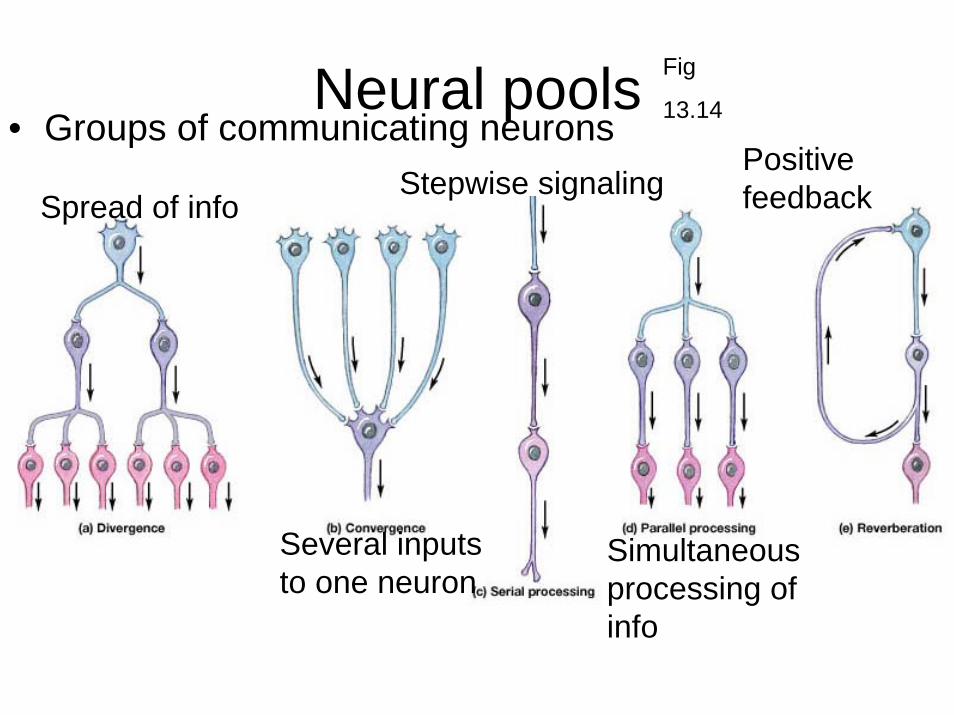
Neural pools• Groups of communicating neurons
Fig
13.14
Spread of info
Several inputs to one neuron
Stepwise signaling
Simultaneous processing of info
Positive feedback

The brain
• Adult Contains 98% of all neural tissue• 3 lbs, feels like jello• 3 primary brain vesicles a 3 weeks
table
15.1

Fig
15.1

Fig
15.11
Grey matter
White matter
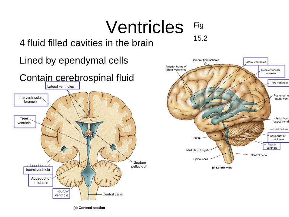
Ventricles4 fluid filled cavities in the brain
Lined by ependymal cells
Contain cerebrospinal fluid
Fig
15.2

CSF
• Cushions the CNS• Supports the brain-the brains is floating in
the CSF• Transport nutrient/wastes etc.
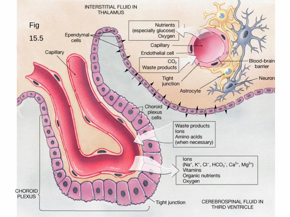
Choroid plexus
• Produces CSF 500 ml/day• Composed of ependymal cells and
capillaries (CSF is very different from plasma)• Found in each ventricle• Floor of lateral ventricles (2)• Roof of 3rd ventricle• Roof of 4th ventricle
Fig
15.5
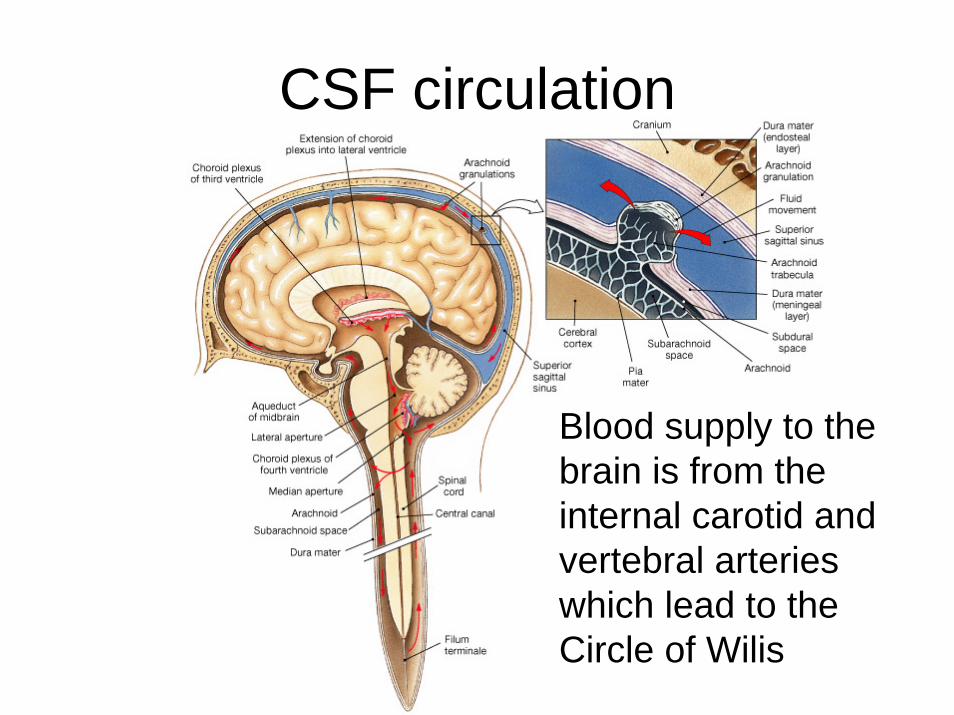
CSF circulation
Blood supply to the brain is from the internal carotid and vertebral arteries which lead to the Circle of Wilis

Blood brain barrier
• Maintained by astrocytes• Not found in:
– the hypothalamus– Pineal gland– Roof of 3rd & 4th ventricles

Cranial Meninges
• Protective layers of the brain & spinal cord– Provide physical stability and shock absorption
• Superficial– Dura mater-Tough fibrous layer– Arachnoid– Pia mater
• Deep

Fig
15.4

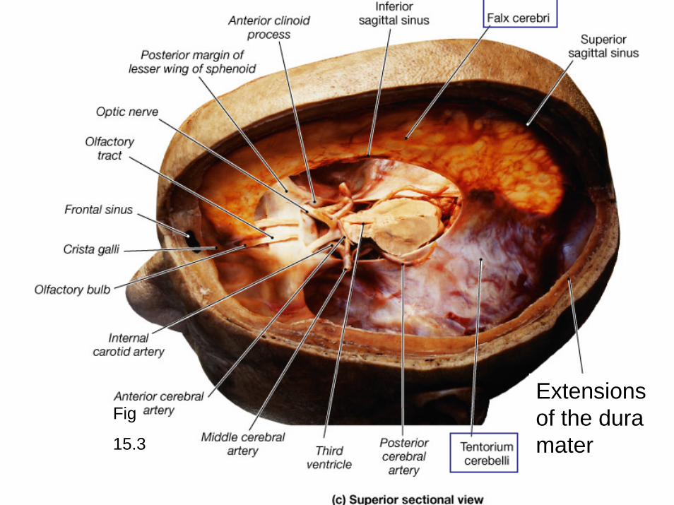
Fig
15.3
Extensions of the duramater

Fig
15.4
• Deep to arachnoidis subarachnoidspace– Network of
collagen and elastin fibers (arachnoidtrabeculae)
– Contains CSF

Fig
15.21
12 pairs of Cranial nerves
Old
Owls
On
Tree
Tops Are Forever
Viewing
Green
Valleys
And
Hills12
11
1-12

Fig
15.22
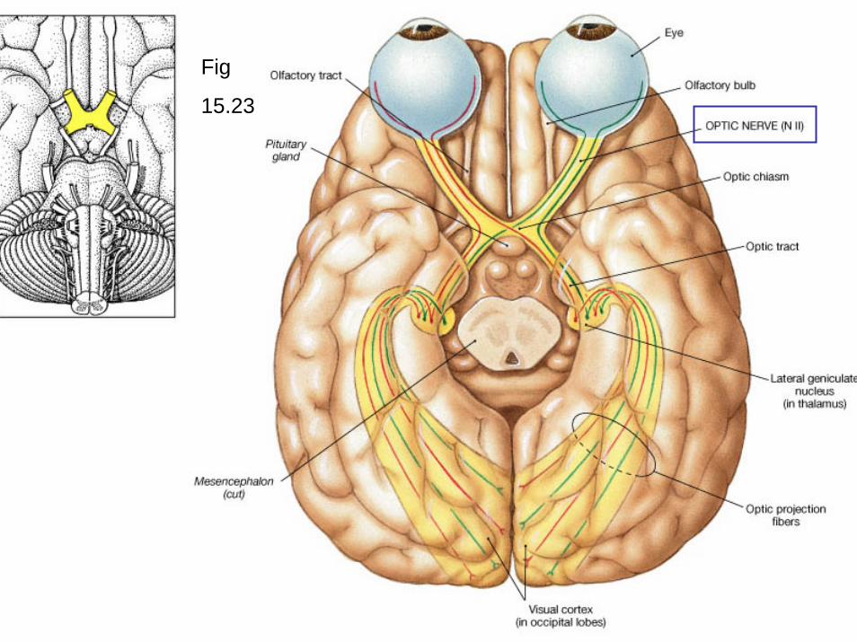
Fig
15.23
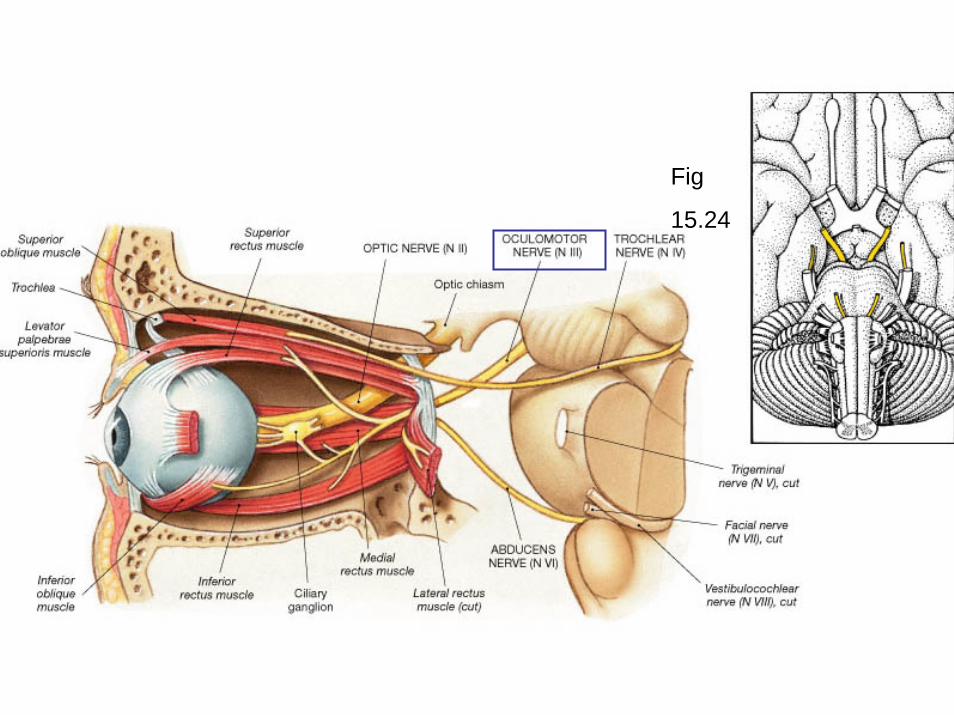
Fig
15.24

Fig
15.24

Fig
15.25

Fig
15.24

Fig
15.26

Fig
15.27

Fig
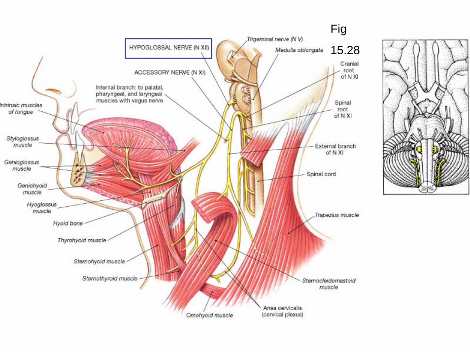
15.28
Fig
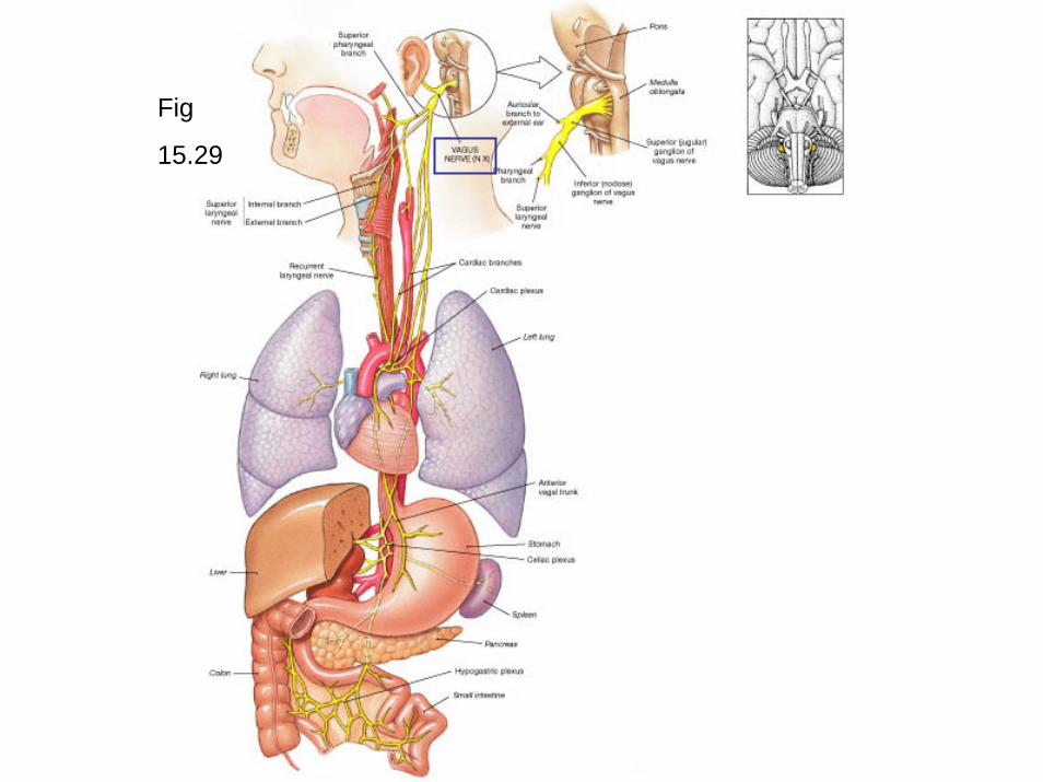
15.29

Fig
15.28
Fig
15.28

oculomotor
facial
glossopharyngeal
trochlear
vagus
Know the primary function of each nerve

break

Cranial dissection video
• By Kevin Petti
Histology CD
Fig
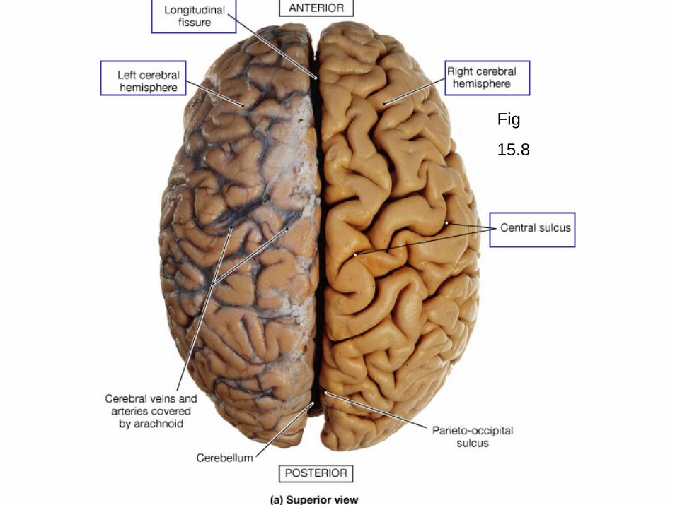
15.8

Fig
15.9
Transverse fissure
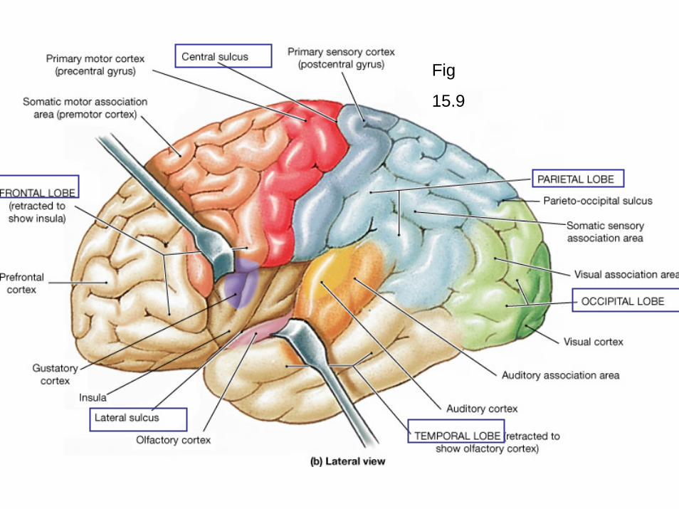
Fig
15.9
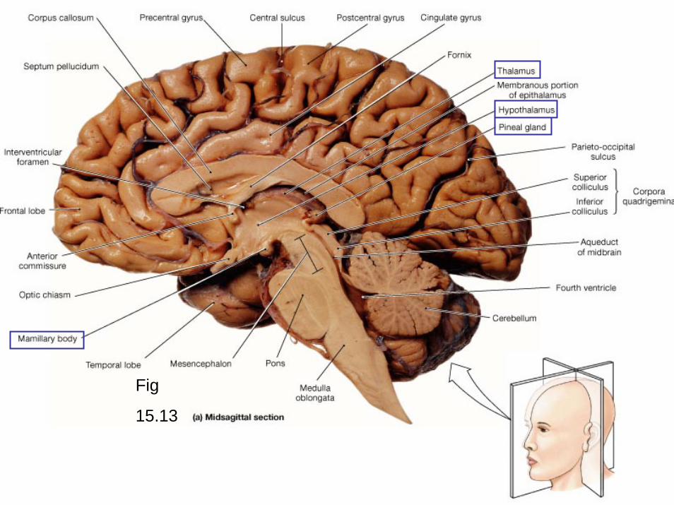
Fig
15.13


Cerebral peduncles
Copora
quadrigemina
Fig
15.15
Aqueduct of midbrain or
Cerebral aqueduct

Fig
15.13

Fig
15.19

Fig
15.13

homunculusFYI

Homunculus
A distorted human figure drawn to reflect the space our body parts occupy on the sensory and motor cortex.
FYI