Embed Size (px)
Citation preview
Chaudhary, Rajneesh, Roy, Kislay, Kanwar, Rupinder Kaur, Walder, Ken and Kanwar, Jagat Rakesh 2016, Engineered atherosclerosis-specific zinc ferrite nanocomplex-based MRI contrast agents, Journal of nanobiotechnology, vol. 14, no. Article number : 6, pp. 1-17. DOI: 10.1186/s12951-016-0157-1 This is the published version. ©2016, The Authors Reproduced by Deakin University under the terms of the Creative Commons Attribution Licence Available from Deakin Research Online: http://hdl.handle.net/10536/DRO/DU:30081881
Chaudhary et al. J Nanobiotechnol (2016) 14:6 DOI 10.1186/s12951-016-0157-1
RESEARCH
Engineered atherosclerosis-specific zinc ferrite nanocomplex-based MRI contrast agentsRajneesh Chaudhary1, Kislay Roy1, Rupinder Kaur Kanwar1, Ken Walder2 and Jagat Rakesh Kanwar1*
Abstract
Background: Cardiovascular diseases are the most prevalent cause of morbidity and mortality affecting millions of people globally. The most effective way to counter cardiovascular complications is early diagnosis and the safest non-invasive diagnostic approach is magnetic resonance imaging (MRI). In this study, superparamagnetic ferrite nanoparticles doped with zinc, exhibiting highly enhanced saturation magnetization and T2 and computed tomography (CT) contrast were synthesized. These nanoparticles have been strategically engineered using bovine lactoferrin (Lf ), polyethylene glycol (PEG), and heat shock protein (Hsp)-70 antibody specifically targeting atherosclerosis with potential therapeutic value. The nanocomplexes were further validated in vitro to assess their cytotoxicity, internalization efficiency, effects on cellular proliferation and were assessed for MRI as well as X-ray CT in ex vivo Psammomys obesus rat model.
Results: Optimized zinc doped ferrite nanoparticles (Zn0.4Fe2.6O4) with enhanced value of maximum saturation magnetization value on 108.4 emu/g and an average diameter of 24 ± 2 nm were successfully synthesized. Success-fully incorporation with bovine lactoferrin, PEG and Hsp-70 (70 kDa) antibody led to synthesis of spherical nanocom-plexes (size 224.8 nm, PDI 0.398). A significantly higher enhancement in T2 (p < 0.05, 1.22-fold) and slightly higher T1 (1.09-fold) and CT (1.08-fold) contrast compared to commercial ferrite nanoparticles was observed. The nanocom-plexes exhibited effective cellular internalization within 2 h in both THP-1 and Jurkat cells. MRI scans of contrast agent injected animal revealed significant arterial narrowing and a significantly higher T2 (p < 0.05, 1.71-fold) contrast in adult animals when compared to juvenile and control animals. The excised heart and aorta agar phantoms exhibited weak MRI contrast enhancement in juvenile animal but significant contrast enhancement in adult animal specifically at the aortic arch, descending thoracic aorta and iliac bifurcation region with X-ray CT scan. Histological investiga-tion of the contrast agent injected aorta and heart confirmed site target-specific accumulation at the atheroscle-rotic aortic arch and descending thoracic aorta of the adult animal with severely damaged intima full of ruptured microatheromas.
Conclusion: Overall, the study demonstrates the strategic development of nanocomplex based bimodal MRI and CT contrast agents and its validation on Psammomys obesus for atherosclerosis diagnostics.
Keywords: Atherosclerosis, Zinc doped ferrite nanoparticles, Heat shock protein, Lactoferrin, Psammomys obesus, Magnetic resonance imaging
© 2016 Chaudhary et al. This article is distributed under the terms of the Creative Commons Attribution 4.0 International License (http://creativecommons.org/licenses/by/4.0/), which permits unrestricted use, distribution, and reproduction in any medium, provided you give appropriate credit to the original author(s) and the source, provide a link to the Creative Commons license, and indicate if changes were made. The Creative Commons Public Domain Dedication waiver (http://creativecommons.org/publicdomain/zero/1.0/) applies to the data made available in this article, unless otherwise stated.
BackgroundAtherosclerosis is a root cause of cardiovascular diseases (CVD) leading to morbidity and mortality in millions of people worldwide [1, 2] and is often symptomatically
silent until the occurrence of a catastrophic clinical event such as myocardial infarction, stroke or peripheral vascu-lar diseases. The most reliable and conventional invasive technique for the visualization of affected blood vessels, either arteries or veins is catheter angiography [3] which is highly invasive, requires a complicated operative proce-dure, exposes the patient to X-ray radiation, is time con-suming and the catheter may injure the artery which may lead to internal bleeding. On the other hand, magnetic resonance imaging (MRI) is a non-invasive, painless,
Open Access
Journal of Nanobiotechnology
*Correspondence: [email protected] 1 Nanomedicine-Laboratory of Immunology and Molecular Biomedical Research (NLIMBR), School of Medicine (SoM), Faculty of Health, Centre for Molecular and Medical Research (C-MMR), Deakin University, Pigdons Road, Waurn Ponds, Geelong, VIC 3217, AustraliaFull list of author information is available at the end of the article
Page 2 of 17Chaudhary et al. J Nanobiotechnol (2016) 14:6
and safe diagnostic technology that uses a magnetic field and radio frequency to produce a detailed image of the body’s organs and structures with high spatial resolu-tion depending on the differences in longitudinal (T1) and transverse (T2) proton relaxation times of contrast agents that can effectively improve the MRI contrast between normal and diseased tissues [4]. Paramagnetic gadolinium chelates are the most widely used commer-cial MRI contrast agents used for MRI based diagnostics [5]. However, Gadolinium exhibits high in vivo toxicity and therefore must be modified with organic chelators such as diethylene triamine pentaacetic acid (Gd-DTPA) and 1,4,7,10-tetraazacyclododecane-1,4,7,10-tetraacetic acid (Gd-DOTA) [6]. Even after chelation gadolinium based contrast agents exhibit nephrogenic fibrosing der-mopathy [7] and nephrogenic systemic fibrosis [8] in patients with renal insufficiency, primarily due to com-petitive transmettalation of divalent or trivalent cations with gadolinium [7, 9].
Engineered nanoparticulate contrast agents progres-sively contribute to the field of cardiovascular diagnostics and molecular imaging by providing several advantages over traditional imaging contrast agents [10, 11]. The most extensively studied class of magnetic nanoparticles for contrast agents development are iron oxide nanopar-ticles, also referred as ferrite nanoparticles. This is due to their superparamagnetic nature, biodegradability, inoffen-sive toxicity profile, low particle dimensions, high surface to volume ratio and reactive surface readily modifiable with biocompatible coatings, targeting/therapeutic mol-ecules as well as other imaging modalities [4, 12–15]. The mode of action of ferrite nanoparticles is by enhancing the relaxation rate or shortening the T2 relaxation time by increasing field inhomogeneity of surrounding protons to creating strong negative MRI contrast [4, 12–15]. Ferrite nanoparticles can be doped with zinc, a micronutrient to considerably enhance the saturation magnetization (MS) for improved MRI contrast while effectively reducing the chances of iron overloading by iron cation replacement [16, 17]. Zinc is the most suitable candidate as a dopant as Food and Drug Administration (FDA) recommends a high reference daily value (DV) for zinc and iron to be 15 and 18 mg respectively, considerably much more than the values for other possible dopants, for example manganese and cobalt at 2 mg (http://www.fda.gov).
In the present study, we developed and validated highly superparamagnetic zinc doped ferrite (ZF) nanoparti-cle based nanocomplexes with minimized in vivo toxic-ity compared to commercial ferrite based MRI contrast agents. Three types of bovine lactoferrin-zinc ferrite (Lf-ZF) nanocomplexes were developed, Hsp-70 targeted lactoferrin embedded ZF nanoparticles (Hsp-70 Lf-ZF), Hsp-70 targeted chitosan encapsulated lactoferrin
embedded ZF nanoparticles (Hsp-70 Ch-Lf-ZF) and Hsp-70 targeted polyethylene glycol assisted lactoferrin embedded ZF (Hsp-70 Lf-PEG-ZF). Lf-ZF forms the core of all nanocomplexes, where superparamagnetic ZF nan-oparticles generate MRI contrast and lactoferrin acts as a targeting and therapeutic protein against atherosclerosis. Lactoferrin is a naturally available protein with anti-bac-terial, anti-viral, anti-inflammatory and anti-carcinogenic properties and also helps in boosting the immune sys-tem by activation of T lymphocytes [18, 19]. Since, the temporal expression of Hsps’ has been demonstrated at atherosclerotic lesion [20], this is the first attempt dem-onstrating Hsp-70 targeted approach for atherosclerosis diagnostics. Validation of the developed nanocomplex contrast agent was performed with Psammomys obesus (P. obesus), a polygenic animal model for type-2 diabetes, obesity and atherosclerosis [21, 22].
Results and discussionCharacterization of zinc doped ferrite based nanocomplexesThe first phase of this study was the synthesis, charac-terization and optimization of zinc-doped ferrite nano-particles (ZF) with their physicochemical properties as the primary criteria in order to procure a highly effec-tive MRI contrast. Selection of zinc, a micronutrient, as the dopant was implemented as a strategy to maximize the characteristic magnetic property of the nanoparti-cles and simultaneously minimizing potential cytotoxic-ity in vivo at higher concentrations. Zinc doping reduces the chances of iron overloading by crystallographic fer-rite iron cation replacement with highly enhanced satura-tion magnetization of 108.4 emu/g at room temperature (data not shown), considerably higher than conventional ferrite nanoparticles employed for MRI contrast agent development. Transmission electron microscopy (TEM) revealed the formation of ZF nanoparticles with an aver-age diameter of 24 ± 2 nm (Fig. 1a) and X-ray diffractom-etry (XRD) confirmed the formation of crystallographic spinel structure (Fig. 1b). The next phase of the study involved the design and development of atherosclerosis-specific nanocomplex MRI contrast agents with empha-sis on their in vitro validation by cellular proliferation, toxicity, internalization and molecular targeting assess-ment. To achieve this goal, three spherical nanosized complexes based on different combinations of ZF nano-particles, bovine lactoferrin, chitosan and PEG were developed. All nanocomplexes were equipped with heat shock protein (Hsp)-70 antibodies for effective targeting of the atherosclerotic inflammatory site. The three types of engineered nanocomplexes developed were named according to their constituents: Hsp-70 targeted lactofer-rin embedded ZF nanoparticles (Hsp-70 Lf-ZF), Hsp-70
Page 3 of 17Chaudhary et al. J Nanobiotechnol (2016) 14:6
Page 4 of 17Chaudhary et al. J Nanobiotechnol (2016) 14:6
targeted chitosan encapsulated lactoferrin embedded zinc ferrite nanoparticles (Hsp-70 Ch-Lf-ZF) and Hsp-70 targeted polyethylene glycol assisted lactoferrin embed-ded ZF nanoparticles (Hsp-70 Lf-PEG-ZF). Lactoferrin is also a known iron modulator internalized by vari-ous cells such as mucosal epithelial cells, hepatocytes, monocytes, macrophages, polymorphonuclear leuko-cytes, lymphocytes, thrombocytes and fibroblasts due to the presence of a number of cellular receptors including transferrin receptors, Lactoferrin receptors and Lipo-protein receptor-related proteins [18, 23–25]. We used chitosan as, chitosan is a naturally occurring mucoadhe-sive polysaccharide having a strong affinity for Mucin-1 receptor (Muc-1) expressing endothelial and epithelial cells [26]. Additionally, chitosan as a nanocomplex car-rier equips the nanocomplex to undertake transcellu-lar and paracellular internalization pathways and also exhibits strong affinity to cholesterol and triglycerides [27], and lipid accumulation is a primary characteristic of the atherosclerotic plaque inflammation [28]. Moreover, it has also been reported to accelerate the wound heal-ing and exert anti-inflammatory effects [29]. Previously published studies have also shown that dietary chitosan inhibits hypercholesterolaemia and atherogenesis in the apolipoprotein E (apoE) deficient mouse model of ath-erosclerosis [30]. Orally administered positively charged chitosan binds to lipids in the intestine, blocking absorp-tion, shown to lower blood cholesterol in animals and humans as a result it has been proposed that dietary sup-plementation with chitosan may inhibit the formation of atherosclerotic plaque [30]. Tail vein injection [31] and intranasal delivery [32] of chitosan based nanoformula-tions have shown to successfully inhibit atherosclerosis in animal models. However, we were unable to synthesize optimum nanoparticles due to the large polydispersity index (PDI). PEG on the other hand, also has several ben-efits as it leads to formation of sterically stabilised ‘stealth’ particles [33] and its hydrophilic nature leads to reduced non-specific protein adsorption [34], avoids aggrega-tion [35] and imparts the property of water solubility to the nanoparticles [36]. Therefore, we went on to replace
chitosan with PEG and were able to synthesize optimum (224.8 nm, PDI 0.398) nanoparticles. A study performed to assess the distribution of Hsp-70 between fibrotic and necrotic plaques in immunostained carotid endarterec-tomy specimens revealed that necrotic plaques and their underlying media contained significantly more Hsp-70 expression than the fibrotic tissues [38]. Their results suggested that inadequate Hsp-70 accumulates with smooth muscle cells residing near necrosis to defend against plaque toxicity. Therefore, all nanocomplexes were conjugated with Hsp-70 for target specific internal-isation. Hsp-70 is also known to protect cellular compo-nents from injury by decreasing oxidation, inflammation and apoptosis and also by refolding damaged proteins [37].
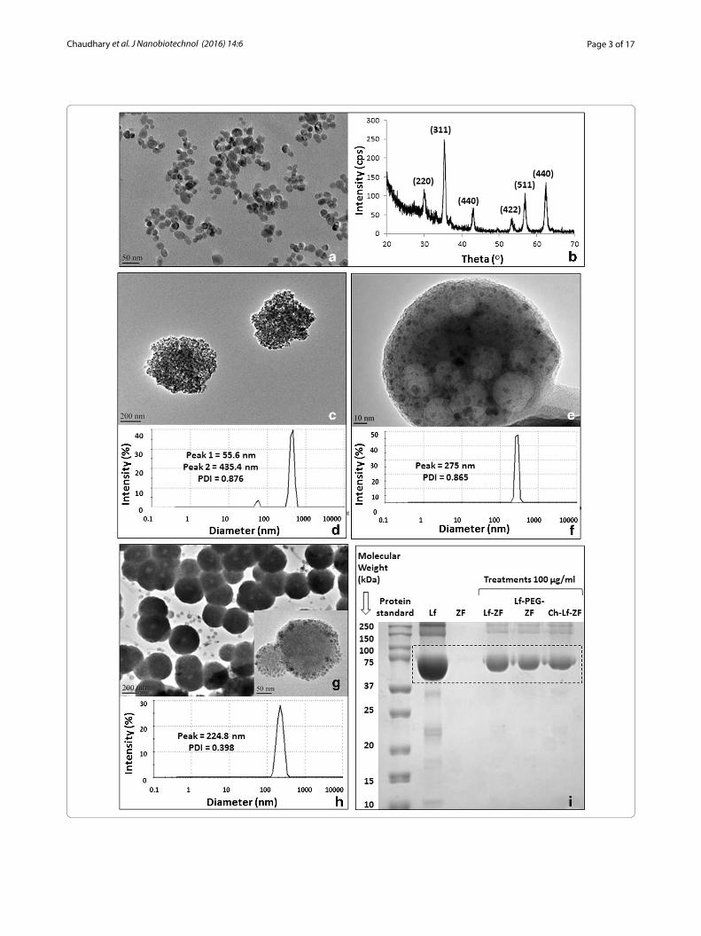
Transmission electron microscopy (TEM) imaging confirmed the formation of all three spherical nano-complexes (Fig. 1c, e, g). Lactoferrin ZF complex can be clearly observed by the TEM images. Furthermore, lactoferrin ZF complex embedded with chitosan can also be clearly observed by the TEM image. Intensity based hydrodynamic size distribution obtained by dynamic light scattering (DLS) exhibited an average diameter of 224.8 and 275 nm for Hsp-70 Lf-PEG-ZF and Hsp-70 Ch-Lf-ZF nanocomplexes respectively (Fig. 1e, g). A bimodal hydrodynamic size distribution was observed for Hsp-70 Lf-ZF with the primary peak at 435.4 nm and secondary peak at 55.6 nm average diameter respectively (Fig. 1f ). Hsp-70 Lf-ZF and Hsp-70 Ch-Lf-ZF nanocomplex exhib-ited a very high polydispersity index (PDI) value of 0.876 and 0.865 respectively. Hsp-70 Lf-PEG-ZF exhibited a relatively narrow size distribution with a PDI value of 0.398. Furthermore, lactoferrin loading was confirmed by band formation around 78 kDa for all nanocomplexes corresponding to lactoferrin as observed by poly acryl amide gel electrophoresis (PAGE) (Fig. 1i). The serum stability studies performed using DLS confirmed that Hsp-70 Lf-PEG-ZF showed least aggregation in 10 % bovine serum albumin (BSA), followed by Hsp-70 Lf-ZF and Hsp-70 Ch-Lf-ZF nanocomplexs (Additional file 1: Figure S1).
(See figure on previous page.) Fig. 1 Characterization of zinc doped ferrite nanoparticles and their nanocomplexes. a Transmission electron microscopy (TEM) image confirmed formation of uniformly sized spherical zinc-doped ferrite nanoparticles (Zn0.4Fe2.6O4) with an average diameter of 24 ± 2.8 nm. b X-ray differacto-gram revealed the formation of crystallographic spinel structure of zinc-doped ferrite nanoparticles. c TEM image revealing the formation of Hsp-70 Lf- ZF nanocomplex. d DLS analysis of Hsp-70 Lf-ZF nanoparticles revealed bimodal hydrodynamic size distribution with the prominent primary peak at 435.4 nm and a secondary peak at 55.6 nm with a polydispersity index of 0.876. e TEM image revealed the formation of Hsp-70 Ch-Lf-ZF nanocomplex. f DLS analysis revealed the hydrodynamic diameter of 275 nm with a PDI of 0.865. g TEM image revealing the formation of Hsp-70 Lf-PEG-ZF nanocomplex and a highly magnified image (inset). h Dynamic light scattering (DLS) revealed the hydrodynamic diameter of 224.8 nm with a polydispersity index of 0.398. i Sodium dodecyl sulfate—polyacrylamide gel electrophoresis (SDS-PAGE) confirming lactoferrin loading of the nanocomplexes. From left to right, standard protein ladder, pure bovine lactoferrin (Lf ), ZF nanoparticles, Hsp-70 Lf-ZF, Hsp-70 Lf-PEG-ZF and Hsp-70 Ch-Lf-ZF nanocomplexes. Nanocomplex lactoferrin loading was confirmed with specific band detection at 75 kDa
Page 5 of 17Chaudhary et al. J Nanobiotechnol (2016) 14:6
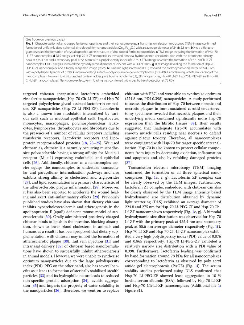
Measurement of MRI/CT contrast and in vitro internalisation assessment of Hsp‑70 Lf‑PEG‑ZF nanoparticlesA slight enhancement of T1 MRI contrast (1.09-fold) was observed in Hsp-70 Lf-PEG-ZF compared to fer-rite nanoparticles (Fig. 2a; Additional file 2: Figure S2a). Significant enhancement of T2 MRI contrast (p < 0.05, 1.22-fold) was observed in Hsp-70 Lf-PEG-ZF compared to ferrite nanoparticles (Fig. 2b, Additional file 2: Figure S2b). Slight enhancement of CT contrast (1.08-fold) was observed in Hsp-70 Lf-PEG-ZF compared to ferrite nan-oparticles (Fig. 2c, Additional file 2: Figure S2c).
All three nanocomplexes were assessed with THP-1 monocyte and T lymphocyte cell lines. The internali-zation efficacy of these nanoparticles was tested using fluorescence confocal microscopy and flow cytometry (Fig. 2d, e). Confocal microscopy revealed very effec-tive cellular internalization by all three nanocomplexes at 6 h after treatment with both cell lines. Flow cytom-etry observation revealed that Hsp-70 Ch-Lf-ZF showed the maximum internalization in THP-1 cells (p < 0.001,
1.41-fold higher than Hsp-70 PEG-Lf-ZF) whereas, Hsp-70 PEG-Lf-ZF showed the maximum internalization in Jurkat cells (p < 0.01, 2.05-fold higher than Hsp-70 Ch-Lf-ZF). This is in accordance with previously published studies where mucoadhesive chitosan nanoparticles have been reported to internalize with very high efficiency in THP-1 cells without inducing any release of pro-inflam-matory cytokines [39].
Monocytes and lymphocytes play central roles in ath-erosclerotic inflammation from the early inflammatory phase to the late thrombotic phase [40]. Therefore, in this study, the nanocomplex effects and internalisation were studied with THP-1 monocytes and Jurkat lymphocytes as in vitro models of cells having a central role in athero-sclerotic plaque inflammation [28].
Flow cytometry also revealed highly efficient inter-nalization by monocytes as compared to lymphocytes primarily due to their Phagocytic characteristic. LDH release (cytotoxicity) and CyQUANT cell prolifera-tion assays were performed in both THP-1 and Jurkat cells following treatment with all three nanocomplexes
Fig. 2 Enhancement in MRI/CT contrast and internalisation efficacy of nanoparticles. a Slight enhancement of T1 MRI contrast was observed in Hsp-70 Lf-PEG-ZF compared to ferrite nanoparticles. b Significant enhancement of T2 MRI contrast was observed in Hsp-70 Lf-PEG-ZF compared to ferrite nanoparticles. c Slight enhancement of CT contrast was observed in Hsp-70 Lf-PEG-ZF compared to ferrite nanoparticles. d Confocal micros-copy internalization images and cell cytometric internalization assessment displaying time-dependent internalization of Hsp-70 Lf-ZF, Hsp-70 Ch-Lf-ZF and Hsp-70 Lf-PEG-ZF nanocomplexes in THP-1 cells and e Jurkat cells. Effective internalization was observed at 2 h Hsp-70 Lf-ZF nanocomplex post-treatment
Page 6 of 17Chaudhary et al. J Nanobiotechnol (2016) 14:6
(Additional file 3: Figure S3a–c). The results revealed that Hsp-70 PEG-Lf-ZF failed to induce any significant cytotoxicity but led to a significant cellular proliferation (p < 0.05) with both THP-1 or Jurkat cells in lower con-centrations (40–2.5 µg/ml). The Hsp-70 Ch-Lf-ZF led to significant cytotoxicity and significant reduction in pro-liferation (p < 0.05) in both THP-1 and Jurkat at 100 µg/ml and led to significant (p < 0.05) increase in prolifera-tion in THP-1 cells at lower concentrations (50–2.5 µg/ml).
Ex vivo MRI/CT of Hsp‑70 Lf‑PEG‑ZF nanoparticles in P. obesus atherosclerotic modelPsammomys obesus exhibit age-dependency and phe-notypic heterogeneity in acquiring diabetes and obesity indicating the polygenic nature of pathogenesis in P. obesus, closely mimicking the human populations [41]. A genetic correlation between type-2 diabetes, athero-sclerosis and inflammation has been well established since the discovery of the role of Tanis gene in P. obesus [22]. Since, we found that Hsp70-Lf-ZF was not suitable due to large particle size and Then we syntheized Hsp-70 Ch-Lf-Zf nanocomplex had very high poydispersity index. Therefore, all further studies were conducted with Hsp70-PEG-Lf-ZF that has a narrower hydrodynamic size distribution.
Therefore, the next phase of ex vivo imaging experi-ments emulated a real-time in vivo scenario by injecting Hsp-70 Lf-PEG-ZF contrast agent directly into the intact animal aorta. Three groups of rats (18 months old Con-trol n = 5, 6 months old injected with Hsp-70 Lf-PEG-ZF n = 5 and 16 months old injected with Hsp-70 Lf-PEG-ZF n = 5) were used with minimal dissection steps prior to MRI/CT scan. Several benefits can be realized by using a direct intra-aortic ex vivo contrast agent injection strat-egy. It imitates an in vivo MRI scan of the animal provid-ing additional internal anatomical information from MRI contrast interferences arising from organs, tissues and other materials and fluids, especially fat. Furthermore, the strategy minimizes the risk of causing any damage to the aorta and heart prior to imaging. There is mini-mal dissection, and pre-treatment electrocautery steps to seal aortic branches can be avoided with no possibility of any unwanted artefacts. The exploratory ex vivo MRI/CT assessment reported in this study paves the way for future in vivo multi-modal imaging studies with P. obesus for development of atherosclerotic inflammation specific contrast or therapeutic agents.
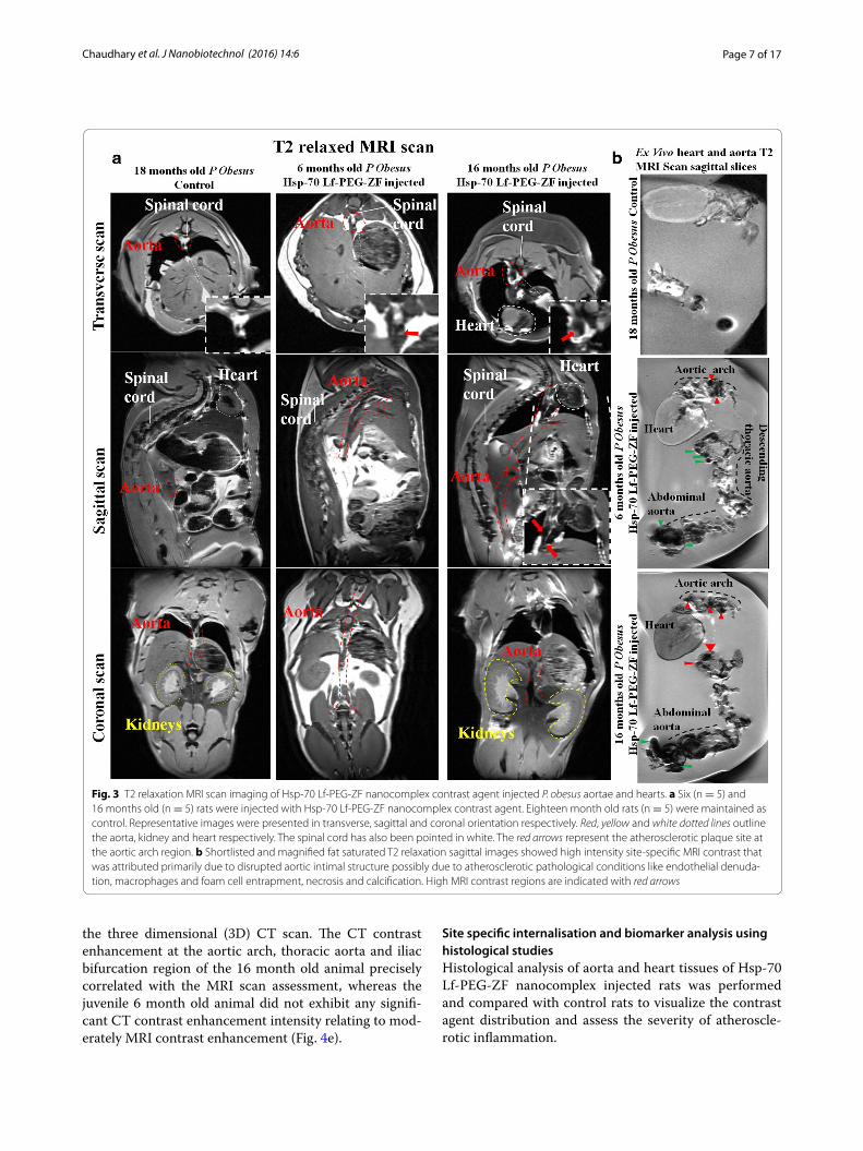
Representative images (Fig. 3a) and the correspond-ing heat map images (Additional file 4: Figure S4a) were presented for simplification. The control animal, per-fused with PBS only showed minimal MRI intensity in all three orientations (Transverse, sagittal and coronal).
High positive contrast exhibiting peri-aortic fat accumu-lation was clearly observable around the arch region. The renal artery had a poorly defined periphery with the sag-ittal scan images. Transverse scan images provided the most prominent information about the aortic peripheral morphology with the contrast agent injected in young and old animals. However, with the PBS injected animal the periphery was very poorly defined to derive any con-clusion about P. obesus cardiovascular health status. A detailed examination of the aortic morphological status was conducted with the MRI assessment with both young (6 months old) and old (16 months old) direct Hsp-70 Lf-PEG-ZF agent injected P. obesus. A higher overall aortic contrast intensity (p < 0.05, 1.71-fold) was observed in the old animals compared to the younger counterparts (Addi-tional file 4: Figure S4b). However, both animals displayed atherosclerotic narrowing with intense T2 relaxed nega-tive contrast at the descending aortic arch site adjacent to the high positive contrast peri-aortic fat accumulation. This mixed positive negative contrast at the arterial nar-rowing sites is caused by negative MRI contrast signal arising from accumulated contrast agent and positive contrast exhibited by lipid at the atherosclerotic plaque site. The young animal MRI images exhibited moderate site-specific accumulation only at the aortic arch site with arterial stenosis and the remaining tissue. Alternatively, the older animal displayed strong contrast enhancement at the aortic arch and descending thoracic aorta regions and moderate contrast at the abdominal aorta.
MRI scan of the aorta from these rats exhibited mod-erate contrast enhancement at the aortic arch and sig-nificantly high contrast enhancement at the descending thoracic aorta region compared to the control. The nanocomplex contrast agent injected heart and aorta agar phantom was made in a customized diamagnetic holder to avoid any scan interference and imaging was performed with a 9.4T MRI scanner (Fig. 3b; Addi-tional file 5: Figure S5). Overall, excellent T2 MRI con-trast enhancement was observed with the contrast agent injected old animal aortas compared to the young animal aortas and the control.
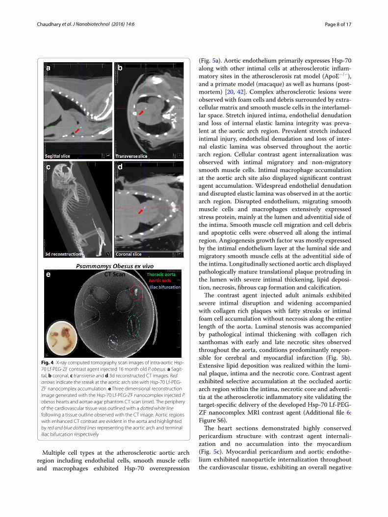
Additionally, contrast agent injected 16 months old (adult) P. obesus were subjected to X-ray CT scan to assess aortic arch contrast agent localization corre-sponding to the T2 MRI scan. Sagittal, transverse and coronal scan exhibited a clear streak with intense CT signal the aortic arch site due to targeted accumula-tion (seen in white contrast) at the aortic arch site reaf-firming the nanocomplex contrast agent accumulation at the aortic arch site (Fig. 4a–d). This was followed by CT imaging of the aorta to assess the potential for CT bimodality. The analysed aortic regions with T2 MRI contrast enhancement image directly corresponded with
Page 7 of 17Chaudhary et al. J Nanobiotechnol (2016) 14:6
the three dimensional (3D) CT scan. The CT contrast enhancement at the aortic arch, thoracic aorta and iliac bifurcation region of the 16 month old animal precisely correlated with the MRI scan assessment, whereas the juvenile 6 month old animal did not exhibit any signifi-cant CT contrast enhancement intensity relating to mod-erately MRI contrast enhancement (Fig. 4e).
Site specific internalisation and biomarker analysis using histological studiesHistological analysis of aorta and heart tissues of Hsp-70 Lf-PEG-ZF nanocomplex injected rats was performed and compared with control rats to visualize the contrast agent distribution and assess the severity of atheroscle-rotic inflammation.
Fig. 3 T2 relaxation MRI scan imaging of Hsp-70 Lf-PEG-ZF nanocomplex contrast agent injected P. obesus aortae and hearts. a Six (n = 5) and 16 months old (n = 5) rats were injected with Hsp-70 Lf-PEG-ZF nanocomplex contrast agent. Eighteen month old rats (n = 5) were maintained as control. Representative images were presented in transverse, sagittal and coronal orientation respectively. Red, yellow and white dotted lines outline the aorta, kidney and heart respectively. The spinal cord has also been pointed in white. The red arrows represent the atherosclerotic plaque site at the aortic arch region. b Shortlisted and magnified fat saturated T2 relaxation sagittal images showed high intensity site-specific MRI contrast that was attributed primarily due to disrupted aortic intimal structure possibly due to atherosclerotic pathological conditions like endothelial denuda-tion, macrophages and foam cell entrapment, necrosis and calcification. High MRI contrast regions are indicated with red arrows
Page 8 of 17Chaudhary et al. J Nanobiotechnol (2016) 14:6
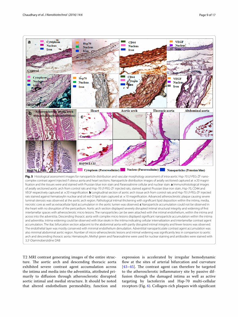
Multiple cell types at the atherosclerotic aortic arch region including endothelial cells, smooth muscle cells and macrophages exhibited Hsp-70 overexpression
(Fig. 5a). Aortic endothelium primarily expresses Hsp-70 along with other intimal cells at atherosclerotic inflam-matory sites in the atherosclerosis rat model (ApoE−/−), and a primate model (macaque) as well as humans (post-mortem) [20, 42]. Complex atherosclerotic lesions were observed with foam cells and debris surrounded by extra-cellular matrix and smooth muscle cells in the interlamel-lar space. Stretch injured intima, endothelial denudation and loss of internal elastic lamina integrity was preva-lent at the aortic arch region. Prevalent stretch induced intimal injury, endothelial denudation and loss of inter-nal elastic lamina was observed throughout the aortic arch region. Cellular contrast agent internalization was observed with intimal migratory and non-migratory smooth muscle cells. Intimal macrophage accumulation at the aortic arch site also displayed significant contrast agent accumulation. Widespread endothelial denudation and disrupted elastic lamina was observed in at the aortic arch region. Disrupted endothelium, migrating smooth muscle cells and macrophages extensively expressed stress protein, mainly at the lumen and adventitial side of the intima. Smooth muscle cell migration and cell debris and apoptotic cells were observed all along the intimal region. Angiogenesis growth factor was mostly expressed by the intimal endothelium layer at the luminal side and migratory smooth muscle cells at the adventitial side of the intima. Longitudinally sectioned aortic arch displayed pathologically mature translational plaque protruding in the lumen with severe intimal thickening, lipid deposi-tion, necrosis, fibrous cap formation and calcification.
The contrast agent injected adult animals exhibited severe intimal disruption and widening accompanied with collagen rich plaques with fatty streaks or intimal foam cell accumulation without necrosis along the entire length of the aorta. Luminal stenosis was accompanied by pathological intimal thickening with collagen rich xanthomas with early and late necrotic sites observed throughout the aorta, conditions predominantly respon-sible for cerebral and myocardial infarction (Fig. 5b). Extensive lipid deposition was realized within the lumi-nal plaque, intima and the necrotic core. Contrast agent exhibited selective accumulation at the occluded aortic arch region within the intima, necrotic core and adventi-tia at the atherosclerotic inflammatory site validating the target-specific delivery of the developed Hsp-70 Lf-PEG-ZF nanocomplex MRI contrast agent (Additional file 6: Figure S6).
The heart sections demonstrated highly conserved pericardium structure with contrast agent internali-zation and no accumulation into the myocardium (Fig. 5c). Myocardial pericardium and aortic endothe-lium exhibited nanoparticle internalization throughout the cardiovascular tissue, exhibiting an overall negative
Fig. 4 X-ray computed tomography scan images of intra-aortic Hsp-70 Lf-PEG-ZF contrast agent injected 16 month old P. obesus. a Sagit-tal, b coronal, c transverse and d 3d reconstructed CT images. Red arrows indicate the streak at the aortic arch site with Hsp-70 Lf-PEG-ZF nanocomplex accumulation. e Three dimensional reconstruction image generated with the Hsp-70 Lf-PEG-ZF nanocomplex injected P. obesus hearts and aortae agar phantom CT scan (inset). The periphery of the cardiovascular tissue was outlined with a dotted white line following a tissue outline observed with the CT image. Aortic regions with enhanced CT contrast are evident in the aorta and highlighted by red and blue dotted lines representing the aortic arch and terminal iliac bifurcation respectively
Page 9 of 17Chaudhary et al. J Nanobiotechnol (2016) 14:6
T2 MRI contrast generating images of the entire struc-ture. The aortic arch and descending thoracic aorta exhibited severe contrast agent accumulation across the intima and media into the adventitia, attributed pri-marily to diffusion through atherosclerotic disrupted aortic intimal and medial structure. It should be noted that altered endothelium permeability, function and
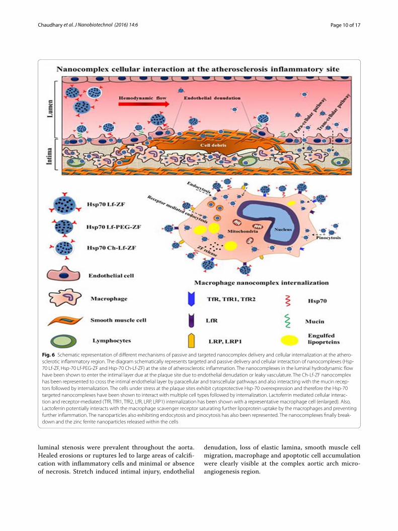
expression is accelerated by irregular hemodynamic flow at the sites of arterial bifurcation and curvature [43–45]. The contrast agent can therefore be targeted to the atherosclerotic inflammatory site by passive dif-fusion through the damaged intima as well as active targeting by lactoferrin and Hsp-70 multi-cellular receptors (Fig. 6). Collagen-rich plaques with significant
Fig. 5 Histological assessment images for nanoparticle distribution and vascular morphology assessment of intra-aortic Hsp-70 Lf-PEG-ZF nano-complex contrast agent injected P. obesus aorta and heart sections. Nanoparticle distribution images of axially sectioned captured at ×20 magni-fication and the tissues were and stained with Prussian blue iron stain and Pararosalinine cellular and nuclear stain. a Immunohistological images of axially sectioned aortic arch from control rats and Hsp-70 Lf-PEG-ZF injected rats, stained against Prussian blue iron stain, Hsp-70, CD44 and VEGF respectively captured at ×20 magnification. b Longitudinal section of aortic arch tissue arch from control rats and Hsp-70 Lf-PEG-ZF injected rats stained against hematoxylin nuclear and oil red-O lipid stain captured at ×10 magnification. Advanced atherosclerotic plaque causing severe luminal stenosis was observed at the aortic arch region. Pathological intimal thickening with significant lipid deposition within the intima, media, necrotic core as well as extracellular lipid accumulation in the aortic lumen was observed. c Nanoparticle accumulation could not be observed in the heart with no disruption of the pericardium. Aortic arch section displayed severely disrupted intimal structural integrity and widening of first interlamellar spaces with atherosclerotic micro lesions. The nanoparticles can be seen attached with the intimal endothelium, within the intima and across into the adventitia. Descending thoracic aorta with complex micro lesions displayed significant nanoparticle accumulation within the intima and adventitia. Intima widening could be observed with blue steaks in the intima indicating cellular internalization and interlamellar contrast agent accumulation. The iliac bifurcation section adjacent to the abdominal aorta with partly disrupted intimal integrity and fewer lesions was observed. The endothelial layer was mostly conserved with minimal endothelium denudation. Adventitial nanoparticulate contrast agent accumulation was also minimal abdominal aortic region. Number of micro-atherosclerotic lesions and intimal widening was significantly less in comparison to aortic arch and descending thoracic aorta. Hematoxylin, Methyl green and Pararosalinine were used for nuclear staining and antibodies were stained with 3,3’-Diaminobenzidine DAB
Page 10 of 17Chaudhary et al. J Nanobiotechnol (2016) 14:6
luminal stenosis were prevalent throughout the aorta. Healed erosions or ruptures led to large areas of calcifi-cation with inflammatory cells and minimal or absence of necrosis. Stretch induced intimal injury, endothelial
denudation, loss of elastic lamina, smooth muscle cell migration, macrophage and apoptotic cell accumulation were clearly visible at the complex aortic arch micro-angiogenesis region.
Fig. 6 Schematic representation of different mechanisms of passive and targeted nanocomplex delivery and cellular internalization at the athero-sclerotic inflammatory region. The diagram schematically represents targeted and passive delivery and cellular interaction of nanocomplexes (Hsp-70 Lf-ZF, Hsp-70 Lf-PEG-ZF and Hsp-70 Ch-Lf-ZF) at the site of atherosclerotic inflammation. The nanocomplexes in the luminal hydrodynamic flow have been shown to enter the intimal layer due at the plaque site due to endothelial denudation or leaky vasculature. The Ch-Lf-ZF nanocomplex has been represented to cross the intimal endothelial layer by paracellular and transcellular pathways and also interacting with the mucin recep-tors followed by internalization. The cells under stress at the plaque sites exhibit cytoprotective Hsp-70 overexpression and therefore the Hsp-70 targeted nanocomplexes have been shown to interact with multiple cell types followed by internalization. Lactoferrin mediated cellular interac-tion and receptor-mediated (TfR, TfR1, TfR2, LfR, LRP, LRP1) internalization has been shown with a representative macrophage cell (enlarged). Also, Lactoferrin potentially interacts with the macrophage scavenger receptor saturating further lipoprotein uptake by the macrophages and preventing further inflammation. The nanoparticles also exhibiting endocytosis and pinocytosis has also been represented. The nanocomplexes finally break-down and the zinc ferrite nanoparticles released within the cells
Page 11 of 17Chaudhary et al. J Nanobiotechnol (2016) 14:6
ConclusionThe study demonstrated development of engineered ath-erosclerosis targeted ZF nanoparticle nanocomplex based T2 relaxation MRI contrast agent which was validated in an ex vivo setting. We also observed that lactoferrin receptor and Hsp-70 antibody induced atherosclerotic inflammatory site specific multi-cellular targeting. Addi-tional insight about aortic contrast agent distribution, accumulation, targeting and internalization was gained by histological examination with both longitudinally and axially oriented aorta and heart sections of contrast agent injected P. obesus rat. Particularly, likely disruptions in the aortic intimal structure due to hemodynamic stress around bifurcation sites were identified, especially in the aorta, a characteristic feature of atherogenesis lead-ing to leaky vasculature. The direct intra-aortic contrast agent injection strategy effectively avoids any artefacts and negates the risk of any damage to the aorta. Juvenile P. obesus (6 months old) aorta and heart images present a significantly lower overall T2 MRI contrast enhancement in comparison to the adult animals (16 months old), attributed to more conserved and intact intimal endothe-lium structure. The adult P. obesus aorta and heart MRI images displayed high overall T2 negative contrast. Sig-nificant site-specific T2 MRI contrast was observed at the aortic arch, abdominal thoracic aorta and iliac bifur-cation region indicating increasingly disrupted aortic peripheral structure and multi-molecular cellular target-ing at these sites. The CT image verified site-specific con-trast enhancement observed by aortic T2 MRI scans of 16 month old P. obesus where a significant contrast was observed at the aortic arch, thoracic aorta and iliac bifur-cation. The point to be noted here is that not the entire contrast enhancement observed by MRI was reflected in the CT scan. Therefore, the CT contrast is generated only at the specific sites with significant contrast agent accumulation, requiring a high concentration to produce effective CT contrast. Accumulation of MRI contrast agent at atherosclerosis inflammation prone sites, spe-cifically the aortic arch, descending thoracic aorta and iliac bifurcation in adult P. obesus, was reaffirmed by the 3D reconstructed CT image. MRI contrast generation is an intricate process and requires manipulation of sev-eral parameters in order to produce contrast depending on the proton spin of the material under external mag-netic fields. On the other hand, X-ray CT is quite straight forward and is based on electron density of the mate-rial. On striking the material the X-rays pass through low electron density material with no loss of intensity as observed by a receiving detector. When the electron den-sity is sufficiently high due to contrast agent accumula-tion, the X-ray shows a change in intensity at the receiver registered as contrast. In the case of Hsp-70 Lf-PEG-ZF
injected cardiovascular tissue of 16 month old rats, the CT contrast enhancement was noted at various sites due to contrast agent accumulation. As mentioned earlier, the contrast agent accumulation at these sites could be due to several factors, primarily disruption in the aortic intima leading to a leaky vasculature, targeted and passive nano-particle internalization. The factors leading to accumula-tion at specific aortic sites was successfully evaluated by thorough histological analysis. Site-specific T2 relaxation signal intensity exhibiting contrast agent would require considerably reduced administrative dose for highly effective diagnosis of atherosclerosis. This is the first ever study reporting enhanced ex vivo bimodal CT contrast with zinc-doped ferrite nanoparticle nanoparticles based MRI contrast agent identifying unexplored new avenues in cardiovascular imaging research. Ex vivo CT/MRI contrast enhancement using Hsp-70 Lf-PEG-ZF contrast agent showed promising results opening multimodal applicability avenues for ferrite nanoparticle based con-trast enhancement.
The imaging experiments reported in this study imi-tated a real-time in vivo scenario by injecting Hsp-70 Lf-PEG-ZF contrast agent directly into the intact ani-mal aortas with minimal dissection steps prior to MRI/CT scan. Several benefits can be realized by using direct intra-aortic ex vivo contrast agent injection strategy. It imitates an in vivo MRI scan of the animals providing additional internal anatomical information from MRI contrast interferences arising from organs, tissues and other materials and fluids, especially fat. The contrast agent validation and atherosclerosis-specific targeting in this study justifies further studies in order to strate-gically observe the contrast agent in in vivo scenarios. Furthermore, the strategy minimizes the risk of causing any damage to the aorta and heart prior to imaging. The exploratory ex vivo MRI/CT assessment reported here paves way for future in vivo multi-modal imaging studies with P. obesus for development of atherosclerotic inflam-mation specific contrast agents with potential for therapy.
MethodsSynthesis of zinc ferrite (ZF) nanoparticlesAn optimized batch of ZF nanoparticles Zn0.4Fe2.6O4 was synthesized using a hydrothermal chemical synthe-sis technique. Laboratory reagent grade ferric chloride (FeCl3·6H2O), ferrous chloride (FeCl2·4H2O), zinc chlo-ride (ZnCl2) and analytical reagent grade sodium hydrox-ide (NaOH) were purchased from Ajax Fine Chemicals (Australia). FeCl3·6H2O 0.125 M was dissolved with cal-culated amounts of 0.125 M FeCl2·4H2O and 0.125 M ZnCl2 in de-oxygenated and de-ionised water to obtain an aqueous solution with Fe3+, Fe2+ and Zn2+ ions respectively. The solution was stirred at 200 rpm and
Page 12 of 17Chaudhary et al. J Nanobiotechnol (2016) 14:6
maintained at room temperature followed by drop wise addition of 1 M NaOH reducing agent. The suspension was immediately autoclaved for annealing at 150 °C for 12 h. The precipitated zinc doped ferrite nanoparticles were washed multiple times by magnetic decantation. Subsequently, the nanoparticles were freeze dried at −80 °C and 0.013 mbar pressure for 24 h.
Synthesis of nanocomplexesFor the synthesis of Hsp-70 Lf-ZF nanocomplex, 20 mg of optimized ZF nanoparticles were dispersed in 10 ml deionised water and ultrasonicated at high intensity for 30 min. Dopamine, 2 mg/ml (Sigma Aldrich) was added to the nanoparticle aqueous suspension and ultrasoni-cation was continued for another 30 min. The particles were washed by magnetic decantation and redispersed in 10 ml water and stirred at 200 rpm at 4 °C. 2.7 ml 1-Ethyl-3-(3-di-methylaminopropyl) carbodiimide (EDC) (2 mg/ml) (Sigma Aldrich) was added to the nanoparti-cle suspension followed by addition of 3.4 ml N-hydrox-ysulfosuccinimide (NHS) (Sigma Aldrich) (2 mg/ml) at 45 min and the aqueous suspension was allowed to stir overnight at 4 °C (EDC/NHS coupling). The unreacted coupling agents were removed by magnetic decanta-tion followed by addition of 5 mg/ml bovine lactoferrin solution to the activated nanoparticles with continuous stirring for 12 h. The nanocomplex was collected by cen-trifugation at 5000 rpm for 30 min. Lactoferrin zinc fer-rite (Lf-ZF) nanocomplexes were redispersed in milliQ water and reactivated by EDC/NHS coupling reaction (Additional file 7: Figure S7). The reaction was quenched by adding 10 µl β-mercaptoethanol followed by addition of 10 ml heat shock protein antibody (Hsp-70) (Sapphire Biosciences) in water (1:1000) and stirred at 200 rpm at 4 °C for 12 h. The final antibody nanoparticle complex was recovered by centrifugation at 5000 rpm for 30 min and freeze dried at −80 °C and 0.013 mbar pressure. Hsp-70 Lf-ZF nanoparticle sample was obtained in powder form and used for further characterization and experi-ments. Synthesis of Hsp-70 Lf-PEG-ZF involved disper-sion of 200 mg ZF nanoparticles in ultrapure water and ultrasonication for 30 min. This was followed by addi-tion of 200 mg carboxylated polyethylene glycol (PEG) (molecular weight 1000) (JenKem, USA) and ultrasoni-cation was continued for 30 min at 80 % amplitude. The particles were washed by magnetic decantation and redispersed in 10 ml water and stirred at 200 rpm at 4 °C. 2.7 ml EDC (2 mg/ml) (Sigma Aldrich) was added to the nanoparticle suspension followed by 3.4 ml NHS (2 mg/ml) after 45 min and the aqueous suspension was allowed to stir overnight at 4 °C. The unreacted coupling agents were removed by magnetic decantation followed by addi-tion of 10 ml aqueous solution containing 5 mg bovine
lactoferrin. After 12 h, the nanocomplex was collected by centrifugation at 5000 rpm for 30 min. The nanopar-ticles were redispersed in 10 ml water followed by EDC/NHS coupling and 50 µl β-mercaptoethanol followed by addition of 10 ml Hsp-70 antibody in water (1:1000) and stirred at 200 rpm at 4 °C for 12 h (Additional file 8: Figure S8). The nanocomplex was recovered by centrif-ugation at 5000 rpm for 30 min and the pellet was fro-zen in liquid nitrogen and freeze dried at −80 °C and 0.013 mbar pressure. The chemical reaction and synthesis steps have been schematically described in supplemen-tary information. For the synthesis of Hsp-70 Ch-Lf-ZF nanocomplex, 40 mg ZF nanoparticles were dispersed in 10 ml ultrapure milliQ water and ultrasonicated at high intensity for 30 min. Ultrasonication was continued for another 30 min in 10 ml of 2 mg/ml aqueous dopamine solution. The nanoparticles were washed by magnetic decantation and redispersed in 10 ml milliQ water. 2.7 ml of 2 mg/ml EDC (Sigma Aldrich) was added to the nano-particle suspension followed by addition of 3.4 ml 2 mg/ml NHS (Sigma Aldrich) (2 mg/ml) to initiate the cou-pling reaction stabilizing the amine reactive intermedi-ate at 45 min and the aqueous suspension was allowed to stir overnight at 4 °C. The nanoparticles were recollected by centrifugation at 5000g for 30 min and redispersed in 90 ml water with 200 mg chitosan. The solution was stirred and maintained at 4 °C with constant stirring at 200 g. 10 ml of 2 mg/ml Sodium tri-polyphosphate solu-tion in acetate buffer was added to the working solution drop wise and the solution was allowed to stir for 2 h. The solution was centrifuged at 5000g for 30 min and the pellet was collected and redispersed in 10 ml water fol-lowed by EDC/NHS activation and the coupling reaction followed quenching using 10μl β-mercaptoethanol. 10 ml of 1:1000 Hsp-70 antibody was added drop wise to the solution and stirred overnight. The nanocomplex solu-tion was washed twice and collected by centrifugation at 5000 rpm for 10 min. Finally the nanocomplex was freeze dried at −80 °C and 0.013 mbar pressure. All nanocom-plexes were stored at 4 °C temperature.
Characterization procedure for nanoparticlesTEM assessment of nanoparticle and nanocomplex char-acterization was performed with JEOL JEM-2100® LaB6 transmission electron microscope. All samples were prepared on carbon coated 400 mesh GSCU400C-50 Formvar® Copper grids and simultaneous sequentially imaging was performed. Standard operating protocol was followed with constant 200 kV high tension volt-age and 110 µA beam current. Hydrodynamic size and zeta potential measurements were performed with Mal-vern Zetasizer Nano® ZS. Crystallographic assessment was performed with Panalytical X’Pert® PRO X-ray
Page 13 of 17Chaudhary et al. J Nanobiotechnol (2016) 14:6
diffractometer. Freeze dried nanoparticle powder was placed in the sample holder and the top surface layer was smoothened. The scans were performed from 10° to 70° 2θ X-ray beam angle with 0.02 unit step size per 2 s. For PAGE, equal amounts of protein and lactofer-rin nanocomplex (with 5× loading dye) were subjected to Sodium Dodecyl Sulfate-Poly acrylamide gel Elec-trophoresis (200 V, 3A, 300 W) on 10 % gel to visualize the bands. Polyacrylamide gel contained 1.5 M (pH 8.8) and 1 M (pH 6.8) Tris buffer (Sigma Aldrich) for sepa-rating and stacking gel respectively, 30 % Acrylamide (Bio-Rad), 10 % Ammonium Persulfate (APS) (Bio-Rad) and 0.2 % TEMED (Sigma Aldrich). The gel was treated with Coomassie staining solution for 1 h followed by de-staining solution treatment for 30 min. The gel was imaged using Bio-rad Chemidoc XRS and the bands were assessed.
The nanocomplexes were dissolved in 10 % BSA in a concentration of 1 mg/ml until a period of 7 days and accumulation in the respective nanocomplexes was observed using DLS at regular time intervals. The T1, T2 and CT contrast of the Hsp-70 Lf-PEG-ZF nanocomplex was compared with commercially available ferrite nano-particles. Equal weight of both nanoparticles was set in 1 % agar in a customized diamagnetic sample holder for nominal magnetic resonance response and the contrast was observed. The MRI was performed at the Monash biomedical imaging centre, Melbourne. The Agilent 9.4 T MRI small animal scanner was used for this study. This instrument is equipped with 1H 63/108 mm coil designed as a standard 16-element coil with 6 cm sample diameter, which is suitable for small animal imaging. The T1 and T2 MRI signal intensities of nanoparticles were measured and analysed using the Vnmr J4 software. The CT scans were obtained using the SOMATOM CT scan-ner (Siemens Australia) also at the Monash Biomedical Imaging Centre, Melbourne.
In vitro methodologies usedHuman THP-1 monocytes (ATCC® TIB-202™) and Jur-kat T-lymphocytes Clone E6-1 (ATCC® TIB-152™) cell lines were obtained from American type culture collec-tion (ATCC), Virginia, United States of America (USA). Both cell lines were grown in Roswell Park Memorial Institute (RPMI) 1640 media supplemented with Dul-becco’s Modified Eagle Medium (DMEM) obtained from Gibco™ Life Technologies (Australia) and 1 % penicillin/streptomycin (P/S). The cells were maintained at 37 °C and 5 % CO2 in a humidified incubator (Heracell® 150i, Thermo Scientific, Australia). The cells were regularly subcultured in 25 and 75 cm2 cell culture flasks pur-chased from Greiner BioOne™ (Australia) after attain-ing 80–90 % confluence. Being non-adherent cell lines,
the suspension cells were directly collected and centri-fuged at 1200 rpm for 5 min. After centrifugation, the cell viability was estimated by trypan blue haemocytom-eter count using a microscope and 1 × 10⁵ cells/ml were resuspended in flasks with fresh media.
Cytotoxicity studies using lactate dehydrogenase (LDH) assayThe cytotoxic effects nanocomplexes were assessed by lactate dehydrogenase (LDH) detection assay kit (Roche, Australia) according to the manufacturer’s instructions. Cells were incubated in 96 well plates at a cell density of 1x10⁵cells/ml and incubated for 24–48 h at 37 °C in a humidified 5 % CO2 atmosphere. Post-incubation the cells were incubated with all the treatments prepared in 1 % serum supplemented RPMI medium. The cells were then incubated at 37 °C for 24 h. Controls were set up as per the manufacturer’s instructions, with a posi-tive control using 100 µl cells with 10 % serum supple-mented media, 100 µl 1 % Triton-X 100 (Sigma Aldrich, Australia) as negative control and treatments containing 100 µl of cells grown in 1 % serum supplemented media. After incubation the plate was centrifuged at 4800 rpm for 5 min at room temperature to settle the cells and debris. The supernatant from each well was then col-lected and transferred to a fresh 96 well plate. To this supernatant, freshly prepared 100 µl of standard LDH reaction solution was added and the plate was incubated in the dark for 30 min at room temperature. After incu-bation the absorbance was measured using a micro plate reader (Corona electric) using a filter of 492 nm with a reference of 620 nm.
Cell proliferation studies using CyQUANT assay kitCell proliferation studies were also conducted using the CyQUANT cell proliferation kit (Life Technolo-gies, Australia) which is a colorimetric based detec-tion method for assessment of proliferating cells as the GR dye component binds directly to the DNA of pro-liferating cells. The cells were cultured at a cell density of 1 × 10⁵ cells/ml in a 96 well plate. The cells were allowed to grow at 37 °C at 5 % CO2 for 48 h. The plates were centrifuged at 1500 rpm and the media was dis-carded. Cells were treated for 24 h and all treatments were prepared in 1 % serum supplemented media. The incubation was done at 37 °C and 5 % CO2. Con-trols were maintained with only media and one group was incubated with 20 % serum supplemented media to achieve maximum proliferation acting as a positive control. After incubation for 24 h, the 96 well plates were centrifuged at 1200g for 5 min. The supernatant was carefully discarded and the cells were then air-dried for 30 min. After drying the plate was covered
Page 14 of 17Chaudhary et al. J Nanobiotechnol (2016) 14:6
in aluminium foil and stored at −80 °C. The plate was removed from −80 °C storage after 48 h and 200 µl of the standard reaction solution was added and fluores-cence was measured at excitation/emission wavelength of 485/530 nm using a microplate reader.
Nanoparticle internalization studies using confocal and flow cytometryFor internalization assessment, cells were cultured in six well plates at an estimated population of around 2 × 10⁵ cells/ml for 24 h. Cells were time-depend-ently treated with 2g/ml Rhodamine labelled 100 µg/ml nanocomplexes in 1 % FBS RPMI-1640 media. Nanocomplex Rhodamine (Sigma) labelling was per-formed using standard EDC/NHS coupling reaction. The nanocomplex treated cells were centrifuged at 1200 rpm for 5 min and the pellet was redispersed in 1 ml PBS. The cell populations were sorted for nano-complex internalization using fluorescence assisted cell sorter and nanocomplex untreated cells were used as control cell population. Confocal microscopy was performed with cells cultured in six well plates at an estimated 2 × 10⁵ cells/ml population. The cell treat-ments including controls including untreated control were centrifuged (1200 rpm, 5 min). The resulting pallet was redispersed in 200 µl PBS and slides were prepared for confocal fluorescence microscopy using the CytoSpin™ cell centrifugation system (1600 rpm, 6 min). The cell layer on the slide was allowed to air dry for 1 h, treated with DAPI mounting media and a coverslip is placed on top avoiding air bubbles. The coverslip was sealed on top of slide and observed under confocal microscope.
Ex vivo assessment for validation of MRI and CT contrast of Hsp‑70 Lf‑PEG‑ZF nanocomplexPsammomys obesus was used as a model for valida-tion of Hsp-70 Lf-PEG-ZF nanocomplex MRI contrast agent, ex vivo. The rat colony was maintained at the animal housing facility, Deakin University (Geelong, Australia). Breeding pairs were fed a standard rodent laboratory diet (Barastoc®, Pakenham, Australia). The animals were weaned at 4 weeks of age and switched to a standard laboratory diet, from which 12 % of energy was derived from fats, 63 % from carbohy-drates, and 25 % from proteins. Animals were housed in a temperature-controlled room (22 ± 1 °C) with a 12 h light–dark cycle (light 0600–1800 h). The experi-mental samples for MRI and CT scans were prepared with two different strategies: extracted aorta and heart nanocomplex injected agar phantom and direct nanocomplex injection in the animal aorta post-sacrifice. For ex vivo assessment, juvenile [6 months
old (n = 5)] and adult [16 (n = 5) and 18 month old (n = 5)] animals were humanely sacrificed in a CO2 chamber. Blood clotting within the cardiovascular vessel was prevented by promptly exposing the aorta followed by PBS perfusion immediately post-sacri-fice. Any damage or dislocation of internal organs was minimized while exposing the aorta to keep the original structure intact. Nanocomplex contrast agent injection was followed right after PBS perfusion at the iliac bifurcation to prevent any blood clot formation in the aorta. Equal concentration of blood was removed prior to PBS perfusion and contrast agent injec-tion. Young 6 month (n = 5) and adult 16 month old animals (n = 5) served as contrast agent treatments (100 µg/ml Hsp-70 Lf-PEG-ZF) and the 18 month old adults (n = 5) served as a control for MRI/com-puted tomography (CT) assessment. Animal thorax was sealed back and imaging was performed with all animals after 4 h of treatment. Scans were performed in coronal, sagittal and transverse orientation in order to have a three dimensional observation assessment of the atherosclerosis plaque sites. Constant scan param-eters were maintained at 6.25 ms gradient echo (TE) and 8000 ms repetition time (TR) with fat saturation for all scans performed.
Later, the thorax and abdomen were rinsed with 70 % ethanol, an incision was made using sterile scis-sors and toothed forceps and the skin was removed. The thorax was opened exposing heart, lungs and the abdomen and the organs were carefully dissected and removed. Electrocautery was performed at all branches to remove the entire aorta and heart, simultaneously sealing all the vessel branches. The aorta was dissected below the iliac bifurcation using sterile micro-dissect-ing scissors. Intact aortae and hearts were moved to a petridish containing phosphate buffered saline (PBS) and PBS perfusion was performed at the iliac bifur-cation end to remove any blood. The peri-aortic fat was meticulously removed preventing any damage to the blood vessel. The nanocomplex suspension was removed after 6 h of treatment and perfused with PBS to remove any unbound nanocomplex from the luminal aortic region. Finally, the tissues were fixed at 4 °C using 4 % Paraformaldehyde and set in 1 % agar in a custom-ized diamagnetic sample holder for nominal magnetic resonance response. Nanocomplex injected aortae and hearts agar phantom were observed with the MRI and X-ray CT scanner.
Histological analysis of plaque, nanocomplex binding and biomarkersIn order to detect the sites of atherosclerotic lesion/fatty streaks detected by MRI/CT imaging, the
Page 15 of 17Chaudhary et al. J Nanobiotechnol (2016) 14:6
vasculature was divided into four parts: heart, aortic arch, descending aorta and abdominal aorta. 10 µm thick longitudinal and axial tissue sections were obtained using a cryotome on poly-lysine coated microscopic slides. The sections were fixed with acetone at −20 °C for 15 min. Standard protocol was followed to stain the sections with haematoxylin and eosin (H&E), methyl green and oil red-O to visualise the structure of cells and lesions/streaks in the aor-tic intima. For H&E staining the tissue sections were stained with haemotoxylin for 8 min and washed with tap water. Counterstaining was done with eosin for 2 min and the tissue was washed again with tap water. The tissue was subjected to acid alcohol for 2 s and washed gently with tap water. The slides were rehydrated/dehydrated in 70, 90 and 100 % ethanol sequentially for 2 min each. The sections were allowed to air dry and mounting media was added followed by placement of a coverslip avoiding any bubble forma-tion. Standard manufacturer’s staining protocol was followed to stain the tissue sections with Prussian blue iron stain and Pararosalinine cellular staining kit (Sigma Aldrich) to observe nanoparticle targeting and localization. Immunohistological analysis was per-formed with 3,3′-Diaminobenzidine (DAB) staining against macrophage-specific CD44, Vascular endothe-lial growth factor (VEGF) and stress protein Hsp-70 for atherosclerosis plaque characterization. For DAB staining, the tissues were surrounded with a hydro-phobic barrier using a barrier pen. To quench endoge-nous peroxidase activity, samples were incubated with 1-3 drops of peroxidase blocking reagent (3 % H2O2 in water or methanol) for 5–15 min. The samples were rinsed and gently washed in wash buffer for 5 min. To reduce non-specific hydrophobic interactions between the primary antibodies and the tissue, the sections were blocked with 1–3 drops of 3 % bovine serum albumin for 15 min. The slides were drained and any excess blocking reagent removed before pro-ceeding to the next steps. The samples were incu-bated with primary antibodies with incubation buffer at 37 °C for 1 h or overnight at 4 °C. Manufacturer’s recommendations were followed regarding working dilution for the primary antibody. The samples were washed 3 times with wash buffer for 5 min each, and drained, then incubated with secondary antibody at 37 °C for 1 h. DAB reagent was then added and incu-bated for 5–10 min. The nucleus was counterstained either with methyl green or haematoxylin. The tissues were fixed with mounting media and a coverslip was placed avoiding any bubbles. Finally, the slides were observed under light microscope and images were captured.
AbbreviationsApoE: apolipoprotein E; BET: Brauner-Emmett-Teller; CVD: cardiovascular diseases; CT: computed tomography; DLS: dynamic light scattering; EDC: 1-ethyl-3-(3-di-methylaminopropyl) carbodiimide; NHS: N-hydroxysulfosuccin-imide; FDA: food and drug administration; H&E: haematoxylin and eosin; Hsp: heat shock protein; LDH: lactate dehydrogenase; MRI: magnetic resonance imaging; MS: saturation magnetization; PBS: phosphate buffer saline; PAGE: poly acryl amide gel electrophoresis; PEG: polyethylene glycol; P. obesus: Psam-momys obesus; SQUID: semiconductor quantum interference device; SPIONs: superparamagnetic iron oxide nanoparticles; TEM: transmission electron microscopy; XRD: X-ray diffraction; ZF: zinc doped ferrite.
Authors’ contributionsRC conducted the synthesis and characterization. JRK, RKK and KW provided important advice for experiments and planning. KR and RC coordinated in vivo and ex vivo experiments. RC and KR prepared the manuscript with final approval and proof reading by all authors. All authors read and approved the final manuscript.
Author details1 Nanomedicine-Laboratory of Immunology and Molecular Biomedical Research (NLIMBR), School of Medicine (SoM), Faculty of Health, Centre for Molecular and Medical Research (C-MMR), Deakin University, Pigdons Road, Waurn Ponds, Geelong, VIC 3217, Australia. 2 Metabolic Research Unit, School of Medicine (SoM), Faculty of Health, Centre for Molecular and Medical
Additional files
Additional file 1: Figure S1. Determination of serum stability of the synthesized MRI/CT contrast agents. The serum stability was conducted in with 1 mg/ml solution of Hsp-70 Lf-PEG-ZF, Hsp-70 Lf-ZF and Hsp-70 Ch-Lf-ZF nanoparticles in 10 % BSA for a period of 7 days. Analysis was performed using DLS.
Additional file 2: Figure S2. Determination of T1, T2 and CT contrast of the synthesized MRI/CT contrast agent. A slight enhancement in T1 contrast (A) was observed in Hsp-70 Lf-PEG-ZF nanoparticles when compared to commercial ferrite nanoparticles. (B) A significant increase in the T2 contrast was observed in Hsp-70 Lf-PEG-ZF nanoparticles when compared to commercial ferrite nanoparticles. (C) A slight enhancement in CT contrast was observed in Hsp-70 Lf-PEG-ZF nanoparticles when compared to commercial ferrite nanoparticles.
Additional file 3: Figure S3. Concentration-dependent lactate dehy-drogenase (LDH) release cell cytotoxicity and CyQUANT cell proliferation assays with THP-1 and Jurkat cells. The cells were incubated with Hsp-70 Lf-PEG-ZF (A), Hsp-70 Lf-ZF (B) and Hsp-70 Ch-Lf-ZF (C) nanocomplexes.
Additional file 4: Figure S4. Heat map images of MRI scans and determination of enhancement in T2 contrast. (A) The heat map images of representative images from Fig. 3 are represented with encircled region of interests (ROI). (B) The enhancement in contrast intensity was calculated from 5 different images of each treatment using imgae J software and is represented in a graph.
Additional file 5: Figure S5. Detailed images from Fig. 3b. More images of ex vivo hart and aorta T2 MRI sagittal scans revealed a higher accumula-tion of Hsp-70 Lf-PEG-ZF nanoparticles in adult mice (16 months) when compared to juvenile mice (6 months old).
Additional file 6: Figure S6. Site specific localisation of Hsp-70 Lf-PEG-ZF nanoparticles. The Hsp-70 Lf-PEG-ZF nanoparticles were found to be specifically bound to the region around the plaque in the aorta in adult mice.
Additional file 7: Figure S7. Diagram schematically represent-ing the steps involved in the chemical synthesis of Hsp-70 Lf-PEG-ZF nanocomplex.
Additional file 8: Figure S8. Diagram schematically depicting the for-mation and steps involved in Hsp-70 Lf-PEG-ZF nanocomplex preparation.
Page 16 of 17Chaudhary et al. J Nanobiotechnol (2016) 14:6
Research (C-MMR), Deakin University, Pigdons Road, Waurn Ponds, Geelong, VIC 3217, Australia.
AcknowledgementsThe authors would like to thank the Australia–India Strategic Research Fund (AISRF), Department of Biotechnology-India (BT/PR/12672/ICD/55/10/2009). Authors would like to thank Dr. Nick Branson, Dr. Rod Collins from AECG, Tania Thorpe, Bruce Newel and Luke Amor from Animal House, Deakin University, for providing their valuable and necessary help in this study. Authors would also like to thank Dr. James Pearson, Dr. Ruth Vreys, Dr. Qi-Zhu Wu and Mr Aldo Besmer from the Monash biomedical imaging center, Melbourne for their help with the MRI and CT imaging. The authors have no other relevant affiliations or financial involvement with any organization or entity with a financial inter-est in or financial conflict with the subject matter or materials discussed in the manuscript apart from those disclosed. No writing assistance was utilized in the production of this manuscript.
Competing interestsThe authors declare that they have no competing interests.
Received: 23 August 2015 Accepted: 5 January 2016
References 1. Lloyd-Jones D, Adams RJ, Brown TM, Carnethon M, Dai S, De Simone G,
et al. Heart disease and stroke statistics—2010 update a report from the American Heart Association. Circulation. 2010;121(7):e46–215.
2. Roger VL, Go AS, Lloyd-Jones DM, Benjamin EJ, Berry JD, Borden WB, et al. Heart disease and stroke statistics–2012 update: a report from the American Heart Association. Circulation. 2012;125(1):e2–220.
3. Hany TF, Debatin JF, Leung DA, Pfammatter T. Evaluation of the aortoiliac and renal arteries: comparison of breath-hold, contrast-enhanced, three-dimensional MR angiography with conventional catheter angiography. Radiology. 1997;204(2):357–62.
4. Liao Z, Wang H, Wang X, Wang C, Hu X, Cao X, et al. Biocompatible surfactin-stabilized superparamagnetic iron oxide nanoparticles as contrast agents for magnetic resonance imaging. Colloids Surf A. 2010;370(1–3):1–5.
5. Caravan P, Ellison JJ, McMurry TJ, Lauffer RB. Gadolinium (III) chelates as MRI contrast agents: structure, dynamics, and applications. Chem Rev. 1999;99(9):2293–352.
6. Cacheris WP, Quay SC, Rocklage SM. The relationship between thermody-namics and the toxicity of gadolinium complexes. Magn Reson Imaging. 1990;8(4):467–81.
7. Grobner T. Gadolinium—a specific trigger for the development of nephrogenic fibrosing dermopathy and nephrogenic systemic fibrosis? Nephrol Dial Transplant. 2006;21(4):1104–8.
8. Swaminathan S, Horn TD, Pellowski D, Abul-Ezz S, Bornhorst JA, Viswami-tra S, et al. Nephrogenic systemic fibrosis, gadolinium, and iron mobiliza-tion. N Engl J Med. 2007;357(7):720–2.
9. Mann JS. Stability of gadolinium complexes in vitro and in vivo. J Comput Assist Tomogr. 1993;17:S19–23.
10. Kanwar RK, Chaudhary R, Tsuzuki T, Kanwar JR. Emerging engineered magnetic nanoparticulate probes for targeted MRI of atherosclerotic plaque macrophages. Nanomedicine. 2012;7(5):735–49.
11. Kanwar RK, Chaudhary R, Tsuzuki T, Kanwar JR. Emerging engineered magnetic nanoparticulate probes for molecular MRI of atherosclerosis: how far have we come? Nanomedicine. 2012;7(6):899–916.
12. Jain TK, Foy SP, Erokwu B, Dimitrijevic S, Flask CA, Labhasetwar V. Magnetic resonance imaging of multifunctional pluronic stabilized iron-oxide nanoparticles in tumor-bearing rat. Biomaterials. 2009;30(35):6748–56.
13. Tsuzuki T, Schäffel F, Muroi M, et al. Magnetic properties of mechano-chemically synthesized gamma-Fe2O3 nanoparticles. J Alloys Compd. 2011;509(17):5420–5.
14. Maeng JH, Lee D-H, Jung KH, Bae Y-H, Park I-S, Jeong S, et al. Multifunc-tional doxorubicin loaded superparamagnetic iron oxide nanoparticles for chemotherapy and magnetic resonance imaging in liver cancer. Biomaterials. 2010;31(18):4995–5006.
15. Kim JS, Kim Y-H, Kim JH, Kang KW, Tae EL, Youn H, et al. Development and in vivo imaging of a PET/MRI nanoprobe with enhanced NIR fluorescence by dye encapsulation. Nanomedicine. 2012;7(2):219–29.
16. Jang JT, Nah H, Lee JH, Moon SH, Kim MG, Cheon J. Critical enhance-ments of MRI contrast and hyperthermic effects by dopant-controlled magnetic nanoparticles. Angewandte Chemie. 2009;121(7):1260–4.
17. Cho MH, Lee EJ, Son M, Lee J-H, Yoo D, J-w Kim, et al. A magnetic switch for the control of cell death signalling in in vitro and in vivo systems. Nat Mater. 2012;11(12):1038–43.
18. Kanwar JR, Roy K, Patel Y, Zhou S-F, Singh MR, Singh D, et al. Multifunc-tional iron bound lactoferrin and nanomedicinal approaches to enhance its bioactive functions. Molecules. 2015;20(6):9703–31.
19. Brock J. Lactoferrin: a multifunctional immunoregulatory protein? Immu-nol Today. 1995;16(9):417–9.
20. Kanwar RK, Kanwar JR, Wang D, Ormrod DJ, Krissansen GW. Temporal expression of heat shock proteins 60 and 70 at lesion-prone sites dur-ing atherogenesis in ApoE-deficient rat. Arterioscler Thromb Vasc Biol. 2001;21(12):1991–7.
21. El Aoufi S, Gendre P, Sennoune S, Rigoard P, Maixent J, Griene L. A high calorie diet induces type 2 diabetes in the desert sand rat (Psammomys obesus). Cell Mol Biol (Noisy-le-Grand, France). 2006;53:943–53.
22. Walder K, Kantham L, McMillan JS, Trevaskis J, Kerr L, de Silva A, et al. Tanis: a link between type 2 diabetes and inflammation? Diabetes. 2002;51(6):1859–66.
23. Iyer S, Lonnerdal B. Lactoferrin, lactoferrin receptors and iron metabolism. Eur J Clin Nutr. 1993;47(4):232–41.
24. Kanwar JR, Mahidhara G, Roy K, Sasidharan S, Krishnakumar S, Prasad N, et al. Fe-bLf nanoformulation targets survivin to kill colon cancer stem cells and maintains absorption of iron, calcium and zinc. Nanomedicine. 2014;10(1):35–55.
25. Jagat R. Nano-lactoferrin in diagnostic, imaging and targeted delivery for cancer and infectious diseases. J Cancer Sci Ther. 2012;4:31–42.
26. Roy K, Kanwar RK, Cheung CHA, Fleming CL, Veedu RN, Krishnakumar S, et al. Locked nucleic acid modified bi-specific aptamer-targeted nanopar-ticles carrying survivin antagonist towards effective colon cancer therapy. RSC Adv. 2015;5(37):29008–16.
27. Koide S. Chitin-chitosan: properties, benefits and risks. Nutr Res. 1998;18(6):1091–101.
28. Ross R. The pathogenesis of atherosclerosis: a perspective for the 1990s. Nature. 1993;363(6423):801–9.
29. Chou T-C, Fu E, Shen E. Chitosan inhibits prostaglandin E2 formation and cyclooxygenase-2 induction in lipopolysaccharide-treated RAW 264.7 macrophages. Biochem Biophys Res Commun. 2003;308(2):403–7.
30. Ormrod DJ, Holmes CC, Miller TE. Dietary chitosan inhibits hypercholes-terolaemia and atherogenesis in the apolipoprotein E-deficient mouse model of atherosclerosis. Atherosclerosis. 1998;138(2):329–34.
31. Park K, Hong H-Y, Moon HJ, Lee B-H, Kim I-S, Kwon IC, et al. A new ath-erosclerotic lesion probe based on hydrophobically modified chitosan nanoparticles functionalized by the atherosclerotic plaque targeted peptides. J Control Release. 2008;128(3):217–23.
32. Yuan X, Yang X, Cai D, Mao D, Wu J, Zong L, et al. Intranasal immunization with chitosan/pCETP nanoparticles inhibits atherosclerosis in a rabbit model of atherosclerosis. Vaccine. 2008;26(29):3727–34.
33. Allen T. The use of glycolipids and hydrophilic polymers in avoiding rapid uptake of liposomes by the mononuclear phagocyte system. Adv Drug Deliv Rev. 1994;13(3):285–309.
34. Horvath J, Dolnik V. Polymer wall coatings for capillary electrophoresis. Electrophoresis. 2001;22(4):644–55.
35. Armstrong JK, Meiselman HJ, Fisher TC. Covalent binding of poly (ethyl-ene glycol) (PEG) to the surface of red blood cells inhibits aggregation and reduces low shear blood viscosity. Am J Hematol. 1997;56(1):26–8.
36. Lee M, Kim SW. Polyethylene glycol-conjugated copolymers for plasmid DNA delivery. Pharm Res. 2005;22(1):1–10.
37. Morimoto RI, Santoro MG. Stress-inducible responses and heat shock proteins: new pharmacologic targets for cytoprotection. Nat Biotechnol. 1998;16(9):833–8.
38. Johnson AD, Berberian PA, Tytell M, Bond MG. Differential distribution of 70-kD heat shock protein in atherosclerosis Its potential role in arterial SMC survival. Arterioscler Thromb Vasc Biol. 1995;15(1):27–36.
39. Chellat F, Grandjean-Laquerriere A, Naour RL, Fernandes J, Yahia LH, Guenounou M, et al. Metalloproteinase and cytokine production by
Page 17 of 17Chaudhary et al. J Nanobiotechnol (2016) 14:6
• We accept pre-submission inquiries
• Our selector tool helps you to find the most relevant journal
• We provide round the clock customer support
• Convenient online submission
• Thorough peer review
• Inclusion in PubMed and all major indexing services
• Maximum visibility for your research
Submit your manuscript atwww.biomedcentral.com/submit
Submit your next manuscript to BioMed Central and we will help you at every step:
THP-1 macrophages following exposure to chitosan-DNA nanoparticles. Biomaterials. 2005;26(9):961–70.
40. Epstein FH, Ross R. Atherosclerosis—an inflammatory disease. N Engl J Med. 1999;340(2):115–26.
41. Walder KR, Fahey RP, Morton GJ, Zimmet PZ, Collier GR. Characterization of obesity phenotypes in Psammomys obesus (Israeli sand rats). J Diabe-tes Res. 2000;1(3):177–84.
42. Johnson AD, Berberian PA, Tytell M, Bond M. Atherosclerosis alters the localization of HSP70 in human and macaque aortas. Exp Mol Pathol. 1993;58(3):155–68.
43. Nerem RM, Levesque MJ, Cornhill J. Vascular endothelial morphol-ogy as an indicator of the pattern of blood flow. J Biomech Eng. 1981;103(3):172–6.
44. Langille B, O’Donnell F. Reductions in arterial diameter produced by chronic decreases in blood flow are endothelium-dependent. Science. 1986;231(4736):405–7.
45. Joris I, Zand T, Majno G. Hydrodynamic injury of the endothelium in acute aortic stenosis. Am J Pathol. 1982;106(3):394.