Embed Size (px)
Citation preview

THIRD LAYER OF AMOEBA VS. TRICHOCYSTS OFPARAMECIUM.*
PHILIP M. JONES,University of Minnesota.
INTRODUCTION.
We are often asked by students and co-workers why amoebaewith no weapons of defense can capture and digest Parameciumcaudatum and other protozoa when these organisms possesssuch weapons as trichocysts (Fig. 3).
We believe we can answer the above question from observa-tions we have made. In these observations, which will bedescribed in detail in the following pages, we have found that,when an Amoeba has succeeded in killing a Paramecium, theformer organism was mature in practically every case. Animmature amoeba was unsuccessful according to our observa-tions, since the third layer was not thick enough to protect itfrom the trichocysts of the Paramecium. We feel the thirdlayer is the essential structure used by the Amoeba in securingfood and, therefore, we have described the characteristics ofthat layer in the following paragraphs.
THE THIRD LAYER.
That a third layer exists on an Amoeba was indicated byobservations made by Butschli (2) in 1892 and by Blochmann (1)in 1894. Several years later Gruber (4) recognized the samestructure and described it as a permanently differentiated layercomposed of gelatinous substance. Schaeffer (12) in 1917 calledthe third layer one of protoplasm which moves faster than theforward advance of the Amoeba and later gave a very cleardescription of its movements as indicated by its ability to carryparticles.
Jennings, (5) on the other hand, concluded from his observa-tions that no third layer existed, but that particles clinging tothe outside of the Amoeba were carried.toward the anterior endby the ectoplasm.
*We are indebted to Doctor H. O. Halvorson for many helpful criticisms inpreparing this paper.
285

286 PHILIP M. JONES Vol. XXX
We are convinced, however, that a third layer does existand that the age of the Amoeba determines the thickness ofthis layer. We are further convinced that the thickness ofthe third layer is an important factor in determining the kindof food secured by the Amoeba. In this connection, we noticedin our cultures that a very young Amoeba lives upon bacteria,
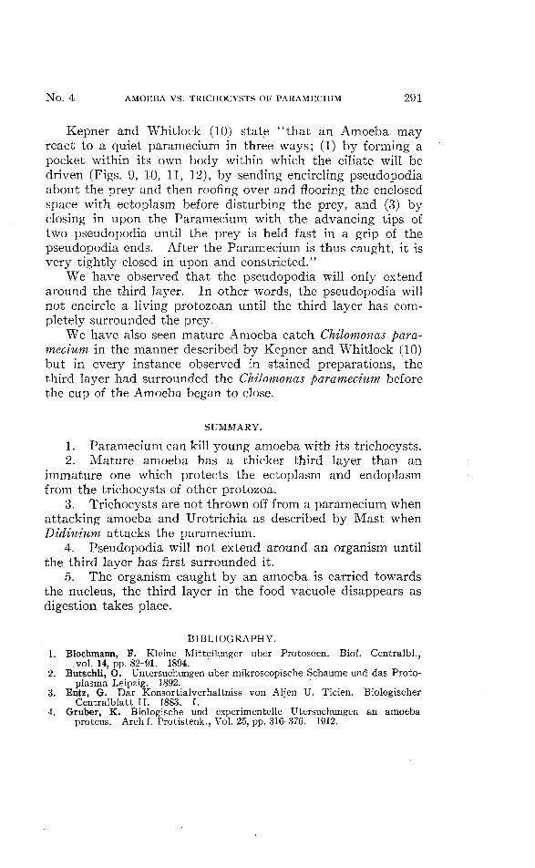
FIG. 1. Amoeba with its third layer surrounding a Chilomonas paramecium.FIG. 2. Chilomonas paramecia with the third layer of an amoeba surrounding
them forming food vacuoles.FIG. 3. The third layer of an amoeba surrounding two Chilomonas paramecia at
the extreme right. The third layer disappears as the food vacuolesapproach the nucleus. At the posterior end of the amoeba there arenumerous Chilomonas paramecia. These were caught when the amoebawas flowing in the direction opposite to the one shown in the diagram.
when older it chooses Chiltnonas paramecium (Text Figs. 1, 2, 3),later paramecia is the principal diet, and finally Rotifers areselected.
According to,our observations practically all conflicts be-tween mature Amoeba and Paramecium were fought to theadvantage of the former organism. On the other hand ayoung Amoeba, in such a battle, always lost. While workingon-the life cycle of Amoeba prokus, (Jones (6), we observed in
N o . 4 AMOEBA VS. TRICHOCYSTS OF PARAMECIUM 287
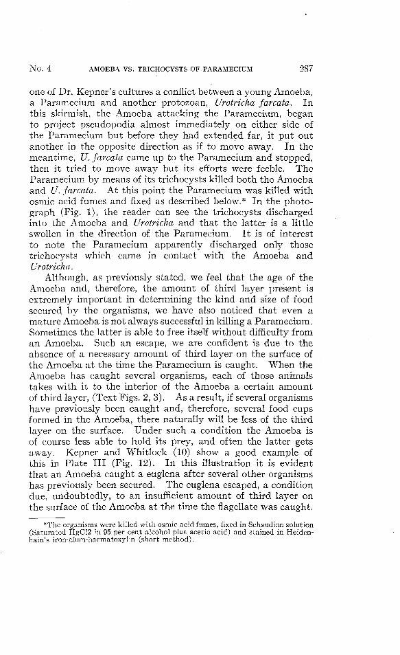
one of Dr. Kepner's cultures a conflict between a young Amoeba,a Paramecium and another protozoan, Urotricha farcata. Inthis skirmish, the Amoeba attacking the Paramecium, beganto project pseudopodia almost immediately on either side ofthe Paramecium but before they had extended far, it put outanother in the opposite direction as if to move away. In themeantime, U. farcata came up to the Paramecium and stopped,then it tried to move away but its efforts were feeble. TheParamecium by means of its trichocysts killed both the Amoebaand U. farcata. At this point the Paramecium was killed withosmic acid fumes and fixed as described below.* In the photo-graph (Fig. 1), the reader can see the trichocysts dischargedinto the Amoeba and Urotricha and that the latter is a littleswollen in the direction of the Paramecium. It is of interestto note the Paramecium apparently discharged only thosetrichocysts which came in contact with the Amoeba andUrotricha.
Although, as previously stated, we feel that the age of theAmoeba and, therefore, the amount of third layer present isextremely important in determining the kind and size of foodsecured by the organisms, we have also noticed that even amature Amoeba is not always successful in killing a Paramecium.Sometimes the latter is able to free itself without difficulty froman Amoeba. Such an escape, we are confident is due to theabsence of a necessary amount of third layer on the surface ofthe Amoeba at the time the Paramecium is caught. When theAmoeba has caught several organisms, each of those animalstakes with it to the interior of the Amoeba a certain amountof third layer, (Text Figs. 2, 3). As a result, if several organismshave previously been caught and, therefore, several food cupsformed in the Amoeba, there naturally will be less of the thirdlayer on the surface. Under such a condition the Amoeba isof course less able to hold its prey, and often the latter getsaway. Kepner and Whitlock (10) show a good example ofthis in Plate III (Fig. 12). In this illustration it is evidentthat an Amoeba caught a euglena after several other organismshas previously been secured. The euglena escaped, a conditiondue, undoubtedly, to an insufficient amount of third layer onthe surface of the Amoeba at the time the flagellate was caught.
*The organisms were killed with osmic acid fumes, fixed in Schaudinn solution(Saturated HgC12 in 95 per cent alcohol plus acetic acid) and stained in Heiden-hain's iron-alum-haematoxylin (short method).
288 PHILIP M. JONES Vol. XXX
The small amount of third layer present on the surface wasneeded by the Amoeba to protect the ectoplasm and endoplasmof its body from the lashing flagellum of the euglena.
There are no indications of the third layer possessing anypower of digestion, since no signs of erosion occur until all thethird layer has left the food vacuoles or has been digested by theenzymes in the endoplasm.
After protozoa have been caught and taken into the Amoeba,the food vacuole containing the imprisoned organisms passestowards the nucleus. During this passage the third layer,taken in the food vacuole with the organisms, gradually dis-appears and is entirely gone by the time the vacuole reachesthe nucleus (Text Figs. 3). The contents of the food vacuoleare then digested and the indigestible particles pass away fromthe nucleus and out of the Amoeba at the posterior end.
When an Amoeba catches protozoa at one end and thenreverses its direction of flow, such as illustrated in Text figure 3,the organisms caught remain at the posterior end until themovements of the Amoeba cease or are reversed again.
TRICHOCYSTS.
Paramecium in defending themselves against Amoeba, pro-ject short stout rods called trichocysts. Although there islittle definite information about these rods it is known thatthey lie within the ectoplasm. A few investigators are of theopinion that trichocysts are used chiefly for protection, othersfeel they must be weapons used for offense while still othershesitate to credit them with either function. Mast (11) sup-porting the theory that the Paramecium uses its trichocystsfor protection, discusses the behavior of the organism whenencountering its worst enemy, the Didinium. The latter attacksits prey by means of a peristome which can be discharged as anelongated tubler proboscis. If it succeeds in fixing this structureto a Paramecium it is usually able to suck much of its prisoner'sbody into its own. According to Mast (11), when the Para-mecium is attacked by a Didinium, it discharges its trichocysts,which form a great tangled mass about the body of its enemy.This discharge of trichocysts, in the cases of the largest Para-mecia forms such a dense mass that the Didinium is pushedfree from the Paramecium and the latter thus escapes death.

N o . 4 AMOEBA VS. TRICHOCYSTS OF PARAMECIUM 289
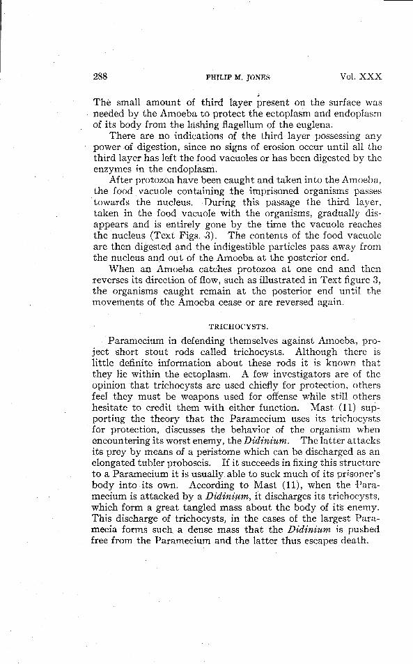
Our observations coincide with those of Mast, but we havealso noticed that the trichocysts are entirely liberated from thebody of the Paramecium when it is attacked by a Didiniumbut are not liberated when it is attacked by other animals.Such a condition, we feel, must be due to a stimulus caused byan injection of material from the proboscis of the Didinium.Figures 1, 3, shows trichocysts discharged but still attached tothe Paramecium.
A MATURE AMOEBA ENGULFS A PARAMECIUM WITH ITSTHIRD LAYER.
When a Paramecium is not moving about but has its ciliain motion, currents are set up by the cilia which become swifteras they pass from the anterior to the posterior end of theanimal. If an Amoeba is near enough the Paramecium toreceive a stimulus from these currents, the first pseudopodiumformed by the Amoeba will be pointed toward the posteriorend of the Paramecium, since this is the source of greateststimulus. As the Amoeba moves nearer the Paramecium, otherpseudopodia of smaller size are formed. These are right andleft of the first pseudopodium. When a pseudopodium touchesthe Paramecium the third layer of that pseudopodium immed-iately engulfs the Paramecium.
In catching a Paramecium or any other animal of similarstructure, it is necessary for the third layer of the Amoeba tofirst surround or engulf the emprisoned organism. Naturallyaccording to this method of securing food, it is necessary forthe Amoeba to have plenty of third layer.
Kepner and Whitlock's (10) drawings, reproduced on PlateIII (Figs. 9, 10, 11), describe the catching of a Paramecium byan Amoeba as follows: The amoeba was advancing in a generalway toward the Paramecium along pseudopodia 1, 2 and 3.As it approached the ciliate, pseudopodia 1 and 2 widened andpartly fused to form a large bi-lobed extremity, m-ml. Whenthis extremity had nearly tbuched the Paramecium, it sent outa small psudopodium, beneath the prey, and b anterior to it(Fig. 10). When the pseudopodia, a and b came in contactwith the detritus, y, they moved apart and become muchstouter (Fig. 11). In the meantime a third pseudopodium, e,appeared projecting from between a and b over the dorsal sideof the Paramecium, while a pocket was formed within the body

290 PHILIP M. JONES Vol. X'XX
proper of the amoeba at the bases of these three pseudopodia.The Paramecium first jumped to position 2, Fig. 11. Theexcited Paramecium next backed into the pocket of the bodyproper, 3 and a, b, and e, closed in and surrounded it com-pletely." .
Our interpretations of the diagrams described above differfrom those given by Kepner and Whitlock, since we bring intouse the third layer. According to our interpretations theParamecium was still, but its cilia were moving violently,judging by the amount of debris that was passing along theside and back of the posterior end of the Paramecium. Thestimulus thus set up caused the Amoeba to put forth pseudo-podium number 2 first, 3 next, then one, since this was the orderof the stimulus as it was received by the Amoeba; notice,number 1 would touch the Paramecium about the center; 2 is offat the posterior end, while 3 would be useless in catching theParamecium. The next in order, number 4, would be out ofconsideration. As 1 and 2 approached nearer the Paramecium,the third layer of either pseudopodia 1 and 2 or both wouldflow around and engulf the Paramecium. Figure 4, text figures1 and 2, shows the third layer of an Amoeba surrounding Chilo-monas paramecium in this same fashion.
The engulfing of a paramecium by the third layer may bedue t© the adhesive forces. The third layer having a strongaffinity for the surface of a paramecium tends to flow over andaround it. This same flow may cause a shifting of surfacetension forces, which will account in part, at least, for thewithdrawal of extended pseudopodia as in Plate III, Fig. 10.Part of the third layer having flowed around the Parameciumwill tend to level out, due to surface tension forces, thus drawingthe Paramecium into the cytoplasm of the Amoeba or, visaversa, the cytoplasm of Amoeba around the Paramecium.As a result, secondary pseudopodia flow up through the thirdlayer and around the paramecium finally engulfing the organismalong with some of the third layer as pictured by Kepner andWhitlock (10), Plate III, Fig. 11. These authors, in observingthis condition, however, pictures the Amoeba surrounded by alayer of water instead of protoplasmic substance. According toour observations, third layer and not water surrounded theengulfed organism as we were able to stain the material, (Fig. 4,Text figures 1, 2, 3).
N o . 4 AMOEBA VS. TRICHOCYSTS OF PARAMECIUM 291
Kepner and Whitlock (10) state "that an Amoeba mayreact to a quiet paramecium in three ways; (1) by forming apocket within its own body within which the ciliate will bedriven (Figs. 9, 10, 11, 12), by sending encircling pseudopodiaabout the prey and then roofing over and flooring the enclosedspace with ectoplasm before disturbing the prey, and (3) byclosing in upon the Paramecium with the advancing tips oftwo pseudopodia until the prey is held fast in a grip of thepseudopodia ends. After the Paramecium is thus caught, it isvery tightly closed in upon and constricted."
We have observed that the pseudopodia will only extendaround the third layer. In other words, the pseudopodia willnot encircle a living protozoan until the third layer has com-pletely surrounded the prey.
We have also seen mature Amoeba catch Chilomonas para-mecium in the manner described by Kepner and Whitlock (10)but in every instance observed in stained preparations, thethird layer had surrounded the Chilomonas paramecium beforethe cup of the Amoeba began to close.
SUMMARY.
1. Paramecium can kill young amoeba with its trichocysts.2. Mature amoeba has a thicker third layer than an
immature one which protects the ectoplasm and endoplasmfrom the trichocysts of other protozoa.
3. Trichocysts are not thrown off from a paramecium whenattacking amoeba and Urotrichia as described by Mast whenDidinium attacks the paramecium.
4. Pseudopodia will not extend around an organism untilthe third layer has first surrounded it.
5. The organism caught by an amoeba is carried towardsthe nucleus, the third layer in the food vacuole disappears asdigestion takes place.
BIBLIOGRAPHY.1. Blochmann, F. Kleine Mitteilunger uber Protosoen. Biol. Centralbl.,
.vol. 14, pp. 82-91. 1894.2. Butschli, O. Untersuchungen uber mikroscopische Schaume und das Proto-
plasma Leipzig. 1892.3. Entz, G. Dar Konsortialverhaltniss von Aljen U. Ticien. Biologischer
Centralblatt II. 1883. I.4. Gruber, K. Biologische und experimentelle Utersuchungen an amoeba
proteus. Arch f. Protistenk., Vol. 25, pp. 316-376. 1912.

292 PHILIP M. JONES Vol. X X X
5. Jennings, H. S. The Behavior of the Lower Organisms. New York, pp. 91.1906. •
6. Jones, P. M. Life Cycle of Amoeba proteus with Sexual Stage. Archi. FurProtestenkunde. Bd. 63:3. 1928.
7. Kepner, W. A. Animals Looking into the Future. MacMillin. 1925.8. Kepner, W. A. and Edwards. Food Reactions of Pelomyxa. Jour, of Expt.
Zool. 24: 383-4. 1917.9. Kepner, W. A. and W. H. Taliaferio. Reactions of amoeba proteus to Food.
Bio. Bull. 24: 411. 1913.10. Kepner, W. A. and C. Whitlock. Food Reactions of Amoeba proteus. Jour.
Exp. Zool. 32: 397-425. 1921.11. Mast, S. O. and Root. Observations on Amoeba Feeding on Rotifers, Nema-
todes and Ciliates and Their Bearing on the Surface Tension Theory.Jour. Exp. Zool. 21:33-49. 1916.
12. Schaeffer, A. A. On the Third Layer of Protoplasm in Amoeba. Anat.Rec, Vol. II, p. 477. 1917.
13. Schaeffer, A. A. Amoeboid Movements. Princeton University Press,pp. 1-156. 1920.
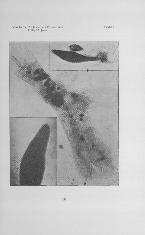
• EXPLANATION OF PLATES.
PLATE I.
Fig. 1. Paramecium with its trichocysts discharged, killing a young amoeba anda Urotricha.
Fig. 2. Amoeba proteus showing the third layer surrounding the amoeba. Theamoeba is filled with food vacuoles. Notice the two Chilomonasparamecia. The one at the posterior end shows signs of erosion,while the one at the large end shows no such change. The dark bodies,except the large one which is the nucleus, are food vacuoles in differentdegrees of digestion.
Fig. 3. Paramecium cordatum with its trichocysts extended; the capsules arestained black with iron heamatoxylin.
PLATE II.
Fig. 4. Amoeba catching active moving Chilomonas paramecia by surroundingthem with the third layer on the arms while it is catching a chilomonasin a food cup at the larger end.
PLATE III .
Figs. 9, 10 and 11. Shows an amoeba catching a paramecium as explained byKepner and Whitlock. (Reproduced by permission of Dr. Kepner.)
Amoeba vs. Trichocysts of ParameciumPhilip M. Jones
PLATE I.
293

Amoeba vs. Trichocysts of ParameciumPhilip M. Jones
PLATE II .
294

Amoeba vs. Trichocysts of ParameciumPhilip M. Jones
PLATE III.