Embed Size (px)
Citation preview

Third Annual
Symposium on
Regenerative
Rehabilitation UPMC Rehabilitation Institute McGowan Institute for Regenerative Medicine University of Pittsburgh School of Health and Rehabilitation Sciences
University of Pittsburgh School of Medicine Center for Continuing Education in the Health Sciences Rehabilitation Research & Development Center of Excellence at the Veterans Affairs Palo Alto Health Care System University of California, San Francisco Department of Physical Therapy And Rehabilitation Sciences
April 10 – 11, 2014
Mission Bay Conference Center
UCSF Mission Bay Campus
San Francisco, CA
2
TABLE OF CONTENTS Welcome Page 3 Course Directors and External Advisory Board Page 4 Organizers Pages 5 - 6 Acknowledgements Page 7 Overview, Objectives, and Continuing Education Page 8-9 Symposium Agenda Pages 10 - 21 Biographies Pages 22 - 34 Disclosures Page 35
Poster Abstracts Pages 36 - 59 Exhibitors Page 61 Travel Awards Pages 62 - 64
3
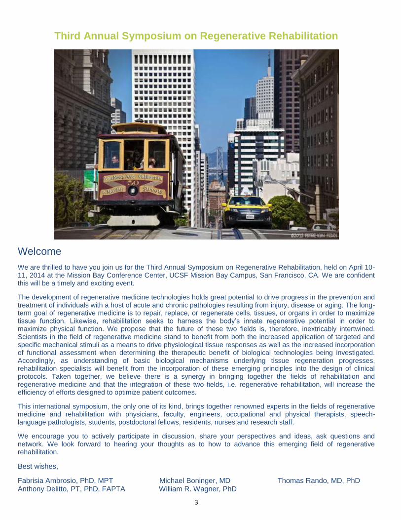
Third Annual Symposium on Regenerative Rehabilitation
Welcome
We are thrilled to have you join us for the Third Annual Symposium on Regenerative Rehabilitation, held on April 10-11, 2014 at the Mission Bay Conference Center, UCSF Mission Bay Campus, San Francisco, CA. We are confident this will be a timely and exciting event.
The development of regenerative medicine technologies holds great potential to drive progress in the prevention and treatment of individuals with a host of acute and chronic pathologies resulting from injury, disease or aging. The long-term goal of regenerative medicine is to repair, replace, or regenerate cells, tissues, or organs in order to maximize tissue function. Likewise, rehabilitation seeks to harness the body’s innate regenerative potential in order to maximize physical function. We propose that the future of these two fields is, therefore, inextricably intertwined. Scientists in the field of regenerative medicine stand to benefit from both the increased application of targeted and specific mechanical stimuli as a means to drive physiological tissue responses as well as the increased incorporation of functional assessment when determining the therapeutic benefit of biological technologies being investigated. Accordingly, as understanding of basic biological mechanisms underlying tissue regeneration progresses, rehabilitation specialists will benefit from the incorporation of these emerging principles into the design of clinical protocols. Taken together, we believe there is a synergy in bringing together the fields of rehabilitation and regenerative medicine and that the integration of these two fields, i.e. regenerative rehabilitation, will increase the efficiency of efforts designed to optimize patient outcomes.
This international symposium, the only one of its kind, brings together renowned experts in the fields of regenerative medicine and rehabilitation with physicians, faculty, engineers, occupational and physical therapists, speech-language pathologists, students, postdoctoral fellows, residents, nurses and research staff.
We encourage you to actively participate in discussion, share your perspectives and ideas, ask questions and network. We look forward to hearing your thoughts as to how to advance this emerging field of regenerative rehabilitation.
Best wishes,
Fabrisia Ambrosio, PhD, MPT Michael Boninger, MD Thomas Rando, MD, PhD Anthony Delitto, PT, PhD, FAPTA William R. Wagner, PhD
4
Course Directors:
Fabrisia Ambrosio, PhD, MPT Assistant Professor Department of Physical Medicine and Rehabilitation & Director, Cellular Rehabilitation Laboratory University of Pittsburgh Pittsburgh, PA Michael Boninger, MD Professor and Chair Department of Physical Medicine and Rehabilitation University of Pittsburgh Pittsburgh, PA Anthony Delitto, PhD, PT, FAPTA Professor and Associate Dean of Research School of Health and Rehabilitation Sciences University of Pittsburgh Pittsburgh, PA Thomas A. Rando, MD, PhD Director, RR&D REAP, VAPAHCS Professor, Department of Neurology and Neurological Sciences Stanford University School of Medicine Stanford, CA William R. Wagner, PhD Director, McGowan Institute for Regenerative Medicine Professor of Surgery, Bioengineering & Chemical Engineering University of Pittsburgh Pittsburgh, PA
Associate Course Director:
Linda J. Noble-Haeusslein, PhD Professor, Departments of Neurological Surgery and Physical Therapy and Rehabilitation Services University of California, San Francisco Box 0112, 513 Parnassus Avenue, Room HSE 772 San Francisco, CA 94143
External Advisory Board:
Steve L. Wolf, PhD, PT, FAPTA, FAHA Professor, Departments of Rehab Medicine, Medicine, Cell Biology Emory University School of Medicine Atlanta, GA Richard K. Shields, PT, PHD, FAPTA Professor and Chair Physical Therapy and Rehabilitation Science University of Iowa Iowa City, IA

5
Organized by: University of Pittsburgh School of Medicine Center for Continuing Education in the Health Sciences The purpose of the Center for Continuing Education in the Health Sciences is to advance the academic, clinical, and service missions of the University of Pittsburgh Schools of the Health Sciences and the University of Pittsburgh Medical Center through the continuing professional development of physicians, pharmacists, and other health professionals and the translation of biomedical knowledge into clinical practice. https://ccehs.upmc.com/
UPMC Rehabilitation Institute The largest rehabilitation provider in Western Pennsylvania, the UPMC Rehabilitation Institute (RI) serves as the hub of a UPMC network of more the 70 rehabilitation facilities that combine clinical care and research to help patients regain independence and enhance their quality of life. The RI’s academic partners include the Department of Physical Medicine and Rehabilitation at the University of Pittsburgh School of Medicine and the School of Health and Rehabilitation Science. These academic partners are national and international leaders in rehabilitation research and education. http://www.upmc.com/Services/rehab/rehab-instiute/Pages/default.aspx
The McGowan Institute for Regenerative Medicine The McGowan Institute for Regenerative Medicine is a partnership between the University of Pittsburgh and UPMC, and serves as a base for scientists and clinical faculty working in tissue engineering and biomaterials, cellular therapies, and medical devices and artificial organs. McGowan’s mission is the development of innovation clinical protocols and the commercial transfer of new technologies. http://www.mcgowan.pitt.edu
University of Pittsburgh School of Health and Rehabilitation Sciences Through academic research, technology design and rigorous training, the School of Health and Rehabilitation Sciences (SHRS) at the University of Pittsburgh educates the next generation of health professionals who will help others reach their fullest potential. At SHRS, we are committed to providing the best learning experience and academic environment possible for our students. Instructional excellence is rigorously pursued. Class sizes are intimate, fostering intellectual exchange and discourse. Students are challenged to not just achieve but to excel. And they do. Graduates of SHRS programs are some of the most sought-after professionals. Our faculty is world class. They are authors, clinicians, noted researchers, speakers and consultants. But foremost, they are teachers… Teachers who care passionately about their field and about their students. They want their students to succeed in the classroom and in their chosen professions. An SHRS education is more than classroom lectures. It’s hands-on learning either in a clinical setting or in the community. Through our strong relationships with the University of Pittsburgh Medical Center and other clinical partners, our students benefit from a wealth of experiences related to their particular field and area of interest. Students train in schools, hospitals, skilled nursing facilities, ambulatory care sites, and in home and community based settings. Our departments and programs listed here offer undergraduate, graduate and certificate degrees:
Clinical Dietetics and Nutrition
Communication Science and Disorders
Speech Language Pathology
Audiology
Emergency Medicine
Health Information Management
Occupational Therapy
Physical Therapy
http://www.shrs.pitt.edu
Physician Assistant Studies
Prosthetics and Orthotics
Rehabilitation Counseling
Rehabilitation Science (undergraduate)
Rehabilitation Science and Technology
Sports Medicine / Athletic Training
University of Pittsburgh Department of Physical Medicine and Rehabilitation Advancing the Science and Practice of Rehabilitation Medicine Our mission is to maximize the health, function and well-being of the people and populations we serve by providing the highest quality rehabilitative medical care, conducting highly relevant, cutting-edge research, and training the next generation of clinicians and researchers. Our research portfolio includes:
Neural Engineering and Neural Prosthetics
Biologics as indicators of pain, injury and recovery
Axon Regeneration
Biomarkers for brain injury
Medical homes for Spinal Cord Injury Care
Motor learning using Transcranial Magnetic Stimulation Our physicians are experts in the fields of traumatic brain injury, spinal cord injury, sports and musculoskeletal medicine, pain medicine, stroke and many conditions that would benefit from rehabilitation care. We partner with patients to design and implement personalized approaches that maximize participation, recovery and well-being. http://www.rehabmedicine.pitt.edu/
Rehabilitation Research and Development Program at the Veterans Affairs Palo Alto Health Care System, Center for Tissue Repair, Regeneration, and Restoration Dr. Thomas Rando directs the Rehabilitation R&D program at the Palo Alto VA. Within that program, the “Center for Tissue Repair, Regeneration, and Restoration" (CTR
3) focuses primarily on the neuromuscular and musculoskeletal systems and pursues research
at the levels of stem cell biology, biomedical engineering, and clinical / translational research. The VA Palo Alto Rehabilitation R & D Program reflects a long-standing commitment by the Department of Veterans Affairs to advance the well-being of American veterans through support of a full spectrum of rehabilitation research, from concept to clinic. A firm scientific understanding of the underlying impairment and a multi-disciplinary team creates a strong basis for developing new clinical treatments that reduce the disability of veterans and improve the effectiveness of healthcare delivery by VA clinicians.
University of California, San Francisco (UCSF), Department of Physical Therapy and Rehabilitation Science The UCSF Department of Physical Therapy and Rehabilitation Science offers high quality patient care through the Outpatient Physical Therapy Faculty Practice and the PhysFit Physical Therapy Health and Wellness Center. The Department also offers two graduate degrees in physical therapy in partnership with San Francisco State University (SFSU): the entry-level Doctor of Physical Therapy (DPT) and the post-professional Doctor of Physical Therapy Science (DPTSc). Additionally, the Department supports faculty and student research in basic, clinical, and translational studies. These research studies are interdepartmental and collaborative, and occur in conjunction with colleagues in departments across UCSF, including the Cancer Center and Gladstone Institute, the Immunology Program, and the Departments of Anatomy, Bioengineering, Radiology, Neurology, Neurosurgery, and Nursing. The mission of the UCSF Department of Physical Therapy and Rehabilitation Science is to provide evidence-based, patient-centered physical therapy services for the community, and to educate scholarly, socially sensitive clinicians, educators, and researchers in physical therapy and rehabilitation science who will lead the profession into the next century. Physical therapy students and clinical faculty participate as part of a team within an environment of health care that is patient-focused, and directed towards building the scientific base of clinical practice, with an eye toward quality, accessibility, and efficiency. The Department aims to 1) provide dynamic and creative educational opportunities for entry and advanced graduate students in physical therapy and rehabilitation science, 2) contribute to the scientific evidence in physical therapy practice, 3) provide high quality, efficient rehabilitation services to clients, and 4) assume an active role in the development of the physical therapy profession within the community at UCSF and SFSU, the state of California, and across the United States.
http://ptrehab.ucsf.edu/
7
In partnership with Stanford University, we gratefully acknowledge the support received from the California Institute for Regenerative Medicine (grant number CG 1-07550). A Special Thanks to…….

8
Course Overview and Objectives: Overview Medical advances in the field of Regenerative Medicine are accelerating at an unprecedented rate. Regrowing a lost limb, restoring function to a diseased organ, or harnessing the body’s natural ability to heal itself are becoming part of our reality instead of a distant promise. Technologies, such as cellular therapies, bioscaffolds, and artificial devices, are now in use or are being tested in clinical trials throughout the country.
How do we as clinicians and rehabilitation professional work with the patient regenerative medicine team to maximize patient outcomes and to help fully translate research?
How do we as investigators in the field of regenerative medicine make the most of these revolutionary results?
Few opportunities are available to bring together scientists and clinicians working in these two currently quite disparate fields: rehabilitation science and regenerative medicine. However rehabilitation science and regenerative therapies have to work closely in order to achieve a successful outcome for the patient. This situation created the need for open cross disciplinary work and collaborative communication. This symposium provides the opportunity for researchers and clinicians from around the world to gather and learn about the latest developments, share ideas and concepts and create sustainable collaborations.
Objectives During this course, participants will:
Interact with cutting-edge researchers.
Learn of the status of translating scientific discoveries into clinical practice.
Network with colleagues and potential collaborators.
Raise questions, debate implications, plan follow-up studies, and discuss results.
Share the status of their own research and clinical observations.
Meet with presenters to learn about their thinking and future research directions.

9
Continuing Education Credit Pennsylvania:
The University of Pittsburgh School of Medicine is accredited by the Accreditation Council for Continuing Medical Education to provide continuing medical education for physicians.
The University of Pittsburgh School of Medicine designates this live activity for a maximum of 10.0 AMA PRA Category 1 Credits™. Physicians should claim only the credit commensurate with the extent of their participation in the activity.
Other health care professionals are awarded 1.0 continuing education units (CEU's) which are equal to 10.0 contact hours.
The University of Pittsburgh is a pre-approved provider of CE for the State Board of Physical Therapy.
California:
This course has been approved by UCSF Rehabilitative Services for 10 continuing education hours (1.0 CEU). Questions regarding this approval should be directed to (415) 514-6777.
Participation by all individuals is encouraged. Advance notification of any special needs
will help us provide better service. Please notify us of your needs at least two weeks in
advance of the program by calling 001-(412) 624 5243

10
Agenda – Day 1 Thursday, April 10
th
6:15am
7:00 to 8:00am
8:00 to 8:30am
Meeting Registration Opens
Sunrise Workshop / Pre-Conference course: ‘Regenerative Medicine 101’ Moderator: Fabrisia Ambrosio, PhD, MPT
Presentations by: Arthur W. English, PhD Department of Cell Biology, Emory University Title: Peripheral Nerves 201: Anatomy and Regeneration of Peripheral nerves At the end of this presentation, participants will be:
Identify the cellular composition of peripheral nerves, including the cell types and connective tissue coverings.
Define the major events in the regeneration of axons after injury to peripheral nerves and identify molecules that are either growth promoting or inhibitors of axon regeneration.
Describe two broad strategies for enhancing regeneration of axons in peripheral nerves. Provide an example of each.
And Carmen M. Terzic, MD, PhD Chair, Department of Physical Medicine and Rehabilitation, Mayo Clinic Title: Stem Cell Therapy and Exercise Driven by patient needs, progress in regenerative sciences will catalyze the next chapters of medicine and surgery. We must therefore accelerate the pace at which discovery translates into clinical practice to provide solutions and hope for our patients and to speed the arrival of the day when organs will be rebuilt rather than replaced. Exercise-induced stem cell activation may enhance overall heart and other organ function and improve the efficacy of cardiac cellular therapeutic protocols. Dissecting the mechanisms for stem/progenitor cell activation with exercise will be instrumental to devise new effective therapies, encompassing myocardial regeneration for a large spectrum of cardiovascular diseases. Break – Continental Breakfast will be available
Second Floor Foyer
Jeanne Robertson Auditorium
Jeanne Robertson Auditorium
Jeanne Robertson Auditorium
Second Floor Foyer

11
8:30am 8:30 to 8:45am
8:45am to 12:00pm
8:45 to 9:20am
9:20 to 9:55am
Open Day 1 Main Meeting
Welcome and Opening Remarks Fabrisia Ambrosio, PhD, MPT
Session 1: Advances in the Biology of Tissue Regeneration & Plasticity and Implications for Clinical Practices Moderator: Michael Boninger, MD Jeffrey A. Kleim, PhD University of Arizona Title: Frontiers in Rehabilitation Sciences and Technology (FIRST): Why Should PTs Care About Genomics? Neurorehabilitation is in the midst of a paradigm shift that is centered around our understanding of neural plasticity and neuroregeneration. Decades of basic science research are now coalescing into clinical principles that are fundamentally changing physical, occupational and speech language therapies. Specifically, cell signaling pathways and genes have been identified as playing key roles in orchestrating neural plasticity and functional improvement after brain injury and disease. These pathways have inspired neurobiologically informed therapies that are being tested both pre clinically and in human patient populations. Recent advances in genomics have revealed the presence of common genetic polymorphisms that may have a significant impact on the efficacy of these new therapies. Evidence that such polymorphisms can impact both the induction of neural plasticity and the potential impact of plasticity promoting treatments will be presented. Linda J. Noble-Haeusslein, PhD University of California, San Francisco Title: Matrix Metalloproteinases and Spinal Cord Injury: Optimizing a Platform for Regenerative Strategies Neurological recovery after spinal cord injury (SCI) is impeded by a “triple threat” that consists of temporally overlapping events, beginning with immediate, irreversible mechanical damage to neural and vascular structures. This is followed by the early emergence of a pro-inflammatory state coupled with oxidative stress that collectively damage structures that were initially intact after the injury. Finally, wound healing, commencing within this inhospitable terrain, defines an environment that is inhibitory to plasticity. Within this complex response to trauma, there is opportunity to intervene both in the acutely injured cord and during wound healing through genetic and pharmacologic modulation of MMP activity and timely administration of exercise programs. We have targeted MMP-9 that is markedly up regulat-
Jeanne Robertson Auditorium
Jeanne Robertson Auditorium
Jeanne Robertson
Auditorium
Jeanne Robertson Auditorium
Jeanne Robertson Auditorium

12
9:55 to 10:00am
10:00 to 10:35am
10:35 to 11:10am
ed in the acutely injured cord. This up-regulation is in part due to inflammatory cells that express this protease and in the case of leukocytes, utilize this protease to transmigrate into the injured spinal cord. Genetic approaches confirm that MMP-9 is a key determinant of long-term neurological recovery after SCI and that this is attributed to an attenuated inflammatory response. Complimentary pharmacological studies, targeting MMPs in the acutely injured cord, reveal injury severity dependent efficacy. The intersection between MMP-9 activation and exercise are also considered in a paradigm of acute and delayed intervention. ‘Poster Teasers’ Naoki Tajiri, PT, PhD, University of Florida Manzhao Hao, MS, Shanghai Jiao Tong University, China Robynne Braun, MD, PhD, University of Washington Morning Break / Poster Set-up David L. Mack, PhD University of Washington Title: Disease-in-a-Dish: The Contribution of Patient-specific iPS Cell Technology to Regenerative Rehabilitation Advances in regenerative medicine technologies will lead to dramatic changes in how patients in rehabilitation medicine clinics are treated in the upcoming decades. For example, the multidisciplinary field of regenerative medicine is developing new tools for disease modeling and drug discovery based on induced pluripotent stem cells (iPSCs). The central idea is to differentiate the stem cells into the specific cell type(s) most likely to be pathologically altered by mutation, thereby recapitulating the disease in a dish. Since differentiating iPS cells in vitro pass through the same sequence of developmental steps as would occur in the embryo, this technology will allow us to initiate and track disease progression at the cellular level. This disease model can then be used as a drug-screening tool, by searching for compounds that can correct the phenotypic defect in the dish. Drugs identified using this personalized medicine approach will have a higher likelihood of working in the patient because the patient’s own cells were used to discover the drug. Therefore, this system proposes a closed loop from sample collection from disease model, to drug discovery and FDA approval, to delivering that drug back to the same patient. As a proof of concept, this the diseased patient, to in vitro talk will present a disease model of the cardiomyopathy associated with Duchenne muscular dystrophy using patient-specific, urine-derived iPS cells differentiated to cardiomyocytes in a dish. The challenges of identifying a clinically relevant assay for the dystrophin-null cardiac phenotype and adapting this model to a high-throughput
Jeanne Robertson
Auditorium
Second Floor Foyer
Jeanne Robertson
Auditorium

13
11:10 to 11:55am
11:55am to 12:00pm
small molecule-screening platform will be discussed. Thomas A. Rando, MD, PhD Director, RR&D REAP, VAPAHCS Stanford University Title: Regenerative Rehabilitation for Volumetric Muscle Loss Stem cell regeneration of injured or diseased tissue holds great promise as a means to restore tissue structure and function. Furthermore, integrating regenerative therapies with rehabilitative physical therapies is likely to provide synergize benefits. One of the approaches we have taken in a path to translation for this regenerative rehabilitation approach has involved a mouse model of volumetric muscle loss (VML). VML is a common consequence of blast injuries in soldiers, leading to sustained impairment of function as there is currently no treatment, either in the acute setting of the chronic phase, for this condition. We have focused on chronic VML to model the more challenging problem for regenerative therapy. Studies from our group and others have demonstrated that the transplantation of myogenic stem or progenitor populations into area of muscle injury result in the engraftment of those cells and their participation and enhancement of a regenerative response. Our studies have focused on the generation of artificial scaffolds on which to seed purified mouse or human muscle stem cells prior to transplantation in order to enhance transplantation efficacy. Using both anatomical and physiological analyses, we have optimized the ex vivo approaches in a xenograft model of regenerative therapy of VML. Furthermore, we are currently combining this approach with the analysis of the effects of physical activity on the efficacy of muscle regeneration. Following transplantation, mice will be placed into two groups – one of which will be allowed to run on a wheel and the other of which will be maintained under normal laboratory conditions. We will analyze the effects of physical activity on the outcome measures to determine whether physical activity could help to promote the regenerative response stimulated by stem cell transplantation. ‘Poster Teasers’ Geetha Mohan, PhD, University of California, San Francisco M. Elise Johanson, DPT, VA Palo Alto Health Care System Shoki Yamaguchi, Kyoto University, Japan
Jeanne Robertson Auditorium
Jeanne Robertson Auditorium

14
12:00 to 1:00pm 12:45 to 1:30pm 1:30 to 1:35pm
1:35 to 2:35pm
Lunch Poster Session Introduction of Keynote Speaker Moderator: Thomas A. Rando, MD, PhD
Keynote Address: Stephen G. Waxman, MD, PhD Director, RR&D Center of Excellence, VACHCS Director, Neuroscience and Regeneration Research Ctr Bridget Marie Flaherty Professor of Neurology, Neurobiology and Pharmacology Yale University Title: Chasing Men on Fire: Sodium Channels as Therapeutic Targets in Rehabilitation Research Although neurophysiology has classically referred to “the” sodium channel as if it might be a singular entity, we now know that nine different genes encode nine distinct voltage-gated sodium channels, each with different physiological and pharmacological properties and each with a different distribution in the nervous system This talk will review several aspects of recent progress in this arena, with an emphasis on developing sodium channels as therapeutic targets: 1. Sodium channels play important roles in conduction in
myelinated and demyelinated axons, and help to drive axonal degeneration. Therapeutic implications will be discussed.
2. Sodium channels are important contributors to hyperexcitability of DRG neurons that underlies neuropathic pain. “Peripheral” sodium channels that are preferentially expressed in peripheral neurons but not functionally essential in heart or brain, have been a holy grail of pain research, since selective blockade of these channels might be expected to attenuate pain with minimal CNS side-effects or addictive potential.
3. Genetic studies have begun to validate specific sodium channel isoforms as therapeutic targets for pain.
4. These studies have identified mutant sodium channels as major contributors to chronic pain, initially in rare genetic disorders and more recently in painful neuropathies.
5. Gene therapy studies have begun to examine knockdown of sodium channels as a therapeutic approach for chronic pain.
6. Recent studies using atomic-level modeling suggest that the goal of pharmacogenomically-guided, individualized pain therapy may not be unrealistic.
Fisher Banquet Rooms
Second Floor Foyer
Jeanne Robertson Auditorium
Jeanne Robertson
Auditorium
15
2:35 to 2:40pm 2:40 to 3:15pm
3:15 to 4:25pm
3:15 to 3:50pm
3:50 to 4:25pm
‘Poster Teasers’ Kristen Stearns, PhD, PT, University of Pittsburgh Ngan F. Huang, PhD, Veterans Affairs Palo Alto Health Care
System Jian Luo, MD, PhD from Stanford Afternoon Break / Poster Viewing
Session 2: Interfacing Rehabilitation Engineering and Regenerative Medicine Moderator: Thomas A. Rando, MD, PhD Michael Boninger, MD University of Pittsburgh and Medical Director. RR&D Center of Excellence, VAPHCS Title: Brain Computer Interfaces (BCI) and Regenerative Rehabilitation: Tapping into “Thoughts” to Increase Independence
A better understanding neural population function would be an important advance in systems neuroscience. The change in emphasis from the single neuron to the neural ensemble has made it possible to extract high-fidelity information about movement. This ability is due to the distributed nature of information processing in the brain. The realization that useful information is embedded in the population has spawned the current success of brain-controlled interfaces. Since multiple movement parameters are encoded simultaneously in the same population of neurons, we have been gradually increasing the degrees of freedom (DOF) that a subject can control through BCI. We have demonstrated the ability to achieve 7 DOF control in a human participant with complete paralysis. This work and other BCI technology will be discuss and related to regenerative rehabilitation. Walt Schneider, PhD University of Pittsburgh Title: High Definition Fiber Tracking (HDFT) an MRI Biomarker for Brain Anatomical Connection Disorders in TBI, Neurosurgery & Autism
High Definition Fiber Tracking (HDFT) is an advanced set of technologies enabling noninvasive MRI diffusion tracking of millimeter tracts over long distances accurately following from source to destination through tract crossings to detail axon projection fields of white matter tracts. Connection disorders are a major medical problem impacting tens of millions of patients with trauma (TBI), neuro-oncology, neurodegeneration (Alzheimer’s) and developmental (autism) pathologies and likely
Jeanne Robertson Auditorium
Second Floor Foyer
Jeanne Robertson Auditorium
Jeanne Robertson Auditorium
Jeanne Robertson Auditorium

16
4:25 to 5:35pm
4:25 to 5:00pm
tract segmentation. It creates a imaging with a 15 minute to play a role in psychiatric disorders. HDFT involves mapping a million microtracts on a single individual with 3T MRI 257d DSI scan using novel computation methods calculating directional axonal volume (dAV), tractography, and personalized circuit diagram of the patient quantifying and visualizing the integrity of twenty brain white matter tracts. The visualization and quantification of tract pathology aids clinical decision. It enables clear detection of damage where past methods typically could not. This Pittsburgh technology has been applied in hundreds of clinical cases and provides the foundation for a more informed patient education in diagnosing and treating connection disorders.
Session 3: The Importance of Mechanical Stimulation in Cellular Therapeutics and Tissue Engineering Moderated by: Thomas Rando, MD, PhD George J. Christ, PhD Wake Forest University Title: Development of a Tissue Engineered Muscle Repair [TERM] Technology Platform for Treatment of Volumetric Muscle Loss [VML] Injuries
Despite the rather well documented capacity of skeletal muscle to repair, regenerate, and remodel following injury, more severe craniofacial injuries, such as those involving the loss of a substantial portion of muscle tissue are not capable of full regeneration on their own. That is, such injuries involve a degree of muscle tissue loss that exceeds the endogenous regenerative capacity of muscle. These injuries are characterized by volumetric muscle loss (VML injury) resulting in permanent cosmetic and functional deficits. Current treatment for VML injuries is limited to surgical muscle transfers, which are associated with poor engraftment and donor site morbidities. To address this unmet medical need we are pursuing proof-of-concept studies for development of a scalable sheet-like, cell-based tissue engineering approach referred to as a Tissue Engineered Muscle Repair (TEMR) technology. Proof of concept for this approach, has been evaluated using a murine model of VML injury, where ≈50% of the latissimus dorsi (LD) muscle is surgically excised and the injury is treated by implantation of a TEMR construct (~1x3cm). TEMR constructs are created by seeding muscle progenitor cells (MPCs) on a decellularized bladder acellular matrix (BAM scaffold), and then subjected to bioreactor preconditioning in vitro. Previous work has shown that bioreactor preconditioning in vitro is critical to increases in the functional regenerative response observed following TEMR implantation in vivo. More specifically,
Jeanne Robertson Auditorium
Jeanne Robertson Auditorium

17
5:00 to 5:35pm
implantation of TEMR constructs at the site of injury restored contractile force generation to ~60-70% of native control within 2 months post implantation. We refer to this as our first generation construct, TEMR1. More recently we documented that an additional round of MPC seeding of the scaffold during bioreactor preconditioning results in enhanced myotube formation due to increased myoblast fusion, a process that may be analogous to exercise in vivo. Implantation of these second generation (TEMR2) constructs resulted in both an accelerated (i.e., increased muscle contraction was now observed at 1 month post-implantation rather than 2 months) as well as prolonged functional recovery, at fully twice the magnitude of TEMR constructs that were seeded only a single time. This recovery was mediated via enhanced repair of native tissue and de novo regeneration of new muscle fibers. Such observations open the door to diverse methods (e.g., pharmacological or gene-based approaches) for manipulating the cellular phenotype and composition of TEMR constructs for improved functional outcomes. In order to optimize development of a more robust TEMR technology platform we are currently exploring a selected range of novel cellular and biomaterials combinations that may result in further improvements in functional muscle tissue regeneration. This approach should lead to a potentially wider range of clinical applications for treatment of VML injuries. Moreover, when coupled with rehabilitative measures, we anticipate that our TEMR technology platform could further advance the field. Finally, following an initial pre-IND conversation with the FDA, we are in the process of finalizing our definitive physiology/toxicology study plan for submission of an IND to the FDA. Our proposed first in man indication will be secondary revision of cleft lip.
Martin Oudega, PhD University of Pittsburgh Title: Bone Marrow Stromal Cell-based Therapy and Rehabilitation for Spinal Cord Repair Animal models of spinal cord injury (SCI) have revealed the potential of bone marrow stromal-derived cell (BMSC) transplants to elicit repair. BMSCs secrete a variety of molecules that through paracrine effects mediate and direct cellular events resulting in restoration of damaged neural tissue, which in turn may lead to recovery of injury-induced impairments in motor and sensory function. Clinically, BMSCs have special interest because their relative easy accessibility allows for autologous transplantation thereby minimizing transplant rejection. Currently, a number of clinical trials are ongoing testing the spinal cord repair potential of BMSCs.
Jeanne Robertson Auditorium

18
5:35 to 5:40pm
6:00- 8:00pm
6:00pm
6:05pm
6:15 to 8:00pm
Intraspinal BMSC transplants elicit nervous tissue sparing which correlates with functional recovery. However, the overall effects of BMSC transplants on tissue sparing and functional recovery are limited. We will present histological and functional data showing that low survival is one of the limiting factors in the repair potential of BMSC transplants. BMSCs injected into an injury epicenter may be lost due to a variety of reasons including oxidative stress, inflammation, and loss of the extracellular matrix component fibronectin. We will present data that highlight the role of these three potential parameters in low intraspinal BMSC transplant survival. We will also demonstrate results revealing the role of endogenous and exogenous fibronectin and the synthetic reverse thermal gel (poly(ethylene glycol)-poly(serinol hexamethylene urethane, ESHU) in promoting BMSC transplant survival. The therapeutic potential of these matrices to enhance the repair effects of intraspinal BMSC transplants will be discussed.
Clinically, locomotor training is employed as a means to promote (activity-dependent) plasticity that may improve recovery after SCI. In the laboratory this approach has been explored alone and in combination with repair-supporting interventions with variable success. We will review the effects of locomotor training alone or in combination with BMSC transplants on spinal cord repair. We will highlight the potential of locomotor training as a means to promote functional recovery or to consolidate functional recovery mediated by concurrently implemented interventions. Closing Remarks Thomas A. Rando, MD, PhD
Reception and tours of the UCSF Faculty Practice and Human Performance Laboratory
Welcome and Opening remarks Linda J. Noble-Haeusslein, PhD
Tours of the NEW Faculty Practice and Human Performance Laboratory start. Each tour will take approximately 15-20 minutes. Tours will consist of groups of 20; pre-registration is requested. Tours times are: 6:05pm, 6:25pm, 6:45pm and 7:05pm
Networking and Poster Viewing
Light refreshments will be served.
Jeanne Robertson Auditorium
Second Floor Foyer
Jeanne Robertson Auditorium
Second Floor Foyer
Second Floor Foyer

19
7:00am
7:15 to 8:15am
7:00 to 8:45am
8:30am 8:30 to 8:35am 8:35 to 10:20am
8:35 to 9:10am
9:10 to 9:45am
Meeting Registration Opens
Sunrise Networking / ‘Meet the Expert/Mentor’ Session This session will enable the students and / or young investigators to meet and interact in a structured format with the senior investigators attending the meeting.
Break - Continental Breakfast will be available Open Day 2 Main Meeting Opening remarks Linda J. Noble-Haeusslein, PhD Session 4: Mechanotransduction as a Tool in Tissue Healing and Repair Moderator: Linda J. Noble-Haeusslein, PhD Richard K. Shields, PT, PhD, FAPTA University of Iowa Title: Mechanical Stress Regulates Musculoskeletal Plasticity following Spinal Cord Injury The effects of disuse from spinal cord injury are studied with respect to skeletal changes (bone density) and muscular adaptations. The extent to which these adaptations are influenced by system based stress are examined during longitudinal and cross-sectional studies.
Grace Griesbach, PhD University of California, Los Angeles Title: Exercise after Mild Traumatic Brian Injury: Implications for Sports and Rehabilitation
Second Floor Foyer
Jeanne Robertson Auditorium
Jeanne Robertson Auditorium
Jeanne Robertson Auditorium
Jeanne Robertson Auditorium
Jeanne Robertson Auditorium
Jeanne Robertson Auditorium
Jeanne Robertson Auditorium
Agenda – Day 2
Friday, April 11th

20
9:45 to10:20am
This talk will focus on the effects of exercise on neuroplasticity following traumatic brain injury (TBI). Those affected with brain injury endure long-lasting impairments that have a strong impact on life quality. Exercise has been proven valuable because it increases proteins that are important in neuronal plasticity and repair. The most prominent mechanisms behind exercise-induced neuroplasticity and neuroprotection will be briefly described. Although exercise helps the brain recover from injury it may impair recovery if it takes place during the early post injury period and is associated with stress. Alterations in the stress response are most prominent during the early post injury period. This will have implication on the timing of rehabilitation as well as the return to athletic activities following a concussion.
Rocky S. Tuan, PhD University of Pittsburgh Title: Stem Cell and Matrix-based Tissue Engineering and Regeneration: Technologies and Models The intrinsically low reparative capacity of cartilage is a clinical challenge to effective treatment of degenerative joint diseases, such as osteoarthritis, the main cause of physical disability. Tissue engineering and regenerative medicine, combining cells, scaffolds, and biological signals, represents a potentially promising approach. Mesenchymal stem cells (MSCs), harvested from adult tissues such as bone marrow and adipose, have multi-lineage differentiation potential, including chondrogenesis, and are considered a promising candidate cell type for cartilage repair. A biocompatible biomaterial scaffold that ideally also enhances proliferation and differentiation of the seeded cells is critical to successful cell-based tissue engineering. We have shown that biomimetic scaffolds that simulate the structure of native extracellular matrix, e.g., the nanoscalar fibrous nature of collagen, are effective in MSC-based skeletal tissue engineering both in vitro and in vivo. Our recent work on the use of custom-designed, photo-crosslinked hydrogel scaffolds, which allows cell encapsulation during fabrication, demonstrates high fidelity reproduction of internal structure and excellent cell retention, viability, and differentiation. Specifically, applying a 3D printing approach and a custom-designed microbioreactor, we have constructed a microtissue analogue of the osteochondral junction, based entirely on MSC-derived components, to model the pathogenesis of osteoarthritis. Adult stem cells, with their multi-differentiation potential and recently discovered trophic activities,
when used in combination with biomimetic scaffolds, present a powerful platform for regenerative, therapeutic, and disease modeling applications in biomedicine.
Jeanne Robertson Auditorium

21
10:20 to 10:45am
10:45 to 11:35am
10:45 to 11:10am
11:10 to 11:35am
11:35 to 11:45am
11:45am
Morning Break
Session 5: Regenerative Rehabilitation in Education Moderator: Linda Noble-Haeusslein, PhD Steve L. Wolf, PhD, PT, FAPTA, FAHA Emory University Title: Frontiers in Rehabilitation Science and Technology (FIRST) Initiative
The Physical Therapy and Society (PASS) meeting in 2009 provided a group of 24 external agencies and professions to critically assess the physical therapy profession and to make recommendations regarding future directions that will be important to optimize quality of care and interdisciplinary collaborations. Amongst those recommendations was the need to: promote the translation and integration of technology and science; foster partnerships with engineering, industry and others; and collaborate in developing and provide leadership in testing new technologies and approaches that optimize health care delivery. As a result, the Frontiers in Rehabilitation Science and Technology (FiRST) initiative was born. This presentation highlights the evolution and progression of FiRST and poses some of the obstacles necessary to assure maximal benefit and compliance in implementing this interdisciplinary effort.
Diane D. Allen, PT, PhD Graduate Program in Physical Therapy University of California, San Francisco / San Francisco State University Title: Roles for Rehabilitation Professionals in Regenerative Medicine
Although research provides evidence of effective rehabilitation in different populations, the role of rehabilitation professionals is less well-documented in regenerative medicine. In this presentation, Dr. Allen will explore the role and potential contribution of rehabilitation professionals in the realm of regenerative medicine. Assessment tools and clinical protocols that focus on patient-centered outcomes can help researchers and clinicians in this realm document meaningful change in the
lives of their patients.
Closing Remarks Fabrisia Ambrosio, PhD, MPT Conclusion of Day 2
Jeanne Robertson Auditorium Jeanne Robertson Auditorium Jeanne Robertson Auditorium
Jeanne Robertson Auditorium Jeanne Robertson Auditorium
Jeanne Robertson Auditorium

22
Biographies
23
Course Directors’ Biographies:
Fabrisia Ambrosio, PhD, MPT Assistant Professor
Department of Physical Medicine and Rehabilitation
McGowan Institute for Regenerative Medicine
University of Pittsburgh
Pittsburgh, PA
Fabrisia Ambrosio, PhD, MPT graduated with a Master of Science in Physiology-Endocrinology from Laval University in Québec City, Québec and a Master of Physical Therapy from the Medical College of Pennsylvania and Hahnemann University. In 2005, Dr. Ambrosio graduated with a PhD in Rehabilitation Science & Technology from the University of Pittsburgh. Also in 2005, she accepted a position as a faculty member in the Department of Physical Medicine & Rehabilitation at the University of Pittsburgh. She holds secondary appointments in the Departments of Physical Therapy, Orthopaedic Surgery, and Microbiology & Molecular Genetics at the University of Pittsburgh, and is a faculty member of the McGowan Institute for Regenerative Medicine. Dr. Ambrosio’s research has the long-term goal of developing regenerative rehabilitation approaches to improve the skeletal muscle healing and functional recovery. Her laboratory investigates the underlying mechanisms by which targeted and specific mechanotransductive signals can be used to enhance donor and/or endogenous stem cell function using mouse and human models.
Michael Boninger, MD Professor and Chair Department of Physical Medicine and Rehabilitation University of Pittsburgh Medical Director, RR&D Center of Excellence, VAPHCS Pittsburgh, PA
Michael Boninger, MD is Professor and Chair in the Department of Physical Medicine & Rehabilitation in the University of Pittsburgh, School of Medicine and director of the UPMC Rehabilitation Institute. Dr. Boninger is a physician researcher for the Department of Veterans Affairs (VA) and is the Medical Director of the Human Engineering Research Laboratories, a VA Rehabilitation Research and Development Center of Excellence. Dr. Boninger has an extensive publication record of over 200 published papers spanning 18 years in the area of spinal cord injury and assistive technology. Dr. Boninger also has extensive experience and publications related to teaching research. Dr. Boninger holds 4 US patents and has won numerous awards, including being inducted into the Institute of Medicine in 2013. Dr. Boninger’s students have also won over 45 national awards.

24
Anthony Delitto, PhD, PT, FAPTA Professor and Associate Dean of Research School of Health and Rehabilitation Sciences University of Pittsburgh Pittsburgh, PA
Anthony Delitto, PhD, PT, FAPTA is a professor and Associate Dean for Research in the School of Health and Rehabilitation Sciences at the University of Pittsburgh. He is also the Director of Research Comprehensive Spine Center at UPMC as well as Vice President for Education and Research Centers for Rehabilitation Services (formerly CORE Network). Dr. Delitto earned his BS in Physical Therapy from SUNY-Buffalo and his MHS/PT and PhD in Psychology from Washington University in St. Louis, Missouri. Dr. Delitto is primarily interested in conducting evidence-based studies in rehabilitation settings, particularly in populations who have musculoskeletal dysfunction (e.g., low back pain). Previous research projects studied the functional impact of PENS for 65+ chronic low back pain, which aimed to test the effectiveness of PENS in reducing the pain intensity in community-dwelling older adults with CLBP and of combining PENS with a general conditioning and aerobic exercise program (GCAE) to improve the pain-related disability of these patients. Dr. Delitto also conducted a randomized clinical trial of treatment for lumbar spinal stenosis, which compared patient outcomes and evaluated gender differences after non-surgical or surgical treatment for lumbar spinal stenosis. Dr. Delitto is the president of the Section on Research in the American Physical Therapy Association as well as on the Doctoral Research Awards Committee and Scientific Review Committee of the Foundation for Physical Therapy. He is a member of the International Advisory Board of the New Zealand Centre for Physiotherapy Research, University of Otago; the Medical and Scientific Committee of the Arthritis Foundation, Western Pennsylvania Chapter; and the ALS Association, Western Pennsylvania Chapter. He is a recipient of the Lucy Blair Service Award, which is presented by the American Physical Therapy Association and which honors the member whose contributions to the Association as a whole have been of exceptional value, as well as the John HP Maley Award, which the Association gives to those who provide outstanding contributions to leadership in research.
Thomas A. Rando, MD, PhD Director, RR&D REAP, VAPAHCS Professor Department of Neurology and Neurological Sciences Stanford University School of Medicine Stanford, CA
Thomas A. Rando, MD, PhD is Director of the Rehabilitation Research & Development Center of Excellence at the VA Palo Alto Health Care System where he is also Chief of Neurology. He is Professor of Neurology and Neurological Sciences and Director of the Glenn Laboratories for the Biology of Aging at Stanford University School of Medicine. Dr. Rando’s research concerns the basic biology of stem cells and how they function in adult tissue homeostasis, in degenerative diseases, and in aging and the application of stem cell therapeutics toward muscle diseases and muscle injury. Groundbreaking work from his lab showed that the age-related decline in stem cell function is due primarily to influences of the aged environmental rather than to intrinsic aging of stem cell themselves. Rando has received numerous awards, including a Paul Beeson Physician Faculty Scholar in Aging from the American Federation for Aging Research and a Scholar Award from the Ellison Medical Foundation. In 2005 he

25
received the prestigious NIH Director’s Pioneer Award for his work at the interface between stem cell biology and the biology of aging and he recently received a Transformative Research Award from the NIH for the study of the regulation of cognitive function by physical activity and exercise.
William R. Wagner, PhD Director, McGowan Institute for Regenerative Medicine Professor of Surgery, Bioengineering & Chemical Engineering University of Pittsburgh Pittsburgh, PA 15219
William R. Wagner, PhD is the Director of the McGowan Institute for Regenerative Medicine as well as a Professor of Surgery, Bioengineering and Chemical Engineering at the University of Pittsburgh. He also serves as the Director of Thrombosis Research for the Artificial Heart and Lung Program, and Deputy Director of the NSF Engineering Research Center on “Revolutionizing Metallic Biomaterials”. He holds a B.S. (Johns Hopkins Univ.) and Ph.D. (Univ. of Texas) in Chemical Engineering. Dr. Wagner is the Coordinator for the Cellular and Organ Engineering track for Bioengineering graduate students, and currently teaches in the areas of biomaterials and tissue engineering. Professor Wagner is the Founding Editor and Editor-in-Chief of one of the leading biomaterials journals, “Acta Biomaterialia”, and currently serves on the editorial boards of the “Journal of Biomedical Materials Research part A”, “Biotechnology and Bioengineering”, and the “Journal of Tissue Engineering and Regenerative Medicine”. Dr. Wagner is also a past president of the American Society for Artificial Internal Organs (ASAIO; 2010-2011) and serves on the Executive Board of the International Federation of Artificial Organs (IFAO). He is a fellow and former vice president of the American Institute for Medical and Biological Engineering (AIMBE; 2000) and has also been elected a fellow of the Biomedical Engineering Society (2007), the International Union of Societies for Biomaterials Science and Engineering (2008) and the American Heart Association (2001). He has served as Chairman for the Gordon Research Conference on Biomaterials: Biocompatibility & Tissue Engineering as well as for the First World Congress of the Tissue Engineering and Regenerative Medicine International Society (TERMIS). In 2006 he was selected to the “Scientific American 50”, the magazine’s annual list recognizing leaders in science and technology from the research, business and policy fields. In 2011 he was awarded the Society for Biomaterials Clemson Award for Applied Research. He has served on numerous NIH and NSF study sections, is a member of the NIH College of Reviewers, and has been a member of external review committees for national and international organizations focused on bioengineering and regenerative medicine. His research has generated numerous patents and patent filings that have resulted in licensing activity, the formation of a company, and University of Pittsburgh Innovator Awards in 2007, 2008, 2009 and 2010. Dr. Wagner's research interests are generally in the area of cardiovascular engineering with projects that address medical device biocompatibility and design, tissue engineering, and targeted imaging. His research group is comprised of graduate students in Bioengineering and Chemical Engineering as well as post-doctoral fellows with backgrounds in surgery, polymer chemistry, or engineering. Dr. Wagner and his group enjoy working across the spectrum from in vitro to clinical studies. The McGowan Institute and the University of Pittsburgh Medical Center are uniquely positioned to allow such broad-based projects to flourish and complement one another. Researchers within Dr. Wagner's group are afforded the opportunity to observe first-hand the clinical successes and failures of currently employed cardiovascular devices while concurrently working on projects that attempt to describe the current

26
modes of failure, test solutions for the current device shortcomings, or develop technologies that may find application as future cardiovascular therapies. The front-line experience afforded by the clinical environment has proven invaluable in the learning experience of group members, not to mention the input such experience has on the creative environment.
Associate Course Director Biography:
Linda J. Noble-Haeusslein, PhD Professor, Departments of Neurological Surgery and Physical Therapy and Rehabilitation Services University of California, San Francisco Box 0112, 513 Parnassus Avenue, Room HSE 772 San Francisco, CA 94143
Linda J. Noble (Haeusslein) obtained her undergraduate degree in physical therapy from the University of Utah and her doctoral degree in anatomy from the University of California at Los Angeles. She is currently Professor and Alvera Kan Endowed Chair, Departments of Neurological Surgery and Physical Therapy and Rehabilitation Science at the University of California, San Francisco. The Noble-Haeusslein laboratory is committed to a strong translational program that investigates the key determinants of recovery after traumatic injury to the developing brain and the adult spinal cord. To date, her laboratory has highlighted the unique vulnerability of the developing murine brain to trauma that is in part attributed to inadequate antioxidant reserves and the emergence of deficits in both cognitive function and socialization, deficits that are likewise seen in brain-injured children. Ongoing studies are defining those anatomical and biochemical substrates that contribute to these behavioral deficits with a long-term goal of developing therapeutics that are specifically designed for the brain-injured child, where injury likely disrupts those developmental processes that are needed to fully assume adult behaviors. Spinal cord injury, a second area of inquiry, relies on both pharmacological and genetic tools to define those secondary pathologic events that emerge in the acutely injured cord and give rise to long-term neurological deficits. Thus far, the laboratory has demonstrated that matrix metalloproteinases and in particular MMM-9 contribute to early vascular dysfunction, secondary demyelination and oxidative stress in the injured murine spinal cord. Efforts to block the early activation of MMPs have resulted in stabilization of the vasculature, reduction in oxidative stress and demyelination and long-term neurological recovery. Given these encouraging findings in a rodent model, ongoing studies are further validating an MMP inhibitor in dogs that sustain naturally occurring spinal cord injuries resulting from the sudden rupture of an intervertebral disk. Dr. Noble-Hauesslein has been an active member of the Society for Neurotrauma and the Society for Neuroscience for over two decades. She has held the office of President, Vice-President, and Secretary of the National Neurotrauma Society and was a Co-Organizer of the First Joint Symposium of the National/International Societies in 2002. She serves on the Editorial Boards of the Journal of Neurotrauma, Development Neuroscience and the International Journal of Developmental Neuroscience and is a reviewer for a number of journals including the Journal of Neuroscience, Experimental Neurology, PNAS, and the Journal of Cerebral Blood Flow and Metabolism. Dr. Noble-Haeusslein is currently chair of the NINDS NSDA study section and has served on three Institute of Medicine Committees that have addressed the long-term consequences of traumatic brain injury, nutrition and traumatic brain injury and the long term effects of blast exposures. Her studies on traumatic brain and spinal cord injury are currently funded by the Department of Defense, the California Institute for Regenerative Medicine, and NIH/NINDS.

27
Speakers’ Biographies:
Diane D. Allen, PT, PhD Associate Clinical Professor, Department of Physical Therapy and Rehabilitation Service, UCSF Associate Professor, Physical Therapy and Rehabilitation Services, San Francisco State University University of California, San Francisco Box 0736, 1318-20 Seventh Avenue San Francisco, CA 94143
Diane D. Allen, PhD, PT is an Associate Professor in the Graduate Program of Physical Therapy at the University of California San Francisco/ San Francisco State University in California. Her areas of research and academic expertise are in qualitative and quantitative tests and measures, neuro-rehabilitation, and evidence-based practice. She is currently collaborating with other researchers in investigating the effects of Balance-Based Torso-Weighting for people with multiple sclerosis. Dr. Allen is also utilizing a computer-adaptive test (CAT) version of a patient-report instrument to examine whether focusing on movement abilities with the largest gaps between current and preferred movement ability improves patient-centered outcomes.
George J. Christ, PhD
Professor, Institute for Regenerative Medicine Wake Forest School of Medicine Richard H. Dean Biomedical Building 391 Technology Way Winston-Salem, NC 27101
George J. Christ, PhD is Professor of Regenerative Medicine and Translational Science and Head of the Program in Cell, Tissue and Organ Physiology, as well as the Director of Education and Training Programs at the Wake Forest Institute for Regenerative Medicine. He is an Affiliate Faculty in the Molecular Medicine and Molecular Genetics Programs, as well as the Virginia Tech/Wake Forest School for Biomedical Engineering and Sciences. He also holds appointments in the Depts. of Urology and Physiology & Pharmacology and the Sticht Center for Aging. He is the former Director and founder of the Institute for Smooth Muscle Biology at the Albert Einstein College of Medicine. Dr. Christ is an expert in muscle physiology. He is the Past Chairman of the Division of Systems and Integrative Pharmacology of the American Society of Pharmacology and Experimental Therapeutics (ASPET), and Past President of the North Carolina Tissue Engineering and Regenerative Medicine Society (NCTERMS), and a member of the AUA Research Council. He currently serves on the Executive Committee of the Division for Integrative Systems, Translational and Clinical Pharmacology of ASPET. He is the Specialty Chief Editor for the Journal of Integrative and Regenerative Pharmacology. Dr. Christ recently retired as an Assistant Editor of Investigative Urology for the Journal of Urology and remains the Associate Editor for Basic Research for the International Journal of Impotence Research, as well as being on the Editorial Board of the American Journal of Pathology. He is an ad-hoc reviewer for numerous other journals, and has authored more than 200 scientific publications. He has served on both national and international committees related to his expertise in muscle physiology, and has also served on NIH study sections in the NIDDK, NICHD, NCRR and NHLBI. He has chaired working groups for both the NIH and the World Health Organization.

28
Dr. Christ is a co-inventor on 24 patents (national and international) that are either issued or pending, related to gene therapy for the treatment of human smooth muscle disorders and tissue engineering technologies. In March 2013 he received was the first recipient of the Wake Forest Innovation Award for his work on a Tissue Engineered Muscle Repair (TEMR) technology for the treatment of traumatic muscle injuries. He is a Co-Founder and Directing Member of Ion Channel Innovations, LLC., a development stage biotechnology company pioneering the use of gene therapy for the treatment of human smooth muscle disorders. Ion Channel Innovations, LLC that has completed a Phase I clinical trial for a gene therapy treatment for smooth muscle dysfunction, and also conducted a Phase I clinical trial for bladder overactivity. In addition, he is a co-founder and board member of Creative Bioreactor Design, Inc., another early stage biotechnology company in the expanding field of regenerative medicine/tissue engineering.
Arthur W. English, PhD Professor, Department of Cell Biology Emory University, School of Medicine 615 Michael Street, Room 405P Atlanta, GA 30322
Arthur W. English, PhD is a Professor of Cell Biology, Associate Professor of Rehabilitation Medicine, Affiliate Scientist, Yerkes Regional Primate Research Center at Emory University. He received his Ph.D. in Neuroscience, at the University of Illinois in 1974. The main interest in my laboratory is enhancing functional recovery following injury to the peripheral nervous system. Peripheral nerve injuries are common clinically but functional recovery from them is rare. Following nerve injury, denervated muscles are deprived of neural control and sensory feedback regulating muscle function is lost. In addition, synaptic inputs onto spinal motoneurons are withdrawn. The slow growth of regenerating axons and the slow reformation of synapses, both in the periphery and in the CNS, are the reasons given for poor functional outcomes. We have found that exercise or electrical stimulation enhances the growth of regenerating axons. Using a combination of transgenic and knockout mice we are investigating the roles played by the neurotrophins BDNF and NT-4/5 in that enhancement, as well as in the reformation of synapses at both neuromuscular junctions and spinal motoneurons. Using chronic electrophysiologic recordings in rats, we are evaluating the effects of exercise or electrical stimulation on functional recovery following peripheral nerve injury.
Grace Griesbach, PhD Assistant Professor, Department of Neurosurgery David Geffen School of Medicine University of California, Los Angeles Room 18-228 Semel Institute, Box 957039 10833 Le Conte Avenue Los Angeles, CA 90095-7039
Grace Griesbach, PhD is currently an Associate Professor in the Department of Neurosurgery, at the University of California, Los Angeles and is a member of the UCLA Brain Injury Research Center and the Federal Advisory Committee for the Scientific Merit Review Board for Brain Injury. She received her doctorate in Behavioral Neuroscience from the University of Texas at Austin under the training of Dr Abram Amsel. She did her post-doctoral studies at the University of California, Los Angeles with Dr. David Hovda. Her research is focused on understanding how pathophysiology of traumatic brain injury

29
influences rehabilitation. Dr. Griesbach’s current research projects involve determining the influence of post-traumatic stress on rehabilitation and enhancing molecular markers of neuroplasticity with exercise.
Jeffrey A. Kleim, PhD Associate Professor, School of Biological and Health Systems Engineering Ira A. Fulton Schools of Engineering Arizona State University P.O. Box 879709 – Engineering Center G Wing, Suite 334 Tempe, AZ 85287-9709
Jeffrey Allan Kleim, PhD received his PhD in Psychology from the University of Illinois in 1997. He completed his postdoctoral fellowship at the Kansas University Medical Center in 1998 before taking a faculty position at the Canadian Center for Behavioral Neuroscience at the University of Lethbridge. In 2005 he moved to the Department of Neuroscience and the Brain Rehabilitation Research Center at the University of Florida. He joined the School of Biological and Health Systems Engineering at Arizona State University as an Associate Professor in 2011. His laboratory examines how plasticity within rat and human motor cortex supports learning in the intact brain and “relearning” after stroke. He uses intracortical microstimulation in rats and transcranial magnetic stimulation in humans to examine how motor training alters the functional organization of motor cortex. This work is funded by several agencies and has demonstrated that rehabilitation-dependent recovery of motor function after stroke is associated with a reorganization of movement representations within motor cortex. These experiments are being used to develop and test novel therapies for enhancing cortical plasticity and motor recovery in stroke patients. He has recently completed a book entitled Neural Plasticity: Foundation For Neurorehabilitation.
David L. Mack, PhD Assistant Professor, Department of Rehabilitation Medicine Institute of Stem Cell & Regenerative Medicine University of Washington 850 Republican Street, Box 358056 Seattle, WA 98109
David L. Mack, PhD earned his bachelor’s degree from Washington University in St. Louis with a major in biology. He went on to earn a master’s degree in molecular microbiology from Indiana University, Bloomington in the laboratory of Dr. Alan Bender studying the regulation of polarity development in yeast. David earned his doctorate in molecular genetics from the Indiana University School of Medicine in Indianapolis, where he cloned a homeobox transcription factor that controls the cytotoxic T-cell, helper-T-cell ratio emerging from the thymus. Postdoctoral work led him to the laboratory of in Dr. Gilbert Smith at the National Cancer Institute, National Institutes of Health in Bethesda where his work focused on the mammary gland microenvironment and its effect on lineage commitment during normal development and how cell/stromal interactions promote or prevent cancer progression. In 2009 the field of regenerative medicine was taking off, so Dr. Mack accepted a senior fellowship at the Wake Forest Institute for Regenerative Medicine in Winston-Salem, NC under the direction of Dr. Anthony Atala, one of the world leaders in the field. Both postdoctoral fellowships involved extensive use of animal models of regeneration and cell culture methods to explore stem cell reprogramming, signal transduction during lineage differentiation as well as cell/cell and cell/microenvironment communication. In late 2012, Dr. Mack joined the faculty of the University of Washington in the

30
Department of Rehabilitation Medicine as an Assistant Professor. The Mack Laboratory, located in the Institute for Stem Cell and Regenerative Medicine, combines stem cell and gene therapy approaches to develop new treatments for neuromuscular diseases.
Martin Oudega, PhD Assistant Professor, Departments of Physical Medicine and Rehabilitation, Neurobiology, & Bioengineering Center for the Neural Basis of Cognition Center for Pain Research Center for Neuroscience University of Pittsburgh School of Medicine W1452 Thomas E. Starzl Biomedical Science Tower 200 Lothrop Street Pittsburgh, PA 15261
Martin Oudega, PhD received his PhD in Medical Biology from the University of Leiden, Leiden, the Netherlands. After postdoctoral fellowships at the University of California at San Diego, La Jolla, California and at the Miami Project to Cure Paralysis at the University of Miami School of Medicine, Miami, Florida, he joined the Neurology faculty at the Johns Hopkins University School of Medicine where he continued his studies at the International Center for Spinal Cord Injury at The Kennedy Krieger Institute, Baltimore, Maryland. Dr. Oudega was then appointed as an Assistant Professor at the University of Pittsburgh School of Medicine in the Department of Physical Medicine and Rehabilitation. He holds secondary appointments at the departments of Neurobiology and Bioengineering and directs the Spinal Cord Repair Laboratory. Dr. Oudega’s research focusses on the efficacy of cellular transplants, alone or in combination with axon growth-supporting interventions, to elicit anatomical and functional restoration after spinal cord injury. He also uses a zebrafish spinal cord injury model to elucidate the genes that are crucial for the failure or success of axon regeneration after spinal cord injury. The overall goal of the Spinal Cord Repair Laboratory is to develop spinal cord repair strategies for translation into the clinic.
Walter Schneider, PhD Professor of Psychology, Neurosurgery & Radiology University of Pittsburgh School of Medicine Pittsburgh, PA 15260
Walter Schneider, PhD is Professor of Psychology, Neurosurgery & Radiology University of Pittsburgh & Medical Center. His research includes basic and actionable neuroscience in the areas of diagnostic diffusion imaging of white matter fiber tracts with High Definition Fiber Tracking (HDFT), fMRI of learning and attention, and training/recover of function. His recent work on HDFT identifies brain networks, quantifies tract integrity, and maps brain areas. HDFT technology is now being used in neurosurgery for both presurgical planning and operating room real time surgical guidance. HDFT is used in diagnostic assessment of Traumatic Brain Injury (TBI) visualizing and quantifying fiber breaks where other MRI imaging methods could not. He uses HDFT and fMRI to localize tasks that can be used in targeted cognitive therapy to regrow damaged tissue. He has over 200 publications and published the 4th and 9th most cited papers in the history of psychology with over 25,000 citations; first functional neuroimaging paper in Nature helping to spark modern era of brain imaging, developed major model of brain executive and control systems (top downloaded paper in Cognitive Science 2003), received the 2010 Editor’s choice award for best imaging methods paper from NeuroImage. His group has developed brain tractographic imaging for mapping brain Connectome, co-developed E-Prime software used by over 10,000 laboratories in 58 countries, he developed the

31
Integrated Functional Imaging Systems (IFIS) (now sold by Phillips) that has been installed by over 150 brain imaging centers around the world. He develops advanced technology for MRI based imaging, patient assessment, data visualization, mobile computing, artificial language natural language processing (of patient clinical reports), and physical MRI phantom engineering. His technology was the basis of the Pittsburgh based Psychology Software Tools Inc spinoff company that employs forty people in high technology jobs in Pittsburgh. He has a well funded laboratory having received support from DARPA, NIH, NSF, ONR, and Army (current active grants as PI/co PI over ten million). His technology is used in clinical neurosurgery and TBI assessments on over a hundred patient per year and has produce improved medical outcomes and helped patients to understand and better deal with their brain pathology and rehabilitation impact. His work was highlighted by First Lady Obama as the most promising new technology for returning TBI war wounded and has appeared in major media reports including 60 Minutes, Discovery Channel, Scientific American, U.S. Medicine as well as traditional news media including AP, CNN, Fox news.
Richard K. Shields, PT, PhD, FAPTA Professor, Chair & DEO, Physical Therapy and Rehabilitation Sciences University of Iowa Carver College of Medicine 1-252 MEB Iowa City, IA 52242
Richard K. Shields, PT, PhD, FAPTA received a bachelor’s degree in biology, a master’s degree in Physical Therapy (Mayo Clinic), and a PhD in Exercise Science (University of Iowa). Dr. Shields developed clinical expertise by managing the acute spinal cord injury (SCI) program at the University of Iowa Hospitals and Clinics for several years. During this time he developed several lines of research exploring the muscular, skeletal, and neural adaptations associated with reduced activity. These studies now include cellular and molecular regulation of tissue in response to therapeutic doses of mechanical stress after paralysis. His work is currently funded by the National Institutes of Health, the Department of Veterans Affairs, the Neilsen Foundation, and the Carver Foundation. Dr. Shields has published over 90 scientific papers and delivered over 500 scientific presentations. He was the recipient of the Iowa Chapter Clinical Research Award, the APTA Neurology Section Research Excellence Award, the University of Iowa Outstanding Mentor and Teaching Award, the Mayo Clinic Outstanding Alumnus Award, the APTA William’s Research Award, the APTA Research Section’s John H Maley Award, and was named an APTA Catherine Worthingham Fellow for his research. He is currently professor and chair of the Physical Therapy and Rehabilitation Science Department, within the Carver College of Medicine, at the University of Iowa.
Carmen M. Terzic, MD, PhD
Chair, Department of Physical Medicine and Rehabilitation Mayo Clinic Research Mayo Building, West 14 200 First Street SW Rochester, MN 55905
Carmen M. Terzic, MD received her MD from Mid-Western University in Barquisimeto, Venezuela, in 1987 and her PhD from Mayo Graduate School of Medicine; Rochester, MN, in 1996. Dr. Terzic is the PM&R Department Chair at Mayo Clinic. She holds a joint appointment in PM&R and the Department of Internal Medicine, Division of Cardiovascular Diseases. Dr. Terzic completed her physical medicine and rehabilitation residency at Mayo Clinic before joining the staff. She is active in teaching and research

32
with more than 60 publications in peer-reviewed journals such as Science. Dr. Terzic’s research team has engaged in a variety of research efforts to direct stem cells toward cardiogenesis, to assess the role of nuclear transport during stem cell differentiation, and optimize their properties for cardiac commitment. These efforts include developing techniques by which direct injection of stem cells in a murine model of cardiac infarction engrafts and repopulates the diseased heart with cardiac cells derived from the stem cells. The ultimate goal is to establish cardiovascular regenerative medicine as the new therapeutic modality for heart disease.
Rocky S. Tuan, PhD Director, Center for Cellular and Molecular Engineering Arthur J. Rooney, Sr. Professor and Executive Vice Chair Department of Orthopaedic Surgery Associate Director, McGowan Institute for Regenerative Medicine Director, Center for Military Medicine Research Professor, Departments of Bioengineering and Material Engineering & Material Sciences University of Pittsburgh
Bridgeside Point Building II, Room 221 450 Technology Drive Pittsburgh, Pennsylvania 15219
Rocky S. Tuan, PhD received his PhD in 1977 from the Rockefeller University in New York, under the mentorship of the late Zanvil A. Cohn, MD. His postdoctoral research fellowship was at Harvard Medical School in Boston, first with Melvin J. Glimcher, MD in the Department of Orthopaedic Surgery at the Children's Hospital, and then from 1978 to 1980 with Jerome Gross, MD, in the Developmental Biology Laboratory at the Massachusetts General Hospital. In 1980, Dr. Tuan was appointed as Assistant Professor in the Department of Biology, University of Pennsylvania in Philadelphia, and was promoted to Associate Professor in 1986. In 1988, Dr. Tuan joined Thomas Jefferson University, Philadelphia, to be the Director of Orthopaedic Research and Professor and Vice Chairman in the Department of Orthopaedic Surgery with a joint appointment in the Department of Biochemistry and Molecular Biology. From 1992-1995, Dr. Tuan was the Academic Director of the MD/PhD program at Jefferson, and in 1997, he established the USA’s first Cell and Tissue Engineering PhD program at Jefferson, with the mission of training the next generation of "cross-cultural" biomedical scientists committed to regenerative medicine and the development of functional tissue substitutes. In the fall of 2001, Dr. Tuan joined the Intramural Research Program of the National Institute of Arthritis, and Musculoskeletal and Skin Diseases (NIAMS), National Institutes of Health (NIH), as Chief of the newly created Cartilage Biology and Orthopaedics Branch. In 2004, Dr. Tuan received the Marshall Urist Award for Excellence in Tissue Regeneration Research of the Orthopaedic Research Society. In the Fall of 2009, Dr. Tuan was recruited by the University of Pittsburgh School of Medicine to be the Founding Director of the Center for Cellular and Molecular Engineering, and as Arthur J. Rooney, Sr Chair Professor and Executive Vice Chairman of the Department of Orthopaedic Surgery, with a joint appointment as Professor in the Department of Bioengineering. Dr. Tuan is currently Co-Director of the Armed Forces Institute of Regenerative Medicine, a U.S. Department of Defense funded, national, multi-institutional consortium focused on developing regenerative therapies for battlefield injuries. Two recent appointments at Pitt include (1) Associate Director of the McGowan Institute for Regenerative Medicine, and (2) Founding Director of the Center for Military Medicine, both at the University of Pittsburgh. Dr. Tuan has published over 400 research papers, has lectured extensively, and is currently Editor of the developmental biology journal, BDRC: EMBRYO TODAY, and the Founding Editor-in-Chief of STEM CELL RESEARCH AND THERAPY.
33
Dr. Tuan directs a multidisciplinary research program, which focuses on orthopaedic research as a study of the biological activities that are important for the development, growth, function, and health of musculoskeletal tissues, and the utilization of this knowledge to develop technologies that will regenerate and/or restore function to diseased and damaged skeletal tissues. Ongoing research projects are directed towards multiple aspects of skeletal and related biology, including skeletal development, stem cells, growth factor signaling, bone-biomaterial interaction, extracellular matrix and cell-matrix interaction, nanotechnology, biomaterials, 3D printing, mechanobiology, regenerative medicine, and tissue engineering, utilizing an integrated experimental approach combining contemporary technologies of biochemistry, cell and molecular biology, embryology and development, cellular imaging, and engineering.
Stephen G. Waxman, MD, PhD Bridget Marie Flaherty Professor of Neurology Professor of Neurobiology and Pharmacology Director, Center for Neuroscience and Regeneration Research Center Center for Neuroscience and Regeneration Research Veterans Affairs Medical Center, West Haven 950 Campbell Avenue, Building 34 West Haven, CT 06516
Stephen Waxman, MD, PhD exemplifies the bridge between basic research and clinical medicine. He is the Bridget Flaherty Professor of Neurology, Neurobiology, and Pharmacology at Yale University and at VA Connecticut. He served as Chairman of Neurology at Yale from 1986 until 2009. He founded and is Director of the Neuroscience & Regeneration Research Center at Yale. He also holds an appointment as Visiting Professor at University College London. Prior to moving to Yale, Dr. Waxman worked at Harvard, MIT, and Stanford.
Dr. Waxman received his BA from Harvard, and his MD and PhD degrees from Albert Einstein College of Medicine. His research, which uses tools from the “molecular revolution” to find new therapies that will promote recovery of function after injury to the brain, spinal cord, and peripheral nerves, has received international recognition.
Dr. Waxman’s research has defined the ion channel architecture of nerve fibers, and demonstrated its importance for axonal conduction (Science, 1985). He demonstrated increased expression of sodium channels in demyelinated axons (Science, 1982), identified the channel isoforms responsible for this remarkable neuronal plasticity which supports remission in multiple sclerosis (PNAS, 2004), and delineated the roles of sodium channels in axonal degeneration (PNAS, 1993). He has made pivotal discoveries that explain pain after nerve injury. Most recently, in translational leaps from laboratory to humans, he carried out molecule-to-man studies combining molecular genetics, molecular biology, and biophysics to demonstrate the contribution of ion channels to human pain (Trends in Molec.Med, 2005; PNAS, 2006), led an international coalition that identified sodium channel mutations as causes of peripheral neuropathy (PNAS, 2012) and has used atomic-level modeling to advance pharmacogenomics (Nature Comm., 2012).
Dr. Waxman has published more than 600 scientific papers. He has as edited nine books, and is the author of Spinal Cord Compression and of Clinical Neuroanatomy (translated into eight languages). He has served on the editorial boards of many journals including The Journal of Physiology, Brain, Annals of Neurology, Trends in Neurosciences, Nature Reviews Neurology, and Trends in Molecular Medicine,

34
and is Editor-in-Chief of Neuroscience Letters. He has trained more than 150 academic neurologists and neuroscientists who lead research teams around the world.
A member of the Institute of Medicine of the National Academy of Sciences, Dr. Waxman’s many awards include the Tuve Award (NIH), the Distinguished Alumnus Award (Albert Einstein College of Medicine), the Dystel Prize and Wartenberg Award (American Academy of Neurology), and the Middleton Award of the Veterans Administration. He received the Annual Prize of the British Physiological Society, an honor he shares with his heroes, Nobel Prize laureates Andrew Huxley, John Eccles, and Alan Hodgkin. He most recently was honored with the Paul Magnuson Award of the Veterans Administration for his achievements in translation of laboratory advances into new therapeutic strategies for restoration of function after injury to the brain, spinal cord and peripheral nerves.
Steven L. Wolf, PhD, PT, FAPTA, FAHA Professor, Departments of Rehabilitation Medicine and Professor, Department of Cell Biology Emory University, School of Medicine Center for Rehabilitation Medicine 1441 Clifton Rd, NE, Room 206 Atlanta, GA 30322 Professor, Health and Elder Care Nell Hodgson Woodruff School of Nursing at Emory University Senior Research Scientist, Atlanta VA Rehab R&D Center
Steven L. Wolf, PhD received his AB in Biology from Clark University, his physical therapy certificate from Columbia University, MS in physical therapy from Boston University and his Ph.D. in neurophysiology from Emory University. He has defined the selection criteria for the application of EMG biofeedback to restore upper extremity function among chronic patients with stroke. These findings became the inclusion criteria for most constraint induced movement therapy stroke studies. He recently completed his role as Principal Investigator for the NIH nationally funded EXCITE Trial, the first multi-center Phase III non–surgical, non-pharmacological, upper extremity stroke rehabilitation study ever funded by the NIH. Steve’s interests in feedback also led to the comparison of center of pressure biofeedback with Tai Chi for falls reduction in older adults. He has over 200 publications and 700 national and international presentations on these topics. He has served in multiple administrative and leadership capacities for the American Physical Therapy Association and for groups associated with the promotion of research and clinical service within neurorehabilitation. He is the recipient of the Marian Williams Award for Research Golden Pen Award, Georgia Merit Award, Physical Therapy Association of Georgia; Catherine Worthingham Fellowship; Robert C. Bartlett Recognition Award, Foundation for Physical Therapy; Distinguished Service Award, Section on Clinical Electrophysiology; Helen J. Hislop Award for Excellence in Contributions to Professional Literature; Lucy Blair Service Award; Section on Geriatrics outstanding published paper award; Neurology Section, Outstanding Researcher Award; Mary McMillan Lecturer. He has been a keynote speaker for many organizations and as a commencement speaker for several institutions and has served on several study sections and advisory boards for the NIH.
35
Faculty Disclosure Faculty for this activity has been required to disclose all relationships with any proprietary entity producing health care goods or services. No relevant financial relationships with commercial entities were disclosed by: Fabrisia Ambrosio, PhD, MPT George J. Christ, PhD Anthony Delitto, Phd, PT, FAPTA Arthur W. English, PhD Grace S. Griebach, PhD Jeffrey A. Kleim, PhD Linda J. Noble-Haeusslien, PhD David. L Mack, PhD Martin Oudega, PhD Thomas A. Rando, MD, PhD Richard K. Shields, PT, PhD, FAPTA Carmen M. Terzic, MD, PhD Rocky S. Tuan, PhD Stephen G. Waxman, MD, PhD William R. Wagner, PhD
The following information was disclosed:
Diane D. Allen, PT, PhD o Grant/ Research support from NIH and PCORI o Consultant for Alameda County Hospital, and
Berkley Bionics o Lecturer for University of Southern California Michael Boninger, MD o Grant/Research support from NIH, VA, DARPA and
NIDRR o Consultant for StemCell, Inc. Walter Schneider, PhD o Stockholder of Psychology Software Tools, Inc. Steven L. Wolf, PhD, PT, FAPTA FAHA o Consultant for SAEBO, Inc.
Disclaimer Statement: The information presented at this CME program represents the views and opinions of the individual presenters, and does not constitute the opinion or endorsement of, or promotion by, the UPMC Center for Continuing Education in the Health Sciences, UPMC / University of Pittsburgh Medical Center or Affiliates and University of Pittsburgh School of Medicine. Reasonable efforts have been taken intending for educational subject matter to be presented in a balanced, unbiased fashion and in compliance with regulatory requirements. However, each program attendee must always use his/her own personal and professional judgment when considering further application of this information, particularly as it may relate to patient diagnostic or treatment decisions including, without limitation, FDA-approved uses and any off-label uses.

36
Poster Abstracts
37
Muscle Progenitor Cell Transplantation Increases Neuromuscular Junction
Formation after Skeletal Muscle Injury Varun Ayyaswami; Ricardo J. Ferrari, MS, PT; Fabrisia Ambrosio, PhD, PT
Department of Physical Medicine & Rehabilitation, University of Pittsburgh, Pittsburgh, PA 15213
Background: Skeletal muscle has the ability to recover after an injury; however, the
healing process is often slow and results in incomplete muscle functional recovery. In addition to
muscle damage, severe muscle injuries frequently result in damage to peripheral nerves
innervating skeletal muscle. The transplantation of muscle progenitor cells (MPCs) is a
promising treatment to promote skeletal muscle regeneration. This study investigates whether
MPC transplantation enhances neuromuscular junction (NMJ) after a severe skeletal muscle
injury.
Methods: MPCs were isolated using a modified pre-plate technique, and transduced to
express the nuclear reporter gene, lacZ, to enable progenitor cell tracking after transplantation.
Mice were divided into three groups, Baseline (B; n=5), Injury (I; n=5), and Injury+MPC
(I+MPC; n=4). The tibialis anterior (TA) muscle was subjected to a severe contusion injury. The
TA muscle of the MPC group was injected with 1.0 × 10⁵ MPCs 24 hours after injury. Following
five weeks of treatment, muscles were harvested, cryosectioned, and stained to visualize AchRs,
lacZ-positive nuclei, and muscle fibers. Additionally, muscle tissue was co-stained to visualize
motor axons, AchRs, and lacZ-positive nuclei. For the I and I+MPC groups, the cross-section
expressing the greatest level of tissue injury was selected for analysis. For the Baseline group,
one cross-section was randomly selected for analysis. Some animals in the MPC group (n=3)
were further analyzed for AchR expression across the entire muscle length. NIS elements
software was used to quantify motor axons, AchRs, and lacZ expression.
Results: A one-way ANOVA revealed a significant difference in AchRs between groups
(p=0.006). Post hoc testing using the Tukey test showed that AchR formation in the I group was
significantly lower when compared to the B group (p=0.006) and I+MPC group (p=0.049).
We also found a significant difference in motor axon regeneration between groups
(p=0.01). Post hoc testing showed there was an increase in motor axons in the I+MPC group
when compared to both the Baseline (p=0.01) and Injury (p=0.04) groups. Moreover, there was a
significant difference between groups in the number of motor axons co-localized with AchRs
(p=0.01). Specifically, the I+MPC group had a significantly greater number of motor axons co-
localized with AchRs when compared to the I group (p=0.01).
Surprisingly, analysis of AchRs in the I+MPC group across the entire muscle length
revealed that AchRs cluster at cross-sections distal to peak areas of lacZ expression. The mean
percentage of AchRs co-localized with lacZ-positive fibers was 15.6 ± 2.0%. Furthermore, the
mean percentage of motor axons co-localized with lacZ-positive nuclei was only 10.1 ± 2.0%.
Discussion: Our results suggest MPC transplantation enhances NMJ formation after
muscle injury. Since the majority of AchRs in the I+MPC group were not co-localized with lacZ-
positive muscle fibers and the majority of motor axons in the I+MPC group were not co-
localized with lacZ-positive nuclei, we hypothesize that most MPCs may not directly
differentiate into muscle fibers expressing AchRs or motor axons. Instead, we hypothesize MPCs
may act through a paracrine mechanism via the secretion of nerve growth factors. Future studies
should examine this hypothesis further by the investigating the specific nerve growth factors that
may be secreted by donor MPCs to promote NMJ formation after a severe muscle injury.

38
PROLOTHERAPY COMBINED WITH PHYSICAL THERAPY IN A RECREATIONALLY ACTIVE MIDDLE-AGED MAN WITH
CHONDROMALACIA PATELLA
Kristin L. Bowne, PT, MS, Kristin Bowne Physical Therapy, Scotts Valley, CA Michael D. Ross, PT, DHSc, Department of Physical Therapy, University of Scranton,
Scranton, PA
INTRODUCTION: Recent evidence suggests that prolotherapy may be beneficial in patients with chondromalacia patella for decreasing pain and stiffness, and increasing strength, range of motion, and functional levels of activity. The purpose of this case report is to describe the use of prolotherapy combined with physical therapy in a recreationally active, middle-aged man with chondromalacia patella. CASE DESCRIPTION: The patient was a 50 year-old male triathlete presenting with a chief complaint of persistent left anterior knee pain and stiffness after injuring his knee 9 months prior while on an elliptical machine. His knee pain progressed to the point that he could not perform any weightbearing exercise without significant discomfort. Previous treatment included corticosteroid and hyaluronic acid injections, as well as physical therapy, with minimal benefit. Physical examination findings revealed an antalgic gait characterized by decreased stance phase on the left. Although knee range of motion was within normal limits, patellofemoral joint crepitus and tenderness to palpation along the medial aspect of the patella were noted. Quadriceps and hamstring muscle weakness was also noted. Ligamentous and meniscal testing was normal. Magnetic resonance imaging findings revealed moderate chondromalacia at the lateral patellar facet. OUTCOMES: The patient received a series of three prolotherapy injections to the knee, each 2 to 3 weeks apart. In addition, physical therapy was initiated, which consisted of manual therapy for soft tissue and joint mobilization, targeted therapeutic exercise to address strength deficits of the quadriceps and hamstring muscles, and a gradual return to weightbearing exercise and activity. At 4 months following the prolotherapy injections and physical therapy, the patient reported no pain during daily activities. Additionally, he had a normal gait, no complaints of stiffness, and full strength of the quadriceps and hamstrings muscles. Additionally, he had returned to swimming with fins, stationary cycling, unlimited walking on all surfaces, and agility drills. DISCUSSION/CONCLUSION: In patients with chondromalacia patella, especially those who have not responded to prior interventions, prolotherapy combined with physical therapy may serve as an effective treatment option.

39
EFFECTS OF STRETCH ON AAV MEDIATED GENE THERAPY IN CULTURED HUMAN SKELETAL MUSCLE
Robynne Braun, MD, PhD1,2, David L. Mack, PhD1,2, Martin K. Childers, DO, PhD1,2
1 Department of Rehabilitation Medicine, 2 Institute for Stem Cell and Regenerative Medicine, University of Washington, Seattle, WA, USA
X-linked myotubular myopathy (XLMTM) is an inherited muscle disorder characterized by severe muscle weakness with progression to early respiratory muscle failure and ventilator dependence. It is caused by mutations in the MTM1 gene encoding the protein myotubularin, and is most often fatal within the first few years of life. Although there is currently no cure, recent preclinical experiments in our laboratory using a canine model of XLMTM have demonstrated the feasibility, safety and efficacy of gene replacement therapy delivered via adeno-associated viral (AAV) vectors. As efforts continue to translate these findings into clinical practice, the effects of gene therapy on functional muscle recovery when delivered in combination with more traditional rehabilitation therapies will need to be examined. It is possible that therapeutic exercise may augment the efficacy of gene therapy targeted to skeletal muscle, thereby reducing the dose required to achieve therapeutic effect. Indeed tensile and compressive mechanical stimuli are known to affect intracellular signaling and gene expression (i.e. “mechanotransduction”), and studies from other research groups have shown that cyclical mechanical stretch can enhance AAV mediated gene transfer to vascular smooth muscle cells, and can also enhance uptake of plasmid mediated gene transfer to skeletal muscle cells. However, the application of mechanical stimuli to enhance AAV vector transduction and transgene expression in skeletal muscle has not been examined to date. The current research is focused on establishing a method to examine the effects of passive stretch on AAV8 vector uptake and transgene expression in cultured skeletal muscle. Human embryonic skeletal muscle cells in culture are cyclically stretched using a vacuum-driven system (FlexCell 4000), and AAV8 uptake in stretched vs. non-stretched cells is measured and compared using fluorescently labeled AAV8 and confocal microscopy. Transgene expression in stretched vs. non-stretched cells is also quantitated and compared using RT-PCR and Western blots.
40
Murine Model of Chronic Volumetric Muscle Loss Suitable for Testing Cell-Based Therapies
Melinda Cromie, Marco Quarta, Bobby Chacon, Jen Go, Thomas Rando Stanford University Department of Neurology and Neurological Science VA Palo Alto Center for Tissue Regeneration, Repair, and Restoration
Volumetric Muscle Loss (VML) is a result of traumatic injury or surgical tissue excision that leaves victims with severely impaired muscle function and limited treatment options. Tissue engineering treatments based on biocompatible materials and muscle stem cells show potential to create new muscle for VML patients. These treatments must first be tested in animal models. A model must test whether the new muscle is innervated and integrated mechanically into the remaining muscle-tendon unit. Furthermore it must test whether the correct muscle architecture is achieved in order to produce the required forces across the range of joint motion. Our tissue engineering treatments are based on primary adult muscle stem cells (satellite cells), which are purified from muscle tissue in small numbers. To test these treatments, we require high sensitivity and repeatability to measure the increased force contributed by the new muscle tissue derived from the primary cells. We desired a chronic injury model because it may not be feasible to treat patients with tissue engineering therapies soon after such traumatic and often multifaceted injuries. Therefore we created and characterized murine models of chronic VML in which we could test the neural and mechanical integration of cell-based engineered muscle tissue with high repeatability and sensitivity. We created two murine models of VML by removing either 25% or 75% of the tibialis anterior muscle mass. We standardized the surgical procedure by quantifying the mass and the geometry of the tissue removed. Four weeks after the injury, we measured the force of the muscle over its length range in two conditions: (1) in the living animal, stimulating the muscle through the sciatic nerve with the tendon attached to a force transducer and (2) ex vivo, stimulating the muscle via electrodes in an oxygenated bath of Krebs-Ringers solution. We characterized the fiber area and collagen area by histology over a timecourse of recovery. We observed no appreciable hypertrophy after the injury. We developed semi-automated histology analysis in MATLAB and ImageJ. We present a repeatable and sensitive murine model for measuring the increase in muscle function of cell-based therapies in chronic VML.
41
The Haptoglobin Deficient Mouse as a Novel Model to Study the
Effects of Oxidative Stress in the Muscle. Stefania Dalise1,2,4, Gaia Scabia2,3, Enrico Bertaggia4, Margherita Maffei2,3, Marco Sandri4,
Bruno Rossi1, Carmelo Chisari1.
1 Functional Myology Lab. – Unit of Neurorehabilitation – Dept. of Neuroscience – University of Pisa,
Italy
2 Dulbecco Telethon Institute, Italy
3 Dept. of Endocrinology and Metabolism - University of Pisa, Italy
4 Molecular Biology Lab. - Venetian Institute of Molecular Medicine and Dulbecco Telethon Institute,
Padova, Italy
Background
Oxidative stress is a relevant cause of sarcopenia and a genetic model to analyze the underlying
mechanisms of this process is missing. Haptoglobin (Hp) removes free Hb, thus contributing to
the prevention of the oxidative damage. During hemolysis Hp deficient mice show an enhanced
renal oxidative damage.
Aim
To study the morphological, functional and molecular modifications undergone by skeletal
muscle upon Hp deficiency.
Materials and Methods
5 months old male Hp-/- and control (Hp+/+) mice were used to evaluate: cross-sectional area
(CSA) of muscle fibers, the abundance of Atrogin-1 and MuRF1 (by real time PCR). Grip test
was employed before and after 3 hrs of rotarod exercise (21 rpm) to assess the effects of fatigue
on muscular strength.
Results Hp-/- mice showed: increased expression of genes involved in muscle atrophy including
Atrogin1 and MuRF1 and decreased (by 10%) tibialis CSA as compared to controls. Muscular
strength, which was similar in basal conditions, was significantly decreased in Hp -/-, but not in
controls following a prolonged exercise.
Conclusions:
Hp deficiency impacts on muscular fiber size and function. The upregulated expression of the
atrogenes found in the muscle of Hp -/- mice is consistent with the activation of a proteolytic
pathway, likely due to the oxidative stress undergone by this model. The Hp -/- mouse can then
be considered as a novel model to investigate the cause/consequences of oxidative stress on
muscular phenotype.

42
IPS Cell-derived Conditioned Medium Alleviate Hypertrophic Scars via Inhibiting Tissue Fibrosis and Local Inflammation
Chenliang Deng, MD, PhD, Department of Plastic Surgery, Shanghai No 6th People Hospital, Shanghai Jiaotong University, Shanghai 200233, China. Tel: 86-13917850463, Fax: 86-021-64701361; Email: [email protected] The molecular mechanisms behind the pathogenesis of hypertrophic scar following cutaneous wounding remain unclear, also there are currently no satisfactory therapies to treat the disease. Induced pluripotent stem cells (iPSCs) have previously been shown to improve tissue repair in different disease models. However, the therapeutic efficacy of iPSC-derived conditioned medium on hypertrophic scar remains unknown. Exaggerated inflammation and tissue fibrosis have been shown to be two main mechanisms of excessive fibrotic diseases. Here, we found that local use of iPSC-CM could significantly attenuate hypertrophic scar proliferation in a mechanical stretch-induced mouse model. iPSC-CM down regulated local inflammation by decreasing CD4 lymphocyte and monocyte/macrophage retention in fibrotic foci and blocked fibroblast adhesion with monocytes. Both in vivo and in vitro studies found that iPSC-CM inhibited the mechanical stress-induced fibrotic effects by suppressing collagen overproduction, proliferation, fibroblast contraction, and inactivating TGF-beta1 signaling. In conclusion, iPSC-CM could simultaneously suppress both the inflammatory and tissue fibrosis procedures, which are the two hinge pathological processes in hypertrophic scar formation, thus suggesting that iPSC-CM can serve as a potential agent for
treating hypertrophic scar formation and other excessive fibrotic diseases.

43
Electrical Stimulation of Cutaneous Afferents for Tremor
Suppression in Parkinson’s Disease
Authors: Manzhao Hao1, Si Li1, Xin He1, Ming Wei2, Qin Xiao2, Ning Lan1
1Institute of Rehabilitation Engineering, Med-X Research Institute, School of Biomedical Engineering,
Shanghai Jiao Tong University, Shanghai, China
2Department of Neurology and Neuroscience Institute, Ruijin Hospital of School of Medicine,
Shanghai JiaoTong University, Shanghai, China
Resting tremor inpatients with Parkinson’s disease (PD) is an involuntary movement in extremities
and is originated from pathological cerebral oscillatory activities. High-frequency electrical
stimulation of subthalamic nucleus (STN) of deep brain has been widely accepted as a therapeutic
treatment for PD tremor suppression. But only a small percent of patients can benefit deep brain
stimulation (DBS) due to its invasiveness and intensive surgery. Functional electrical stimulation of
antagonist muscles using an artificial oscillatory signal as feedback has also been suggested for
tremor suppression. But these biomechanical methods may cause muscle fatigue for the patient.
Neurophysiological studies have suggested that there is a strong cortico-muscular coupling in PD
tremor via propriospinal neurons (PN) in C3-C4 spinal cord. However, there has been no peripheral
therapy based on this neural mechanism. In this study, we investigated the effects of stimulating
cutaneous afferents on the dorsal skin of the hand, which inhibits the PNs in C3-C4, to alleviate
tremor symptom in PD patients. The PN network has been suggested as an important relay in cortico-
spinal transmission of tremor signals. Cutaneous afferents that modulate inhibition of PNs could
subdue the transmission of involuntary tremor signals, thus reducing the tremor in PD patients. We
verified this idea with a computational model first, and tested cutaneous stimulation in PD patients.
In experiments, kinematic data of resting tremor and EMGs of biceps, triceps, flexor digitorum
superficialis (FDS), extensor digitorum (ED), flexor carpi ulnaris (FCU) and extensor carpi radialis
(ECR) muscles were simultaneously recorded while transcutaneous electrical stimulation was applied
to the dorsal hand skin of PD subjects. Preliminary results indicated that instant suppression of
tremor amplitude and EMGs occurred immediately at the onset of stimulation. The tremor and
EMGs were observed to recover to the prior level at the end of stimulation. Preliminary results in PD
subjects agreed with neurophysiological mechanisms of PN network and computational analysis.

44
Extracellular Matrix Enhance Generation of Human Induced Pluripotent
Stem Cell- Derived Endothelial Cell for Rehabilitative Tissue Engineering
Luqia Hou, PhD 1,2
, Ngan F. Huang, PhD 1,2,3
1 Cardiovascular Institute, Stanford University, Stanford, CA
2 Veterans Affairs Palo Alto Health Care System, Palo Alto, CA
3 Department of Cardiothoracic Surgery, Stanford, CA
Tissue vascularization is an important component of three-dimensional engineered bone or
skeletal muscle. We previously demonstrated that human induced pluripotent stem cell (iPSC)-
derived endothelial cells (iPSC-ECs) can serve as a promising source to enhance vascularization.
However, the low differentiation efficiency of iPSC-ECs is a major limitation to their clinical
use. Although extracellular matrix (ECM) is generally accepted as an important factor in
modulating stem cell fate commitment, currently no systematic studies have been performed to
optimize the ECM composition that maximizes the yield of endothelial differentiation.
Therefore, the purpose of this study was to investigate the role of ECMs and cell-ECM
interactions in endothelial differentiation of iPSCs using high throughput ECM microarrays. We
developed a high-throughput ECM microarray that contains circular areas of ECMs covalently
conjugated onto glass slides. Multi-component mixtures of purified ECMs components (gelatin,
fibronectin, laminin, heparan sulfate proteoglycans, collagen IV, and matrigel) were deposited
onto glass slides at fixed distances. A total of 280 spotted ECM features were fabricated with
multi-component mixtures of the ECM components. After dissociating and culturing iPSCs onto
ECM microarrays, differentiation was induced by adding VEGF (50ng/ml) for 5 days.
Endothelial differentiation was confirmed with CD31 staining, whose expression was scored
semi-quantitatively and averaged among the six replicates on the slide. A markedly higher score
was quantified in ECM features composed of gelatin + laminin + fibronectin tri-component
mixtures, in comparison to other conditions, suggesting that this tri-component mixture could
preferentially induce endothelial differentiation. Gene expressions of integrin subunits were
compared between iPSCs and iPSC-ECs to elucidate the role of cell-ECM interactions. The
expression of integrins α1 through α6 and β1 through β4 were elevated in iPSC-ECs comparing
to iPSCs. On the other hand, β5 shows a significant decrease in iPSC-ECs. Applications to
engineer vascularized skeletal muscle using iPSC-ECs are currently underway. Together, this
data suggests that ECMs differentially promote iPSC differentiation into iPSC-ECs, which are of
therapeutic value for rehabilitative tissue engineering applications.

45
Engineering Vascularized Skeletal Muscle with Physiologically-Relevant
Cellular Organization Karina Nakayama, PhD1,2; Zachary Strassberg, BS1; Thomas A. Rando, MD, PhD2; Ngan F. Huang,
PhD1,2,3 1 Veterans Affairs Palo Alto Health Care System, 3801 Miranda Avenue, Palo Alto, CA 94304,
USA 2 The Cardiovascular Institute, Stanford, CA, USA 3 Department of Cardiothoracic Surgery, Stanford University, Stanford, CA, USA
Abstract
Traumatic injury, surgical procedures, or disease may result in impaired endogenous regeneration
and revascularization capacity of skeletal muscle. Restoration of vascular and skeletal muscle
function using tissue engineering is a promising therapeutic approach. The long-term goal is to
engineer skeletal muscle that mimics the physiological orientation of native muscle tissue, in order to
examine its therapeutic potential for improving muscle function in a volumetric muscle loss model.
As a starting point, the purpose of this study is to engineer an in vitro muscle that consists of parallel
aligned skeletal myotubes interspersed with parallel-aligned capillaries. We hypothesized that
parallel-aligned nanofibrillar scaffolds will guide the organization of skeletal myoblasts and vascular
endothelial cells to produce aligned multi-nucleated myotubes interspersed with aligned capillary-
like structures. We developed a facile shear-based extrusion technique to create parallel aligned
nanofibrillar scaffolds composed of collagen I. Based on scanning electron microscopy, the
nanofibrils were uniformly aligned with nanofibril diameters of about 50 nm. When the scaffolds
were seeded with skeletal myoblasts and endothelial cells, both cell types rapidly organized their cell
bodies along the direction of the nanofibrils. Current studies to quantify cellular alignment and
myotube formation by protein and gene expression assays are underway. Based on the results of this
study, aligned nanofibrillar scaffolds are a potent modulator of cellular organization and are a useful
approach to create oriented skeletal muscle.

46
MILD THERMAL ENVIRONMENT ENHANCES REDIFFERENTIATION
AND CARTILAGE EXTRACELLULAR MATRIX FORMATION OF
EXPANDED ELDERLY HUMAN CHONDROCYTES
Akira Ito1, 2
, Shoki Yamaguchi1, Junichi Tajino
1, Momoko Nagai
1, Hirotaka Iijima
1, Xiangkai
Zhang1, Tomoki Aoyama
1, Hiroshi Kuroki
1
1Dept. Phys. Ther., Grad. Sch. Med., Kyoto Univ., Kyoto, Japan
2JSPS Research Fellow
Purpose: Regenerative medicine has been already utilized to restore articular cartilage as clinical
treatment. However, exploration of post-operative rehabilitation in regenerative medicine on
articular cartilage has just initiated. We assume that thermal environment could enhance the
cartilage extracellular matrix (ECM) formation. The purpose of this study was to investigate the
effects of different thermal environment on the ECM formation using elderly human
chondrocytes in vitro.
Methods: The Ethics Committee of the Kyoto University approved the procedure and informed
consent was obtained from the donor. Human chondrocytes (62-year-old, female) were cultured
using a pellet culture method to provide a three-dimensional environment. Culturing
temperatures were set at 32°C as approximately normal intra-articular temperature, 37°C as inner
body temperature, and 41°C as threshold temperature of mammal cell survival. The ability of
ECM formation was evaluated by measuring wet weight and assessing production of collagen
and sulfated glycosaminoglycan (GAG) with mRNA expression analysis, histological and
biochemical analysis, and scanning electron microscope (SEM) observation.
Results: The wet weight was increased over time at 32°C and 37°C, and was heavier at 37°C
than at 32°C on day 14 and 21. However, that of 41°C was significantly lighter than that of 32°C
and 37°C. The production of type 2 collagen and GAG were superior at 37°C than at 32°C, and
was significantly inhibited at 41°C. In SEM observation at day 21, dense and layered collagen
fiber formations were observed in the peripheral region at 32°C and at 37°C whereas no collagen
formations were observed at 41°C.
Conclusions: Our results showed that cartilage ECM formation was dramatically inhibited at
41°C. Interestingly, 32°C, which is comparatively lower temperature condition, was suitable for
the ECM formation equivalent to inner body temperature (37°C). Our results also indicated that
type 2 collagen and GAG, which are the hyaline cartilage marker, were enhanced at 37°C
compared to 32°C, therefore, mild thermal environment of 37°C might enhance redifferentiation
of the expanded elderly human chondrocytes.

47
Neural Correlates of Functional Performance in Motor Rehabilitation Authors: Johanson ME1, Westlake KP2, Dairaghi C1, Lateva ZC1, McGill KC1
1 VA Palo Alto Health Care System, Rehabilitation R&D REAP and Spinal Cord Injury Service, Palo Alto, CA 2 University of Maryland, Dept of Physical Therapy and Rehabilitation Science, Baltimore MD Introduction: Understanding the mechanisms associated with muscle re-education and motor learning in response to novel therapeutic approaches will improve functional outcomes. One model that can be used to understand the mechanisms by which a muscle can be re-trained to perform a new function is tendon transfer surgery. For individuals with cervical spinal cord injuries (SCI), a common procedure to restore lateral (key) pinch is to transfer the distal tendon of the brachioradialis (Br), one of the three elbow flexors, to the tendon of the paralyzed thumb flexor, the flexor pollicis longus (FPL). Recovery of functional pinch depends on how well the patient learns to activate the Br to flex the thumb through its new distal attachment, and also to control flexion at the elbow through its proximal attachment. The purpose of this study was to investigate the postoperative activation of the Br and its synergists and to evaluate neural activation patterns in participants with successful surgical outcomes. Participants: Two individuals with cervical SCI and tendon transfer to restore lateral pinch strength (ages 29 and 42) and two healthy controls (ages 43 and 57), participated. The participants with SCI had cervical 5-7 level injuries and were greater than one year post-op. Methods: Fine-wire electromyography (EMG) was used to record activation of the transferred Br, brachialis, and biceps brachii muscles during pinch and during elbow flexion. EMG signals were expressed as the number of active motor units (MUs) in Br times the mean firing rate divided by the sum of the number of active MUs times the mean firing rate in all 3 elbow flexors. During fMRI, subjects performed isometric elbow flexion and lateral pinch in a visually guided block design. Tasks were chosen as the pre and postoperative functions of the Br muscle. Results: In both subjects with SCI, postoperative pinch strength was restored to a level that is adequate to perform most functional tasks (33N and 47N). Both participants had similar EMG activation patterns with the relative Br activation at 84% and 100% in pinch and 0% during elbow flexion. Preliminary fMRI findings show that in both elbow flexion and lateral pinch, non-impaired individuals (control) have more localized activation in the primary sensorimotor cortex compared to the individuals with SCI and tendon transfer. The fMRI data showed bilateral activation that included the dorsal-lateral prefrontal cortex, premotor areas, and primary motor cortices (M1). The cortical activation during pinch overlapped with elbow motor representations in M1 despite independent elbow and pinch EMG activation. Conclusion: These preliminary findings indicate that muscle re-education after tendon transfer involves motor learning and that positive surgical outcomes may be associated with substantial plasticity. Both subjects learned to isolate the Br from the elbow flexor synergy in pinch. Bilateral activation and activity in the secondary motor areas is typically associated with motor planning and increased task complexity. The transferred muscle was recognized and activated as a thumb flexor by the motor cortex. The shift in cortical activity is similar for elbow and pinch suggesting that peripheral inputs enabled cortical reorganization to occur in an orderly manner. This knowledge may apply more generally to retraining motor function after nerve transfer, peripheral nerve repair, and in response to regenerative therapies. Funding: Department of Veterans Affairs, R&D Service, Career Development Award (B6857W).

48
Long-Term Behavioral Impairments and Pathological Alterations in a Mouse Model of Repetitive Mild Traumatic Brain Injury (mTBI) and
Treatment with CSF1
Jian Luo1, 2
, Andy Nguyen1, 2
, Saul Villeda1, 2
, Hui Zhang1, 2
, Zhaoqing Ding1, 2
, Derek Lindsey2,
Gregor Bieri1, 2
, Joseph M. Castellano1, 2
, Gary S. Beaupre2, Tony Wyss-Coray
1, 2
1Department of Neurology and Neurological Sciences, Stanford University School of Medicine,
Stanford, CA 94305. 2Center for Tissue Regeneration, Repair and Restoration, VA Palo Alto
Health Care System, Palo Alto, CA 94304.
Mild traumatic brain injury (mTBI, also referred to as concussion) accounts for the majority of
all TBIs. The consequences of repetitive mTBI have become of particular concern for individuals
engaged in certain sports or in military operations. Many mTBI patients suffer long-lasting
neurobehavioral impairments. In order to expedite pre-clinical research and therapy
development, there is a need for animal models that reflect the long-term cognitive and
pathological features seen in patients. We developed and characterized a mouse model of
repetitive mTBI, induced onto the closed head over the left frontal hemisphere with an
electromagnetic stereotaxic impact device. Using GFAP-luciferase bioluminescence reporter
mice that provide a readout of astrocyte activation, we observed an increase in bioluminescence
relative to the force delivered by the impactor after single impact and cumulative effects of
repetitive mTBI. Using the injury parameters established in the reporter mice, we induced a
repetitive mTBI in wild-type C57BL/6J mice and characterized the long-term outcome. Animals
received repetitive mTBI showed a significant impairment in spatial learning and memory when
tested at 2 and 6 months after injury. A robust astrogliosis and increased p-Tau immunoreactivity
was observed upon post-mortem pathological examinations. Furthermore, a single bolus
treatment of colony stimulating factor 1 (CSF1) 24 hours after injury significantly reduces
memory impairment and astrogliosis assessed 3 months later. These findings are consistent with
the deficits and pathology associated with mTBI in humans and support the use of this model to
evaluate potential therapeutic approaches. (Supported by the Center for Tissue Regeneration,
Repair and Restoration, VA Palo Alto Health Care System and a grant from the Department of
Veterans Affairs).
49
Electrophysiological Assessment of White and Gray Matter Damage in Cervical Spinal Cord Injury
1McGill KC , 1Johanson ME, 1Lateva ZC, 2Jaramillo J, 1Dairaghi C, 2Kiratli BJ 1Rehabilitation R&D REAP and 2Spinal Cord Injury Service, VA Palo Alto Health Care System, Palo Alto, CA Introduction: Individuals with cervical spinal cord injury (cSCI) have highly heterogeneous pathophysiology, and as a consequence there is considerable variability in their potential to respond to rehabilitative therapy. One major factor is the extent of white matter damage (interruption of descending pathways) versus gray matter damage (loss of lower motoneurons). The objective of this study was to develop a practical method for assessing the relative extent of white and gray matter damage in chronic cSCI. Methods: We tested 4 individuals with chronic cSCI (injury levels C5-C7; two complete, two incomplete; two men, two women; 46 ± 5 years old; 18 ± 4 years since injury) and two able-bodied controls (25 years old). Fine-wire EMG signals were recorded from the lateral head of the triceps brachii muscle during isometric elbow extension contractions. Motor-unit recruitment, amplitude, and firing rates were measured. The maximum level of voluntary muscle activation was also estimated using twitch interpolation. This involved electrically stimulating the muscle before, during, and after maximal and submaximal voluntary contractions. Results: The subjects with cSCI fell into two groups. Two of the subjects had motor-unit recruitment and firing rates comparable to a 25% MVC contraction of the control subjects, normal or high motor-unit amplitudes, and incomplete (40%) voluntary muscle activation. These findings are consistent with a predominantly white matter lesion: the descending drive is impaired, limiting the ability to recruit motor units, increase firing rates, and fully activate the muscle. The other two cSCI subjects had very low recruitment, very high amplitudes (> 5 mV), very high firing rates (> 50 Hz), and full voluntary muscle activation. These findings are consistent with a predominantly gray matter lesion: the descending drive is adequate, but there is a severe lack of surviving motoneurons to recruit. Conclusions: These findings suggest that EMG may provide a practical way to assess white and gray matter damage at specific segmental levels in cSCI. We would predict that subjects with signs of predominantly white matter damage have the potential to respond to exercise therapy since it may be possible for them to reestablish impaired descending pathways or to strengthen alternate pathways. On the other hand, subjects with signs of severe gray matter damage will probably not respond well to exercise therapy, but they will be appropriate candidates for regenerative therapies to regrow lower motoneurons.

50
Effect of Kartogenin on Cartilage Degradation in a Rat Model of
Osteoarthritis Using in vivo High-resolution Magnetic Resonance Imaging
Geetha Mohan1, Gerd Melkus
1, Karupppasamy Subburaj
1, Sergey Magnitsky
1, Alexis Dang
2,
Nancy E Lane3 and Sharmila Majumdar
1
1Department of Radiology and Biomedical Imaging, University of California, San Francisco
2Department of Orthopaedic Surgery, University of California, San Francisco
3Department of Internal Medicine, University of California at Davis Medical Center,
Sacramento
Purpose: Osteoarthritis (OA) is the most common joint disorder and a major cause of chronic
pain and physical disability. This disease is mainly characterized by cartilage degeneration and
subchondral bone changes. Currently there is no treatment available to halt the disease
progression. Recently Johnston et al. have shown that a small molecule, Kartogenin selectively
directed mesenchymal stem cell differentiation into chondrocytes for cartilage regeneration in
cell cultures. The purpose of this study was to use high-resolution in vivo MRI with advanced
quantitative imaging techniques to determine the effect of Kartogenin on articular cartilage in a
small animal model of post-traumatic OA.
Methods: Eighteen male Wistar Rats, mean age 12 weeks at the start of the experiment, were
randomized into three groups: surgical intervention group (OA) or sham surgery group and
Kartogenin treatment group. Rats from the OA group and Kartogenin treatment group (n=6
each) underwent anterior cruciate ligament transection (ACLT) surgery. The control group rats
(n=6), underwent a sham surgery. All the rats underwent MRI of the knee joint at 3, 6 and 12
weeks after surgery on a 7T 300 MHz horizontal bore Varian MR system, using a dedicated 63
mm volume transmit coil with a 20 mm diameter surface receive coil. Quantitative T1ρ, T2, and
T2* images were acquired. Cartilage was segmented semiautomatically into the following
compartments: lateral femoral condyle (LFC), medial femoral condyle (MFC), lateral tibia (LT)
and medial tibia (MT). T1ρ, T2 and T2* relaxation times were determined in those regions of
interest to evaluate changes in articular cartilage at different time points.
Results: Higher T1ρ, T2 and T2* values were observed in the knee joints of OA group rats
compared to the control group. The values were also high at week 3 compared to 12 weeks in
the OA group (p<0.05 for MFC). No significant differences in T1ρ, T2 and T2* values were
observed over time in the control group. T1ρ, T2 and T2* values of Kartogenin treatment were
lower compared to OA group.
Conclusions: We have demonstrated in this study that T1ρ, T2 and T2* relaxation times were
sensitive to early changes in cartilage induced by ACL transection in a rat model. Higher T1ρ,
T2 and T2* values in OA group indicates loss of proteoglycans, collagen and hydration changes
at 3 weeks after surgery. Kartogenin treatment restored the T1ρ, T2 and T2* values. Evaluation
of the longitudinal changes in the subchondral bone compartment in this animal model will
provide further insight into the pathological changes during the progression of OA.
51
Regenerative Rehabilitation in the Physical Therapy DPT Curriculum
Matthew Muchnick
Institute of Physical Therapy Education, School of Human Services Profession, Widener University,
Chester, PA 19013
The Doctor of Physical Therapy program is an intense program covering the most relevant material to prepare each student for entry level position in the many fields available. Although space for added material is extremely limited, a more in depth look into regenerative rehabilitation will be beneficial to students. Through lectures, labs, and speakers, application of regenerative rehabilitation can be taught as it relates to each specialty of physical therapy: neurological, orthopedic, integumentary, pediatric, geriatric, and cardiopulmonary. This study will take a qualitative look at the views of regenerative rehabilitation from professors as well as recommendations as to what may be added into the curriculum. The goal of the study is to start the dialogue with professors on what regenerative rehabilitation is and what contributions it has to offer students if infused into the DPT curriculum.

52
Cartilage Degeneration in Immobilization Rat Knee Joint
Deteriorated with Re-mobilization
M. Nagai1, T. Aoyama
1, A. Ito
1, J. Tajino
1, S. Yamaguchi
1, H. Iijima1, X. Zhang
1, H. Akiyama
2,
H. Kuroki1
1Dept. Phys. Ther., Grad. Sch. Med., Kyoto Univ., Kyoto, Japan
2Dept. Orthop. Surg., Grad. Sch. Med., Gifu Univ., Gifu, Japan
Purpose: Joint contracture after immobilization causes impairment disability. Understanding of
the pathological changes about joint after immobilization is necessary in the process of
developing rehabilitation strategies to prevent and treat joint disuses. There were little
understanding of the cartilage and joint range of motion (ROM) changes after re-mobilization.
The purpose of this study was to examine the effects of re-mobilization on the ROM and
cartilage changes after immobilization at rat knee joint.
Methods: Bilateral knee joint of Wistar rats was immobilized at 140 ± 5 degrees of knee flexion
for 8 weeks. Thereafter the external fixator was removed and bred again for 3-day, 1-, 2-, 4- and
8-week. Rats were sacrificed after re-mobilization. The loss of extension of ROM was confirmed
in the knee joint. The knees were extirpated, fixed, decalcified and embedded in paraffin. The
sections were stained with hematoxylin-eosin and safranin-O. The modified Mankin’s
histological grading scheme (score) was applied for evaluation of the cartilage degeneration. In
the four areas (apposed and unapposed in femur and tibia) was evaluated in histologically.
Results: ROM extension restrictions remained in the experimental group 8 weeks post re-
mobilization approximately 11 degrees. Although there was a progressive recovery of extension
ROM in the knee joints with time in experimental groups. In histology, thickness of cartilage
apposed areas of tibia was significantly less than that of the control groups. The scores apposed
areas of the tibia and femur in the experimental groups were higher than that in the control group
at 3-day and 1-week and in the tibia at 1-week. Furthermore same area in experimental group at
8-week in the tibia, surface irregularities or cleft, cell degeneration and cyst formation in which
is not observed normally were seen frequently. The scores apposed areas of the tibia and femur in
all experimental groups were higher than unapposed areas and not significantly changed with
time, indicating that the apposed areas were not recover in histology.
Conclusions: Current results might suggest that re-load and re-joint motion of knee joint after
immobilization exaggerated cartilage degeneration.

53
Treating Volumetric Muscle Loss with Tissue-Engineered Skeletal Muscle
from Adult Stem Cells
Marco Quarta PhD 1,2, Melinda Cromie PhD 1,2, Robert Chacon MS 1,2; Jennifer Go 2; Victor Garcia 2;
Michael Gibbons MS 1,2; Merel Stok PhD 2; Thomas Rando MD PhD 1,2
(1) Dept. of Neurology, School of Medicine, Stanford University; (2) Center for Tissue Regeneration, Restoration
and Repair (CTR3), VAPAHCS
Skeletal muscle is the most abundant tissue of the body. It has a remarkable capacity to regenerate after injury or
diseases such as muscular dystrophy. Muscle Stem Cells (MuSCs), in particular satellite cells, are resident quiescent
cells that are able to respond to injuries by activating, proliferating and differentiating into fully regenerated tissue.
However, after traumatic injuries or surgical removal which result in large tissue loss (i.e. Volumetric Muscle Loss
or VML), MuSCs permanently fail to regenerate the lost tissue volume due to the absence of a supporting scaffold.
VML results in a serious loss of function and aesthetic impairments.
Tissue engineering has the potential to generate new muscle to repair VML. However, in vitro approaches primarily
use two dimensional cell cultures which do not have the potential to scale up to generate large muscle volumes. In
vivo, normal muscle regeneration occurs in a complex environment with multiple cell types present. Therefore, we
designed an engineered bioconstruct to recreate the 3D geometry and multicellular environment to generate new
muscle tissue in vivo in a murine model of VML.
We generated and characterized two different murine models of chronic VML (25% and 75% muscle loss). We then
treated it using a 3D bioconstruct comprised of scaffolds and stem cells, resulting in newly formed muscle tissue in
the implanted bioconstruct. The bioconstruct was based on decellularized scaffolds derived from donor muscle
tissue. Different cell populations resident in the muscle, including MuSCs, were purified by FACS sorting and
microinjected into the decellularized scaffold together with hydrogels based on extracellular matrix proteins. The
bioconstruct was then perfused with media in a bioreactor to increase cell viability before transplantation and
regenerative potency assessed by increased bioluminescence. Furthermore, we traced the contribution of MuSCs and
the other muscle cell subpopulations in the de novo muscle tissue formation using cells sorted from genetic mouse
models or MuSCs transduced with a lentivirus which expressed the reporters GFP and Luciferase .
After implantation of the 3D bioconstruct, we observed new large fibers in the bioconstruct area and functional
muscle tissue integrated with the host tissue. Moreover, the newly formed muscle tissue was vascularized and
innervated. We measured the muscle force and twitch kinetics of the muscles in our VML models to establish the
restoration of physiological parameters upon treatment. Bioluminescence imaging of luciferase-expressing cell
subpopulations revealed the time course of proliferation and survival of each cell type. Histological analysis of
lineage traced populations showed that transplanted MuSCs contributed to new fiber formation.
We present an engineered 3D bioconstruct based on decellularized muscle scaffolds, ECM-derived hydrogels, and
stem cells, to generate new muscle tissue in vivo. This VML model and treatment paradigm can be employed to
develop translational strategies for regenerative medicine for in vivo tissue repair.

54
The Effect of Age on Structural and Mechanical Properties of Skeletal Muscle Extracellular Matrix
Stearns K (1,2), Beezhold K (3), D’Amore A (1,4), Wagner W (1,4,5,6), Barchowsky A (3), Ambrosio F (1,2) (1) McGowan Institute for Regenerative Medicine, (2) Department of Physical Medicine and Rehabilitation, (3) Department of Environmental and Occupational Health, (4) Department of Bioengineering, (5) Department of Chemical and Petroleum Engineering, (6) Department of Surgery, University of Pittsburgh, Pittsburgh, PA Background: The age-related loss of skeletal muscle strength and function has been well documented. Of particular concern is the loss of muscle stem cell function, which ultimately contributes to the loss of regenerative capacity and increased fibrosis formation following injury in aged skeletal muscle. Skeletal muscle extracellular matrix (ECM) functions to permit force transmission and provides a physical niche for muscle stem cells. Recently, it was demonstrated that increased matrix stiffness might drive terminal stem cell differentiation toward a fibrogenic or osteogenic lineage; effects we hypothesize may be related to the previously reported deficits in stem cell function and tissue regeneration with increasing age. The purpose of this study was to characterize age-related changes in the structural and mechanical properties of skeletal muscle ECM. Methods: The gastrocnemius muscle was isolated from 16 week and 24 month old mice and decellularized for 48 hours in 1% SDS, followed by serial rinses in PBS and DiH2O. Muscles were fixed in 10% neutral buffered formalin, embedded in paraffin, and sectioned for Herovici (type III collagen) and Verhoeff (elastin) staining. Biaxial mechanical testing was performed on whole muscle to evaluate ECM stiffness in the longitudinal and circumferential directions. Second harmonic generation images of whole muscle obtained from multiphoton microscopy were used to evaluate collagen fiber orientation (orientation index (OI)). The presence of matrix-associated microRNAs and ECM proteins were analyzed from whole tissue RNA extracts. Independent samples t-tests were used to evaluate between group differences in type III collagen, elastin, and OI, and peak ECM stiffness in the longitudinal and circumferential directions was evaluated using an ANOVA. Results: The ECM from old mice had significantly less type III collagen (p=0.003) as compared to the ECM from young mice. There was a trend towards old ECM having less total elastin, however this difference was not significant (p=0.057). Skeletal muscle from old mice demonstrated less overall anisotropy and there was significantly greater stiffness in the circumferential direction, as compared to young mice (p=0.033), however there was no significant difference in the longitudinal direction (p=0.699). There was no significant difference in the OI between groups, however we did notice that collagen fibers in young skeletal muscle appeared more torturous than those in old. Overall, miR29, 451, and 144 were downregulated in old mice as compared to young, as were type I and IV collagen, MMP9, and MMP13. Conclusions: Analysis of the ECM from old skeletal muscle demonstrated changes consistent with increased tissue stiffness and altered structural integrity. While there were no significant changes in collagen fiber orientation, there did appear to be differences in collagen fiber tortuosity, which may contribute to the increased stiffness observed in old muscle. Further analysis is needed to quantify differences in tortuosity between groups. The observed downregulation of matrix associated microRNA’s and ECM proteins may explain some of the age associated changes in skeletal muscle ECM, however, these changes need to be confirmed via real time PCR and Western blotting. Future studies are needed to examine changes in collagen fiber architecture, the activity of specific microRNAs, and the influence of the observed age-related biophysical changes on muscle stem cell function. Overall, these findings may contribute to an improved understanding of the poor regenerative capacity and diminished strength and function observed in aged skeletal muscle.

55
Hyperbaric Oxygen Treatment and Exercise Therapy Attenuate Behavioral and Histological Deficits in Adult Rats Exposed to
Experimental Traumatic Brain Injury Naoki Tajiri1, 2, Raffaele Pilla3, Martin R. Steele4, Christopher Metcalf1, Paul R. Sanberg4, William S. Quillen2, Jay Dean3, and Cesar V. Borlongan1
1 Center of Excellence for Aging & Brain Repair, Department of Neurosurgery and Brain Repair, University of South Florida Morsani College of Medicine, Tampa, Florida, 33612 2 School of Physical Therapy & Rehabilitation Sciences, University of South Florida Morsani College of Medicine, Tampa, Florida, 33612 3 Department of Molecular Pharmacology and Physiology, University of South Florida Morsani College of Medicine, Tampa, Florida, 33612 4 Office of Research and Innovation, University of South Florida, Tampa, Florida, 33612 The primary injury induced by traumatic brain injury (TBI) is largely unavoidable, but triggers secondary brain cell death that may be readily treatable. This study examined an urgent health-related issue of high military significance, which will have direct impact on treating soldiers at risk of presenting TBI or have succumbed to TBI. Here, we evaluated therapeutic effects in TBI either by stand-alone treatments or combination therapy. Adult, male Sprague-Dawley rats were exposed to the TBI model of controlled cortical impact (CCI). Rats were then randomly assigned to hyperbaric oxygen therapy (HBOT) of 1.5 atmospheres absolute (ATA) for 90-minutes either as a single treatment (day 3 post-TBI) or multiple treatments (days 3, 4, and 5 post-TBI). In addition, randomly selected animals were subsequently exposed to rehabilitation therapy (RT) either as single (90-minute running wheel exercise) or multiple exposure (days 3, 4, and 5 post-TBI). Results revealed that TBI-induced histological deficits in the frontal cortex were significantly reduced in TBI animals exposed to a single HBOT and/or RT. Quantitative analyses revealed that the single exposure to HBOT with or without RT resulted in reduction of the impacted cortical damage. RT alone did not significantly reduce cortical damage. The results have also shown a trend in protection of the peri-impact cortical area with HBOT with or without RT, but did not reach statistical significance. These histological benefits corresponded with significant amelioration of TBI-induced motor and neurological deficits in animals exposed to HBOT with or without RT. Preliminary data indicate that neurogenesis is enhanced by combination therapy. These results demonstrated the efficacy of combination therapy of HBOT and RT in TBI.

56
mTORC1 Promotes Denervation-Induced Muscle Atrophy by Feedback Activation of FoxO and E3 Ubiquitin Ligases Huibin Tang 1,2, †, Ken Inoki 3, Myung Lee 1,2, Erika Wright 1,2, Andy Khuong 1, Amanda Khuong 1, Sista Sugiarto 1, Matthew Garner 1, Jihye Paik 6, Ronald DePinho 6, Daniel Goldman 4, Kun-Liang Guan 5, and Joseph B. Shrager 1,2† 1Division of Thoracic Surgery, Department of Cardiothoracic Surgery, Stanford University School of Medicine, Stanford, CA; 2VA Palo Alto Healthcare System, Palo Alto, CA; 3Life Science Institute, Department of Molecular and Integrative Physiology and Internal Medicine, 4Molecular and Behavioral Neuroscience Institute, University of Michigan, Ann Arbor, MI; 5Department of Pharmacology and Moores Cancer Center, University of California San Diego, CA; 6Department of Medicine and Genetics, Harvard Medical School, Boston, MA. Abstract
Skeletal muscle mass and function are regulated by motor innervation, and
denervation results in muscle atrophy. mTORC1 activity is substantially increased in denervated muscle, but its regulatory role in denervation-induced atrophy remains unclear. The class II HDACs-myogenin pathway is rapidly activated at the early stage of denervation and is known to subserve denervation-induced muscle atrophy. We showed that activation of mTORC1 in denervated fast muscle contributed to atrophy at a later stage, through the induction of the E3 ubiquitin ligases atrogin (MAFbx) and MuRF1. We found that denervation-induced atrophy was mitigated by inhibition of mTORC1 with rapamycin. Activation of mTORC1 through genetic deletion of its inhibitor TSC1 sensitized mice to denervation-induced muscle atrophy by activating FoxO transcription factors through suppression of the kinase activity of Akt, thereby increasing the expression of genes encoding E3 ubiquitin ligases. Rapamycin treatment of mice restored Akt activity and mitigated the denervation-induced changes by inhibiting FoxO transcription factors. Genetic deletion of three FoxO isoforms induced muscle hypertrophy and abolished the late induction of E3 ubiquitin ligases after denervation, thereby preventing denervation atrophy. These data reveal that mTORC1, which is generally considered to be an important component of anabolism, is in fact central to muscle catabolism and atrophy after denervation. This mTORC1-FoxO axis represents a potential therapeutic target in neurogenic muscle atrophy.

57
Role of Interneuron Subtypes in the Induction of Cortical Plasticity
Yunshuo Tang1,2, Sebastian Espinosa3, Michael Stryker3, Arturo Alvarez-Buylla2
1. Medical Scientist Training Program, Biomedical Sciences Graduate Program, the University of California, San Francisco 2. Department of Neurological Surgery, the University of California, San Francisco 3. Center for Integrative Neuroscience, Department of Physiology, the University of California, San Francisco
The medial ganglionic eminence (MGE), an embryonic neurogenic zone in the subpallium, is the
source of most forebrain GABAergic interneurons. During development, interneurons migrate
extensively from the MGE into cortex, hippocampus, striatum, and amygdala to form local
inhibitory circuits. When transplanted into the postnatal mouse cortex, MGE cells retain the
ability to migrate, functionally integrate, and differentiate in the host brain. Transplanted MGE
cells primarily become parvalbumin (PV) and somatostatin (SOM) expressing interneurons.
Previous work has indicated that GABAergic inhibition plays a pivotal role in the induction of
ocular dominance plasticity (ODP) during the developmental critical period. Recent work from
our laboratories has shown that MGE cells transplanted into postnatal mouse visual cortex can
open a new period of ODP after the closure of the normal critical period. To determine which
subtype(s) of interneuron within the transplant is responsible for the induction of cortical
plasticity, we have developed a genetic approach to ablate PV, SOM, or both cell types from the
MGE grafts using subtype-specific cre recombinase and cre-dependent expression of diphtheria
toxin. We have shown that MGE transplants depleted of PV cells can still induce ODP. While
previous research suggests that PV cells may be responsible for the induction of ODP, our
results show that PV cells may not be necessary for plasticity, and non-PV interneurons may
also be capable of inducing ODP. This novel finding provides new candidate cells for cell
therapy and brain repair. Furthermore, it hints at a shared mechanism of inducing plasticity by
different GABAergic interneurons that has yet to be explored.

58
Estrogen Alters Macrophage and Neutrophil Recruitment and Response
Following Traumatic Skeletal Muscle Injury
Authors: Tara L. Mader1, Coco Le
1, Dawn A. Lowe
1, and Gordon L. Warren
2
Affiliations: 1 Program in Physical Therapy, University of Minnesota, Minneapolis, MN
2 Department of Physical Therapy, Georgia State University, Atlanta, GA
Inflammation is a necessary component for skeletal muscle to recover from injury; alterations of
this response can lead to prolonged or incomplete recovery which can have devastating effects.
Certain aspects of the inflammatory response have been shown to be estrogen responsive.
Furthermore, when estrogen is lacking, skeletal muscle recovery from injury is impaired. The
purpose of this study was to determine the extent to which estradiol modulates the recruitment
and response of neutrophils and macrophages following traumatic skeletal muscle injury. Adult
female C57BL/6 mice were ovariectomized and then randomly assigned to implantation of a 17-
estradiol time-release or placebo pellet. Three weeks following surgery, tibialis anterior (TA)
muscles were freeze injured and mice were allowed to recover for either 1, 2, 3, or 4 days. Gene
expression levels for chemokines related to recruitment of macrophages and neutrophils, and cell
surface markers for these inflammatory cells, were assessed using qPCR. Expression of markers
for recruitment and activation of monocytes and macrophages (Spp1/MCP-1), recruitment of
neutrophils (Cxcl1/Cxcl5), pro-inflammatory macrophages (CD68), anti-inflammatory
macrophages (CD163/CD206), and neutrophils (Ly6g), were affected by estrogen treatment
although the marker for total leukocytes (Mac-1) was not. Compared to estradiol-deficient mice:
Spp1 expression was 65% greater by 3 days (p=0.001); Cxcl5 and Cxcl1 expression was
upregulated by 2.5-3.2 fold at 1-2 days (p≤0.014); and CD68 expression was downregulated by
up to 29% (p=0.008) while CD163 expression was upregulated by 2-5 fold (p≤0.032), and Ly6g
expression was 9.5-18 fold greater at 1-4 days (p≤0.008) in estradiol-treated mice. The results of
this study indicate that the presence of estrogen significantly affects the early inflammatory
response to injury in skeletal muscle. When estradiol is present, the neutrophil response is more
robust and there is a shift to a more anti-inflammatory macrophage subpopulation than when
estradiol is lacking.
59
Efficacy of Exercise Following Bone Marrow Mesenchymal Stromal Cell
Transplantation in an Osteochondral Defect Model of Rats.
Shoki Yamaguchi1)
, Tomoki Aoyama1)
, Akira Ito1)
, Momoko Nagai1)
, Hirotaka Iijima1)
, Junichi Tajino1)
,
Xiangkai Zhang1)
, Haruhiko Akiyama2)
, Hiroshi Kuroki1)
1 Dept. Phys ther, Grad. Sch. Med., Kyoto Univ. Japan 2Dept. Orthop Surg., Grad Sch. Med., Gifu Univ.
Purpose: The purpose was to assess efficacy of exercise on cartilage regeneration following
mesenchymal stromal cell (MSC) transplantation for osteochondral defects.
Methods: Bone marrow-derived MSCs were obtained from 1 male Wistar rat. An osteochondral
defect was created on the femoral groove of both legs of Wistar rats. After 4 weeks, MSCs were
injected into the right knee joint, while phosphate-buffered saline (PBS) into the left knee joint.
The rats were randomized into 2 groups: treadmill exercise and a no exercise group. The femurs
were classified into 4 groups: control group (left knee, no exercise with PBS injection), MSC
group (right knee, no exercise with MSC injection), exercise group (left knee, exercise with PBS
injection), and MSCEx group (right knees, exercise with MSC injection). At 2, 4 and 8 weeks
after injection, the femurs were sectioned and histologically graded using the Wakitani scoring
system. Staining intensity of type II collagen was evaluated after the histological sections were
treated with anti-type II collagen immunohistochemical stains.
Results: At 2 weeks after injection, the histological scores from the MSCEx group were
significantly improved compared with the control group. In regenerated cartilage, the cells
primarily observed were fibrocartilage-like tissuein the control and exercise groups, while
hyaline cartilage morphological cells in the MSC and MSCEx group. At 4 weeks post injection,
the scores of both the MSC and MSCEx groups were significantly higher than the control group;
the scores in the MSCE group were higher than the MSC group. At 4 weeks, the most intense
staining of type II collagen was seen in the MSCEx group. At 8 weeks after injection, the score
of both the MSC and MSCEx groups were significantly higher than the control group, but there
was no difference between MSC and MSCEx groups.
Conclusions: MSCs injection is effective in stimulating cartilage regeneration on osteochondral
defects 4 weeks after injection. Compared with normal activity, exercise might enhance cartilage
regeneration for at least 4 weeks after MSC transplantation.

60
Evaluation of Bone Regeneration Potential of Dental Follicle Stem Cells
(DFSCs) for Treatment of Craniofacial Defects
Maryam Rezai Rad, Jonathan Bova, Fabio Del Piero, Mahdi Orooji, Jenny Pepping,
Chunhong Li and Shomian Yao
School of Veterinary Medicine, Louisiana State University
Abstract
Background: The concept of harvesting adult stem cells (ASCs) followed by expansion
and their transplantation to patients for tissue regeneration has been proposed, and utilization of
stem cells for treatment of craniofacial defects has been attempted. The dental follicle (DF), a
loose connective tissue surrounding unerupted teeth, has been shown containing progenitor/stem
cells. Dental follicle stem cells (DFSCs) possess strong osteogenesis capability, which makes
them suitable for repairing skeletal defects. The objective of this study was to evaluate bone
regeneration capability of DFSCs for treatment of calvarial defects. Methods: DFSCs were
isolated from molars of Sprague Dawley rats and seeded onto PCL (polycaprolactone) or PCL-
HA (mixture of 75% polycaprolactone and 25% hydroxyapatite) scaffolds. Cell attachment and
cell viability on the scaffolds were examined using scanning electron microscopy (SEM) and
Alamar blue assay, respectively. For in vivo transplantation, critical-size defects were created on
the skulls of 5 month-old rats, and the cell–scaffold constructs were transplanted into the defects.
The control defects were untreated or transplanted with blank scaffolds without DFSCs. Skulls
were collected at 4 and 8 weeks post-transplantation. Bone regeneration in the defects was
evaluated with micro-CT and histological analysis. Experiments were repeated in 4 litters of
animals. Results: SEM and Alamar blue assay revealed that DFSCs could attach and proliferate
in the PCL and PCL-HA scaffolds. Bone regeneration was observed in the defects treated with
DFSC transplantation, but not in the controls. Transplanting DFSCs-PCL achieved
approximately 50 % wound healing of the defects whereas transplanting DFSCs-PCL/HA
resulted in 5.0 % wound healing at 8 weeks. Formation of woven bone was observed in the
DFSCs-PCL treatment group. Conclusion: This study demonstrated that transplantation of
DFSCs seeded on scaffolds is effective in repairing craniofacial defects using normal rats as the
experiment animal model for the first time. Our experimental data demonstrate that PCL
scaffold appears to be suitable for seeding DFSCs for bone regeneration in craniofacial defects.

61
Exhibitors: We gratefully acknowledge support from the following to support this activity:
Aurora Scientific, Inc.
http://www.aurorascientific.com/
We gratefully acknowledge all the hard work done by the UCSF personnel in partnership Dr. Fabrisia Ambrosio, Patrick Cantini and Katy Wharton of McGowan Institute for Regenerative Medicine at the University of Pittsburgh in the planning
of this symposium.

62
Travel Awards: Thanks to the generous support by the American Association of Anatomists, we were able to provide registration and / or travel grant to a total of 17 attendees. This grant is intended to provide opportunities for graduate students, medical fellows and residents, post-doctoral fellows, rehabilitation clinicians, and junior investigators to participate in the Third Annual Symposium on Regenerative Rehabilitation, to be held in San Francisco, CA on April 10-11, 2014. Congratulations to all of our recipients!
Travel Award Recipients:
Domestic Awardees
Varun Ayyaswami
University of Pittsburgh
Pittsburgh, PA
Michael Borich, PhD
Emory University
Atlanta, GA
Robynne Braun, MD, PhD
University of Washington Medical Center
Seattle, WA
Jillian Coddington
University of California, San Francisco
San Francisco State University
Stockton, CA

63
Zachary Harmon
Marymount
Washington, DC
Daniel Keller
University of California, San Francisco
San Francisco, CA
Rebecca Krow-Boniske
University of California, San Francisco
San Francisco State University
San Francisco, CA
Matthew Muchnick
Widener University
Media, PA
Ryan Norland
Ithaca College
Ithaca, NY
Ana E. Rodriguez – Soto
University of California, San Diego
San Diego, CA
Eugene Sato
University of California, San Diego
San Diego, CA
64
Jian Shin, PhD
University of California, San Francisco
San Francisco VA Medical Center
San Francisco, CA
Kristen Stearns, PhD
University of Pittsburgh
Pittsburgh, PA
Naoki Tajiri, PhD
University of South Florida
Morsani College of Medicine
Tampa, FL
Vincenzo Verardi, MD
University of Pittsburgh
Pittsburgh, PA
Kenton Wan
University of California, San Francisco
San Francisco, CA
International Awardees:
Ms. Manzhoa Hao
Institute for Rehabilitation Engineering
Shanghai Jiao Tong University
Shanghai City
China

65
NOTES: