Embed Size (px)
Citation preview

TURKISH REPUBLIC OF NORTHEN CYPRUS
NEAR EAST UNIVERSITY
INSTITUTE OF HEALTH SCIENCES
Comparison of Hard Palatal Volume and Lips Prominence
in Patients with Nasal and Mouth Breathing
Dt. Mohammed TURK
Orthodontic Program
Ph.D. THESİS
THESİS SUPERVISER
Assistants Professor DR. Levent VAHDETTİN
NICOSIA
2019


i
Near East University Institute of Health Sciences
This thesis, which was conducted within the framework of Orthodontics Department
Program, was accepted by the following jury as a dissertation / majority vote as a
Ph.D. thesis.
Thesis Presentation Date: 23.09.2019
Signature
Head Of The Jury
Prof. Dr. Zahir Altuğ
Juri Juri
Assoc.Prof. Dr. Ulaş ÖZ Prof Dr. Mete Özer
Juri Juri
Ass.Prof. Dr.Beste Kamiloğlu Ass.Prof. Dr. Levent Vahdettin
APPROVAL:
This thesis has been approved by the above jury members in accordance with the relevant
articles of the Near East University Graduate Education and Examination Regulation and
accepted by the Board of Directors of the Institute.
Director of health Institute
Prof. Dr. Hüsnü CAN BAŞER

ii
Acknowledgement
First of all i would like to thank my father Asaad and my mother Nada for their love
,prayers, caring and sacrifices for educating and preparing me for my future,
sisters Rawan , Omnia and brother Dia for their wise counsel and sympathetic ear.
You were always there for me, without you i could not reach this point of my life.
I would like to express my sincere gratitude to my advisor Assistants Professor
Dr.Levent Vahdettin for the continuous support of my Ph.D study and related
research, for his patience, motivation, and immense knowledge. His guidance helped
me in all the time of research and writing of this thesis.
My sincere thanks also goes to Prof. Dr. Hakan Gögen and Assistants Professor Dr.
Beste Kamiloğlu for their Patience , kindest all this four years.
Expressing my heartfelt gratitude, I would like to thank my teacher, friend and my
doctor Associate Professor Dr. Ulaş Öz for his , vision, sincerity and motivation have
deeply inspired me.
I would like to say thanks to my friends who standing beside me all the time and
never say no whatever i asked them.
I am extending my thanks to my colleagues , all of them the youngest and oldest
who i learned from them and gained from their experience.
Last but not the least, to my caring, loving, and supportive , Maria : my deepest
gratitude. Your encouragement when the times got rough are much appreciated and
duly noted. İt was enough for me to know that you beside me all the time .

iii
Özet
Son on yıldan beri, nefes alma şeklinin orofasiyal büyüme üzerindeki etkisi,
ortodontide önemli bir çalışma konususur . Fonksiyonel matris Moss teorisinin
önceki çalışmalarına göre, sadece burun solunumunun dentofasiyal kompleksin
uygun bir şekilde büyümesini sağladığı görülmüştür. Moss teorisi, normal burun
solunum işlemlerinden etkilenen kraniyofasiyal yapıların gelişimini ortaya
koymaktadır. Nazal hava yolu yetersizliği ile dentofasiyal deformiteler arasındaki
ilişkiyi belirlemek önemlidir. Bazı araştırmalar bu ilişkiyi anlamaya çalışmış ancak
dentofasiyal gelişme ve ağız solunumu arasındaki ilişki hala araştırılması
gerekmektedir. Bu çalışmanın amacı,ağız solunumu ve burun solunumu yapan
bireylerde dudak konumunun palatal yüzey anatomisine (maksiller ark)bağlı olarak
üç boyutlu dijital ortodontik modeller ve sefalometrik filmler üzerinde
karşılaştırılmasıdır.
Araştırma, Yakın Doğu Üniversitesi Ortodonti Anabilim Dalı'nda 40 hasta üzerinde
gerçekleştirilmiştir. Bu çalışma, araştırmalar için kullanılan malzeme ve yöntemleri
,bunların sonuçlarındaki istatistik sonuçlarını sunmaktadır.
Damak hacım ve damak alanının ortalama değerleri, ağız solunumunun sert damak
boyutlarında ciddi değişikliklere yol açtığını göstermektedir. ağızdan nefes alan’ın
ortalama değerlerinin burundan nefes alan’ın ortalama değerlerine kıyasla daha
düşük seyretmesi, ağız solunumuyapan hastalarda burun solunumu yapanlara kıyasla
sert damak boyutlarında küçülmye neden olduğunu göstermektedir.
Üst dudak ve alt dudak ortalama değerleri, ağız solunumuna bağlı olarak dudak
belirginliğinin değiştiğini göstermektedir. ağızdan nefes alanın ortalama değerlerinin
burundan nefes alanın ortalama değerlerine göre yüksek olması, ağız solunumu
yapan hastalarda burun solunumu yapan hastalara kıyasla dudak pozisyonunun daha
protruziv konumlandığını belirtmektedir.
Anahtar Sözcükler: Anatomik özellikler, palatal hacim, buru solunumu, ağız
solunumu, ortodonti, dudak konumu.
iv
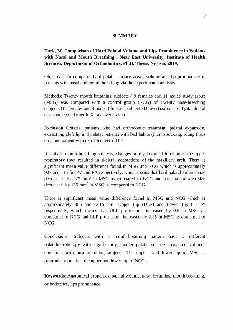
SUMMARY
Turk, M. Comparison of Hard Palatal Volume and Lips Prominence in Patients
with Nasal and Mouth Breathing . Near East University, Institute of Health
Sciences, Department of Orthodontics, Ph.D. Thesis, Nicosia, 2019.
Objective: To compare hard palatal surface area , volume and lip prominence in
patients with nasal and mouth breathing via the experimental analysis.
Methods: Twenty mouth breathing subjects ( 9 females and 11 males study group
(MSG) was compared with a control group (NCG) of Twenty nose-breathing
subjects (11 females and 9 males ) for each subject 3D investigations of digital dental
casts and cephalometric X-rays were taken .
Exclusion Criteria: patients who had orthodontic treatment, palatal expansion,
extraction, cleft lip and palate, patients with bad habits (thump sucking, toung thrus
etc.) and patient with extracted teeth .This
Results:In mouth-breathing subjects, changes in physiological function of the upper
respiratory tract resulted in skeletal adaptations of the maxillary arch. There is
significant mean value difference found in MSG and NCG which is approximately
927 and 115 for PV and PA respectively, which means that hard palatal volume size
decreased by 927 mm³ in MSG as compared to NCG and hard palatal area size
decreased by 115 mm2 in MSG as compared to NCG.
There is significant mean value difference found in MSG and NCG which is
approximately -0.5 and -2.15 for Upper Lip (ULP) and Lower Lip ( LLP)
respectively, which means that ULP protrusion increased by 0.5 in MSG as
compared to NCG and LLP protrusion increased by 2.15 in MSG as compared to
NCG.
Conclusions: Subjects with a mouth-breathing pattern have a different
palatalmorphology with significantly smaller palatal surface areas and volumes
compared with nose-breathing subjects. The upper and lower lip of MSG is
protruded more than the upper and lower lop of NCG .
Keywords: Anatomical properties, palatal volume, nasal breathing, mouth breathing,
orthodontics, lips prominence.

v
Contents
Approval page i
Acknowledgement ii
Summary iii
Contents iv
List Of Symbols And Abbreviations viii
List of figures x
List of tables xiii
1.Introduction 1
1.2 research motivation 2
1.3 Problem Statement 3
1.4 Aim 3
2. General Informatıon 4
2.1 Mouth Breathing 4
2.2 Causes Of Mouth Breathing 7
2.2.1 Stress And Anxiety 8
2.2.2 Nasal Congestion 8
2.2.3 Enlarged Adenoids 9

vi
2.2.4 Enlarged Tonsils 10
2.2.5 Deviated Septum 11
2.2.6 Nasal Polyps 11
2.2.7 Enlarged Turbinate’s 12
2.2.8 Nose Shape, Jaw Shape, And Tumors 12
2.3 Literature Survey 12
2.3.1 Studies (1990-2010) On Dentofacial And Physiologic Effects 13
3. Mouth Breathıng Dıagnosıs And Treatment 17
3.1 Diagnosis Based On Symptoms 18
3.1.1 Snoring 19
3.1.2 Dry Mouth 19
3.1.3 Bad Breath (Halitosis) 19
3.1.4 Chronic Fatigue 20
3.2 Questions Based Diagnosis 20
4. Effects Of Mouth Breathıng 22
4.1 Craniofacial Development And Head Postured 23
4.2 Hyoid Bone Position 24
4.3 Long And Narrow Faces 25

vii
4.4 Tongue Position 26
4.5 Growth Of Maxilla 26
4.6 Hard Palate Dimension 27
4.7 Growth Of Mandible 28
4.8 Dental Maloclussion 28
4.9 Periodontal Disease 29
4.10 Gummy Smiles 30
5. Materıal And Methods 31
5.1 Materials 31
5.2 Methods 31
6. Results 36
7. Dıscussıons 44
8. Conclusion 47
9. Reference 48
viii
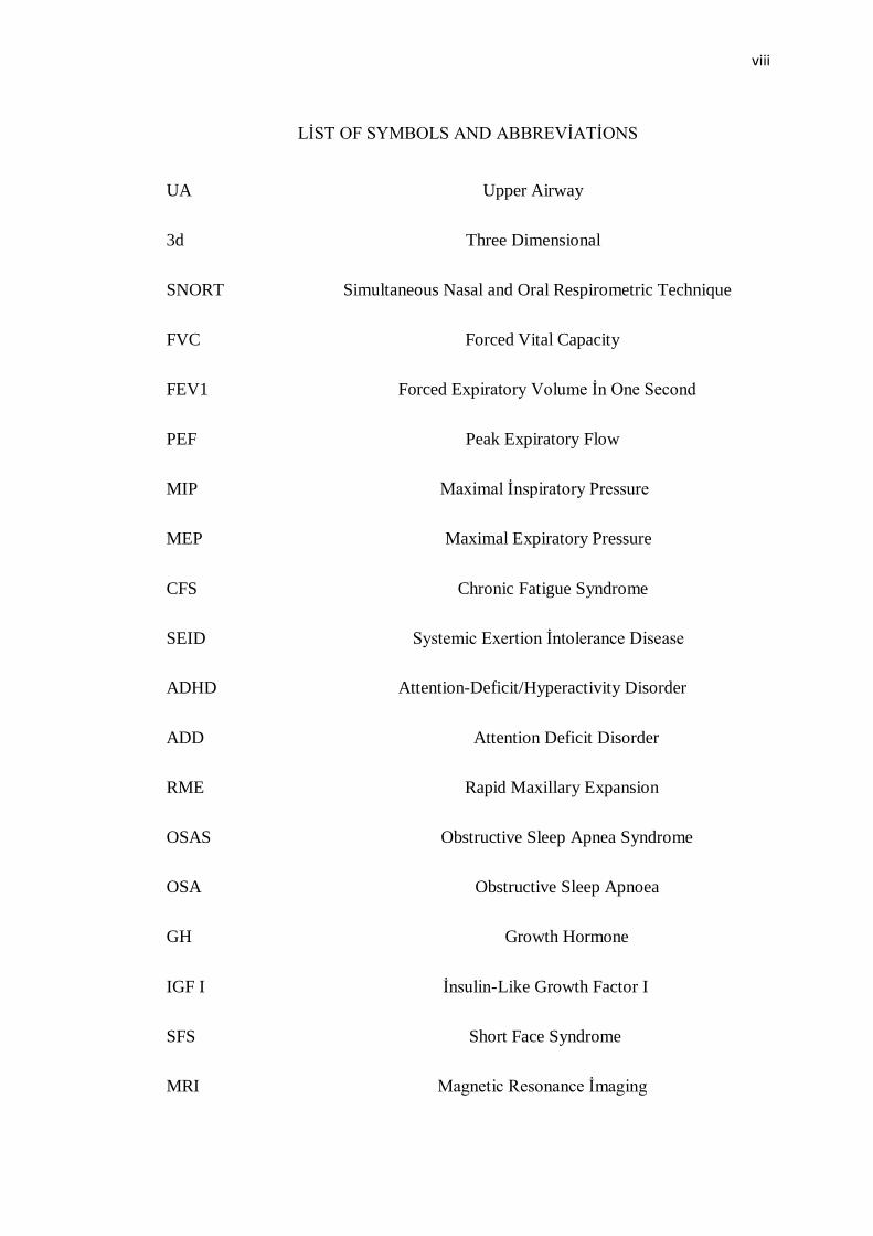
LİST OF SYMBOLS AND ABBREVİATİONS
UA Upper Airway
3d Three Dimensional
SNORT Simultaneous Nasal and Oral Respirometric Technique
FVC Forced Vital Capacity
FEV1 Forced Expiratory Volume İn One Second
PEF Peak Expiratory Flow
MIP Maximal İnspiratory Pressure
MEP Maximal Expiratory Pressure
CFS Chronic Fatigue Syndrome
SEID Systemic Exertion İntolerance Disease
ADHD Attention-Deficit/Hyperactivity Disorder
ADD Attention Deficit Disorder
RME Rapid Maxillary Expansion
OSAS Obstructive Sleep Apnea Syndrome
OSA Obstructive Sleep Apnoea
GH Growth Hormone
IGF I İnsulin-Like Growth Factor I
SFS Short Face Syndrome
MRI Magnetic Resonance İmaging
ix
CT Computed Tomography
MSG Mouth Study Group
NCG Nose Control Group
DCS Direct Conversion Sensor
μm Micrometre
cm Centimeter
mm Millimeter
STA Soft Tissues Analysis
PV Palatal Volume
PA Palatal Area

x
LİST OF FİGURES
Figure 1. Sagittal occlusal relationship in mouth breathing patients 5
Figure 2.. A posterior crossbite and an anterior open-bite 6
malocclusion in a mouth-breathing patient
Figure 3. Lateral cephalometric radiograph of mouth breathing patient 7
Figure 4: Palatal surface area and volume computations 33
Figure 5: Measurement of upper lips prominence using 34
VistaDentsoftware
Figure 6. Measurement of lower lips prominence using 35
VistaDent OC software
Figure 7. Graphical representation for mean statistical 37
comparison of PV and PA measures between MSG and NCG (N=20)
Figure 8. Graphical representation for SD statistical 38
comparison of PV and PA measures between MSG and NCG (N=20)
Figure 9. Graphical representation for outcomes of t-test 39
Figure 10. Graphical representation for mean statistical 41
comparison of ULP and LLP measures between MSG and NCG (N=20)
Figure 11: Graphical representation for SD statistical 42
comparison of ULP and LLP measures between MSG and NCG (N=20)
xi
LİST OF TABELS
Table 1 Mean statistical comparison of PV and PA measures 37
between MSG and NCG (N=20)
Table 2 SD statistical comparison of PV and PA measures 38
between MSG and NCG (N=20)
Table 3 t-test analysis for MSG and NCG using PV and PA differences 39
Table 4 Mean statistical comparison of ULP and LLP measures 40
between MSG and NCG (N=20)
Table 5 SD statistical comparison of ULP and LLP measures 41
between MSG and NCG (N
Table 6: t-test analysis for MSG and NCG using ULP and LLP differences 43
1
1. INTRODUCTION
1.1 Research Background
The main function of human body is breathing and hence it mainly depends
on the adequate permeability of the nasal route. The physiologic breathing mode in
the human being is nasal, regardless of age. The nasal cavity has a fundamental role
in the physiology of respiration (Brant TCS. et al. 2008). It promotes filtering,
heating and humidification of the inhaled air, causing it to reach the lungs at the ideal
temperature and favoring the adequate oxygenation of the body (Yi LC et al. 2008).
Any factor leading to the upper airway (UA) obstruction causes nasal breathing to be
replaced by mouth breathing, among which mechanical events, allergic and
nonallergic inflammatory diseases, congenital malformation and tumoral lesions.
Mouth breathing has been studied since the beginning of the twentieth century, with
scientific publications directed to the scope of dentistry emphasizing the occlusal
consequences (Hartsook JT 1946). This condition, considered as a public health
problem, is attracting growing scientific interest in recent years, and greater coverage
in the multidisciplinary aspects surrounding it.
As discussed earlier, the nose breathing allows the proper performance of
oral sensorimotor system of other human functions as well as orofacial structures
physiological position. In such situations, balance achieved by muscles in facial’s
hard tissue and hence it is stimulus for the harmonious craniofacial development and
growth. The naso-respiratory function can be replaced by a compensatory oral
pattern, due to obstructive or habitual causes. The obstructive mouth breathing
occurs when there is a mechanical hindrance to the airflow passage through the upper
airways, because of enlarged adenotonsillar tissues, among other causes. On the
other hand, in the habitual mouth breathing there is no upper airways obstruction,
and it occurs as a result of flaccidity or bad positioning of orofacial muscles,
transitory swelling of the nasal mucosa, and repaired airway obstruction. Being a
multifactorial pathology, studies have been carried out in order to verify the effects
of the different etiological factors of mouth breathing in the orofacial complex (de
2
Freitas FCN et al. 2001, Di Francesco RC et al. 2004, Ghasempour M et al. 2009,
Souki BQ et al. 2009). In general, the establishment of mouth breathing mode may
alterations in myofunctional aspects, body posture, craniofacial morphology, and
dental occlusion, as well as in the behavior and the quality of life of the subjects.
Among the morphological alterations mentioned, there are the hard palate
modifications, which are expressed by the following classification: deep and atretic;
high and narrow (Marchesan IQ et al. 1996, deep and narrow (Castelluci e Barbosa et
al. 2009, Bianchini AP et al. 2007), ogival and narrow (Cattoni DM et al. 2007)
ogival (Gouveia SA et al. 2009), and deep (Coelho AR et al. 2010).
Thus, the mouth breathing is nothing but the act of inhaling and exhaling
using the mouth. This respiratory phenomenon is considered normal under increased
physical activity because the increased need for oxygen under such circumstances
can only be supplied by breathing through the mouth and nose simultaneously.
However, using one’s mouth to breathe in daily life or during sleep is a serious
ailment which occurs in the presence of an obstruction in nasal and nasopharyngeal
regions of the upper airway. Nasal obstructions can be due to physiologic factors like
allergic rhinitis and polyps or anatomical factors such as a deviated septum and a
narrow nasal area. Enlarged adenoids and tonsils are the most common causes of
obstruction in the nasopharyngeal area, especially at ages 5–6 years. As compared to
nasal breathing, the mouth breathing caused several negative impacts on the
dentofacial deformities in human beings, however justifying the relationship between
dentofacial deformities and mouth breathing still the research problem due to
inconclusive results. This research presents the novel study that demonstrates the
effects of nasal breathing and mouth breathing comparatively by considering the hard
palatal and lip prominence.
1.2 Research Motivation
The resting position and functions based on structure of morphologically
altered hard palatal may be adapted. Thus appropriate anatomical investigations and
analysis is essential. The researchers required to conduct the statistical investigations
of hard palatal in order to gain the more efficiency and accuracy in medical diagnosis
3
as well as assists the medical expert this structure assessment. According to the
hypothesis, mouth breathing and mixed breathing may lead the alterations in hard
palatal area (maxillary arch) or volume as well as lip prominence, and various
manifestations noticed as per the etiology of the mouth breathing. Several researches
already presented on relationship between dentofacial deformities and nasal
breathing, however the accurate study over the relationship between dentofacial
deformities and mouth breathing is still challenging research problem for the
researchers. Defining the appropriate elaboration of mouth breathing and it’s related
with dentofacial deformities based on experimental study is motivating factor of this
research.
1.3 Problem Statement
The relationship between mouth breathing and dentofacial growth is still
being debated after more than a century. Despite the existence of an extensive body
of literature on this subject, the inconclusive results so far may be explained by
different population selection criteria and the various diagnostic methods used for
differentiating mouth breathers from nasal breathers. In order to determine this
assumed relationship, the meaning of the term mouth breathing needs to be clearly
established at first. Then to justify the negative effects of mouth breathing on
dentofacial growth as compared to nasal breathing, preparation of the appropriate
and conclusive experimental analysis is required.
1.4 Aim
To compare the anatomical characteristics of the maxillary arch, identified
as palatal Surface area and volume, and lips prominence between mouth-breathing
and nose-breathing subjects using a three dimensional(3d) analysis of digital dental
casts and cephalometric X-rays.
4
2. General Informatıon
2.1 Mouth breathing
Mouth breathing is a serious ailment -which occurs in the presence of an
obstruction in nasal and nasopharyngeal -regions of the upper airway. Chronic mouth
breathing can cause problems in facial structures and oral health. Changes in facial
structures include long anterior facial height, narrow facial width, a retrognathic
mandible, open-mouthed posture, an incompetent and short upper lip, loss of tonus in
perioral muscles, a pinched looking nose and a dull appearance. The intraoral
consequences of mouth breathing are openbite, a Class II molar occlusion with
increased overjet, protruding maxillary anterior teeth, and a V-shaped maxillary arch.
Not every patient has the same growth changes due to oral breathing. The heritable
characteristics of anatomical structures also seem to play a role in determining which
patients will be most affected. A careful evaluation of the patient by
otolaryngologists, pediatricians and orthodontists is needed before treatment
decision.
If chronic mouth breathing develops due to obstruction of the airways, this
can cause a multitude of problems in facial structures and oral health. Such patients
have been described as having special facial characteristics generally referred to as
adenoid facies (Emslie et al. 1952 ; Linder-Aronson 1970 ; Koski and Lähdemäki
1975 ), long face syndrome (Schendel et al. 1976 ), and respiratory obstruction
syndrome (Ricketts 1968 ). This type of face reportedly features long anterior facial
height, narrow facial width, a retrognathic mandible, open-mouthed posture, an
incompetent and short upper lip, loss of tonus in perioral muscles, a pinched looking
nose,. The intraoral consequences of mouth breathing are said to include open bite, a
Class II molar occlusion with increased overjet, protruding maxillary anterior teeth,
and a V-shaped maxillary arch with deep palatal vault showing in figures 2.3, 2.4,.
Mouth breathers may also extend their heads in order to maintain a patent airway
observed in figures 2.6 and 2.7.

5
In addition to these structural changes, mouth breathing can cause halitosis
(breath malodor), increased incidence of caries, and marginal gingivitis around the
maxillary anterior teeth. If diagnosed early, orthodontic and dentofacial orthopedic
treatment of the patients is possible. However, in adult patients, orthognathic surgical
treatment in addition to orthodontic treatment may be necessary.
Figure 1: Sagittal occlusal relationship in mouth breathing patients (Carlson, D.S.
(2005).
As noticed in figure 2.3, Sagittal occlusal relationship in mouth breathing patients is
said to include a Class II molar occlusion with increased overjet and protruding
maxillary anterior teeth.

6
Figure 2 posterior crossbite and an anterior open-bite malocclusion in a mouth-
breathing patient (Peltomaki,T. 2007)
The posterior crossbite is suggested to be due to transverse maxillary
deficiency caused by an imbalance of forces between the muscle forces acting on
these structures. The open bite is caused by extruded molar teeth due to an open-
mouthed posture in response to an increased nasal resistance.
7
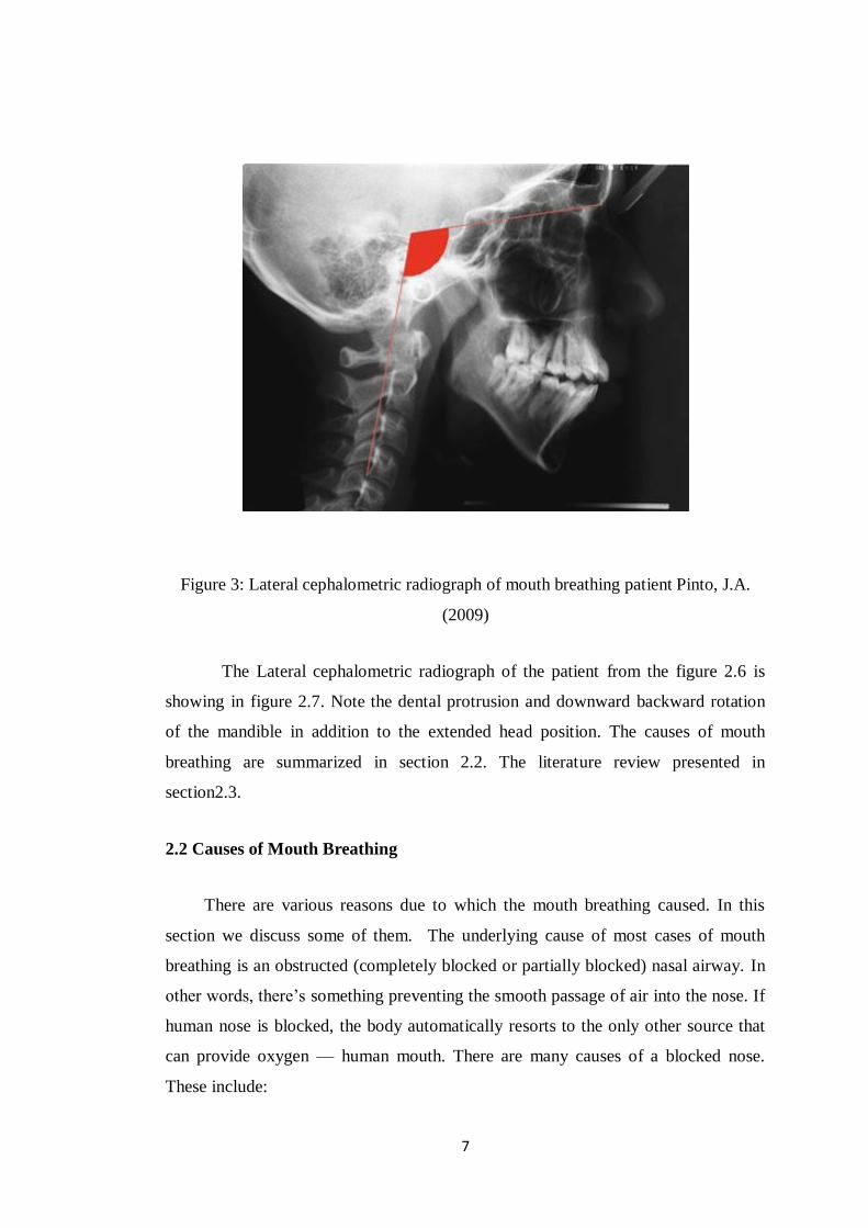
Figure 3: Lateral cephalometric radiograph of mouth breathing patient Pinto, J.A.
(2009)
The Lateral cephalometric radiograph of the patient from the figure 2.6 is
showing in figure 2.7. Note the dental protrusion and downward backward rotation
of the mandible in addition to the extended head position. The causes of mouth
breathing are summarized in section 2.2. The literature review presented in
section2.3.
2.2 Causes of Mouth Breathing
There are various reasons due to which the mouth breathing caused. In this
section we discuss some of them. The underlying cause of most cases of mouth
breathing is an obstructed (completely blocked or partially blocked) nasal airway. In
other words, there’s something preventing the smooth passage of air into the nose. If
human nose is blocked, the body automatically resorts to the only other source that
can provide oxygen — human mouth. There are many causes of a blocked nose.
These include:

8
Stress and anxiety
Nasal Congestion caused by allergies, a cold, or a sinus infection
Enlarged adenoids
Enlarged tonsils
Deviated septum
Nasal polyps
Enlarged turbinates
Nose shape, Jaw Shape, and Tumors (Rare cases)
Some people develop a habit of breathing through their mouth instead of their nose
even after the nasal obstruction clears. For some people with sleep apnea, it may
become a habit to sleep with their mouth open to accommodate their need for
oxygen.
2.2.1 Stress and Anxiety
Stress and anxiety can also cause a person to breathe through their mouth
instead of their nose. Stress activates the sympathetic nervous system leading to
shallow, rapid, and abnormal breathing. Stress is human body's response to certain
situations. It’s subjective, so something that is stressful for human being may not be
stressful for someone else. There are many different kinds of stress and not all of
them are bad. Stress can help individual act quickly in an emergency or help
individual meet a deadline. Stress can affect human physical and mental health, and
human behavior. Human body responds to stress by producing chemicals and
hormones to help individual rise to the challenge. Human heart rate increases, human
brain works faster, and individual have a sudden burst of energy. This response is
basic and natural and is what kept our ancestors from falling victim to hungry
predators.
2.2.2 Nasal Congestion
It is one of main and common cause for the mouth breathing. Nasal
congestion, also called a stuffy nose, is often a symptom of another health problem

9
such as a sinus infection. It may also be caused by the common cold. Nasal
congestion is marked by:
a stuffy or runny nose
sinus pain
mucus buildup
swollen nasal tissue
Home remedies may be enough to alleviate nasal congestion, particularly if
it’s caused by the common cold. However, if you experience long-term congestion,
2.2.3 Enlarged Adenoids
Enlarged adenoids are common in children. The adenoids can become
enlarged due to an infection or may be enlarged from birth. The adenoids are glands
that sit behind the nose above the roof of the mouth. When they grow large, they can
cause snoring and breathing problems. The adenoids are glands that sit above the
roof of the mouth and the tonsils. They are part of the immune system. These glands
help trap germs that enter the nose or the mouth, in an effort to prevent infections.
The size of the adenoids increases until a child is 6 years old, and then they slowly
shrink. The adenoids usually disappear by the time a person is 16. Enlarged adenoids
are rare in adults.
Most of the time, the adenoids become enlarged when the body is trying to fight off
infection. They can remain enlarged, even after the infection is gone. Some children
have enlarged adenoids from birth. Allergies can also cause this enlargement.
Although it is rare, adults' adenoids can become enlarged, due to a chronic infection
or allergy, pollution, or smoking. Even less common are enlarged adenoids resulting
from a cancerous tumor.

10
Thus, the enlarged adenoids is one of cause that leads the mouth breathing in
which breathing more through the mouth than the nose and bad breath or dry,
cracked lips resulting from mouth breathing.
2.2.4 Enlarged Tonsils
Similar to enlarged adenoids, the enlarged tonsils commonly noticed in
children’s. Some children have enlarged palatine tonsils (often simply referred to as
"tonsils," on the left and right sides at the back of the throat , and others have
enlarged adenoids (also called the pharyngeal tonsil, found at the back of the nose).
The medical terms for these enlarged areas of tissue are "tonsil hypertrophy" and
"adenoid hypertrophy." Sometimes both are enlarged. The possible signs of enlarged
tonsils include the following:
Snoring
Pauses in breathing during sleep
Mainly breathing through the mouth
Strained breathing
Restless sleep, waking frequently, bedwetting
Unusual sleeping position (head bent back, knees drawn up to chest while
lying on your front)
Trouble swallowing, “hot potato” speech
Frequent colds
The enlarged tonsils mainly affect ability to breathe through nose. Pauses in
breathing during sleep, on the other hand, are mainly caused by enlarged palatine
tonsils.
11
2.2.5 Deviated Septum
A deviated septum can be congenital. This means that a person was born
with it. It can also occur as a result of an injury to the nose. People often get these
injuries from contact sports, fighting, or car accidents. A deviated septum can also
worsen with age. The septum is the cartilage in the nose that separates the nostrils.
Typically, it sits at the center and divides the nostrils evenly. However, in some
people, this isn’t the case. Many people have an uneven septum, which makes one
nostril larger than the other. Severe unevenness is known as a deviated septum. It can
cause health complications such as a blocked nostril or difficulty breathing. An
uneven septum is very common. According to the American Academy of
Otolaryngology - Head and Neck Surgery, 80 % of all septums are deviated to some
degree. A deviated septum requires medical attention only if it causes other health
issues or negatively impacts quality of life.
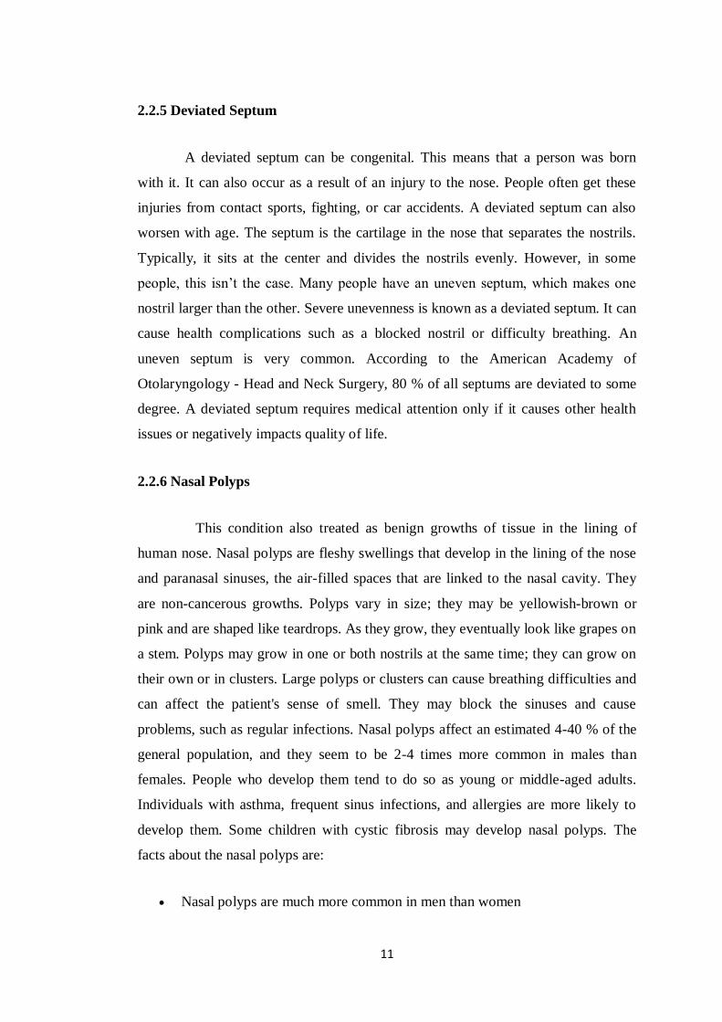
2.2.6 Nasal Polyps
This condition also treated as benign growths of tissue in the lining of
human nose. Nasal polyps are fleshy swellings that develop in the lining of the nose
and paranasal sinuses, the air-filled spaces that are linked to the nasal cavity. They
are non-cancerous growths. Polyps vary in size; they may be yellowish-brown or
pink and are shaped like teardrops. As they grow, they eventually look like grapes on
a stem. Polyps may grow in one or both nostrils at the same time; they can grow on
their own or in clusters. Large polyps or clusters can cause breathing difficulties and
can affect the patient's sense of smell. They may block the sinuses and cause
problems, such as regular infections. Nasal polyps affect an estimated 4-40 % of the
general population, and they seem to be 2-4 times more common in males than
females. People who develop them tend to do so as young or middle-aged adults.
Individuals with asthma, frequent sinus infections, and allergies are more likely to
develop them. Some children with cystic fibrosis may develop nasal polyps. The
facts about the nasal polyps are:
Nasal polyps are much more common in men than women

12
Often nasal polyps do not present any symptoms, making it difficult to know
exactly how common they are
Sometimes, nasal polyps can cause sleep apnea
Nasal polyps can arise at any age
2.2.7 Enlarged Turbinates
The enlarged turbinates one of the cause of mouth breathing. The nasal
turbinates are long, narrow passageways that help to warm and moisten the air that
flows in through the nose. The turbinates are also called the nasal conchae. If the
turbinates are too large, they can actually block airflow. Doctors call this condition
turbinate hypertrophy. This condition can cause breathing problems (in such cases
peoples does the mouth breathing), frequent infections, and nosebleeds. Some people
have three while other people have four. Most people have superior, middle, and
inferior turbinates. Enlargement of the inferior and middle turbinates most commonly
causes turbinate hypertrophy. Both over-the-counter and surgical treatments can treat
turbinate hypertrophy.
2.2.8 Nose Shape, Jaw Shape, and Tumors
The nose shape causes the mouth breathing. As noticed in figure, at age 10 the
normal nasal breathing operations performed, however as the shape changes at age
17 leads the problem of mouth breathing in child. Sometimes, the shape of nose
enlarges over the years than the regular progress which cause the difficulties for
breathing. Similarly, the size and shape of jaw and presence of tumors also leads the
mouth breathing. However this are the rare causes for the mouth breathing.
2.3 Literature Survey
There is still controversial issue within orthodontics and otolaryngology
despite many attempts to establish a cause-and-effect relationship between nasal
respiratory impairments and dentofacial deformities. The most prevalent view among
13
clinicians is that a change in the mode of respiration, such as mouth breathing due to
an inadequate nasal airway, could cause changes in craniocervical posture,
maxillomandibular relationship, and position of the tongue. This in turn could affect
dentofacial growth and positions of the teeth. However, clinicians from both sides of
controversy can find ample evidence in the literature supporting their opinions, and
the results of studies relating dentofacial features with respiratory pattern are far
from being conclusive. Since there are treatment decisions that revolve around the
degree of interplay between nasal obstruction and dentofacial development, the
relationship must be further elucidated. If nasal obstruction has an effect on
dentofacial development, early treatment for removal of the cause of this obstruction
would be necessary. On the other hand, if dentofacial growth and development is not
signifi cantly affected by the respiratory mode, then treatment of nasal obstruction in
order to prevent abnormal orofacial development would not be indicated.
2.3.1 Studies (1990-2010) on Dentofacial and Physiologic Effects
The studies reported during the last two decades presented in this section.
Heredity and environmental factors are both effective in the development of dental
arches and postnatal determination of craniofacial features. One of the most
important environmental factors is the predominant respiratory pattern. Nasal
breathing is associated with normal posture of the tongue and lips and normal muscle
activity. If there is nasal obstruction, this would likely affect the muscle forces acting
on the dentofacial region. This change in muscular action may cause abnormal facial
growth and development. It has been shown that during oral breathing masseter
muscle activity is inhibited (Ono et al. 1998 ). Increased airway resistance also
stimulates mechanoreceptors in the upper airway and increases the activities of the
genioglossus and mylohyoid muscles due to forward positioning of the tongue and
opening of the mouth to maintain the airway (Song and Pae 2001 ).
Long-term mouth breathing seems to affect the occlusion and facial
morphology during periods of rapid growth. However, not every patient has the same
growth changes due to oral breathing. The heritable characteristics of anatomical
structures also seem to play a role in determining which patients will be most

14
affected. In some patients a slight opening of the lips may be enough to provide the
necessary airway, while in others a more exaggerated postural response of the
mandible, tongue, and head will be necessary.
Children with narrow facial patterns also may be more susceptible to growth
changes due to mouth breathing than children with broad facial features.
It is also possible for patients with a vertical facial growth pattern to be more
likely to be mouth breathers. The severity of the obstruction will also determine if the
child is a chronic mouth breather or a partial one. If the obstruction is severe, the
changed postural responses will be in place longer causing more extensive growth
changes in dentofacial structures.
The most common cause of oral breathing in children is enlarged pharyngeal
lymphoid tissue. The enlargement of these tissues may adversely affect pharyngeal
patency (Gross and Harrison 2000 ). Normally, the size of the adenoid tissue is
dependent on the associated skeletal structures. However, abnormal growth of this
tissue may predispose the patient to upper airway obstruction causing oral
respiration. As a matter of fact, any reason that causes nasal resistance to increase for
long periods of time, such as allergies or nasal eptal deformity, has the potential to
cause chronic oral respiration.
During oral respiration, the mandible rotates open and the tongue is positioned
lower in the mouth and no longer contacts the palate causing eruption of the molars
and transverse maxillary deficiency .
When the tongue is in its correct position, it exerts transverse pressure on the
teeth and alveolus, allowing proper growth . If the tongue is in a lower position,
equilibrium of forces is disturbed. As a result the palate becomes narrow . Thus, the
mandible rotates in a clockwise direction, losing contact with the soft palate, causing
open bite and mandibular retrognathism.

15
However, this is only a mechanistic view of the possible interactions between
mandibular growth and oral breathing. Complex epigenetic events may also be
responsible for the growth changes in the mandible. It has been hypothesized that
children with significantly enlarged adenoids will develop obstructive sleep apnea
causing abnormal nocturnal growth hormone secretion causing somatic growth
impairment (Peltomaki 2007 ). Due to this abnormal hormonal balance, mandibular
ramus growth may be less than in healthy subjects causing the observed mandibular
rotation in these children. This mandibular rotation also causes backward and
downward displacement of the tongue (L’Estrange et al. 1996 ). Postural changes of
the tongue lead to altered muscle forces to act on maxillary arch causing a
constricted maxillary arch, high palate, and posterior crossbite in the transverse
direction.
Craniocervical posture and oral breathing have also been the subject of
various investigations. Oral breathing has been shown to be associated with head
extension (Cuccia et al. 2008 ; Chaves et al. 2010 ; Neiva et al. 2009 ). This postural
change moves the hyoid bone upward establishing an adequate airway (Gonzalez and
Manns 1996 ). Since there is a relationship between head posture and altered muscle
activity, long-term craniocervical changes may influence craniofacial growth as well
as putting undue load on the neck and upper shoulders.
Mouth breathing in cleft palate patients is a clinically relevant subject as well
(Hairfi eld et al. 1988 ). The airway size is reduced and nasal resistance is higher in
cleft patients compared to noncleft subjects (Warren et al. 1984 , 1969 ). The large
craniocervical angulation indicates an extended head position in these cases
(Oosterkamp et al. 2007 ). The high prevalence of mouth breathing in these patients
may be caused by various factors, such as septal deviation and effects of surgical
techniques. Thus, a normal breathing pattern may not be established ever because of
the open communication between the nose and mouth at birth (Warren et al. 1988 ).
In addition to these structural changes in the dentofacial region, mouth
breathing may cause other oral diseases. Mouth-breathing patients often have
inflamed labial gingival tissues around the maxillary incisors. The gingiva becomes

16
inflamed and hyperplastic because the mouth remains open and the salivary flow is
reduced. Since saliva performs essential roles including antimicrobial action and
protection of oral tissues, a reduction in salivary flow will have negative impact on
teeth and gingival tissues as well as generating odoriferous volatile compounds.
Clinically, the gingiva appears swollen, red, and shiny with the classic rolled
up appearance.This lack of lip seal causes the mouth to become dry. Maxillary
anterior teeth and gingiva are most at risk for the negative effects of this open-
mouthed posture. The gingiva is inflamed, and there are decalcifications on teeth due
to poor oral hygiene and mouth breathing.
Note the classic shiny red, swollen look of the gingival. There can be bone
loss and pocket formation in the interproximal area if proper oral hygiene is not
maintained. There is also an increased incidence of caries and halitosis (breath
malodor). Correction of mouth breathing along with necessary dental treatments will
improve the health of the oral cavity.
It has been difficult to determine the relationship between dentofacial
morphology and oral breathing because respiration is a complex act which cannot be
easily classified with the current techniques. There are multiple factors which
determine if dentofacial growth of a person will be affected by the changes in the
nasal airway. Thus, a careful evaluation of the patient by otolaryngologists,
pediatricians, and orthodontists is needed before treatment decisions.

17
3. Mouth Breathing Diagnosıs And Treatment
As discussed earlier, mouth breathing has been reported to cause abnormal
facial growth, attention problems associated with sleep disorders, and of reduction in
quality of life. In addition, we highlight that increased oxygen load in the prefrontal
cortex when changing from nasal breathing to mouth breathing. In this way, in the
field of dentistry, the characteristics of mouth breathing have been investigated
mainly for the purpose of discovering the effects of mouth-breathing and identifying
habitual mouth breathers.
There are a number of findings reported as characteristic of mouth breathing,
but they differ in different reports. For example, there are reports focused on rhinitis
and open mouth at rest. Rhinitis is reported as a finding related to mouth breathing in
some studies, but not in others. Open mouth at rest is also found to be characteristic
of mouth breathing in some studies but not others, because there are some cases in
which the mouth is habitually open without mouth breathing. Mouth breathing is
determined by a combination of predisposing factors (i.e., facial type) and
precipitating factors (i.e., local factors). In other words, all of the findings associated
with mouth breathing may not apply to a given individual. Accordingly, to identify
whether a person is a habitual mouth breather, he or she should be evaluated based
not on the presence (or absence) of certain specified characteristics, but on the extent
to which the various different characteristics in the spectrum of characteristics
associated with mouth breathing apply.
Clinically, visual assessment is most commonly used (97.2%) to identify the
characteristic findings of mouth breathing (Pacheco MCT et al. 2015). In visual
assessment, the dentist (or other clinician) observes the patient for the presence of
factors causing increased breathing resistance, such as adenoid facies, pharynx or
palatine tonsil hypertrophy and deviated nasal septum (Wieler WJ et al. 2007); and
observes whether the mouth-breathing route is closed at rest. Facial morphology and
the condition of the front teeth and gingiva are also observed. Next in frequency after
visual assessment, interviews (87.2%) and respiratory tests (59%) have been used to
evaluate for mouth breathing (Pacheco MCT et al. 2015). In interviews, subjects are

18
questioned about symptoms or habits that may induce mouth breathing, such as
allergic rhinitis, nasal congestion, snoring, and open mouth during sleeping or resting
(Fujimoto S et al. 2009, Pacheco MCT et al. 2015, Masahiro Sano et al. 2018).
Respiratory tests used include a lip seal test, which evaluates whether a subject can
keep his or her lips closed; a mirror test, which assesses the extent of clouding on a
mirror held below the nose; and a water retention test, which evaluates the ability to
hold water in one’s mouth (Fujimoto S et al. 2009).
Namely, the methods and content of evaluation for detecting habitual mouth
breathing are left to the discretion of the dentist. There is currently no unified
screening method for detecting habitual mouth breathing. Moreover, there is little
general awareness of the need for intervention in the case of habitual mouth
breathing, and people in general do not know how to identify habitual mouth
breathing. For this reason, early detection of habitual mouth breathing is delayed, and
people are unlikely to ask their dentist about mouth breathing, or visit a dentist with
mouth breathing as their main complaint. Creation of a screening process for
detecting habitual mouth breathing without the use of special equipment would thus
be useful not only for dentists, enabling them to detect mouth breathing on a uniform
scale, but also for the general public, by raising awareness of the importance of
mouth breathing prevention. In short, doctors mainly relay on two cases for the
diagnosis of mouth breathing such as symptoms and questionnaires.
3.1 Diagnosis based on Symptoms
A doctor will often inspect a person's mouth, throat, and nose to identify any
areas of swelling or abnormalities and observe their breathing pattern. They may also
order imaging studies to examine the nasal passages and perform lung function tests
to see if the lungs are impacted by asthma or other conditions. If a doctor suspects a
person may have sleep apnea, they may order a sleep study.

19
This involves the individual going to a sleep center where monitoring
equipment can identify if, when, and how often a person stops breathing while they
are sleeping. Both children and adults can have sleep apnea.
You may not realize that you’re breathing through your mouth instead of
your nose, especially while you sleep. People who breathe through their mouth at
night may have the following symptoms:
3.1.1 Snoring: Snoring is a common condition that can affect anyone, although it
occurs more frequently in men and people who are overweight. Snoring has a
tendency to worsen with age. Occasional snoring is usually not very serious and is
mostly a nuisance for your bed partner. However, if you are a habitual snorer, you
not only disrupt the sleep patterns of those close to you, but you also impair your
own sleep quality. Snoring occurs when the flow of air through the mouth and nose
is physically obstructed.
3.1.2 Dry Mouth
The dry mouth (xerostomia) might seem like an annoying thing that happens
at night from time to time. But if it occurs regularly, It is one of symptom of the
mouth breathing. Saliva is necessary for tooth and gum health, and enzymes in saliva
help aid indigestion. If your mouth is dry throughout the night, your oral health
might be affected without you even knowing it.
3.1.3 Bad Breath (Halitosis)
Breath odor affects everyone at some point. Bad breath is also known as
halitosis or fetor oris. Odor can come from the mouth, teeth, or as a result of an
underlying health problem. Bad breath odor can be a temporary problem or a chronic
condition.
According to the American Dental Association, at least 50 % of adults have
had halitosis in their lifetime. In addition to a bad smell in mouth, we may also notice

20
a bad taste in mouth. If the taste is due to an underlying condition and isn’t because
of trapped food particles, it may not disappear even if brush teeth and use
mouthwash.
3.1.4 Chronic Fatigue
The chronic fatigue syndrome (CFS) is a disorder characterized by extreme
fatigue or tiredness that doesn’t go away with rest and can’t be explained by an
underlying medical condition. CFS can also be referred to as myalgic
encephalomyelitis (ME) or systemic exertion intolerance disease (SEID). The causes
of CFS aren’t fully understood yet. Some theories include viral infection,
psychological stress, or a combination of factors. Because no single cause has been
identified, and because many other conditions produce similar symptoms, CFS can
be difficult to diagnose. There are no tests for CFS. The doctor will have to rule out
other causes for fatigue when determining a diagnosis. While CFS was previously a
controversial diagnosis, it’s now widely accepted as a medical condition. CFS can
affect anyone, though it’s most common among women Trusted Source in their 40s
and 50s. There’s currently no cure, but treatment can relieve symptoms. However,
CFS is considered one of the symptoms to diagnose the mouth breathing, it means
that some studies reveals that mouth breathing cause the CFS in human beings.
3.2 Questions based Diagnosis
A doctor will ask questions to obtain a full medical history if they suspect mouth
breathing is a problem for someone. They will ask when the person first noticed their
symptoms, what makes their symptoms worse, and if anything makes them better.
Medical professionals may ask one or more than one questions to diagnose the
possibility of mouth breathing disease. The 10 questions are more commonly used by
doctors for the prediction as they delivered the higher accuracy of diagnosis..Which
the respondents were asked about a variety of items conventionally reported to be
characteristic of habitual mouth breathing. Of the 50 questions, 27 had 3 possible
responses (yes, sometimes, or no), and 23 had 2 possible responses (yes or no).

21
Additional questions intended to reveal possible attributes of the mouth breathing
group were also included, concerning gender, age, height, weight, history of asthma,
incidence of allergic rhinitis, and history of smoking.
22
4. Effects Of Mouth Breathıng
Nasal obstruction, chronic allergic rhinitis and hypertrophic adenoids
decrease capacity for nasal breathing (NB) and compensating for this by mouth
breathing (MB) might be necessary (Oulis et al., 1994). Respiratory airway function
influences facial morphology and both craniofacial (Gungor and Turkkahraman,
2009) and cervical functions (Huggare and Laine-Alava, 1997; McNamara, 1981).
The breathing pattern may influence the development of the transverse relationship
between the maxilla mandible, resulting in the development of a posterior cross bite
(Rubin, 1980).
MB can affect the form of the jaw or cause malocclusions (Hartsook, 1946), and
it has been shown to lead to the so-called “adenoid face”, which is characterized by a
narrow upper dental arch, retroclined mandibular incisors, an incompetent lip seal, a
steep mandibular plane angle and increased anterior facial height (Lessa et al., 2005;
Peltomäki, 2007; Linder-Aronson, 1970). Ricketts (1968) suggested that head
extension represents a functional response in MB patients to compensate for nasal
obstruction.
There were several effects of MB studied. MB has been reported to cause
changes in human head posture (Cuccia et al., 2008). The treatment of hypertrophic
adenoids (Linder-Aronson, 1970) and nasal obstruction (Vig et al., 1980) with a
nasal clip has been shown to alter head posture. Children with MB who have
enlarged tonsils can develop the extension of their head posture and the low position
of hyoid bone position (Behlfelt et al., 1990a,b). However, some authors have
concluded that the hyoid position is maintained in a stable position in children with
MB (Bibby, 1984; Ferraz et al., 2007). MB is associated with a low tongue posture
and the absence of a contact surface between the tongue and soft palate; this latter
factor was termed “posterior oral incompetence” by Ballard (1951). This problem is
caused by enlarged adenoid tissue that reduces the airway space and leads to postural
adaptations at the level of the oropharynx.
23
The hyoid bone drops in relation to the mandible, and creates a relatively
constant air-space diameter in the anteroposterior direction. This neuromuscular
recruitment may cause changes in the mandibular resting position and neck extension
(Tourné, 1991). Thus, the breathing pattern could represent a major factor that
underlies the hyoid bone position (Graber, 1978).
In this section, the aim is to present the study on various effects caused by the
MB on human beings such as Craniofacial Development and Head Postured, Hyoid
bone position, Long-narrow faces, Tongue Position, Growth of Maxilla, Hard Palate
Dimension, Growth of Mndibule, Dental Malocclusion etc.
4.1 Craniofacial Development and Head Postured
In all individuals, muscular activity is reduced and upper airway resistance
increased during sleep compared with wakefulness (Worsnop et al., 2000). This does
not have a notable effect on breathing in anatomically and functionally ‘healthy’
individuals. On the other hand, reduction of muscular tonus in children with large
adenoids and tonsils, or with other underlying abnormal upper airway anatomy, may
lead to airway obstruction and eventually to obstructive sleep apnoea (OSA).
Interestingly, these children have been found to have similar craniofacial
characteristics as adenoid face children (Guilleminault et al., 1996; Shintani et al.,
1997; Agren et al., 1998; Zucconi et al., 1999; Kawashima et al., 2000, 2002;
Zettergren-Wijk et al., 2006). The first treatment of choice of OSA children is
removal of adenoids and tonsils (Nieminen, 2002; Guilleminault et al., 2004).
It can thus be postulated that some children with a clinical diagnosis of an
adenoid face could nowadays be diagnosed as having OSA. Of particular interest is
the recent cephalometric study on 5-year-old children with polysomnographically
verified OSA (Zettergren-Wijk et al., 2006). This study showed that OSA children
have a different facial morphology compared with age-matched controls. The
mandibular plane angle was found to be posteriorly inclined, anterior face height to
be greater, and posterior face height smaller, in the OSA than in the control children.

24
At the 5-year follow-up after adenotonsillectomy, no major craniofacial and Head
postured differences were noted in many studies.
In a closer look at the growth changes it becomes evident that anterior face
height remained greater in the OSA children than in the control children (difference
on average 2.5 mm), but it increased on average by a comparable amount in both
groups of children. Yet, the mandibular plane angle was decreased in the OSA
children. This may be explained by the described greater posterior face height growth
(ramus growth) in the OSA than in the control children (OSA children 5 mm, control
children 3 mm).
OSA children with large adenoids and tonsils have also been found to have
somatic growth impairment due to abnormal nocturnal growth hormone (GH)
secretion (Goldstein et al., 1987; Bar et al., 1999; Nieminen et al., 2002). Following
adenotonsillectomy, a significant increase in the serum levels of GH mediators, i.e.
insulin-like growth factor I (IGF I) and its binding protein, has been reported.
Consequently, normalization and even catch-up of somatic growth have been
observed (Bar et al., 1999; Nieminen et al., 2002).
4.2 Hyoid Bone Position
The MB having severe effects on hyoid bone position as well. The
importance of the hyoid bone is related to its single relation, nonetheless, it provides
connections to pharynx, mandible and cranial muscles, ligaments and fascia. Many
of the characteristics of the so called Long Face Syndrome (LFS) group and the
Short Face Syndrome (SFS) group could be explained based on the clockwise and
counter-clock wise rotation of the mandible "in harmony" with the hyoid bone,
tongue, pharynx and neck. The vital need to keep the air space pattern at the tongue
base may explain this rotation in the LFS.
Adenoid tissue or tongue mass may reduce the air space and cause postural
adaptations at the level of the oropharynx. A hyoid bone drop in relation to the
mandible would represent an attempt to assure a relatively constant air-space
25
diameter in the antero-posterior direction. This neuromuscular recruiting could cause
changes in mandibular rest position and neck extension, thus influencing the
craniofacial growth pattern.
Thus, air space pattern and stability would represent
major factors responsible for hyoid bone position. Since malocclusion may be caused
by inadequate oral habit, such as atypical swallowing and oral breathing - hyoid bone
position could serve as an important diagnostic guide.
Numerous authors have
studied the hyoid bone morphology and function, and others have investigated hyoid
bone position in relation to the cranium and the cervical spine by means of
cephalometric techniques.
4.3 Long and Narrow Faces
As discussed earlier this research, the mouth breathing leads another effects
on dentofacial development of patients called long and narrow faces. Long and
narrow face effects commonly noticed in mouth breathers. In medical terms the long
and narrow face conditions is called as long face syndrome.
It is also referred as skeletal open bite, is a relatively common condition
characterised by excessive vertical facial development. Its causes may be either
genetic or mouth breathing. Long face syndrome is a common dentofacial
abnormality. Its diagnosis, symptomology and treatments are complex and
controversial. Indeed, even its existence as a "syndrome" is disputed.The Habitual
mouth breathing can lead to long face syndrome "long face" or elongated facial
features during a child's development.
26
4.4 Tongue Position
The effects of mouth breathing leads the changes in tongue position as well.
The Mouth breathing changes the position of tongue called as a tongue “thrust.” This
negatively affects speech, swallowing, and chewing. This could lead to a child
feeling self-conscious. Depending on the severity of the speech impediment(s), a
speech pathologist may be necessary to correct speech alterations and slurs. This
effect is medically called as Tongue Trust syndrome.
Since 1958, the term "tongue thrust" has been described and discussed in
speech and dental publications by many writers. Many school-age children have
tongue thrust. For example, according to recent literature, as many as 67–95 percent
of children 5–8 years old exhibit tongue thrust, which may be associated with or
contributing to an orthodontic or speech problem. Up to the age of four, there is a
possibility that the child will outgrow tongue thrust. However, if the tongue thrust
swallowing pattern is retained beyond that age, it may be strengthened.
4.5 Growth of Maxilla
“All growth changes in size, shape, and spatial position, and the maintenance
of all skeletal units are always secondary to specific functional matrices.” (i.e.
capsular and periosteal matrix) (Moss M.L et.al, 1965). The capsular matrix includes
oral, pharyngeal and nasal cavity. Whenever function carried out by capsular matrix
is hampered the growth of skeletal units will be affected.
Assuming this, the growth of the sinus should also be dependent on functions
carried out by nasal capsule. When the functioning of the nasal capsule is hampered
due to mouth breathing, changes in the form of maxilla and paranasal sinuses should
be expected. The four sets of paranasal sinuses – maxillary, frontal, sphenoid and
ethmoidal could show poor development. These paranasal sinuses show growth by
pneumatisation (Enlow D.H et al. 1990). In presence of stunted growth, impaired
pneumatization is also a possibility which indicates the likelihood of dimensional

27
changes in the sinuses especially maxillary sinus as it is largest of all the paranasal
sinuses.
Maxillary sinus volume reaches nearly adult size between the ages of 12
and 15 years with biphasic growth spurts i.e. rapid growth during the first 3 years and
then again from the age of 7–12. The growth of maxilla is also considered to be
completed by 12–14 years (Enlow D.H et al. 1990). Therefore, it is decided to
evaluate maxillary sinus in the age group of 12–14 years. Evaluation of maxillary
sinus can be performed using radiographs (lateral and frontal), MRI and CT scans
(Oktay H, 1992, Ikeda A et al. 1998). Lateral and frontal head films offer limited
information about the maxillary sinus, with the inherent errors of a 2D representation
of a 3D structure. Although MRI is most superior in soft tissue rendering, its use is
limited by its cost and restricted accessibility.
The effects of mouth breathing on the growth maxillilary sinus are summarized as:
The volume of maxillary sinus in mouth breathers was significantly lower
than normal breathers. However it remains unclear whether it is because of
improper functioning of nasal cavity or due to the underlying pathological
condition.
Another finding in our study was that the maxillary sinus volume of right and
left side differed significantly amongst the mouth breathers, it suggests that
chronic inflammation of the sinus is more likely the cause for this. The
chronic inflammation causes thickening of bony wall of the sinus, thereby
reducing its volume. However, chronic inflammation is prone to occur in
poorly growing sinus, hence both these factors are interrelated and it is
difficult to say which the main causative factor is.
4.6 Hard Palate Dimension
The bony structure which builds the division among the nasal cavity and
oral cavity called the hard palate. Nasal breathing can stimulate the lateral growth of
the maxilla, thus making the palate flat, affecting the breathing. Any factors that

28
promote obstruction in the upper airway lead to a change from nose breathing to
mouth breathing. In children, the causes of nasal obstruction are rhinitis allergy,
adenoid hypertrophy, nasal polyps, rhinosinusitis, and obstructive sleep apnea
syndrome.
Apart from the other effects, the mouth breathing also produces alterations in
hard palate morphology, such as a narrow V-shaped maxillary arch and a high palatal
vault (IS Indiarti et al. 2017). Many studies have shown that there is a relationship
between mouth breathing and a high palatal vault.
4.7 Growth of Mandible
Mouth breathing leads to a new posture in order to compensate the
decrease in nasal airflow and to allow respiration. The changes include a lower
position of the mandible, and an anterior or a lower position of the tongue, usually
associated with lower orofacial muscles tonicity. As a result, there is disharmony in
the growth and development of orofacial structures, including narrowing of the
maxilla, lower development of the mandible, alterations in the position of the head in
relation to the neck, protrusion of the upper incisors and distal position of the
mandible in relation to the maxilla.
4.8 Dental Maloclussion
As noticed earlier the mouth breathing is nothing but the para-functional
habit whereby air passes exclusively or partially through the mouth instead of the
nose, and it is accompanied by skeletal and functional alterations in the orofacial
district. The causes of oral breathing are classified as both congenital and acquired.
The former comprise: choanal atresia, nostril atresia and nasal septum deviations.
The latter include: outcomes of nasal fractures, rhinopharyngitis, allergic rhinitis,
nasal polyposis, chronic sinusitis, chronic adenotonsillitis, chronic hypertrophic
rhinitis, adenotonsillar hypertrophy, malignant and benign tumours.

29
The most evident consequences of chronic oral breathing are represented by
craniomaxillo facial alterations, mainly caused by abnormal mandible displacement
and subsequent dysmorphism of the oralstructures and modified posture (Bernkoff et
al. 1997). The tongue lies against the bottom of the mouth, thus allowing the passage
of air via the oralroute. In the event of concomitant tonsillar hypertrophy, the tongue
undergoes further anteriorisation, since its base is forced to move away from the
posterior pharyngeal wall due to spatial constraints. The most frequent dysgnathia is
represented by both dental and skeletal Class II, with reduction of the transverse
diameter of the upper arch, ogival palate, mono- or bilateral posterior crossbite, and
anterioropen bite with buccally inclined upper incisors.
It must be clarified that the malocclusion is not a consequence but, rather,
the cause of oral breathing through incorrect mandible and tongue positioning due to
the presence of dysmorphosis. Facial morphology is typical (the so-called “facies
adenoidea”), characterised by labial incompetence with a short upper lip that has
accentuated lower concavity, and a protruding and often erythematous lower lip. The
tongue placed between the arches and hypotonia of the alar cartilages due to limited
use can also be observed. In the most severe and complicated forms, the patient’s
general appearance is asthenic and longilineal, with an underdeveloped thoracic
cage, sunken or keeled sternum, winged scapulas, kyphosis and rachitic traits.
4.9 Periodontal Disease
As compared to the nasal breathers, 42 % water lost in mouth breather which leads
the periodontal disease (Svensson S et al. 2006). Dryness is linked to various
inflammatory conditions such as dry eye disease, exercise-induced asthma, and
atopic dermatitis (Stevenson W et al. 2012).
In dry eye disease, decreased formation of tears or increased rate of
evaporation causes inflammation of the ocular surface and associated damage.
During exercise, lower airways are recruited in conditioning inspired air, especially
at times of increased ventilation and prolonged duration of exercise (Daviskas E et
30
al. 1991). This process of humidifying typically cold and dry air results in
dehydration (Daviskas E et al. 1991).
Dehydration evokes release of inflammatory mediators and thereby leads to
exercise-induced broncho constriction and asthma. Hydration status of the skin
during wound healing influences expression of inflammatory signals in the epidermis
with exaggerated response of pro-inflammatory cytokines in healing wounds under
dry conditions compared to wounds that restore to normal healthy state in an
optimally hydrated environment. Application of emollients on dry skin in individuals
with or at an increased risk for developing atopic dermatitis decreases transepidermal
water loss as well as improving hydration status of skin and providing relief in
pruritis (Lio PA 2016).
In the oral cavity, tissues are protected from dessication by salivary mucins
that bind with water and form a coating over the oral mucosa, thereby maintaining
the tissue’s hydration. In individuals with xerostomia, increased plaque and gingival
inflammation is reported. Mouth breathing has also been reported to play a role in
gingival inflammation
4.10 Gummy Smile
This is another effect of mouth breathing in patients.It may also cause
gingivitis (inflamed gums) and halitosis (bad breath), especially upon waking if
mouth breathing occurs during sleeping. The term "mouth-breather" is sometimes
utilized as an insult to imply low intelligence.Apart from the above effects of mouth
breathing, there other effects also such as Bad Breath, Facial Soft Tissue, Chewing
Efficiency, Learning Process, Speech (Disorders), Throat And Ear Infections, and
Quality Of Life. Such factors are more or less affected in mouth breathers.
According to different studies, the facial soft tissue of severely obstructed mouth
breathing children is different than in nasal breathing children. Changes in lips,
nasolabial angle, nasal prominence, and chin thickness are associated with severe
airway obstruction in children. Mouth breathing also leads the various infections at
throat and ear.

31
5. Materıal And Methods
For this research as per the aim we received the ethical approval received
from the Department of Orthodontics at the Near East University, and the subject’s
permission granted from patient themselves or parents prior to inclusion in this study.
The section 5.1 presents the information of subjects as materials used for the
comparative analysis. The section 5.2 demonstrates the background of software used
to conduct the experiments and then presented the methodology to measure the
readings for palatal dimensions and lips prominence of nasal breathers and mouth
breathers.
5.1 Materials
The subjects include patient of both genders for this study. These subjects
were sought for orthodontic treatment at the Department of Orthodontics at the Near
East University. The maxillary cast and cephalometric radiograph were
retrospectively selected from the records of treated cases, those are underwent
treatment in the outpatient Department of Orthodontics at Near East University in
Turkish republic of North Cyprus. In short, the subjects of this study described in
two groups MSG and NCG with below properties:
MSG: 20 patients for study group include 9 female and 11 male.
NCG: 20 patients for control group include 11 female and 9 male
Subject Exclusion Criterio: patients who had orthodontic treatment, palatal
expansion, extraction, cleft lip and palate, patients with bad habits (thump sucking,
toung thrus etc.) and patient with extracted teeth are selected in this study. Mode of
breathing was defined according to history and complete physical examination,
lateral nasopharyngeal x-ray, and confirmed by questionnaire answered by the
parents or by Patients themselves classifying them as clinically normal respiratory
pattern or as exclusive mouth breathers. The two groups did not have a previous
history of nasal respiratory surgery or orthodontic treatment. No significant
differences were found between age and sex in the different mode of breathing.

32
5.2 Methods
For each subject we have to measure the palatal surface area and then
compute the palatal volume at first. Secondly we need to compute the lips
prominence as well for each subject of this research. First we present the study over
the methods/tools used in this study, and then present the methodology used to get
the results.
In order to measure palatal surface area and calculate palatal volume, Sirona
Orthophos SL 3D/2D, (NC, USA) has been used to scan the casts in this work (as
described above), the boundaries for the palate must be defined. The gingival plane
and a distal plane were used as boundaries for the palate. The gingival plane was
obtained by connecting the centre of the dentogingival junction of all erupted
permanent teeth. The distal plane was created through two points at the distal of the
second primary molars perpendicular to the gingival plane. Figure 5.7 demonstrates
the outcome of palatal surface area measurement and palatal volume computations.
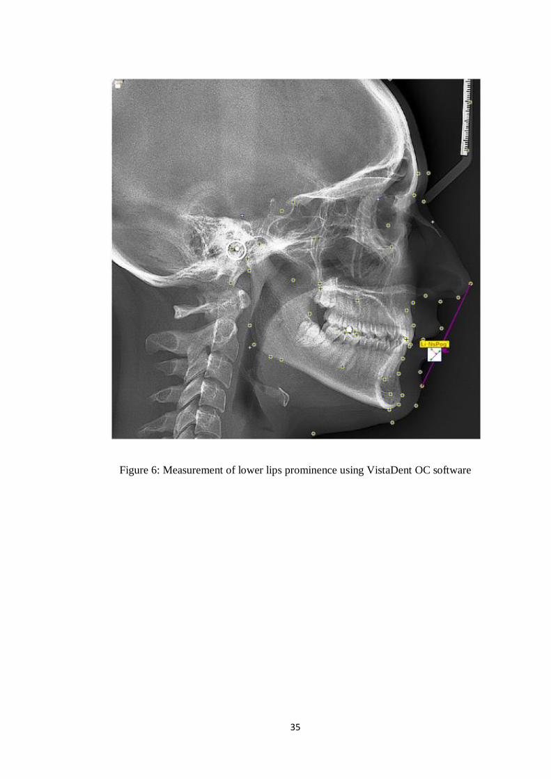
This is first research were we measuring the lips prominences of subjects
for investigation of nasal and mouth breathers influences. We used the technique of
Soft Tissues Analysis (STA) using the VistaDent OC Complete software. Using this
tool we measured the values for both upper and lower lips prominence. The Ls-
NsPog which stands for the upper lip prominence and Li-NsPog stands for the lower
lip prominence. Figure 5.8 demonstrate the measurement process of upper lips
prominence using VistaDent OC software. Figure 5.9 demonstrate the measurement
process of lower lips prominence using VistaDent OC software.

33
Figure 4: Palatal surface area and volume computations
As observed in figure 5.7, the gingival plane is constructed by connecting
the midpoints of the dentogingival junction of all erupted teeth. The distal plane is
built perpendicular to the dentogingival plane and passing from the two most distal
points corresponding to the distal surface of the second permanent molars.

34
Figure 5: Measurement of upper lips prominence using VistaDent OC software
The figures 5.7 to 5.9 shows the process of measuring the various parameters
such as upper lips prominence value, lower lips prominence value, hard palatal
volume value, and hard palatal area value for both categories of groups such as MSG
and NCG in this study. We record each subjects values under both categories using
the methodology mentioned in this chapter for the comparative and statistical
analysis between the nasal breathing and mouth breathing candidates.
35
Figure 6: Measurement of lower lips prominence using VistaDent OC software

36
6. RESULTS
In this study, main aim is to present the novel comparative study of palatal
measurements and lip prominence for both mouth breathers and nasal breathers to
estimate the difference and effectively justify the relationship between mouth
breathers and dentofacial deformities. In literature, some attempts presented to find
the relationship between mouth breathing and dentofacial deformities, however some
shown the differences and some did not, it is still research topic of discussions.
The mean values of palatal volume and palatal areas shows that sever changes in
hard palatal dimensions due to the mouth breathing. The mean values for MSG are
lower than the NCG which indicating the decreasing size of hard patatal dimensions
due to mouth breathing as compared to the nasal breathers.
The computed mean and SD values for both groups to analyze the statistical
differences. Table 1 (figure 7) and table 2 (figure 8) shows the statistical
comparisons on PV and PA between MSG and NCG for mean and SD respectively
- There is significant mean value difference found in MSG and NCG which is
approximately 927 and 115 for PV and PA respectively, which means that
hard palatal volume size decreased by 927 mm³ in MSG as compared to
NCG and hard palatal area size decreased by 115 mm2 in MSG as compared
to NCG.

37
Table 1: Mean statistical comparison of PV and PA measures between MSG and
NCG (N=20)
Variables NCG MSG Difference
PV (mm³) 7809.23 6836.26 927.97
PA (mm2) 1688.61 1573.32 115.29
Figure 7 : Graphical representation for mean statistical comparison of PV and PA
measures between MSG and NCG (N=20)
0
1000
2000
3000
4000
5000
6000
7000
8000
9000
NCG MSG Difference
PV (mm³)
PV (mm2)
38
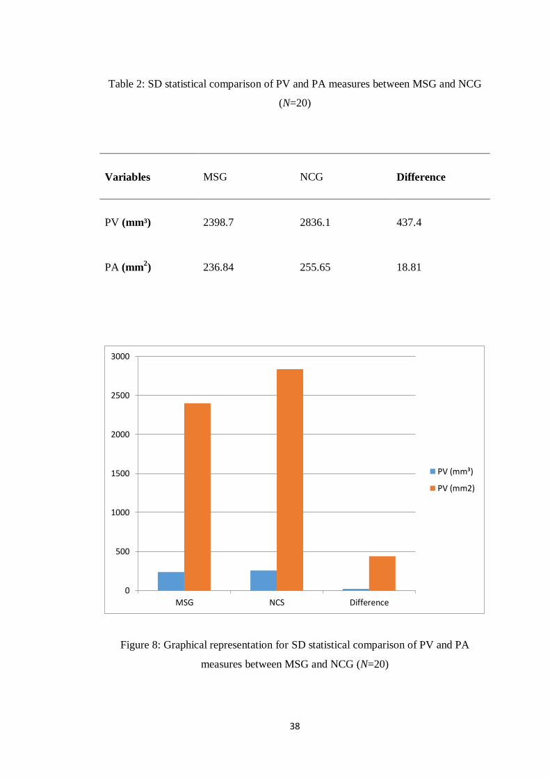
Table 2: SD statistical comparison of PV and PA measures between MSG and NCG
(N=20)
Variables MSG NCG Difference
PV (mm³) 2398.7 2836.1 437.4
PA (mm2) 236.84 255.65 18.81
Figure 8: Graphical representation for SD statistical comparison of PV and PA
measures between MSG and NCG (N=20)
0
500
1000
1500
2000
2500
3000
MSG NCS Difference
PV (mm³)
PV (mm2)
39
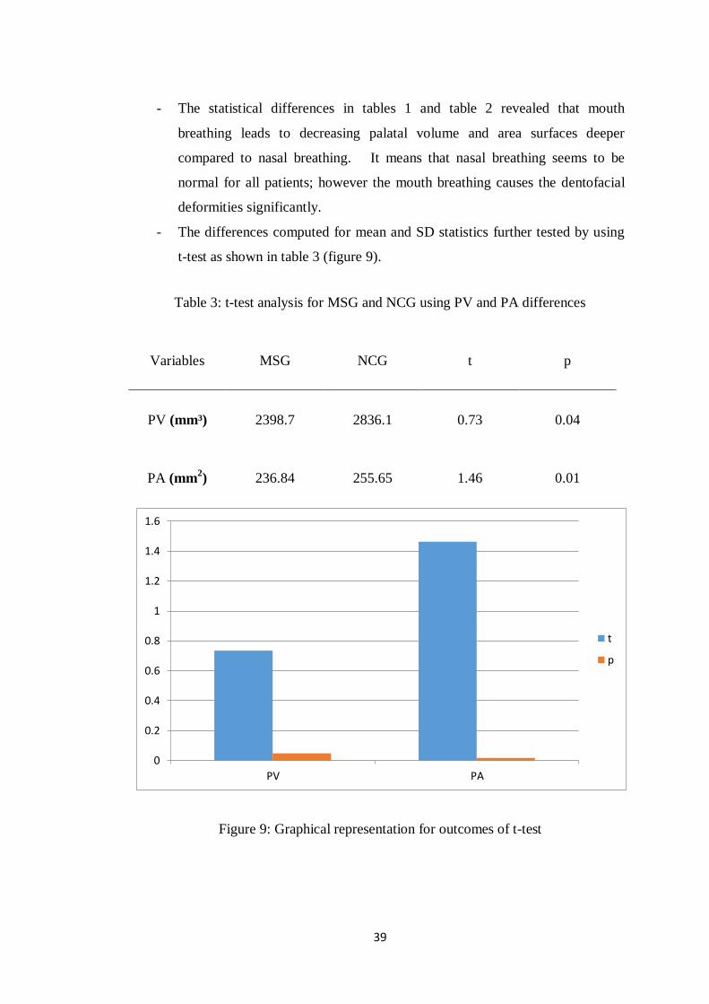
- The statistical differences in tables 1 and table 2 revealed that mouth
breathing leads to decreasing palatal volume and area surfaces deeper
compared to nasal breathing. It means that nasal breathing seems to be
normal for all patients; however the mouth breathing causes the dentofacial
deformities significantly.
- The differences computed for mean and SD statistics further tested by using
t-test as shown in table 3 (figure 9).
Table 3: t-test analysis for MSG and NCG using PV and PA differences
Variables MSG NCG t p
PV (mm³) 2398.7 2836.1 0.73 0.04
PA (mm2) 236.84 255.65 1.46 0.01
Figure 9: Graphical representation for outcomes of t-test
0
0.2
0.4
0.6
0.8
1
1.2
1.4
1.6
PV PA
t
p

40
- The t-test outcomes over the PV and PA readings confirmed that there is
significant systematic error noticed among MSG and NCG which means that
there are effects of mouth breathing by considering the values of PV and PA.
- In this study, along with the hard palatal dimensions we measured and
evaluated the lips prominence also.
- Further to determine the comparative study and reliability of methods we
performed the statistical analysis based on the obtained readings under the
both groups of subjects. The computation of the descriptive statistics in terms
mean and standard deviation (SD) for the measurements in each group is
presented in this chapter.
- the results in table 4 (figure 10) and table 5 (figure 11) demonstrate the mean
and SD statistical computations of ULP and LLP measures for both control
groups respectively.
Table 4 Mean statistical comparison of ULP and LLP measures between MSG and
NCG (N=20)
Variables MSG NCG Difference
ULP -2.6 -3.1 -0.5
LLP 0.55 -1.6 -2.15
41
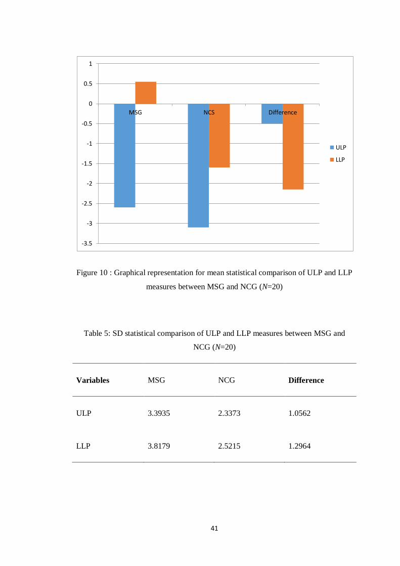
Figure 10 : Graphical representation for mean statistical comparison of ULP and LLP
measures between MSG and NCG (N=20)
Table 5: SD statistical comparison of ULP and LLP measures between MSG and
NCG (N=20)
Variables MSG NCG Difference
ULP 3.3935 2.3373 1.0562
LLP 3.8179 2.5215 1.2964
-3.5
-3
-2.5
-2
-1.5
-1
-0.5
0
0.5
1
MSG NCS Difference
ULP
LLP
42
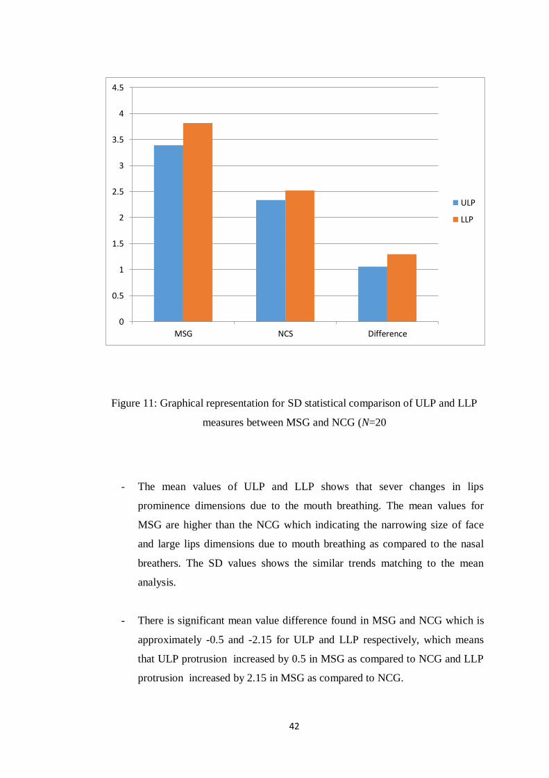
Figure 11: Graphical representation for SD statistical comparison of ULP and LLP
measures between MSG and NCG (N=20
- The mean values of ULP and LLP shows that sever changes in lips
prominence dimensions due to the mouth breathing. The mean values for
MSG are higher than the NCG which indicating the narrowing size of face
and large lips dimensions due to mouth breathing as compared to the nasal
breathers. The SD values shows the similar trends matching to the mean
analysis.
- There is significant mean value difference found in MSG and NCG which is
approximately -0.5 and -2.15 for ULP and LLP respectively, which means
that ULP protrusion increased by 0.5 in MSG as compared to NCG and LLP
protrusion increased by 2.15 in MSG as compared to NCG.
0
0.5
1
1.5
2
2.5
3
3.5
4
4.5
MSG NCS Difference
ULP
LLP

43
- The differences computed for mean and SD statistics further tested by using
t-test as shown in table 6.
-
- The t-test outcomes over the ULP and LLP readings confirmed that there is
significant systematic error noticed among MSG and NCG which means that
there are effects of mouth breathing by considering the values of ULP and
LLP.
Table 6: t-test analysis for MSG and NCG using ULP and LLP differences
Variables MSG NCG t p
ULP 3.3935 2.3373 4.3 0.07
LLP 3.8179 2.5215 5.5 0.04
44
7. DISCUSSION
Is known that mouth breathing influenced the development of a different
palatal morphology with a narrower and higher palatal vault compared to subjects
with a nose-breathing pattern. These modifications in the anatomy of the maxilla
were clearly quantified by 3D measurements of the palatal surface area and volume
that have been reported as reliable indicators of palatal growth.
Conflicting conclusions are reported in the literature about the influence of the
mode of breathing on the development of maxillofacial complex. This could be due
to the fact that nasal airway inadequacy is usually subjective and the judgement of
breathing mode differs among investigators.
In order to evaluate respiratory function and its effects on the morphology of the
palatal region, a clear differentiation between nose and mouth breathers is required.
However, this distinction is not easily made since most mouth breathers usually have
some nasal respiratory capacity as well . Therefore, in this study, only data from a
group of subjects classified at the end of complete physical examination as exclusive
mouth breathers were included.
Moreover, the morphology of the palate and of the maxillaryarch has been
assessed usually by measuring transverse dental distanceson study casts giving
incomplete information about the 3D morphology of the palatal vault. To overcome
this limitation, an evaluation of 3D characteristics of the maxillary arch by means of
3D technology has been used.
In previous studies, the PV and PA statistical analysis performed for MSG and NCG
by taking the difference of mean and SD values.We first computed the mean
statistics of all 20 observations under both categories (MSG and NCG), and then
estimate the difference between mean values for PV and PA for both categories.
R. Lione et al. messuared the palatal volume and palatal area , and the
difference between mouth group and nose group palatal volume is 1017 mm³ . the

45
difference between mouth group and nose group palatal area is 124.2 mm2
.
In our study total difference 927.97mm³ for PV and 115.29mm2 for PA, it
approximately similar to R. Lione et al reasult.
It is significant to justify the mouth breathing and nasal breathing relationships with
dentofacial deformities in patients. Similarly, the Standard Deviation statistics shows
that 437.4 (PV) and 18.81 (PA) statistical differences (table 2).
Moreover, Bresolin et al. , Harari et al. , and Berwig et al. compared plaster casts of
nasal and mouth breathers at the age of 8–12 years demonstrating that a change in the
breathing pattern of children can lead to a narrowing of both intermolar and
intercanine widths .
R. Lione et al had Statistical analysis and the P value was (0.000) for both of surface
area and palatal volume . which that mean there is a significant difference between
two groups.
In our study the P value (0.04) for the palatal volume and (0.01) for the palatal areea
. It is also mean there is a significant difference between tww groups.
The results of the current investigation are in agreement with previous studies that
showed that nasal deformities and maxillary growth deficiencies were correlated
with increased nasal airway resistance .
However, our findings are in disagreement with those reported by Primozic et al.
who did not find differences in palatal surface area and volume between mouth and
the nose breathers. These conflicting results may be due to a different group selection
since in this study the primary inclusion criteria was the abnormal respiratory pattern
and not the dentition stage and occlusal characteristics.
The differences computed for both upper lip and lower lip justified the
influence of mouth breathers on dentofacial deformities as compared to NCG group,
that both of upper lip and lower lip are protruded in patient with mouth breathing.

46
The previous studies mainly based on palatal volume and surface area measurements
to estimate the changes due to mouth breathing and diagnose the accurate dentofacial
deformities in human beings. The outcomes of this study show the enough
information to stop the debate over the relationship between mouth breathing and
dentofacial deformities at large extend.
The value indicates that there is significant changes of mouth breathing lips
prominence in payient with mouth breathing than the others in nasal breathing .
47
8. CONCLUSİON
Subjects with a mouth-breathing pattern have a different palatalmorphology
with significantly smaller palatal surface areas and volumes compared with nose-
breathing subjects.
The soft tissue changes are more prominence in MB patient .
The upper and lower lip of MB is protruded more than the upper and lower lip of
NB .

48
9. REFERENCES
Angle EH. Treatment of malocclusion of the teeth and fractures of the maxillae:
Angle’s system. 6th ed. Philadelphia: The SS White Dental Manufacturing Co; 1900.
p. 32.
Brant TCS, Parreira VF, Mancini MC, Becker HMG, Reis AFC, Brito RR. Breathing
pattern and thoracoabdominal motion in mouth-breathing children. Rev Bras Fisioter.
2008;12(6):495-501.
Bernardo Q.Souki Petrus B.Lopes, Natalia C.Veloso, Ricardo A.Avelino, Tatiana
B.J.Pereira, Paulo E.A., Souza Leticia P., Franco Helen, M.G.Becker. Facial soft
tissues of mouth-breathing children: Do expectations meet reality?. International
Journal of Pediatric Otorhinolaryngology, Volume 78, Issue 7, July 2014, Pages
1074-1079.
Babacan H, Sokucu O, Doruk C, Ay S. Rapid maxillary expansion and surgically
assisted maxillary expansion effects on nasal volume. Angle Orthod. 2006;76: 66–
71.
Baik UB, Suzuki M, Ikeda K, Sugawara J, Mitani H. Relationship between
cephalometric characteristics and obstructive sites in obstructive sleep apnea
syndrome. Angle Orthod. 2002;72:124–34.
Bailey LJ, Simmons KE, Warren DW. Orthodontic problems in children. In:
Bluestone CD, Stool SE, Kenna MA, editors. Pediatric otolaryngology, vol. 2. 3rd
ed. Philadelphia: W.B. Saunders Company; 1996. p. 1036.
Balyeat RM, Bowen R. Facial and dental deformities due to perennial nasal allergy
in children. Int J Orthod Dent Child. 1934;20:445–60.
49
Basciftci FA, Mutlu N, Karaman AI, Malkoc S, Küçükkolbasi H. Does the timing
and method of rapid maxillary expansion have an effect on the changes in nasal
dimensions? Angle Orthod. 2002;72:118–23.
Bicakci AA, Agar U, Sökücü O, Babacan H, Doruk C. Nasal airway changes due to
rapid maxillary expansion timing. Angle Orthod. 2005;75:1–6.
Bresolin D, Shapiro PA, Shapiro GG, Chapko MK, Dassel S. Mouth breathing in
allergic children: its relationship to dentofacial development. Am J Orthod. 1983;83:
334–40.
Barbiero EF, Vanderlei LCM, Neto AC, Nascimento PC. Influence of respiratory
biofeedback associated to re-expansive ventilation patterns in individuals with
functional mouth breathing. Int J Pediatr Otorhinolaryngol. 2008;72:1683-91.
Bianchini AP, Guedes ZC, Vieira MM. A study on the relationship between mouth
breathing and facial morphological pattern. Braz J Otorhinolaryngol.
2007;73(4):500-5.
Castelluci e Barbosa M, Knop LA, Lessa MM, de Araujo TM. Avaliação da
radiografia cefalométrica lateral como meio de diagnóstico da hipertrofia de
adenóide. R Dental Press Ortodon Ortop Facial. 2009;14(4):83-91.
C. Grippaudo,corresponding E.G. Paolantonio, G. Antonini, R. Saulle, G. La Torre,
and R. Deli. Association between oral habits, mouth breathing and malocclusion.
Acta Otorhinolaryngol Ital. 2016 Oct; 36(5): 386–394.
Chambi-Rocha, Cabrera-Domínguez, Domínguez-Reyes. Breathing mode influence
on craniofacial development and head posture. J Pediatr (Rio J). 2018 Mar -
Apr;94(2):123-130.
50
Cattoni DM, Fernandes FD, Di Francesco RC, Latorre MR. Characteristics of the
stomatognathic system of mouth breathing children: anthroposcopic approach. Pró-
Fono. 2007;19(4):347-51.
Chaaban M, Corey JP. Assessing nasal air flow: options and utility. Proc Am Thorac
Soc. 2011;8:70–8.
Chaves TC, de Andrade e Silva TS, Monteiro SA, Watanabe PC, Oliveira AS, Grossi
DB. Craniocervical posture and hyoid bone position in children with mild and
moderate asthma and mouth breathing. Int J Pediatr Otorhinolaryngol.
2010;74:1021–7.
Cohen MB. Orthodontic problems associated with allergy. Angle Orthod.
1937;7:150–4. Compadretti GC, Tasca I, Bonetti GA. Nasal airway measurements in
children treated by rapid maxillary expansion. Am J Rhinol. 2006;20:385–93.
Cross DL, McDonald JP. Effect of rapid maxillary expansion on skeletal dental, and
nasal structures: a posteroanterior cephalometric study. Eur J Orthod. 2002;22:519–
28.
Campanha SMA, Fontes MJF, Camargos PAM, Freire LMS. The impact of speech
therapy on asthma and allergic rhinitis control in mouth breathing children and
adolescents. J Pediatr. 2010;86(3):203-8.
Crouse U, Laine-Alava MT, Warren DW, Wood CL. A longitudinal study of nasal
airway size from age 9 to age 13. Angle Orthod. 1999;69:413–8.
Cuccia AM, Lotti M, Caradonna D. Oral breathing and head posture. Angle Orthod.
2008;78:77–82.
Coelho AR, Tanaka O, Ribeiro JS, Machado MA, Camargo ES. Transverse
craniofacial dimensions in Angle Class II, Division 1 malocclusion according to
breathing mode. Braz Oral Res. 2010;24(1):70-5.
51
De Freitas FCN, Bastos EP, Primo LS, de Freitas VL. Evaluation of the palate
dimensions of patients with perennial allergic rhinitis. Int J Paediatr Dent.
2001;11(5):365-71.
Di Francesco RC, Passerotii G, Paulucci B, Miniti A. Respiração oral na criança:
repercussões diferentes de acordo com o diagnóstico. Rev Bras Otorrinolaringol.
2004;70(5):665-70.
Daviskas E, Gonda I, Anderson SD. Local airway heat and water vapour losses.
Respir. Physiol. 1991;84:115–132. doi: 10.1016/0034-5687(91)90023-C.
Doruk C, Sökücü O, Sezer H, Canbay E. Evaluation of nasal airway resistance
during rapid maxillary expansion using acoustic rhinometry. Eur J Orthod.
2004;26:397–401.
Duke WW. Deformity of the face caused by nasal allergy in childhood. Arch
Otolaryngol. 1930;12:493–8.
Enlow D.H., Poston W.R. W B Saunders; Philadelphia: 1990. Facial Growth.
Ellingsen R, Vandevanter C, Shapiro P, Shapiro G. Temporal variation in nasal and
oral breathing in children. Am J Orthod Dentofacial Orthop. 1995;107:411–7.
Emslie RD, Massler M, Zwemer JD. Mouth breathing: etiology and effects. J Am
Dent Assoc. 1952;44:506–21.
Ferreira FS, Weber P, Correa ECR, Milanesi JM, Borin GS, Dias MF. Efeito da
fisioterapia sobre os parâmetros ventilatórios e a dinâmica tóraco-abdominal de
crianças respiradoras bucais. Fisioter Pesq. 2012;19(1):8-13.
Fields HW, Warren DW, Black K, Phillips CL. Relationship between vertical
dentofacial morphology and respiration in adolescents. Am J Orthod Dentofacial
Orthop. 1991;99:147–54.
52
Fujimoto S, Yamaguchi K, Gunjikake K. Clinical estimation of mouth breathing. Am
J Orthod Dentofac Orthop. 2009;136:630.e1–7.
Ghasempour M, Mohammadzadeh I, Garakani S. Palatal arch diameters of patients
with allergic rhinitis. Iran J Allergy Asthma Immunol. 2009;8(1):63-4.
Goldsmith JL, Stool SE. George Catlin’s concepts on mouth-breathing, as presented
by Dr. Edward H. Angle. Angle Orthod. 1994;64:75–8.
Gonzalez HE, Manns A. Forward head posture: its structural and functional infl
uence on the stomatognathic system, a conceptual study. Cranio. 1996;14:71–80.
Gordon JM, Rosenblatt M, Witmans M, Carey JP, Heo G, Major PW, et al. Rapid
palatal expansion effects on nasal airway dimensions as measured by acoustic
rhinometry. A systematic review. Angle Orthod. 2009;79: 1000–7.
Gouveia SA, Nahás AC, Cotrim-Ferreira FA. Estudo cefalométrico das alterações
dos terços médio e inferior da face em pacientes com diferentes padrões respiratórios
e tipos faciais. R Dental Press Ortodon Ortop Facial. 2009;14(4):92-100.
Gross CW, Harrison SE. Tonsils and adenoids. Pediatr Rev. 2000;21:75–8.
Gurley WH, Vig PS. A technique for the simultaneous measurement of nasal and
oral respiration. Am J Orthod. 1982;82:33–41.
Gwynne-Evans E, Ballard CF. Discussion on the mouthbreather. Proc R Soc Med.
1959;51:279–85.
Haas AJ. Rapid expansion of the maxillary dental arch and nasal cavity by opening
the midpalatal suture. Angle Orthod. 1961;31:73–90.
Hairfield WM, Warren DW, Hinton VA, Seaton D. Inspiratory and expiratory effects
on nasal breathing. Cleft Palate J. 1987;24:183–9.
53
Hairfield WM, Warren DW, Seaton DL. Prevalence of mouthbreathing in cleft lip
and palate. Cleft Palate J. 1988;25:135–8.
Hannuksela A. The effect of moderate and severe atrophy on the facial skeleton. Eur
J Orthod. 1981;3:187–93.
Hannuksela A. The effect of atrophy on the dentition. Eur J Orthod. 1983;5:279–85.
Harvold EP. The role of function in the etiology and treatment of malocclusion. Am
J Orthod. 1968;54:883–98.
Harvold EP, Chierici G, Vargervik K. Experiments on the development of dental
malocclusions. Am J Orthod. 1972;61:38–44.
Harvold EP, Vargervik K, Chierici G. Primate experiments on oral sensation and
dental malocclusions. Am J Orthod. 1973;63:494–508.
Harvold EP, Tomer BS, Vargervik K, Chierici G. Primate experiments on oral
respiration. Am J Orthod. 1981;79:359–72.
Hershey HG, Stewart BL, Warren DW. Changes in nasal airway resistance
associated with rapid maxillary expansion. Am J Orthod. 1976;69:274–84.
Howard CC. Inherent growth and its infl uence on malocclusion. J Am Dent Assoc.
1932;19:642–8.
Hartsook JT. Mouth breathing as a primary etiologic factor in the production of
malocclusion. J Dent Child. 1946;13(4):91-4.
Huang ZL, Ong KL, Goh SY, Liew HL, Yeoh KH, Wang DY. Assessment of nasal
cycle by acoustic rhinometry and rhinomanometry. Otolaryngol Head Neck Surg.
2003;128:510–6.

54
Huber RE, Reynolds JW. A dentofacial study of male students at the University of
Michigan in the physical hardening program. Am J Orthod Oral Surg. 1946;32: 1–21.
Humphreys HF, Leighton BC. A survey of anteroposterior abnormalities of the jaws
in children between the ages of 2 and 5 1/2 years of age. Br Dent J. 1950;88:3–15.
I S Indiarti, D B Setyanto, A Kusumaningrum, S B Budiardjo. Changes in the palatal
dimensions of mouth breathing children caused by nasal obstruction. Journal of
Physics: Conference Series. Volume 884, conference 1, 2017.
Isabel Chung Leng Muñoz , Paola Beltri Orta. Comparison of cephalometric patterns
in mouth breathing and nose reathing children. International Journal of Pediatric
Otorhinolaryngology 78 (2014) 1167–1172.
Ikeda A., Ikeda M., Komatsuzaki Atsushi. A CT study of the course of growth of
maxillary sinus: normal subjects and subjects with chronic sinusitis. ORL.
1998;60:147–152
James WW, Hastings S. Discussion on mouth-breathing and nasal obstruction. Proc
R Soc Med. 1932;25:1343–55.
Kingsley WS. A treatise on oral deformities as a branch of mechanical surgery. New
York: D. Appleton Company; 1889.
Koski K, Lähdemäki P. Adaptation of the mandible in children with adenoids. Am J
Orthod. 1975;68:660–5.
Luana Cristina Berwig, Ana Maria Toniolo da Silva, Eliane Castilhos Rodrigues
Côrrea, Anaelena Bragança de Moraes, Márlon Munhoz Montenegro, Rodrigo Agne
Ritze. Hard palate dimensions in nasal and mouth breathers from different etiologies.
Jornal da Sociedade Brasileira de Fonoaudiologia. J. Soc. Bras. Fonoaudiol. vol.23
no.4 São Paulo Dec. 2011.

55
L’Estrange PR, Battagel JM, Harkness B, Spratley MH, Nolan PJ, Jorgensen GI. A
method of studying adaptive changes of the oropharynx to variation in mandibular
position in patients with obstructive sleep apnoea. J Oral Rehabil. 1996;23:699–711.
Laine T, Warren DW. Effects of age, gender and body size on nasal cross-sectional
area in children. Eur J Orthod. 1991;13:311–6.
Lio PA. Efficacy of a moisturizing foam in skin barrier regeneration and itch relief in
subjects prone to atopic dermatitis. J. Drugs Dermatol. 2016;15:s77–s80.
Leech HL. A clinical analysis of orofacial morphology and behavior of 500 patients
attending an upper respiratory research clinic. Dent Pract. 1958;9:57–68.
Linder-Aronson S. Adenoids. Their effect on mode of breathing and nasal airfl ow
and their relationship to characteristics of the facial skeleton and the dentition. A
biometric, rhino-manometric and cephalometroradiographic study on children with
and without adenoids. Acta Otolaryngol Suppl. 1970;265:1–132.
Linder-Aronson S. Effects of adenoidectomy on dentition and nasopharynx. Am J
Orthod. 1974;65:1–15.
Linder-Aronson S, Woodside DG, Lundström A. Mandibular growth direction
following adenoidectomy. Am J Orthod. 1986;89:273–84.
Milanesi JM, Weber P, Berwig LC, Ritzel RA, Silva AMT, Correa ECR. Childhood
mouth-breathing consequences at adult age: entilator function and quality of life.
Fisioter Mov. 2014;27(2) 211-8.
Masahiro Sano, Sayaka Sano Hiromasa Kato, Ken Arakawa, Masaaki Arai. Proposal
for a screening questionnaire for detecting habitual mouth breathing, based on a
mouth-breathing habit score. BMC Oral Health, December 2018, 18:216.
56
Marchesan IQ, Krakauer LR. The importance of respiratory activity in
myofunctional therapy. Int J Orofacial Myology. 1996;22:23-7.
Marks MB. Allergy in relation to orofacial dental deformities in children: a review. J
Allergy. 1965;36: 293–302.
Maw AR, Jeans WD, Fernando DCJ. Inter-observer variability in the clinical and
radiological assessment of adenoid size, and the correlation with adenoid volume.
Clin Otolaryngol. 1981;2:317–22.
Melsen B, Attina L, Santuari M, Attina A. Relationships between swallowing
pattern, mode of respiration, and development of malocclusion. Angle Orthod.
1987;57: 113–20.
Mergen DC, Jacobs RM. The size of nasopharynx associated with normal and class
II malocclusions. Angle Orthod. 1970;40:342–6. Miller HI. The relation of long-
continued respiratory allergy to occlusion. Am J Orthod. 1949;35:780–9.
Miller AJ, Vargervik K, Chierici G. Sequential neuromuscular changes in rhesus
monkeys during the initial adaptation to oral respiration. Am J Orthod. 1982;81: 99–
107.
Miller AJ, Vargervik K, Chierici G. Experimentally induced neuromuscular changes
during and after nasal airway obstruction. Am J Orthod. 1984;85: 385–92.
Monini S, Malagola C, Villa MP, Tripodi C, Tarentini S, Malagnino I, et al. Rapid
maxillary expansion for the treatment of nasal obstruction in children younger than
12 years. Arch Otolaryngol Head Neck Surg. 2009;135:22–7.
Morrison WW. The interrelationship between nasal obstruction and oral deformities.
Int J Orthod. 1931;17:453–8.

57
Moss ML. The functional matrix. In: Kraus B, Riedel R, editors. Vistas in
orthodontics. Philadelphia: Lea & Febiger; 1962. p. 85–98.
Moss ML. The veloepiglottic sphincter and obligate nose breathing in the neonate. J
Pediatr. 1965;67:330–1.
Moss ML. The primary role of functional matrices in facial growth. Am J Orthod.
1969;55:566–77.
Neiva PD, Kirkwood RN, Godinho R. Orientation and position of head posture,
scapula and thoracic spine in mouth-breathing children. Int J Pediatr
Otorhinolaryngol. 2009;73:227–36.
Niinimaa V, Cole P, Mintz S, Shephard RJ. Oronasal distribution of respiratory airfl
ow. Respir Physiol. 1981;43:69–75.
Ono T, Ishiwata Y, Kuroda T. Inhibition of masseteric electromyographic activity
during oral respiration. Am J Orthod Dentofacial Orthop. 1998;113:18–25.
Oosterkamp BC, Remmelink HJ, Pruim GJ, Hoekema A, Dijkstra PU.
Craniocervical, and pharyngeal morphology in bilateral cleft lip and palate and
obstructive sleep apnea patients. Cleft Palate Craniofac J. 2007;44: 1–7.
Oktay H. Study of maxillary sinus areas in different malocclusions. Am J Orthod.
1992;102:143–145
Paul JL, Nanda RS. Effect of mouth breathing on dental occlusion. Angle Orthod.
1973;43:201–6.
Pacheco MCT, Casadrande CF, Teixeira LP, Finck NS, Martins de Araujo MT.
Guidelines proposal for clinical recognition of mouth breathing children. Dental
Press Journal of Orthodontics. 2015;20:39–44.
58
Peltomaki T. The effect of mode of breathing on craniofacial growth-revisited. Eur J
Orthod. 2007;29: 426–9.
Roberta Lione, Lorenzo Franchi, Luis Tomas Huanca Ghislanzoni, Jasmina
Primozic, Marco Buongiorno, Paola Cozza. Palatal surface and volume in mouth-
breathing subjects evaluated with three-dimensional analysis of digital dental casts—
a controlled study. European Journal of Orthodontics, Volume 37, Issue 1, February
2015, Pages 101–104.
Rasmus RL, Jacobs RM. Mouth breathing and malocclusion: quantitative technique
for measurement of oral and nasal air-flow velocities. Angle Orthod. 1969;39: 296–
302.
Ricketts RM. Respiratory obstruction syndrome. Am J Orthod. 1968;54:495–507.
Rodenstein DO, Stanescu DC. Soft palate and oronasal breathing in humans. J Appl
Physiol. 1984;57:651–7.
Souki BQ, Pimenta GB, Souki MQ, Franco LP, Becker HM, Pinto JA. Prevalence of
malocclusion among mouth breathing children: do expectations meet reality? Int J
Pediatr Otorhinolaryngol. 2009;73(5):767-73.
Scarano E, Ottaviani F, Di Girolamo S, Galli A, Deli R, Paludetti G. Relationship
between chronic nasal obstruction and craniofacial growth: an experimental model.
Int J Pediatr Otorhinolaryngol. 1998;45: 125–31.
Schendel SA, Eisenfeld J, Bell WH, Epker BN, Mishelevich DJ. The long face
syndrome: vertical maxillary excess. Am J Orthod. 1976;70:398–408.
Schlenker WL, Jennings BD, Jeiroudi MT, Caruso JM. The effects of chronic
absence of active nasal respiration on the growth of the skull: a pilot study. Am J
Orthod Dentofacial Orthop. 2000;117:706–13.

59
Shanker S, Fields HW, Beck FM, Vig PS, Vig KWL. A longitudinal assessment of
upper respiratory function and dentofacial morphology in 8- to 12-year-old children.
Semin Orthod. 2004;10:45–53.
Shapiro GG, Shapiro PA. Nasal airway obstruction and facial development. Clin
Rev. 1984;2:225–35.
Silveira W, Mello FCQ, Guimarães FS, Menezes SLS. Postural alterations and
pulmonary function of mouth-breathing children. Braz J Otorhinolaryngol.
2010;76(6):683-6.
Solow B, Kreiberg S. Soft-tissue stretching: a possible control factor in craniofacial
morphogenesis. Scand J Dent Res. 1977;85:505–7.
Souki BQ, Lopes PB, Pereira TB, Franco LP, Becker HM, Oliveira DD. Mouth
breathing children and cephalometric pattern: does the stage of dental development
matter?. Int J Pediatr Otorhinolaryngol. 2012 Jun;76(6):837-41.
Solow B, Sandham A. Cranio-cervical posture: a factor in the development and
function of the dentofacial structures. Eur J Orthod. 2002;24:447–56.
Solow B, Tallgren A. Head posture and craniofacial morphology. Am J Phys
Antropol. 1976;44:417–36.
Song HG, Pae EK. Changes in orofacial muscle activity in response to changes in
respiratory resistance. Am J Orthod Dentofacial Orthop. 2001;119:436–42.
Svensson S, Olin AC, Hellgren J. Increased net water loss by oral compared to nasal
expiration in healthy subjects. Rhinology. 2006;44:74–77.
Stevenson W, Chauhan SK, Dana R. Dry eye disease. An immune-mediated ocular
surface disorder. Arch. Ophthalmol. 2012;130:90–100. doi:
10.1001/archophthalmol.2011.364

60
Sosa FA, Graber TM, Muller TP. Postpharyngeal lymphoid tissue in Angle class I
and class II malocclusions. Am J Orthod. 1982;81:299–309.
Subtelny JD. The signifi cance of adenoid tissue in orthodontia. Angle Orthod.
1954;24:59–69.
Tulin Taner and Banu Saglam-Aydinatay. Physiologic and Dentofacial Effects of
Mouth Breathing Cmpared to Nasal Breathing. T.M. Önerci (ed.), Nasal Physiology
and Pathophysiology of Nasal Disorders, © Springer-Verlag Berlin Heidelberg 2013.
Tarvonen PL, Koski K. Craniofacial skeleton of 7-year old children with enlarged
adenoids. Am J Orthod. 1987;91:300–4.
Tecco S, Festa F, Tete S, Longhi V, D’Attilio M. Changes in head posture after rapid
maxillary expansion in mouth-breathing girls: a controlled study. Angle Orthod.
2005;75:171–6.
Tomes CS. On the developmental origin of the v-shaped contracted maxilla. Mon
Rev Dent Surg. 1872;1: 2–5.
Trotman CA, McNamara JA, Dibbets JMH, Van der Weele LT. Association of lip
posture and the dimensions of the tonsils and sagittal airway with facial morphology.
Angle Orthod. 1997;67:425–32.
Ung N, Koenig J, Shapiro PA, Shapiro G, Trask G. A quantitative assessment of
respiratory patterns and their effects on dentofacial development. Am J Orthod
Dentofacial Orthop. 1990;98:523–32.
Vargervik K, Miller AJ, Chierici G, Harvold E, Tomer BS. Morphologic response to
changes in neuromuscular patterns experimentally induced by altered modes of
respiration. Am J Orthod. 1984;85:115–24.
61
Vig P, Hall D. The inadequacy of cephalometric radiographs for airway assessment.
Letter to the editor. Am J Orthod. 1980;77:230–2.
Vig PS, Zajac DJ. Age and gender effects on nasal respiratory function in normal
subjects. Cleft Palate Craniofac J. 1993;30:279–84.
Vig PS, Sarver DM, Hall DJ, Warren DW. Quantitative evaluation of nasal airfl ow
in relation to facial morphology. Am J Orthod. 1981;79:263–72.
Vig PS, Spalding PM, Lints RR. Sensitivity and specificity of diagnostic tests for
impaired nasal respiration. Am J Orthod Dentofacial Orthop. 1991;99:354–60.
Wieler WJ, Barros AM, Barros LA. Tanaka OM. A combined protocol to aid
diagnosis of breathing mode. Rev Clín Pesq. Odontology. 2007;3:101–14
Warren DW, Spalding PM. Dentofacial morphology and breathing: a century of
controversy. In: Melsen B, editor. Current controversies in orthodontics. 1st ed.
Chicago: Quintessence Publishing Company; 1991. p. 45.
Warren DW, Duany LF, Fischer ND. Nasal airway resistance in normal and cleft lip
and palate subjects. Cleft Palate J. 1969;6:134–40.
Warren DW, Lehman MD, Hinton V. Analysis of simulated upper airway breathing.
Am J Orthod. 1984;86:197–206.
Warren DW, Hinton VA, Hairfi eld WM. Measurement of nasal and oral respiration
using inductive plethysmography. Am J Orthod. 1986;89:480–4.
Warren DW, Turvey TA, Hairfi eld WM. The nasal airway following maxillary
expansion. Am J Orthod Dentofacial Orthop. 1987;91:111–6.
Warren DW, Hairfi eld WM, Seaton D, Hinton VA. The relationship between size of
the nasal airway and nasal-oral breathing. Am J Orthod Dentofacial Orthop.
1988;93:289–93.

62
Warren DW, Hairfi eld WM, Dalston ET. Nasal airway impairment: the oral
response in cleft palate patients. Am J Orthod. 1991;99:346–53.
Wenzel A, Henriksen J, Melsen B. Nasal respiratory resistance and head posture:
effect of intranasal corticosteroid (Budesonide) in children with asthma and perennial
rhinitis. Am J Orthod. 1983;84: 422–6.
Wenzel A, Højensgaard E, Hendriksen JM. Craniofacial morphology and head
posture in children with asthma and perennial rhinitis. Eur J Orthod. 1985;7:83–9.
Woodside DG, Linder-Aronson S, Lundström A, McWilliam J. Mandibular and
maxillary growth after changed mode of breathing. Am J Orthod Dentofacial Orthop.
1991;100:1–18.
Woodside DG, Linder-Aronson S. The channelization of upper and lower anterior
face heights compared to population standard in males between ages 6 to 20 years.
Eur J Orthod. 1979;1:25–40.
Yi LC, Jardim JR, Inoue DP, Pignatari SSN. The relationship between excursion of
the diaphragm and curvatures of the spinal column in mouth breathing children. J
Pediatr. 2008;84(2):171-7.
Yamada T, Tanne K, Miyamoto K, Yamauchi K. Influences of nasal respiratory
obstruction on craniofacial growth in young Macaca fuscata monkeys. Am J Orthod
Dentofacial Orthop. 1997;111:38–43.
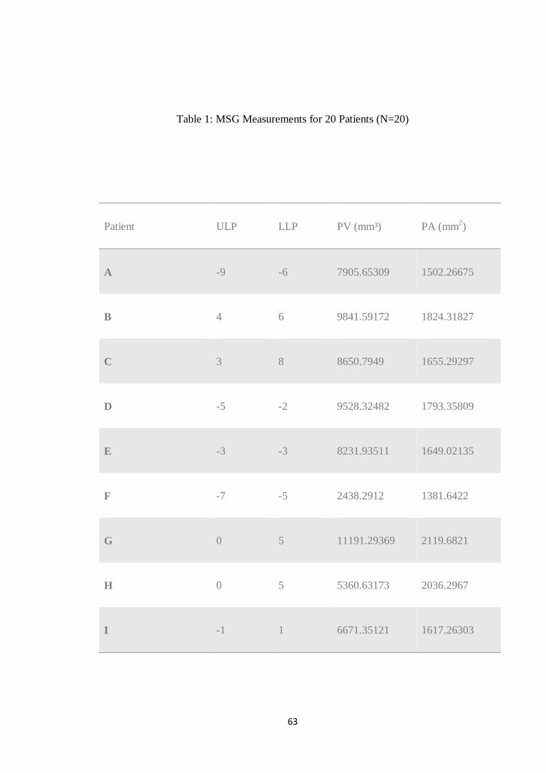
63
Table 1: MSG Measurements for 20 Patients (N=20)
Patient ULP LLP PV (mm³) PA (mm2)
A -9 -6 7905.65309 1502.26675
B 4 6 9841.59172 1824.31827
C 3 8 8650.7949 1655.29297
D -5 -2 9528.32482 1793.35809
E -3 -3 8231.93511 1649.02135
F -7 -5 2438.2912 1381.6422
G 0 5 11191.29369 2119.6821
H 0 5 5360.63173 2036.2967
I -1 1 6671.35121 1617.26303

64
J -6 0 8961.82743 1703.62671
K -3 2 5753.11015 1328.06191
L -3 0 9937.26529 1754.08804
M -6 -3 8809.98194 1703.98025
N -1 2 6272.64054 1701.45392
O -3 -1 7554.66735 1570.03989
P -1 1 4964.74683 1244.47222
Q -5 -2 4925.55984 1734.2665
R -3 1 8501.06261 1942.91861
S 2 5 8046.70292 1462.5683
T -5 -3 12637.2826 2047.67621
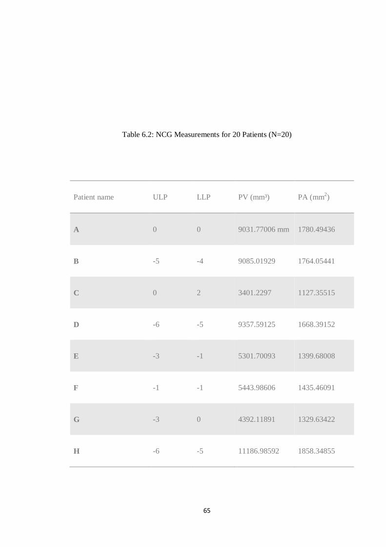
65
Table 6.2: NCG Measurements for 20 Patients (N=20)
Patient name ULP LLP PV (mm³) PA (mm2)
A 0 0 9031.77006 mm 1780.49436
B -5 -4 9085.01929 1764.05441
C 0 2 3401.2297 1127.35515
D -6 -5 9357.59125 1668.39152
E -3 -1 5301.70093 1399.68008
F -1 -1 5443.98606 1435.46091
G -3 0 4392.11891 1329.63422
H -6 -5 11186.98592 1858.34855

66
I -3 -3 8617.46862 1759.96918
J -3 1 7432.6011 1523.59417
K -3 -1 4719.6382 1563.31231
L -6 -5 4872.50949 1157.21544
M -3 -1 9785.11721 1751.66576
N -3 -2 7328.53941 1517.01654
O 3 4 2102.65871 1309.19861
P -5 -5 7971.21853 1483.40491
Q -6 -3 9875.08101 1743.0571
R -3 -2 6984.09835 1399.28094
S -2 1 11678.94697 2173.57484
T -4 -2 4642.22823 1721.83115

67