Embed Size (px)
Citation preview
THERMAL BURNS

DR.UĞUR ANIL BİNGÖL

TREATMENT
DNB
• Burn wounds can be classified into 6 separate groups based on the mechanism of injury:
• scalds• contact burns• Fire• chemical,• electrical• radiation. • The first 4types of burns are addressed in this lesson. • The mechanism of burn injury can be used as a predictor
of outcome. For example, patients with flame burns and electrical burn injuries often require hospitalization. In contrast, most patients with burns caused either by contact with hot surfaces or sun exposure are managed as outpatients.
WHO IS SUSCEPTIBLE?
• People of all ages are susceptible to minor burn injury. • The highest incidence occurs during the first few
years of life and in those aged 20-29 years.• Minor burns in children younger than 4 years are
caused primarily by contact with hot surfaces and by liquid scalds• These varied heat sources reflect the many
different daily experiences of older children and adults.
SKIN ANATOMY AND FUNCTION
• Skin is the largest organ of the body. It has 3 major tissue layers.
• Epidermis• The outermost layer, the epidermis, is composed of stratified
epithelium. Epidermis has 2 components, an outer layer of anucleate cornified cells (stratum corneum) that covers inner layers of viable cells (Malpighian layers) from which the cornified surface cells arise by differentiation. The stratum corneum acts as a barrier to impede the entrance of microorganisms and toxic substances while allowing the body to retain water and electrolytes. Malpighian layers provide a continuous production of cornified cells. Malpighian layers can be further subdivided into the germinal basal cell layer, stratum spinosum, and stratum granulosum.
SKIN ANATOMY AND FUNCTION
• Dermis• Beneath the epidermis is the dermis, which is composed of a
dense fibroelastic connective-tissue stroma containing collagen and elastic fibers and an extracellular gel termed ground substance. The dermal layer contains an extensive vascular and nerve network, special glands, and appendages that communicate with the overlying epidermis.
• The dermis is divided into 2 parts.• The most superficial portion, the papillary dermis, is molded
against the epidermis and contains superficial elements of the microcirculation of the skin
• In the reticular portion of the dermis, collagen and elastic fibers are thicker and greater in number. Fewer cells and less ground substance are found in the reticular dermis than in the papillary dermis.
• Thickness of the dermis varies from 1-4 mm in different anatomic regions and is thickest in the back, followed by the thigh, abdomen, forehead, wrist, scalp, palm, and eyelid
SKIN ANATOMY AND FUNCTION
• Subcutaneous tissue• The third layer of skin is subcutaneous tissue,
which is composed primarily of areolar and fatty connective tissue. • It contains skin appendages, glands, and
hair follicles .
SKIN ANATOMY AND FUNCTION
• Apocrine and eccrine sweat glands• There are 2 types of sweat glands in skin: apocrine and eccrine.• Apocrine glands are epitrichial because they have a duct that
opens into a hair follicle. Apocrine glands are largely confined to the axillary and perineal region and do not become functional until just after puberty.
• Eccrine glands are simple, coiled, tubular glands usually extending into the papillary dermis. Eccrine glands are atrichial because their duct opens onto the skin surface independently of a hair follicle. Eccrine glands are found over the entire body surface, except the margins of the lips, eardrum, inner surface of the prepuce, and glans penis.
• Sebaceous glands are simple or branched alveolar glands, usually connected to the hair follicles. Sebaceous glands unconnected with hair follicles occur along the margin of the lips, in the nipples, in the glans and prepuce of the penis, and in the labia minora. Depending on the depth of burn injury, epithelial repair can be accomplished from local epithelial elements and skin appendages.
• The ultimate outcome of a burn injury also is influenced by the depth of epidermal appendages in the burned tissue, which varies according to the age of the patient.• Very young and old individuals have superficial
appendages, which make both groups more susceptible to full-thickness burn injury. • By contrast, the epidermal appendages of the
human scalp and male beard are very deep, making these sites more refractory to severe burn injury.
HEAT TRANSFER FROM HEATING AGENT TO SKIN
• Severity of burn injury is related to the rate at which heat is transferred from the heating agent to the skin. Rate of heat transfer depends on the heat capacity of the agent, temperature of the agent, duration of contact with the agent, transfer coefficient, and specific heat and conductivity of the local tissues.
• Heat capacity: Capacity of a material to hold heat energy is determined by both the specific heat and the heat capacity of the material.
• Specific heat of a material: This is defined as the ratio of the amount of heat required to raise a specific mass of the material 1 degree in temperature, to the amount of heat required to raise an equal mass of a reference substance (usually water) 1 degree in temperature.
• Heat capacity: This refers to a quantity of heat a material contains when it comes in contact with skin. Quantity of heat stored depends on the specific heat of the material and the amount and temperature of the material.
WATER VS COPPER
• If the temperature of each material decreases by 60°C, water gives up 2530 W xsec of heat, whereas copper transfers only 230 W xsec of heat. Even if the initial temperatures of the 2 materials are identical, heat available from water is much more likely to produce a severe injury. The specific heat of water (most common cause of scald burns) is the highest of all the gases, metals, and solids tested to date, with the exception of ammonia and ether.
TEMPERATURE
• Initial temperature of a material at the instant of contact is also an important determinant of burn severity. Many materials (eg, water) cannot be heated beyond a certain temperature without changing state. Water can only be heated to 100°C at atmospheric pressure before it ceases to be a liquid and vaporizes. Because more joules are required to produce steam, this additional heat transfer accounts for the severe burns caused by steam injury. When other liquids reach a specific temperature, they ignite or oxidize by combining with oxygen.
DURATION OF CONTACT
• Human skin can tolerate temperatures as high as 44°C (111°F) for a relatively long time (6 hours) before irreversible injury occurs.• Temperatures
greater than this level cause an almost logarithmic increase in tissue destruction. Duration of contact between a liquid and skin depends on both the viscosity of the liquid and the manner in which it is applied to the victim's skin.
CHILD ABUSE AND IMMERSION SCALD BURNS
• Immersion burns caused by child abuse can be distinguished from accidental burns by the pattern and site of the burn, histories given by the caretaker and patient, and a medical history of scars representing previous abuse. • Nonaccidental burns often have clear-cut
edges, as found in "stocking" scalds
BURN WOUND INJURY
• During the first day after burn injury, 3 concentric zones of tissue injury characterize a full-thickness burn: zones of coagulation, stasis, and hyperemia.
• The central zone of coagulation has the most intimate contact with the heat source. It consists of dead or dying cells as a result of coagulation necrosis and absent blood flow. The intermediate zone of stasis usually is red and may blanch on pressure, appearing to have an intact circulation; however, after 24 hours, circulation through its superficial vessels often has ceased. Petechial hemorrhages may be present. By the third day, the intermediate zone of stasis becomes white because its superficial dermis is avascular and necrotic. The outer zone of hyperemia is a red zone that blanches on pressure, indicating that it has intact circulation. By the fourth day, this zone has a deeper red color. Healing is present by the seventh day.
BURN WOUND INJURY
• Transformation of the zone of stasis to coagulation occurs and has been related to many factors, including progressive dermal ischemia.
• Experimental studies have implicated prostaglandins, histamine, and bradykinin as the chemical mediators of this progressive vascular occlusion.
• They can produce edema by altering endothelial cell and basement membrane function to enhance permeability.
• When this ischemia persists, the zone of stasis eventually becomes a full-thickness burn injury
SYSTEMIC INFLAMMATORY RESPONSE
• In patients whose burns exceed 30% of TBSA, cytokines and other mediators are released into the systemic circulation, causing a systemic inflammatory response. • Because vessels in burned tissue exhibit
increased vascular permeability, an extravasation of fluids into the burned tissues occurs.
• Hypovolemia is the immediate consequence of this fluid loss, which accounts for decreased perfusion and oxygen delivery. In patients with serious burns, release of catecholamines, vasopressin, and angiotensin causes peripheral and splanchnic bed vasoconstriction that can compromise in-organ perfusion. • Myocardial contractility also may be reduced by
the release of inflammatory cytokine tumor necrosis factor-alpha.
• A decrease in pulmonary function can occur in severely burned patients without evidence of inhalation injury from the bronchoconstriction caused by humoral actors, such as histamine, serotonin, and thromboxane A2. A decrease in lung and tissue compliance is a manifestation of this reduction in pulmonary function.
• A significant proportion of the morbidity and mortality of severe burns is attributable to the ensuing hypermetabolic response. This response can last as long as a year after injury and is associated with impaired wound healing, increased infection risk, erosion of lean body mass, impaired rehabilitation, and delayed integration of the burn patient into society.
NUTRITIONAL SUPPORT
• Because burn injury causes a hypermetabolic state that is characterized by a dramatic increase in resting energy expenditure, nutritional support is essential, especially via the enteral route, to reduce intestinal villous atrophy. • decreased bowel mucosal integrity, capillary
leak, and decreased mesenteric blood flow, which allowed bacterial translocation into the portal circulation.• These translocated bacteria significantly alter
hepatocyte function and spread systemically to cause systemic sepsis.
NUTRITIONAL SUPPORT
• Adequate resuscitation that ensures mesenteric blood flow can prevent potential development of multisystem organ failure. Enteral nutrition with glutamine has a tropic effect on the enterocytes that preserve mucosal integrity.
INFECTION
• In patients with major burn injuries, infection remains the major cause of death. Immune consequences of this injury have been identified and are specific deficits in neutrophil chemotaxis, phagocytosis, and intracellular bacterial killing. Cell-mediated immunity, as measured by skin testing, also is compromised and has been related to both decreased lymphocyte activation and suppressive mediators present in the serum of burn patients. A reduction in immunoglobulin synthesis also has been encountered in these seriously ill patients.
BURN SHOCK
• Severe burn injury causes a coagulation necrosis of tissue that initiates a physiologic response in every organ system that is directly proportional to the size of the burn. Tissue destruction results in increased capillary permeability with profound egress of fluid from the intravascular space to the tissues adjacent to the burn wound. Inordinate amounts of fluid are lost by evaporation from the damaged surface that is no longer able to retain water. This increase in capillary permeability, coupled with evaporative water loss, causes hypovolemic shock
• GI dysfunction also appears to be proportional to the magnitude of thermal injury. In patients with burned areas in excess of 25% TBSA, gastroparesis is commonly noted until the third to fifth postburn day.
QUANTIFYING BURN SEVERITY
• Burn Depth• Burned patients' survival is related to the
following factors: • burn size/depth• age• presence of inhalation injury• and patient comorbidity. • Depth of burn injury is usually classified
according to degrees.
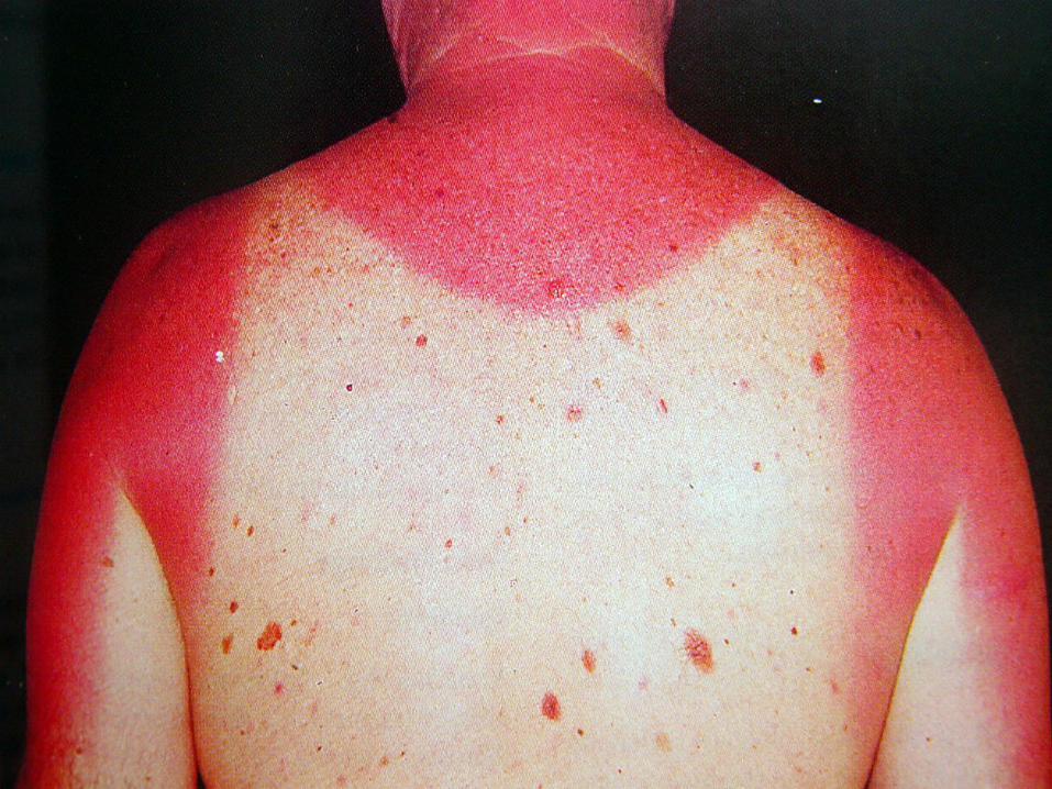
FIRST-DEGREE BURNS
• In first-degree burns, minor epithelial damage of the epidermis exists. Redness, tenderness, and pain are the hallmarks of this injury. Blistering does not occur, and 2-point discrimination remains intact. Healing takes place after several days without scarring. Because the epidermal barrier remains intact, metabolic response and risk of infection are minimal. Most common causes of first-degree burns are flash burns and sunburns.

SECOND-DEGREE BURNS
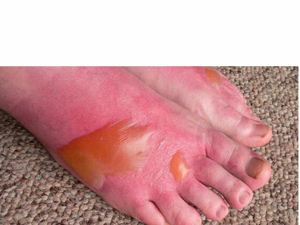
• Superficial partial-thickness and deep partial-thickness burns are the 2 types of second-degree burns. In these burn injuries, some portion of the skin appendages remains viable, allowing epithelial repair of the burn wound without skin grafting. Superficial partial-thickness burn involves the epidermis and superficial (papillary) dermis, often resulting in thin-walled, fluid-filled blisters. These burns appear pink, moist, and soft and are exquisitely tender when touched by a gloved hand. They heal in approximately 2-3 weeks, usually without scarring, by outgrowth of epithelial buds from the viable pilosebaceous units and sweat glands residing in the papillary and reticular dermis.
SECOND-DEGREE BURNS
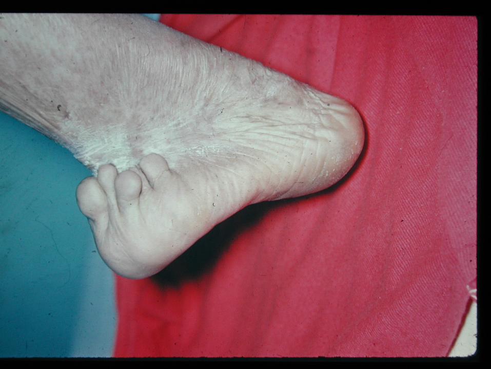
• Deep partial-thickness burns extend into the reticular dermis. Skin color is usually a mixture of red and blanched white, and capillary refill is slow. Blisters are thick-walled and commonly ruptured. Two-point discrimination may be diminished, but pressure and pinprick applied to the burned skin can be felt. Superficial partial-thickness burns usually re-epithelialize 7-10 days after injury. Risk of hypertrophic scarring is very small. For deep partial-thickness burns, tissue may undergo spontaneous epithelialization from the few viable epithelial appendages at this deepest layer of dermis and heal within 3-6 weeks (if no infection arises).
SECOND-DEGREE BURNS
• Because these burns have less capacity for re-epithelializing, a greater potential for hypertrophic scar formation exists. In deep partial-thickness burns, treatment with topical antimicrobial dressings is necessary to prevent infection as the burn wound heals. Contraction across joints, with resulting limitation in range of motion, is a common sequela. Splash scalds often cause second-degree burns.
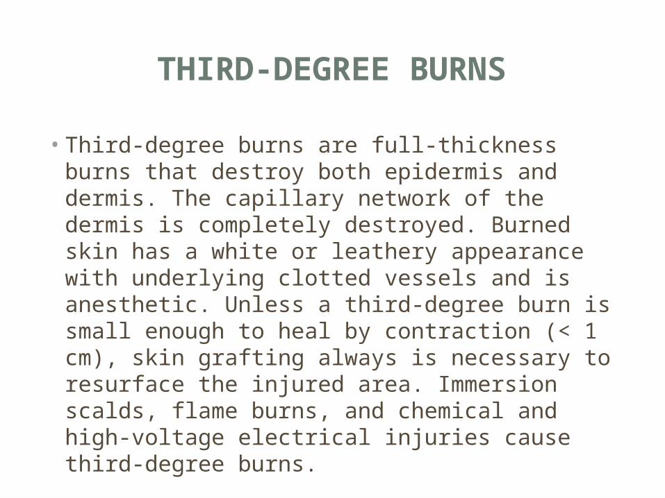
THIRD-DEGREE BURNS
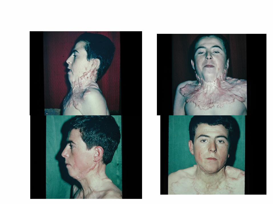
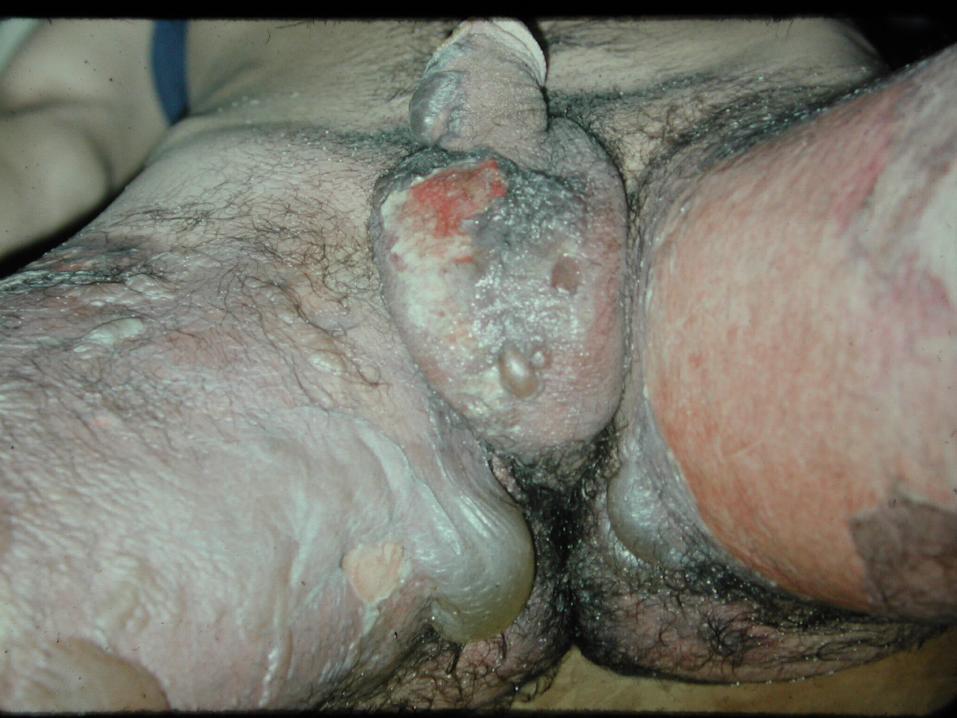
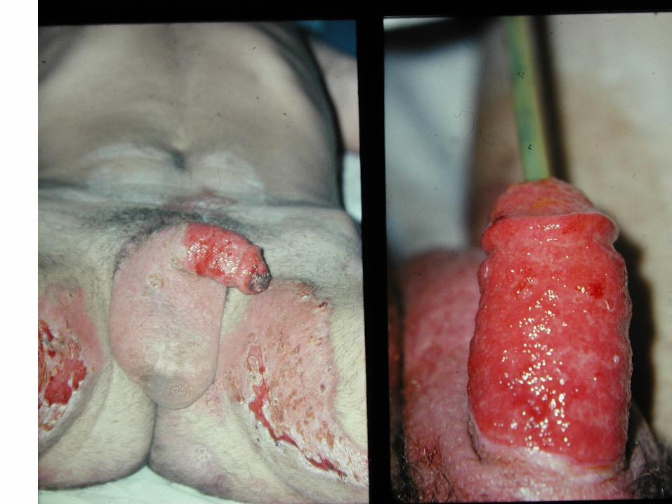
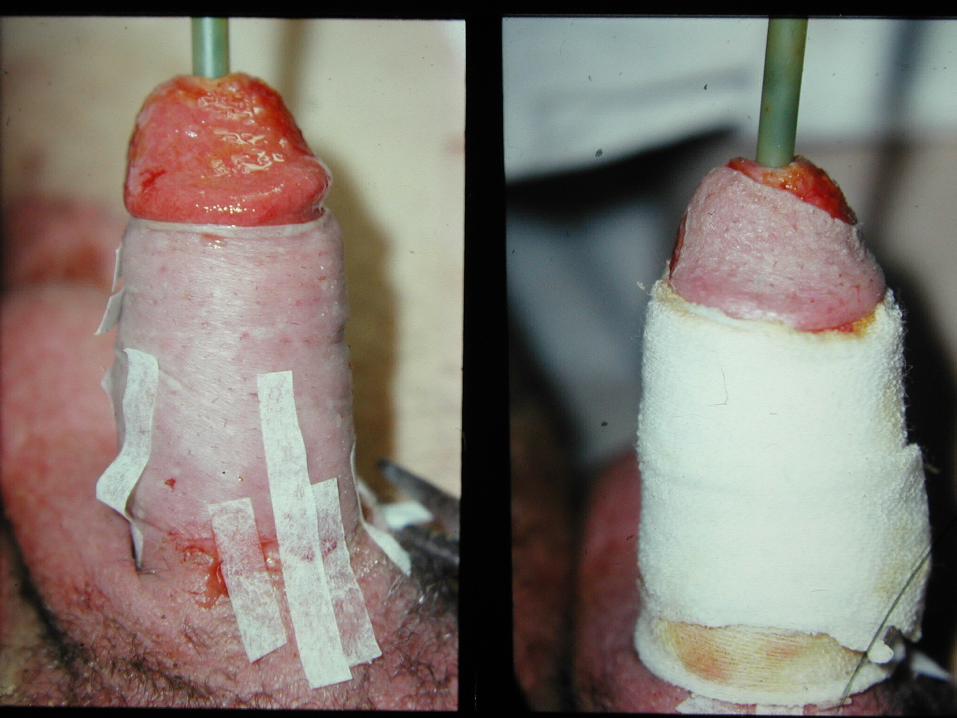
• Third-degree burns are full-thickness burns that destroy both epidermis and dermis. The capillary network of the dermis is completely destroyed. Burned skin has a white or leathery appearance with underlying clotted vessels and is anesthetic. Unless a third-degree burn is small enough to heal by contraction (< 1 cm), skin grafting always is necessary to resurface the injured area. Immersion scalds, flame burns, and chemical and high-voltage electrical injuries cause third-degree burns.
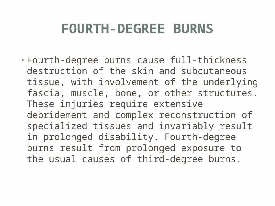
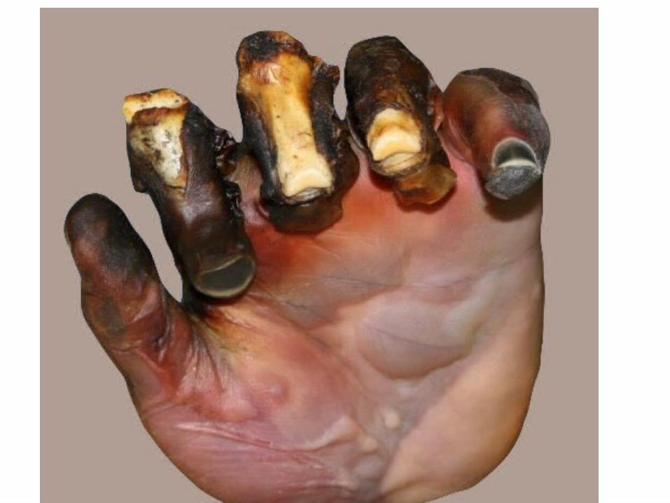
FOURTH-DEGREE BURNS
• Fourth-degree burns cause full-thickness destruction of the skin and subcutaneous tissue, with involvement of the underlying fascia, muscle, bone, or other structures. These injuries require extensive debridement and complex reconstruction of specialized tissues and invariably result in prolonged disability. Fourth-degree burns result from prolonged exposure to the usual causes of third-degree burns.
BURN SIZE
• The "rule of nines" is a practical technique for estimating the extent of TBSA involved in a burn injury. The patient's palm area represents approximately 1% of TBSA and can be helpful in calculating scattered areas of involvement.
• In estimating the extent of burn injury, the extent of involvement of each anatomic area (eg, an arm or leg) must be calculated separately, and the total is derived from the simple addition of the burned anatomic sites. A small difference between TBSA of the adult and infant reflects the size of the infant's head (18%), which is proportionally larger than that of the adult, and the lower extremities (14%), which are proportionally smaller than those of the adult. Lund-Brower charts with age-appropriate diagrams can be used to better estimate the area of burn injury in children. Remember that first-degree burns are not included in the calculation of burn size.
PATIENT CATEGORIZATION
• Severity of burn injury depends on • (1) extent, depth, and location of burn injury• (2) age of patient • (3) etiologic agents involved• (4) presence of inhalation injury• (5) coexisting injuries or preexisting illnesses
MAJOR BURN INJURY
• Major burn injury is defined as partial-thickness burns involving more than 25% of TBSA in adults
• 20% of TBSA in children younger than 10 years or adults older than 50 years
• full-thickness burns involving more than 10% of TBSA• burns involving the face, eyes, ears, hands, feet, or perineum that
may result in functional or cosmetic impairment• burns caused by caustic chemical agents• high-voltage electrical injury• burns complicated by inhalation injury or major trauma;• burns sustained by high-risk patients (those with underlying
debilitating diseases). These injuries are best managed in a specialized burn center staffed by a team of professionals with expertise in the care of burn patients, including both acute care and rehabilitation.
MODERATE BURN INJURY
• Moderate burn injury includes partial-thickness burns of 15-25% of TBSA in adults or 10-20% of TBSA in children or older adults
• full-thickness burns involving 2-10% of TBSA that do not present serious threat of functional or cosmetic impairment of the eyes, ears, face, hands, feet, or perineum.
• This category excludes high-voltage electrical injury, all burns complicated by inhalation injury or other trauma, and burns sustained by high-risk patients. Patients with moderate burn injuries should be hospitalized for their initial care but not necessarily at a burn center.
MINOR BURN INJURY
• Minor burn injury includes burns involving less than 15% of TBSA in adults or 10% of TBSA in children or older persons, • full-thickness burns involving less than 2% of
TBSA that do not present a serious threat of functional or cosmetic risk to eyes, ears, face, hands, feet, or perineum. • These burns usually can be managed safely in the
outpatient setting.
PREHOSPITAL TREATMENT
• Smoke inhalation can cause both pulmonary parenchymal damage and carbon monoxide and other toxic poisonings, which may have life-threatening consequences. The prehospital care provider should look for signs of inhalation injury (eg, dyspnea, burns of the mouth and nose, singed nasal hairs, sooty sputum, brassy cough). If one or more of these signs is present, administer humidified oxygen with a nonrebreathing reservoir mask at a rate of 10-12 L/min. A patient who is not breathing should be intubated and ventilated with 100% oxygen.
TREATMENT OF BURN SHOCK
• Treatment of burn shock in the prehospital setting should consist of elevating the patient's legs and administering humidified oxygen
• intravenous (IV) fluid administration. Fluid resuscitation need not be initiated if patient is transported to the hospital in less than 30 minutes. When transport time is longer than 30 minutes, the indications for fluid resuscitation are thermal injuries involving greater than 20% of TBSA or evidence of burn shock.
• Fluid resuscitation is not recommended for children at the scene of the accident because of the difficulties encountered in cannulating small veins. When fluid resuscitation is indicated in an adult, administer lactated Ringer solution or normal saline without glucose though a large-bore percutaneous catheter, preferably inserted through unburned skin. The arm is the preferred site for cannulation. Determine IV flow rates by the patient's clinical status.
PERFORM SECONDARY SURVEY
• When ventilatory and circulatory competence is restored, perform a secondary survey. Wash all burned clothing and skin with cool water. Immerse the burn wound in cold water (1-5°C) for approximately 30 minutes if transport cannot be undertaken immediately. This must be initiated as soon as possible because cooling has no therapeutic benefit if delayed more than 30 minutes after the burn injury. Local cooling of less than 9% of TBSA can be continued longer than this 30-minute interval to relieve pain; however, prolonged cooling of a larger TBSA can cause severe hypothermia, which may result in cardiac arrest. Do not apply ice directly to the burn wound because it may result in increased tissue injury through frostbite.
EMERGENCY DEPARTMENT TREATMENT
• Initial Evaluation• When the patient arrives in the ED, perform a
rapid initial assessment of respiratory and cardiovascular status, establish the extent of burn injury, and determine the need for special procedures. ED treatment focuses on airway and respiratory care and fluid resuscitation.
INHALATION INJURY
• Inhalation injury has a significant impact on the survival of burn patients. It has 3 components: upper airway swelling, acute respiratory failure, and carbon monoxide intoxication. The natural history of upper airway burn injury is the development of edema that narrows the airway 12-24 hours after injury. Consequently, intubation rather than observation is recommended in patients with signs of upper airway injury, such as stridor, inspiratory grunting, wheezing, or tachypnea.
• Fiberoptic bronchoscopy is a simple, safe, and accurate method of diagnosing acute inhalation injury. It reveals the anatomic level and severity of large airway injury; identification of supraglottic and infraglottic involvement is helpful in predicting ultimate pulmonary complications.
• When collagen is burned, it loses its elasticity, shortens its fibers, and becomes rigid. This can occur very quickly in fourth-degree and severe third-degree burns. When combined with accumulation of burn edema in interstitial spaces, respiratory insufficiency or ischemia of an extremity may result.
• The compressive effect of a full-thickness burn of the neck may contribute to airway compromise. Without tracheostomy, tight neck eschar accentuates pharyngeal edema and draws the neck into flexion, compressing the pharyngeal airway. A vertical incision through the eschar extending from the sternal notch to the chin helps maintain a patent airway. If respiratory insufficiency is caused by a constricting eschar of the anterior thorax that limits respiratory excursion, escharotomy is imperative. Lateral incisions are made in the anterior axillary lines that extend 2 cm below the clavicle to the 9th or 10th rib. The top and bottom of the incisions are then joined to form a square across the anterior chest.
CARBON MONOXIDE
• Carbon monoxide (CO) is present in smoke and has 280 times the affinity for hemoglobin as oxygen. Obtain a CO level in all patients with suspected inhalation injury. Patients should receive 100% oxygen until their carboxyhemoglobin (COHb) level is less than 10% because the elimination half-life for COHb depends on oxygen tension. In room air, the half-life of CO-bound hemoglobin is 4 hours. Under 100% oxygen, the half-life of CO-bound hemoglobin decreases to 45 minutes. Administration of 100% oxygen increases the gradient for oxygen binding to hemoglobin, and unbound CO is exhaled through the lungs.
• Patients who have elevated COHb levels associated with a pH of less than 7.4 should be treated with hyperbaric oxygenation. Because serum COHb levels do not reflect tissue levels, evaluate clinical symptoms when considering hyperbaric oxygen therapy. These include a history of unconsciousness, the presence of neuropsychiatric abnormalities, and the presence of cardiac instability or cardiac ischemia.
CYANIDE POISONING
• Specific therapy for cyanide poisoning in patients with inhalation injury is another consideration. Cyanide causes tissue hypoxia by uncoupling oxidative phosphorylation by binding to mitochondrial cytochrome a-a3. Consider empiric treatment for cyanide toxicity for patients with unexplained severe metabolic acidosis associated with elevated central venous oxygen content, normal arterial oxygen content, and a low COHb level.
FLUID RESUSCITATION
• Percent TBSA• All patients with a major burn injury must be
subjected to fluid resuscitation that is influenced by the percent TBSA as well as the presence of inhalation injury. Patients with burn wounds smaller than 20% TBSA can be treated with a combination of oral and IV fluid. For larger burns, the Parkland formula and its variations have become the standard method for resuscitating the burned patient.
• Moderate burn victims should have at least one large-bore intravenous line placed through unburned skin, and severe burn victims should have at least 2 lines initiated. If necessary, venous catheters may be placed through burned skin or via venous cutdown using the saphenous vein at the groin or ankle. When a burn patient requires considerable fluid resuscitation or has evidence of cardiopulmonary disease, a central venous line is indicated. Patients with massive burns or respiratory injury and elderly patients with severe burns or cardiac disease should be monitored with a Swan-Ganz catheter to avoid fluid overload or inadequate replacement of volume.
• Microvascular injury caused by a burn leads to increased vascular permeability with edema formation that results in ongoing plasma volume loss. Maximal edema formation occurs at 8-12 hours after burn injury for small burns, and 24-48 hours for large burns. The purpose of fluid resuscitation is to restore effective plasma volume, avoid microvascular ischemia, and maintain vital organ function. The amount of fluid required varies with the patient's age, body weight, and extent of burned TBSA.
•Burns greater than 20% of TBSA require intravenous fluid resuscitation because the accompanying GI ileus precludes sufficient oral intake.
• Parkland formula to estimate resuscitation volume • lactated Ringer solution • urinary output ADULT-NEWBORN-CHILD
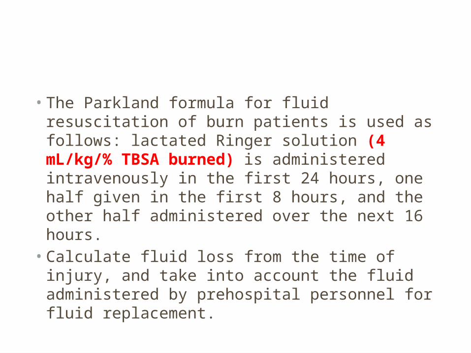
• The Parkland formula for fluid resuscitation of burn patients is used as follows: lactated Ringer solution (4 mL/kg/% TBSA burned) is administered intravenously in the first 24 hours, one half given in the first 8 hours, and the other half administered over the next 16 hours. • Calculate fluid loss from the time of injury, and
take into account the fluid administered by prehospital personnel for fluid replacement.
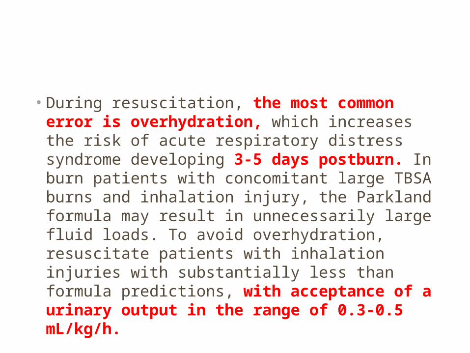
• During resuscitation, the most common error is overhydration, which increases the risk of acute respiratory distress syndrome developing 3-5 days postburn. In burn patients with concomitant large TBSA burns and inhalation injury, the Parkland formula may result in unnecessarily large fluid loads. To avoid overhydration, resuscitate patients with inhalation injuries with substantially less than formula predictions, with acceptance of a urinary output in the range of 0.3-0.5 mL/kg/h.
PROTEIN LOSSES
• After a burn injury, significant intravascular protein is lost through endothelial leaks in the burned vessels. When endothelial integrity is restored 24 hours postinjury, some clinicians favor the administration of 5% albumin at 0.5 mL/kg/% TBSA to maintain dynamic forces between the extracellular spaces and the intravascular system. In addition, a low-dose dopamine infusion (3-5 mcg/kg/min) is beneficial in restoring renal and splanchnic blood flow in patients with major burn injury.
• The Parkland formula is effective in estimating fluid loss in adults, yet it underestimates the evaporative fluid loss and maintenance needs in children. Compared to adults, children have a larger TBSA relative to weight than do adults and generally have somewhat greater fluid needs during resuscitation. Use the Galveston formula for fluid resuscitation in children as follows: 5% dextrose in lactated Ringer (5000 mL/m2 of TBSA burned plus 2000 mL/2) is administered intravenously in the first 24 hours. One half is given in the first 8 hours, and the other half is given over the next 16 hours. Add dextrose to the resuscitation fluid in children to prevent hypoglycemia, because children have smaller glycogen stores than adults.
• prolonged hypothermia in burned infants may result in excessive lactate production and acidosis. • After 6 months, infants and children are able to
shiver. Because they have a greater evaporative water loss relative to weight than do adults, infants and children are especially prone to hypothermia; therefore, keep the ambient temperature high to decrease radiant and evaporative heat loss from burned infants and children to the environment.
CATHETERIZE PATIENT
• Place a Foley catheter into the bladder to monitor the effectiveness of intravenous fluid replacement. Burns of the perineum also are best cared for with an indwelling Foley catheter to decrease urinary soilage of the wound. In patients with major burn injuries who require intravenous fluid resuscitation
• pass a nasogastric (NG) tube for initial evacuation of fluid and air from the stomach and feeding access. Removal of the gastric contents prevents vomiting and aspiration, sequelae of the ileus that commonly occur soon after burn injuries involving more than 20% TBSA. Early feeding through the NG tube within 6-8 hours of the burn injury diminishes the hypermetabolic response and maintains intestinal integrity.
• Consider the burn wound a dirty wound, and institute tetanus prophylaxis accordingly.
• Prophylactic antibiotics are not indicated.• Treatment of the patient in the burn center
involves 3 important considerations: supportive care, burn wound management, and nutritional support.
SUPPORTIVE CARE
• Upon arrival in the burn center, each patient should receive a standard regimen of supportive care that involves pain management and prevention of gastric erosions as well as renal failure.• The requirement of pain medication is inversely
proportional to the depth of burn injury.• morphine has been advocated for the
management of pain in burn patients.• Any respiratory depression caused by morphine
can be rapidly reversed by small doses of naloxone.
• Acute upper GI erosions and ulcers may occur in patients with severe burn injuries.
• These lesions are often termed stress ulcers or erosions (Curling ulcer). The most common clinical finding in such patients is painless GI hemorrhage.
• Treatment of acute stress ulceration is principally preventive.
• In high-risk patients, antacids can reduce the occurrence of stress ulcerations by neutralizing gastric contents, and H2-receptor antagonists can inhibit gastric acid secretion. This prophylaxis against acute stress ulceration usually is initiated immediately after admission to the burn center.
• Renal failure can occur after burn injury, as manifested by an elevated serum creatinine and a fall in creatinine clearance. • Prevention of this complication involves adequate
resuscitation, treatment of infection in the wound and other sites, and avoidance of nephrotoxic drugs (eg, aminoglycoside antibiotics, vancomycin, loop diuretics).• When renal function deteriorates with resultant
fluid and electrolyte imbalance, dialysis may be indicated.
BURN WOUND MANAGEMENT
• using powder-free gloves• Ruptured blisters are removed with scissors. After
wound cleansing, cover with a topical antimicrobial dressing.• Prophylactic systemic antibiotics are not
recommended because they do not prevent wound sepsis. Systemic antibiotics may be indicated when cellulitis is evident in surrounding unburned tissue.
DRESSINGS
• Apply daily dressings to the wound continuously until complete healing occurs or surgical intervention is required for wound closure. • In the past, many burn surgeons dressed wounds
twice daily. • twice-daily dressing changes are still indicated in
patients with wounds that are or have been infected or those with excessive amounts of exudate.
• Most partial-thickness burns of less than 10% TBSA respond satisfactorily to daily antibiotic dressings. Silver sulfadiazine (Silvadene) remains the most popular antimicrobial cream.
• The current formulation of silver sulfadiazine contains a lipid soluble carrier, polypropylene glycol, which has certain disadvantages, including pseudoeschar formation.
• Mafenide is an alternate agent that penetrates burn eschar more effectively than silver sulfadiazine. Consequently, it is frequently used on infected wounds that do not respond to silver sulfadiazine. Mafenide is the treatment of choice for serious burns of the ears to prevent infectious chondritis. Use Mafenide with caution because it can induce metabolic acidosis
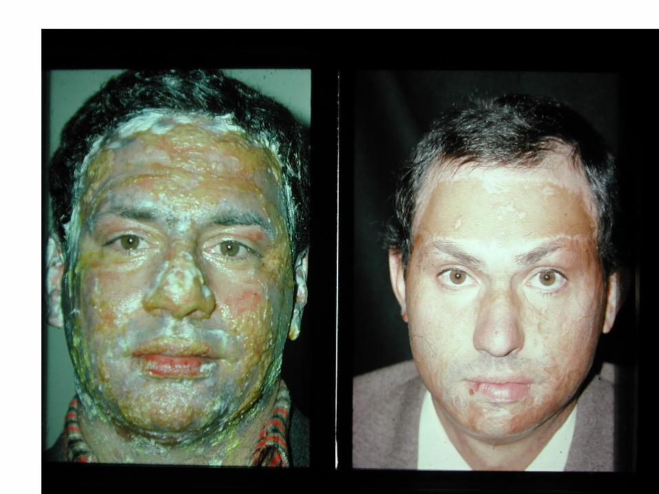
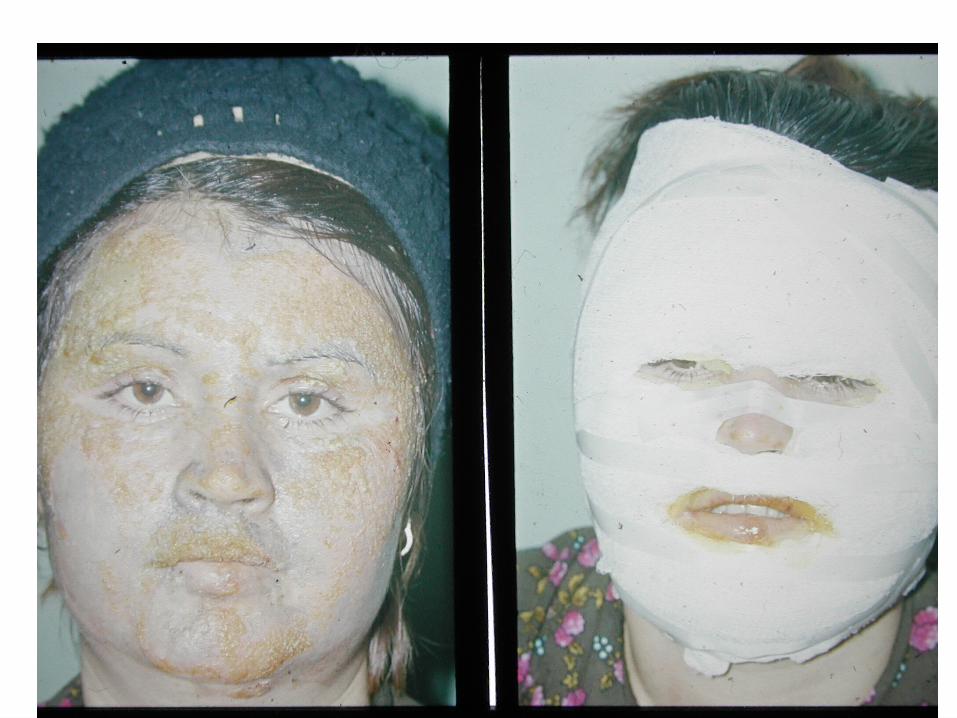
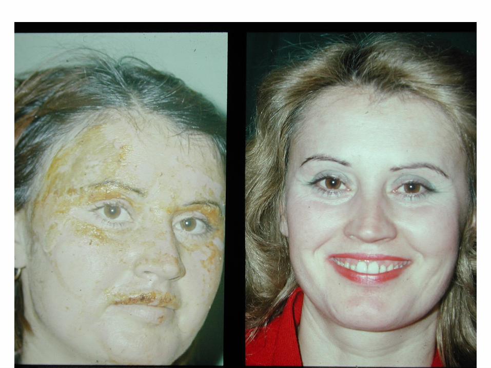
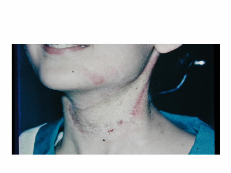
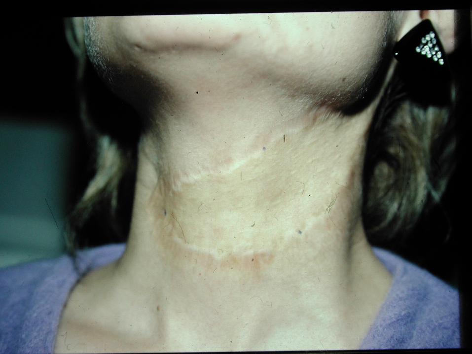
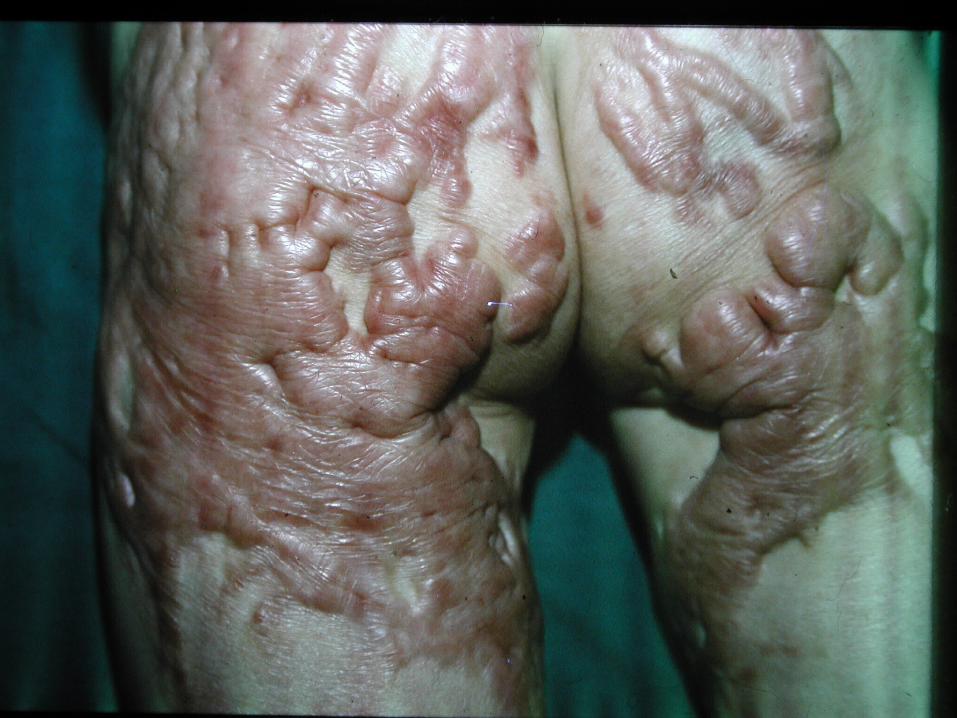
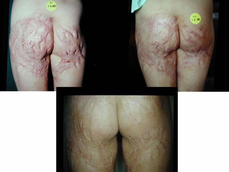
• Indications for surgery are full-thickness burns or partial-thickness burns that are unlikely to heal within 3 weeks. If the burn fails to heal in 3 weeks, the risk for hypertrophic scar and contracture formation increases and the healed wound exhibits an aesthetically displeasing scar.
• In regions with a dense cross section of dermal appendages (such as the face, scalp, and ears), observe the burn wound for at least 3 weeks to clearly identify its healing potential.
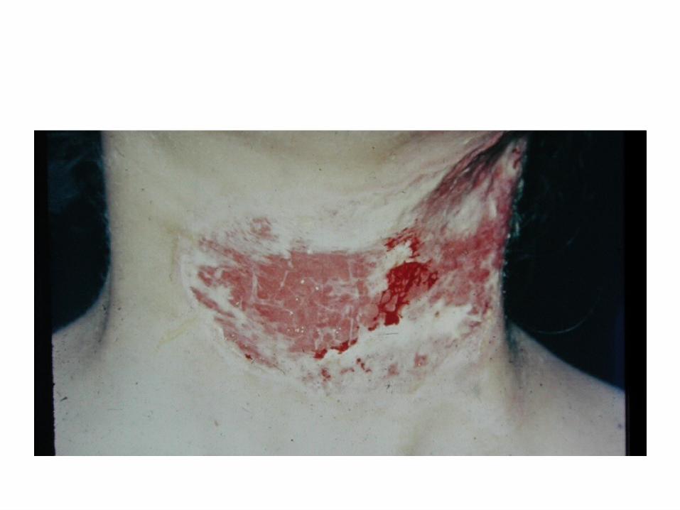
• When circumferential burns of the extremity exist, emergency escharotomy can salvage an ischemic limb.
ESCHAROTOMY
• After burn injury, a rise in interstitial tissue pressure first occludes venous outflow, then arterial capillary inflow. A period of 3-8 hours is required for edema to develop sufficiently to increase tissue pressure. When tissue compartment pressures are greater than 40 mm Hg, escharotomies of the full-thickness burn prevent this ischemic injury. Note that the most common cause of absent pulses in an extremity is hypovolemia with peripheral vasoconstriction, not increased interstitial pressure.
• Escharotomies are performed on the medial and lateral aspects of the extremity and extend the length of the constricting eschar. Incisions are made using either a scalpel or high-frequency electric current, with release of the edematous tissues ensuring adequate depth. • After prolonged vascular compromise, an
escharotomy may cause reperfusion injury to the extremity with reactive hyperemia and edema of the compartment muscles. In this case, a fasciotomy is required to restore perfusion to the extremity.
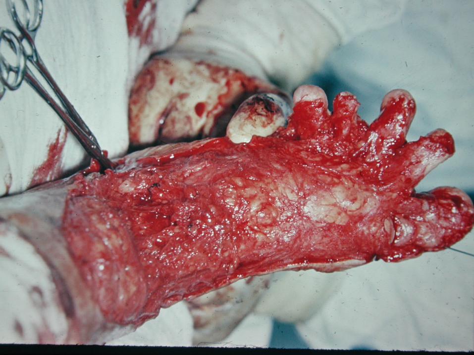
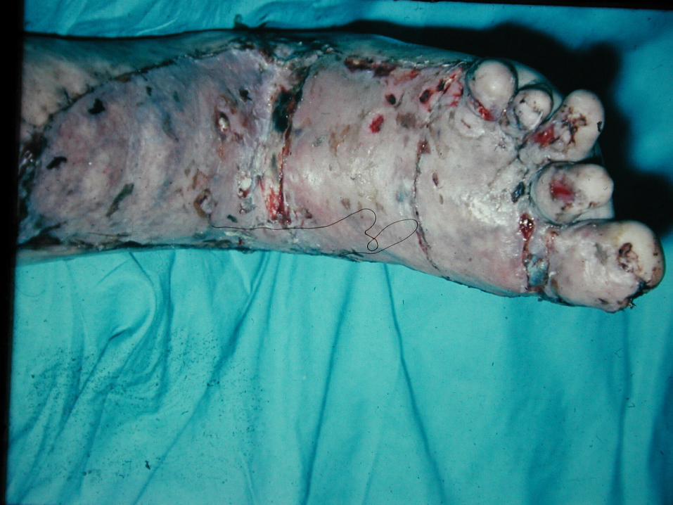
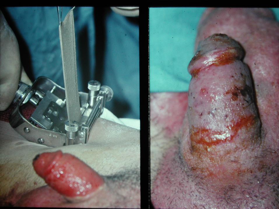
• For full-thickness circumferential burns of the upper extremity, first decompress the fingers by a digital escharotomy that is performed along each side of the burned finger, cutting down to fat.
• Decompress the palm by an incision along the palmar crease. At the wrist, continue the incision ulnarward to avoid injury to the palmar cutaneous branch of the median nerve. When intrinsic muscle involvement is suspected, decompress the interossei through short longitudinal skin incisions made in the intermetacarpal spaces carried down to the dorsal interossei.
• Decompress the leg by midmedial and midlateral incisions. Decompress each toe in a manner similar to that used for the fingers.
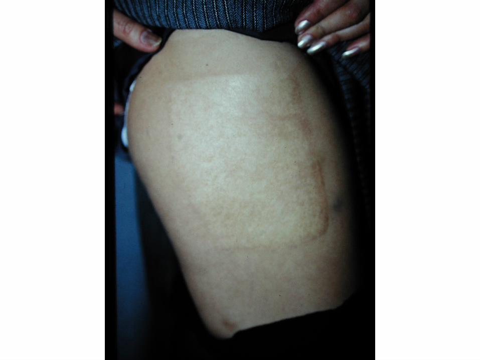
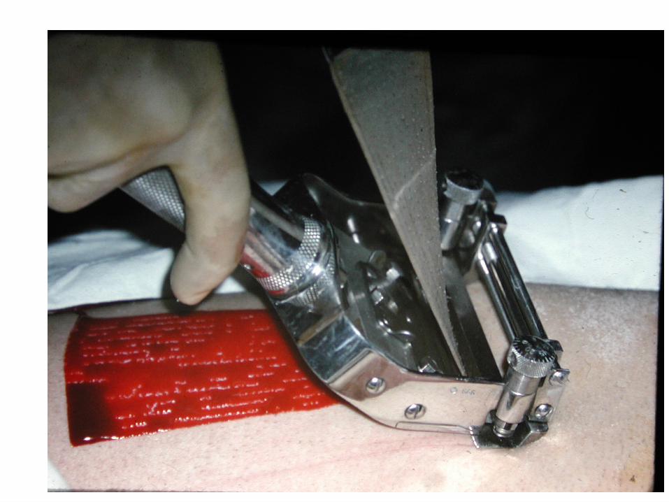
• The standard of care for full-thickness burns is burn wound excision and grafting.
• The mortality of patients with massive burns is reduced by early tangential excision of the entire wound, followed by skin closure with an autograft from unburned areas on the patient or an allograft from a donor cadaver.
• In burns less than 30% TBSA, wound closure is accomplished in one operation using split-thickness skin grafts from unburned areas on the patient, taken in sheets or meshed either 1:1 or 2:1. In burns greater than 40% TBSA, split-thickness skin grafts are meshed 3:1 or 4:1, complemented by cadaver allograft used to temporarily cover the residual open wound areas. Cadaver allograft skin adheres to the wound and serves as a partial barrier to infection
• Allograft, xenograft, or artificial coverings, such as Integra or Dermagraft-TC, are routinely used for burns involving over 40% TBSA. These burn dressings accomplish 4 functions:
• (1) protect the damaged epithelium• (2) splint the area into the desired position to maximize long-
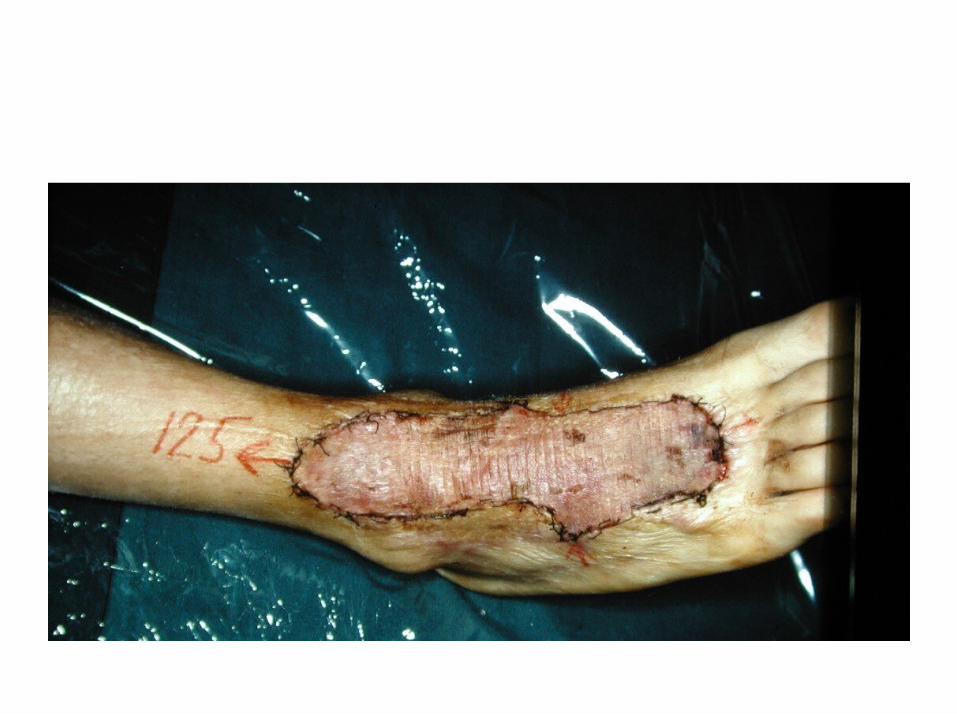
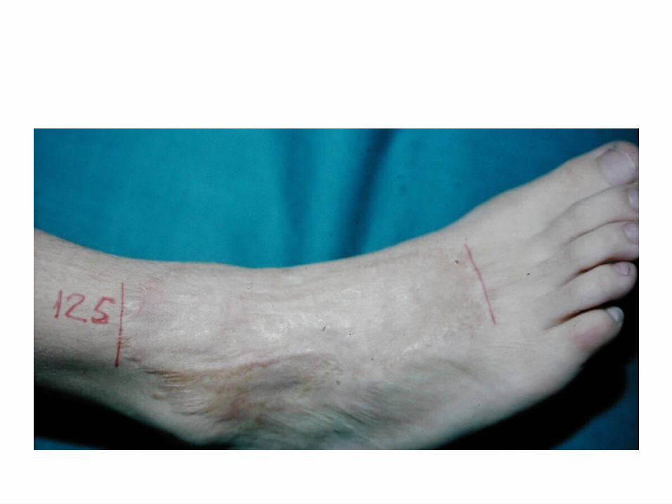
term function• (3) occlude the wound and prevent evaporative heat loss, • (4) provide comfort. • Donor sites for autograft require 1-2 weeks to heal. At that
time, temporary burn dressings are removed and residual open wound areas are closed with split-thickness skin grafts from these same donor sites. For larger than 90% TBSA burns, up to 10 cycles of autografting may be required to completely close the wounds.
MINOR BURNS
• cleanse all minor burns with sterile saline • The treatment of burn blisters remains controversial.
Exposing an unbroken blister can lead to local wound infection but studies have demonstrated that burn blister fluid may be deleterious to wound healing, and undrained fluid confined by necrotic skin can act as a source for closed space infection.
• Most surgeons recommend leaving blisters on the palms or soles intact. Other blisters, particularly when large enough to preclude the application of an adequate dressing, should be aspirated sterilely. Alternatively, open blister with a No. 15 knife blade and remove the surface of the blister.
• Provide tetanus prophylaxis if indicated.• Prophylactic antibiotics are not recommended. • Treat burn wounds either by the open or closed
technique. Open therapy of minor burn injuries usually is reserved for burns of the face. These burns are covered by bacitracin ointment, which is reapplied every 6 hours after gently washing the skin.
• Burn injury causes the release of massive amounts of amino acids from muscle. This response is caused by increases in cortisol and decreases in growth hormone and insulin, with resultant increased proteolysis of muscle protein and release of amino acids. Anabolic growth hormone treatment is shown to increase protein synthesis in muscle, increase muscle mass, and accelerate wound healing after burn injury. Potential anabolic hormones (eg, insulinlike growth factor, insulin, dehydroepiandrosterone, oxandrolone) are being evaluated for their effects on wound healing.
• because basal energy expenditure is increased 3-fold above normal, early and aggressive nutritional support via the enteral route is important in preventing bacterial translocation from the gut and systemic sepsis.
• the patient's caloric requirements can be estimated using the Curreri formula (25 kcal/kg+40 kcal/% TBSA) or twice the Harris-Benedict estimate.
• Measuring the prealbumin level is another approach to documenting the effectiveness of nutritional support.