Embed Size (px)
Citation preview

ii
Therapeutic properties of the lantibiotic nisin F
by
Anneke Mari Brand
March 2013
Dissertation presented for the degree of Doctor of Science in the Faculty
of Science at
Stellenbosch University
Promoter Prof Leon Milner Theodore Dicks
Co-promoter Prof Carine Smith
75
The antimicrobial activity of nisin F was determined by using the agar-spot test method as described
by de Kwaadsteniet et al (2008) Staph aureus Xen 36 (1 x 10⁶ CFU) was imbedded in BHI soft agar
(1 wv) supplemented with 200 microg ml-1
kanamycin
Antimicrobial activity of nisin F was also recorded by monitoring changes in bioluminescence of
Staph aureus Xen 36 The strain was grown in BHI broth supplemented with 200 microg ml-1
kanamycin to an optical density (OD600nm) of 15 Wells of a sterile titre plate were filled with 100 microl
(3 x 10⁷ CFU) of the culture and then treated with two concentrations of nisin F Wells A1-3 received
75 microl (240 AU) nisin F and wells A4-6 150 microl (480 AU) nisin F The control received nothing
Bioluminescent readings were taken at 490 nm at T0 (before the addition of nisin F) T010 (6 min after
addition) T24 (24 h later) and T48 (48 h later) using the Xenogen In Vivo Imaging System (IVISreg 100)
of Caliper Life Sciences Living Imagereg
software version 30 (Caliper Life Sciences) was used to
quantify the average radiances (avg rad) of photons emitted from regions of interest (ROIs) The
ROI was selected by using the free draw method with the threshold set at zero percent Values
recorded from each well were expressed as log10 photons second-1
cm-2
steridian-1
(p s-1
cm-2
sr-1
) The
correlation between p s-1
cm-2
sr-1
and viable cell numbers (CFU) was obtained from a standard curve
For the standard curve cells were grown in BHI supplemented with 200 microg ml-1
kanamycin to an
optical density (OD600nm) of 15 which represented 3 x 10⁸ CFU ml
-1 Sterile titre plates were filled
with 100 microl of the cell suspension (3 x 10⁷ CFU) and incubated at 37degC for 48 h At specific time
intervals the number of photons emitted per second was recorded and converted to p s-1
cm-2
sr-1
by the
Living Imagereg
software The number of viable cells (CFU) was determined by plating onto BHI agar
Antimicrobial activity of nisin F in vivo
Approval to conduct this research on mice was granted by the Ethics Committee of the University of
Stellenbosch (ethics reference number 2008B02003A2)
C57BL6 mice weighing between 20 and 30 g were fed a standard rodent diet and kept under
controlled environmental conditions Twenty-four mice were anaesthetized with 2 (vv) isoflurane
(Isofor Safe Saline pharmaceuticals Florida South Africa) Twelve mice were intraperitoneally
injected with 200 microl (1 x 10⁸ viable cells) of Staph aureus Xen 36 and divided into two groups of six
mice each After 4 h of infection mice in group 1 were again anaesthetized and intraperitoneally
injected with 200 microl nisin F (640 AU) Mice in group 2 were also anaesthetized but intraperitoneally
injected with 200 microl sterile saline Images were taken at T0 (before infection with Staph aureus Xen
36) T025 (15 min after infection) T4 (4 h after infection and immediately before treatment with nisin
F) T425 (15 min after treatment) and T8 T24 and T48 (ie 4 h 20 h and 44 h after treatment with nisin
F) Mice in group 3 (not infected with Staph aureus) were treated with nisin F (640 AU) 4 h after
infection Mice in group 4 were not infected and were not treated with nisin F All mice were
76
anaesthetized with the same dosage of isoflurane Bioluminescent readings of each were taken with
the IVISreg 100 at 490 nm as described before The avg rad of mice in each group was calculated
Where no ROIs could be detected values of 0 were changed to 0001 to allow for log10 calculations
Histology
At the end of the two-day experiment the mice were sacrificed by cardiac puncture after an overdose
of sodium pentobarbitone (Euthanaze Centaur Labs) administered intraperitoneally Samples of the
intestinal tract heart liver kidneys lungs and spleen were kept in 4 (vv) formaldehyde (PBS) for
24 h at 25ordmC embedded in paraffin sectioned and then stained with hematoxylin and eosin (HampE)
Microscopic analysis was done at Pathcare Veterinary Pathologists (Pathcare Dietrich Voigt Mia and
Partners Goodwood South Africa)
Statistical Analysis
Mixed model repeated measures ANOVA was used to determine the effects of treatments over time on
results presented in Fig1 and Table 1
Results
In vitro activity of nisin F
Nisin F prepared from an 18-h-old culture yielded 3 200 AU ml-1
as recorded with the agar-spot
method This correlated to 6672 AU mg-1
protein Separation by SDS-tricine PAGE yielded a single
protein band of 35 kDa (not shown) corresponding to the 3 457 Da reported for nisin F (de
Kwaadsteniet et al 2008) Bioluminescent readings recorded for Staph aureus Xen 36 treated with
240 AU nisin F (A1-A3) and 480 AU nisin F (A4-A6) changed from 575 p s-1
cm-2
sr-1
(3 x 10⁷ CFU)
before treatment to 480 p s-1
cm-2
sr-1
(25 x 107 CFU) within 6 min (not shown) On the other hand
bioluminescence of the control group (B1-B6) not treated with nisin F increased from 576 p s-1
cm-2
sr-1
(3 x 10⁷ CFU) to 592 p s
-1 cm
-2 sr
-1 (31 x 10
7 CFU) over the same period and to approximately
630 p s-1
cm-2
sr-1
(33 x 107 CFU) over the next 48 h (not shown) Results recorded for Staph aureus
Xen 36 treated with nisin F differed significantly (p 001) from Staph aureus Xen 36 not treated
with nisin F (not shown) A decrease in photons indicates that either fewer Staph aureus cells are
present or that the cells present are not metabolically active
Stellenbosch University httpscholarsunacza
77
In vivo activity of nisin F
Bioluminescent images recorded for a representative mouse from each of the 4 groups is shown in Fig
1
Figure 1 Bioluminescent images of selected mice from different groups Group1 infected with 1 x
108
viable cells of Staph aureus Xen 36 for 4 h treated with 640 AU nisin F and monitored for a
further 44 h Group 2 infected with the same number of Staph aureus Xen 36 for 4 h but treated
with sterile saline after 4 h Group 3 Not infected with Staph aureus Xen 36 and treated with 640
AU nisin F Group 4 Not infected and not treated Arrows indicate time of infection and treatment
Bioluminescence emitted from regions of interest (ROI) was measured as p s-1
cm-2
sr-1
and the total
average radiation for each mouse calculated Readings are listed in Table 1 Symbols = infection dagger
= treatment
Gro
up
1G
rou
p 2
Gro
up
3G
rou
p 4
0 025 4 425 8 24 48
Time (h)
dagger
Stellenbosch University httpscholarsunacza
78
Mice injected with Staph aureus Xen 36 (groups 1 and 2) showed clear signs of infection in the upper
and lower sections of the peritoneal cavity after 15 min of inoculation (T025 Fig 1) In both groups
the infection increased over the next 3 h and 45 min as observed by changes in photon counts (Table
1)
Table 1 Log10 values of average radiance (p s-1
cm-2
sr-1
) measured from ROIs in bioluminescent
images Values listed are the average recorded for six mice in each group
Group Time (h)
025 400 425 800 2400 4800
1 387 plusmn 028 473 plusmn 026 455 plusmn 022 455 plusmn 025 404 plusmn 032 417 plusmn 036
2 385 plusmn 021 488 plusmn 021 490 plusmn 020 464 plusmn 053 348 plusmn 038 350 plusmn 054
Group 1 infected with Staph aureus Xen 36 and treated with nisin F
Group 2 infected with Staph aureus Xen 36 and treated with sterile saline (control)
Variations recorded between the different readings are indicated by plusmn values
Fifteen minutes after treatment with nisin F (T425) Staph aureus bioluminescence in the mid to upper
section of the peritoneal cavity decreased slightly (an average decrease in photons from 473 to 455 p
s-1
cm-2
sr-1
Table 1) At the same time interval bioluminescence in the lower section of the peritoneal
cavity closer to the tail was slightly suppressed (group 1 Fig 1) Staph aureus bioluminescence
gradually decreased over the following 20 h in mice treated with nisin F (Table 1) but
bioluminescence re-emerged 24 h later specifically in the mid to upper section of the peritoneal cavity
(T48 Fig 1) Levels of Staph aureus bioluminescence in mice treated with saline (group 2) remained
unchanged for 4 h (up to T8) decreased over the next 16 h (T24) but re-emerged 24 h later (T48) in the
mid to upper section of the peritoneal cavity (Fig 1 Table 1) There were overall no significant
differences (p = 048) observed between mice treated with nisin F and mice treated with sterile saline
However a more significant difference in bioluminescence (post hoc p value of 026) has been
recorded 15 min after administration of nisin F (Fig 2)
Histology
Lymphoid hyperplasia was observed in the spleen samples of mice from all four groups Infiltration of
polymorphonuclear cells and mild degenerative changes of hepatocytes were observed in the livers of
mice from groups 1 and 2 (not shown) Segmented filamentous bacteria were observed in various
sections of the intestinal tract of mice in groups 1 and 2 (not shown)
Stellenbosch University httpscholarsunacza
79
Figure 2 No significant differences (p = 048) were recorded between mice treated with nisin F
(plotted as ndashndash) and mice treated with sterile saline (plotted as ndashndash) However according to the post
hoc value (p = 026) of results 15 min after administration of nisin F a significant difference has been
recorded The data used is from Table 1 Vertical bars denote 095 confidence intervals of Least
Squares (LS) means Letters indicates significant differences on a 5 level (plt005) for the post hoc
tests Where associated letters are different (no letters overlap) the difference is significant Where at
least one letter appears above both of the means to be compared the difference is not significant
Discussion
Nisin F inhibits a number of Gram-positive bacteria (de Kwaadsteniet et al 2008) and proved
successful in the treatment of Staph aureus infection in the respiratory tract of Wistar rats (de
Kwaadsteniet et al 2009) However nisin F could not control subcutaneous infections caused by
Staph aureus (de Kwaadsteniet et al 2010) Based on these results the authors hypothesised that the
innate immune system of mice has been more successful in combating the infection or that
administration of nisin F could have modulated the innate immune system
Readings recorded with the IVISreg 100 indicated that all cells of Staph aureus Xen 36 have lost their
bioluminescence within 6 min of treatment with either 240 AU or 480 AU nisin F with no recovery
towards the end of the 48 h experiment This suggests that 240 AU nisin F is sufficient to suppress
cell growth in vitro for at least 48 h
Stellenbosch University httpscholarsunacza
80
Nisin F suppressed a certain percentage of Staph aureus Xen 36 for up to 15 min in the peritoneal
cavity (Table 1) Furthermore the re-emergence of Staph aureus Xen 36 bioluminescence 44 h after
treatment suggests that the peptide has been degraded or inactivated possibly by proteolytic enzymes
or that the strain became resistant to nisin F Overall bioluminescence of the cells decreased but was
not eliminated This suggests that the mode of activity of nisin F against Staph aureus Xen 36 is
bacteriostatic in vivo Photon emission readings were taken from the same animal over a short time
period (48 h) Thus changes in p s-1
cm-2
sr-1
readings recorded are directly related to the physiological
state of strain Xen 36 in a specific animal and are not influenced by changes in body mass It is
important to realise that photons emitted from strain Xen 36 lose signal strength as they migrate
through the tissue Thus contrary to readings taken in vitro photons emitted in vivo are indirectly
related to the level of infection but no direct correlation can be drawn between photons emitted (p s-1
cm-2
sr-1
) and number of viable cells of strain Xen 36 in situ
Growth of segmented filamentous bacteria (SFB) in the intestinal tract could be due to secondary
infection induced by Staph aureus Growth of these bacteria in the gastro-intestinal tract could not be
controlled with nisin F suggesting that the peptide did not migrate across the epithelial cells and
mucus of the intestinal wall or that the SFB are resistant to the lantibiotic The peptide could also
have been degraded These bacteria naturally colonize the intestinal tract of rodents and their numbers
often increase in response to the presence of a pathogen (Davis and Savage 1974) Snel et al (1995)
described SFB as autochthonous strictly anaerobic spore-forming Gram-positive bacteria
representing a cluster within the Clostridium subphylum and proposed the name ldquoCandidatus
Arthomitusrdquo These bacteria may stimulate the formation of intraepithelial lymphocytes (Umesaki et
al 1995 Talham et al 1999) and lead to an increase in the mucosal immune system (Klaasen et al
1993 Umesaki et al 1995) No visible abnormalities were observed in any of the organs that have
been studied This however does not rule out the possibility of cell damage or mutations that could
have taken place
Acknowledgements
Dr Lucia Lange Pathcare Veterinary Pathologists (Pathcare Dietrich Voigt Mia and Partners
Goodwood South Africa) for histology
Dr Rob Smith Department of Physiology Stellenbosch University for assistance with animals
Prof M Kidd Centre for Statistical Consultation Stellenbosch University for statistical analyses
The National Research Foundation (NRF) South Africa for funding
Stellenbosch University httpscholarsunacza
81
References
Awad SS Elhabash SI Lee L Farrow B and Berger DH (2007) Increasing incidence of
methicillin-resistant Staphylococcus aureus skin and soft-tissue infections reconsideration of
empiric antimicrobial therapy Am J Surg 194 606-610
Contag CH Contag PR Mullins JI Spilman SD Stevenson DK and Benaron DA (1995)
Photonic detection of bacterial pathogens in living hosts Mol Microbiol 18 593-603
Davis CP and Savage DC (1974) Habitat succession attachment and morphology of segmented
filamentous microbes indigenous to the murine gastrointestinal tract Infect Immun 10 948ndash956
De Kwaadsteniet M Ten Doeschate K and Dicks LMT (2008) Characterization of the structural
gene encoding nisin F a new lantibiotic produced by a Lactococcus lactis subsp lactis isolate
from freshwater fish (Clarias gariepinus) Appl Environ Microbiol 74 547-549
De Kwaadsteniet M Ten Doeschate KT and Dicks LMT (2009) Nisin F in the treatment of
respiratory tract infections caused by Staphylococcus aureus Lett Appl Microbiol 48 65-70
De Kwaadsteniet M Van Reenen CA and Dicks LMT (2010) Evaluation of nisin F in the
treatment of subcutaneous skin infections as monitored by using a bioluminescent strain of
Staphylococcus aureus Probiotics Antimicro Prot 2 61-65
Francis KF Joh D Bellinger-Kawahara C Hawkinson MJ Purchio TF and Contag PR
(2000) Monitoring bioluminescent Staphylococcus aureus infections in living mice using a
novel luxABCDE construct Infect Immun 68 3594-3600
Klaasen HL Van der Heijden BMPJ Stok W Poelma FGJ Koopman JP Van den Brink
ME Bakker MH Eling WMC and Beynen AC (1993) Apathogenic intestinal
segmented filamentous bacteria stimulate the mucosal immune system of mice Infect Immun
61 303ndash306
Lowy FD (1998) Staphylococcus aureus infections N Engl J Med 339 520-532
Sambrook JE Fritsch F and Maniatis J (1989) Molecular cloning a laboratory manual 2nd
edn
Cold Spring Harbor Laboratory Press Cold Spring Harbor NY
Snel J Heinen PP Blok HJ Carman RJ Duncan AJ Allen PC and Collins MD (1995)
Comparison of 16S rRNA sequences of segmented filamentous bacteria isolated from mice
rats and chickens and proposal of ldquoCandidatus Arthomitusrdquo Int J Syst Bacteriol 45 780-782
Talham GL Jiang H-Q Bos NA and Cebra JJ (1999) Segmented filamentous bacteria are
potent stimuli of a physiologically normal state of the murine gut mucosal immune system
Infect Immun 67 1992ndash2000
Umesaki Y Okada Y Matsumoto S Imaoka A and Setoyama H (1995) Segmented filamentous
bacteria are indigenous intestinal bacteria that activate intraepithelial lymphocytes and induce
MHC class II molecules and fucosyl asialo GM1 glycolipids on the small intestinal epithelial
cells in the ex-germ-free mouse Microbiol Immunol 39 555-62
Stellenbosch University httpscholarsunacza
82
Van Reenen CA Dicks LMT Chikindas ML (1998) Isolation purification and partial
characterization of plantaricin 423 a bacteriocin produced by Lactobacillus plantarum J Appl
Microbiol 84 1131ndash1137
Stellenbosch University httpscholarsunacza
83
Chapter 5
Stellenbosch University httpscholarsunacza
84
The effects of continuous in vivo administration of nisin F on
Staphylococcus aureus infection and immune response in mice
AM Brand1 C Smith
2 and LMT Dicks
1
1Department of Microbiology and
2Department of Physiological Sciences University of Stellenbosch
Stellenbosch 7600 South Africa
Stellenbosch University httpscholarsunacza
85
Abstract
Mice were intraperitoneally infected with 2 x 10⁸ cfu Staphylococcus aureus Xen 36 and treated with
2130 AU (arbitrary units) Nisin F a class Ia lantibiotic over seven days Nisin F was delivered at a
constant rate to the intraperitoneal cavity by using osmotic minipumps The metabolic activity of S
aureus Xen 36 concluded from changes in cell bioluminescence declined for the first 35 h but
increased over the next 24 h and remained constant during the rest of the trial Similar results were
obtained with inactive (autoclaved) nisin F This suggests that the decline in metabolic activity of S
aureus Xen 36 was not due to the presence of nisin F The drastic increase in neutrophil versus
lymphocyte numbers (NL ratio) observed in mice infected with S aureus Xen 36 and treated with
nisin F opposed to mice not infected and treated with nisin F suggested that the decline in metabolic
activity of S aureus Xen 36 was due to an immune response triggered by the infection In uninfected
mice treatment with nisin F elicited increased plasma IL-6 and decreased IL-10 levels and the
magnitude of these responses was significantly larger in the group treated with active nisin F when
compared to inactive nisin F However the overall immune response triggered by nisin F was too
small to suggest an antigenic immune reaction
Keywords bioluminescence Staphylococcus aureus immune response
Stellenbosch University httpscholarsunacza
86
1 Introduction
Staphylococcus aureus is a normal inhabitant of the human skin nose and mucosal surfaces but some
strains may cause serious infections and even death if cell numbers are not controlled [1] The
prevalence of methicillin-resistant strains of S aureus (MRSA) in hospitals and the community is of
global concern and the need to discover novel antimicrobial compounds is of critical importance
Nisin F a class Ia lantibiotic showed promising results in the treatment of respiratory tract [2] and
intraperitoneal [3] infections but not in subcutaneous infections [4] A single dosage of nisin F
injected into the intraperitoneal cavity suppressed the metabolic activity of a bioluminescent strain of
S aureus (strain Xen 36) for at least 15 min [3] The subsequent increase in bioluminescence was
ascribed to the possible degradation of nisin F by proteolytic enzymes or acquired resistance to the
treatment [3] Nisin F incorporated into bone cement prevented the growth of S aureus Xen 36 in
vivo [5] Although these studies have shown that nisin F may be used to treat S aureus infections
little is known about the effect the lantibiotic has on the immune system
In this study nisin F was administered in vivo to BALBC mice over 7 days A constant flow of nisin
F was delivered into the peritoneal cavity by subcutaneously implanted osmotic minipumps These
osmotic pumps have been used for more than 40 years for the delivery of various compounds
including antibodies cytokines growth factors neurotrophic factors insulin and numerous proteins
To our knowledge this is the first time these pumps were used to deliver a bacteriocin Implementation
of the pumps has various advantages over repeated manual administration especially in the case of
proteins and peptides with short half-lives Advantages include a maintained therapeutic level
throughout the study convenience minimized experimental variables reduced handling and stress to
laboratory animals and delivery of agents to most bodily sites
Changes in the immune system of mice in response to nisin F were monitored by differential blood
counts (total white blood cells neutrophils lymphocytes monocytes eosinophils and basophils) and
by determining the levels of interleukin-6 (IL-6) interleukin-10 (IL-10) monocyte chemoattractant
protein-1 (MCP-1) interferon-γ (IFN-γ) tumor necrosis factor (TNF) and interleukin-12p70 (IL-
12p70) in plasma of blood sampled from non-infected mice These are the most important cytokines
(immunomodulating cell-signalling molecules produced by white blood cells but also various other
cell types) involved in the inflammatory response in mice and were therefore chosen for assessment
In addition changes in the metabolic activity (bioluminescence) of S aureus Xen 36 was monitored in
mice treated with either active or inactive (autoclaved) nisin F
Stellenbosch University httpscholarsunacza
87
2 Materials and methods
21 Preparation of nisin F
Lactococcus lactis subsp lactis F10 producer of nisin F was cultured at 30ordmC in De Man Rogosa
Sharpe (MRS) broth (Biolab Biolab Diagnostics Midrand South Africa) as described by Brand et al
[3] Cells were harvested at 11300 x g (20 min 4degC) Nisin F was precipitated from the cell-free
supernatant with 80 saturated ammonium sulphate [6] and dialysed against sterile distilled water
through a 1000 Da cut-off regenerated cellulose membrane (SpectraPorreg Spectrum Laboratories Inc
Rancho Dominguez CA USA) The dialysate was concentrated by freeze-drying and then
redissolved in sterile distilled water The antimicrobial activity of nisin F was determined using the
agar spot-test method [7] and expressed as AU (arbitrary units) per mL Staphylococcus aureus Xen
36 cultured at 37degC in Brain Heart Infusion (BHI Biolab Diagnostics) supplemented with 200 microgmL
kanamycin was used as target strain
22 In vivo antimicrobial activity and immunological effect of nisin F
221 Preparation of osmotic minipumps and polyethylene tubing
Active nisin F (200 microL per pump equivalent to 2130 AU) and the same volume of inactivated
(autoclaved) nisin F were injected into Alzetreg model 2001 osmotic minipumps (Durect Cupertino
CA USA) The outer surfaces of the minipumps were sterilized by wiping the outside surface with
70 (vv) isopropanol Pumps were placed in sterile petri dishes and incubated at 37degC for 24 h to
initialise capillary flow Sterile polyethylene tubing was attached to the flow moderators of
minipumps to deliver nisin F directly into the peritoneal cavity To prevent adherence of S aureus Xen
36 to the outer surface of the polyethylene tubing the tubing was coated with a thin layer of nisin F
(10650 AUmL) encapsulated in a combination of poly (D-L) lactide dissolved in dimethylformamide
(20 wv) Inactive nisin F served as control Three coatings of the nisin F-imbedded polymer were
applied with 2 h of drying (at room temperature) in between The coated tubes were left in a laminar
flow cabinet overnight to dry at room temperature before they were attached to minipumps and
surgically implanted into the mice
222 Infection with S aureus and treatment with nisin F
Ethical approval was granted by the Ethics Committee of the University of Stellenbosch (reference
number 10NMDIC01)
Twenty-four BALBc mice (20 to 30 g each) were divided into four equal groups and housed in
sterilized type II short filter cages under controlled environmental conditions The mice had free
access to water and feed and were anaesthetised with 2 (vv) isofluorane (Isofor Safeline
Pharmaceuticals South Africa) before all injections or imaging On day two the intraperitoneal
Stellenbosch University httpscholarsunacza
88
cavities of mice in groups 1 and 2 were infected with 200 microL (2 x 10⁸ cfu) S aureus Xen 36 as
described previously [3] Four hours later minipumps filled with 200 microL (2130 AU) nisin F were
surgically implanted in a subcutaneous pocket on the dorsal side of mice in group 1 Mice in group 2
received minipumps filled with 200 microL inactivated nisin F The procedure was repeated on two other
groups of mice (groups 3 and 4 respectively) that were not infected with S aureus Xen 36 The
polyethylene tube attached to each minipump was inserted through a small incision made in the
peritoneum Peritoneum and skin incisions were closed with CliniSorb 15 (40) sutures (CliniSut
Akacia health Port Elizabeth South Africa) and the skin wounds sterilized with 10 (vv) Betadinereg
(Purdue Products LP Stamford CT USA) Mice were injected with the analgesic Temgesic
(Schering-Plough Woodmead South Africa) 003 mgkg bodyweight immediately after surgery and
12 h later
223 Bioluminescent imaging
Bioluminescent images were recorded with an In Vivo Imaging System (IVISreg 100 Caliper Life
Sciences Hopkinton MA USA) All mice were imaged at the same time points ie before infection
with S aureus Xen 36 15 min 35 h 8 h and 24 h after infection and every 24 h thereafter for a
further 6 days Photons were quantified by manually selecting the border of each mouse and defining
the whole body as a region of interest (ROI) Images were captured after 5 min of exposure analysed
with Living Imagereg
software (Caliper Life Sciences) and expressed as average radiance
(photonscm2secondsteridian) per ROI After 7 days of monitoring the mice were killed by cardiac
puncture after an overdose of sodium pentobarbitone (Euthapent Kyron Laboratories Benrose South
Africa)
224 Immunological analyses
Blood (ca 250 microL) was collected from the ventral tail vein of mice in all four groups on days 1 3 4 6
and 8 Blood samples were collected in paediatric K2EDTA Vacutainerreg tubes (BD Diagnostics
Franklin Lakes NJ USA) Within 4 h after collection full blood counts were determined using an
automated hematology analyser (Cell-Dyn 3700 CS Abbott Diagnostics Santa Clara California
USA) and analysed with HEMO VetSoft v 11 (Veterinary Software Davis CA USA) The
remaining blood samples were centrifuged (1500 x g 10 min 25ordmC) and the plasma stored at ndash80degC
The effect of nisin F and inactive nisin F on the cytokine (IL-6 IL-10 MCP-1 IFN-γ TNF and IL-
12p70) levels of uninfected mice (groups 3 and 4) was determined using the BD Mouse Inflammation
cytometric bead array (CBA) kit (BD Diagnostics) The cytokine levels of infected mice were not
analysed since we were interested in the effect nisin F (active and inactive form) has on the immune
response in the absence of additional antigenic agents
Stellenbosch University httpscholarsunacza
89
23 Data analyses
Data was analysed with Statistica software (v10 StatSoft Inc OK USA) For bioluminescent data t-
tests assuming unequal variances were performed Results for blood parameters were analyzed for
effects of time and treatment using repeated measures ANOVA or one-way ANOVA with Bonferroni
post hoc testing P-values lt 005 were considered as significant
3 Results
31 Effect of nisin F on S aureus Xen 36
The average radiance (photonscm2secondsteridian) values recorded for the six mice in group 1
(infected with S aureus Xen 36 and treated with nisin F) and the six mice in group 2 (infected with S
aureus Xen 36 and treated with inactive nisin F) is shown in Fig 1
Fig 1 Bioluminescent readings (photonscm2secondsteridian) recorded from mice infected with S
aureus Xen 36 and then treated with nisin F for the following 7 days Readings were taken before
infection (not shown) and 15 min 35 h 8 h and 24 h after infection and every 24 h thereafter for a
further 6 days Each data point is an average of readings recorded for six mice in each group --diams-- =
mice infected with S aureus Xen 36 and treated with nisin F (group 1) ∙∙∙∙ = mice infected with S
aureus Xen 36 and treated with inactive (autoclaved) nisin F (group 2) Nisin F was injected via
surgically implanted Alzet osmotic minipumps on day 2 4 h after infection with S aureus Xen 36
The point at which nisin F was injected is indicated by
Stellenbosch University httpscholarsunacza
90
No significant differences in bioluminescence were recorded between the two treatments One mouse
in group 1 and three mice in group 2 died within 24 h after infection Since mice in groups 3 and 4
were not infected with S aureus Xen 36 no bioluminescence was observed
32 Immune response
Total white blood cell monocyte eosinophil and basophil counts were on average within normal
ranges irrespective of the treatment (results not shown) On day 3 24 h after infection with S aureus
Xen 36 the relative neutrophil count in group 1 mice increased from 23 to 53 which represents an
average increase of 130 (Fig 2A)
Fig 2 Distribution of neutrophils and lymphocytes in mice recorded on day 1 (before infection with
S aureus Xen 36) on day 3 (24 h after infection) and on days 4 6 and 8 Mice in groups 1 and 3 were
infected with S aureus Xen 36 on day 2 = group 1 (infected with S aureus Xen 36 and treated with
nisin F) = group 2 (infected with S aureus Xen 36 and treated with inactive nisin F) = group 3
(not infected and treated with active nisin F) = group 4 (not infected and treated with inactive nisin
F) Data are presented as mean plusmn SD (n=6group) Values without common post hoc letters (a-e)
differ significantly (p lt 005)
Stellenbosch University httpscholarsunacza
91
On the same day the relative neutrophil count in mice of group 2 increased from 22 to 67 which
represents an increase of 200 (Fig 2A) On day 4 the relative neutrophil count of group 1 mice
increased to a similar level (approximately 65) as that recorded for group 2 mice (Fig 2A) The
neutrophil count of mice in both groups remained at more-or-less this level for the next four days The
neutrophil levels of mice in groups 3 and 4 basically remained the same throughout the trial (Fig 2A)
One day after infection with S aureus Xen 36 the relative lymphocyte count in group 1 mice
decreased from 70 (day 1) to 43 (day 3) which represents a decrease of 39 (Fig 2B) The
lymphocyte count in group 2 mice decreased from 69 (day 1) to 25 (day 3) which represents a
decrease of 64 (Fig 2B) On day 4 the relative lymphocyte count of group 1 mice decreased to
almost the same level (27) as that recorded for group 2 mice (25 Fig 2B) The lymphocyte count
of mice in both groups remained at this level for the next four days (Fig 2B) No significant
differences in lymphocyte counts were recorded in mice not infected with S aureus Xen 36
irrespective of whether they received active nisin F (group 3) or inactive nisin F (group 4) (Fig 2B)
The leukocyte response expressed as a ratio between neutrophil and lymphocyte counts (NL) is
shown in Fig 2C The NL values of mice in group 2 was exactly double compared to that recorded
for mice in group 1 on day 3 ie 24 h after infection with S aureus Xen 36 (Fig 2C) However on
day 4 the NL values in both groups were similar and remained more-or-less equal over the following
four days (Fig 2C) No significant differences in NL levels were recorded between mice not infected
with S aureus Xen 36 irrespective of whether they received active nisin F (group 3) or inactive nisin
F (group 4 Fig 2C)
Significant changes from baseline levels (day 1) were observed on day 3 and 4 for all cytokines tested
(Fig 3) Most notably the average levels of IL-6 in group 3 mice increased from 101 pgmL on day 1
to 3085 pgmL within 24 h of nisin administration and remained similar (326 pgmL) on day 4 The
IL-6 levels in group 4 mice showed a slow but sustained increase from 79 pgmL to 198 pgmL on
day 3 and 191 pgmL on day 4 In contrast average levels of IL-10 decreased in mice of groups 3 and
4 but to a lesser extend in mice of group 4 The MCP-1 levels in group 3 decreased with 3138 plusmn
409 whilst that in group 4 increased to 162 plusmn 167 from baseline (day 3)
Stellenbosch University httpscholarsunacza
92
Fig 3 Percentage change from baseline (day 1) in plasma levels of IL-6 IL-10 MCP-1 IFN-γ TNF
and IL-12p70 in mice on days 1 3 and 4 Results are the average calculated for six mice in each
group = not infected with S aureus and treated with active nisin F (group 3) = not infected with
S aureus and treated with inactive nisin F (group 4) Values are displayed as means plusmn SD and values
with uncommon letters (a-e) differ significantly (plt005)
4 Discussion
In a previous study [3] we have shown that a single dose of nisin F (640 AU) to the intraperitoneal
cavity reduced the metabolic activity of S aureus Xen 36 (1 x 10⁸ cfu) for 15 min but that some cells
survived the treatment and that their metabolic activity increased over the next 48 h The conclusion
drawn by Brand et al [3] was that the short-lived antimicrobial effect of nisin F could be attributed to
degradation of the peptide by proteolytic enzymes in the peritoneal cavity With the design of the
present study we have argued that the continuous administration of nisin F to the peritoneal cavity
may effectively repress the growth of S aureus for a longer period Although the metabolic activity of
S aureus Xen 36 declined for the first 35 h after treatment with nisin F which is longer than the 15
min recorded by Brand et al [3] the cells recovered and after two days their metabolic activity
increased to a level equivalent to that before treatment (Fig 1) Similar results were recorded when
the infection was treated with inactive nisin F (p gt 005) (Fig 1) These results suggested that the
initial growth inhibition of S aureus Xen 36 was not due to the antimicrobial activity of nisin F but
rather an immune response triggered by the bacterial cells This contradicts our previous findings [3]
namely that nisin controlled the growth of S aureus Xen 36 in the peritoneal cavity for as long as the
Stellenbosch University httpscholarsunacza
93
peptide remained intact in other words not degraded by proteolytic enzymes Concluded from the
results in Fig 1 the immune system showed transient efficiencyactivation lasting only 35 hours
The reason why nisin F did not repress the metabolic activity (and thus growth) of S aureus Xen 36 is
not clear Nisin F could have been degraded by proteolytic enzymes as speculated by Brand et al [3]
A variety of nisin resistance mechanisms have been described which includes degradation by
proteolytic enzymes the export of nisin by ATP-binding cassette (ABC)-transporters and the
acquisition of resistance genes from other bacteria [89] A nisin resistance (NSR) protease has been
described for Lactococcus lactis [10] and a glutamate decarboxylase (gad)-mediated nisin resistance
enzyme for Listeria monocytogenes [11] We did however not test for the presence of these enzymes
in S aureus Xen 36 Another possibility is that S aureus Xen 36 could have adhered to the epithelial
cells to form a protective biofilm within the first 4 h before nisin F was injected into the peritoneal
cavity The biofilm could have lasted throughout the 6-day trial It is also possible that nisin F had
been encapsulated by mucus or other proteins in the peritoneal cavity as suggested by Breukink and
de Kruijff [8] and Rink et al [9] Another possibility could be that a higher dosage of nisin F is needed
to overcome bacterial infection in the complex immunological environment in vivo
Changes recorded in the neutrophil counts suggest that the immune response was activated during the
first 24 h after infection with S aureus Xen 36 (groups 1 and 2 day 3 Fig 2A) The neutrophil
counts of mice in groups 1 and 2 did not differ significantly (Fig 2A) suggesting that active and
inactive nisin F elicited the same immune response The neutrophil counts in uninfected mice treated
with active and inactive nisin F (groups 3 and 4 respectively) were much lower compared to results
recorded for infected mice and remained at more-or-less the same level throughout the 7-day trial (Fig
2A) This suggested that the activation of neutrophils (increase in counts) was not due to a response to
nisin F but rather due to a response to the infection caused by S aureus Xen 36
In contrast lymphocytes seemed to exhibit effects in response to both bacterial infection and nisin F
Firstly the decline in relative lymphocyte counts from pre-infection with S aureus Xen 36 (day 1 Fig
2B) to 24 h after infection on day 2 and treatment with nisin F (groups 1 and 2 day 3 Fig 2B) may be
due to apoptosis This is usually observed in the case of severe infection [12] A decrease in
lymphocytes may limit the production of pro-inflammatory cytokines which in turn leads to a decline
in the defence against a pathogen The lymphocyte counts were much higher and constant in mice that
have not been infected with S aureus Xen 36 (groups 3 and 4 Fig 2B) and also similar to pre-
infection counts in mice infected with S aureus Xen 36 (groups 1 and 2 Fig 2B) Treatment of non-
infected mice with active and inactive nisin F had no effect on lymphocyte counts (groups 3 and 4
Fig 2B) This proved that the major decrease in lymphocyte counts observed in infected mice was
due to the presence of S aureus Xen 36 Of interest however is that the infection-associated decline
in lymphocyte counts were of smaller magnitude after treatment with active nisin F compared to
Stellenbosch University httpscholarsunacza
94
inactive nisin F This may indicate that nisin F somehow limited the sepsis-induced apoptosis of
lymphocytes
The NL ratio provides an accurate indication of bacterial infection as observed with studies on
bacteraemia [13] Similar to the lymphocyte response the NL ratio calculated for mice that have not
been infected with S aureus Xen 36 but treated with active and inactive nisin F (groups 3 and 4
respectively) remained constant throughout the 7-day trial (Fig 2C) However infected animals had
hugely increased NL ratio although this response was also attenuated (to the benefit of the host) in
animals treated with active nisin F (Fig 2C) The overall immune cell data suggests that not only did
nisin F not have any detrimental effect on the immune system but that it may have protective benefits
to this system
Statistically significant effects of the treatment was seen up to day 4 for cytokines IL-6 IL-10 MCP-1
and IFN-γ and up to day 3 for TNF and IL-12-p70 (Fig 3) The drastic increase in IL-6 levels of
uninfected mice treated with active nisin F (Fig 3) suggests that the lantibiotic did initiate some
degree of an immune response The increase in IL-6 levels recorded in the presence of inactive nisin F
was however not that profound (Fig 3) The increase in IL-6 together with a decrease in IL-10 and
TNF levels suggests that nisin F had an immune boosting effect Heat treatment breaks the disulphide
bridges of nisin [14] and changes the tertiary structure Concluded from the results shown in Fig 3
nisin F elicits a stronger immune response in terms of IL-6 release when intact However the data
presented here suggests that nisin F in its active form may also trigger an IL-6 response This could be
through protection of lymphocytes (allowing them to produce IL-6) in an infection situation as seen in
Fig 2 Recently nisin Z was shown to modulate the innate immune system through the stimulation of
IL-6 production together with various other cytokines including MCP-1 IL-8 and Gro-α in human
peripheral blood mononuclear cells (PBMCs) ex vivo [15] Through immunomodulatory stimulation
the peptide (200 microgmouse) was also able to protect mice against S aureus (2 x 10⁸ cfumouse) and
Salmonella typhimurium (2 x 10⁴ cfumouse) infections
In conclusion nisin F active or inactive presented a mild immune boosting effect as indicated by IL-
6 IL-10 and TNF levels The overall immune response triggered by nisin F (administered at 2130 AU
over 7 days) was however too small to suggest that an inappropriate immune reaction of clinical
relevance would occur in the absence of infection The fact that inactive nisin F has the same effect
on the metabolic activity of S aureus Xen 36 than active nisin F is a clear indication that nisin F has
no ability to directly control S aureus infection in the peritoneal cavity
Stellenbosch University httpscholarsunacza
95
Acknowledgements
Mr Noeumll Markgraaff for surgical assistance
The National Research Foundation (NRF) South Africa for funding
References
[1] Lowy FD Staphylococcus aureus infections N Engl J Med 1998339520-532
[2] De Kwaadsteniet M Ten Doeschate KT Dicks LMT Nisin F in the treatment of respiratory tract
infections caused by Staphylococcus aureus Lett Appl Microbiol 20094865-70
[3] Brand AM De Kwaadsteniet M Dicks LMT The ability of Nisin F to control Staphylococcus
aureus infection in the peritoneal cavity as studied in mice Lett Appl Microbiol 201051645-649
[4] De Kwaadsteniet M Van Reenen CA Dicks LMT Evaluation of nisin F in the treatment of
subcutaneous skin infections as monitored by using a bioluminescent strain of Staphylococcus
aureus Probiotics Antimicrob Proteins 2010261-65
[5] Van Staden AD Brand AM Dicks LMT Nisin F-loaded brushite bone cement prevented the
growth of Staphylococcus aureus in vivo J Appl Microbiol 2012112831-840
[6] Sambrook JE Fritsch F Maniatis J Molecular cloning a laboratory manual 2nd edn Cold Spring
Harbor Cold Spring Harbor Laboratory Press 1989
[7] De Kwaadsteniet M Ten Doeschate K Dicks LMT Characterization of the structural gene
encoding nisin F a new lantibiotic produced by a Lactococcus lactis subsp lactis isolate from
freshwater fish (Clarias gariepinus) Appl Environ Microbiol 200874547-549
[8] Breukink E de Kruijff B Lipid II as a target for antibiotics Nat Rev Drug Discov 20065321-332
[9] Rink R Arkema-Meter A Baudoin I Post E Kuipers A Nelemans SA et al To protect peptide
pharmaceuticals against peptidases J Pharmacol Toxicol Methods 201061 210-218
[10] Sun Z Zhong J Liang X Liu J Chen X Huan L Novel mechanism for nisin resistance via
proteolytic degradation of nisin by the nisin resistance protein NSR Antimicrob Agents Chemother
2009531964-1973
[11] Begley M Cotter PD Hill C Ross RP Glutamate decarboxylase-mediated nisin resistance in
Listeria monocytogenes Appl Environ Microbiol 2010766541-6546
[12] Hotchkiss RS Swanson PE Freeman BD Tinsley KW Cobb JP Matuschak GM et al Apoptotic
cell death in patients with sepsis shock and multiple organ dysfunction Crit Care Med 1999271230-
1251
[13] De Jager CPC Van Wijk PTL Mathoera RB De Jongh-Leuvenink J Van der Poll T Wever PC
Lymphocytopenia and neutrophil-lymphocyte count ratio predict bacteremia better than conventional
infection markers in an emergency care unit Crit Care 201014R192
[14] Zacharof MP Lovitt RW Investigation of shelf life of potency and activity of the Lactobacilli
produced bacteriocins through their exposure to various physicochemical stress factors Probiotics
Antimicro Prot 20124187-197
Stellenbosch University httpscholarsunacza
96
[15] Kindrachuk J Jenssen H Elliott M Nijnik A Magrangeas-Janot L Pasupuleti M et al
Manipulation of innate immunity by a bacterial secreted peptide Lantibiotic nisin Z is selectively
immunomodulatory Innate Immun 201201-13
Stellenbosch University httpscholarsunacza
97
Chapter 6
Stellenbosch University httpscholarsunacza
98
Antimicrobial activity of nisin F nisin A and Nisaplinreg in the presence of
serum and the effect of these lantibiotics on leukocyte functional capacity
AM Brand1 C Smith
2 and LMT Dicks
1
1Department of Microbiology and
2Department of Physiological Sciences University of Stellenbosch
Stellenbosch 7600 South Africa
Stellenbosch University httpscholarsunacza
99
Abstract
Antimicrobial peptides from various origins including human animal fungal and bacterial have
protective properties Nisin a lantibiotic has been used as preservative in the food industry for
decades Recently nisin has also been evaluated as an antibacterial agent in the control of infections
Here we evaluated the ex vivo functional capacity of rat peripheral blood mononuclear cells (PBMCs)
through interleukin-1β (IL-1β) and IL-6 production in response to exogenous stimulus (LPS from
Escherichia coli) and three nisin variants nisin F nisin A and a commercially purified form of nisin
A Nisaplinreg The effect of rat serum on the antimicrobial activity of the three variants was also
analysed with a newly developed multi-well bioluminescence assay The antimicrobial activity of
nisin F and nisin A decreased within 10 h of exposure to 40 and 80 (vv) serum The activity of
Nisaplinreg however remained unchanged after 18 h in the presence of serum Nisin F induced IL-1β
secretion in PBMCs in a dose-dependent manner and at high concentration (5 x MIC) led to a decrease
in the number of viable lymphocytes (cell counts decreased with 82 without LPS and 90 with
LPS) No significant effect of treatment or dosage was observed for Nisin A or Nisaplinreg These
results suggest that the in sera effect of nisin is influenced by the preparation of the peptide and that
nisin-induced cytotoxicity is variant-dependent
Keywords nisin PBMCs cytotoxicity serum bioluminescence
Stellenbosch University httpscholarsunacza
100
Introduction
Nisin is a lantibiotic defined as small ribosomally synthesized posttranslationaly modified class I
bacteriocin [1] Nisin A in purified form commercially distributed as Nisaplinreg is used as food
preservative Nisin F is structurally similar to nisin A [1] and is also produced by Lactococcus lactis
subsp lactis Nisin is active against Staphylococcus aureus and Listeria monocytogenes and has been
studied for its application in the medical field [2-4]
Nisin F controlled S aureus infection in mice for 15 min after one intraperitoneal (ip) administration
[4] Continuous administration of nisin F to the ip cavity failed to alleviate S aureus infection
despite an increase in immune response elicited by the lantibiotic [5] The reason for this is not
entirely clear Althought it is an important aspect of drug development only a limited number of
studies have been conducted on the immune response of bacteriocins Cytotoxicity studies have been
done on the bacteriocins pediocin PA-1 [67] divercin V41 [8] carnobacteriocins Cbn BM1 and B2
[9] the antimicrobial peptides P40 [10] and P34 [1112] nisin A [13-16] and nisin Z [17] The
immunomodulatory effect of nisin A [1819] and nisin Z [17] has been studied and variable results
were obtained which suggests that no general assumptions can be made regarding immune
modulation
It is also important to investigate the degree of protein binding (PB) during drug development [20] PB
has two main negative effects on in vivo drug efficiency Firstly only the unbound fraction penetrates
the site of infection [21] and is cleared from the body [22] Secondly PB can have a detrimental effect
on the antimicrobial activity of a drug (2023-25) Various methods have been used to determine the
degree of PB Parameters that should always be taken into consideration are growth media
concentration of the antimicrobial agent temperature pH electrolytes and supplements [26] Current
PB assays involve either the study of bacterial growth (CFUml) at different time points that is
laborious or minimum inhibitory concentration (MIC) testing that can be less accurate [26] The
method we have developed allows the simultaneous reading of 96 samples over an extended period
According to the National Committee for Clinical Laboratory Standards (NCCLS) guidelines [27] a
drug should be able to exert its antimicrobial activity for up to 6 h In the present study the effect of
nisin F nisin A and Nisaplinreg on the growth of the target strain S aureus Xen 36 (expressed as
bioluminescence and optical density) was recorded over 18 h
In this study the immunomodulatory effect of partially purified nisin F was compared to partially
purified nisin A and Nisaplinreg a purified form of nisin A The ability of peripheral blood mononuclear
cells (PBMCs) to produce interleukin-1β (IL-β) and IL-6 two pro-inflammatory cytokines in the
presence of the bacteriocins as well as an additional external stimulus lipopolysaccharides (LPS) from
Escherichia coli was investigated Lastly the effect of different concentrations of rat serum on the
antimicrobial effect of the bacteriocins was determined with bioluminescence imaging (BLI) using the
Stellenbosch University httpscholarsunacza
101
In Vivo Imaging System (IVISreg 100 Caliper Life Sciences Hopkinton MA) in a newly developed
assay
Materials and methods
Preparation of nisin F nisin A and Nisaplinreg and MIC determinations
Nisin F was produced by growing L lactis subsp lactis F10 [1] in De Man Rogosa Sharpe (MRS)
broth (Biolab Biolab Diagnostics Midrand South Africa) for 18 h at 30ordmC Nisin A was produced by
growing L lactis subsp lactis QU51 (obtained from Prof K Sonomoto Kyushu University Japan)
under the same conditions The two lantibiotics were concentrated from the culture-free supernatant
with 80 saturated ammonium sulphate [28] Nisaplinreg the commercial form of nisin A was
purchased from Aplin amp Barret (Dorset England) dissolved in sterile distilled water to a final
concentration of 02 mgml and centrifuged (3000 x g 10 min 4ordmC) to remove milk solids and
stabilizers The three suspensions were dialysed against sterile distilled water concentrated by freeze-
drying and stored at -80 ordmC
Freeze-dried peptides were resuspended in sterile distilled water and the antimicrobial activity of nisin
A nisin F and Nisaplinreg determined against S aureus strain Xen 36 (Caliper Life Sciences
Hopkinton MA) Activity was expressed in arbitrary units per ml (AUml) The MIC required to
inhibit the growth of S aureus Xen 36 was determined using a 96-well microtitre plate Each well
was filled with 100 microl Mueller Hinton (MH) broth (Sigma-Aldrich St Louis Mo USA) and seeded
with 1 x 10⁶ CFUml S aureus Xen 36 Nisin F nisin A and Nisaplin
reg were serially diluted (2-fold)
and 100 microl added to each well The MIC value was expressed as the lowest concentration (AUml) of
the lantibiotic required to inhibit the growth of 5 x 10⁵ CFUml S aureus Xen 36 as suggested by the
NCCLS guidelines [24] Total protein concentrations (mgml) were determined with the colorimetric
Piercereg Bicinchoninic Acid (BCA) Protein Assay kit (Thermo Scientific Waltham MA USA)
The purity of nisin F nisin A and Nisaplinreg (125 AU each) was determined by tricine-sodium dodecyl
sulphate polyacrylamide gel electrophoresis (SDS-PAGE) as described by Schaumlgger [29] but
modified by using 48 acrylamide 15 bis-acrylamide for the stacking gel and 465 acrylamide
15 bis-acrylamide for the separating gel The acrylamide bis-acrylamide gel buffer 50 glycerol
and water were used at a ratio of 111078 for the SDS separating gel and at 1308 for the stacking
gel The gels were polymerized with 10 ammonium persulphate (APS) and TEMED (101) The
bacteriocins were each mixed with 2x sample buffer consisting of 0125 M Tris (pH 68) 4 (wv)
SDS 5 (vv) 2-mercaptoethanol 10 (vv) glycerol and 002 (wv) bromophenol blue at a ratio
of 11 and heated at 95degC for 5 min The protein samples and a low-range rainbow marker RPN755E
(Amersham International United Kingdom) were separated at 180V for 4 h Protein bands were
visualised after silver staining as described by Ilmeacuten et al [30]
Stellenbosch University httpscholarsunacza
102
Effect of nisin F nisin A and Nisaplinreg on leukocyte counts and functional capacity
Whole blood from healthy male Wistar rats was collected into BD Vacutainerreg K2EDTA tubes (BD
Diagnostics Franklin Lakes NJ USA) pooled and kept at room temperature Full blood counts of
pooled blood were measured using a Cell-Dyn hematology analyser (Cell-Dyn 3700 CS Abbott
Diagnostics Santa Clara California USA) and HEMO VetSoft Veterinary Software (Davis CA
USA) The wells in Multiwell (BD Falcontrade) tissue culture plates (BD Diagnostics) were each filled
with 200 microl Roswell Park Memorial Institute (RPMI) 1640 medium (Sigma-Aldrich St Louis MO
USA) Nisin F A and Nisaplinreg each dissolved in 50 microl RPMI medium was added to wells to
correspond to final concentrations of 05 10 25 and 50 x MIC Corresponding volumes of RPMI
medium were added to control wells To one set of tissue culture plates 50 microl RPMI containing
lipopolysaccharide (LPS final concentration 15 microgml) from E coli 0111B4 (Sigma-Aldrich) was
added per well to stimulate the production of cytokines The control set received 50 microl RPMI medium
without LPS All reactions were performed in triplicate Whole blood (200 microl) was added to each well
to a final volume of 500 microl and the plates were incubated at 37degC (98 humidity 5 CO2) for 4 h
The content of each well was centrifuged (1500 x g 10 min 25ordmC) and the supernatants collected and
stored at -80degC Volumes removed from the wells were replaced with equal volumes of sterile
phosphate buffered saline (PBS) The blood cells were resuspended and cell counts determined as
described before The PBMC values were calculated as the sum of the monocyte and lymphocyte
counts in each sample Initial and final values (before and after incubation) were analysed to assess the
net effect of changes in cell death andor proliferation on viable PBMC count Levels of IL-6 and IL-
1β (produced by PBMCs during incubation) in the supernatants were determined using Platinum
Enzyme-linked Immunosorbent (ELISA) kits (eBioscience Inc SanDiego USA)
Effect of serum on the antimicrobial activity of nisin A nisin F and Nisaplinreg
Blood was collected from healthy male Wistar rats in BD Vacutainerreg Plus Plastic Serum tubes (BD
Diagnostics) pooled and centrifuged at 1500 x g (10 min 25ordmC) Media with different serum
concentrations were prepared as follows To obtain a 40 serum content two parts of serum was
added to one part 5 x MH broth (Sigma-Aldrich) and two parts sterile saline (085 NaCl wv)
Medium containing 80 serum was prepared by adding four parts of serum to one part of 5 x MH
broth Medium without serum was prepared by adding one part of 5 x MH broth to four parts sterile
saline Wells of a microtitre plate were filled with the serum-containing media respectively Nisin A
nisin F and Nisaplinreg each suspended in sterile distilled water were added to media to yield final
levels of 10 x MIC 25 x MIC and 50 x MIC respectively in total volumes of 50 microl per well Sterile
distilled water was used as control for the lantibiotics Staphylococcus aureus Xen 36 cultured at
37degC in 1 x MH broth supplemented with 200 microgml kanamycin to maintain selective pressure on the
plasmid containing the luxABCDE operon were harvested (11300 x g 10 min 4degC) resuspended into
MH broth and 50 microl added to each well to yield a final cell number of 38 x 105
CFUml The plates
Stellenbosch University httpscholarsunacza
103
were incubated in the IVIS at 37degC for 18 h and bioluminescent images were recorded every 30 min
(10 sec exposure) for 18 h The entire size of each well was selected as one ROI and analysed with
Living Imagereg
software (Caliper Life Sciences) Optical density (A600nm) readings were recorded for
each well immediately after inoculation with S aureus Xen 36 and after 18 h of incubation
Statistical analyses
IL-1β and IL-6 production were analysed separately for nisin A nisin F and Nisaplinreg using one-way
ANOVA with Bonferroni post hoc testing (Statistica software v10 StatSoft Inc OK USA) Serum
data were analysed with the PROC GLIMMIX mixed model with time as the repeated measure (SAS
software v 93 SAS Institute Inc USA) P-values lt 005 were considered as significant
Results
MIC levels of nisin A nisin F and Nisaplinreg
The lantibiotic preparations showed different degrees of purity as observed with trycine SDS-PAGE
(not shown) The specific activity of nisin A nisin F and Nisaplinreg (expressed per total protein
content) was 7661 AUmg 3567 AUmg and 88889 AUmg respectively as determined against S
aureus Xen 36 grown in MH broth The MIC values against S aureus Xen 36 were 1477 microgml
2800 microgml and 113 microgml for nisin A nisin F and Nisaplinreg respectively
Changes in leukocyte counts and cytokine secretion
The total white blood cell count of pooled blood was 117 x 106ml The differential cell counts were
also within expected normal range ie 95 neutrophils 840 lymphocytes 30 monocytes 12
eosinophils and 23 basophils The initial PBMC (sum total of monocyte and lymphocyte) count in
each well was 53 x106ml The PBMC counts remained unchanged in the presence of 05 x 10 x 25
x and 50 x MIC levels of nisin A and Nisaplinreg irrespective of the absence or presence of LPS (not
shown) Nisin F at 05 x 10 x and 25 x MIC had no effect on PBMC levels (Fig 1) However the
highest dosage of nisin F (5 x MIC) caused a significant decrease in PBMC counts (Fig 1) At 5 x
MIC in the absence of LPS PBMC counts decreased by 70 (lymphocytes by 82) At the same
nisin dosage but in the presence of LPS the decreases in cell counts were substantially greater
(PBMC decreased by 865 and lymphocytes by 90)
Stellenbosch University httpscholarsunacza
104
Figure 1 Peripheral blood mononuclear cell (PBMC) counts after incubation with different
concentrations of nisin F in the presence and absence of E coli LPS -- = spontaneous PBMCsml -
- = stimulated PBMCsml Data are presented as means plusmn SD of PBMCsml (n=3) Values without
common letters (a-b) differ significantly (p lt 0001)
No significant differences in IL-1β and IL-6 levels were recorded in the presence of 05 10 or 25 x
MIC nisin A nisin F or Nisaplinreg when compared to the control (Fig 2) Nisin F showed a dose-
dependent pattern of IL-1β production Nisin F at 50 x MIC resulted in a 501 increase of
spontaneous IL-1β (Fig 2A) while no significant difference in stimulated IL-1 β or any IL-6 levels
was seen (Fig 2B) The averages for stimulated production of IL-1 β in response to nisin A were
consistently higher than the spontaneous production recorded for all MIC levels although not
statistically significantly (Fig 1A) A similar consistent effect was not observed for nisin F or
Nisaplinreg
Stellenbosch University httpscholarsunacza
105
Figure 2 Spontaneous () and stimulated () production of (A) IL-1β and (B) IL-6 by peripheral
blood mononuclear cells (PBMCs) after 4 h of incubation in the presence of 05 1 25 and 5 x MIC
nisin F nisin A and Nisaplinreg IL-1β and IL-6 production was stimulated by the addition of 15 microgml
lipopolysaccharide (LPS) from E coli Data are presented as means plusmn SD (n=3) Values with different
letters (a-c) differ significantly (p lt 005)
The effect of serum on the antimicrobial activity of bacteriocins
Ten hours of incubation in the presence of 40 (vv) serum had no effect on the antimicrobial activity
of nisin F at MIC values of 10 25 and 50 (Fig 3B) Similar results were observed with nisin F
incubated in the presence of 80 serum except that the antimicrobial activity of 10 x MIC nisin F
Stellenbosch University httpscholarsunacza
106
decreased rapidly after 4 h of incubation (p lt 005 Fig 3C) With a further 8 h of incubation in the
presence of 40 serum the antimicrobial activity of 10 x MIC nisin F declined whereas the activity
of 25 x and 50 x MIC nisin F remained unchanged (Fig 3B insert) A further 8 h of incubation in the
presence of 80 serum resulted in a decline of 25 x MIC nisin F activity whereas the activity of 50 x
MIC nisin F remained unaffected (p lt 005 insert Fig 3C)
Figure 3 Effect of serum on the antimicrobial activity of nisin F Changes in the metabolic activity
(growth) of S aureus Xen 36 were observed by bioluminescence A no serum B 40 (vv) serum
C 80 (vv) serum Data are presented as means plusmn SD (n=3) and values without common letters (a-b)
differ significantly (p ˂ 005) The insert in each graph represents the optical density readings (A600nm)
recorded 18 h after incubation Significantly different values (p lt 005) are indicated by
The activity of 10 x MIC nisin A was completely inhibited after 10 h of incubation in the presence of
40 (vv) and 80 (vv) serum and remained inactivated for a further 8 h (Fig 4 B and C
respectively) Higher concentrations of nisin A (25 x and 50 x MIC) were inactivated after 10 h of
incubation in the presence of 40 serum (Fig 4E and H) Incubation of 25 x and 50 x MIC nisin A
in the presence of 80 (vv) completely inactivated 25 x MIC while 50 x MIC nisin A was slightly
more resistant (Fig 4 F and I) These results were confirmed by optical density readings recorded for S
aureus Xen 36 (Fig 5A) The antimicrobial activity of 10 x 25 x and 50 x MIC Nisaplinreg was not
inhibited by 40 serum (Fig 4 B E and H) A slight inhibition of 5 x MIC Nisaplinreg
was observed in
the presence of 80 (vv) serum (p lt 005 Fig 4 I) According to optical density readings the growth
of S aureus was significantly inhibited for 18 h by all MIC dosages of Nisaplinreg irrespective of
serum concentration (Fig 5B)
Stellenbosch University httpscholarsunacza
107
Figure 4 Bioluminescence of S aureus Xen 36 incubated with different concentrations of serum (A
D G 0 B E H 40 and C F I 80) and antimicrobial proteins (A-C 1 x MIC D-F 25 x MIC
G-I 5 x MIC) Log10 values of average radiance (pscm2sr) are shown as mean plusmn SD (n = 3) and
curves without common letters (a-c) differ significantly (p lt 005) Negative control () nisin A ()
and Nisaplinreg ()
Figure 5 Optical density (A600nm) of different concentrations control 1 x MIC 25 x MIC and 5
x MIC of nisin A (A) and Nisaplinreg (B) incubated with S aureus Xen 36 and different
concentrations of rat serum (0 40 and 80) Values were obtained after 18 h of incubation and are
presented as means plusmn SD (n = 3) Asterisks () indicate values with significant differences (p lt 005)
Stellenbosch University httpscholarsunacza
108
When comparing the three nisin variants at 1 x MIC in media containing 80 (vv) serum clear
differences in antimicrobial activity can be observed (Fig 6) The activity of nisin F and nisin A was
totally inhibited (p lt 005) while Nisaplinreg (commercially purified) was not affected Optical density
(A600) data obtained after 18 h confirmed results obtained by BLI (insert Fig 6)
Figure 6 Bioluminescent values of nisin F () nisin A () Nisaplinreg () and water as negative
control () during incubation with S aureus Xen36 over a period of 18 h The proteins were added to
a final concentration of 1 x MIC and media contained 80 serum Curves without common letters (a-
b) differ significantly (p lt 005) A600 values of the same samples 18 h after incubation are shown in
the insert graph Samples with significantly the same values (p lt 005) are indicated with an asterisk
()
Discussion
Of the three nisin preparations Nisaplinreg
was the purest This is also evident from the low
concentration Nisaplinreg required compared to the other preparations to yield the same antimicrobial
activity
Only the highest dosage of nisin F was cytotoxic to PBMCs (Fig 1) The decrease in cell counts was
mainly due to lymphocyte death In a study by Bedge et al [16] cytotoxicity towards lymphocytes
and neutrophils was observed after exposure of human whole blood to nisin [16] When continuously
administered to uninfected mice nisin F did not cause any significant changes in relative neutrophil or
lymphocyte counts [5] However the lantibiotic in its active form partly protected S aureus infected
mice against lymphocyte death In another study De Pablo et al [34] observed an increase in CD4
Stellenbosch University httpscholarsunacza
109
and CD8 T-lymphocytes and a decrease in B-lymphocytes in mice orally administered with Nisaplinreg
in their diet for 30 and 75 days The effects were however neutralized over time with long-term
administration of Nisaplinreg (100 days) The phagocytic activity of peritoneal cells also increased after
long-term administration [31]
Based on lactate dehydrogenase (LDH) secretion purified nisin Z as high as 200 microgml did not cause
toxicity (through membrane permeabilisation) to human PBMCs [17] The same concentration of
gallidermin on the other hand caused roughly 40 cytotoxicity The authors did not assess leukocyte
viability Nisin showed an immunostimulatory effect through head kidney macrophages after ip
administration to fish [18] The purity of the nisin used in previous studies are however not clear An
exception is an in vivo toxicity study done by Gupta et al [15] where RP-HPLC purified nisin (free of
milk solids and high salt concentration usually found in commercial versions of nisin) was applied as a
vaginal microbicide in rats No toxic effect was observed In the case of toxicity the host will often
break down foreign peptides into nontoxic amino acids that can be metabolized [32] Nisin [33]
pediocin [34] and peptide AS-48 [35] also showed an immunogenic effect in antibody studies
Recently nisin Z was proposed to be similar in action as the natural host-defense peptide (HDP) and
synthetic innate defense peptide (IDR) [17] Nisin was able to enhance innate immunity both ex vivo
in human PBMCs as well as in vivo in mice Nisin Z (50 microgml) and not Pep5 or gallidermin caused
human PBMCs to produce the cytokines monocyte chemoattractant protein-1 (MCP-1) gro-α and IL-8
in a dose-dependent manner after 24 h incubation In the prescence of 2 ngml LPS from
Pseudomonas aeruginosa 50 microgml nisin Z caused a significant reduction in production of the pro-
inflammatory cytokine TNF-α by PBMCs after 24 h incubation More significantly it was
determined that stimulation of IL-6 production together with various pathways modulates the immune
system When administrated 1 h post-infection these immunomodulatory properties offered partial
protection against infection by Gram-negative organisms such as E coli and Salmonella thyphimurium
in mice even though nisin Z is not active against these organisms In another study the bacteriocin
CBT-SL5 also suppressed IL-8 production in human keratinocytes exposed to Propionibacterium
acnes [36] In our case cell death was preceded by a burst of IL-1β production (Fig 2) which is also
a pro-inflammatory cytokine This might suggest that nisin F has a different mechanism of action
compared to nisin Z although conditions differed greatly from this study
The inhibitory effect serum has on the antimicrobial activity of nisin F and nisin A (Figs 3-6) is
contradicting to the study done by Ghobrial et al [37] where the MIC of the lantibiotic MU114 against
S aureus decreased from 64 microgml to 16 microgml in media containing 25 and 50 human serum
Purified nisin A (Nisaplinreg) was less affected by serum MIC dosages are usually determined based
on visible changes recorded in bacterial growth Because the IVIS used in this study is based on
bioluminescence (metabolic activity) it is more accurate Even though bacterial cells might not be
Stellenbosch University httpscholarsunacza
110
visible with the eye metabolic activity will produce a bioluminescent signal that can be detected with
the IVIS This might explain the slight increase in bioluminescence observed at the end of incubation
(Fig 3 4 6) even though all dosages were equal to or higher than 1 x MIC These facts further
illustrate the sensitivity of the assay developed here Binding and inactivation of nisin F by serum
could limit the in vivo therapeutic application of the lantibiotic Future studies could include
development of ways to overcome this problem
From bioluminescent results we can conclude that preparation (as seen between Nisaplinreg and the
semi-purified nisin A and F) plays a significant role in PB in serum (Fig 6) However cytotoxicity
was more dependent on the type of nisin (Fig 1) The semi-purified form of nisin F which was used
in all our previous studies [4538] showed similar SDS-PAGE separation profiles and antimicrobial
activity in rat serum (Fig 6) as nisin A Thus further purification of nisin F might decrease PB and
antigenicity of the lantibiotic allowing more effective in vivo application
Acknowledgements
Mr C Truter for assistance with statistical analyses
Ms R Adams for assistance with cytokine assays
The National Research Foundation (NRF) of South Africa for funding
References
1 De Kwaadsteniet M Ten Doeschate K Dicks LMT Characterization of the structural gene
encoding nisin F a new lantibiotic produced by a Lactococcus lactis subsp lactis isolate from
freshwater fish (Clarius gariepinus) Appl Environ Microbiol 2008 74 547-549
2 De Kwaadsteniet M Ten Doeschate KT Dicks LMT Nisin F in the treatment of respiratory
tract infections caused by Staphylococcus aureus Lett Appl Microbiol 2009 48 65-70
3 De Kwaadsteniet M Van Reenen CA Dicks LMT Evaluation of nisin F in the treatment of
subcutaneous skin infections as monitored by using a bioluminescent strain of Staphylococcus
aureus Probiotics Antimicrob Proteins 2010 2 61-65
4 Brand AM De Kwaadsteniet M Dicks LMT The ability of Nisin F to control Staphylococcus
aureus infection in the peritoneal cavity as studied in mice Lett Appl Microbiol 2010 51
645-649
5 Brand AM Smith C Dicks LMT The effects of continuous in vivo administration of nisin F
on Staphylococcus aureus infection and immune profile in mice Prepared for publishing in J
Antimicrob Agents
6 Bhunia AK Johnson MC Ray B Belden EL Antigenic property of Pediocin AcH produced
by Pediococcus acidilactici H J Appl Bacteriol 1990 69 211-215
Stellenbosch University httpscholarsunacza
111
7 Dabour N Zihler A Kheadr E Lacroix C Fliss I In vivo study on the effectiveness of
pediocin PA-1 and Pediococcus acidilactici UL5 at inhibiting Listeria monocytogenes Int J
Food Microbiol 2009 133 225-233
8 Rihakova J Cappelier JM Hue I Demnerova K Feacutedeacuterighi M Preacutevost H Drider D In vivo
activities of recombinant divercin V41 and its structural variants against Listeria
monocytogenes Antim Agents Chemother 2010 54 563-564
9 Jasniewski J Cailliez-Grimal C Chevalot I Milliere JB Revol-Junelles AM Interactions
between two carnobacteriocins Cbn BM1 and Cbn B2 from Carnobacterium maltaromaticum
CP5 on target bacteria and Caco-2 cells Food Chem Toxicol 2009 47 893-897
10 Vaucher RA Teixeira ML Brandelli A Investigation of the cytotoxicity of antimicrobial
peptide P40 on eukaryotic cells Curr Microbiol 2010 60 1-5
11 Vaucher RA Motta AS Brandelli A Evaluation of the in vitro cytotoxicity of the
antimicrobial peptide P34 Cell Biol Int 2010 34 317-323
12 Vaucher RA Gewehr CCV Correa APF SantlsquoAnna V Ferreira J Brandelli A Evaluation of
the immunogenicity and in vivo toxicity of the antimicrobial peptide P34 Int J Pharm 2011
421 94-98
13 Hara S Yakazu K Nakakawaji K Takeuchi T Kobayashi T Sata M Imai Z Shibuya T An
investigation of toxicity of nisin with a particular reference to experimental studies of its oral
administration and influences by digestive enzymes J Tokyo Med Coll 1962 20 176-207
14 Hagiwara A Imai N Nakashima H Toda Y Kawabe M Furukawa F Delves-Broughton J
Yasuhara K Hayashi S A 90-day oral toxicity study of nisin A an anti-microbial peptide
derived from Lactococcus lactis subsp lactis in F344 rats Food Chem Toxicol 2010 48
2421-2428
15 Gupta SM Aranha CC Reddy KV Evaluation of developmental toxicity of microbicide nisin
in rats Food Chem Toxicol 2008 46 598-603
16 Begde D Bundale S Mashitha P Rudra J Nashikkar N Upadhyay A Immunomodulatory
efficacy of nisin ndash a bacterial lantibiotic peptide J Pept Sci 2011 17 438-444
17 Kindrachuk J Jenssen H Elliott M Nijnik A Magrangeas-Janot L Pasupuleti M Thorson L
Ma S Easton DM Bains M Finlay B Breukink EJ Sahl H Hancock REW Manipulation of
innate immunity by a bacterial secreted peptide Lantibiotic nisin Z is selectively
immunomodulatory Innate Immun 2012 0 1-13
18 De Pablo MA Gaforio JJ Gallego AM Ortega E Gaacutelvez AM Loacutepez AC Evaluation of
immunomodulatory effects of nisin-containing diets on mice FEMS Immunol Med
Microbiol 1999 24 35-42
19 Villamil L Figueras A Novoa B Immunomodulatory effects of nisin in turbot (Scophthalmus
maximus L) Fish Shellfish Immunol 2003 14 157-169
20 Beer J Wagner CC Zeitlinger M Protein binding of antimicrobials methods for
quantification and for investigation of its impact on bacterial killing AAPS J 2009 111-12
Stellenbosch University httpscholarsunacza
112
21 Bergogne-Berezin E Clinical role of protein binding of quinolones Clin Pharmacokinet
2002 41 741-750
22 Lindup W E Orme MC Clinical pharmacology plasma protein 632 binding of drugs Br
Med J (Clin Res Ed) 1981 282 212-214
23 Craig WA Kunin CM Significance of serum protein and tissue binding of antimicrobial
agents Annu Rev Med 1976 27 287-300
24 Merrikin D J Briant J Rolinson GN Effect of protein binding on antibiotic activity in vivo
J Antim Chemother 1983 11 233-238
25 Wise RThe clinical relevance of protein binding and tissue concentrations in antimicrobial
therapy Clin Pharmacokinet 1986 11 470-482
26 Zeitlinger MA Derendorf H Mouton JW Cars O Craig WA Andes D Theuretzbacher U
Protein binding ndash do we ever learn Antimicrob Agents Chemother 2011 55 3067-3074
27 National Committee for Clinical Laboratory Standards (NCCLS) Performance standards for
antimicrobial susceptibility testing twelfth informational supplement NCCLS Document
M100-S12 NCCLS Wayne PA USA 2002
28 Sambrook JE Fritsch F Maniatis J Molecular cloning a laboratory manual 2nd edn Cold
Spring Harbor Laboratory Press Cold Spring Harbor New York 1989
29 Schaumlgger H Tricine-SDS-PAGE Nature Protocols 2006 1 16-22
30 Ilmeacuten M Den Haan R Brevnova H McBride J Wiswall E Froehlich A Koivula A
Voutilainen SP Siika-aho M La Grange DC Thorngren N Ahlgren S Mellon M Deleault K
Rajgarhia V Van Zyl WH Penttilauml M High level secretion of cellobiohydrolases by
Saccharomyces cerevisiae Biotechnol Biofuels 2011 430-45
31 Abts A Mavaro A Stindt J Bakkes PJ Metzger S Driessen AJM Smits SHJ Schmitt L
Easy and Rapid Purification of Highly Active Nisin Int J Peptides 2011 2011 1-9
32 Lohans CT Vederas JC Development of Class IIa Bacteriocins as Therapeutic Agents Int J
Microbiol 2012 2012 1-13
33 Suaacuterez AM Rodregraveguez JM Hernaegravendez PE Azcona-Olivera JI Generation of polyclonal
antibodies against nisin immunization strategies and immunoassay development Appl
Environ Microbiol 1996 62 2117-2121
34 Martiacutenez MI Rodregraveguez JM Suaacuterez A Martiacutenez JM Azcona JI Hernaacutendez PE Generation
of polyclonal antibodies against a chemically synthesized N-terminal fragment of the
bacteriocin pediocin Lett Appl Microbiol 1997 24 488-492
35 Maqueda M Gaegravelvez A Martegravenez-Bueno M Guerra I Valdivia E Neutralizing antibodies
against the peptide antibiotic AS-48 immunocytological studies Antimicrob Agents
Chemother 1993 37 148-151
36 Kitazawa H Harata T Uemura J Saito T Kaneko T Itoh T Phosphate group requirement for
mitogenic activation of lymphocytes by an extracellular phosphopolysaccharide from
Lactobacillus delbrueckii ssp bulgaricus Int J Food Microbiol 1998 40169-175
Stellenbosch University httpscholarsunacza
113
37 Ghobrial O Derendorf H Hillman JD Human serum binding and its effect on the
pharmacodynamics of the lantibiotic MU1140 Eur J Pharm Sci 2010 41 658-664
38 Brand AM De Kwaadsteniet M Smith R Dicks LMT Development of a murine model with
optimal routes for bacterial infection and treatment as determined with bioluminescent
imaging in C57BL6 mice Probiotics Antimicrob Proteins 2011 3 125-131
Stellenbosch University httpscholarsunacza
114
Chapter 7
Stellenbosch University httpscholarsunacza
115
General Discussion and Conclusions
The global pharmaceutical market experienced rapid growth over the last ten years and was estimated
at euro614583 million ($855500 million) in 2011 (EFPIA 2012) Although the annual growth rate of
research and development (RampD) in leading countries like Europe and the United States of America
suffered a decrease of 818 and 759 respectively over the last ten years (EFPIA) South Africa has
been ranked as one of seventeen ldquoPharmergingrdquo countries with high-growth pharmaceutical markets
(IMS 2011) Unfortunately this economic asset also comes with several drawbacks The increase in
novel antibiotics leads to over-subscription of antibiotics by medical practitioners and that leads to
increased antibiotic resistant pathogens which in turn lead to an even greater need for novel
antibiotics - in essence the start of a vicious cycle
Antimicrobial peptides like nisin F have various attributes rendering them attractive for use as
pharmaceuticals Because they are small they can reach infected areas more easily and increase
possibilities for drug engineering They also show high specificity and potency Peptide
pharmaceuticals are widely used in fields like oncology diagnostics diabetes obesity arthritis
cardiovascular disease and central nervous system disorders (Ayoub and Scheidegger 2006) One
major disadvantage of such peptides is the short half-life and rapid degradation by proteolytic enzymes
like aminopeptidases and carboxypeptidases followed by endopeptidases cleaving to specific sites
within the peptide (Rink et al 2010)
From in vitro work in this study it was determined that the lantibiotic nisin F has the best activity
against S aureus Xen 36 compared to other pathogens in our bioluminescent culture collection After
exploring different in vivo routes of infection (ig ip im iv) we concluded that intraperitoneal
administration was the most effective in establishing systemic infection and treatment (Chapter 3
Brand et al 2011) Dosages for infection and treatment applied in subsequent studies were also
optimized C57BL6 mice were used to determine the in vivo antimicrobial effect of nisin F against S
aureus Xen 36 infection Results showed that nisin F suppressed S aureus growth in the peritoneal
cavity for at least 15 min (Chapter 4 Brand et al 2010) In addition no abnormalities were observed
in histological analysis of the hearts lungs livers spleens kidneys and intestinal organs of the mice
The in vivo stability of nisin F was further examined in BALBc mice intraperitoneally infected with S
aureus Xen36 (Chapter 5) Four hours after infection mice were surgically implanted with Alzet
osmotic minipumps filled with active and inactive nisin F Nisin F-coated polyethylene tubing was
connected to each minipump and inserted through an incision in the peritoneum of the mouse to ensure
the delivery of nisin F inside the peritoneal cavity and its uptake into the bloodstream Infection was
monitored with bioluminescent imaging (BLI) for seven days and no significant differences could be
recorded between groups treated with active and inactive nisin F Differential blood counts
Stellenbosch University httpscholarsunacza
116
(specifically neutrophils and lymphocytes) suggest that nisin F might significantly enhance the
inflammatory response in the presence of a pathogen Similarly but at much lower magnitude nisin F
also caused an increase in IL-6 and decrease in IL-10 levels 24 h after initiation of treatment in the
absence of an added pathogen Taken together these data suggest that while the lantibiotic itself does
not cause an inappropriate immune response or intolerance when administered in the absence of
bacterial infection it may indeed have immune boosting functions in the presence of bacterial
infection
The interpretation of the in vivo cytokine response (Chapter 5) is further substantiated by the results of
the ex vivo assay in Chapter 6 PBMCs incubated with nisin F produced IL-1β in a dose-dependent
way Cytotoxicity to leukocytes (especially lymphocytes) observed after exposure to the highest
concentration (5 x MIC) of nisin F could be overcome through further purification of the peptide
Cells cultured with nisin F and an added pathogen E coli LPS were still able to elicit an immune
response similar to controls (without nisin F)
A clear difference was observed between the purity of semi-purified extracts of nisin F and nisin A
when compared to Nisaplinreg a commercial form of nisin A (Chapter 6) These results were confirmed
when the three above-mentioned proteins were incubated with S aureus Xen 36 and media containing
three different serum concentrations (0 40 and 80) The metabolic activity of S aureus in the
presence of serum and different dosages of the proteins (1 x MIC 25 x MIC and 5 x MIC) was
measured with a bioluminescent assay The results were confirmed with optical density measurements
Serum tolerance was significantly less in semi-purified nisin F and nisin A compared to Nisaplinreg
Because they are foreign to the animalpatientrsquos body peptides are often degraded or lysed (Rink et al
2010) The combination of bacteriocins and other treatments like pulsed electric fields high
hydrostatic pressure and organic acids enhances potency of the bacteriocins and treatment of Gram-
negative bacteria (Taylor et al 1985 Mazzotta and Montville 1997 De Martinis et al1997)
Nanotechnology can be applied to protect peptides or antibiotics inside nanoparticles or polymer
coatings that will enable slow release of the compounds (Guillaume et al 2012) FDA- approved
water-soluble polymers can be used and manipulated for sustained release
This was the first time the in vivo antimicrobial effect and stability of nisin F was monitored in the
bloodstream After determining a model for systemic infection and treatment we established that nisin
F controlled S aureus growth in the peritoneal cavity for at least 15 min after a single administration
However after administration of the peptide for seven days active nisin F had the same effect on S
aureus growth as inactive nisin F according to bioluminescence Blood counts of infected mice
showed that active nisin F exerted protection against lymphocyte death The immune boosting effect
of nisin F in terms of IL-6 and IL-10 when administered in the absence of infection shows that the
Stellenbosch University httpscholarsunacza
117
peptide might act in an immunomodulatory way to aid its host against infection Future studies could
involve further purification of the peptide followed by novel application methods
References
Ananou S Valdivia E Martiacutenez Bueno M Gaacutelvez A and Maqueda M (2004) Effect of
combined physico-chemical preservatives on enterocin AS-48 activity against the
enterotoxigenic Staphylococcus aureus CECT 976 strain J Appl Microbiol 97 48-56
Ayoub M and Scheidegger D (2006) Peptide drugs overcoming the challenges a growing business
Chemistry Today 24 46-48
Brand AM De Kwaadsteniet M and Dicks LMT (2010) The ability of Nisin F to control
Staphylococcus aureus infection in the peritoneal cavity as studied in mice L Appl Microbiol
51 645-649
Brand AM De Kwaadsteniet M Smith R and Dicks LMT (2011) Development of a murine
model with optimal routes for bacterial infection and treatment as determined with
bioluminescent imaging in C57BL6 mice Probiotics Antimicrob Prot 3 125-131
De Martinis ECP Crandall AD Mazzotta AS and Montville TJ (1997) Influence of pH salt
and temperature on nisin resistance in Listeria monocytogenes J Food Prot 60420-423
European Federation of Pharmaceutical Industries and Associations (EFPIA) The Pharmaceutical
Industry in Figures Key Data 2012
(httpwwwefpiaeusiteswwwefpiaeufilesEFPIA20Figures20201220Finalpdf
Accessed on 9 August 2012)
Guillaume O Garric X Lavigne JP Van Den Berghe H and Coudane J (2012) Multilayer
degradable coating as a carrier for the sustained release of antibiotics Preparation and
antimicrobial efficacy in vitro J Control Release 162 492-501
IMS Institute for Healthcare Informatics MIDAS Report May 2011
(httpwwwimshealthcomdeployedfilesimsGlobalContentInsightsIMS20Institute20f
or20Healthcare20InformaticsGlobal_Use_of_Medicines_Reportpdf Accessed on 9
August 2012)
Mazzotta AS Crandall AD and Montville TJ (1997) Nisin resistance in Clostridium botulinum
spores and vegetative cells Appl Environ Microbiol 63 2654-2659
Rink R Arkema-Meter A Baudoin I Post E Kuipers A Nelemans SA Haas Jimoh Akanbi
M and Moll GN (2010) To protect peptide pharmaceuticals against peptidases J Pharmacol
Toxicol Methods 61 210-218
Taylor SL Somers EB and Krueger LA (1985) Antibotulinal effectiveness of nisin-nitrite
combinations in culture medium and chicken frankfurter emulsions J Food Prot 48 234-239
Stellenbosch University httpscholarsunacza
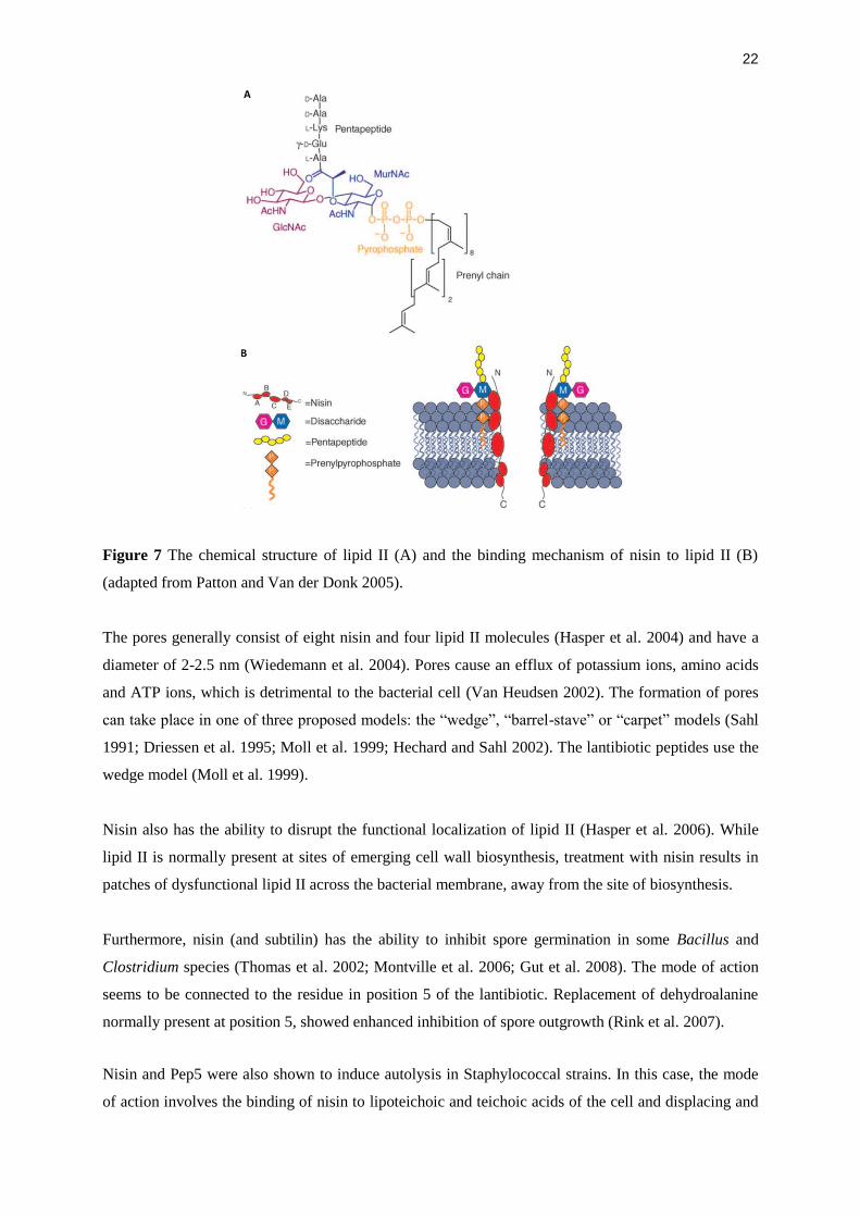