Embed Size (px)
Citation preview

Cellular/Molecular
The mTOR Substrate S6 Kinase 1 (S6K1) Is a NegativeRegulator of Axon Regeneration and a Potential Drug Targetfor Central Nervous System InjuryX Hassan Al-Ali,1,2,3* Ying Ding,5,6* Tatiana Slepak,1* X Wei Wu,5 Yan Sun,5,7 Yania Martinez,1 Xiao-Ming Xu,5
Vance P. Lemmon,1,3 and X John L. Bixby1,3,4
1Miami Project to Cure Paralysis, University of Miami Miller School of Medicine, Miami, Florida 33136, 2Peggy and Harold Katz Family Drug DiscoveryCenter, University of Miami Miller School of Medicine, Miami, Florida 33136, 3Department of Neurological Surgery, University of Miami MillerSchool of Medicine, Miami, Florida 33136, 4Department of Molecular and Cellular Pharmacology, University of Miami Miller School of Medicine, Miami,Florida 33136, 5Spinal Cord and Brain Injury Research Group, Stark Neurosciences Research Institute, Department of Neurological Surgery, IndianaUniversity School of Medicine, Indianapolis, Indiana 46202, 6Department of Histology and Embryology, Zhongshan School of Medicine, Sun Yat-senUniversity, Guangzhou, Guangdong 510080, China, and 7Department of Anatomy, Histology and Embryology, School of Basic Medical Sciences,Fudan University, Shanghai, 200032, China
The mammalian target of rapamycin (mTOR) positively regulates axon growth in the mammalian central nervous system (CNS). Although axonregeneration and functional recovery from CNS injuries are typically limited, knockdown or deletion of PTEN, a negative regulator of mTOR,increases mTOR activity and induces robust axon growth and regeneration. It has been suggested that inhibition of S6 kinase 1 (S6K1, genesymbol: RPS6KB1), a prominent mTOR target, would blunt mTOR’s positive effect on axon growth. In contrast to this expectation, we demon-strate that inhibition of S6K1 in CNS neurons promotes neurite outgrowth in vitro by twofold to threefold. Biochemical analysis revealed that anmTOR-dependent induction of PI3K signaling is involved in mediating this effect of S6K1 inhibition. Importantly, treating female mice in vivowith PF-4708671, a selective S6K1 inhibitor, stimulated corticospinal tract regeneration across a dorsal spinal hemisection between the cervical5 and 6 cord segments (C5/C6), increasing axon counts for at least 3 mm beyond the injury site at 8 weeks after injury. Concomitantly, treatmentwith PF-4708671 produced significant locomotor recovery. Pharmacological targeting of S6K1 may therefore constitute an attractive strategy forpromoting axon regeneration following CNS injury, especially given that S6K1 inhibitors are being assessed in clinical trials for nononcologicalindications.
Key words: axon regeneration; drug discovery; drug target; kinase; S6K; spinal cord injury
IntroductionThe PI3K/mammalian target of rapamycin (mTOR) pathway is akey regulator of axon growth and regeneration in the CNS (Park
et al., 2008). Typically, axons in the adult mammalian CNS havelimited capacity for regeneration following injury. Activating thePI3K/mTOR pathway by knockdown or genetic deletion of itsnegative regulator, PTEN, results in a marked increase in regen-erative capacity (Zerhouni, 2004; Park et al., 2008, 2010; Zukor et
Received April 4, 2017; revised May 18, 2017; accepted May 27, 2017.Author contributions: H.A., Y.D., T.S., W.W., Y.S., X.-M.X., V.P.L., and J.L.B. designed research; H.A., Y.D., T.S.,
W.W., Y.S., and Y.M. performed research; H.A., Y.D., and T.S. analyzed data; H.A., T.S., X.-M.X., V.P.L., and J.L.B.wrote the paper.
This work was supported by Department of Defense Grant W81XWH-13–1-077 and the National Institutes ofHealth Grant HD057632 to J.L.B. and V.P.L., and National Institutes of Health Grant NS059622, Department ofDefense Grant CDMRP W81XWH-12-1-0562, U.S. Department of Veterans Affairs Merit Review Award I01 BX002356,and Craig H. Neilsen Foundation 296749 to X.-M.X., V.P.L. holds the Walter G. Ross Distinguished Chair in Develop-mental Neuroscience. X.-M.X. holds the Mari Hulman George Chair in Neurological Surgery. We thank Yan Shi forhelp with cell imaging and high content analysis.
The authors declare no competing financial interests.*H.A., Y.D., and T.S. contributed equally to this work as co-first authors.Correspondence should be addressed to either of the following: Dr. John L. Bixby, University of Miami Miller
School of Medicine, 1400 NW 10th Avenue, DT 1205, Miami, FL 33136, E-mail: [email protected]; or Dr. VanceP. Lemmon, University of Miami Miller School of Medicine, 1095 NW 15th Terrace, LPLC 4-16, Miami, FL 33136,E-mail: [email protected].
DOI:10.1523/JNEUROSCI.0931-17.2017Copyright © 2017 the authors 0270-6474/17/377079-17$15.00/0
Significance Statement
Despite mTOR’s well-established function in promoting axon regeneration, the role of its downstream target, S6 kinase 1 (S6K1),has been unclear. We used cellular assays with primary neurons to demonstrate that S6K1 is a negative regulator of neuriteoutgrowth, and a spinal cord injury model to show that it is a viable pharmacological target for inducing axon regeneration. Weprovide mechanistic evidence that S6K1’s negative feedback to PI3K signaling is involved in axon growth inhibition, and show thatphosphorylation of S6K1 is a more appropriate regeneration indicator than is S6 phosphorylation.
The Journal of Neuroscience, July 26, 2017 • 37(30):7079 –7095 • 7079

al., 2013), and mTOR activity is critical for the regeneration in-duced by loss of PTEN (Park et al., 2010). The mechanismsthrough which mTOR promotes axon growth/regeneration areless clear; in particular, the role(s) of S6K1, a downstream effectorof mTOR, are incompletely understood (Hubert et al., 2014;Yang et al., 2014).
We previously screened a large number of kinase inhibitors ina neurite outgrowth assay using primary hippocampal neurons,and identified “hit” compounds that promote neurite outgrowth(Al-Ali et al., 2013b). This phenotypic assay is reliable (Z� fac-tor � 0.7) and has identified both chemical and genetic pertur-bagens that promote axon growth from a variety of CNS neurons(Al-Ali et al., 2013a, 2017). In a follow-up study, we used machinelearning to relate data from the phenotypic screen of kinase in-hibitors to kinase profiling data, which allowed us to identify(and verify) “target” kinases whose inhibition induces neuriteoutgrowth (Al-Ali et al., 2015). These target kinases includedrepresentatives of the family of ribosomal S6 protein kinases(RPS6Ks).
Two types of S6 kinases have been described based on theirdomain topology: the p70 ribosomal S6 kinases (S6K1 and S6K2,which phosphorylate S6 at S 235/236/240/244/247) and the p90 ribo-somal S6 kinases (RSK1, RSK2, RSK3, and RSK4, which phos-phorylate S6 at S 235/236) (Meyuhas, 2015). Two additional p90kinases, MSK1 and MSK2, are included in the family by virtue ofsequence similarity (Pearce et al., 2010b) but do not appear tohave substantial activity toward S6. Previous studies have shownthat RSKs and MSKs negatively regulate neurite outgrowth (Lohet al., 2008; Fischer et al., 2009; Buchser et al., 2010; Hubert et al.,2014). Therefore, the finding that their inhibition promotes neu-rite outgrowth might be expected. The observation that neuronalS6K1 activity may be negatively correlated with neurite out-growth was interesting, however, given the well-established roleof its upstream activator mTOR as a positive regulator of axongrowth.
In dividing cells, S6K1 acts as a negative feedback regulator ofthe PI3K/mTOR pathway, such that inhibition of S6K1 leads toinduction of PI3K signaling and subsequent activation of mTOR(Pende et al., 2004; Um et al., 2004; Magnuson et al., 2012). In thisstudy, we show that a similar regulatory mechanism occurs in neu-rons: inhibition of S6K1 induces neurite outgrowth in an mTOR-dependent manner. Importantly, we demonstrate that treating micewith a selective S6K1 inhibitor following transection of the cortico-spinal tract (CST) promoted robust CST axonal regeneration acrossand beyond the lesion site. This regeneration was accompanied byimproved behavioral recovery, suggesting that axon regenerationinduced by S6K1 inhibition may be helpful in recovery from CNSinjury.
Materials and MethodsAntibodies, reagents, and compounds. Pan Akt (#2920), pT308Akt (#4056),pS473Akt (#4058), pan S6 ribosomal protein (#2317), pS240/244S6 (#5364),pan S6K1 (#2708), and pT389S6K1 (#9205) antibodies (working dilution1:1000, except for S6 pan, which was diluted 1:300) were purchased fromCell Signaling Technology. GAPDH (#IMG-5019A-1) antibody (workingdilution 1:500) was purchased from Imgenex. �III-tubulin (#T2200) anti-body (working dilution 1:2000) was purchased from Sigma-Aldrich. Alexafluorophore-conjugated secondary antibodies (working dilution 1:1000)were purchased form Invitrogen. IRDye-700- and IRDye-800-conjugated sec-ondary antibodies (working dilutions 1:15000) were purchased from LiCor.Poly-D-lysine (P7886-500MG) and sterile DMSO (D2650) were purchasedfrom Sigma-Aldrich. Hippocampal tissue was incubated in Hibernate E(without calcium) from BrainBits, supplemented with NeuroCult SM1(05711) from Stem Cell Technologies. Neurons were cultured in NbActive4
media from BrainBits. Accell siRNAs (working concentration 1 �M) werepurchased from GE Healthcare/Dharmacon (scramble SMARTPool #D-001910-10-20, S6K1 siRNA SMARTPool #E-099323-00-0003, scrambleoligo #D-001910-04-05, S6 set of 4 siRNA oligos #EU-089542-00-0002).Kinase inhibitors PF-4708671, ML-7, ROCK inhibitor IV, IKK inhib-itor VII, Flt3 inhibitor III, and Bisindolylmaleimide I, and rapamycinwere purchased from EMD Millipore. GW784684X, GW659386A,GSK1511931A, GSK269962B, and SB-750140 were acquired via col-laboration from GlaxoSmithKline. RO0480500-002 was acquired viacollaboration from Roche. VCC379989:02 and VCC781016:01 werepurchased from Vichem. Torin-2 was purchased from Tocris Biosci-ence. All other reagents were purchased from Invitrogen.
Neurite outgrowth assay for small-molecule kinase inhibitors. The neu-rite outgrowth assay was performed as previously described (Al-Ali et al.,2013b), with minor modifications. Briefly, compounds were tested withrat embryonic (E18) hippocampal neurons cultured at low density for48 h. Neurons were plated at 1500 –1800 cells/well in 96-well platescoated overnight with poly-D-lysine. For the pretreatment condition,mTOR inhibitors were added to the cells 1 h before the addition of S6K1inhibitor. Plates were fixed, immunostained, and imaged in a CellomicsArrayScan VTI. Images were automatically traced using the NeuronalProfiling Bioapplication (version 3.5). Data were analyzed usingMATLAB (R2014b, MathWorks).
Neurite outgrowth assay for siRNAs. Dissociated hippocampal neuronswere seeded in 96-well poly-D-lysine-coated plates at 1500 –1800 cells perwell in 150 �l of NbActive4 and incubated overnight. The following day,siRNA solutions were prepared in 96-well plates at 2 �M in 100 �l ofmedia and allowed to equilibrate for 1 h in a CO2 incubator. Immediatelybefore treatment, 100 �l of media was removed from the cells and re-placed with 50 �l of siRNA solution (duplicate wells for each treatment)to bring the final siRNA reagent concentration to 1 �M (per vendorrecommendation) in a total volume of 100 �l. Cells were incubated for96 h, then fixed and analyzed for neurite outgrowth as previously de-scribed for the neurite outgrowth assay (Al-Ali et al., 2013b).
Assessing protein phosphorylation. Hippocampal (E18) neurons werecultured in NbActive4 (Invitrogen). Cells were seeded into 24-well platesat 0.25–0.5 � 106 cells/well in 0.5 ml of media. Compound stocks (10 mM inDMSO) were diluted in cell culture media at 2� final concentration andadded at 1:1 volume to cells (final volume � 1 ml). Control wells receivedthe corresponding volume of DMSO, which was kept constant across alltreatments at 0.2% v/v. Each plate contained two DMSO control wells(one normalization control and one treatment control). After indicatedtime points, cells were washed once with PBS and collected in 120 �l oflysis buffer containing 100 mM Tris-HCl, pH 6.8, 20% glycerol, 4% SDS,5% �-mercaptoethanol, phosphatase inhibitor mixture (Clontech), pro-tease inhibitor mixture (Roche), and 0.2% bromophenol blue. Lysateswere boiled for 5–7 min, and 20 �l of each sample was loaded on a 10%polyacrylamide gel. Protein was transferred from the gel to a nitrocellu-lose membrane in a wet transfer apparatus using 25 mM sodium bicar-bonate as transferring solution. Membranes were blocked with Odysseyblocking buffer for at least 1 h, then incubated overnight in primaryantibody solution (1:1 PBS, Odyssey blocking buffer with 0.1% Tween20) at 4°C on a shaker. Membranes were washed 3� in PBS-Tween (10min washes) and incubated in secondary antibody solution (secondaryantibodies in 1:1 PBS, Odyssey blocking buffer with 0.1% Tween and0.02% SDS) at room temperature for 2 h with shaking. The 800-IR-dye-conjugated secondary antibodies were used to develop total proteinbands, and the 680-IR-dye-conjugated secondary antibodies were usedto develop phosphorylated protein bands. Finally, membranes werewashed 5� in PBS Tween (10 min washes). Blots were scanned using anOdyssey system. Relative change in phosphorylation in response to treat-ment was computed as follows:
Change in phosphorylation
�Sample 700 signal
Sample 800 signal�control 700 signal
control 800 signal(1)
Assessing target protein knockdown. Neurons were plated in 48-wellplates at �0.25 � 10 6 cells/well and transfected with siRNAs at 1 �M in a
7080 • J. Neurosci., July 26, 2017 • 37(30):7079 –7095 Al-Ali et al. • Inhibiting S6 Kinase 1 Promotes Axon Regeneration

total volume of 200 �l. Three days following transfection, cells were lysedand analyzed by SDS-PAGE followed by Western blotting as above.
Assessing S6 mRNA knockdown. Neurons were plated in 48-well platesat �0.25 � 10 6 cells/well and transfected with siRNAs at 1 �M in a totalvolume of 200 �l. One day following transfection, RNA was isolated fromthe neurons using the micro RNAeasy (QIAGEN, #74004) kit. Total amountof isolated RNA was quantified using a NanoDrop 1000 (NanoDrop Prod-ucts). Reverse transcription (RT) reaction was performed using theAdvantage RT PCR kit (Clontech) with 1 �g of isolated RNA and botholigo dT and random hexamer primers. Samples were collected from twoindependent experiments and qPCR for each sample was set up in trip-licates using the Power SYBR Green PCR Master Mix (Applied Biosys-tems) according to manufacturer suggestions. Controls included aNo-RT control to ensure absence of contaminating DNA, cDNA plasmidcontrol (1 pg/ml) to verify the correct product size, and qPCR of GAPDHand �-actin housekeeping genes for normalization. Reactions were runon 7300 RealTime PCR System (Applied Biosystems), and the resultswere analyzed using Sequence Detection System version 1.2.3 (AppliedBiosystems) software.
Animal studies. A total of 30 adult C57BL/6 mice (7 weeks, HarlanLaboratories) were used in the in vivo studies. Only female mice wereused because males often get bladder infections and develop kidneyproblems after this kind of spinal cord injury. All behavior testing, sur-gical interventions, and postoperative animal care were performed inaccordance with the Guide for the care and use of laboratory animals(National Research Council) and were approved by the Indiana Univer-sity Institutional Animal Care and Use Committee. The mice were pre-trained for behavior analysis for 4 weeks before the surgery (see Fig. 8A).Two mice were unable to learn the task and were excluded from theexperiment. The remaining 28 animals received dorsal hemisection sur-gery at the end of the pretraining and baseline behavioral testing.
Spinal cord dorsal hemisection and cortical injection of PF-4708671. Atthe 10th week of age (4 weeks after training), mice (20–30 g) were anesthe-tized with an intraperitoneal injection of a ketamine/xylazine/acepromazinemixture (120 mg/3.3 mg/1.67 mg /kg) provided by Laboratory Animal Re-source Center at Indiana University School of Medicine, Indianapolis. Micewere then subjected to stabilization of the C5/C6 vertebra with a vertebralstabilizer. After the exposure of the C5-C6 dorsal laminae, a transverse du-rotomy at the interlaminar space between C5 and C6 was performed using a30 G needle followed by cutting with a pair of microscissors. A midline dorsalhemisection was performed using a VibraKnife attached to the LouisvilleInjury System Apparatus (Louisville Impactor System, Louisville, KY) ac-cording to a previously published method (Sivasankaran et al., 2004; Y. Liu etal., 2008; Zhang et al., 2013) with modifications. Briefly, the blade was 1.2mm wide and was advanced 0.9 mm ventrally from the dorsal surface of thecord, extending beyond the central canal (see Fig. 1B). Such a lesion com-pletely transected the entire dorsal corticospinal tract (dCST) and lateralcorticospinal tract (lCST) on both sides. In the C57BL/6 mice, we did notobserve the presence of the ventral CST (vCST). Thus, a dorsal hemisectionin our model (1.2 mm wide, 0.9 mm deep) transected all descending CSTaxons at the lesion site. In this case, any labeled CST axons growing beyondthe lesion would be interpreted as coming from cut axons via either regen-eration from the tips or by regenerative sprouting.
Compounds were injected bilaterally into sensorimotor cortex afterthe C5-C6 dorsal hemisection according to a previously establishedmethod (Wang et al., 2014). Briefly, after the dorsal hemisection, animalswere randomly assigned into four groups that receive the following cor-tical injections (n � 7/group): Group 1 (control): DMSO (0.1% DMSOin 0.1 M PBS); Group 2 (low-dose PF): 1 mM PF-4708671 (in 0.1% DMSOin 0.1 M PBS); Group 3 (medium-dose PF-4708671): 5 mM PF-4708671(in 0.1% DMSO in 0.1 M PBS); and Group 4 (high-dose): 10 mM PF-4708671 (in 0.1% DMSO in 0.1 M PBS). Five stereotaxic injections ofPF-4708671 were made into the sensorimotor cortex on each side at adepth of 0.5 mm and coordination of 2 mm/1.5 mm, 1.25 mm/1.5 mm,0.5 mm/1.5 mm, �0.25 mm/1 mm, and �1 mm/1 mm (anteroposterior/mediolateral to the bregma) with 1 �l/injection site. After the surgery andPF-4708671 injections, an analgesic agent, buprenorphine, was givenintraperitoneally every 12 h for 2 d (Laboratory Animal Resource Center,Indiana University, Indianapolis).
Single-pellet reaching task. The single-pellet reaching task is a simplerepetitive training protocol used to test the precise and coordinated mo-tor movements of the forelimb. The single-pellet reaching task was per-formed in accordance with a previously described method (Chen et al.,2014). In this test, a clear Plexiglas training chamber (15 cm long � 8.5cm wide � 20 cm tall) contains three vertical slits (one slit on “shaping”edge and two slits on the opposite “training” edge). The vertical slits are0.5 cm wide and 13 cm tall and are located on the front wall of the box: inthe center, on the left side, and on the right side. A food platform (8.5 cmlong � 4.4 cm wide � 0.9 cm tall) is placed in the front side (facing thetrainer) of the training chamber during training sessions. There are twodivot slots on the food platform for positioning seeds: one slot on the leftand the other slot on the right side. The divots are 0.3 cm from the longedge and 2.4 cm from the width edge (Chen et al., 2014). The left andright divot slots correspond to the left and right slits in the mouse trainingchamber and are used for training of dominant forelimbs. The purpose ofhaving these divot slots is to ensure that the pellet (millet seed) is placedconsistently at the same place for each reaching attempt. The trainingphase contained food deprivation (2 d), shaping (7 d), and training (9 d).The mice were trained for 30 reaching attempts on the preferred limb for20 min per day. Reaching behavior and scoring were recorded accordingto the following four categories: (1) Success: The mouse reaches withpreferred paw, grasps and retrieves the seed, and feeds it into its mouth.Success (%) � (the number of success/total number of feeding seeds) �100. (2) Grasp: The mouse reaches with preferred paw, grasps the seed,but drops it before putting it into its mouth. Grasp (%) � (the number ofgrasp/total number of feeding seeds) � 100. (3) Reach: The mouse reacheswith preferred paw toward the seed, but it either misses the seed or knocks itoff from the holding plate. Reach (%) � (the number of reach/total numberof feeding seeds) � 100. (4) No reach: The mouse attempts to grasp the seed,but it cannot reach the seed in the holding plate. No reach (%) � (thenumber of no reach/total number of feeding seeds) � 100. In all mice, abaseline task was performed at 3 d before the surgery. The single-pellet reach-ing task was also assessed at 1, 2, 4, and 6 weeks after the dorsal hemisectionand PF-4708671 treatment (see Fig. 8A).
Rotarod test. The Rotarod test was used to assess motor coordination inrodents following procedures described previously with minor modifi-cation (N. K. Liu et al., 2013). Briefly, mice were placed on a rotating rod(diameter 30 mm) in a 5-lane Rotarod device (IITC Life Science) thataccelerated from 0 to 30 rpm within a 90 s period, with each trial lastinga maximum of 120 s. Trials ended when the animal either fell off the rodor clung to the rod as it made one complete rotation. The animals weretrained five times per day for 3 d. The Rotarod tests were performedbefore injury to score the baseline latencies for each animal. The baselinevalue was scored as the mean of five trials at 3 d before surgery (see Fig.1A). The Rotarod test was performed at 1, 2, 4, and 6 weeks after thesurgery. Data were expressed as the ratio of duration relative to baseline.
Adhesive removal test. Animals were trained for the adhesive removaltest for 3 d, and the baseline was recorded at 3 d before the dorsal he-misection (see Fig. 8A) according to a protocol previously described(Bouet et al., 2009; N. K. Liu et al., 2013). This concise sensorimotorfunction assay was performed by applying adhesive tape on the forepawof the animal and measuring the time to contact and the time to remove it. A 3mm � 4 mm adhesive tape strip (waterproof adhesive tape, ACME/Chaston)was, respectively, pressed on the left and right sides of the forepaw coveringthe whole palm and proximal part of the digits. The time to contact and timeto remove the adhesive tape were collected with a maximum of 3 min/animal. The time to contact is defined as the point at which the mouse reactsto the affected paw by either shaking its paw or bringing the paw to its mouth.The time to remove is the point when the tape is removed by its mouth,combined with assistance from the forelimb in loosening it. The adhesiveremoval test was also assessed at 1, 2, 4, and 6 weeks after the dorsal hemisec-tion (see Fig. 8A).
Grid-walking test. The grid-walking test assesses the ability to place theforepaws on the rungs of a grid accurately during spontaneous explora-tion. The mice were trained to walk on a wire grid (0.35 m long with 10mm squares) for 3 d according to an established protocol with modifica-tions (N. K. Liu et al., 2013). The percentage of forepaw drops below thegrid plane during a 3 min observation period was calculated at 3 d before
Al-Ali et al. • Inhibiting S6 Kinase 1 Promotes Axon Regeneration J. Neurosci., July 26, 2017 • 37(30):7079 –7095 • 7081

surgery (as a baseline) and at 1, 2, 4, and 6 weeks after the dorsal hemisec-tion (see Fig. 8A).
Anterograde tracing with biotinylated-dextran amine. Six weeks afterthe dorsal hemisection, mice received bilateral injections of biotinylated-dextran amine (BDA, 10,000 MW, 10% in saline; Invitrogen) into thesensorimotor cortex to trace the CST axons anterogradely. Five injec-tions of BDA (0.5 �l/each) were made into the sensorimotor cortex oneach side using the same coordinates as the PF-4708671 injection. Foreach injection, 0.5 �l of BDA was delivered for a period of 5 min via a glasspipette attached to a 10 �l syringe connected to the Stoelting motorizedintegrated stereotaxic injectors system (Stoelting). Mice were humanelykilled by a lethal dose of anesthesia and transcardially perfused with salinefollowed by 4% PFA 2 weeks after the BDA injection (see Fig. 1A).
BDA visualization and immunofluorescent labeling. After perfusion, thespinal cords containing the lesion epicenter were removed and postfixedin 4% PFA overnight, and were then equilibrated in 30% sucrose forcryoprotection. Sagittal sections at 25 �m were cut using a cryostat.BDA-labeled CST axons were visualized by using avidin-biotin peroxi-dase incubation followed by biotinyl tyramide and Extra-Avidin-FITC(SAT700, PerkinElmer) (Wang et al., 2014). Lesion gaps were identifiedin the same sections by immunostaining for GFAP (1:1000; Millipore).Quantification of BDA-labeled CST axons was performed with the ex-perimenter blind to the treatment groups. Briefly, 7 sagittal sections ofthe spinal cord spaced 125 �m apart were chosen. The middle section wasdefined as the one that cut through the central canal (see Fig. 3A). Theother 6 sections were selected at 125, 250, and 375 �m lateral to themiddle section on each side. The number of BDA-immunoreactive (IR)CST axon fragments was counted at defined zones spaced 0.5 mm apart.The axon numbers were converted into CST Axon Index defined as aratio of the BDA-IR axon number at a given zone over the total numberof BDA-IR axons in the dCST on a cross section of the C1 spinal cord.The lesion epicenter on the sagittal section is defined as “0 mm,” and allother zones are defined as their distances from the lesion epicenter.
Statistical and computational analyses. All computational analysesand tools were implemented in MATLAB (R2013b and R2014b,
MathWorks). Statistical analyses were performed in GraphPad Prism(5.03), MATLAB, or Excel (2007). Data were presented as mean � SEM,unless otherwise noted. Statistical analyses were performed using theGraphPad Software (Prism 5.0) with ANOVA followed by the appropri-ate post-test correction, or two-way repeated-measures ANOVA withBonferroni post-test. Statistical significance was accepted with p 0.05.
ResultsPhosphorylation of ribosomal S6 protein is reduced by kinaseinhibitors that promote neurite outgrowthA set of small-molecule kinase inhibitors was selected from thehits in our phenotypic screen to represent a variety of chemicalscaffolds, distinct kinase inhibition profiles, and potentially di-verse mechanisms for promoting neurite outgrowth (Al-Ali et al.,2013b, 2015). In the present study, we investigated the effect ofthese representative compounds on S6 phosphorylation, in pri-mary hippocampal neurons. We observed that the majority (9 of14) of these compounds induced a significant reduction in S6phosphorylation at S 240/244 (Fig. 1A), sites phosphorylated exclu-sively by S6K1/2 (Pende et al., 2004). Interestingly, only 4 of the14 compounds directly inhibit S6 kinase activity in enzymaticassays in vitro (Fig. 1B). This finding suggests that some of thecompounds reduce S6K1 phosphorylation of S6 without directlyinhibiting the enzyme.
Highly selective S6K1 inhibitor promotes neurite outgrowthBecause the catalytic kinase domains of S6 kinases are structurallysimilar, most inhibitors of one S6 kinase tend to inhibit otherkinases within the S6 kinase family (Fig. 1B). One compound(PF-4708671), however, has unusual selectivity for S6K1, bothwithin the group of S6 kinases (Pearce et al., 2010a) and across thekinome (Fig. 2A). PF-4708671 was shown to inhibit S6K1 with
Figure 1. Ribosomal S6 protein phosphorylation is reduced by most kinase inhibitors that promote neurite outgrowth in primary hippocampal neurons. A, Fourteen small-molecule kinaseinhibitors were selected, from a set of neurite outgrowth-promoting compounds, to represent distinct chemical scaffolds and diverse kinase activity profiles (Al-Ali et al., 2015). Neurons were treatedwith the compounds at 1 �M (or DMSO vehicle as a control) for 10 h before lysis and analysis by Western blotting. Most compounds (9 of 14) resulted in significantly reduced S6 phosphorylation(relative to DMSO control) at Serine 240/244, the substrate site for S6K1, and four others exhibited a trend toward reduced S6 phosphorylation. Data are mean � SEM. n � 4 experimental replicatesfrom two separate preparations of primary neurons. *p 0.05 (one-way ANOVA with Dunnett’s multiple comparisons test). **p 0.01 (one-way ANOVA with Dunnett’s multiple comparisonstest). ***p 0.001 (one-way ANOVA with Dunnett’s multiple comparisons test). B, In vitro kinase activity assays for RSK1 (RPS6KA1), RSK3 (RPS6KA2), RSK2 (RPS6KA3), MSK1 (RPS6KA5), MSK2(RPS6KA4), RSK4 (RPS6KA6), and S6K1 (RPS6KB1) with the 14 representative compounds. Increasing inhibition is indicated by increased intensity of yellow. Only four of the compounds directlyinhibited catalytic activity of S6 kinases in vitro (red arrowheads).
7082 • J. Neurosci., July 26, 2017 • 37(30):7079 –7095 Al-Ali et al. • Inhibiting S6 Kinase 1 Promotes Axon Regeneration

Figure 2. A selective S6K1 inhibitor promotes neurite outgrowth in primary hippocampal neurons. A, in vitro kinase inhibition profiling of PF-4708671 with a panel of 200 kinases (Al-Ali et al.,2015) shows activity against S6K1 and its isoform S6K2, and little to no activity against other kinases, including other members of the RPS6K family of kinases. B, Hippocampal neurons treated with10 �M PF-4708671 for 48 h (right) show longer neurites than those treated with vehicle (left). Green represents �III-tubulin immunostaining. Blue represents nuclear stain. Scale bar, 50 �m.C, Western blot of cell lysates from neurons treated for 1 h with PF-4708671 at the indicated concentrations. D, Red circles represent quantification of NTL of neurons treated with PF-4708671expressed as percentage of DMSO controls. Neurons were treated at the indicated concentrations for 48 h. Data are mean � SEM. n � 3 separate preparations of hippocampal neurons with threeexperimental replicates for each. Blue squares represent quantification of phospho S6 to pan S6 levels in C expressed as percentage of DMSO control. Data are mean � SEM. E, Western blot of celllysates from neurons treated for the indicated times with DMSO or 10 �M PF-4708671. F, Blue squares represent quantification of phospho- to pan-S6 band intensities in E and expressed aspercentage of DMSO control. Data are mean � SEM. n � 3 experimental replicates from a single preparation of primary neurons.
Al-Ali et al. • Inhibiting S6 Kinase 1 Promotes Axon Regeneration J. Neurosci., July 26, 2017 • 37(30):7079 –7095 • 7083

�400-fold selectivity over S6K2 (IC50 0.16�M vs 65 �M) and �5– 60-fold selectivityover RSKs and MSKs. Treating cells with10 �M PF-4708671 strongly inhibits S6K1activity with no detectable inhibition ofRSKs or MSKs, suggesting that intracellu-lar S6K1 is selectively inhibited at orbelow that treatment concentration (Pearce etal., 2010a). We treated primary hippocam-pal neurons with PF-4708671 and foundthat it induced a large increase in neuriteoutgrowth relative to DMSO (vehicle)controls, reaching �200% at 10 �M (Fig.2B). Western blot analysis of lysates fromneurons treated for 1 h with PF-4708671showed a dose-dependent decrease inS 240/244S6 phosphorylation (Fig. 2C,D),which was correlated with induction ofneurite outgrowth (Fig. 2D). With contin-uous PF-4708671 treatment, S6 phos-phorylation remained strongly inhibitedfor the full duration of the neurite out-growth assay (Fig. 2E,F).
SiRNA-mediated knockdown of S6K1promotes neurite outgrowthWe sought an independent line of evi-dence that the correlation between S6K1inhibition and neurite outgrowth can beattributed to S6K1 itself, rather than topharmacological linkage within the S6 ki-nase family or another “off target” effect.We previously determined that Accell siR-NAs can be used for efficient protein knockdown in culturedhippocampal neurons (Al-Ali et al., 2015). We therefore usedAccell siRNAs directed against S6K1 (�-S6K1 siRNA) and foundthat, by 2 d following transfection, S6K1 protein abundance wasreduced by �60% (Fig. 3A,B). As expected, this was accompa-nied by a decrease in S6 phosphorylation at S 240/244 (Fig. 3A,C).To investigate the effect of S6K1 knockdown on neurite out-growth, we treated neurons with �-S6K1 siRNA using the sameformat as the compound screening assay with two modifications:(1) the number of replicates per condition was increased from 2to 6 to account for variations inherent to siRNA knockdownexperiments; and (2) assay duration was increased from 2 to 5 dto allow time for protein knockdown to take effect. Consistentwith the previous inhibitor data, S6K1 knockdown led to a sig-nificant increase in neurite total length (NTL) compared withcontrols (Fig. 3D).
Inhibiting S6K1 in primary neurons induces PI3K signalingand stimulates mTOR activityStudies in non-neuronal cells have shown that S6K1 inhibitsPI3K activity, thus serving as a negative feedback regulator of thePI3K/mTOR pathway (Zoncu et al., 2011; Shimobayashi andHall, 2014). We therefore investigated the effect of S6K1 inhibi-tion on PI3K/mTOR signaling in neurons. PI3K is recruited tothe plasma membrane by a variety of transmembrane receptors(e.g., insulin or growth factor receptors), where it catalyzes the phos-phorylation of phosphatidylinositol-4,5-bisphosphate (PIP2) intophosphatidylinositol-3,4,5-trisphosphate (PIP3) (Vivanco and Saw-yers, 2002). PIP3 recruits and activates kinases like PDK1 and Aktthrough pleckstrin homology domains, allowing PDK1 to phos-
phorylate and activate Akt at T308 (Sarbassov et al., 2005). PDK1 alsophosphorylates S6K1 at T229; however, this step occurs indepen-dently of PIP3 and is therefore not directly regulated by PI3K (Moraet al., 2004). Activated Akt leads to activation of mTOR complex 1(mTORC1), composed of mTOR and its binding partner Raptor(Zoncu et al., 2011). mTORC1 phosphorylates S6K1 at T389, leadingto its activation (Q. Liu et al., 2010, 2011). In addition to mTORC1,mTOR forms a second complex with Rictor, mTOR complex 2(mTORC2) (Sarbassov et al., 2004). PI3K activity (and PIP3 produc-tion) stimulates mTORC2, which phosphorylates Akt at S473 andfacilitates its activation by PDK1 (Sarbassov et al., 2005). PTEN, adual-specificity protein/lipid phosphatase, silences this pathway byhydrolyzing PIP3 into PIP2 (Song et al., 2012).
Treating hippocampal neurons with 10 �M PF-4708671 rap-idly inhibited S6K1 activity (S6 phosphorylation at S 240/244; Fig.4A,B, blue line). A simultaneous increase was observed inmTORC1 activity (phosphorylation of S6K1 at T 389) (Fig. 4A,C).Additionally, PF-4708671 treatment increased activities of PDK1(Akt phosphorylation at its T 308) and mTORC2 (Akt phosphor-ylation at its S 473) (Fig. 4A,D,E). In agreement with previouswork showing that PDK1 phosphorylation of S6K1 is indepen-dent of PI3K (Mora et al., 2004), we did not observe a strongincrease in S6K1 T 229 phosphorylation (Fig. 4A). These resultsdemonstrate that inhibiting S6K1 in neurons induces the PI3Kpathway and leads to the activation of both mTOR complexes.
Inhibiting both mTOR complexes abolishes PF-4708671’seffect on neurite outgrowthTo investigate whether mTOR activity is required for neurite out-growth promotion by PF-4708671, we used two mTOR inhibitors:
Figure 3. siRNA-mediated knockdown of S6K1 promotes neurite outgrowth in primary hippocampal neurons. A, Western blotof cell lysates (3 d after transfection) from neurons transfected with S6K1-targeting or scrambled siRNAs. B, C, Quantification ofA expressed as percentage of scramble control. Data are mean � SD. D, Quantification of NTL in neurons transfected for 5 d with�-S6K1 siRNA or scrambled control. Data are mean � SEM. n � 6 experimental replicates from a single preparation of primaryneurons. **p 0.01 (Student’s t test, one-tailed). ***p 0.001 (Student’s t test, one-tailed).
7084 • J. Neurosci., July 26, 2017 • 37(30):7079 –7095 Al-Ali et al. • Inhibiting S6 Kinase 1 Promotes Axon Regeneration
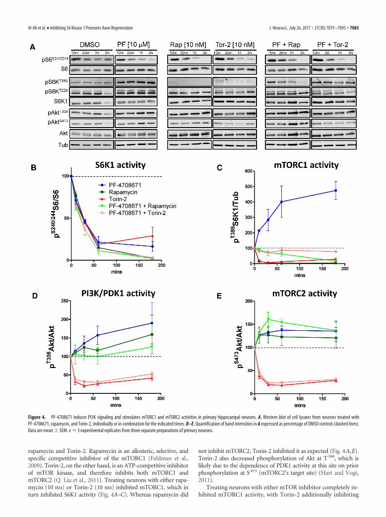
rapamycin and Torin-2. Rapamycin is an allosteric, selective, andspecific competitive inhibitor of the mTORC1 (Feldman et al.,2009). Torin-2, on the other hand, is an ATP-competitive inhibitorof mTOR kinase, and therefore inhibits both mTORC1 andmTORC2 (Q. Liu et al., 2011). Treating neurons with either rapa-mycin (10 nM) or Torin-2 (10 nM) inhibited mTORC1, which inturn inhibited S6K1 activity (Fig. 4A–C). Whereas rapamycin did
not inhibit mTORC2, Torin-2 inhibited it as expected (Fig. 4A,E).Torin-2 also decreased phosphorylation of Akt at T308, which islikely due to the dependence of PDK1 activity at this site on priorphosphorylation at S473 (mTORC2’s target site) (Hart and Vogt,2011).
Treating neurons with either mTOR inhibitor completely in-hibited mTORC1 activity, with Torin-2 additionally inhibiting
Figure 4. PF-4708671 induces PI3K signaling and stimulates mTORC1 and mTORC2 activities in primary hippocampal neurons. A, Western blot of cell lysates from neurons treated withPF-4708671, rapamycin, and Torin-2, individually or in combination for the indicated times. B–E, Quantification of band intensities in A expressed as percentage of DMSO controls (dashed lines).Data are mean � SEM. n � 3 experimental replicates from three separate preparations of primary neurons.
Al-Ali et al. • Inhibiting S6 Kinase 1 Promotes Axon Regeneration J. Neurosci., July 26, 2017 • 37(30):7079 –7095 • 7085
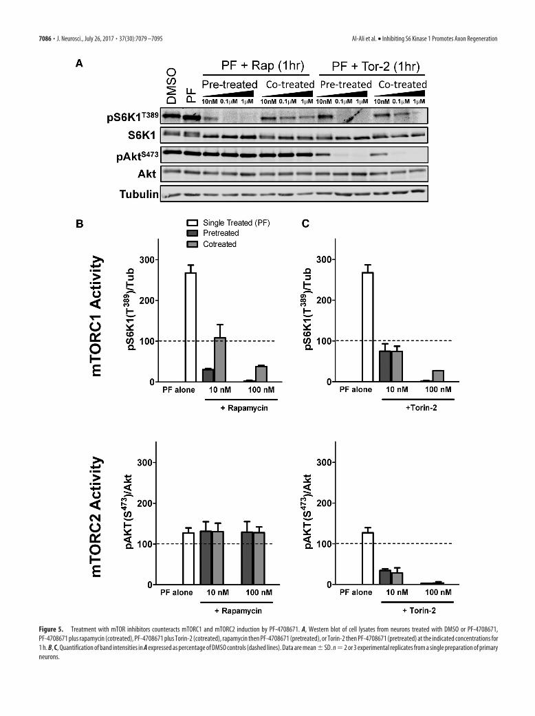
Figure 5. Treatment with mTOR inhibitors counteracts mTORC1 and mTORC2 induction by PF-4708671. A, Western blot of cell lysates from neurons treated with DMSO or PF-4708671,PF-4708671 plus rapamycin (cotreated), PF-4708671 plus Torin-2 (cotreated), rapamycin then PF-4708671 (pretreated), or Torin-2 then PF-4708671 (pretreated) at the indicated concentrations for1 h. B, C, Quantification of band intensities in A expressed as percentage of DMSO controls (dashed lines). Data are mean�SD. n�2 or 3 experimental replicates from a single preparation of primaryneurons.
7086 • J. Neurosci., July 26, 2017 • 37(30):7079 –7095 Al-Ali et al. • Inhibiting S6 Kinase 1 Promotes Axon Regeneration
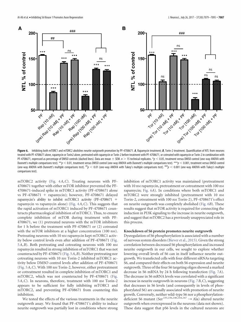
mTORC2 activity (Fig. 4A,C). Treating neurons with PF-4708671 together with either mTOR inhibitor prevented the PF-4708671-induced spike in mTORC1 activity (PF-4708671 alonevs PF-4708671 rapamycin); however, PF-4708671 delayedrapamycin’s ability to inhibit mTORC1 activity (PF-4708671 rapamycin vs rapamycin alone) (Fig. 4A,C). This suggests thatthe rapid activation of mTORC1 induced by PF-4708671 coun-teracts pharmacological inhibition of mTORC1. Thus, to ensurecomplete inhibition of mTOR during treatment with PF-4708671, we (1) pretreated neurons with the mTOR inhibitorsfor 1 h before the treatment with PF-4708671 or (2) cotreatedwith the mTOR inhibitors at a higher concentration (100 nM).Pretreating neurons with 10 nM rapamycin kept mTORC1 activ-ity below control levels even after addition of PF-4708671 (Fig.5A,B). Both pretreating and cotreating neurons with 100 nM
rapamycin resulted in strong inhibition of mTORC1 that was notcounteracted by PF-4708671 (Fig. 5A,B). Neither pretreating norcotreating neurons with 10 nM Torin-2 inhibited mTORC1 ac-tivity below DMSO control levels after addition of PF-4708671(Fig. 5A,C). With 100 nM Torin-2, however, either pretreatmentor cotreatment resulted in complete inhibition of mTORC1 andmTORC2, which was not counteracted by PF-4708671 (Fig.5A,C). In neurons, therefore, treatment with 100 nM Torin-2appears to be sufficient for fully inhibiting mTORC1 andmTORC2, and preventing PF-4708671 from countering thisinhibition.
We tested the effects of the various treatments in the neuriteoutgrowth assay. We found that PF-4708671’s ability to induceneurite outgrowth was partially lost in conditions where strong
inhibition of mTORC1 activity was maintained (pretreatmentwith 10 nM rapamycin, pretreatment or cotreatment with 100 nM
rapamycin; Fig. 6A). In conditions where both mTORC1 andmTORC2 were strongly inhibited (pretreatment with 10 nM
Torin-2, cotreatment with 100 nM Torin-2), PF-4708671’s effecton neurite outgrowth was completely abolished (Fig. 6B). Theseresults suggest that mTOR activity is required for connecting theinduction in PI3K signaling to the increase in neurite outgrowth,and suggest that mTORC2 has a previously unappreciated role inthis process.
Knockdown of S6 protein promotes neurite outgrowthDysregulation of S6 phosphorylation is associated with a numberof nervous system disorders (Biever et al., 2015). Given the strongcorrelation between decreased S6 phosphorylation and increasedneurite outgrowth in our cells, we sought to explore whetherlowering overall levels of S6 can in itself influence neurite out-growth. We transfected cells with four different siRNAs targetingS6, and compared their effects on both S6 expression and neuriteoutgrowth. Three of the four S6 targeting oligos showed a markeddecrease in S6 mRNA by 24 h following transfection (Fig. 7A).The decrease in S6 mRNA levels was correlated with a significantincrease in neurite outgrowth in neurons (Fig. 7B,C), suggestingthat decreases in S6 levels (and consequently in levels of phos-phorylated S6) are causally associated with promotion of neuritegrowth. Conversely, neither wild-type S6 nor a phosphorylation-deficient S6 mutant (Ser 235,236,240,244,247 ¡ Ala) altered neuriteoutgrowth when overexpressed in the neurons (data not shown).These data suggest that pS6 levels in the cultured neurons are
Figure 6. Inhibiting both mTORC1 and mTORC2 abolishes neurite outgrowth promotion by PF-4708671. A, Rapamycin treatment; B, Torin-2 treatment. Quantification of NTL from neuronstreated with PF-4708671 alone, rapamycin or Torin2 alone, pretreated with rapamycin or Torin-2 before treatment with PF-4708671, or cotreated with rapamycin or Torin-2 in combination withPF-4708671, expressed as percentage of DMSO controls (dashed lines). Data are mean � SEM. n � 15 technical replicates. *p 0.05, treatment versus DMSO control (one-way ANOVA withDunnett’s multiple comparisons test). **p 0.01, treatment versus DMSO control (one-way ANOVA with Dunnett’s multiple comparisons test). ***p 0.001, treatment versus DMSO control(one-way ANOVA with Dunnett’s multiple comparisons test). ##p 0.01 (one-way ANOVA with Tukey’s multiple comparisons test). ###p 0.001 (one-way ANOVA with Tukey’s multiplecomparisons test).
Al-Ali et al. • Inhibiting S6 Kinase 1 Promotes Axon Regeneration J. Neurosci., July 26, 2017 • 37(30):7079 –7095 • 7087
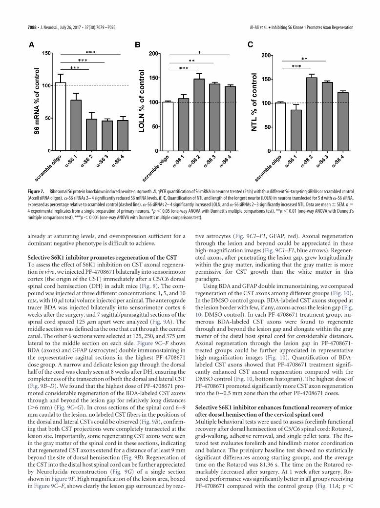
already at saturating levels, and overexpression sufficient for adominant negative phenotype is difficult to achieve.
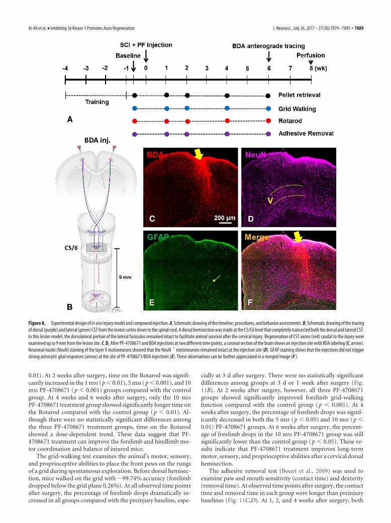
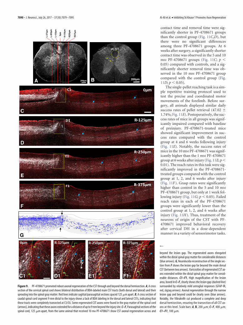
Selective S6K1 inhibitor promotes regeneration of the CSTTo assess the effect of S6K1 inhibition on CST axonal regenera-tion in vivo, we injected PF-4708671 bilaterally into sensorimotorcortex (the origin of the CST) immediately after a C5/C6 dorsalspinal cord hemisection (DH) in adult mice (Fig. 8). The com-pound was injected at three different concentrations: 1, 5, and 10mM, with 10 �l total volume injected per animal. The anterogradetracer BDA was injected bilaterally into sensorimotor cortex 6weeks after the surgery, and 7 sagittal/parasagittal sections of thespinal cord spaced 125 �m apart were analyzed (Fig. 9A). Themiddle section was defined as the one that cut through the centralcanal. The other 6 sections were selected at 125, 250, and 375 �mlateral to the middle section on each side. Figure 9C–F showsBDA (axons) and GFAP (astrocytes) double immunostaining inthe representative sagittal sections in the highest PF-4708671dose group. A narrow and delicate lesion gap through the dorsalhalf of the cord was clearly seen at 8 weeks after DH, ensuring thecompleteness of the transection of both the dorsal and lateral CST(Fig. 9B–D). We found that the highest dose of PF-4708671 pro-moted considerable regeneration of the BDA-labeled CST axonsthrough and beyond the lesion gap for relatively long distances(�6 mm) (Fig. 9C–G). In cross sections of the spinal cord 6 –9mm caudal to the lesion, no labeled CST fibers in the positions ofthe dorsal and lateral CSTs could be observed (Fig. 9B), confirm-ing that both CST projections were completely transected at thelesion site. Importantly, some regenerating CST axons were seenin the gray matter of the spinal cord in these sections, indicatingthat regenerated CST axons extend for a distance of at least 9 mmbeyond the site of dorsal hemisection (Fig. 9B). Regeneration ofthe CST into the distal host spinal cord can be further appreciatedby Neurolucida reconstruction (Fig. 9G) of a single sectionshown in Figure 9F. High magnification of the lesion area, boxedin Figure 9C–F, shows clearly the lesion gap surrounded by reac-
tive astrocytes (Fig. 9C1–F1, GFAP, red). Axonal regenerationthrough the lesion and beyond could be appreciated in thesehigh-magnification images (Fig. 9C1–F1, blue arrows). Regener-ated axons, after penetrating the lesion gap, grew longitudinallywithin the gray matter, indicating that the gray matter is morepermissive for CST growth than the white matter in thisparadigm.
Using BDA and GFAP double immunostaining, we comparedregeneration of the CST axons among different groups (Fig. 10).In the DMSO control group, BDA-labeled CST axons stopped atthe lesion border with few, if any, axons across the lesion gap (Fig.10; DMSO control). In each PF-4708671 treatment group, nu-merous BDA-labeled CST axons were found to regeneratethrough and beyond the lesion gap and elongate within the graymatter of the distal host spinal cord for considerable distances.Axonal regeneration through the lesion gap in PF-4708671-treated groups could be further appreciated in representativehigh-magnification images (Fig. 10). Quantification of BDA-labeled CST axons showed that PF-4708671 treatment signifi-cantly enhanced CST axonal regeneration compared with theDMSO control (Fig. 10, bottom histogram). The highest dose ofPF-4708671 promoted significantly more CST axon regenerationinto the 0 – 0.5 mm zone than the other PF-4708671 doses.
Selective S6K1 inhibitor enhances functional recovery of miceafter dorsal hemisection of the cervical spinal cordMultiple behavioral tests were used to assess forelimb functionalrecovery after dorsal hemisection of C5/C6 spinal cord: Rotarod,grid-walking, adhesive removal, and single pellet tests. The Ro-tarod test evaluates forelimb and hindlimb motor coordinationand balance. The preinjury baseline test showed no statisticallysignificant differences among starting groups, and the averagetime on the Rotarod was 81.36 s. The time on the Rotarod re-markably decreased after surgery. At 1 week after surgery, Ro-tarod performance was significantly better in all groups receivingPF-4708671 compared with the control group (Fig. 11A; p
Figure 7. Ribosomal S6 protein knockdown induced neurite outgrowth. A, qPCR quantification of S6 mRNA in neurons treated (24 h) with four different S6-targeting siRNAs or scrambled control(Accell siRNA oligos). �-S6 siRNAs 2– 4 significantly reduced S6 mRNA levels. B, C, Quantification of NTL and length of the longest neurite (LOLN) in neurons transfected for 5 d with �-S6 siRNA,expressed as percentage relative to scrambled control (dashed line). �-S6 siRNAs 2– 4 significantly increased LOLN, and �-S6 siRNAs 2–3 significantly increased NTL. Data are mean � SEM. n �4 experimental replicates from a single preparation of primary neurons. *p 0.05 (one-way ANOVA with Dunnett’s multiple comparisons test). **p 0.01 (one-way ANOVA with Dunnett’smultiple comparisons test). ***p 0.001 (one-way ANOVA with Dunnett’s multiple comparisons test).
7088 • J. Neurosci., July 26, 2017 • 37(30):7079 –7095 Al-Ali et al. • Inhibiting S6 Kinase 1 Promotes Axon Regeneration
0.01). At 2 weeks after surgery, time on the Rotarod was signifi-cantly increased in the 1 mM (p 0.01), 5 mM (p 0.001), and 10mM PF-4708671 (p 0.001) groups compared with the controlgroup. At 4 weeks and 6 weeks after surgery, only the 10 mM
PF-4708671 treatment group showed significantly longer time onthe Rotarod compared with the control group (p 0.01). Al-though there were no statistically significant differences amongthe three PF-4708671 treatment groups, time on the Rotarodshowed a dose-dependent trend. These data suggest that PF-4708671 treatment can improve the forelimb and hindlimb mo-tor coordination and balance of injured mice.
The grid-walking test examines the animal’s motor, sensory,and proprioceptive abilities to place the front paws on the rungsof a grid during spontaneous exploration. Before dorsal hemisec-tion, mice walked on the grid with �99.74% accuracy (forelimbdropped below the grid plane 0.26%). At all observed time pointsafter surgery, the percentage of forelimb drops dramatically in-creased in all groups compared with the preinjury baseline, espe-
cially at 3 d after surgery. There were no statistically significantdifferences among groups at 3 d or 1 week after surgery (Fig.11B). At 2 weeks after surgery, however, all three PF-4708671groups showed significantly improved forelimb grid-walkingfunction compared with the control group (p 0.001). At 4weeks after surgery, the percentage of forelimb drops was signif-icantly decreased in both the 5 mM (p 0.05) and 10 mM (p 0.01) PF-4708671 groups. At 6 weeks after surgery, the percent-age of forelimb drops in the 10 mM PF-4708671 group was stillsignificantly lower than the control group (p 0.05). These re-sults indicate that PF-4708671 treatment improves long-termmotor, sensory, and proprioceptive abilities after a cervical dorsalhemisection.
The adhesive removal test (Bouet et al., 2009) was used toexamine paw and mouth sensitivity (contact time) and dexterity(removal time). At observed time points after surgery, the contacttime and removal time in each group were longer than preinjurybaselines (Fig. 11C,D). At 1, 2, and 4 weeks after surgery, both
Figure 8. Experimental design of in vivo injury model and compound injection. A, Schematic drawing of the timeline, procedures, and behavior assessments. B, Schematic drawing of the tracingof dorsal (purple) and lateral (green) CST from the motor cortex down to the spinal cord. A dorsal hemisection was made at the C5/C6 level that completely transected both the dorsal and lateral CST.In this lesion model, the dorsolateral portion of the lateral funiculus remained intact to facilitate animal survival after the cervical injury. Regeneration of CST axons (red) caudal to the injury wereexamined up to 9 mm from the lesion site. C, D, After PF-4708671 and BDA injections at two different time points, a coronal section of the brain shows an injection site with BDA labeling (C, arrow).Neuronal nuclei (NeuN) staining of the layer V motoneurons showed that the NeuN motoneurons remained intact at the injection site (D). GFAP staining shows that the injections did not triggerstrong astrocytic glial responses (arrow) at the site of PF-4708671/BDA injections (E). These observations can be further appreciated in a merged image (F ).
Al-Ali et al. • Inhibiting S6 Kinase 1 Promotes Axon Regeneration J. Neurosci., July 26, 2017 • 37(30):7079 –7095 • 7089
contact time and removal time were sig-nificantly shorter in PF-4708671 groupsthan the control group (Fig. 11C,D), butthere were no significant differencesamong three PF-4708671 groups. At 6weeks after surgery, a significantly shortercontact time was observed in the 5 and 10mM PF-4708671 groups (Fig. 11C; p 0.05) compared with controls, and a sig-nificantly shorter removal time was ob-served in the 10 mM PF-4708671 groupcompared with the control group (Fig.11D; p 0.05).
The single-pellet reaching task is a sim-ple repetitive training protocol used totest the precise and coordinated motormovements of the forelimb. Before sur-gery, all animals displayed similar dailysuccess rates of pellet retrieval (47.02 �1.74%; Fig. 11E). Postoperatively, the suc-cess rates of mice in all groups was signif-icantly impaired compared with baselineof preinjury. PF-4708671-treated miceshowed significant improvement in suc-cess rates compared with the controlgroup at 4 and 6 weeks following injury(Fig. 11E). Notably, the success rates ofmice in the 10 mM PF-4708671 was signif-icantly higher than the 1 mM PF-4708671group at 6 weeks after injury (Fig. 11E; p 0.01). The reach rates in this task were sig-nificantly improved in the PF-4708671-treated groups compared with the controlgroup at 1, 2, and 6 weeks after injury(Fig. 11F). Grasp rates were significantlyhigher than control in the 5 and 10 mM
PF-4708671 group, but only at 1 week fol-lowing injury (Fig. 11G; p 0.05). Failedreach rates in each of the PF-4708671groups were significantly lower than thecontrol group at 1, 2, and 6 weeks afterinjury (Fig. 11H). Thus, treatment of theneurons of origin of the CST with PF-4708671 improved behavioral recoveryafter cervical DH in a dose-dependentmanner in a variety of sensorimotor tasks.
Figure 9. PF-4708671 promoted robust axonal regeneration of the CST through and beyond the dorsal hemisection. A, A crosssection of the cervical spinal cord shows bilateral distribution of BDA-labeled main CST tracts (both dorsal and lateral) and theirsprouting into the spinal gray matter. Red lines indicate sagittal/parasagittal sections spaced 125 �m apart. B, A cross section ofcaudal spinal cord segment 9 mm distal to the injury shows a lack of BDA labeling in the dorsal and lateral CSTs, indicating thatthese tracts were completely transected at C5/C6. Some regenerated CST axons were found in the gray matter of the spinal cord(arrows), indicating that these axons extended for a distance of up to 9 mm beyond the injury site. C–F, Parasagittal sections of thespinal cord, 125 �m apart, from the same animal that received 10 mM PF-4708671 show CST axonal regeneration across and
4
beyond the lesion gap. The regenerated axons elongatedwithin the distal spinal gray matter for considerable distances(blue arrows). G, Neurolucida reconstruction of the single sec-tion from F shows the lesion gap far beyond the main dorsalCST (between two arrows). Varicosities of regenerated CST ax-ons extended within the distal spinal gray matter for consid-erable distances. C1–F1, High magnification of the lesionarea, boxed in C–F, clearly shows the lesion gap (dashed line)surrounded by relatively mild astroglial responses (GFAP-IR,red, zigzag arrows). Axonal regeneration through the narrowlesion gap and beyond could be clearly seen (blue arrows).Notably, the Vibraknife cut produced a complete and deepdorsal hemisection, ensuring the transection of all CST ax-ons at this level. Scale bars: A, B, 200 �m; C–F, 400 �m;C1–F1, 100 �m.
7090 • J. Neurosci., July 26, 2017 • 37(30):7079 –7095 Al-Ali et al. • Inhibiting S6 Kinase 1 Promotes Axon Regeneration

DiscussionKnockdown or genetic deletion of PTENpromotes substantial axon regenerationin an mTOR-dependent manner, both inthe optic nerve and in the CST (Park et al.,2008, 2010; K. Liu et al., 2010; Zukor et al.,2013; Du et al., 2015). It has thereforebeen suggested that the activity of S6K1,an effector of mTOR, is required for re-generation in this paradigm (Yang et al.,2014). Nevertheless, experimental resultson this point are conflicting (Hubert et al.,2014; Yang et al., 2014). In our study, wefound that decreased phosphorylation ofS6K1’s substrate, S6, strongly correlateswith promotion of neurite outgrowth inprimary neurons treated with a variety ofsmall-molecule kinase inhibitors, andthat pharmacological inhibition of S6K1promotes neurite outgrowth in primaryneurons. Activation of PI3K/mTOR sig-naling, in response to the release ofS6K1-mediated negative feedback on thispathway, may be driving the induction ofneurite outgrowth. Consistent with this,we observed that inhibiting S6K1 in neu-rons induced a rapid increase in PI3K sig-naling and led to the induction of bothmTORC1 and mTORC2 activities.
Previous studies on S6K1’s role in axongrowth/regeneration have led to conflict-ing conclusions. It is plausible that this isdue to the complexity of S6K1 expressionand regulation. Overexpressing a nativeform of S6K1 does not readily yield acatalytically active form of the enzyme(Keshwani et al., 2008). Overexpressinga phosphomimetic mutant kinase (Maand Blenis, 2009), co-overexpressing anactivating kinase (Keshwani et al.,2008), or overexpressing a truncated ki-nase lacking the autoinhibitory domains(Al-Ali et al., 2007) can result in a fullycatalytically active enzyme; however,these alterations may disrupt regulationand substrate specificity (Pearce et al.,2010b), potentially distancing the over-expressed kinase from native biology.We thus chose siRNA-mediated knock-down to obtain independent evidenceon S6K1’s role in the regulation of neu-rite outgrowth. Consistent with our
Figure 10. Concentrations of PF-4708671 from 1 to 10 mM increase CST regeneration after a C5/C6 dorsal hemisection. In theDMSO control group, BDA-labeled CST axons stopped at the lesion border (DMSO control). In all three PF-4708671 treatmentgroups (1, 5, and 10 mM PF-4708671), numerous BDA-labeled CST axons were found to regenerate through and beyond the lesiongap and elongated within the distal spinal cord gray matter for considerable distances. High magnifications of boxed areas inrepresentative sections show CST axonal regeneration across the lesion gap only in the PF-4708671-treated groups. Neurolucidadrawings under the same representative images of the four experimental groups show detailed growth patterns of the CST axonsacross and beyond the lesion gap. Bottom, Quantitative analysis of BDA-labeled CST axons regenerated at different distance zonesfrom the lesion site. In general, PF-4708671 significantly enhanced CST axonal regeneration beyond the lesion gap; 10 mM
4
PF-4708671 promoted higher numbers of regenerative axonsat different zones distal to the injury, but a statistically signif-icant difference was only found at the 0 – 0.5 mm zone com-pared with the 1 and 5 mM PF-4708671 groups. Data aremean � SEM. *p 0.05 (two-way repeated-measuresANOVA with Bonferroni post-test). **p 0.01 (two-wayrepeated-measures ANOVA with Bonferroni post-test).***p 0.001 (two-way repeated-measures ANOVA withBonferroni post-test).
Al-Ali et al. • Inhibiting S6 Kinase 1 Promotes Axon Regeneration J. Neurosci., July 26, 2017 • 37(30):7079 –7095 • 7091

pharmacological data, we found that knocking down S6K1promotes neurite outgrowth.
A variety of experimental approaches has led to the conclusionthat mTORC1 activation is a key element in the induction of axon
growth (Park et al., 2010). We therefore investigated whethermTORC1 activity is required for neurite outgrowth induced byS6K1 inhibition. Indeed, inhibiting mTORC1 with rapamycinreduced, but did not completely abolish, the amount of neurite
Figure 11. PF-4708671 enhanced functional recoveries of mice after a C5/C6 dorsal hemisection. Rotarod (A) forelimb drops at grid-walking (B) adhesive removal (C, D) and pellet retrieval(E–H) show improved behavioral recoveries in groups treated with 1, 5, and 10 mM PF-4708671, compared with the control groups. At one or more time points for several tests, 10 mM PF-4708671showed significant differences from control when 1 and 5 mM treatments did not (e.g., Rotarod at 4 and 6 weeks). Data are mean � SEM. *p 0.05 (two-way repeated-measures ANOVA withBonferroni post-test). **p 0.01 (two-way repeated-measures ANOVA with Bonferroni post-test). ***p 0.001 (two-way repeated-measures ANOVA with Bonferroni post-test).
7092 • J. Neurosci., July 26, 2017 • 37(30):7079 –7095 Al-Ali et al. • Inhibiting S6 Kinase 1 Promotes Axon Regeneration
outgrowth induced by PF-4708671. Inhibiting both mTORC1and mTORC2 with Torin-2, on the other hand, completely ab-rogated PF-4708671’s ability to promote neurite growth. Previ-ous studies have shown that the mTORC2 regulates actinpolymerization and cytoskeletal dynamics (Jacinto et al., 2004;Angliker and Ruegg, 2013). Mice deficient in mTORC2 exhibitreduced actin polymerization in the hippocampus and presentwith disrupted LTP and impaired memory (Huang et al., 2013).Our results suggest that mTORC2 may also play a role in neuriteoutgrowth and axon regeneration within the PI3K/PTEN net-work. Multiple effectors and interaction partners of mTORC2 areknown regulators of axon growth and may be involved in medi-ating its effects, including the kinases Akt and PKC, and theGTPase Rac1 (Ng et al., 2002; Sivasankaran et al., 2004; Park et al.,2008; Quinn et al., 2008; Oh and Jacinto, 2011).
The strong regulation of neurite growth by manipulation ofS6K1 suggests that phosphorylation of S6, S6K1’s substrate, isimportant in neurite outgrowth. Despite the large number ofstudies using phosphorylation of S6 as a marker for protein trans-lational activity, the biological significance of S6’s phosphoryla-tion has remained controversial, suggesting the presence ofcomplex and likely cell-specific regulatory mechanisms (Mag-nuson et al., 2012; Biever et al., 2015). S6 phosphorylation wasoriginally proposed to be required for the translation of the so-called TOP mRNAs, which encode ribosomal protein and trans-lation factors (Meyuhas and Dreazen, 2009), but was later foundto be merely a correlated event (Patursky-Polischuk et al., 2009).We manipulated intracellular levels of S6 to alter availability ofthe protein for phosphorylation, and found that S6 knockdowninduced neurite outgrowth in cultured neurons. Althoughknocking down S6 is not equivalent to specifically decreasing theavailability of phosphorylated S6, it demonstrates that decreasingproduction of S6 (and presumably, of its phosphorylated form)can promote neurite outgrowth.
In studies of PTEN knockdown or genetic deletion, activationof mTORC1 is consistently correlated with activation of S6K1,and with phosphorylation of ribosomal S6. Consequently, S6phosphorylation has been used as a marker of mTORC1 activa-tion and, by extension, axon growth ability. Conclusions fromthese studies, however, are complicated by the fact that someinvestigators examined S6 phosphorylation sites that are not ex-clusive to S6K1 (Biever et al., 2015), whereas others have notspecified which S6 phosphorylation sites were probed. In thisstudy, we demonstrate that perturbagens that promote neuriteoutgrowth through inhibition of S6K1 activity can produce bothmTOR activation and a reduction in S6 phosphorylation. Wetherefore propose that the mTOR target site on S6K1, T 389S6K1,is a more reliable marker for mTORC1 activation vis-a-vis axonregeneration than is S6 phosphorylation.
Because inhibiting S6K1 can induce neurite growth in cul-tured neurons, we investigated the effect of S6K1 inhibition onaxon regeneration (and on behavioral recovery) in vivo, in a dor-sal hemisection model of spinal cord injury. Notably, our modelproduces a precise and definitive anatomical lesion that com-pletely transects the dCST and dlCST (Sivasankaran et al., 2004;Y. Liu et al., 2008; Zhang et al., 2013). Thus, the model can pro-vide clean and definitive anatomical evidence of CST axonal re-generation. We injected PF-4708671 once, bilaterally, intosensorimotor cortex, following a previously established method(Wang et al., 2014). This single injection had a pronounced effecton CST regeneration across and beyond the lesion gap. Previ-ously, a single cortical injection of Gö6976, an inhibitor of PKCand other kinases, was shown to promote CST axonal regenera-
tion and forelimb functional recovery after a C4 dorsal hemisec-tion in adult rats (Wang et al., 2014). It appears that deliveringthese kinase inhibitors to the cell bodies of the CST neurons isimportant because Gö6976 delivery to the transected CST axonsat the lesion site did not elicit axonal regeneration after a similarinjury (Sivasankaran et al., 2004; Wang et al., 2014). Supportingthe idea that the CST axons in our model were regenerating,rather than spared, was the fact that caudal to the transection,axons were observed in the gray matter and not in the dorsal orlateral CST tracts. This is the same pattern observed with othertreatments that promote CST regeneration, such as PTEN knock-out, PTEN-SOCS3 double knockout, or overexpression of VP16-KLF7 or Sox11 (Blackmore et al., 2012; Du et al., 2015; Jin et al.,2015; Wang et al., 2015). Although it is not possible to directlycompare experiments done in different laboratories, it is intrigu-ing that the density and distance from the lesion of regeneratingCST fibers after a single PF treatment appear to equal or exceedaxon growth observed with PTEN knockout, or with overexpres-sion of VP16-KLF7 or Sox11 (Wang et al., 2011; Blackmore et al.,2012; Zukor et al., 2013; Jin et al., 2015). In this context, a singleinjection of a small-molecule inhibitor provides advantages overthese direct manipulations of gene expression because it is con-venient and easier to produce and deliver.
CST damage in rodents causes deficits primarily in fine con-trol of the forelimbs, which is required for grasping and holdingobjects (Anderson et al., 2007; Courtine et al., 2007). We per-formed a variety of behavioral assessments to investigate the re-covery of forelimb function following dorsal spinal hemisection.Using injection of PF-4708671 to inhibit S6K1, we found im-provements in functional outcomes for tests of pellet retrieval,grid walking, Rotarod, and adhesive removal, with clear evidenceof a dose–response relationship. These tasks measure differentaspects of sensorimotor function, suggesting that S6K1 inhibi-tion in corticospinal neurons helps to rebuild the neural networkcontrolling forelimb movements. Because a number of the func-tional improvements associated with PF-4708671 administrationwere evident as early as 1 week after injury, the relationship be-tween regeneration of CST axons beyond the injury site and re-covery of function may be complex and multifactorial.
The fact that single PF-4708671 injections into sensorimotorcortex allow extensive axon growth beyond a spinal cord lesionraises the possibility that strong inhibition of S6K1 (and activa-tion of mTOR) produces long-term changes in the intrinsic stateof the CST neurons. Indeed, activation of the mTOR pathway, inaddition to effects on protein synthesis, is known to affect genetranscription by a variety of mechanisms (Laplante and Sabatini,2013). It may be that treatment of cortical projection neuronswith PF-4708671 can lead to sustained changes in transcriptionfactor activity and/or in epigenetic regulation. If so, it would beimportant to understand the mechanisms underlying theselonger-term changes.
Although our data provide specific evidence on S6K1’s in-volvement in regulating axon growth, we cannot rule out a rolefor S6K2. Under normal conditions, S6K1 and S6K2 have distinctbiological roles and subcellular localization patterns (Pardo andSeckl, 2013). However, knockout studies suggest that the twoisoforms have overlapping functions, and that S6K2 can partiallycompensate for the S6K1 deletion (Pende et al., 2004). It will beinteresting to investigate whether S6K2 can compensate for S6K1inhibition in the context of axon growth promotion, and whethersimultaneous targeting of both isoforms is required for long-term treatment efficacy.
Al-Ali et al. • Inhibiting S6 Kinase 1 Promotes Axon Regeneration J. Neurosci., July 26, 2017 • 37(30):7079 –7095 • 7093

A potential advantage of targeting S6K1, compared with phar-macological or genetic inhibition of PTEN, is that it can inducemTOR activation and transient increases in axon growth andregeneration, without the potentially tumor-promoting effects ofPTEN inactivation (Song et al., 2012). The potential use of S6K1inhibitors as drugs for neurodegeneration is further encouragedby the fact that S6K1 inhibitors are in Phase III clinical trials forother nononcological indications, such as liver cirrhosis and in-sulin resistance (Hameed and Terrault, 2016). Our results, to-gether, implicate S6K1 as a potential target for drugs that improveaxon regeneration and functional recovery following CNS injury.
ReferencesAl-Ali H, Ragan TJ, Gao X, Harris TK (2007) Reconstitution of modular
PDK1 functions on trans-splicing of the regulatory PH and catalytic ki-nase domains. Bioconjug Chem 18:1294 –1302. CrossRef Medline
Al-Ali H, Blackmore M, Bixby JL, Lemmon VP (2013a) High contentscreening with primary neurons. In: Assay guidance manual. Bethesda,MD: Eli Lilly.
Al-Ali H, Schurer SC, Lemmon VP, Bixby JL (2013b) Chemical interroga-tion of the neuronal kinome using a primary cell-based screening assay.ACS Chem Biol 8:1027–1036. CrossRef Medline
Al-Ali H, Lee DH, Danzi MC, Nassif H, Gautam P, Wennerberg K, ZuercherB, Drewry DH, Lee JK, Lemmon VP, Bixby JL (2015) Rational polyphar-macology: systematically identifying and engaging multiple drug targetsto promote axon growth. ACS Chem Biol 10:1939 –1951. CrossRefMedline
Al-Ali H, Beckerman SR, Bixby JL, Lemmon VP (2017) In vitro models ofaxon regeneration. Exp Neurol 287:423– 434. CrossRef Medline
Anderson KD, Gunawan A, Steward O (2007) Spinal pathways involved inthe control of forelimb motor function in rats. Exp Neurol 206:318 –331.CrossRef Medline
Angliker N, Ruegg MA (2013) In vivo evidence for mTORC2-mediated ac-tin cytoskeleton rearrangement in neurons. Bioarchitecture 3:113–118.CrossRef Medline
Biever A, Valjent E, Puighermanal E (2015) Ribosomal protein S6 phos-phorylation in the nervous system: from regulation to function. FrontMol Neurosci 8:75. CrossRef Medline
Blackmore MG, Wang Z, Lerch JK, Motti D, Zhang YP, Shields CB, Lee JK,Goldberg JL, Lemmon VP, Bixby JL (2012) Kruppel-like Factor 7 engi-neered for transcriptional activation promotes axon regeneration in theadult corticospinal tract. Proc Natl Acad Sci U�S�A 109:7517–7522.CrossRef Medline
Bouet V, Boulouard M, Toutain J, Divoux D, Bernaudin M, Schumann-BardP, Freret T (2009) The adhesive removal test: a sensitive method to as-sess sensorimotor deficits in mice. Nat Protoc 4:1560 –1564. CrossRefMedline
Buchser WJ, Slepak TI, Gutierrez-Arenas O, Bixby JL, Lemmon VP (2010)Kinase/phosphatase overexpression reveals pathways regulating hip-pocampal neuron morphology. Mol Syst Biol 6:391. CrossRef Medline
Chen CC, Gilmore A, Zuo Y (2014) Study motor skill learning by single-pellet reaching tasks in mice. J Vis Exp 85:e51238-e51238. CrossRefMedline
Courtine G, Bunge MB, Fawcett JW, Grossman RG, Kaas JH, Lemon R, MaierI, Martin J, Nudo RJ, Ramon-Cueto A, Rouiller EM, Schnell L, WannierT, Schwab ME, Edgerton VR (2007) Can experiments in nonhumanprimates expedite the translation of treatments for spinal cord injury inhumans? Nat Med 13:561–566. CrossRef Medline
Du K, Zheng S, Zhang Q, Li S, Gao X, Wang J, Jiang L, Liu K (2015) Ptendeletion promotes regrowth of corticospinal tract axons 1 year after spinalcord injury. J Neurosci 35:9754 –9763. CrossRef Medline
Feldman ME, Apsel B, Uotila A, Loewith R, Knight ZA, Ruggero D, ShokatKM (2009) Active-site inhibitors of mTOR target rapamycin-resistantoutputs of mTORC1 and mTORC2. PLoS Biol 7:e38. CrossRef Medline
Fischer M, Pereira PM, Holtmann B, Simon CM, Hanauer A, Heisenberg M,Sendtner M (2009) P90 Ribosomal s6 kinase 2 negatively regulates axongrowth in motoneurons. Mol Cell Neurosci 42:134 –141. CrossRefMedline
Hameed B, Terrault N (2016) Emerging therapies for nonalcoholic fattyliver disease. Clin Liver Dis 20:365–385. CrossRef Medline
Hart JR, Vogt PK (2011) Phosphorylation of AKT: a mutational analysis.Oncotarget 2:467– 476. CrossRef Medline
Huang W, Zhu PJ, Zhang S, Zhou H, Stoica L, Galiano M, Krnjevic K, RomanG, Costa-Mattioli M (2013) mTORC2 controls actin polymerization re-quired for consolidation of long-term memory. Nat Neurosci 16:441–448. CrossRef Medline
Hubert T, Wu Z, Chisholm AD, Jin Y (2014) S6 kinase inhibits intrinsicaxon regeneration capacity via AMP kinase in Caenorhabditis elegans.J Neurosci 34:758 –763. CrossRef Medline
Jacinto E, Loewith R, Schmidt A, Lin S, Ruegg MA, Hall A, Hall MN (2004)Mammalian TOR complex 2 controls the actin cytoskeleton and is rapa-mycin insensitive. Nat Cell Biol 6:1122–1128. CrossRef Medline
Jin D, Liu Y, Sun F, Wang X, Liu X, He Z (2015) Restoration of skilledlocomotion by sprouting corticospinal axons induced by co-deletion ofPTEN and SOCS3. Nat Commun 6:8074. CrossRef Medline
Keshwani MM, Ross DB, Ragan TJ, Harris TK (2008) Baculovirus-mediatedexpression, purification, and characterization of a fully activated catalytickinase domain construct of the 70 kDa 40S ribosomal protein S6 kinase-1alphaII isoform (S6K1alphaII). Protein Expr Purif 58:32– 41. CrossRefMedline
Laplante M, Sabatini DM (2013) Regulation of mTORC1 and its impact ongene expression at a glance. J Cell Sci 126:1713–1719. CrossRef Medline
Liu K, Lu Y, Lee JK, Samara R, Willenberg R, Sears-Kraxberger I, Tedeschi A,Park KK, Jin D, Cai B, Xu B, Connolly L, Steward O, Zheng B, He Z(2010) PTEN deletion enhances the regenerative ability of adult cortico-spinal neurons. Nat Neurosci 13:1075–1081. CrossRef Medline
Liu NK, Zhang YP, O’Connor J, Gianaris A, Oakes E, Lu QB, Verhovshek T,Walker CL, Shields CB, Xu XM (2013) A bilateral head injury that showsgraded brain damage and behavioral deficits in adult mice. Brain Res1499:121–128. CrossRef Medline
LiuQ,ChangJW,WangJ,KangSA,ThoreenCC,MarkhardA,HurW,ZhangJ,SimT, Sabatini DM, Gray NS (2010) Discovery of 1-(4-(4-propionylpiperazin-1-yl)-3-(trifluoromethyl)phenyl)-9-(quinolin-3-yl)benzo[h][1,6]naphthyridin-2(1H)-oneasahighlypotent, selectivemammaliantargetofrapamycin(mTOR)inhibitor for the treatment of cancer. J Med Chem 53:7146–7155. CrossRefMedline
Liu Q, Wang J, Kang SA, Thoreen CC, Hur W, Ahmed T, Sabatini DM,Gray NS (2011) Discovery of 9-(6-aminopyridin-3-yl)-1-(3-(trifluoromethyl)phenyl)benzo[h][1,6]naphthyridin-2(1H)-one(Torin2) as a potent, selective, and orally available mammalian targetof rapamycin (mTOR) inhibitor for treatment of cancer. J Med Chem54:1473–1480. CrossRef Medline
Liu Y, Wang X, Lu CC, Kerman R, Steward O, Xu XM, Zou Y (2008) Repul-sive Wnt signaling inhibits axon regeneration after CNS injury. J Neurosci28:8376 – 8382. CrossRef Medline
Loh SH, Francescut L, Lingor P, Bähr M, Nicotera P (2008) Identification ofnew kinase clusters required for neurite outgrowth and retraction by aloss-of-function RNA interference screen. Cell Death Differ 15:283–298.CrossRef Medline
Ma XM, Blenis J (2009) Molecular mechanisms of mTOR-mediated trans-lational control. Nat Rev Mol Cell Biol 10:307–318. CrossRef Medline
Magnuson B, Ekim B, Fingar DC (2012) Regulation and function of ribo-somal protein S6 kinase (S6K) within mTOR signalling networks.Biochem J 441:1–21. CrossRef Medline
Meyuhas O (2015) Ribosomal protein S6 phosphorylation: four decades ofresearch. Int Rev Cell Mol Biol 320:41–73. CrossRef Medline
Meyuhas O, Dreazen A (2009) Ribosomal protein S6 kinase from TOPmRNAs to cell size. Prog Mol Biol Transl Sci 90:109–153. CrossRef Medline
Mora A, Komander D, van Aalten DM, Alessi DR (2004) PDK1, the masterregulator of AGC kinase signal transduction. Semin Cell Dev Biol 15:161–170. CrossRef Medline
Ng J, Nardine T, Harms M, Tzu J, Goldstein A, Sun Y, Dietzl G, Dickson BJ,Luo L (2002) Rac GTPases control axon growth, guidance and branch-ing. Nature 416:442– 447. CrossRef Medline
Oh WJ, Jacinto E (2011) mTOR complex 2 signaling and functions. CellCycle 10:2305–2316. CrossRef Medline
Pardo OE, Seckl MJ (2013) S6K2: the neglected S6 kinase family member.Front Oncol 3:191. CrossRef Medline
Park KK, Liu K, Hu Y, Smith PD, Wang C, Cai B, Xu B, Connolly L, KramvisI, Sahin M, He Z (2008) Promoting axon regeneration in the adult CNSby modulation of the PTEN/mTOR pathway. Science 322:963–966.CrossRef Medline
7094 • J. Neurosci., July 26, 2017 • 37(30):7079 –7095 Al-Ali et al. • Inhibiting S6 Kinase 1 Promotes Axon Regeneration
Park KK, Liu K, Hu Y, Kanter JL, He Z (2010) PTEN/mTOR and axonregeneration. Exp Neurol 223:45–50. CrossRef Medline
Patursky-Polischuk I, Stolovich-Rain M, Hausner-Hanochi M, Kasir J, Cy-bulski N, Avruch J, Ruegg MA, Hall MN, Meyuhas O (2009) The TSC-mTOR pathway mediates translational activation of TOP mRNAs byinsulin largely in a raptor- or rictor-independent manner. Mol Cell Biol29:640 – 649. CrossRef Medline
Pearce LR, Alton GR, Richter DT, Kath JC, Lingardo L, Chapman J, Hwang C,Alessi DR (2010a) Characterization of PF-4708671, a novel and highlyspecific inhibitor of p70 ribosomal S6 kinase (S6K1). Biochem J 431:245–255. CrossRef Medline
Pearce LR, Komander D, Alessi DR (2010b) The nuts and bolts of AGCprotein kinases. Nat Rev Mol Cell Biol 11:9 –22. CrossRef Medline
Pende M, Um SH, Mieulet V, Sticker M, Goss VL, Mestan J, Mueller M,Fumagalli S, Kozma SC, Thomas G (2004) S6K1(�/�)/S6K2(�/�)mice exhibit perinatal lethality and rapamycin-sensitive 5�-terminal oli-gopyrimidine mRNA translation and reveal a mitogen-activated proteinkinase-dependent S6 kinase pathway. Mol Cell Biol 24:3112–3124.CrossRef Medline
Quinn CC, Pfeil DS, Wadsworth WG (2008) CED-10/Rac1 mediates axonguidance by regulating the asymmetric distribution of MIG-10/lamelli-podin. Curr Biol 18:808 – 813. CrossRef Medline
Sarbassov DD, Ali SM, Kim DH, Guertin DA, Latek RR, Erdjument-BromageH, Tempst P, Sabatini DM (2004) Rictor, a novel binding partner ofmTOR, defines a rapamycin-insensitive and raptor-independent pathwaythat regulates the cytoskeleton. Curr Biol 14:1296 –1302. CrossRefMedline
Sarbassov DD, Guertin DA, Ali SM, Sabatini DM (2005) Phosphorylationand regulation of Akt/PKB by the rictor-mTOR complex. Science 307:1098 –1101. CrossRef Medline
Shimobayashi M, Hall MN (2014) Making new contacts: the mTOR net-work in metabolism and signalling crosstalk. Nat Rev Mol Cell Biol 15:155–162. CrossRef Medline
Sivasankaran R, Pei J, Wang KC, Zhang YP, Shields CB, Xu XM, He Z (2004)PKC mediates inhibitory effects of myelin and chondroitin sulfate pro-teoglycans on axonal regeneration. Nat Neurosci 7:261–268. CrossRefMedline
Song MS, Salmena L, Pandolfi PP (2012) The functions and regulation ofthe PTEN tumour suppressor. Nat Rev Mol Cell Biol 13:283–296.CrossRef Medline
Um SH, Frigerio F, Watanabe M, Picard F, Joaquin M, Sticker M, Fumagalli S,Allegrini PR, Kozma SC, Auwerx J, Thomas G (2004) Absence of S6K1protects against age- and diet-induced obesity while enhancing insulinsensitivity. Nature 431:200 –205. CrossRef Medline
Vivanco I, Sawyers CL (2002) The phosphatidylinositol 3-kinase AKT path-way in human cancer. Nat Rev Cancer 2:489 –501. CrossRef Medline
Wang D, Ichiyama RM, Zhao R, Andrews MR, Fawcett JW (2011) Chon-droitinase combined with rehabilitation promotes recovery of forelimbfunction in rats with chronic spinal cord injury. J Neurosci 31:9332–9344.CrossRef Medline
Wang X, Hu J, She Y, Smith GM, Xu XM (2014) Cortical PKC inhibitionpromotes axonal regeneration of the corticospinal tract and forelimbfunctional recovery after cervical dorsal spinal hemisection in adult rats.Cereb Cortex 24:3069 –3079. CrossRef Medline
Wang Z, Reynolds A, Kirry A, Nienhaus C, Blackmore MG (2015) Overex-pression of Sox11 promotes corticospinal tract regeneration after spinalinjury while interfering with functional recovery. J Neurosci 35:3139 –3145. CrossRef Medline
Yang L, Miao L, Liang F, Huang H, Teng X, Li S, Nuriddinov J, Selzer ME, HuY (2014) The mTORC1 effectors S6K1 and 4E-BP play different roles inCNS axon regeneration. Nat Commun 5:5416. CrossRef Medline
Zerhouni E (2004) PTEN delection enhances the regenerative ability ofadult corticospinal neurons. Science 13:1075–1081.
Zhang YP, Walker MJ, Shields LB, Wang X, Walker CL, Xu XM, Shields CB(2013) Controlled cervical laceration injury in mice. J Vis Exp 75:e50030.CrossRef Medline
Zoncu R, Efeyan A, Sabatini DM (2011) mTOR: from growth signal inte-gration to cancer, diabetes and ageing. Nat Rev Mol Cell Biol 12:21–35.CrossRef Medline
Zukor K, Belin S, Wang C, Keelan N, Wang X, He Z (2013) Short hairpinRNA against PTEN enhances regenerative growth of corticospinal tractaxons after spinal cord injury. J Neurosci 33:15350 –15361. CrossRefMedline
Al-Ali et al. • Inhibiting S6 Kinase 1 Promotes Axon Regeneration J. Neurosci., July 26, 2017 • 37(30):7079 –7095 • 7095