Embed Size (px)
Citation preview

TheDual Role ofThymidinePhosphorylase in Cancer Development
and Chemotherapy
Annelies Bronckaers,1 Federico Gago,2 Jan Balzarini,1 andSandra Liekens1
1Rega Institute for Medical Research, K.U.Leuven, B-3000 Leuven, Belgium2Departamento de Farmacologıa, Universidad de Alcala, 28871 Alcala de Henares, Spain
Published online 11 May 2009 in Wiley InterScience (www.interscience.wiley.com).DOI 10.1002/med.20159
.
Abstract: Thymidine phosphorylase (TP), also known as ‘‘platelet-derived endothelial cell growth
factor’’ (PD-ECGF), is an enzyme, which is upregulated in a wide variety of solid tumors including
breast and colorectal cancers. TP promotes tumor growth and metastasis by preventing apoptosis and
inducing angiogenesis. Elevated levels of TP are associated with tumor aggressiveness and poor
prognosis. Therefore, TP inhibitors are synthesized in an attempt to prevent tumor angiogenesis
and metastasis. TP is also indispensable for the activation of the extensively used 5-fluorouracil prodrug
capecitabine, which is clinically used for the treatment of colon and breast cancer. Clinical trials that
combine capecitabine with TP-inducing therapies (such as taxanes or radiotherapy) suggest that in-
creasing TP expression is an adequate strategy to enhance the antitumoral efficacy of capecitabine.
Thus, TP plays a dual role in cancer development and therapy: on the one hand, TP inhibitors can
abrogate the tumorigenic and metastatic properties of TP; on the other, TP activity is necessary for the
activation of several chemotherapeutic drugs. This duality illustrates the complexity of the role of TP in
tumor progression and in the clinical response to fluoropyrimidine-based chemotherapy. & 2009 Wiley
Periodicals, Inc. Med Res Rev, 29, No. 6, 903–953, 2009
Key words: thymidine phosphorylase (TP); angiogenesis; cancer chemotherapy; fluoropyrimidines;thymidine phosphorylase inhibitors
Contract grant sponsor: Centers of Excellence of the K.U.Leuven; contract grant number:Krediet no. 05/15;Contract grant spon-sor: GeconcerteerdeOnderzoeksacties of the K.U.Leuven; contract grant number:GOA 05/19;Contract grant sponsor: ComisioŁ nInterministerial de Ciencia yTecnolog|¤a; contract grant number: SAF2006-12713-C02; Contract grant sponsor: Comunidad de
Madrid; contract grant number:S-BIO/0214/2006.
Correspondence to: Sandra Liekens, Rega Institute for Medical Research, K.U.Leuven, B-3000 Leuven, Belgium.
E-mail: [email protected]
Medicinal Research Reviews,Vol. 29,No. 6, 903--953, 2009
& 2009 Wiley Periodicals, Inc.
1. INTRODUCTION
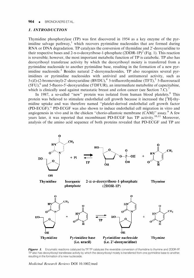
Thymidine phosphorylase (TP) was first discovered in 1954 as a key enzyme of the pyr-imidine salvage pathway,1 which recovers pyrimidine nucleosides that are formed duringRNA or DNA degradation. TP catalyzes the conversion of thymidine and 20-deoxyuridine totheir respective bases and 2-a-D-deoxyribose-1-phosphate (2DDR-1P)2 (Fig. 1). This reactionis reversible; however, the most important metabolic function of TP is catabolic. TP also hasdeoxyribosyl transferase activity by which the deoxyribosyl moiety is transferred from apyrimidine nucleoside to another pyrimidine base, resulting in the formation of a new pyr-imidine nucleoside.3 Besides natural 20-deoxynucleosides, TP also recognizes several pyr-imidines or pyrimidine nucleosides with antiviral and antitumoral activity, such as5-(E)-(2-bromovinyl)-20-deoxyuridine (BVDU),4 5-trifluorothymidine (TFT),5 5-fluorouracil(5FU),6 and 5-fluoro-50-deoxyuridine (50DFUR), an intermediate metabolite of capecitabine,which is clinically used against metastatic breast and colon cancer (see Section 7.C).7
In 1987, a so-called ‘‘new’’ protein was isolated from human blood platelets.8 Thisprotein was believed to stimulate endothelial cell growth because it increased the [3H]-thy-midine uptake and was therefore named ‘‘platelet-derived endothelial cell growth factor(PD-ECGF).’’ PD-ECGF was also shown to induce endothelial cell migration in vitro andangiogenesis in vivo and in the chicken ‘‘chorio-allantoic membrane (CAM)’’ assay.9 A fewyears later, it was reported that recombinant PD-ECGF has TP activity.10,11 Moreover,analysis of the amino acid sequence of both proteins revealed that PD-ECGF and TP are
Figure 1. Enzymatic reactions catalyzed byTP.TP catalyzes the reversible conversion of thymidine to thymine and 2DDR-1P.
TPalso has deoxyribosyl transferase activity by which the deoxyribosyl moiety is transferred from one pyrimidine base to another,
resulting in the formation of a newnucleoside.
904 K BRONCKAERS ETAL.
Medicinal Research Reviews DOI 10.1002/med

identical.12 This leads to the conclusion that the observed increased thymidine uptake was anartifact, caused by the TP activity of PD-ECGF. TP in cell supernatant hydrolyzes serum-derived thymidine, depleting the cells of this metabolite. When the cells are subsequentlyincubated with [3H]-thymidine, the cells treated with TP take up more of the radiolabelledthymidine than the control cells.10 Thus, TP is not a growth factor.
A third role for TP has also been described and in this context TP is called gliostatin.In 1992, gliostatin was extracted from human neurofibroma. This protein inhibits the growthof both astrocytes and glial tumor cells.13 Gliostatin is also shown to promote the survivaland neurite outgrowth of rat cortical neurons.14
Thus, TP, PD-ECGF, and gliostatin are all synonyms that refer to the same, identicalprotein. Throughout the literature TP and PD-ECGF are used interchangeably, while the useof the word ‘‘gliostatin’’ is restricted to the context of rheumatoid arthritis (RA) and neu-rological research.
2. STRUCTURE OF TP
In the mid-1970s TP was purified from both Escherichia coli and Salmonella typhimurium.15,16
Several years later, human TP was extracted from the amniochorion.17 The amino acidsequence of TP is highly conserved during evolution. For example, human TP shares 39%sequence identity with E. coli TP.18
TP functions as a homodimer consisting of two identical subunits (Fig. 2), with a dimermolecular mass ranging from 90 kDa in E. coli to 110 kDa in mammals.19,20 Detailedstructural information on TP was first provided in 1990 by Walter et al. who solved thecrystal structure of E. coli TP.21 This analysis revealed that each subunit contains a largemixed a-helical and b-sheet domain (a/b domain) separated from a smaller a-helical domain(a-domain) by a large cleft. The active site consists of the thymine-binding site in thea-domain and the phosphate-binding site across the cleft in the a/b domain. The finding thatboth sites were about 8 A apart immediately suggested that a hinge motion of one domainrelative to the other was necessary to generate a closed conformation of the enzyme con-taining a catalytically competent active site,21,22 as that seen in the crystal structure of therelated pyrimidine nucleoside phosphorylase from Bacillus stearothermophilus.23 This clos-ing/opening motion in the presence of substrate, product, and transition state has beensimulated using steered molecular dynamics.24
It took several more years to have the structure of human TP solved because manycrystallization trials failed to produce well-diffracting crystals.25 Thus, Spraggon et al.reported crystals of human TP for which, despite using a synchroton X-ray source, diffractionwas limited to 3.5 A resolution.26 Finally, in 2004, Norman et al. successfully solved at 2.1 Aresolution the structure of human TP in complex with the small and potent inhibitor 5-chloro-6-[1-(2-iminopyrrolidinyl)methyl] uracil (TPI) (see Section 6.A).25 Nonetheless, limited pro-teolysis with trypsin was found to be necessary and this treatment yielded a structure in whichamino acids 409 and 410 were missing and the loop formed by amino acid residues 405–416was disordered. In these crystals, TP was found as a dimer, with each monomer in the closed,active conformation, and TPI mimicking the substrate transition state. This work providedthe first structural insight into the binding mode of an inhibitor to a pyrimidine nucleosidephosphorylase.25 In 2006, El Omari et al. managed to determine the structure of un-proteolyzed human TP at 2.3 A resolution with the aid of the small-molecule inhibitor KIN59(see Section 6.B), which helped to obtain good quality diffracting crystals although it couldnot be located in the electron density map.27 The asymmetric unit revealed two dimers eachdisplaying the same inter-subunit contacts that were observed in the previous structure and, in
ROLEOF TP INCANCERDEVELOPMENTANDCHEMOTHERAPY K 905
Medicinal Research Reviews DOI 10.1002/med
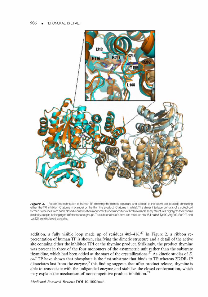
addition, a fully visible loop made up of residues 405–416.27 In Figure 2, a ribbon re-presentation of human TP is shown, clarifying the dimeric structure and a detail of the activesite containg either the inhibitor TPI or the thymine product. Strikingly, the product thyminewas present in three of the four monomers of the asymmetric unit rather than the substratethymidine, which had been added at the start of the crystallizations.27 As kinetic studies of E.coli TP have shown that phosphate is the first substrate that binds to TP whereas 2DDR-1Pdissociates last from the enzyme,3 this finding suggests that after product release, thymine isable to reassociate with the unliganded enzyme and stabilize the closed conformation, whichmay explain the mechanism of noncompetitive product inhibition.27
Figure 2. Ribbon representation of humanTP showing the dimeric structure and a detail of the active site (boxed) containing
either theTPI inhibitor (C atoms in orange) or the thymine product (C atoms in white). The dimer interface consists of a coiled coil
formedbyhelices fromeachclosed-conformationmonomer.Superimpositionofbothavailable X-ray structureshighlights theiroverall
similaritydespitebelonging todifferent spacegroups.Theside chainsofactive-site residuesHis116,Leu148,Tyr199, Arg202,Ser217, and
Lys221are displayedas sticks.
906 K BRONCKAERS ETAL.
Medicinal Research Reviews DOI 10.1002/med
3.EXPRESSION OF TP IN HEALTH AND DISEASE
A. The Physiological Role of TP
TP is found in many normal tissues and cells, with high levels in macrophages, stromal cells,glial cells, reticulocytes, some epithelia, tissues of the digestive tract (oesophagus and therectum), salivary gland, brain, bladder, spleen, lymph, and the lungs.28–30 Within the cell, TPis present in both the cytoplasm and the nucleus.28
Blood platelets are one of the richest sources of TP, which suggests a role for the enzymein wound healing. TP activity is also detected in plasma and serum, where its presence isprobably due to blood platelet damage or cell turnover.31
Furthermore, TP plays an important role in the female reproductive cycle. Largequantities of TP are found in the placenta, where two alternative forms of the protein aredetected. One is a 27 kDa splice variant of TP,32 while the other form contains five additionalamino acids on the N-terminus and is processed at Thr-6 instead of Ala-11.33 Whether thesestructural differences also result in functional differences is not clear. High amounts of TPwere also discovered in the endometrium, which undergoes extensive angiogenesis duringeach menstrual cycle. TP shows a characteristic pattern of distribution dependent on thephase of the menstrual cycle: TP expression moves from stroma to epithelium as the cycleprogresses34 and is inversely correlated with oestradiol concentrations.35 Endometrial TPexpression was also raised by human chorionic gonadotropin36 and by a combination ofprogesterone and transforming growth factor b1.34 A marked increase of TP was also de-tected in decidualized endometrium.37 During the first trimester of pregnancy, TP is found inthe trophoblast together with vascular endothelial growth factor (VEGF), which indicatesthat both factors play an active role during gestation.32
B. TP in Inflammatory Diseases
Various studies indicate that TP is also involved in a wide variety of chronic inflammatorydiseases (see Table I). This may be explained by the fact that (i) inflammatory cells such asmacrophages contain large amounts of TP (cf. Section 3.D) and (ii) inflammatory cytokines(such as interleukin-1 and tumor-necrosis factor-a) induce TP expression (see Section 5).
TP levels are raised in psoriasis.38,39 Accordingly, a 20-fold increase in TP activity wasfound in psoriatic lesions.39 In inflammatory bowel disease, strong TP expression wasobserved, predominantly in macrophages and fibroblasts of the inflamed colonic mucosa andthe grade of expression augmented with an increasing grade of inflammation.40,41 In addition,TP was found in the endothelial cells of the inflamed colonic mucosa.40,41 Furthermore, TP isupregulated in chronic glomerulonephritis (a renal disease characterized by inflammation ofthe glomeruli) where it probably plays a critical role in the progression of interstitial fibrosis.42
Moreover, TP is expressed in atherosclerosis. Macrophages, foam cells, and giant cells fromboth aortic and coronary plaques were found to be immunoreactive for this angiogenic factor,suggesting that TP may play a role in the pathogenesis of atherosclerosis.43
TP is also implicated in RA. Higher levels of TP were found in the synovial fluid or seraof patients with RA than in patients with osteoarthritis or normal healthy individuals andserum TP was found to be a useful clinical marker for RA.44–46 Furthermore, the intra-articular injection of recombinant TP into the knees of rabbits induced RA-like synovitis.47
Both wild-type TP or mutant (K115E) TP, which lacks enzymatic activity, caused this effect,indicating that not the enzymatic activity of TP but rather the protein itself is implicated inthe pathology of RA.47 Further studies revealed that TP augments its own synthesis throughan autocrine mechanism in fibroblast-like synoviocytes (FLS).48 TP also induced the extra-cellular secretion of matrix metalloproteinase-1 (MMP-1) and MMP-3, which are the major
ROLEOF TP INCANCERDEVELOPMENTANDCHEMOTHERAPY K 907
Medicinal Research Reviews DOI 10.1002/med

triggers of cartilage degeneration.48,49 TP was also found to upregulate both mRNA andprotein levels of VEGF, suggesting that both factors have synergistic effects on angiogenesisin RA.50
C. The Involvement of TP in MNGIE
Mitochondrial neurogastrointestinal encephalomyopathy (MNGIE) is an autosomal re-cessive human disorder associated with multiple deletions of skeletal muscle mitochondrialDNA.51 This disease is characterized clinically by ptosis, progressive external ophthalmo-plegia, gastrointestinal dysmotility, thin body habitus, peripheral neuropathy, myopathy,leukoencephalopathy, and lactic acidosis. Loss-of-function mutations of the TP gene wereidentified as the possible cause of this disease.52,53 In MNGIE, the severely reduced TPenzyme activity leads to an increase in the plasma and tissue thymidine and 20-deoxyuridinelevels.53–55 This may cause unbalanced mitochondrial nucleoside and nucleotide pools, whichlead to impaired mitochondrial DNA replication and repair. Therefore, therapies that de-crease thymidine levels may be beneficial to MNGIE patients.53
In order to unravel the role of TP in MNGIE, mice deficient in the TPgene were generated. As mouse uridine phosphorylase (UP) may also cleave thymidine,TP�/�UP�/� double knockout mice were constructed. In these TP�/�UP�/� mice, all TPactivity was abrogated and the plasma thymidine levels were 5-fold higher than in wild-typemice. Surprisingly, no alterations in mtDNA or pathological changes in the muscles of theseknockout mice could be observed.56,57 Only in the brain, mitochondrial DNA depletion,respiratory chain defects and histological alterations could be detected.57 The brain-specificphenotype in TP�/�UP�/�mice may be due to the relatively modest increases in thymidine or20-deoxyuridine levels in mutant mice (5-fold increase in mice versus 100-fold increase inhumans). Another possible explanation is the shorter lifespan of mice compared to humansbecause in humans the average age-at-onset of symptoms of MNGIE is 18.7 years.58
Nevertheless, other studies suggest that functional mutations of the TP gene are notsufficient to induce MNGIE. Kumagai et al. postulated that TP gene mutation is not theprimary cause of this mitochondrial disease because TP mutations are also found in un-related healthy individuals.59 Examination of chromosome 22q13.32 where the TP gene islocated, revealed that exon 10 of TP overlaps with the SCO2 gene,56 which is a cytochrome coxidase (COX) assembly gene.60 Mutations in SCO2 have been reported to cause severe COX
Table I. Biological Functions of TP
References
Physiological roleSalvage of pyrimidines 1
Female reproductive cycle 32–37
Wound healing 31
Pathological role
Cancer 63–205
Inflammatory diseases
Reumatoid arthritis 44–50
Atherosclerosis 43
Psoriasis 38, 39
Inflammatory bowel disease 40, 41
Chronic glomerulonephritis 42
MNGIE 51–58
908 K BRONCKAERS ETAL.
Medicinal Research Reviews DOI 10.1002/med

deficiency in skeletal muscle leading to mitochondrial disorders characterized by hyper-trophic cardiomyopathy and encephalopathy.61 Thus, more investigations are needed toclarify the exact role of TP deficiency in MNGIE development.
So far no vascular abnormalities have been reported in MNGIE patients nor inTP�/�UP�/� mice.56,57,62 These data suggest that TP is not of fundamental importance fordevelopmental angiogenesis.
Figure 3. Correlationof TPexpression invarious cancerswith the clinicopathological factorsmicrovesseldensity, tumor stage,
and grade.
ROLEOF TP INCANCERDEVELOPMENTANDCHEMOTHERAPY K 909
Medicinal Research Reviews DOI 10.1002/med
D. TP Overexpression in Cancer
Increased TP expression in tumor tissues compared to corresponding nonneoplastic tissuewas found in breast,63 bladder,64,65 gastric,29,66,67 colorectal,29,68 lung,68,69 esophageal,68,70
and cervical68,71 cancers but not in cancers of the liver,68,72 common bile duct,68 and thethyroid.68 TP expression is not only upregulated in solid tumors, elevated levels of TP werealso observed in lymph nodes of patients with classical Hodgkin lymphoma where TP levelsincreased with disease progression.73 Recently, tumor-reactive T cells from a patient withrelapsed multiple myeloma (who was successfully treated with donor lymphocyte infusionafter allogeneic stem cell transplantation) were found to be directed against TP.74 These dataidentify TP as a potential target for the immunotherapy of hematological tumors.
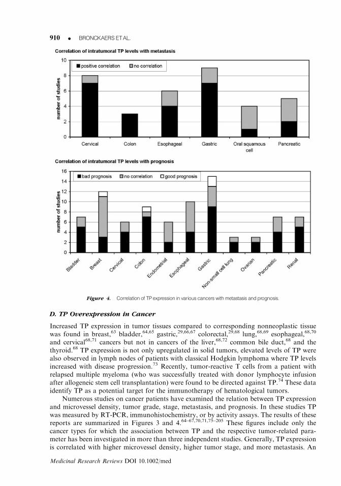
Numerous studies on cancer patients have examined the relation between TP expressionand microvessel density, tumor grade, stage, metastasis, and prognosis. In these studies TPwas measured by RT-PCR, immunohistochemistry, or by activity assays. The results of thesereports are summarized in Figures 3 and 4.64–67,70,71,75–205 These figures include only thecancer types for which the association between TP and the respective tumor-related para-meter has been investigated in more than three independent studies. Generally, TP expressionis correlated with higher microvessel density, higher tumor stage, and more metastasis. An
Figure 4. Correlation of TPexpression in various cancers withmetastasis and prognosis.
910 K BRONCKAERS ETAL.
Medicinal Research Reviews DOI 10.1002/med

association of TP with tumor grade is evident in bladder, cervical, and renal cell cancer, butnot in the other investigated cancers. Furthermore, in most cases, TP appeared to beassociated with poor prognosis, although there are conflicting reports for some cancers.For example, seven of the nine studies on colon cancer reported a significant correlationbetween TP and bad prognosis, while Saito et al. demonstrated that TP is associated withgood prognosis.142 These discrepancies might be caused by differences in the histological typeof cancer, stage (early versus advanced stage of disease), number of patients examined,assay for TP and different methodology for the evaluation of the immunohistochemistryresults.142,206
Tumors are heterogeneous tissues consisting of unknown variable contributions of tu-mor, stromal, and infiltrating cells. Besides tumor cells, also endothelial cells, fibroblasts,lymphocytes and especially tumor-associated macrophages (TAM) express TP.138 TAM arethought to play a key role in stimulating tumor growth and metastasis through the pro-duction of various growth factors, proteinases, chemokines, and cytokines.207 High levelsof TP have been demonstrated in TAM of melanoma,208 gastric,100,140 glioblastoma,110
breast,135,151 colon,137,209 astrocytic,178 uterine endometrial,104 and prostate210 cancer.In gastric adenocarcinoma,100 astrocytic tumors,178 breast,135 and uterine endometrialcancer,104 TP expressed in macrophages has been suggested to be correlated with microvesseldensity and to play an important role in tumor invasiveness.
Elevated TP levels are not only found in the tumor tissue but also in the plasma of cancerpatients.211,212 Already in 1977, Pauly et al. demonstrated that cancer patients had muchhigher TP activity in the plasma than healthy individuals.213 He also reported that tumor-bearing animals have elevated TP activity in their ascites and plasma.214 More recent dataindicate that plasma TP concentrations in cancer patients may have a prognostic value. Inuterine cervical cancer high serum TP levels correlate with clinical stage, tumor size, lymphnode metastasis, and an extremely poor prognosis.163 High TP concentrations in the bloodare also associated with depth of tumor invasion and poor response to treatment in patientswith esophageal squamous cell carcinoma.215 Furthermore, in patients with colorectal can-cers, the TP serum level is suggested to be a novel marker to predict occurrence of hema-togenous metastasis.216
4. TP AND TUMOR DEVELOPMENT
A. Role of TP in Angiogenesis (Fig. 5)
Angiogenesis, the formation of new capillaries from existing blood vessels, is of fundamentalimportance in several physiological processes, such as embryonic development, woundrepair, and reproduction (see Table II). It is a multistep process that involves degradation ofthe surrounding extracellular matrix (ECM) by proteases, endothelial cell migration, pro-liferation, and differentiation into mature blood vessels. Cytokines, growth factors, growthfactor receptors, enzymes (like TP), components of the ECM, and adhesion molecules eachhave their own specific role in this well-coordinated process.217–220 The equilibrium betweenangiogenic and angiostatic proteins, the so-called angiogenic balance, in the microenviron-ment controls the rate of new blood vessel formation.220 Alteration of this angiogenic bal-ance, for example by the uncontrolled release of angiogenic regulators, can lead to severalpathological conditions including inflammation, RA, tumor growth, and metastasis.218,220
TP induces endothelial cell migration and tube formation in vitro.9,63,221 This enzyme wasalso shown to stimulate angiogenesis in the CAM9,221,222 and in several in vivo models, such asthe freeze-injured skin graft,63 rat corneal,223 and mouse dorsal air sac223 assays and in gelatine
ROLEOF TP INCANCERDEVELOPMENTANDCHEMOTHERAPY K 911
Medicinal Research Reviews DOI 10.1002/med

sponges subcutaneously implanted in rats or mice.9,63,221,224 Recently, proteomic research hasidentified TP as a key regulator of the angiogenic potential of endothelial progenitor cells(EPC).225 EPC are bone marrow-derived cells, which can differentiate into endothelial cells and
Table II. Molecular Mediators of TP and/or 2DDR in Angiogenesis
Molecular mediators Cell type References
MMP-1 Human bladder RT-112 cells 48, 234
RA-associated synoviocytes
MMP-2 Cervical carcinoma cells 256
MMP-3 RA-associated synoviocytes 48
MMP-7 Human PC-3 prostate cancer cells 258
KK47 bladder cancer cells
MMP-9 Human PC-3 prostate cancer cells 257, 258
KK47 bladder cancer cells
KB epidermoid cells
VEGF Human bladder RT-112 cells 234
Integrins a5b1 and avb3 HUVEC 225, 240
EPC
IL-8 Human bladder RT-112 cells 234
P-selectin HUVEC 260
Figure 5. The role of TP in tumor progression.TPexpression in tumors canbeupregulatedby various stress-inducible factors,
suchas radio- andchemotherapy, inflammatory cytokines, hypoxia, and lowpH.Withinthe tumor tissue,TP is found inboth tumor-
associatedmacrophages and tumorcells.TPand itsmetabolite 2DDR stimulate tumorgrowthby promotingangiogenesis.TPand
2DDR stimulate the secretion and/or expression of the angiogenic molecules VEGF, IL-8, P-selectin, and various MMPs. 2DDR,
which can diffuse outside of the cell, also directly induces endothelial cell migration through activation of the integrins a5b1 andavb3.TPand 2DDRmayalso induce tumor progression by protecting the tumoragainst apoptosis induced by hypoxia, Fas,DNA-
damage, andmicrotubuli-interfering agents.Conversely, tumors do not always profit from elevated TP levels asTP plays a crucial
role in the activation of chemotherapeutic drugs such as capecitabine. [Color figure can be viewed in the online issue, which is
available at www.interscience.wiley.com.]
912 K BRONCKAERS ETAL.
Medicinal Research Reviews DOI 10.1002/med
contribute to the repair of blood vessels after a myocardial attack.226,227 These cells thus offer apromising strategy for the treatment of various cardiovascular diseases.228 Furthermore, be-cause these cells have the capacity to home to, and invade, tumor tissues, they also showpotential as a target for gene therapy against malignant tumors.229
Numerous experimental data indicate that the catalytic activity of TP is indispensable forits angiogenic properties: (i) unlike wild-type TP, TP mutants that lack enzymatic activity didnot induce the formation of new blood vessels in the gelatine sponge assay;63,224 (ii) theangiogenic activities of TP could be abolished by using TP-directed neutralizing antibodies,63
by addition of a specific TP inhibitor such as 5-amino-6-chlorouracil224 or by downregulatingTP by siRNA;227 and (iii) 2-deoxy-D-ribose (2DDR), which is a degradation product of theTP-metabolite 2DDR-1P, also induces endothelial cell migration and angiogenesis.230–232
Besides 2DDR, other metabolites of TP have been shown to possess angiogenic properties invitro. b-amino-iso-butyric acid, which is a degradation product of thymine, stimulated tubeformation in the rat aortic assay,233 while 2DDR-1P induced endothelial cell migra-tion.230,231,234 Nevertheless, 2DDR is considered to be responsible for the angiogenic activ-ities of TP. de Bruin et al. showed that 2DDR-1P produced by TP-overexpressing Colo320colon carcinoma cells is rapidly converted to 2DDR.235 Therefore, it can not be excluded thatthe biological activity obtained for 2DDR-1P is a result of the conversion of 2DDR-1P to2DDR. Moreover, Hotchkiss et al. showed that the conversion of 2DDR-1P to 2DDR isindispensable for the induction of endothelial cell migration as addition of an alkalinephosphatase inhibitor, which blocks the dephosphorylation of 2DDR-1P, completely abro-gated the chemotactic effects of 2DDR-1P.230 Moreover, a neutralizing antibody to TP hadno effect on endothelial cell migration stimulated by TP-overexpressing cells, even thoughthis antibody completely inhibited migration mediated by recombinant, extracellular TP.These studies demonstrate that TP-mediated endothelial cell migration relies on the in-tracellular catabolism of thymidine and subsequent extracellular release of 2DDR, whichforms a chemotactic gradient.230 In spite of this, the enzyme purine nucleoside phosphor-ylase, which also produces 2DDR, has never been reported to possess angiogenic properties.
In several ways, TP is a very exceptional angiogenic molecule. Indeed, usually, angio-genic factors are released into the extracellular space to activate endothelial cells. However,TP lacks an amino-terminal hydrophobic leader sequence required for cell secretion and istherefore mainly found inside the cell.9 Nevertheless, some tumor cell lines such as theepidermoid carcinoma A431 and stomach cancer MKN74 cell lines do release the proteininto the cell culture medium.30 Also cytokine-treated FLS have been shown to actively secreteTP.46 The mechanism behind the secretion of TP is possibly a posttranslational processwhereby serine residues of TP are covalently linked to phosphate groups of nucleotides,leading to the formation of a nucleotidylated protein that can be secreted.236 While TPmostly remains inside the cell, its metabolite 2DDR is able to cross the cell membrane andexert its biological effects on other cells. Furthermore, angiogenic factors usually bind to aspecific cell surface receptor, which induces a signal transduction cascade followed by abiological response of the cell. However, mammalian cells do not seem to have a receptor forTP nor for 2DDR. Several bacterial receptors for carbohydrates, including for D-ribose, havebeen identified that play a role in chemotaxis.237,238 These receptors are histidine kinases,which is a family of receptors that is found in prokaryotes and eukaryotes but not in theanimal kingdom.239 Thus, TP and 2DDR most likely induce angiogenesis through a non-receptor-mediated mechanism.
1. TP stimulates endothelial cell migrationThe molecular mechanisms through which TP and 2DDR stimulate endothelial cell migrationin vitro are not completely understood. Hotchkiss et al.240 revealed that TP and 2DDR affect
ROLEOF TP INCANCERDEVELOPMENTANDCHEMOTHERAPY K 913
Medicinal Research Reviews DOI 10.1002/med

endothelial cell migration through activation of integrins and their downstream signallingpathways. In human umbilical vein endothelial cells (HUVEC), it was shown that both TP and2DDR stimulate the formation of focal adhesions and the phosphorylation of tyrosine 397 offocal adhesion kinase (FAK). FAK is a nonreceptor protein–tyrosine kinase that is recruited tofocal adhesions by integrin engagement with the ECM. Thus, FAK plays an important role inendothelial cell attachment and migration.240 Hotchkiss et al. also demonstrated that VEGF,TP, and 2DDR all stimulate HUVEC migration, although through different integrins. TP- and2DDR-mediated endothelial cell migration and FAK phosphorylation were blocked by anti-bodies against either integrin a5b1 or avb3, whereas VEGF-induced migration was only in-hibited by the avb3 antibody.
225,240 The cell surface expression of integrin a5b1 and the cellularexpression of integrin b3 were increased by TP and 2DDR.
Also other investigators tried to unravel the signalling pathways through which 2DDRactivates endothelial cells. Seeliger et al. demonstrated that rapamycin completely abrogates2DDR-mediated HUVEC migration and tube formation in the rat aortic ring assay, prob-ably by blocking 2DDR-induced p70/s6 kinase activation.241 The intracellular p70/s6 kinaseis known to induce endothelial cell migration after activation of the mammalian target ofrapamycin.242 It has been shown that p70/s6 kinase activation is induced after interaction ofintegrins with ECM components and that this activation requires FAK.243,244
2. TP induces the expression and/or secretion of other angiogenic factorsVarious studies have demonstrated that TP and 2DDR promote the expression and secretionof several angiogenic factors (see Table II). Human bladder carcinoma cells transfected withTP (RT112-TP) secrete higher amounts of VEGF, interleukin-8, and MMP-1 than mock-transfected RT112 cells in the presence of thymidine.234 RT112-TP cells incubated withthymidine also showed an elevated expression of heme oxygenase-1 (HO-1), HO-1 is anenzyme that catalyzes the degradation of heme to carbon monoxide, iron, and biliverdin.245
The expression of HO-1 can be induced by hypoxia, cytokines, and several angiogenic factorssuch as VEGF and stromal cell derived factor-1 (SDF-1).246–249 Recent data indicate thatHO-1 also possesses proangiogenic properties: it promotes endothelial cell proliferation,protects endothelial cells from apoptosis, and induces the secretion of several angiogenicmediators such as VEGF.248–250 Among the different end products of the enzyme reaction ofHO-1, carbon monoxide is proposed to be responsible for the angiogenic actions ofHO-1,248,249 although recently also biliverdin has been reported to stimulate the synthesis ofangiogenic mediators.251 Not only in RT112-TP cells an elevated expression of HO-1 oc-curred, also in TP-overexpressing vascular smooth muscle cells (VSMC) an induction ofHO-1 was observed.252 An excess of thymine, which acts as a scavenger for the formed2DDR-1P, prevented the induction of HO-1. Brown et al.234 suggested that 2DDR is astrongly reducing sugar that may generate oxygen radical species during the early stages ofprotein glycation. It was hypothesized that 2DDR binds to an amino group (preferentially ata lysine, arginine or the N-terminal amino acid) of a protein during a nonenzymatic reaction,the so-called Maillard reaction. This may lead to the formation of a Schiff base, which canthen rearrange to an a-hydroxyketone. This unstable reaction intermediate autoxidizesduring which reaction free oxygen radicals are produced. Thus, through the formation of2DDR, TP may induce oxidative stress in TP-overexpressing tumor cells causing these cellsto secrete angiogenic factors, such as VEGF.234 A recent study demonstrates that TP mayinduce VEGF secretion through another mechanism. Transcription of VEGF is known to bedriven by hypoxia-inducible factor-1a (HIF-1a). Under hypoxic conditions, the transcriptionfactor HIF-1a is upregulated and increases the expression of several target genes by forminga dimer with HIF-1b, which recognizes the hypoxia responsive elements in the promoter
914 K BRONCKAERS ETAL.
Medicinal Research Reviews DOI 10.1002/med

region.253 In RT112 cells, TP activity augments the levels of HIF-1a during in vitro hypoxiaand TP and HIF-1a acted together to induce VEGF secretion.254
Not only MMP-1, but also other MMPs have been shown to be upregulated by TP.MMPs degrade the ECM surrounding tumor and endothelial cells and therefore promotetumor cell invasion, migration, and metastasis.255 In several cervical carcinoma cell lines, TPexpression correlated significantly with the mRNA level of MMP-2 and with cell invasionin vitro.256 Human epidermoid carcinoma cells (KB) transfected with TP expressed moreMMP-9,257 while the TP-overexpressing PC-3 prostate and KK47 bladder cancer cells hadhigher levels of MMP-7 and MMP-9 under hypoxia than the mock-transfected controlcells.258 TP induced the expression and extracellular secretion of MMP-1 and MMP-3 incultured human RA-associated synoviocytes.48 Also clinical data provide evidence for acorrelation between TP and MMP expression. In breast cancer, TP was associated withhigher levels of activated MMP-9.259 Moreover, in human bladder cancers, TP correlatedsignificantly with the expression of MMP-1, MMP-9, and urokinase plasminogen acti-vator,258 which also plays a role in ECM degradation.
cDNA microarray analysis showed that the cell adhesion molecule P-selectin isupregulated in HUVEC treated with TP.260 A correlation between tumor cell P-selectinexpression and TP was also assessed in human breast cancers by immunohistochemistry.260
P-selectin is a vascular adhesion molecule mostly found on endothelial cells that mediates theinteractions of endothelial cells and leukocytes during inflammation.261 It is also believed toplay a vital role in tumor growth and metastasis, including the promotion of angiogenesis262
and the movement of tumor cells through the endothelial cell layer.261
A study on breast cancers revealed that angiopoietin-1 (Ang-1) is inversely related toTP.263 Ang-1, which is a ligand of the tyrosine kinase receptor Tie-2,264 is a survival factor forendothelium and stabilizes vascular networks.265–267 It maintains the integrity of the capil-laries by recruiting and stabilizing nonendothelial support cells such as pericytes.265 Thus, theloss of Ang-1 results in pericyte–endothelial cell contact destabilization, which possibly en-ables TP and 2DDR to act on the ‘‘free’’ endothelial cells and induce angiogenesis.263
3. TP is a promising target for the treatment of vascular obstructive diseasesAs TP is an angiogenic molecule, its upregulation could be applied to treat diseases caused byreduced angiogenesis or a disturbed blood flow. In 2005, Li et al.268 showed that gene therapyusing TP is an effective treatment against chronic myocardial ischemia in dogs. The plasmid-mediated gene transfer of TP stimulated angiogenesis and arteriogenesis in chronically is-chemic myocardium, decreased the infarct size, restored the myocardial blood flow, andimproved myocardial function.268 This experimental gene therapy also proved to have a long-term beneficial effect in the treatment of chronic ischemic myocardium.269
As VSMC play an important role in vessel maturation during angiogenesis, and de-regulated growth or motility of VSMC contributes to the pathogenesis of vascular obstructivediseases (such as ischemia), the effect of TP on VSMC migration and proliferation wasinvestigated. TP-overexpressing VSMC migrated and proliferated more slowly than mock-transfected VSMC. The decreased VSMC proliferation was correlated with TP-inducedHO-1 expression. In TP-overexpressing VSMC the cyclin-dependent kinase inhibitor(p27KIP1) was upregulated and the cell cycle was arrested at the G1 phase. Thus, surprisingly,TP inhibits proliferation and migration of VSMC, while it stimulates the migration ofendothelial cells. This apparently conflicting role of TP may be due to the opposite effect ofHO-1 in endothelial cells and VSMC. As described above, TP induces HO-1 both in en-dothelial cells and VSMC. HO-1 has been shown to induce endothelial cell proliferation andmigration,249,250 while it inhibits VSMC growth.270 In balloon-injured rat carotid arteriesadventitial TP gene delivery significantly reduced neointimal VSMC migration and neointima
ROLEOF TP INCANCERDEVELOPMENTANDCHEMOTHERAPY K 915
Medicinal Research Reviews DOI 10.1002/med

formation.252 Furthermore, adventitial delivery of the TP gene also prevented intimalhyperplasia of vein grafs in rabbits.271 TP thus reduces the neointimal mass and inhibitsfurther neointimal outgrowth and is therefore a promising target in occlusive vascular diseases.
B. TP Induces Metastasis
TP was found to increase the metastatic potential of several experimental and human tumors.Moreover, in various cancers high TP expression correlates with metastasis (Fig. 4). Takaoet al. demonstrated that TP-expressing KB carcinoma cells show more basement membraneinvasion than their mock-transfected counterparts.272 Intrasplenic injection of KB/TP cells innude mice resulted 4 weeks after injection in significantly more metastatic nodules in thelivers than injection with KB/CV cells.272,273 The stimulation of metastasis by TP-over-expressing cells could be dramatically inhibited by the TP inhibitor TPI or by 2-deoxy-L-ribose (2DLR), a stereoisomer of 2DDR.272,273 Finally, in mice xenografted with the humanmelanoma cancer cell line A-07, lung colonization and spontaneous metastasis were inhibitedby treatment with neutralizing antibodies against TP.274
C. TP Protects Cancer Cells Against Apoptosis
Moghaddam et al. reported in 1995 that TP-expressing breast carcinomas have a highergrowth rate without an increase in microvessel density than breast cancers that do notexpress TP.63 Furthermore, a clinical study of human colon carcinomas showed that TP is aprognostic factor independent of angiogenesis.121 These data suggest that TP may stimulatetumor growth through a mechanism different than angiogenesis. Uchimiya et al.223 in-vestigated in a mouse model the anti-apoptotic effect of TP by injecting KB/TP cells ormock-transfected KB cells (KB/CV) into nude mice.223 The apoptotic index in KB/TP tu-mors was significantly lower than in KB/CV tumors, indicating that TP protects cells againstapoptosis.223,273,275 Also numerous clinical studies give evidence for the anti-apoptotic effectof TP. TP expression is correlated with a reduction in apoptotic cells in colon,144 gastric,276
esophageal,82,184 ovarian,277 and oral squamous cell175 carcinomas but not in cervical can-cers169,184 or in astrocytic tumors.165,178
A correlation between TP expression and apoptosis was first demonstrated in vitro byusing human epidermoid carcinoma KB cells. KB cells transfected with TP (KB/TP) wereresistant to hypoxia-induced apoptosis. This advantage was abrogated when the cells weretreated with TPI, which inhibits the enzymatic activity of TP, leading to the conclusion thatthe enzymatic activity of TP is indispensable for protection against hypoxia-induced apop-tosis.278 Also the metabolites of the TP reaction, 2DDR, and thymine, partially preventedhypoxia-induced apoptosis in KB cells. A potential molecular basis for the inhibition ofhypoxia-induced apoptosis was first suggested by Ikeda et al.279 In human leukemia (HL-60)cells, 2DDR inhibited numerous hypoxia-induced pro-apoptotic events, such as activation ofcaspase 3 and 9, mitochondrial cytochrome c release, loss of mitochondrial transmembranepotential, phosphorylation of p38 mitogen-activated protein kinase, and downregulation ofthe anti-apoptotic proteins Bcl-2 and Bcl-xl.279,280 Furthermore, 2DDR also prevented theupregulation of the transcription factor HIF-1a.279 Also in the human leukemia cell line(Jurkat cells) overexpression of TP inhibited the upregulation of HIF-1a and the HIF-1a-inducible, pro-apoptotic factor BNIP3.281 This is in contradiction with the results of Brownet al. who showed that TP-activity augments the levels of HIF-1a in RT-112 cells during invitro hypoxia.254 It is however possible that different tumor cell lines demonstrate con-siderable variation in induction of HIF-1a when subjected to TP. Another explanation mightbe that in HL-60 cells, 2DDR was added extracellularly, while in RT-112 cells the effect wasobserved in cells transfected with TP.
916 K BRONCKAERS ETAL.
Medicinal Research Reviews DOI 10.1002/med
Besides hypoxia-induced apoptosis, TP suppresses apoptosis induced by Fas, micro-tubule-interfering, and DNA damage-inducing agents such as cisplatin (see Table III).282–286
TP exerts these protective effects independent of its enzymatic activity.282,283,286 Further-more, in the presence of UV-light, which causes DNA damage, KB/TP cells had higheramounts of both Akt and phosphorylated Akt than KB/CV cells. Akt activation was sig-nificantly decreased by phosphatidylinositol 3 kinase (PI3K) inhibitors, suggesting that theAkt/PI3K pathway is implicated in TP-induced resistance against DNA damage.284 Finally,it was demonstrated that TP protects cells against Fas-induced apoptosis by inhibiting cas-pase-8 cleavage, Bcl-2 phosphorylation, and cytochrome c release.285
5. REGULATION OF TP EXPRESSION
The TP gene is localized on chromosome 22q13287 and is composed of ten exons dispersedover a 4.3 kb region. The TP promoter lacks a ‘‘TATA’’ and a ‘‘CCAAT’’ box, sequencesrecognized by RNA polymerase II, prevalent in most eukaryotic genes.288 Instead, it containsa cluster of six to nine SP1-binding motifs, just upstream of the transcription start site.288,289
The transcription factor SP1 is activated by protein kinase A, which is in turn activated bycyclic adenosine monophosphate (cAMP).290 SP1 sites are also involved in the transcriptionof VEGF.291 Indeed, various studies confer the tendency for VEGF and TP to be co-ex-pressed. A significant correlation was found between expression of VEGF and TP inbreast,118,124,292 colorectal,293 non-small cell lung,69,133 head and neck squamous cell,294
endometrial,79 astrocytic,165 lung,295 and cervical296 carcinomas. The co-expression of TPand VEGF may also be explained by the fact that TP increases the expression of VEGF byinducing oxidative stress or by upregulating the transcription factor HIF-1a, as described inSection 4.A.2. However, no relation between VEGF and TP expression was found in gas-tric,80 gallbladder,297 bladder,298 and esophageal squamous cell191 carcinomas.
Besides SP1 sites, the promoter region of the TP gene contains other transcription factor-binding sites, such as an interferon-stimulated response element (ISRE)299 and a g-activatedsequence-like element (GAS).300 In HT29 colon carcinoma cells, interferon-g (IFN-g) inducesTP expression through these ISRE sequences,299 while in U937 monocytes IFN-g promotesTP expression by increasing the binding of the signal transducer and activator of tran-scription 1 (STAT1) to the GAS sequence, suggesting that IFN-g induces TP expression byactivation of the Janus kinase (JAK)/STAT pathway.300 This observation is in line with thefindings of Yao et al. who reported that the JAK inhibitor AG-490 blocks both IFN-inducedSTAT1 phosphorylation and TP expression in glioblastoma cells.301 Furthermore, in clini-cally resected colon carcinomas eight of nine tumors tested had both high STAT1 proteinlevels and TP activity.302 The IFN-induced TP gene expression is also regulated by post-transcriptional mechanisms. Schwartz et al. demonstrated that IFN induces an increase in TPmRNA and that the TP mRNA levels remained elevated for up to 72 hr, suggesting that IFNpromotes TP mRNA stability. Analysis of the TP mRNA sequence revealed the presence of apyrimidine-rich sequence at the 30- end that is similar to a motif that has been reported toincrease the mRNA stability in other genes such as VEGF.299
Interferons are not the only inflammatory cytokines that upregulate TP. Also tumornecrosis factor-a (TNF-a) and interleukins induce the expression of TP.46,289,303,304 In THP-1monocytes TP expression was increased by TNF-a and this induction could be mimicked byan antibody against TNF-a receptor 2.303,304 It was also shown that the TNF-a-inducedincrease in TP mRNA was inhibited by an inhibitor of transcription factor nuclear factor-kB(NF-kB).304 Correspondingly, de Bruin et al. showed that prolonged exposure of humanmacrophage THP1 and U937 cells to sulfasalazine, an anti-inflammatory drug and inhibitor
ROLEOF TP INCANCERDEVELOPMENTANDCHEMOTHERAPY K 917
Medicinal Research Reviews DOI 10.1002/med

of NF-kB, resulted in downregulation of TP and IL-8 along with elimination of their in-duction by TNF-a and IFN-g.305 Thus, the transcription factor NF-kB is involved in theinduction of TP expression.
In cultured FLS, TNF-a, IL-1a, IL-1b, IL-6, and IL-8 stimulated the expression ofTP.46,306 The IL-1b-induced expression of TP was inhibited by treatment with the anti-rheumatic drugs dexamethasone and aurothioglucose, while methotrexate or sulfasalazinehad no influence on the TP levels.306 This suggests that dexamethasone and aurothioglucosemay inhibit RA through inhibition of TP expression. Also in OUMS-27 chondrosarcomacells the secretion of TP was augmented by IL-1b in a dose-dependent manner. This effectcould be blocked by selective inhibition of the p38 mitogen-activated protein kinase (p38MAPK) pathway.49
Cancer treatments, such as X-ray irradiation and chemotherapeutic agents (includingpaclitaxel, docetaxel, doxorubicin, oxaliplatin, cyclophosphamide, and mitomycin C) havebeen reported to dramatically increase the tumor TP levels (see Table III). This is probablydue to the fact that these therapies induce cytokines (such as TNF-a, IFN-g, IL-1) thatstimulate TP expression.302,307–309
Recently, evidence was provided that TP expression is also regulated at the transcriptionlevel by epigenetic modifications, such as methylation and histone deacetylation.310 High
Table III. Molecular Mechanisms of the Anti-Apoptotic Effect of TP
Apoptosis
induced by
Involvement
of catalytic
activity of TP Cell type Anti-apoptotic actions of TP Ref.
Hypoxia Yes KB epidermoid
cells
– 278
HL-60 leukemia
cells
Inhibition of 279, 280
Caspases 3 and 9 activation
Cytochrome c release
p38 MAPK phosphorylation
Bcl-2 and Bcl-xl downregulation
HIF-1a upregulation
Jurkat leukemia
cells
Prevention of upregulation of 281
HIF-1aBNIP3
DNA damage caused
by cisplatin
No Jurkat leukemia
cells
Inhibition of 282
Caspases 3 and 9 activation
Cytochrome c release
DNA damage caused
by UV
No KB epidermoid
cells
Akt activation 284
Fas No KB epidermoid
cells
Inhibition of 285
Caspase-8 cleavage
Bcl-2 phosphorylation
Cytochrome c release
Microtubule
interfering agents
No Jurkat leukemia
cells
Suppression of 283
Bcl-2 phosphorylation
FasL expression
918 K BRONCKAERS ETAL.
Medicinal Research Reviews DOI 10.1002/med
expression of TP was associated with complete demethylation of the CpG dinucleotides locatedin the TP promoter, which was demonstrated in breast carcinoma SKBR-3 cells. Low TPexpression was correlated with hypermethylation of the CpG islands as in DLD-1 coloncarcinoma cells. In DLD-1 cells, the expression of TP could be activated by demethylation with5-aza-20deoxycytidine and to a lesser extent by histone deacetylation with trichostatin-A.310
Finally, also microenvironmental stress conditions, such as hypoxia and low pH stimulatethe expression of TP.311 This clarifies why TP can be found in those parts of the tumor that areadjacent to necrotic areas or after occlusion of the tumor blood supply.311 In conclusion, stress-related factors, such as hypoxia and cytokines, induce the expression of TP, which indicatesthat this enzyme is a product of inflammation or microenvironmental stress.
6. TP INHIBITORS
Already in 1971, Judah Folkman postulated that tumor growth is angiogenesis-dependentand that tumor development and metastasis could be abolished by blocking the tumor bloodsupply.312 Currently, anti-angiogenic drugs, such as the VEGF-antibody bevacizumab(Avastin, Genentech/Roche, Basel Switzerland) and the kinase inhibitors sorafenib (Nex-avar, Bayer, Leverkusen, Germany), and sunitinib (Sutent, Pfizer, New York, NY) are beingused in cancer treatment while dozens of other anti-angiogenic molecules are evaluatedclinically219,313 (for an update see: http://www.cancer.gov/cancertopics/factsheet/Therapy/angiogenesis-inhibitors). However, the benefits from these anti-angiogenic therapies are atthe best temporary and mostly followed by resistance development of the tumor. Althoughtumor resistance may be caused by various mechanisms such as poor pharmacokinetics,limited drug uptake, increased drug efflux, and mutation of the target proteins, tumor re-sistance may also be caused by circumvention of the angiogenic blockade by activation and/or upregulation of alternative pro-angiogenic pathways in the tumor.313,314 For example, astudy on glioblastoma patients treated with the VEGF receptor inhibitor cedinarib (Re-centin, Astra Zeneca, London, UK) showed that the tumors evaded the anti-angiogenictherapy by upregulating the angiogenic fibroblast growth factor-2 (FGF-2) and stromal cellderived factor-1a (SDF-1a).315 Therefore, there is an urgent need to develop anti-angiogenicdrugs directed at different angiogenic targets. As TP plays a fundamental role in cancerangiogenesis, many laboratories have tried to synthesize potent TP inhibitory drugs. Some ofthese molecules have been tested preclinically and clinically, but currently no product hasbeen approved yet for clinical use. Therefore, only a few relevant inhibitors will be discussed.For a more extensive review on TP inhibitors, we refer to reference.316
A. Pyrimidine Analogues
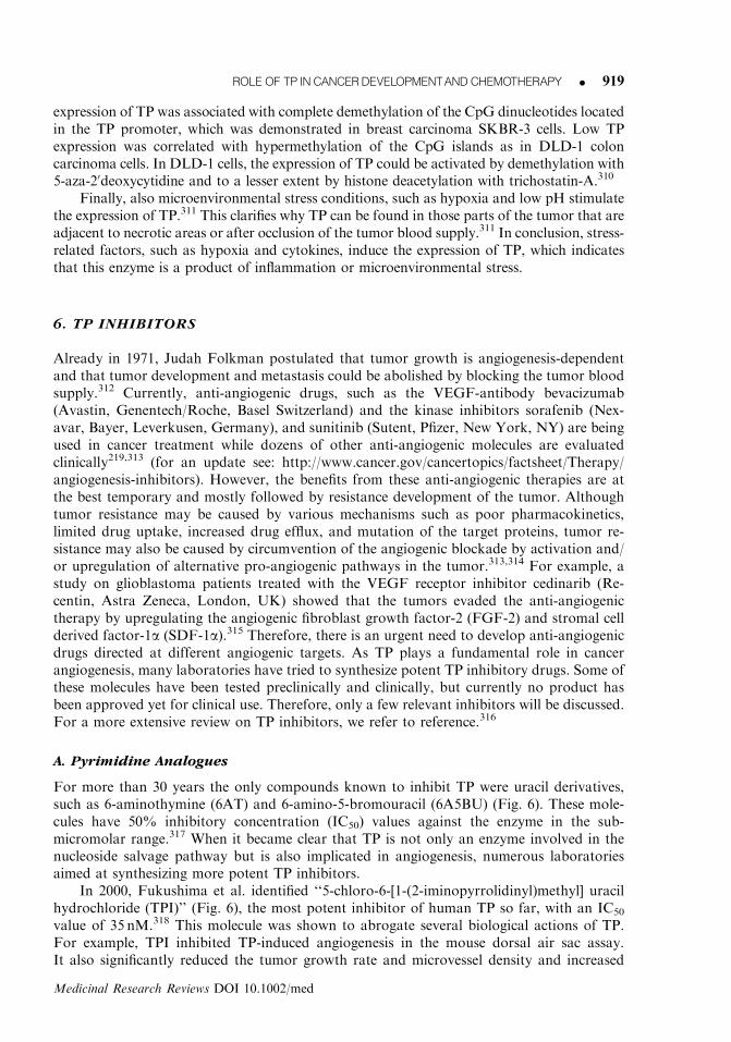
For more than 30 years the only compounds known to inhibit TP were uracil derivatives,such as 6-aminothymine (6AT) and 6-amino-5-bromouracil (6A5BU) (Fig. 6). These mole-cules have 50% inhibitory concentration (IC50) values against the enzyme in the sub-micromolar range.317 When it became clear that TP is not only an enzyme involved in thenucleoside salvage pathway but is also implicated in angiogenesis, numerous laboratoriesaimed at synthesizing more potent TP inhibitors.
In 2000, Fukushima et al. identified ‘‘5-chloro-6-[1-(2-iminopyrrolidinyl)methyl] uracilhydrochloride (TPI)’’ (Fig. 6), the most potent inhibitor of human TP so far, with an IC50
value of 35 nM.318 This molecule was shown to abrogate several biological actions of TP.For example, TPI inhibited TP-induced angiogenesis in the mouse dorsal air sac assay.It also significantly reduced the tumor growth rate and microvessel density and increased
ROLEOF TP INCANCERDEVELOPMENTANDCHEMOTHERAPY K 919
Medicinal Research Reviews DOI 10.1002/med

the apoptotic index of KB/TP xenografted tumors.275 Furthermore, oral administrationof TPI suppressed macroscopic liver metastases of highly metastatic KB/TP cellsand also the level of human b-globin as a molecular marker of micrometastases in thelivers of the mice.272 The fact that TPI is orally bio-available and has a strong nanomolar
Figure 6. Chemical structure of some illustrative inhibitors of TP.
920 K BRONCKAERS ETAL.
Medicinal Research Reviews DOI 10.1002/med

inhibitory activity against TP suggests that this molecule might be a promisingantitumor agent.
B. Purine Analogues
In 1998, Balzarini et al.319 described 7-deazaxanthine (7-DX) (Fig. 6) as the first purinederivative with inhibitory activity against a pyrimidine nucleoside phosphorylase (i.e. TP).The three-dimensional structure of E. coli TP was used for the rational modelling and designof 7-DX, which can be regarded as a pyrimidine at which a second ring was added to createextra stabilization. 7-DX not only efficiently inhibited the enzymatic activity of TP; it wasalso able to prevent neovascularization in the CAM assay.319
The available crystal structure of E. coli TP has also led to the rational design ofcompounds that interact both with the thymine and the phosphate-binding site, the so-calledmultisubstrate analogue inhibitors of TP. These types of molecules consist of a base,interacting with the nucleoside-binding site and a phosphonate moiety that may bind to thephosphate-binding site. The distance between the thymine- and the phosphate-binding site ofE. coli TP is estimated to be around 10 A, therefore the thymine and the phosphonate moietyof these novel inhibitors were linked to each other with a spacer of 6–9 methylene entities.These compounds interact with both substrate-binding sites, and thus ‘‘freeze’’ the enzyme inan open, inactive conformation.320,321 TP65, which contains an alkyl phosphonate moietycovalently linked to 7-DX (Fig. 6), is such a multisubstrate inhibitor of TP, with an IC50
value in the micromolar range. This molecule could also abrogate biological activities of TP,such as angiogenesis in the CAM assay and the formation of microvascular sprouts fromendothelial cell aggregates in a fibrin gel.222
Another purine derivative that inhibits TP is 50-O-tritylinosine (KIN59) (Fig. 6). KIN59consists of a purine base (hypoxanthine), a ribose sugar and a trityl group at the 50-positionof the ribose. The trityl group of KIN59 has proven to be crucial for its inhibitory activityagainst TP and its anti-angiogenic effect in the CAM assay.322,323 KIN59 is in several ways avery unusual TP inhibitor. In the CAM assay, KIN59 not only prevented the formation ofnew blood vessels but also promoted the degradation of small pre-existing immature bloodvessels. This effect was not due to unspecific cell toxicity. Furthermore, in contrast to allpreviously described TP inhibitors, this molecule does not compete with the naturalsubstrates for binding to either the nucleoside- or the phosphate-binding site of TP, butinteracts with a new, yet unknown, allosteric site of the enzyme in a non-competitive fash-ion.322 In order to identify the amino acids that interact with KIN59, computer-assistedmodelling of the KIN59-TP complex was performed (unpublished data). This in silicoanalysis revealed a cavity in which KIN59 could fit in the vicinity of the Gly405-Val419 loop.In this pocket the amino acid Asp203 was found to play an important role for loop stabi-lization required for efficient enzyme catalysis. Site-directed mutagenesis of Asp203 to ala-nine yielded a TP with �60-fold reduction in phosphorolytic capacity (Vmax/Km) relative tothe wild-type enzyme. Furthermore, KIN59 was not able to inhibit the enzymatic activity ofthe mutant TP, while the competitive inhibitors 6AT and 6A5BU maintained their inhibitorycapacity.
C. Prodrugs of TP Inhibitors
Reigan et al. have explored a xanthine oxidase (XO) prodrug strategy. XO activity andexpression are increased in hypoxic conditions. Moreover, increased XO activities are foundin colorectal and prostate tumors as compared to their corresponding normal tissues.Therefore, 20-nitro prodrugs of potent 20-aminoimidazolyl TP inhibitors were developed.These prodrugs may become selectively activated by XO in the tumors and thus may exert
ROLEOF TP INCANCERDEVELOPMENTANDCHEMOTHERAPY K 921
Medicinal Research Reviews DOI 10.1002/med
their TP inhibitory activity specifically within the hypoxic regions of the tumors.324 Also XO-sensitive prodrugs of 6A5BU, 7-DX, and TPI (Fig. 6) have been synthesized.325,326 The invivo efficacy of these prodrug molecules remains to be investigated.
D. 2-Deoxy-L-Ribose
2-Deoxy-L-ribose (2DLR) is a stereoisomer of 2DDR. It is not a direct TP inhibitor becauseit does not inhibit the enzymatic activity of TP. However, this molecule is able to affect thebiological effects of TP. Indeed, 2DLR suppresses the anti-apoptotic effect of 2DDR andprevents 2DDR-induced chemotaxis and tubulogenesis of bovine aortic endothelial cells invitro. In vivo, 2DLR was able to abrogate TP-induced angiogenesis in the rat corneal assayand in the mouse dorsal air sac assay.223 Moreover, oral administration of 2DLR couldsignificantly reduce the growth of KB/TP cells transplanted into nude mice and suppressedinvasion and metastasis induced by KB/TP cells.273 Also the TP-induced activation of MMP-9 and secretion of IL-8 and VEGF could be blocked by 2DLR.257,273 The molecular basis ofthe biological effects of 2DLR remains, however, to be resolved.
7. TP IN FLUOROPYRIMIDINE CHEMOTHERAPY
A. 5-Fluorouracil (5-FU)
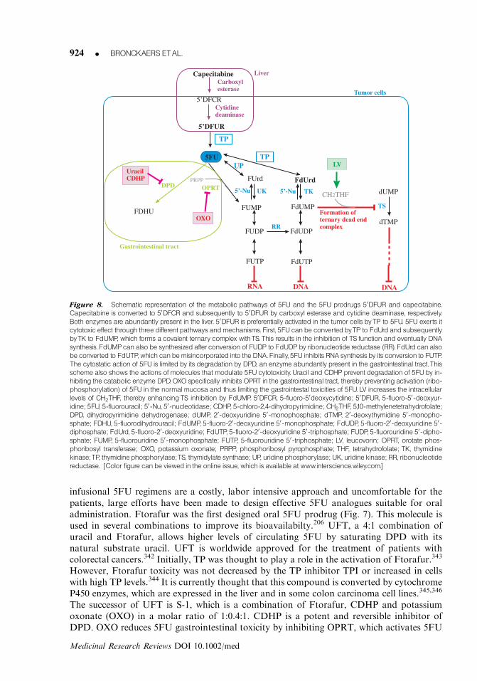
In 1957, Heidelberger et al. discovered the antitumor activity of 5-fluorouracil (5FU)(Fig. 7).327 Fifty years after its first synthesis, 5FU remains extensively used in the treatmentof colorectal cancer. The possible metabolic pathways of 5FU are depicted in Figure 8.5FU can elicit its antitumor activity by inhibiting thymidylate synthase (TS), which is re-sponsible for the de novo thymidylate production and is thus a rate-limiting enzyme in DNAsynthesis.328 First, 5FU needs to be converted by TP to 5-fluoro-20-deoxyuridine (FdUrd).This action of TP can only take place if there is enough co-substrate (i.e. 2DDR-1P) for TP.Indeed, increase in the 2DDR-1P availability by addition of 20-deoxy-pyrimidine nucleosidesor 20-deoxypurine nucleosides greatly enhances TP-mediated 5FU sensitivity of tumors.329–331
FdUrd is further converted by thymidine kinase (TK) to 5-fluoro-20-deoxyuridine 50-mono-phosphate (FdUMP). FdUMP can also be derived from FdUDP that is formed by reductivesynthesis of FUDP, a reaction catalyzed by ribonucleotide reductase. FdUMP binds to both5,10-methylenetetrahydrofolate (CH2THF) and TS leading to the formation of a ternarycomplex, which inhibits TS activity.332,333 As a result, dTTP pools get depleted, affecting DNAsynthesis. FdUMP can also be further phosphorylated to FdUTP, which can be mis-incorporated into the DNA. Alternatively, 5FU may be converted directly or indirectly to 5-fluorouridine 50-monophosphate (FUMP), which can be incorporated into RNA (upon con-version to its 50-triphosphate derivative), resulting in inhibition of protein synthesis.328 Thedirect conversion of 5FU to FUMP by orotate phosphoribosyl transferase (OPRT) is con-sidered to be the most important pathway for the activation of 5FU. 5FU cytotoxicity is notonly determined by the above-described anabolic pathways but also by 5FU catabolism, i.e.5FU is degraded by dihydropyrimidine dehydrogenase (DPD), which is abundantly found inthe liver. This reaction is so fast that the plasma half-life of 5FU is only 6–20min, i.e. more than80% of the administered 5FU is catabolized by DPD.334
Several strategies have been explored to increase the anticancer activity of 5FU. One ofthese strategies is to enhance the binding of FdUMP to TS, which increases the cytotoxicityof 5FU. Administration of leucovorin (LV, Fig. 9), which is converted to CH2THF, increasesthe intracellular pools of CH2THF and stabilizes the FdUMP/TS complex. Studies havedemonstrated that the addition of LV to bolus 5FU improves response rates compared to
922 K BRONCKAERS ETAL.
Medicinal Research Reviews DOI 10.1002/med

single agent 5FU treatment (23 versus 11%) in patients with advanced colorectal cancer.335
5FU/LV can be combined with oxaliplatin in a formulation known as FOLFOX, which is afrequently used therapy against metastatic colorectal cancer.336 Another approach to boostthe bioavailability of 5FU is the inhibition of DPD, which causes the rapid breakdown of thefluorinated nucleobase derivative. The use of DPD inhibitors enables the oral use of 5FUbecause they almost completely prevent 5FU degradation in the gastrointestinal tract.5-chloro-2,4-dihydropyrimidine (CDHP) is an example of such a frequently used DPD in-hibitor,337 but also other DPD inhibitors such as (E)-5-(2-bromovinyl)uracil (BVU)338,339
and 5-ethynyluracil (eniluracil)340 have been reported. Efficient inactivation of DPD by oraladministration of eniluracil has been observed in primary and metastatic colorectal cancer.341
However, eniluracil is currently withdrawn from further development because several studiesshowed that the combination of oral eniluracil and 5FU had lower activity compared tointravenous 5FU/LV treatment.342
B. 5FU-Prodrugs
As 5FU has a poor oral bioavailability and is rapidly degraded by DPD, 5FU has to beadministered via bolus injection or continuous intravenous administration. As these
Figure 7. Chemical structure of TFT, 5FU, and 5FUprodrugs.
ROLEOF TP INCANCERDEVELOPMENTANDCHEMOTHERAPY K 923
Medicinal Research Reviews DOI 10.1002/med
infusional 5FU regimens are a costly, labor intensive approach and uncomfortable for thepatients, large efforts have been made to design effective 5FU analogues suitable for oraladministration. Ftorafur was the first designed oral 5FU prodrug (Fig. 7). This molecule isused in several combinations to improve its bioavailabilty.206 UFT, a 4:1 combination ofuracil and Ftorafur, allows higher levels of circulating 5FU by saturating DPD with itsnatural substrate uracil. UFT is worldwide approved for the treatment of patients withcolorectal cancers.342 Initially, TP was thought to play a role in the activation of Ftorafur.343
However, Ftorafur toxicity was not decreased by the TP inhibitor TPI or increased in cellswith high TP levels.344 It is currently thought that this compound is converted by cytochromeP450 enzymes, which are expressed in the liver and in some colon carcinoma cell lines.345,346
The successor of UFT is S-1, which is a combination of Ftorafur, CDHP and potassiumoxonate (OXO) in a molar ratio of 1:0.4:1. CDHP is a potent and reversible inhibitor ofDPD. OXO reduces 5FU gastrointestinal toxicity by inhibiting OPRT, which activates 5FU
FdUrdFUrd
FUDP
FUTP
FdUMP
FdUDP
FdUTP
OPRT
UP
UK TK
TS
dUMP
Cytidinedeaminase
FUMP
RR
DPD
CapecitabineCarboxylesterase
DNA DNARNA
dTMP
UracilCDHP
Formation ofternary dead endcomplex
CH THF2
TPLV
5’DFCR
5’DFUR
5FU
FDHU
TP
Liver
Tumor cells
Gastrointestinal tract
OXO
PRPP
5’-Nu 5’-Nu
Figure 8. Schematic representation of the metabolic pathways of 5FU and the 5FU prodrugs 50DFUR and capecitabine.
Capecitabine is converted to 50DFCR and subsequently to 50DFUR by carboxyl esterase and cytidine deaminase, respectively.
Both enzymes are abundantly present in the liver. 50DFUR is preferentially activated in the tumor cells byTP to 5FU. 5FU exerts it
cytotoxic effect through three different pathways andmechanisms. First, 5FU canbe converted byTP to FdUrd and subsequently
byTK to FdUMP, which forms a covalent ternary complex withTS.This results in the inhibition of TS function and eventually DNA
synthesis. FdUMPcanalso be synthesized after conversion of FUDP to FdUDP by ribonucleotide reductase (RR). FdUrd canalso
be converted to FdUTP, which canbemisincorporated into the DNA.Finally, 5FU inhibits RNA synthesis by its conversion to FUTP.
The cytostatic action of 5FU is limited by its degradation by DPD, an enzyme abundantly present in the gastrointestinal tract.This
scheme also shows the actions of molecules that modulate 5FU cytotoxicity. Uracil and CDHP prevent degradation of 5FU by in-
hibiting the catabolic enzyme DPD.OXO specifically inhibits OPRT in the gastrointestinal tract, thereby preventing activation (ribo-
phosphorylation) of 5FU in the normal mucosa and thus limiting the gastrointestal toxicities of 5FU. LV increases the intracellular
levels of CH2THF, thereby enhancingTS inhibition by FdUMP. 50DFCR, 5-fluoro-50deoxycytidine; 50DFUR, 5-fluoro-50-deoxyur-
idine; 5FU, 5-fluorouracil; 50-Nu, 50-nucleotidase; CDHP, 5-chloro-2,4-dihydropyrimidine; CH2THF, 5,10-methylenetetrahydrofolate;
DPD, dihydropyrimidine dehydrogenase; dUMP, 20-deoxyuridine 50-monophosphate; dTMP, 20-deoxythymidine 50-monopho-
sphate; FDHU, 5-fluorodihydrouracil; FdUMP, 5-fluoro-20-deoxyuridine 50-monophosphate; FdUDP, 5-fluoro-20-deoxyuridine 50-
diphosphate; FdUrd, 5-fluoro-20-deoxyuridine; FdUTP, 5-fluoro-20-deoxyuridine 50-triphosphate; FUDP, 5-fluorouridine 50-dipho-
sphate; FUMP, 5-fluorouridine 50-monophosphate; FUTP, 5-fluorouridine 50-triphosphate; LV, leucovorin; OPRT, orotate phos-
phoribosyl transferase; OXO, potassium oxonate; PRPP, phosphoribosyl pyrophosphate; THF, tetrahydrofolate; TK, thymidine
kinase;TP, thymidine phosphorylase;TS, thymidylate synthase; UP, uridine phosphorylase; UK, uridine kinase; RR, ribonucleotide
reductase. [Color figure canbe viewed in the online issue, which is available at www.interscience.wiley.com.]
924 K BRONCKAERS ETAL.
Medicinal Research Reviews DOI 10.1002/med

in the digestive tract.337 OXO specifically accumulates in the gastrointestinal tract, thuspreventing the activation of 5FU in the normal mucosa but not in the tumor.347
5-fluoro-20-deoxyuridine (FdUrd, floxuridine) is the deoxyribose metabolite of 5FU. Asseen in Figure 8 it is a precursor of FdUMP, which inhibits TS.348 FdUrd can also beconverted to 5FU in the liver by TP.349 Like 5FU, this molecule has a short plasma half-life(15min) and causes gastrointestinal toxicity.348 Due to the higher toxicity, higher costs, andthe equal efficacy of bolus injection of FdUrd compared to bolus 5FU, the use of FdUrd hasbeen very limited.348 It is only occasionally used as a chemotherapeutic agent for hepaticarterial infusion in the treatment of unresectable liver metastases caused by colorectalcancer.349
Another prodrug of 5FU is doxifluridine (50-deoxy-5-fluorouridine, 50DFUR), whichrequires TP for its one-step conversion to 5FU. Numerous in vitro studies showed thattransfection of TP cDNA into tumor cells dramatically increases the sensitivity of the cells to50DFUR.6,350–353 Some studies also reported that TP enhances the activity of 5FU, but to alesser extent than that of 50DFUR. This is due to the fact that 50DFUR only requires TP forthe conversion to 5FU, while 5FU activation is mediated by at least three different pathways.As TP expression is also high in the gastrointestinal tract, 50DFUR therapy resulted in dose-limiting toxicity, such as diarrhea.354,355
Capecitabine (Xeloda, N4-pentyloxycarbonyl-50-deoxy-5-fluorocytidine), an oral pro-drug of 5FU, was designed to circumvent the gastrointestinal toxicity of 50DFUR and togenerate 5FU preferentially at the tumor site.7 The conversion of capecitabine to 5FU re-quires three distinct steps (see Fig. 8). Once oral capecitabine has passed the intestines in itsintact form, it is hydrolyzed to 50-deoxy-5-fluorocytidine (50DFCR) by carboxylesterase inthe liver. The second step is the conversion to 50DFUR by cytidine deaminase, which is
Figure 9. Chemical structure of products that modulate 5FU cytotoxicity: leucovorin,CDHP,OXO.
ROLEOF TP INCANCERDEVELOPMENTANDCHEMOTHERAPY K 925
Medicinal Research Reviews DOI 10.1002/med

localized in the liver and in various tumor types. Finally, 50DFUR can be converted to 5FUin the tumors by TP7 (and UP356,357). As TP is highly expressed in the tumor tissue, it permitsthe targeted intratumoral release of 5FU and consequently minimizes systemic toxicity.358–360
For example, in patients with colorectal cancer, it was proven that following oral adminis-tration of capecitabine, the 5FU concentration in the tumor tissue was 3.2 times higher thanin adjacent nontumorigenic tissue and 21 times more elevated than in plasma.358 Further-more, phase III trials enrolling patients with metastatic colorectal cancer showed that singleagent capecitabine treatment is at least as effective as 5FU/LV therapy and is associated withsignificantly fewer clinically relevant toxicities.359–362 As capecitabine is at least as active asthe 5FU/LV standard and better tolerated by the patients, it has become one of the mostprescribed oral chemotherapeutic agents. Currently, capecitabine is approved by the USFood and Drug Administration (FDA) as an adjuvant in stage III Dukes’ C colorectal cancerand as first-line monotherapy in metastatic colon cancer. The drug is also accepted for useagainst metastatic breast cancer in combination with docetaxel after failure of anthracycline-based treatment or as monotherapy when patients have failed paclitaxel-based therapy.At the moment, the combination of capecitabine with different other anticancer agents suchas bevacizumab, enzastaurin, and sorafenib is being evaluated in clinical trials.303,363–365
As TP is the rate-limiting enzyme for the activation of capecitabine, it might be a usefulpredictor of tumor response to capecitabine-based chemotherapy. In colorectal366 and ad-vanced non-small cell lung367 cancer, TP expression was associated with tumor response tocapecitabine, while in patients with breast368 and gastric369 cancer the TP/DPD ratio showeda significant correlation with response to capecitabine therapy.
C. Combination of TP-Inducible and TP-Targeted Therapy
As it has been shown in numerous transfection experiments that the antitumoral activity of50DFUR and capecitabine depends on their activation by TP, this enzyme is used as a targetto enhance the anticancer activity of these fluoropyrimidines. As described in Section 5, TPlevels can be elevated by several anticancer treatments, such as X-ray irradiation andchemotherapeutic agents (i.e. taxanes, mitomycin C, cyclophosphamide). It has beenhypothesized that combination of TP-inducing therapies (such as taxanes) with TP-targetedtherapy (such as capecitabine) would improve the clinical efficacy of these fluoropyrimidines.In the WiDr colon and MX-1 mammary human cancer xenograft models, the combination ofX-ray irradiation with either capecitabine or 50-DFUR showed a better antitumor effectthan either radiation or chemotherapy alone.309 Furthermore, several clinical trials havedemonstrated a synergy between capecitabine and TP-inducible chemotherapy. For example,in a large randomized phase-III trial on metastatic breast cancer it was demonstrated that theaddition of capecitabine to docetaxel therapy results in an increased response rate, time toprogression, and survival compared to standard treatment alone.370 Also the combinationwith other TP-inducible therapies, such as irinotecan, oxaliplatin, cisplatin, cyclopho-sphamide, paclitaxel, mitomycin C, and irradiation resulted in improved survival andtime-to-progression compared to the monotherapy.7 The combination of capecitabine plusoxaliplatin (XELOX regimen) now represents a new standard of care for metastatic coloncarcinoma.371
D. TFT
The fluoropyrimidine nucleoside 5-trifluorothymidine (TFT) was originally synthesized byHeidelberger et al. in 1964.372 TFT is phosphorylated by TK to its active monophosphateTFT-MP, which inhibits TS.373 In contrast to FdUMP, TFT-MP does not form a ternarycomplex with TS, but binds covalently to the active site of TS.374 TFT-MP can also be
926 K BRONCKAERS ETAL.
Medicinal Research Reviews DOI 10.1002/med

further phosphorylated to TFT-TP, which can be incorporated into the DNA leading to celldeath due to DNA strand break formation.375 Since 1980, TFT has been used for the topicaltreatment of epithelial keratitis caused by herpes simplex virus.376 This molecule has alsobeen evaluated as an antitumor agent but the clinical studies were discontinued because ofthe high toxicity of TFT and its rapid degradation by TP that inactivates TFT by convertingit to its inactive base.5,377 Given as a single agent, the plasma half-life of TFT is less then20min.378 Therefore, TFT was recently chosen to be combined with TPI, a very potentinhibitor of TP (see Section 6.A). This oral combination therapy, called TAS-102, combinesTFT and TPI in a 1:0.5 molar ratio.379 The application of TFT together with TPI bypassesTFT degradation by TP resulting in increased TFT plasma levels compared to TFT alone.TAS-102 thus improves the bioavailability and thereby the efficacy of TFT.318 Anotheradvantage of TAS-102 is that TPI might also abrogate the angiogenic properties of TP.Furthermore, TAS-102 can also be used against cancers that are resistant to 5FU, as shownby in vitro studies and tumor implants in nude mice.375,380 So far, several phaseI clinical trials using TAS-102 have been completed. The toxicities observed were granulo-cytopenia, nausea, vomiting, diarrhea, fatigue, and rash. A phase I clinical trial demonstratedthat TAS-102 is active against heavily pretreated metastatic breast cancers.381 However, ina recent phase I trial, where TAS-102 was administered daily on a 5-day-a-week schedule topatients with solid (mostly colorectal) tumors, patients treated with TAS-102 showed noobjective response although stable disease was seen in 18 of 61 patients.382 Currently, phaseII trials of TAS-102 alone or in combination with other therapies against breast, gastro-intestinal, and other solid tumors are ongoing.381,382 The combination of TFT together withoxaliplatin was tested in vitro in various colon carcinoma cell lines and strong synergism wasobserved. These results provide a motivation for the clinical study of TAS-102 together withoxaliplatin in the treatment of colorectal cancer.383
E. Mycoplasmal TP and Fluoropyrimidines
TP is not only present in humans; TP activity was also detected in different Mycoplasmaspecies.318,384,385 Mycoplasmas are the smallest self-replicating bacteria, which can causerespiratory and urogenital diseases.386 Most mycoplasmal infections remain, however, uni-dentified, because many people seem to be chronically infected without apparent clinicalsymptoms.387 Mycoplasmas might also play a role in cancer.388,389 Mycoplasmal infectionsare associated with leukaemia and ovarian and cervical cancer.390–392 In particular, thespecies M. hyorhinis is frequently found in tissues of gastric, colon, esophageal, lung, andbreast cancer, but not in analogous nontumorigenic tissue.393 Chronic and persistentinfections with mycoplasmas affect many biological characteristics of mammalian cells andcan even lead to malignant transformations.394–396 The M. hyorhinis-encoded protein p37was shown to alter gene expression, growth, and migratory potential of prostate cancercell lines in vitro.397–399 p37 was also found to promote cancer cell invasiveness and metas-tasis by activation of MMP-2 and by phosphorylation of the epidermal growth factorreceptor.399 Moreover, in an experimental metastasis mouse model, p37-encoding adeno-virus-infected mouse melanoma B16F10 cells formed more metastatic lesions than the par-ental cell line.399
Recently, it was shown that TP encoded by M. hyorhinis not only catalyzes the con-version of thymidine to thymine, but also efficiently recognizes FdUrd, TFT, and50DFUR.389,400 As a result, the cytostatic activity of FdUrd and TFT was significantlydecreased in MCF-7 breast carcinoma cells infected with M. hyorhinis compared to controlMCF-7 cells. The sensitivity to 5FU was not altered by mycoplasma infection while 50DFURwas at least 30-fold more cytostatic in mycoplasma-infected MCF-7 cells, suggesting that
ROLEOF TP INCANCERDEVELOPMENTANDCHEMOTHERAPY K 927
Medicinal Research Reviews DOI 10.1002/med

mycoplasma-encoded TP activated this molecule. Addition of the TP inhibitor TPI or themycoplasma-specific antibiotic plasmocin could restore the altered cytostatic activity.389,400
Also HCT116 colon cancer cells infected with mycoplasma were 5- and 100-fold more re-sistant to 5-FU and FdUrd, respectively, than the parental noninfected cells.401 These datademonstrate that the presence of mycoplasma and thus mycoplasmal TP may severely affectthe cytostatic efficacy of FdUrd, TFT (and 5FU), suggesting that the combination of theseanticancer agents with a specific antibiotic against mycoplasmas might improve the efficacyof these drugs.389,400
8. CONCLUSIONS AND PERSPECTIVES
There is compelling evidence that the intracellular enzyme TP plays an essential role in tumorprogression. TP stimulates tumor growth by protecting tumor cells from apoptosis and byinducing angiogenesis. Although many investigators have tried to unravel the molecularmechanisms through which TP exerts these biological effects, more research needs to be doneto gain more detailed insight into the signal transduction cascades induced by TP and itsproduct metabolite 2DDR. This knowledge could offer crucial information for the rationaldevelopment of strategies to inhibit the protumoral actions of TP. Indeed, as TP expression iselevated in numerous tumor types, inhibition of TP activity might offer a potential strategy inthe battle against cancer. In this respect, more potent TP inhibitors need to be synthesizedand (pre)clinically evaluated as TPI is currently the only available TP inhibitor that is beinginvestigated in clinical trials. As such, the results of the phase II clinical trials with TAS-102will be of particular interest, as they will provide important information on the impact of TPinhibitors on tumor growth in cancer patients.
Recent data have shown that tumors treated with anti-angiogenic monotherapy seem toescape drug treatment by upregulating and/or activating alternative angiogenic pathways.Therefore, future strategies should favor the concomittant attack of multiple targets bycombining different anti-angiogenic molecules or anti-angiogenic molecules with radio- and/or chemotherapy. Thus, TP presents an alternative target or an additional target to existinganticancer therapies. The potential use of TP inhibitors is not limited to cancer as TP is alsoinvolved in many inflammatory diseases, such as RA. Also these patients might thus benefitfrom treatment with TP-inhibitory drugs and the application of TP inhibitors in the treat-ment of inflammatory diseases should be further explored.
However, the use of TP inhibitors in the treatment of cancer should be carefully andcautiously considered, because TP activity is required for the activation of the commonlyused 5FU-prodrug capecitabine. Thus, TP inhibitors should not be combined with such TP-activation-dependent therapeutic agents. One possible strategy to overcome this problemmight be to inhibit the downstream mediators of TP instead of directly inhibiting TP activityper se. 2DLR has been shown to inhibit the biological actions of TP without affecting itsenzymatic activity and therefore it might be a good candidate for combination with fluor-opyrimidine chemotherapy. However, as TP fulfills a key role in capecitabine activation,most investigations have explored the combination of TP-upregulating therapies with ca-pecitabine Clinical trials combining capecitabine with TP-inducible therapies showed thatcombination of both therapies results in a higher antitumor efficacy than monotherapy ofeither agents.
In conclusion, TP may play a dual role in cancer development and therapy: TP stimu-lates tumor growth but at the same time it is also required to activate the chemotherapeuticagent capecitabine. Depending on the type of the tumor and the nature of the therapeuticagents, cancer patients may benefit from TP-inducible therapies or TP inhibitory drugs.
928 K BRONCKAERS ETAL.
Medicinal Research Reviews DOI 10.1002/med

9. ABBREVIATIONS
BVDU 5-(E)-(2-bromovinyl)-20-deoxyuridineCAM chorio-allantoic membraneCDHP 5-chloro-2,4-dihydropyrimidineCH2THF 5,10-methylenetetrahydrofolate2DDR 2-deoxy-D-ribose2DDR-1P 2-deoxy-D-ribose-1-phosphate50DFUR 5-fluoro-50-deoxyuridine2DLR 2-deoxy-L-riboseDPD dihydropyrimidine dehydrogenaseECM extracellular matrixEPC endothelial progenitor cellFAK focal adhesion kinaseFdUMP 5-fluoro-20-deoxyuridine 50-monophosphateFdUrd 5-fluoro-20-deoxyuridineFLS fibroblast-like synoviocytes5FU 5-fluorouracilHIF-1a hypoxia-inducible factor-1aHO-1 heme-oxygenase-1HUVEC human umbilical vein endothelial cellIC50 50% inhibitory concentrationIFN-g interferon-gIL interleukinLV leucovorinMMP matrix metalloproteinaseMNGIE mitochondrial neurogastrointestinal encephalomyopathyOPRT orotate phosphoribosyl transferaseOXO potassium oxonatePD-ECGF platelet-derived-endothelial cell growth factorPRPP phosphoribosyl pyrophosphateRA rheumatoid arthritisRR ribonucleotide reductaseTAM tumor-associated macrophageTFT 5-trifluorothymidineTK thymidine kinaseTP thymidine phosphorylaseTPI 5-chloro-6-[1-(2-iminopyrrolidinyl)methyl] uracil hydrochlorideTS thymidylate synthaseUP uridine phosphorylaseVEGF vascular endothelial growth factorVSMC vascular smooth muscle cellXO xanthine oxidase
ACKNOWLEDGMENTS
Annelies Bronckaers benefits from a Ph.D. scholarship of the FWO. Sandra Liekens is apostdoctoral fellow of the FWO. The research of the authors on TP was supported by grantsof the Centers of Excellence of the K.U. Leuven (Krediet no. 05/15) (to J.B. and S.L.), the
ROLEOF TP INCANCERDEVELOPMENTANDCHEMOTHERAPY K 929
Medicinal Research Reviews DOI 10.1002/med
Geconcerteerde Onderzoeksacties of the K.U. Leuven (GOA 05/19) (to J.B. and S.L.), the‘‘Comision Interministerial de Ciencia y Tecnologıa’’ (SAF2006-12713-C02) (to F.G.) andthe ‘‘Comunidad de Madrid’’ (S-BIO/0214/2006).
REFERENCES
1. Friedkin M, Roberts D. The enzymatic synthesis of nucleosides. I. Thymidine phosphorylase in
mammalian tissue. J Biol Chem 1954;207:245–256.
2. Iltzsch MH, El Kouni MH, Cha S. Kinetic studies of thymidine phosphorylase from mouse liver.
Biochemistry 1985;24:6799–6807.
3. Schwartz M. Thymidine phosphorylase from Escherichia coli. Properties and kinetics. Eur J
Biochem 1971;21:191–198.
4. Desgranges C, Razaka G, Rabaud M, Bricaud H, Balzarini J, De Clercq E. Phosphorolysis of (E)-5-
(2-bromovinyl)-20-deoxyuridine (BVDU) and other 5-substituted-20-deoxyuridines by purified
human thymidine phosphorylase and intact blood platelets. Biochem Pharmacol 1983;32:3583–3590.
5. Heidelberger C, Anderson S. Fluorinated pyrimidines. XXI. The tumor-inhibitory activity of
5-triflyoromethyl-2’-deoxyuridine. Cancer Res 1964;24:1979–1985.
6. Schwartz EL, Baptiste N, Wadler S, Makower D. Thymidine phosphorylase mediates the
sensitivity of human colon carcinoma cells to 5-fluorouracil. J Biol Chem 1995;270:19073–19077.
7. Walko CM, Lindley C. Capecitabine: A review. Clin Ther 2005;27:23–44.
8. Miyazono K, Okabe T, Urabe A, Takaku F, Heldin CH. Purification and properties of an
endothelial cell growth factor from human platelets. J Biol Chem 1987;262:4098–4103.
9. Ishikawa F, Miyazono K, Hellman U, Drexler H, Wernstedt C, Hagiwara K, Usuki K, Takaku F,
Risau W, Heldin CH. Identification of angiogenic activity and the cloning and expression of
platelet-derived endothelial cell growth factor. Nature 1989;338:557–562.
10. Moghaddam A, Bicknell R. Expression of platelet-derived endothelial cell growth factor in
Escherichia coli and confirmation of its thymidine phosphorylase activity. Biochemistry
1992;31:12141–12146.
11. Usuki K, Saras J, Waltenberger J, Miyazono K, Pierce G, Thomason A, Heldin CH. Platelet-
derived endothelial cell growth factor has thymidine phosphorylase activity. Biochem Biophys Res
Commun 1992;184:1311–1316.
12. Furukawa T, Yoshimura A, Sumizawa T, Haraguchi M, Akiyama S, Fukui K, Ishizawa M,
Yamada Y. Angiogenic factor. Nature 1992;356:668.
13. Asai K, Hirano T, Kaneko S, Moriyama A, Nakanishi K, Isobe I, Eksioglu YZ, Kato T. A novel
glial growth inhibitory factor, gliostatin, derived from neurofibroma. J Neurochem 1992;59:307–317.
14. Asai K, Nakanishi K, Isobe I, Eksioglu YZ, Hirano A, Hama K, Miyamoto T, Kato T.
Neurotrophic action of gliostatin on cortical neurons. Identity of gliostatin and platelet-derived
endothelial cell growth factor. J Biol Chem 1992;267:20311–20316.
15. Blank JG, Hoffee PA. Purification and properties of thymidine phosphorylase from Salmonella
typhimurium. Arch Biochem Biophys 1975;168:259–265.
16. Voytek P. Purification of thymidine phosphorylase from Escherichia coli and its photoinactivation in
the presence of thymine, thymidine, and some halogenated analogs. J Biol Chem 1975;250:3660–3665.
17. Kubilus J, Lee LD, Baden HP. Purification of thymidine phosphorylase from human
amniochorion. Biochim Biophys Acta 1978;527:221–228.
18. Barton GJ, Ponting CP, Spraggon G, Finnis C, Sleep D. Human platelet-derived endothelial cell
growth factor is homologous to Escherichia coli thymidine phosphorylase. Protein Sci
1992;1:688–690.
19. Schwartz M. Thymidine phosphorylase from Escherichia coli. Methods Enzymol 1978;51:
442–445.
930 K BRONCKAERS ETAL.
Medicinal Research Reviews DOI 10.1002/med
20. Desgranges C, Razaka G, Rabaud M, Bricaud H. Catabolism of thymidine in human blood
platelets: Purification and properties of thymidine phosphorylase. Biochim Biophys Acta
1981;654:211–218.
21. Walter MR, Cook WJ, Cole LB, Short SA, Koszalka GW, Krenitsky TA, Ealick SE. Three-
dimensional structure of thymidine phosphorylase from Escherichia coli at 2.8 A resolution. J Biol
Chem 1990;265:14016–14022.
22. Pugmire MJ, Cook WJ, Jasanoff A, Walter MR, Ealick SE. Structural and theoretical studies
suggest domain movement produces an active conformation of thymidine phosphorylase. J Mol
Biol 1998;281:285–299.
23. Pugmire MJ, Ealick SE. The crystal structure of pyrimidine nucleoside phosphorylase in a closed
conformation. Structure 1998;6:1467–1479.
24. Mendieta J, Martin-Santamaria S, Priego EM, Balzarini J, Camarasa MJ, Perez-Perez MJ,
Gago F. Role of histidine-85 in the catalytic mechanism of thymidine phosphorylase as assessed
by targeted molecular dynamics simulations and quantum mechanical calculations. Biochemistry
2004;43:405–414.
25. Norman RA, Barry ST, Bate M, Breed J, Colls JG, Ernill RJ, Luke RW, Minshull CA,
McAlister MS, McCall EJ, McMiken HH, Paterson DS, Timms D, Tucker JA, Pauptit RA.
Crystal structure of human thymidine phosphorylase in complex with a small molecule inhibitor.
Structure 2004;12:75–84.
26. Spraggon G, Stuart D, Ponting C, Finnis C, Sleep D, Jones Y. Crystallization and X-ray
diffraction study of recombinant platelet-derived endothelial cell growth factor. J Mol Biol
1993;234:879–880.
27. El Omari K, Bronckaers A, Liekens S, Perez-Perez MJ, Balzarini J, Stammers DK. Structural
basis for non-competitive product inhibition in human thymidine phosphorylase: Implications for
drug design. Biochem J 2006;399:199–204.
28. Fox SB, Moghaddam A, Westwood M, Turley H, Bicknell R, Gatter KC, Harris AL. Platelet-
derived endothelial cell growth factor/thymidine phosphorylase expression in normal tissues: An
immunohistochemical study. J Pathol 1995;176:183–190.
29. Yoshimura A, Kuwazuru Y, Furukawa T, Yoshida H, Yamada K, Akiyama S. Purification and
tissue distribution of human thymidine phosphorylase; high expression in lymphocytes,
reticulocytes and tumors. Biochim Biophys Acta 1990;1034:107–113.
30. Matsukawa K, Moriyama A, Kawai Y, Asai K, Kato T. Tissue distribution of human gliostatin/
platelet-derived endothelial cell growth factor (PD-ECGF) and its drug-induced expression.
Biochim Biophys Acta 1996;1314:71–82.
31. Shaw T, Smillie RH, MacPhee DG. The role of blood platelets in nucleoside metabolism: Assay,
cellular location and significance of thymidine phosphorylase in human blood. Mutat Res
1988;200:99–116.
32. Jackson MR, Carney EW, Lye SJ, Ritchie JW. Localization of two angiogenic growth factors
(PDECGF and VEGF) in human placentae throughout gestation. Placenta 1994;15:341–353.
33. Usuki K, Norberg L, Larsson E, Miyazono K, Hellman U, Wernstedt C, Rubin K, Heldin CH.
Localization of platelet-derived endothelial cell growth factor in human placenta and purification
of an alternatively processed form. Cell Regul 1990;1:577–584.
34. Zhang L, MacKenzie IZ, Rees MC, Bicknell R. Regulation of the expression of the angiogenic
enzyme platelet-derived endothelial cell growth factor/thymidine phosphorylase in endometrial
isolates by ovarian steroids and cytokines. Endocrinology 1997;138:4921–4930.
35. Fujimoto J, Ichigo S, Sakaguchi H, Hirose R, Tamaya T. Expression of platelet-derived
endothelial cell growth factor and its mRNA in uterine endometrium during the menstrual cycle.
Mol Hum Reprod 1998;4:509–513.
36. Abbas MM, Evans JJ, Sykes PH, Benny PS. Modulation of vascular endothelial growth factor
and thymidine phosphorylase in normal human endometrial stromal cells. Fertil Steril
2004;82:1048–1053.
ROLEOF TP INCANCERDEVELOPMENTANDCHEMOTHERAPY K 931
Medicinal Research Reviews DOI 10.1002/med
37. Osuga Y, Toyoshima H, Mitsuhashi N, Taketani Y. The presence of platelet-derived endothelial
cell growth factor in human endometrium and its characteristic expression during the menstrual
cycle and early gestational period. Hum Reprod 1995;10:989–993.
38. Creamer D, Jaggar R, Allen M, Bicknell R, Barker J. Overexpression of the angiogenic factor
platelet-derived endothelial cell growth factor/thymidine phosphorylase in psoriatic epidermis.
Br J Dermatol 1997;137:851–855.
39. Hammerberg C, Fisher GJ, Voorhees JJ, Cooper KD. Elevated thymidine phosphorylase activity
in psoriatic lesions accounts for the apparent presence of an epidermal ‘‘growth inhibitor,’’ but is
not in itself growth inhibitory. J Invest Dermatol 1991;97:286–290.
40. Giatromanolaki A, Sivridis E, Maltezos E, Papazoglou D, Simopoulos C, Gatter KC, Harris AL,
Koukourakis MI. Hypoxia inducible factor 1alpha and 2alpha overexpression in inflammatory
bowel disease. J Clin Pathol 2003;56:209–213.
41. Saito S, Tsuno NH, Sunami E, Hori N, Kitayama J, Kazama S, Okaji Y, Kawai K, Kanazawa T,
Watanabe T, Shibata Y, Nagawa H. Expression of platelet-derived endothelial cell growth factor
in inflammatory bowel disease. J Gastroenterol 2003;38:229–237.
42. Wang EH, Goh YB, Moon IS, Park CH, Lee KH, Kang SH, Kang CS, Choi YJ. Upregulation of
thymidine phosphorylase in chronic glomerulonephritis and its role in tubulointerstitial injury.
Nephron Clin Pract 2006;102:c133–c142.
43. Boyle JJ, Wilson B, Bicknell R, Harrower S, Weissberg PL, Fan TP. Expression of angiogenic
factor thymidine phosphorylase and angiogenesis in human atherosclerosis. J Pathol
2000;192:234–242.
44. Takeuchi M, Otsuka T, Matsui N, Asai K, Hirano T, Moriyama A, Isobe I, Eksioglu YZ,
Matsukawa K, Kato T. Aberrant production of gliostatin/platelet-derived endothelial cell growth
factor in rheumatoid synovium. Arthritis Rheum 1994;37:662–672.
45. Asai K, Hirano T, Matsukawa K, Kusada J, Takeuchi M, Otsuka T, Matsui N, Kato T. High
concentrations of immunoreactive gliostatin/platelet-derived endothelial cell growth factor in
synovial fluid and serum of rheumatoid arthritis. Clin Chim Acta 1993;218:1–4.
46. Waguri Y, Otsuka T, Sugimura I, Matsui N, Asai K, Moriyama A, Kato T. Gliostatin/platelet-
derived endothelial cell growth factor as a clinical marker of rheumatoid arthritis and its
regulation in fibroblast-like synoviocytes. Br J Rheumatol 1997;36:315–321.
47. Waguri-Nagaya Y, Otsuka T, Sugimura I, Matsui N, Asai K, Nakajima K, Tada T, Akiyama S,
Kato T. Synovial inflammation and hyperplasia induced by gliostatin/platelet-derived endothelial
cell growth factor in rabbit knees. Rheumatol Int 2000;20:13–19.
48. Muro H, Waguri-Nagaya Y, Mukofujiwara Y, Iwahashi T, Otsuka T, Matsui N, Moriyama A,
Asai K, Kato T. Autocrine induction of gliostatin/platelet-derived endothelial cell growth factor
(GLS/PD-ECGF) and GLS-induced expression of matrix metalloproteinases in rheumatoid
arthritis synoviocytes. Rheumatology (Oxford) 1999;38:1195–1202.
49. Ieda Y, Waguri-Nagaya Y, Iwahasi T, Otsuka T, Matsui N, Namba M, Asai K, Kato T. IL-1beta-
induced expression of matrix metalloproteinases and gliostatin/platelet-derived endothelial cell
growth factor (GLS/PD-ECGF) in a chondrosarcoma cell line (OUMS-27). Rheumatol Int
2001;21:45–52.
50. Tanikawa T, Waguri-Nagaya Y, Kusabe T, Aoyama M, Asai K, Otsuka T. Gliostatin/thymidine
phosphorylase-regulated vascular endothelial growth-factor production in human fibroblast-like
synoviocytes. Rheumatol Int 2007;27:553–559.
51. Hirano M, Silvestri G, Blake DM, Lombes A, Minetti C, Bonilla E, Hays AP, Lovelace RE,
Butler I, Bertorini TE. Mitochondrial neurogastrointestinal encephalomyopathy (MNGIE):
Clinical, biochemical, and genetic features of an autosomal recessive mitochondrial disorder.
Neurology 1994;44:721–727.
52. Nishino I, Spinazzola A, Papadimitriou A, Hammans S, Steiner I, Hahn CD, Connolly AM,
Verloes A, Guimaraes J, Maillard I, Hamano H, Donati MA, Semrad CE, Russell JA, Andreu AL,
Hadjigeorgiou GM, Vu TH, Tadesse S, Nygaard TG, Nonaka I, Hirano I, Bonilla E, Rowland LP,
932 K BRONCKAERS ETAL.
Medicinal Research Reviews DOI 10.1002/med
DiMauro S, Hirano M. Mitochondrial neurogastrointestinal encephalomyopathy: An autosomal
recessive disorder due to thymidine phosphorylase mutations. Ann Neurol 2000;47:792–800.
53. Spinazzola A, Marti R, Nishino I, Andreu AL, Naini A, Tadesse S, Pela I, Zammarchi E,
Donati MA, Oliver JA, Hirano M. Altered thymidine metabolism due to defects of thymidine
phosphorylase. J Biol Chem 2002;277:4128–4133.
54. Marti R, Nishigaki Y, Hirano M. Elevated plasma deoxyuridine in patients with thymidine
phosphorylase deficiency. Biochem Biophys Res Commun 2003;303:14–18.
55. Valentino ML, Marti R, Tadesse S, Lopez LC, Manes JL, Lyzak J, Hahn A, Carelli V, Hirano M.
Thymidine and deoxyuridine accumulate in tissues of patients with mitochondrial neurogas-
trointestinal encephalomyopathy (MNGIE). FEBS Lett 2007;581:3410–3414.
56. Haraguchi M, Tsujimoto H, Fukushima M, Higuchi I, Kuribayashi H, Utsumi H, Nakayama A,
Hashizume Y, Hirato J, Yoshida H, Hara H, Hamano S, Kawaguchi H, Furukawa T,
Miyazono K, Ishikawa F, Toyoshima H, Kaname T, Komatsu M, Chen ZS, Gotanda T,
Tachiwada T, Sumizawa T, Miyadera K, Osame M, Yoshida H, Noda T, Yamada Y, Akiyama S.
Targeted deletion of both thymidine phosphorylase and uridine phosphorylase and consequent
disorders in mice. Mol Cell Biol 2002;22:5212–5221.
57. Lopez LC, Akman HO, Garcia-Cazorla A, Dorado B, Marti R, Nishino I, Tadesse S, Pizzorno G,
Shungu D, Bonilla E, Tanji K, Hirano M. Unbalanced deoxynucleotide pools cause
mitochondrial DNA instability in thymidine phosphorylase deficient mice. Hum Mol Genet
2009;18:714–722.
58. Hirano M, Nishigaki Y, Marti R. Mitochondrial neurogastrointestinal encephalomyopathy
(MNGIE): A disease of two genomes. Neurologist 2004;10:8–17.
59. Kumagai Y, Sugiura Y, Sugeno H, Takebayashi Y, Takenoshita S, Yamamoto T. Thymidine
phosphorylase gene mutation is not a primary cause of mitochondrial neurogastrointestinal
encephalomyopathy (MNGIE). Intern Med 2006;45:443–446.
60. Dickinson EK, Adams DL, Schon EA, Glerum DM. A human SCO2 mutation helps define the
role of Sco1p in the cytochrome oxidase assembly pathway. J Biol Chem 2000;275:26780–26785.
61. Jaksch M, Ogilvie I, Yao J, Kortenhaus G, Bresser HG, Gerbitz KD, Shoubridge EA. Mutations
in SCO2 are associated with a distinct form of hypertrophic cardiomyopathy and cytochrome c
oxidase deficiency. Hum Mol Genet 2000;9:795–801.
62. Nishino I, Spinazzola A, Hirano M. Thymidine phosphorylase gene mutations in MNGIE, a
human mitochondrial disorder. Science 1999;283:689–692.
63. Moghaddam A, Zhang HT, Fan TP, Hu DE, Lees VC, Turley H, Fox SB, Gatter KC, Harris AL,
Bicknell R. Thymidine phosphorylase is angiogenic and promotes tumor growth. Proc Natl Acad
Sci USA 1995;92:998–1002.
64. Arima J, Imazono Y, Takebayashi Y, Nishiyama K, Shirahama T, Akiba S, Furukawa T,
Akiyama S, Ohi Y. Expression of thymidine phosphorylase as an indicator of poor prognosis for
patients with transitional cell carcinoma of the bladder. Cancer 2000;88:1131–1138.
65. O’Brien TS, Fox SB, Dickinson AJ, Turley H, Westwood M, Moghaddam A, Gatter KC,
Bicknell R, Harris AL. Expression of the angiogenic factor thymidine phosphorylase/
platelet-derived endothelial cell growth factor in primary bladder cancers. Cancer Res
1996;56:4799–4804.
66. Yoshikawa T, Suzuki K, Kobayashi O, Sairenji M, Motohashi H, Tsuburaya A, Nakamura Y,
Shimizu A, Yanoma S, Noguchi Y. Thymidine phosphorylase/platelet-derived endothelial cell growth
factor is upregulated in advanced solid types of gastric cancer. Br J Cancer 1999;79:1145–1150.
67. Takebayashi Y, Miyadera K, Akiyama S, Hokita S, Yamada K, Akiba S, Yamada Y,
Sumizawa T, Aikou T. Expression of thymidine phosphorylase in human gastric carcinoma. Jpn J
Cancer Res 1996;87:288–295.
68. Takebayashi Y, Yamada K, Miyadera K, Sumizawa T, Furukawa T, Kinoshita F, Aoki D,
Okumura H, Yamada Y, Akiyama S, Aikou T. The activity and expression of thymidine
phosphorylase in human solid tumours. Eur J Cancer 1996;32A:1227–1232.
ROLEOF TP INCANCERDEVELOPMENTANDCHEMOTHERAPY K 933
Medicinal Research Reviews DOI 10.1002/med

69. O’Byrne KJ, Koukourakis MI, Giatromanolaki A, Cox G, Turley H, Steward WP, Gatter K,
Harris AL. Vascular endothelial growth factor, platelet-derived endothelial cell growth factor and
angiogenesis in non-small-cell lung cancer. Br J Cancer 2000;82:1427–1432.
70. Takebayashi Y, Natsugoe S, Baba M, Akiba S, Fukumoto T, Miyadera K, Yamada Y, Takao S,
Akiyama S, Aikou T. Thymidine phosphorylase in human esophageal squamous cell carcinoma.
Cancer 1999;85:282–289.
71. Fujimoto J, Sakaguchi H, Hirose R, Wen H, Tamaya T. Clinical implication of expression of
platelet-derived endothelial cell growth factor (PD-ECGF) in metastatic lesions of uterine cervical
cancers. Cancer Res 1999;59:3041–3044.
72. Yamamoto A, Dhar DK, El Assal ON, Igarashi M, Tabara H, Nagasue N. Thymidine
phosphorylase (platelet-derived endothelial cell growth factor), microvessel density and clinical
outcome in hepatocellular carcinoma. J Hepatol 1998;29:290–299.
73. Mainou-Fowler T, Angus B, Miller S, Proctor SJ, Taylor PR, Wood KM. Micro-vessel density and
the expression of vascular endothelial growth factor (VEGF) and platelet-derived endothelial cell
growth factor (PdEGF) in classical Hodgkin lymphoma (HL). Leuk Lymphoma 2006;47:223–230.
74. Slager EH, Honders MW, van der Meijden ED, Luxemburg-Heijs SA, Kloosterboer FM,
Kester MG, Jedema I, Marijt WA, Schaafsma MR, Willemze R, Falkenburg JH. Identification of
the angiogenic endothelial-cell growth factor-1/thymidine phosphorylase as a potential target for
immunotherapy of cancer. Blood 2006;107:4954–4960.
75. Fujimoto J, Ichigo S, Sakaguchi H, Hirose R, Tamaya T. Expression of platelet-derived
endothelial cell growth factor (PD-ECGF) and its mRNA in uterine endometrial cancers. Cancer
Lett 1998;130:115–120.
76. Fujimoto K, Hosotani R, Wada M, Lee JU, Koshiba T, Miyamoto Y, Tsuji S, Nakajima S,
Doi R, Imamura M. Expression of two angiogenic factors, vascular endothelial growth factor and
platelet-derived endothelial cell growth factor in human pancreatic cancer, and its relationship to
angiogenesis. Eur J Cancer 1998;34:1439–1447.
77. Fujioka S, Yoshida K, Yanagisawa S, KawakamiM, Aoki T, Yamazaki Y. Angiogenesis in pancreatic
carcinoma: Thymidine phosphorylase expression in stromal cells and intratumoral microvessel density
as independent predictors of overall and relapse-free survival. Cancer 2001;92:1788–1797.
78. Fujiwaki R, Hata K, Iida K, Koike M, Miyazaki K. Immunohistochemical expression of
thymidine phosphorylase in human endometrial cancer. Gynecol Oncol 1998;68:247–252.
79. Fujiwaki R, Hata K, Iida K, Maede Y, Watanabe Y, Koike M, Miyazaki K. Co-expression of
vascular endothelial growth factor and thymidine phosphorylase in endometrial cancer. Acta
Obstet Gynecol Scand 1999;78:728–734.
80. Han HS, Hwang TS. Angiogenesis in gastric cancer: Importance of the thymidine phosphorylase
expression of cancer cells as an angiogenic factor. Oncol Rep 2007;17:61–65.
81. Ikeda N, Adachi M, Taki T, Huang C, Hashida H, Takabayashi A, Sho M, Nakajima Y,
Kanehiro H, Hisanaga M, Nakano H, Miyake M. Prognostic significance of angiogenesis in
human pancreatic cancer. Br J Cancer 1999;79:1553–1563.
82. Ikeguchi M, Sakatani T, Ueta T, Fukuda K, Yamaguchi K, Tsujitani S, Kaibara N. The
expression of thymidine phosphorylase suppresses spontaneous apoptosis of cancer cells in
esophageal squamous cell carcinoma. Pathobiology 2001;69:36–43.
83. Jinfeng M, Kimura W, Sakurai F, Moriya T, Mizutani M, Hirai I. Prognostic role of angiogenesis
and its correlations with thymidine phosphorylase and p53 expression in ductal adenocarcinoma
of the pancreas. Hepatogastroenterology 2007;54:1635–1640.
84. Kakeji Y, Maehara Y, Tomoda M, Kabashima A, Oda S, Ooshiro T, Baba H, Kohnoe S,
Sugimachi K. Thymidine phosphorylase activity and angiogenesis in gastric cancer. Oncol Rep
1999;6:995–999.
85. Kikuyama S, Inada T, Shimizu K, Miyakita M. Thymidine phosphorylase expression in gastric
cancer in association with proliferative activity and angiogenesis. Anticancer Res 2000;20:
2081–2086.
934 K BRONCKAERS ETAL.
Medicinal Research Reviews DOI 10.1002/med

86. Kimura H, Konishi K, Nukui T, Kaji M, Maeda K, Yabushita K, Tsuji M, Miwa A. Prognostic
significance of expression of thymidine phosphorylase and vascular endothelial growth factor in
human gastric carcinoma. J Surg Oncol 2001;76:31–36.
87. Kimura H, Konishi K, Kaji M, Maeda K, Yabushita K, Miwa A. Correlation between expression
levels of thymidine phosphorylase (dThdPase) and clinical features in human gastric carcinoma.
Hepatogastroenterology 2002;49:882–886.
88. Konno S, Takebayashi Y, Aiba M, Akiyama S, Ogawa K. Clinicopathological and prognostic
significance of thymidine phosphorylase and proliferating cell nuclear antigen in gastric
carcinoma. Cancer Lett 2001;166:103–111.
89. Konno S, Takebayashi Y, Higashimoto M, Katsube T, Kanzaki A, Kawahara M, Takenoshita S,
Aiba M, Ogawa K. Thymidine phosphorylase expression in gastric carcinoma as a marker for
metastasis. Anticancer Res 2003;23:5011–5014.
90. Kuwahara K, Sasaki T, Kuwada Y, Murakami M, Yamasaki S, Chayama K. Expressions of
angiogenic factors in pancreatic ductal carcinoma: A correlative study with clinicopathologic
parameters and patient survival. Pancreas 2003;26:344–349.
91. Liakakos T, Troupis T, Ghiconti I, Triantafyllidis S, Macheras A, Karatzas G, Pavlakis K.
Immunohistochemical localization of thymidine phosphorylase in gastric cancer: Is there a role of
the differential expression in tumor cells and associated stromal cells? Anticancer Res
2006;26:3899–3903.
92. Mazurek A, Kuc P, Terlikowski S, Laudanski T. Evaluation of tumor angiogenesis and
thymidine phosphorylase tissue expression in patients with endometrial cancer. Neoplasma
2006;53:242–246.
93. Mazurek A, Kuc P, Mazurek-Wadolkowska E, Laudanski T. A role of thymidine phosphorylase
and P53 tissue protein expression in biology of endometrial cancer. Neoplasma 2008;55:261–265.
94. Miyake K, Imura S, Yoshizumi T, Ikemoto T, Morine Y, Shimada M. Role of thymidine
phosphorylase and orotate phosphoribosyltransferase mRNA expression and its ratio to
dihydropyrimidine dehydrogenase in the prognosis and clinicopathological features of patients
with pancreatic cancer. Int J Clin Oncol 2007;12:111–119.
95. Noguchi T, Fujiwara S, Takeno S, Kai S, Mizuta A, Nagao Y, Uchida Y. Clinical impact of
thymidine phosphorylase expression in gastric cancer. Oncol Rep 2003;10:561–566.
96. Ogawa K, Konno S, Takebayashi Y, Miura K, Katsube T, Kajiwara T, Aiba M, Aikou T,
Akiyama S. Clinicopathological and prognostic significance of thymidine phosphorylase
expression in gastric carcinoma. Anticancer Res 1999;19:4363–4367.
97. Sakamoto H, Shirakawa T, Izuka S, Igarashi T, Kinoshita K, Ohtani K, Takami T, Nakayama Y,
Teramoto K, Satoh K. Thymidine phosphorylase expression is predominantly observed in stroma
of well-differentiated adenocarcinoma of endometrium and correlates with a frequency of vascular
involvement. Gynecol Oncol 1999;72:298–305.
98. Seki N, Kodama J, Hongo A, Miyagi Y, Yoshinouchi M, Kudo T. Angiogenesis and platelet-
derived endothelial cell growth factor/thymidine phosphorylase expression in endometrial cancer.
Int J Oncol 1999;15:781–786.
99. Seki N, Kodama J, Hongo A, Miyagi Y, Yoshinouchi M, Kudo T. Vascular endothelial growth
factor and platelet-derived endothelial cell growth factor expression are implicated in the
angiogenesis of endometrial cancer. Eur J Cancer 2000;36:68–73.
100. Shimaoka S, Matsushita S, Nitanda T, Matsuda A, Nioh T, Suenaga T, Nishimata Y, Akiba S,
Akiyama S, Nishimata H. The role of thymidine phosphorylase expression in the invasiveness of
gastric carcinoma. Cancer 2000;88:2220–2227.
101. Sivridis E, Giatromanolaki A, Koukourakis MI, Bicknell R, Harris AL, Gatter KC. Thymidine
phosphorylase expression in endometrial carcinomas. Clin Exp Metastasis 1999;17:445–450.
102. Suda Y, Kuwashima Y, Tanaka Y, Uchida K, Sakamoto H, Hashiguchi Y, Sekine T. Expression
of thymidylate synthase and thymidine phosphorylase in recurrence and survival rates of advanced
gastric cancer. Gastric Cancer 1999;2:165–172.
ROLEOF TP INCANCERDEVELOPMENTANDCHEMOTHERAPY K 935
Medicinal Research Reviews DOI 10.1002/med
103. Takao S, Takebayashi Y, Che X, Shinchi H, Natsugoe S, Miyadera K, Yamada Y, Akiyama S,
Aikou T. Expression of thymidine phosphorylase is associated with a poor prognosis in patients
with ductal adenocarcinoma of the pancreas. Clin Cancer Res 1998;4:1619–1624.
104. Tanaka Y, Kobayashi H, Suzuki M, Kanayama N, Suzuki M, Terao T. Thymidine phosphorylase
expression in tumor-infiltrating macrophages may be correlated with poor prognosis in uterine
endometrial cancer. Hum Pathol 2002;33:1105–1113.
105. Tokumo K, Kodama J, Seki N, Nakanishi Y, Miyagi Y, Kamimura S, Yoshinouchi M, Okuda H,
Kudo T. Different angiogenic pathways in human cervical cancers. Gynecol Oncol 1998;68:
38–44.
106. Tsujitani S, Saito H, Maeta Y, Yamaguchi K, Tatebe S, Kondo A, Kaibara N. Neoangiogenesis in
patients with gastric carcinoma in relation to the expression of vascular endothelial growth factor
and thymidine phosphorylase. Anticancer Res 2004;24:1853–1859.
107. Giatromanolaki A, Koukourakis MI, Kakolyris S, Kaklamanis L, Barbatis K, O’Byrne KJ,
Theodosssiou D, Harris AL, Gatter KC. Focal expression of thymidine phosphorylase associates
with CD31 positive lymphocytic aggregation and local neo-angiogenesis in non-small cell lung
cancer. Anticancer Res 1998;18:71–76.
108. Fujimoto J, Ichigo S, Sakaguchi H, Hirose R, Tamaya T. Expression of platelet-derived
endothelial cell growth factor (PD-ECGF) and its mRNA in ovarian cancers. Cancer Lett
1998;126:83–88.
109. Igarashi M, Dhar DK, Kubota H, Yamamoto A, El Assal O, Nagasue N. The prognostic
significance of microvessel density and thymidine phosphorylase expression in squamous cell
carcinoma of the esophagus. Cancer 1998;82:1225–1232.
110. Nakayama Y, Sueishi K, Oka K, Kono S, Tomonaga M. Stromal angiogenesis in human glioma:
A role of platelet-derived endothelial cell growth factor. Surg Neurol 1998;49:181–187.
111. Maeda K, Kang SM, Ogawa M, Onoda N, Sawada T, Nakata B, Kato Y, Chung YS, Sowa M.
Combined analysis of vascular endothelial growth factor and platelet-derived endothelial cell
growth factor expression in gastric carcinoma. Int J Cancer 1997;74:545–550.
112. Saeki T, Tanada M, Takashima S, Saeki H, Takiyama W, Nishimoto N, Moriwaki S. Correlation
between expression of platelet-derived endothelial cell growth factor (thymidine phosphorylase) and
microvessel density in early-stage human colon carcinomas. Jpn J Clin Oncol 1997;27:227–230.
113. Engels K, Fox SB, Whitehouse RM, Gatter KC, Harris AL. Up-regulation of thymidine
phosphorylase expression is associated with a discrete pattern of angiogenesis in ductal
carcinomas in situ of the breast. J Pathol 1997;182:414–420.
114. Imazano Y, Takebayashi Y, Nishiyama K, Akiba S, Miyadera K, Yamada Y, Akiyama S, Ohi Y.
Correlation between thymidine phosphorylase expression and prognosis in human renal cell
carcinoma. J Clin Oncol 1997;15:2570–2578.
115. Kubota Y, Miura T, Moriyama M, Noguchi S, Matsuzaki J, Takebayashi S, Hosaka M.
Thymidine phosphorylase activity in human bladder cancer: Difference between superficial and
invasive cancer. Clin Cancer Res 1997;3:973–976.
116. Fox SB, Engels K, Comley M, Whitehouse RM, Turley H, Gatter KC, Harris AL. Relationship of
elevated tumour thymidine phosphorylase in node-positive breast carcinomas to the effects of
adjuvant CMF. Ann Oncol 1997;8:271–275.
117. Relf M, LeJeune S, Scott PA, Fox S, Smith K, Leek R, Moghaddam A, Whitehouse R,
Bicknell R, Harris AL. Expression of the angiogenic factors vascular endothelial cell growth
factor, acidic and basic fibroblast growth factor, tumor growth factor beta-1, platelet-derived
endothelial cell growth factor, placenta growth factor, and pleiotrophin in human primary breast
cancer and its relation to angiogenesis. Cancer Res 1997;57:963–969.
118. Toi M, Gion M, Biganzoli E, Dittadi R, Boracchi P, Miceli R, Meli S, Mori K, Tominaga T,
Gasparini G. Co-determination of the angiogenic factors thymidine phosphorylase and vascular
endothelial growth factor in node-negative breast cancer: Prognostic implications. Angiogenesis
1997;1:71–83.
936 K BRONCKAERS ETAL.
Medicinal Research Reviews DOI 10.1002/med

119. Koukourakis MI, Giatromanolaki A, O’Byrne KJ, Comley M, Whitehouse RM, Talbot DC,
Gatter KC, Harris AL. Platelet-derived endothelial cell growth factor expression correlates with
tumour angiogenesis and prognosis in non-small-cell lung cancer. Br J Cancer 1997;75:477–481.
120. Tanigawa N, Amaya H, Matsumura M, Katoh Y, Kitaoka A, Aotake T, Shimomatsuya T,
Rosenwasser OA, Iki M. Tumor angiogenesis and expression of thymidine phosphorylase/platelet
derived endothelial cell growth factor in human gastric carcinoma. Cancer Lett 1996;108:281–290.
121. Takebayashi Y, Akiyama S, Akiba S, Yamada K, Miyadera K, Sumizawa T, Yamada Y,
Murata F, Aikou T. Clinicopathologic and prognostic significance of an angiogenic factor,
thymidine phosphorylase, in human colorectal carcinoma. J Natl Cancer Inst 1996;88:1110–1117.
122. Maeda K, Chung YS, Ogawa Y, Takatsuka S, Kang SM, Ogawa M, Sawada T, Onoda N,
Kato Y, Sowa M. Thymidine phosphorylase/platelet-derived endothelial cell growth factor
expression associated with hepatic metastasis in gastric carcinoma. Br J Cancer 1996;73:884–888.
123. Fox SB, Westwood M, Moghaddam A, Comley M, Turley H, Whitehouse RM, Bicknell R,
Gatter KC, Harris AL. The angiogenic factor platelet-derived endothelial cell growth factor/
thymidine phosphorylase is up-regulated in breast cancer epithelium and endothelium. Br J
Cancer 1996;73:275–280.
124. Toi M, Hoshina S, Taniguchi T, Yamamoto Y, Ishitsuka H, Tominaga T. Expression of platelet-
derived endothelial cell growth factor/thymidine phosphorylase in human breast cancer. Int J
Cancer 1995;64:79–82.
125. Sugamoto T, Tanji N, Nishio S, Yokoyama M. Expression of platelet-derived endothelial cell
growth factor in prostatic adenocarcinoma. Oncol Rep 1999;6:519–522.
126. Yamagata M, Mori M, Mimori K, Mafune KI, Tanaka Y, Ueo H, Akiyoshi T. Expression of
pyrimidine nucleoside phosphorylase mRNA plays an important role in the prognosis of patients
with oesophageal cancer. Br J Cancer 1999;79:565–569.
127. Mimori K, Ueo H, Shirasaka C, Shiraishi T, Yamagata M, Haraguchi M, Mori M. Up-regulated
pyrimidine nucleoside phosphorylase in breast carcinoma correlates with lymph node metastasis.
Ann Oncol 1999;10:111–113.
128. Mizutani Y, Okada Y, Yoshida O. Expression of platelet-derived endothelial cell growth factor in
bladder carcinoma. Cancer 1997;79:1190–1194.
129. Alcalde RE, Terakado N, Otsuki K, Matsumura T. Angiogenesis and expression of platelet-derived
endothelial cell growth factor in oral squamous cell carcinoma. Oncology 1997;54:324–328.
130. Kodama J, Seki N, Tokumo K, Hongo A, Miyagi Y, Yoshinouchi M, Okuda H, Kudo T. Platelet-
derived endothelial cell growth factor is not implicated in progression of cervical cancer. Oncol
Rep 1999;6:617–620.
131. Fujimoto J, Sakaguchi H, Hirose R, Ichigo S, Tamaya T. Expression of platelet-derived
endothelial cell growth factor (PD-ECGF) and its mRNA in uterine cervical cancers. Br J Cancer
1999;79:1249–1254.
132. Hata K, Nagami H, Iida K, Miyazaki K, Collins WP. Expression of thymidine phosphorylase in
malignant ovarian tumors: Correlation with microvessel density and an ultrasound-derived index
of angiogenesis. Ultrasound Obstet Gynecol 1998;12:201–206.
133. Volm M, Mattern J, Koomagi R. Expression of platelet-derived endothelial cell growth factor in non-
small cell lung carcinomas: Relationship to various biological factors. Int J Oncol 1998;13:975–979.
134. Sawase K, Nomata K, Kanetake H, Saito Y. The expression of platelet-derived endothelial cell
growth factor in human bladder cancer. Cancer Lett 1998;130:35–41.
135. Nagaoka H, Iino Y, Takei H, Morishita Y. Platelet-derived endothelial cell growth factor/
thymidine phosphorylase expression in macrophages correlates with tumor angiogenesis and
prognosis in invasive breast cancer. Int J Oncol 1998;13:449–454.
136. Fujieda S, Sunaga H, Tsuzuki H, Tanaka N, Saito H. Expression of platelet-derived endothelial
cell growth factor in oral and oropharyngeal carcinoma. Clin Cancer Res 1998;4:1583–1590.
137. Matsumura M, Chiba Y, Lu C, Amaya H, Shimomatsuya T, Horiuchi T, Muraoka R,
Tanigawa N. Platelet-derived endothelial cell growth factor/thymidine phosphorylase expression
ROLEOF TP INCANCERDEVELOPMENTANDCHEMOTHERAPY K 937
Medicinal Research Reviews DOI 10.1002/med

correlated with tumor angiogenesis and macrophage infiltration in colorectal cancer. Cancer Lett
1998;128:55–63.
138. Koukourakis MI, Giatromanolaki A, Kakolyris S, O’Byrne KJ, Apostolikas N, Skarlatos J,
Gatter KC, Harris AL. Different patterns of stromal and cancer cell thymidine phosphorylase
reactivity in non-small-cell lung cancer: Impact on tumour neoangiogenesis and survival. Br J
Cancer 1998;77:1696–1703.
139. Leek RD, Landers R, Fox SB, Ng F, Harris AL, Lewis CE. Association of tumour necrosis factor
alpha and its receptors with thymidine phosphorylase expression in invasive breast carcinoma.
Br J Cancer 1998;77:2246–2251.
140. Takahashi Y, Bucana CD, Akagi Y, Liu W, Cleary KR, Mai M, Ellis LM. Significance of platelet-
derived endothelial cell growth factor in the angiogenesis of human gastric cancer. Clin Cancer
Res 1998;4:429–434.
141. Aikawa H, Takahashi H, Fujimura S, Sato M, Endo C, Sakurada A, Kondo T, Tanita T,
Matsumura Y, Ono S, Saito Y, Sagawa M. Immunohistochemical study on tumor angiogenic
factors in non-small cell lung cancer. Anticancer Res 1999;19:4305–4309.
142. Saito S, Tsuno N, Nagawa H, Sunami E, Zhengxi J, Osada T, Kitayama J, Shibata Y, Tsuruo T,
Muto T. Expression of platelet-derived endothelial cell growth factor correlates with good
prognosis in patients with colorectal carcinoma. Cancer 2000;88:42–49.
143. Ueda M, Terai Y, Kumagai K, Ueki K, Okamoto Y, Ueki M. Correlation between tumor
angiogenesis and expression of thymidine phosphorylase, and patient outcome in uterine cervical
carcinoma. Hum Pathol 1999;30:1389–1394.
144. Matsuura T, Kuratate I, Teramachi K, Osaki M, Fukuda Y, Ito H. Thymidine phosphorylase
expression is associated with both increase of intratumoral microvessels and decrease of apoptosis
in human colorectal carcinomas. Cancer Res 1999;59:5037–5040.
145. Saito H, Tsujitani S, Oka S, Kondo A, Ikeguchi M, Maeta M, Kaibara N. The expression
of thymidine phosphorylase correlates with angiogenesis and the efficacy of chemotherapy
using fluorouracil derivatives in advanced gastric carcinoma. Br J Cancer 1999;81:
484–489.
146. Shomori K, Sakatani T, Goto A, Matsuura T, Kiyonari H, Ito H. Thymidine phosphorylase
expression in human colorectal mucosa, adenoma and carcinoma: Role of p53 expression. Pathol
Int 1999;49:491–499.
147. Ikeguchi M, Oka S, Saito H, Kondo A, Tsujitani S, Maeta M, Kaibara N. Clinical significance of
the detection of thymidine phosphorylase activity in esophageal squamous cell carcinomas. Eur
Surg Res 1999;31:357–363.
148. Yamashita J, Ogawa M, Abe M, Nishida M. Platelet-derived endothelial cell growth factor/
thymidine phosphorylase concentrations differ in small cell and non-small cell lung cancer. Chest
1999;116:206–211.
149. Kodama J, Yoshinouchi M, Seki N, Hongo A, Miyagi Y, Kudo T. Angiogenesis and platelet-
derived endothelial cell growth factor/thymidine phosphorylase expression in cervical cancer. Int J
Oncol 1999;15:149–154.
150. Koide N, Watanabe H, Yazawa K, Adachi W, Amano J. Immunohistochemical expression of
thymidine phosphorylase/platelet-derived endothelial cell growth factor in squamous cell
carcinoma of the esophagus. Hepatogastroenterology 1999;46:944–951.
151. Toi M, Ueno T, Matsumoto H, Saji H, Funata N, Koike M, Tominaga T. Significance of
thymidine phosphorylase as a marker of protumor monocytes in breast cancer. Clin Cancer Res
1999;5:1131–1137.
152. Hata K, Takebayashi Y, Iida K, Fujiwaki R, Fukumoto M, Miyazaki K. Expression of thymidine
phosphorylase in human cervical cancer. Anticancer Res 1999;19:709–716.
153. Volm M, Koomagi R, Mattern J. PD-ECGF, bFGF, and VEGF expression in non-small
cell lung carcinomas and their association with lymph node metastasis. Anticancer Res
1999;19:651–655.
938 K BRONCKAERS ETAL.
Medicinal Research Reviews DOI 10.1002/med

154. Hata K, Kamikawa T, Arao S, Tashiro H, Katabuchi H, Okamura H, Fujiwaki R, Miyazaki K,
Fukumoto M. Expression of the thymidine phosphorylase gene in epithelial ovarian cancer. Br J
Cancer 1999;79:1848–1854.
155. Tanioka K, Takeshima H, Hirano H, Kimura T, Nagata S, Akiyama S, Kuratsu J. Biological role
of thymidine phosphorylase in human astrocytic tumors. Oncol Rep 2001;8:491–496.
156. Hata K, Fujiwaki R, Nakayama K, Maede Y, Fukumoto M, Miyazaki K. Expression of
thymidine phosphorylase and vascular endothelial growth factor in epithelial ovarian cancer:
Correlation with angiogenesis and progression of the tumor. Anticancer Res 2000;20:3941–3949.
157. Okada K, Yokoyama K, Okihara K, Ukimura O, Kojima M, Miki T, Takamatsu T.
Immunohistochemical localization of platelet-derived endothelial cell growth factor expression
and its relation to angiogenesis in prostate. Urology 2001;57:376–381.
158. Matsumoto Y, Ishiko O, Deguchi M, Ogita S, Haba T, Wakasa K. Platelet-derived endothelial
cell growth factor in keratinizing-type squamous cell uterine cervical cancer. Oncol Rep
2001;8:93–97.
159. Terai Y, Ueda M, Kumagai K, Ueki K, Ueki M. Tumor angiogenesis and thymidine
phosphorylase expression in ovarian carcinomas including serous surface papillary adenocarci-
noma of the peritoneum. Int J Gynecol Pathol 2000;19:354–360.
160. Fujiwaki R, Hata K, Nakayama K, Fukumoto M, Miyazaki K. Gene expression for
dihydropyrimidine dehydrogenase and thymidine phosphorylase influences outcome in epithelial
ovarian cancer. J Clin Oncol 2000;18:3946–3951.
161. Dobbs SP, Brown LJ, Ireland D, Abrams KR, Murray JC, Gatter K, Harris A, Steward WP,
O’Byrne KJ. Platelet-derived endothelial cell growth factor expression and angiogenesis in cervical
intraepithelial neoplasia and squamous cell carcinoma of the cervix. Ann Diagn Pathol
2000;4:286–292.
162. Li Z, Shimada Y, Uchida S, Maeda M, Kawabe A, Mori A, Itami A, Kano M, Watanabe G,
Imamura M. TGF-alpha as well as VEGF, PD-ECGF and bFGF contribute to angiogenesis of
esophageal squamous cell carcinoma. Int J Oncol 2000;17:453–460.
163. Fujimoto J, Sakaguchi H, Aoki I, Tamaya T. The value of platelet-derived endothelial cell growth
factor as a novel predictor of advancement of uterine cervical cancers. Cancer Res 2000;60:3662–3665.
164. Sakatani T, Okamoto E, Tsujitani S, Ikeguchi M, Kaibara N, Ito H. Expressions of thymidine
phosphorylase (dThdPase) and vascular endothelial growth factor on angiogenesis in intestinal-
type gastric carcinoma. Oncol Rep 2000;7:831–836.
165. Takano S, Tsuboi K,Matsumura A, Tomono Y,Mitsui Y, Nose T. Expression of the angiogenic factor
thymidine phosphorylase in human astrocytic tumors. J Cancer Res Clin Oncol 2000;126:145–152.
166. Van Triest B, Pinedo HM, Blaauwgeers JL, van Diest PJ, Schoenmakers PS, Voorn DA, Smid K,
Hoekman K, Hoitsma HF, Peters GJ. Prognostic role of thymidylate synthase, thymidine
phosphorylase/platelet-derived endothelial cell growth factor, and proliferation markers in
colorectal cancer. Clin Cancer Res 2000;6:1063–1072.
167. Tang W, Wang X, Utsunomiya H, Nakamuta Y, Yang Q, Zhang Q, Zhou G, Tsubota Y,
Mabuchi Y, Li L, Kakudo K. Thymidine phosphorylase expression in tumor stroma of uterine
cervical carcinomas: Histological features and microvessel density. Cancer Lett 2000;148:153–159.
168. Tanaka T, Yoshiki T, Arai Y, Higuchi K, Kageyama S, Ogawa Y, Isono T, Okada Y. Expression
of platelet-derived endothelial cell growth factor/thymidine phosphorylase in human bladder
cancer. Jpn J Cancer Res 1999;90:1344–1350.
169. Fujiwaki R, Hata K, Iida K, Maede Y, Koike M, Miyazaki K. Thymidine phosphorylase
expression in progression of cervical cancer: Correlation with microvessel count, proliferating cell
nuclear antigen, and apoptosis. J Clin Pathol 1999;52:598–603.
170. Hirano Y, Takayama T, Kageyama S, Ushiyama T, Suzuki K, Fujita K. Thymidine
phosphorylase and dihydropyrimidine dehydrogenase in renal cell carcinoma:
Relationship between histological parameters and chemosensitivity to 5-fluorouracil. Eur Urol
2003;43:45–51.
ROLEOF TP INCANCERDEVELOPMENTANDCHEMOTHERAPY K 939
Medicinal Research Reviews DOI 10.1002/med

171. Hirano Y, Kageyama S, Ushiyama T, Suzuki K, Fujita K. Clinical significance of thymidine
phosphorylase and dihydropyrimidine dehydrogenase expression in transitional cell cancer.
Cancer Chemother Pharmacol 2003;51:29–35.
172. Kaio E, Tanaka S, Kitadai Y, Sumii M, Yoshihara M, Haruma K, Chayama K. Clinical
significance of angiogenic factor expression at the deepest invasive site of advanced colorectal
carcinoma. Oncology 2003;64:61–73.
173. Ranieri G, Labriola A, Achille G, Florio G, Zito AF, Grammatica L, Paradiso A. Microvessel
density, mast cell density and thymidine phosphorylase expression in oral squamous carcinoma.
Int J Oncol 2002;21:1317–1323.
174. Aoki T, Katsumata K, Tsuchida A, Tomioka H, Koyanagi Y. Correlation between malignancy
grade and p53 gene in relation to thymidine phosphorylase activity in colorectal cancer patients.
Oncol Rep 2002;9:1267–1271.
175. Yao L, Itoh S, Furuta I. Thymidine phosphorylase expression in oral squamous cell carcinoma.
Oral Oncol 2002;38:584–590.
176. Kanzaki A, Takebayashi Y, Bando H, Eliason JF, Watanabe SS, Miyashita H, Fukumoto M,
Toi M, Uchida T. Expression of uridine and thymidine phosphorylase genes in human breast
carcinoma. Int J Cancer 2002;97:631–635.
177. Yang Q, Barbareschi M, Mori I, Mauri F, Muscara M, Nakamura M, Nakamura Y,
Yoshimura G, Sakurai T, Caffo O, Galligioni E, Dalla PP, Kakudo K. Prognostic value of
thymidine phosphorylase expression in breast carcinoma. Int J Cancer 2002;97:512–517.
178. Yao Y, Kubota T, Sato K, Kitai R. Macrophage infiltration-associated thymidine phosphorylase
expression correlates with increased microvessel density and poor prognosis in astrocytic tumors.
Clin Cancer Res 2001;7:4021–4026.
179. Makoto O, Takeda A, Ting-Leig L, Shinnichi O, HisahiroM, Yutaka F, Yoshihiro N, Kobayashi S,
Gunji Y, Suzuki T, Takenori O, Hideaki S. Prognostic significance of thymidine phosphorylase and
p53 co-expression in esophageal squamous cell carcinoma. Oncol Rep 2002;9:23–28.
180. Yano T, Takeo S. Thymidine phosphorylase activity in nonsmall cell lung carcinoma tissues.
Cancer 2001;92:2658–2661.
181. Li S, Nomata K, Sawase K, Noguchi M, Kanda S, Kanetake H. Prognostic significance of
platelet-derived endothelial cell growth factor/thymidine phosphorylase expression in stage pT1
G3 bladder cancer. Int J Urol 2001;8:478–482.
182. Hirano H, Tanioka K, Yokoyama S, Akiyama S, Kuratsu J. Angiogenic effect of thymidine
phosphorylase on macrophages in glioblastoma multiforme. J Neurosurg 2001;95:89–95.
183. van Halteren HK, Peters HM, van Krieken JH, Coebergh JW, Roumen RM, van der WE,
Wagener JT, Vreugdenhil G. Tumor growth pattern and thymidine phosphorylase expression are
related with the risk of hematogenous metastasis in patients with Astler Coller B1/B2 colorectal
carcinoma. Cancer 2001;91:1752–1757.
184. Okamoto E, Osaki M, Kase S, Adachi H, Kaibara N, Ito H. Thymidine phosphorylase expression
causes both the increase of intratumoral microvessels and decrease of apoptosis in human
esophageal carcinomas. Pathol Int 2001;51:158–164.
185. Suzuki K, Morita T, Hashimoto S, Tokue A. Thymidine phosphorylase/platelet-derived
endothelial cell growth factor (PD-ECGF) associated with prognosis in renal cell carcinoma.
Urol Res 2001;29:7–12.
186. Ioachim E. Thymidine phoshorylase expression in breast cancer: The prognostic significance and
its association with other angiogenesis related proteins and extracellular matrix components.
Histol Histopathol 2008;23:187–196.
187. Kobayashi Y, Wada Y, Ohara T, Okuda Y, Suzuki N, Hasegawa K, Kiguchi K, Ishizuka B.
Enzymatic activities of uridine and thymidine phosphorylase in normal and cancerous uterine
cervical tissues. Hum Cell 2007;20:107–110.
188. Nonomura N, Nakai Y, Nakayama M, Inoue H, Nishimura K, Hatanaka E, Arima R,
Kishimoto T, Miki T, Kuroda H, Okuyama A. The expression of thymidine phosphorylase is a
940 K BRONCKAERS ETAL.
Medicinal Research Reviews DOI 10.1002/med

prognostic predictor for the intravesical recurrence of superficial bladder cancer. Int J Clin Oncol
2006;11:297–302.
189. Takayama T, Mugiya S, Sugiyama T, Aoki T, Furuse H, Liu H, Hirano Y, Kai F, Ushiyama T,
Ozono S. High levels of thymidine phosphorylase as an independent prognostic factor in renal cell
carcinoma. Jpn J Clin Oncol 2006;36:564–569.
190. Aoki S, Yamada Y, Nakamura K, Taki T, Tobiume M, Honda N. Thymidine phosphorylase
expression as a prognostic marker for predicting recurrence in primary superficial bladder cancer.
Oncol Rep 2006;16:279–284.
191. Nomiya T, Nemoto K, Nakata E, Takai Y, Yamada S. Expression of thymidine phosphorylase
and VEGF in esophageal squamous cell carcinoma. Oncol Rep 2006;15:1497–1501.
192. Lassmann S, Hennig M, Rosenberg R, Nahrig J, Schreglmann J, Krause F, Poignee-Heger M,
Nekarda H, Hofler H, Werner M. Thymidine phosphorylase, dihydropyrimidine dehydrogenase
and thymidylate synthase mRNA expression in primary colorectal tumors-correlation to tumor
histopathology and clinical follow-up. Int J Colorectal Dis 2006;21:238–247.
193. Stavropoulos NE, Bouropoulos C, Ioachim E, Michael M, Hastazeris K, Tsimaris I, Kalogeras D,
Liamis Z, Kafarakis V, Stefanaki S, Malamou-Mitsi V. Prognostic significance of thymidine
phosphorylase in superficial bladder carcinoma. Int Urol Nephrol 2005;37:55–60.
194. Nakamura T, Ozawa S, Kitagawa Y, Shih CH, Ueda M, Kitajima M. Expression of basic
fibroblast growth factor is associated with a good outcome in patients with squamous cell
carcinoma of the esophagus. Oncol Rep 2005;14:617–623.
195. Li C, Shintani S, Terakado N, Klosek SK, Ishikawa T, Nakashiro K, Hamakawa H. Microvessel
density and expression of vascular endothelial growth factor, basic fibroblast growth factor, and
platelet-derived endothelial growth factor in oral squamous cell carcinomas. Int J Oral Maxillofac
Surg 2005;34:559–565.
196. Kataoka A, Yuasa T, Kageyama S, Iwaki H, Higuchi K, Tanaka T, Okada Y, Yoshiki T.
Expression of thymidine phosphorylase correlates with microvessel density in prostate cancer.
Oncol Rep 2005;13:597–600.
197. Yasuno M, Mori T, Koike M, Takahashi K, Toi M, Takizawa T, Shimizu S, Yamaguchi T,
Matsumoto H. Importance of thymidine phosphorylase expression in tumor stroma as a
prognostic factor in patients with advanced colorectal carcinoma. Oncol Rep 2005;13:405–412.
198. Miszczak-Zaborska E, Wojcik-Krowiranda K, Kubiak R, Bienkiewicz A, Bartkowiak J. The activity
of thymidine phosphorylase as a new ovarian tumor marker. Gynecol Oncol 2004;94:86–92.
199. Li H, Suo Z, Zhang Y, Risberg B, Karlsson MG, Villman K, Nesland JM. The prognostic
significance of thymidine phosphorylase, thymidylate synthase and dihydropyrimidine dehydro-
genase mRNA expressions in breast carcinomas. Histol Histopathol 2004;19:129–136.
200. Watanabe Y, Nakai H, Ueda H, Nozaki K, Hoshiai H, Noda K. Platelet-derived endothelial cell
growth factor predicts of progression and recurrence in primary epithelial ovarian cancer. Cancer
Lett 2003;200:173–176.
201. Morita T, Matsuzaki A, Tokue A. Quantitative analysis of thymidine phosphorylase and
dihydropyrimidine dehydrogenase in renal cell carcinoma. Oncology 2003;65:125–131.
202. Sato J, Sata M, Nakamura H, Inoue S, Wada T, Takabatake N, Otake K, Tomoike H, Kubota I.
Role of thymidine phosphorylase on invasiveness and metastasis in lung adenocarcinoma. Int J
Cancer 2003;106:863–870.
203. Mizutani Y, Wada H, Yoshida O, Fukushima M, Kawauchi A, Nakao M, Miki T. The
significance of thymidine phosphorylase/platelet-derived endothelial cell growth factor activity in
renal cell carcinoma. Cancer 2003;98:730–736.
204. Kikuno N, Yoshino T, Urakami S, Shigeno K, Kishi H, Hata K, Shiina H, Igawa M. The role of
thymidine phosphorylase (TP) mRNA expression in angiogenesis of prostate cancer. Anticancer
Res 2003;23:1305–1312.
205. Wada S, Yoshimura R, Naganuma T, Yoshida N, Narita K, Ikemoto S. Thymidine
phosphorylase levels as a prognostic factor in renal cell carcinoma. BJU Int 2003;91:105–108.
ROLEOF TP INCANCERDEVELOPMENTANDCHEMOTHERAPY K 941
Medicinal Research Reviews DOI 10.1002/med

206. Morita T, Matsuzaki A, Suzuki K, Tokue A. Role of thymidine phosphorylase in biomodulation
of fluoropyrimidines. Curr Pharm Biotechnol 2001;2:257–267.
207. Ono M. Molecular links between tumor angiogenesis and inflammation: Inflammatory stimuli of
macrophages and cancer cells as targets for therapeutic strategy. Cancer Sci 2008;99:1501–1506.
208. Torisu-Itakura H, Furue M, Kuwano M, Ono M. Co-expression of thymidine phosphorylase and
heme oxygenase-1 in macrophages in human malignant vertical growth melanomas. Jpn J Cancer
Res 2000;91:906–910.
209. Zhang JM, Mizoi T, Shiiba K, Sasaki I, Matsuno S. Expression of thymidine phosphorylase by
macrophages in colorectal cancer tissues. World J Gastroenterol 2004;10:545–549.
210. Sivridis E, Giatromanolaki A, Papadopoulos I, Gatter KC, Harris AL, Koukourakis MI.
Thymidine phosphorylase expression in normal, hyperplastic and neoplastic prostates: Correlation
with tumour associated macrophages, infiltrating lymphocytes, and angiogenesis. Br J Cancer
2002;86:1465–1471.
211. Poon RT, Fan ST, Wong J. Clinical implications of circulating angiogenic factors in cancer
patients. J Clin Oncol 2001;19:1207–1225.
212. Shimada H, Hoshino T, Okazumi S, Matsubara H, Funami Y, Nabeya Y, Hayashi H, Takeda A,
Shiratori T, Uno T, Ito H, Ochiai T. Expression of angiogenic factors predicts response to
chemoradiotherapy and prognosis of oesophageal squamous cell carcinoma. Br J Cancer
2002;86:552–557.
213. Pauly JL, Schuller MG, Zelcer AA, Kirss TA, Gore SS, Germain MJ. Identification and
comparative analysis of thymidine phosphorylase in the plasma of healthy subjects and cancer
patients. J Natl Cancer Inst 1977;58:1587–1590.
214. Pauly JL, Paolini NS, Ebarb RL, Germain MJ. Elevated thymidine phosphorylase activity
in the plasma and ascitis fluids of tumor-bearing animals. Proc Soc Exp Biol Med 1978;157:262–267.
215. Shimada H, Takeda A, Shiratori T, Nabeya Y, Okazumi S, Matsubara H, Funami Y, Hayashi H,
Gunji Y, Kobayashi S, Suzuki T, Ochiai T. Prognostic significance of serum thymidine
phosphorylase concentration in esophageal squamous cell carcinoma. Cancer 2002;94:1947–1954.
216. Haraguchi M, Komuta K, Akashi A, Furui J, Kanematsu T. Occurrence of hematogenous
metastasis and serum levels of thymidine phosphorylase in colorectal cancer. Oncol Rep
2003;10:1207–1212.
217. Ferrara N, Kerbel RS. Angiogenesis as a therapeutic target. Nature 2005;438:967–974.
218. Carmeliet P. Angiogenesis in life, disease and medicine. Nature 2005;438:932–936.
219. Folkman J. Angiogenesis: An organizing principle for drug discovery? Nat Rev Drug Discov
2007;6:273–286.
220. Bergers G, Benjamin LE. Tumorigenesis and the angiogenic switch. Nat Rev Cancer 2003;3:401–410.
221. Haraguchi M, Miyadera K, Uemura K, Sumizawa T, Furukawa T, Yamada K, Akiyama S,
Yamada Y. Angiogenic activity of enzymes. Nature 1994;368:198.
222. Liekens S, Bilsen F, De Clercq E, Priego EM, Camarasa MJ, Perez-Perez MJ, Balzarini J. Anti-
angiogenic activity of a novel multi-substrate analogue inhibitor of thymidine phosphorylase.
FEBS Lett 2002;510:83–88.
223. Uchimiya H, Furukawa T, Okamoto M, Nakajima Y, Matsushita S, Ikeda R, Gotanda T,
Haraguchi M, Sumizawa T, Ono M, Kuwano M, Kanzaki T, Akiyama S. Suppression of
thymidine phosphorylase-mediated angiogenesis and tumor growth by 2-deoxy-L-ribose. Cancer
Res 2002;62:2834–2839.
224. Miyadera K, Sumizawa T, Haraguchi M, Yoshida H, Konstanty W, Yamada Y, Akiyama S. Role
of thymidine phosphorylase activity in the angiogenic effect of platelet derived endothelial cell
growth factor/thymidine phosphorylase. Cancer Res 1995;55:1687–1690.
225. Pula G, Mayr U, Evans C, Prokopi M, Vara DS, Yin X, Astroulakis Z, Xiao Q, Hill J, Xu Q, Mayr
M. Proteomics identifies thymidine phosphorylase as a key regulator of the angiogenic potential of
colony-forming units and endothelial progenitor cell cultures. Circ Res 2009;104:32–40.
942 K BRONCKAERS ETAL.
Medicinal Research Reviews DOI 10.1002/med

226. Asahara T, Murohara T, Sullivan A, Silver M, van der ZR, Li T, Witzenbichler B, Schatteman G,
Isner JM. Isolation of putative progenitor endothelial cells for angiogenesis. Science 1997;275:964–967.
227. Asahara T, Masuda H, Takahashi T, Kalka C, Pastore C, Silver M, Kearne M, Magner M, Isner
JM. Bone marrow origin of endothelial progenitor cells responsible for postnatal vasculogenesis in
physiological and pathological neovascularization. Circ Res 1999;85:221–228.
228. Marsboom G, Janssens S. Endothelial progenitor cells: New perspectives and applications in
cardiovascular therapies. Expert Rev Cardiovasc Ther 2008;6:687–701.
229. Debatin KM, Wei J, Beltinger C. Endothelial progenitor cells for cancer gene therapy. Gene Ther
2008;15:780–786.
230. Hotchkiss KA, Ashton AW, Klein RS, Lenzi ML, Zhu GH, Schwartz EL. Mechanisms by which
tumor cells and monocytes expressing the angiogenic factor thymidine phosphorylase mediate
human endothelial cell migration. Cancer Res 2003;63:527–533.
231. Sengupta S, Sellers LA, Matheson HB, Fan TP. Thymidine phosphorylase induces angiogenesis in
vivo and in vitro: An evaluation of possible mechanisms. Br J Pharmacol 2003;139:219–231.
232. Stevenson DP, Milligan SR, Collins WP. Effects of platelet-derived endothelial cell growth factor/
thymidine phosphorylase, substrate, and products in a three-dimensional model of angiogenesis.
Am J Pathol 1998;152:1641–1646.
233. Stevenson DP, Collins WP, Farzaneh F, Hata K, Miyazaki K. Thymidine phosphorylase activity
and prodrug effects in a three-dimensional model of angiogenesis: Implications for the treatment
of ovarian cancer. Am J Pathol 1998;153:1573–1578.
234. Brown NS, Jones A, Fujiyama C, Harris AL, Bicknell R. Thymidine phosphorylase induces
carcinoma cell oxidative stress and promotes secretion of angiogenic factors. Cancer Res
2000;60:6298–6302.
235. de Bruin M, Smid K, Laan AC, Noordhuis P, Fukushima M, Hoekman K, Pinedo HM,
Peters GJ. Rapid disappearance of deoxyribose-1-phosphate in platelet derived endothelial cell
growth factor/thymidine phosphorylase overexpressing cells. Biochem Biophys Res Commun
2003;301:675–679.
236. Usuki K, Miyazono K, Heldin CH. Covalent linkage between nucleotides and platelet-derived
endothelial cell growth factor. J Biol Chem 1991;266:20525–20531.
237. Brown NS, Bicknell R. Thymidine phosphorylase, 2-deoxy-D-ribose and angiogenesis. Biochem J
1998;334:1–8.
238. Groarke JM, Mahoney WC, Hope JN, Furlong CE, Robb FT, Zalkin H, Hermodson MA. The
amino acid sequence of D-ribose-binding protein from Escherichia coli K12. J Biol Chem
1983;258:12952–12956.
239. Wolanin PM, Thomason PA, Stock JB. Histidine protein kinases: Key signal transducers outside
the animal kingdom. Genome Biol 2002;3:REVIEWS3013.
240. Hotchkiss KA, Ashton AW, Schwartz EL. Thymidine phosphorylase and 2-deoxyribose stimulate
human endothelial cell migration by specific activation of the integrins alpha 5 beta 1 and alpha V
beta 3. J Biol Chem 2003;278:19272–19279.
241. Seeliger H, Guba M, Koehl GE, Doenecke A, Steinbauer M, Bruns CJ, Wagner C, Frank E,
Jauch KW, Geissler EK. Blockage of 2-deoxy-D-ribose-induced angiogenesis with rapamycin
counteracts a thymidine phosphorylase-based escape mechanism available for colon cancer under
5-fluorouracil therapy. Clin Cancer Res 2004;10:1843–1852.
242. Seeliger H, Guba M, Kleespies A, Jauch KW, Bruns CJ. Role of mTOR in solid tumor systems: A
therapeutical target against primary tumor growth, metastases, and angiogenesis. Cancer
Metastasis Rev 2007;26:611–621.
243. Malik RK, Parsons JT. Integrin-dependent activation of the p70 ribosomal S6 kinase signaling
pathway. J Biol Chem 1996;271:29785–29791.
244. Gan B, Yoo Y, Guan JL. Association of focal adhesion kinase with tuberous sclerosis
complex 2 in the regulation of s6 kinase activation and cell growth. J Biol Chem
2006;281:37321–37329.
ROLEOF TP INCANCERDEVELOPMENTANDCHEMOTHERAPY K 943
Medicinal Research Reviews DOI 10.1002/med

245. Tenhunen R, Marver HS, Schmid R. Microsomal heme oxygenase. Characterization of the
enzyme. J Biol Chem 1969;244:6388–6394.
246. Bussolati B, Ahmed A, Pemberton H, Landis RC, Di Carlo F, Haskard DO, Mason JC.
Bifunctional role for VEGF-induced heme oxygenase-1 in vivo: Induction of angiogenesis and
inhibition of leukocytic infiltration. Blood 2004;103:761–766.
247. Deshane J, Chen S, Caballero S, Grochot-Przeczek A, Was H, Li CS, Lach R, Hock TD, Chen B,
Hill-Kapturczak N, Siegal GP, Dulak J, Jozkowicz A, Grant MB, Agarwal A. Stromal cell-
derived factor 1 promotes angiogenesis via a heme oxygenase 1-dependent mechanism. J Exp Med
2007;204:605–618.
248. Loboda A, Jazwa A, Grochot-Przeczek A, Rutkowski AJ, Cisowski J, Agarwal A, Jozkowicz A,
Dulak J. Heme oxygenase-1 and the vascular bed: From molecular mechanisms to therapeutic
opportunities. Antioxid Redox Signal 2008;10:1767–1812.
249. Dulak J, Deshane J, Jozkowicz A, Agarwal A. Heme oxygenase-1 and carbon monoxide in
vascular pathobiology: Focus on angiogenesis. Circulation 2008;117:231–241.
250. Deramaudt BM, Braunstein S, Remy P, Abraham NG. Gene transfer of human heme oxygenase
into coronary endothelial cells potentially promotes angiogenesis. J Cell Biochem 1998;68:
121–127.
251. Jazwa A, Loboda A, Golda S, Cisowski J, Szelag M, Zagorska A, Sroczynska P, Drukala J,
Jozkowicz A, Dulak J. Effect of heme and heme oxygenase-1 on vascular endothelial growth
factor synthesis and angiogenic potency of human keratinocytes. Free Radic Biol Med
2006;40:1250–1263.
252. Li W, Tanaka K, Morioka K, Uesaka T, Yamada N, Takamori A, Handa M, Tanabe S,
Ihaya A. Thymidine phosphorylase gene transfer inhibits vascular smooth muscle cell
proliferation by upregulating heme oxygenase-1 and p27KIP1. Arterioscler Thromb Vasc Biol
2005;25:1370–1375.
253. Carmeliet P. VEGF as a key mediator of angiogenesis in cancer. Oncology 2005;69:4–10.
254. Brown NS, Streeter EH, Jones A, Harris AL, Bicknell R. Cooperative stimulation of vascular
endothelial growth factor expression by hypoxia and reactive oxygen species: The effect of
targeting vascular endothelial growth factor and oxidative stress in an orthotopic xenograft model
of bladder carcinoma. Br J Cancer 2005;92:1696–1701.
255. Hu J, Van den Steen PE, Sang QX, Opdenakker G. Matrix metalloproteinase inhibitors as therapy
for inflammatory and vascular diseases. Nat Rev Drug Discov 2007;6:480–498.
256. Ueda M, Terai Y, Kumagai K, Ueki K, Kanemura M, Ueki M. Correlation between thymidine
phosphorylase expression and invasion phenotype in cervical carcinoma cells. Int J Cancer
2001;91:778–782.
257. Nakajima Y, Haraguchi M, Furukawa T, Yamamoto M, Nakanishi H, Tatematsu M, Akiyama S.
2-Deoxy-L-ribose inhibits the invasion of thymidine phosphorylase-overexpressing tumors by
suppressing matrix metalloproteinase-9. Int J Cancer 2006;119:1710–1716.
258. Gotanda T, Haraguchi M, Tachiwada T, Shinkura R, Koriyama C, Akiba S, Kawahara M,
Nishiyama K, Sumizawa T, Furukawa T, Mimata H, Nomura Y, Akiyama S, Nakagawa M.
Molecular basis for the involvement of thymidine phosphorylase in cancer invasion. Int J Mol
Med 2006;17:1085–1091.
259. Kurizaki T, Toi M, Tominaga T. Relationship between matrix metalloproteinase expression and
tumor angiogenesis in human breast carcinoma. Oncol Rep 1998;5:673–677.
260. Gunningham SP, Currie MJ, Morrin HR, Tan EY, Turley H, Dachs GU, Watson AI,
Frampton C, Robinson BA, Fox SB. The angiogenic factor thymidine phosphorylase up-regulates
the cell adhesion molecule P-selectin in human vascular endothelial cells and is associated with
P-selectin expression in breast cancers. J Pathol 2007;212:335–344.
261. Borsig L, Wong R, Feramisco J, Nadeau DR, Varki NM, Varki A. Heparin and cancer revisited:
Mechanistic connections involving platelets, P-selectin, carcinoma mucins, and tumor metastasis.
Proc Natl Acad Sci USA 2001;98:3352–3357.
944 K BRONCKAERS ETAL.
Medicinal Research Reviews DOI 10.1002/med
262. Morbidelli L, Brogelli L, Granger HJ, Ziche M. Endothelial cell migration is induced by soluble
P-selectin. Life Sci 1998;62:L7–11.
263. Currie MJ, Gunningham SP, Han C, Scott PA, Robinson BA, Harris AL, Fox SB. Angiopoietin-1
is inversely related to thymidine phosphorylase expression in human breast cancer, indicating a
role in vascular remodeling. Clin Cancer Res 2001;7:918–927.
264. Davis S, Aldrich TH, Jones PF, Acheson A, Compton DL, Jain V, Ryan TE, Bruno J,
Radziejewski C, Maisonpierre PC, Yancopoulos GD. Isolation of angiopoietin-1, a ligand for the
TIE2 receptor, by secretion-trap expression cloning. Cell 1996;87:1161–1169.
265. Papapetropoulos A, Garcia-Cardena G, Dengler TJ, Maisonpierre PC, Yancopoulos GD,
Sessa WC. Direct actions of angiopoietin-1 on human endothelium: Evidence for network
stabilization, cell survival, and interaction with other angiogenic growth factors. Lab Invest
1999;79:213–223.
266. Suri C, Jones PF, Patan S, Bartunkova S, Maisonpierre PC, Davis S, Sato TN, Yancopoulos GD.
Requisite role of angiopoietin-1, a ligand for the TIE2 receptor, during embryonic angiogenesis.
Cell 1996;87:1171–1180.
267. Kwak HJ, So JN, Lee SJ, Kim I, Koh GY. Angiopoietin-1 is an apoptosis survival factor for
endothelial cells. FEBS Lett 1999;448:249–253.
268. Li W, Tanaka K, Ihaya A, Fujibayashi Y, Takamatsu S, Morioka K, Sasaki M, Uesaka T,
Kimura T, Yamada N, Tsuda T, Chiba Y. Gene therapy for chronic myocardial ischemia using
platelet-derived endothelial cell growth factor in dogs. Am J Physiol Heart Circ Physiol
2005;288:H408–H415.
269. Li W, Tanaka K, Morioka K, Takamori A, Handa M, Yamada N, Ihaya A. Long-term effect of
gene therapy for chronic ischemic myocardium using platelet-derived endothelial cell growth
factor in dogs. J Gene Med 2008;10:412–420.
270. Deng YM, Wu BJ, Witting PK, Stocker R. Probucol protects against smooth muscle cell
proliferation by upregulating heme oxygenase-1. Circulation 2004;110:1855–1860.
271. Handa M, Li W, Morioka K, Takamori A, Yamada N, Ihaya A. Adventitial delivery of platelet-
derived endothelial cell growth factor gene prevented intimal hyperplasia of vein graft. J Vasc Surg
2008;48:1566–1574.
272. Takao S, Akiyama SI, Nakajo A, Yoh H, Kitazono M, Natsugoe S, Miyadera K, Fukushima M,
Yamada Y, Aikou T. Suppression of metastasis by thymidine phosphorylase inhibitor. Cancer
Res 2000;60:5345–5348.
273. Nakajima Y, Gotanda T, Uchimiya H, Furukawa T, Haraguchi M, Ikeda R, Sumizawa T,
Yoshida H, Akiyama S. Inhibition of metastasis of tumor cells overexpressing thymidine
phosphorylase by 2-deoxy-L-ribose. Cancer Res 2004;64:1794–1801.
274. Rofstad EK, Halsor EF. Vascular endothelial growth factor, interleukin 8, platelet-derived
endothelial cell growth factor, and basic fibroblast growth factor promote angiogenesis and
metastasis in human melanoma xenografts. Cancer Res 2000;60:4932–4938.
275. Matsushita S, Nitanda T, Furukawa T, Sumizawa T, Tani A, Nishimoto K, Akiba S,
Miyadera K, Fukushima M, Yamada Y, Yoshida H, Kanzaki T, Akiyama S. The effect of a
thymidine phosphorylase inhibitor on angiogenesis and apoptosis in tumors. Cancer Res
1999;59:1911–1916.
276. Ikeguchi M, Cai J, Fukuda K, Oka S, Katano K, Tsujitani S, Maeta M, Kaibara N. Correlation
between spontaneous apoptosis and the expression of angiogenic factors in advanced gastric
adenocarcinoma. J Exp Clin Cancer Res 2001;20:257–263.
277. Hata K, Fujiwaki R, Maede Y, Nakayama K, Fukumoto M, Miyazaki K. Expression of
thymidine phosphorylase in epithelial ovarian cancer: Correlation with angiogenesis, apoptosis,
and ultrasound-derived peak systolic velocity. Gynecol Oncol 2000;77:26–34.
278. Kitazono M, Takebayashi Y, Ishitsuka K, Takao S, Tani A, Furukawa T, Miyadera K,
Yamada Y, Aikou T, Akiyama S. Prevention of hypoxia-induced apoptosis by the angiogenic
factor thymidine phosphorylase. Biochem Biophys Res Commun 1998;253:797–803.
ROLEOF TP INCANCERDEVELOPMENTANDCHEMOTHERAPY K 945
Medicinal Research Reviews DOI 10.1002/med
279. Ikeda R, Furukawa T, Kitazono M, Ishitsuka K, Okumura H, Tani A, Sumizawa T,
Haraguchi M, Komatsu M, Uchimiya H, Ren XQ, Motoya T, Yamada K, Akiyama S.
Molecular basis for the inhibition of hypoxia-induced apoptosis by 2-deoxy-D-ribose. Biochem
Biophys Res Commun 2002;291:806–812.
280. Ikeda R, Che XF, Ushiyama M, Yamaguchi T, Okumura H, Nakajima Y, Takeda Y,
Shibayama Y, Furukawa T, Yamamoto M, Haraguchi M, Sumizawa T, Yamada K, Akiyama S.
2-Deoxy-D-ribose inhibits hypoxia-induced apoptosis by suppressing the phosphorylation of p38
MAPK. Biochem Biophys Res Commun 2006;342:280–285.
281. Ikeda R, Tajitsu Y, Iwashita K, Che XF, Yoshida K, Ushiyama M, Furukawa T, Komatsu M,
Yamaguchi T, Shibayama Y, Yamamoto M, Zhao HY, Arima J, Takeda Y, Akiyama S,
Yamada K. Thymidine phosphorylase inhibits the expression of proapoptotic protein BNIP3.
Biochem Biophys Res Commun 2008;370:220–224.
282. Ikeda R, Furukawa T, Mitsuo R, Noguchi T, Kitazono M, Okumura H, Sumizawa T,
Haraguchi M, Che XF, Uchimiya H, Nakajima Y, Ren XQ, Oiso S, Inoue I, Yamada K,
Akiyama S. Thymidine phosphorylase inhibits apoptosis induced by cisplatin. Biochem Biophys
Res Commun 2003;301:358–363.
283. Jeung HC, Che XF, Haraguchi M, Furukawa T, Zheng CL, Sumizawa T, Rha SY, Roh JK,
Akiyama S. Thymidine phosphorylase suppresses apoptosis induced by microtubule-interfering
agents. Biochem Pharmacol 2005;70:13–21.
284. Jeung HC, Che XF, Haraguchi M, Zhao HY, Furukawa T, Gotanda T, Zheng CL,
Tsuneyoshi K, Sumizawa T, Roh JK, Akiyama S. Protection against DNA damage-
induced apoptosis by the angiogenic factor thymidine phosphorylase. FEBS Lett
2006;580:1294–1302.
285. Mori S, Takao S, Ikeda R, Noma H, Mataki Y, Wang X, Akiyama S, Aiko T. Role of thymidine
phosphorylase in Fas-induced apoptosis. Hum Cell 2001;14:323–330.
286. Mori S, Takao S, Ikeda R, Noma H, Mataki Y, Wang X, Akiyama S, Aikou T. Thymidine
phosphorylase suppresses Fas-induced apoptotic signal transduction independent of its enzymatic
activity. Biochem Biophys Res Commun 2002;295:300–305.
287. Stenman G, Sahlin P, Dumanski JP, Hagiwara K, Ishikawa F, Miyazono K, Collins VP,
Heldin CH. Regional localization of the human platelet-derived endothelial cell growth factor
(ECGF1) gene to chromosome 22q13. Cytogenet Cell Genet 1992;59:22–23.
288. Hagiwara K, Stenman G, Honda H, Sahlin P, Andersson A, Miyazono K, Heldin CH,
Ishikawa F, Takaku F. Organization and chromosomal localization of the human platelet-derived
endothelial cell growth factor gene. Mol Cell Biol 1991;11:2125–2132.
289. Zhu GH, Lenzi M, Schwartz EL. The Sp1 transcription factor contributes to the tumor necrosis
factor-induced expression of the angiogenic factor thymidine phosphorylase in human colon
carcinoma cells. Oncogene 2002;21:8477–8485.
290. Rohlff C, Ahmad S, Borellini F, Lei J, Glazer RI. Modulation of transcription factor Sp1 by
cAMP-dependent protein kinase. J Biol Chem 1997;272:21137–21141.
291. Finkenzeller G, Sparacio A, Technau A, Marme D, Siemeister G. Sp1 recognition sites in the
proximal promoter of the human vascular endothelial growth factor gene are essential for platelet-
derived growth factor-induced gene expression. Oncogene 1997;15:669–676.
292. Toi M, Inada K, Hoshina S, Suzuki H, Kondo S, Tominaga T. Vascular endothelial growth factor
and platelet-derived endothelial cell growth factor are frequently coexpressed in highly
vascularized human breast cancer. Clin Cancer Res 1995;1:961–964.
293. Amaya H, Tanigawa N, Lu C, Matsumura M, Shimomatsuya T, Horiuchi T, Muraoka R.
Association of vascular endothelial growth factor expression with tumor angiogenesis, survival
and thymidine phosphorylase/platelet-derived endothelial cell growth factor expression in human
colorectal cancer. Cancer Lett 1997;119:227–235.
294. Fukuiwa T, Takebayashi Y, Akiba S, Matsuzaki T, Hanamure Y, Miyadera K, Yamada Y,
Akiyama S. Expression of thymidine phosphorylase and vascular endothelial cell growth factor in
946 K BRONCKAERS ETAL.
Medicinal Research Reviews DOI 10.1002/med

human head and neck squamous cell carcinoma and their different characteristics. Cancer
1999;85:960–969.
295. Kojima H, Shijubo N, Abe S. Thymidine phosphorylase and vascular endothelial growth factor in
patients with Stage I lung adenocarcinoma. Cancer 2002;94:1083–1093.
296. Fujiwaki R, Hata K, Iida K, Maede Y, Miyazaki K. Vascular endothelial growth factor expression
in progression of cervical cancer: Correlation with thymidine phosphorylase expression,
angiogenesis, tumor cell proliferation, and apoptosis. Anticancer Res 2000;20:1317–1322.
297. Harino Y, Imura S, Kanemura H, Morine Y, Fujii M, Ikegami T, Uehara H, Shimada M. Role of
tumor angiogenesis in gallbladder carcinoma: With special reference to thymidine phosphorylase.
Int J Clin Oncol 2008;13:452–457.
298. O’Brien T, Cranston D, Fuggle S, Bicknell R, Harris AL. Different angiogenic pathways
characterize superficial and invasive bladder cancer. Cancer Res 1995;55:510–513.
299. Schwartz EL, Wan E, Wang FS, Baptiste N. Regulation of expression of thymidine
phosphorylase/platelet-derived endothelial cell growth factor in human colon carcinoma cells.
Cancer Res 1998;58:1551–1557.
300. Goto H, Kohno K, Sone S, Akiyama S, Kuwano M, Ono M. Interferon gamma-dependent
induction of thymidine phosphorylase/platelet-derived endothelial growth factor through gamma-
activated sequence-like element in human macrophages. Cancer Res 2001;61:469–473.
301. Yao Y, Kubota T, Sato K, Takeuchi H, Kitai R, Matsukawa S. Interferons upregulate thymidine
phosphorylase expression via JAK-STAT-dependent transcriptional activation and mRNA
stabilization in human glioblastoma cells. J Neurooncol 2005;72:217–223.
302. Fukushima M, Okabe H, Takechi T, Ichikawa W, Hirayama R. Induction of thymidine
phosphorylase by interferon and taxanes occurs only in human cancer cells with low thymidine
phosphorylase activity. Cancer Lett 2002;187:103–110.
303. Eda H, Fujimoto K, Watanabe S, Ura M, Hino A, Tanaka Y, Wada K, Ishitsuka H. Cytokines
induce thymidine phosphorylase expression in tumor cells and make them more susceptible to
50-deoxy-5-fluorouridine. Cancer Chemother Pharmacol 1993;32:333–338.
304. Zhu GH, Schwartz EL. Expression of the angiogenic factor thymidine phosphorylase in THP-1
monocytes: Induction by autocrine tumor necrosis factor-alpha and inhibition by aspirin. Mol
Pharmacol 2003;64:1251–1258.
305. de Bruin M, Peters GJ, Oerlemans R, Assaraf YG, Masterson AJ, Adema AD, Dijkmans BA,
Pinedo HM, Jansen G. Sulfasalazine down-regulates the expression of the angiogenic factors
platelet-derived endothelial cell growth factor/thymidine phosphorylase and interleukin-8 in
human monocytic-macrophage THP1 and U937 cells. Mol Pharmacol 2004;66:1054–1060.
306. Kusabe T, Waguri-Nagaya Y, Tanikawa T, Aoyama M, Fukuoka M, Kobayashi M, Otsuka T,
Asai K. The inhibitory effect of disease-modifying anti-rheumatic drugs and steroids on gliostatin/
platelet-derived endothelial cell growth factor production in human fibroblast-like synoviocytes.
Rheumatol Int 2005;25:625–630.
307. Blanquicett C, Gillespie GY, Nabors LB, Miller CR, Bharara S, Buchsbaum DJ, Diasio RB,
Johnson MR. Induction of thymidine phosphorylase in both irradiated and shielded, contralateral
human U87MG glioma xenografts: Implications for a dual modality treatment using capecitabine
and irradiation. Mol Cancer Ther 2002;1:1139–1145.
308. Sawada N, Ishikawa T, Fukase Y, Nishida M, Yoshikubo T, Ishitsuka H. Induction of thymidine
phosphorylase activity and enhancement of capecitabine efficacy by taxol/taxotere in human
cancer xenografts. Clin Cancer Res 1998;4:1013–1019.
309. Sawada N, Ishikawa T, Sekiguchi F, Tanaka Y, Ishitsuka H. X-ray irradiation induces thymidine
phosphorylase and enhances the efficacy of capecitabine (Xeloda) in human cancer xenografts.
Clin Cancer Res 1999;5:2948–2953.
310. Guarcello V, Blanquicett C, Naguib FN, El Kouni MH. Suppression of thymidine phosphorylase
expression by promoter methylation in human cancer cells lacking enzyme activity. Cancer
Chemother Pharmacol 2008;62:85–96.
ROLEOF TP INCANCERDEVELOPMENTANDCHEMOTHERAPY K 947
Medicinal Research Reviews DOI 10.1002/med
311. Griffiths L, Dachs GU, Bicknell R, Harris AL, Stratford IJ. The influence of oxygen tension and
pH on the expression of platelet-derived endothelial cell growth factor/thymidine phosphorylase in
human breast tumor cells grown in vitro and in vivo. Cancer Res 1997;57:570–572.
312. Folkman J. Tumor angiogenesis: Therapeutic implications. N Engl J Med 1971;285:
1182–1186.
313. Bergers G, Hanahan D. Modes of resistance to anti-angiogenic therapy. Nat Rev Cancer
2008;8:592–603.
314. Fernando NT, Koch M, Rothrock C, Gollogly LK, D’Amore PA, Ryeom S, Yoon SS. Tumor
escape from endogenous, extracellular matrix-associated angiogenesis inhibitors by up-regulation
of multiple proangiogenic factors. Clin Cancer Res 2008;14:1529–1539.
315. Batchelor TT, Sorensen AG, di Tomaso E, Zhang WT, Duda DG, Cohen KS, Kozak KR,
Cahill DP, Chen PJ, Zhu M, Ancukiewicz M, Mrugala MM, Plotkin S, Drappatz J, Louis DN,
Ivy P, Scadden DT, Benner T, Loeffler JS, Wen PY, Jain RK. AZD2171, a pan-VEGF receptor
tyrosine kinase inhibitor, normalizes tumor vasculature and alleviates edema in glioblastoma
patients. Cancer Cell 2007;11:83–95.
316. Perez-Perez MJ, Priego EM, Hernandez AI, Camarasa MJ, Balzarini J, Liekens S. Thymidine
phosphorylase inhibitors: Recent developments and potential therapeutic applications. Mini Rev
Med Chem 2005;5:1113–1123.
317. Langen P, Etzold G, Barwolff D, Preussel B. Inhibition of thymidine phosphorylase by
6-aminothymine and derivatives of 6-aminouracil. Biochem Pharmacol 1967;16:1833–1837.
318. Fukushima M, Suzuki N, Emura T, Yano S, Kazuno H, Tada Y, Yamada Y, Asao T. Structure
and activity of specific inhibitors of thymidine phosphorylase to potentiate the function of
antitumor 20-deoxyribonucleosides. Biochem Pharmacol 2000;59:1227–1236.
319. Balzarini J, Gamboa AE, Esnouf R, Liekens S, Neyts J, De Clercq E, Camarasa MJ,
Perez-Perez MJ. 7-Deazaxanthine, a novel prototype inhibitor of thymidine phosphorylase. FEBS
Lett 1998;438:91–95.
320. Balzarini J, Degreve B, Esteban-Gamboa A, Esnouf R, De Clercq E, Engelborghs Y,
Camarasa MJ, Perez-Perez MJ. Kinetic analysis of novel multisubstrate analogue inhibitors of
thymidine phosphorylase. FEBS Lett 2000;483:181–185.
321. Esteban-Gamboa A, Balzarini J, Esnouf R, De Clercq E, Camarasa MJ, Perez-Perez MJ. Design,
synthesis, and enzymatic evaluation of multisubstrate analogue inhibitors of Escherichia coli
thymidine phosphorylase. J Med Chem 2000;43:971–983.
322. Liekens S, Hernandez AI, Ribatti D, De Clercq E, Camarasa MJ, Perez-Perez MJ, Balzarini J.
The nucleoside derivative 50-O-trityl-inosine (KIN59) suppresses thymidine phosphorylase-
triggered angiogenesis via a noncompetitive mechanism of action. J Biol Chem 2004;279:
29598–29605.
323. Liekens S, Bronckaers A, Hernandez AI, Priego EM, Casanova E, Camarasa MJ, Perez-Perez MJ,
Balzarini J. 50-O-tritylated nucleoside derivatives: Inhibition of thymidine phosphorylase and
angiogenesis. Mol Pharmacol 2006;70:501–509.
324. Cole C, Reigan P, Gbaj A, Edwards PN, Douglas KT, Stratford IJ, Freeman S, Jaffar M.
Potential tumor-selective nitroimidazolylmethyluracil prodrug derivatives: Inhibitors of the
angiogenic enzyme thymidine phosphorylase. J Med Chem 2003;46:207–209.
325. Reigan P, Edwards PN, Gbaj A, Cole C, Barry ST, Page KM, Ashton SE, Luke RW,
Douglas KT, Stratford IJ, Jaffar M, Bryce RA, Freeman S. Aminoimidazolylmethyluracil
analogues as potent inhibitors of thymidine phosphorylase and their bioreductive nitroimidazolyl
prodrugs. J Med Chem 2005;48:392–402.
326. Reigan P, Gbaj A, Stratford IJ, Bryce RA, Freeman S. Xanthine oxidase-activated prodrugs of
thymidine phosphorylase inhibitors. Eur J Med Chem 2008;43:1248–1260.
327. Heidelberger C, Chaudhuri N, Danneberg P, Mooren D, Griesbach L, Duschinsky R,
Schnitzer R, Pleven E, Scheiner J. Fluorinated pyrimidines, a new class of tumour-inhibitory
compounds. Nature 1957;179:663–666.
948 K BRONCKAERS ETAL.
Medicinal Research Reviews DOI 10.1002/med

328. Longley DB, Harkin DP, Johnston PG. 5-fluorouracil: Mechanisms of action and clinical
strategies. Nat Rev Cancer 2003;3:330–338.
329. Peters GJ, Laurensse E, Leyva A, Pinedo HM. Purine nucleosides as cell-specific modulators of
5-fluorouracil metabolism and cytotoxicity. Eur J Cancer Clin Oncol 1987;23:1869–1881.
330. Schwartz EL, Baptiste N, O’Connor CJ, Wadler S, Otter BA. Potentiation of the antitumor
activity of 5-fluorouracil in colon carcinoma cells by the combination of interferon and
deoxyribonucleosides results from complementary effects on thymidine phosphorylase. Cancer
Res 1994;54:1472–1478.
331. Ciccolini J, Cuq P, Evrard A, Giacometti S, Pelegrin A, Aubert C, Cano JP, Iliadis A.
Combination of thymidine phosphorylase gene transfer and deoxyinosine treatment greatly
enhances 5-fluorouracil antitumor activity in vitro and in vivo. Mol Cancer Ther 2001;1:133–139.
332. Santi DV, McHenry CS, Sommer H. Mechanism of interaction of thymidylate synthetase with
5-fluorodeoxyuridylate. Biochemistry 1974;13:471–481.
333. Sommer H, Santi DV. Purification and amino acid analysis of an active site peptide from
thymidylate synthetase containing covalently bound 5-fluoro-20-deoxyuridylate and methylenete-
trahydrofolate. Biochem Biophys Res Commun 1974;57:689–695.
334. Di Paolo A, Lencioni M, Amatori F, Di Donato S, Bocci G, Orlandini C, Lastella M, Federici F,
Iannopollo M, Falcone A, Ricci S, Del Tacca M, Danesi R. 5-fluorouracil pharmacokinetics
predicts disease-free survival in patients administered adjuvant chemotherapy for colorectal
cancer. Clin Cancer Res 2008;14:2749–2755.
335. Modulation of fluorouracil by leucovorin in patients with advanced colorectal cancer: Evidence in
terms of response rate. Advanced colorectal cancer meta-analysis project. J Clin Oncol
1992;10:896–903.
336. Douillard JY, Bennouna J, Senellart H. Is XELOX equivalent to FOLFOX or other continuous-
infusion 5-fluorouracil chemotherapy in metastatic colorectal cancer? Clin Colorectal Cancer
2008;7:206–211.
337. Takechi T, Nakano K, Uchida J, Mita A, Toko K, Takeda S, Unemi N, Shirasaka T. Antitumor
activity and low intestinal toxicity of S-1, a new formulation of oral tegafur, in experimental tumor
models in rats. Cancer Chemother Pharmacol 1997;39:205–211.
338. Kanamitsu SI, Ito K, Okuda H, Ogura K, Watabe T, Muro K, Sugiyama Y. Prediction of
in vivo drug–drug interactions based on mechanism-based inhibition from in vitro data: Inhibition
of 5-fluorouracil metabolism by (E)-5-(2-bromovinyl)uracil. Drug Metab Dispos 2000;28:
467–474.
339. Ogura K, Nishiyama T, Takubo H, Kato A, Okuda H, Arakawa K, Fukushima M, Nagayama S,
Kawaguchi Y, Watabe T. Suicidal inactivation of human dihydropyrimidine dehydrogenase by
(E)-5-(2-bromovinyl)uracil derived from the antiviral, sorivudine. Cancer Lett 1998;122:107–113.
340. Porter DJ, Chestnut WG, Merrill BM, Spector T. Mechanism-based inactivation of dihydropyr-
imidine dehydrogenase by 5-ethynyluracil. J Biol Chem 1992;267:5236–5242.
341. Heslin MJ, Yan J, Weiss H, Shao L, Owens J, Lucas VS, Diasio RB. Dihydropyrimidine
dehydrogenase (DPD) rapidly regenerates after inactivation by eniluracil (GW776C85) in primary
and metastatic colorectal cancer. Cancer Chemother Pharmacol 2003;52:399–404.
342. Hoff PM. Practical considerations in the use of oral fluoropyrimidines. Semin Oncol 2003;30:
88–92.
343. Sugata S, Kono A, Hara Y, Karube Y, Matsushima Y. Partial purification of a thymidine
phosphorylase from human gastric cancer. Chem Pharm Bull (Tokyo) 1986;34:1219–1222.
344. de Bruin M, van Capel T, van der BK, Kruyt FA, Fukushima M, Hoekman K, Pinedo HM,
Peters GJ. Role of platelet-derived endothelial cell growth factor/thymidine phosphorylase in
fluoropyrimidine sensitivity. Br J Cancer 2003;88:957–964.
345. Yu LJ, Matias J, Scudiero DA, Hite KM, Monks A, Sausville EA, Waxman DJ. P450
enzyme expression patterns in the NCI human tumor cell line panel. Drug Metab Dispos
2001;29:304–312.
ROLEOF TP INCANCERDEVELOPMENTANDCHEMOTHERAPY K 949
Medicinal Research Reviews DOI 10.1002/med

346. Komatsu T, Yamazaki H, Shimada N, Nakajima M, Yokoi T. Roles of cytochromes P450 1A2,
2A6, and 2C8 in 5-fluorouracil formation from tegafur, an anticancer prodrug, in human liver
microsomes. Drug Metab Dispos 2000;28:1457–1463.
347. Yoshisue K, Masuda H, Matsushima E, Ikeda K, Nagayama S, Kawaguchi Y. Tissue distribution
and biotransformation of potassium oxonate after oral administration of a novel antitumor agent
(drug combination of tegafur, 5-chloro-2,4-dihydroxypyridine, and potassium oxonate) to rats.
Drug Metab Dispos 2000;28:1162–1167.
348. Poorter RL, Bakker PJ, Veenhof CH. Continuous infusion of chemotherapy: Focus on
5-fluorouracil and fluorodeoxyuridine. Pharm World Sci 1998;20:45–59.
349. Mocellin S, Pilati P, Lise M, Nitti D. Meta-analysis of hepatic arterial infusion for
unresectable liver metastases from colorectal cancer: The end of an era? J Clin Oncol
2007;25:5649–5654.
350. Patterson AV, Zhang H, Moghaddam A, Bicknell R, Talbot DC, Stratford IJ, Harris AL.
Increased sensitivity to the prodrug 50-deoxy-5-fluorouridine and modulation of 5-fluoro-
20-deoxyuridine sensitivity in MCF-7 cells transfected with thymidine phosphorylase. Br J Cancer
1995;72:669–675.
351. Kato Y, Matsukawa S, Muraoka R, Tanigawa N. Enhancement of drug sensitivity and a
bystander effect in PC-9 cells transfected with a platelet-derived endothelial cell growth factor
thymidine phosphorylase cDNA. Br J Cancer 1997;75:506–511.
352. Haraguchi M, Furukawa T, Sumizawa T, Akiyama S. Sensitivity of human KB cells expressing
platelet-derived endothelial cell growth factor to pyrimidine antimetabolites. Cancer Res
1993;53:5680–5682.
353. Evrard A, Cuq P, Robert B, Vian L, Pelegrin A, Cano JP. Enhancement of 5-fluorouracil
cytotoxicity by human thymidine-phosphorylase expression in cancer cells: In vitro and in vivo
study. Int J Cancer 1999;80:465–470.
354. Bajetta E, Colleoni M, Rosso R, Sobrero A, Amadori D, Comella G, Marangolo M, Scanni A,
Lorusso V, Calabresi F. Prospective randomised trial comparing fluorouracil versus doxifluridine
for the treatment of advanced colorectal cancer. Eur J Cancer 1993;29A:1658–1663.
355. Bollag W, Hartmann HR. Tumor inhibitory effects of a new fluorouracil derivative: 50-deoxy-5-
fluorouridine. Eur J Cancer 1980;16:427–432.
356. Temmink OH, de Bruin M, Turksma AW, Cricca S, Laan AC, Peters GJ. Activity and substrate
specificity of pyrimidine phosphorylases and their role in fluoropyrimidine sensitivity in colon
cancer cell lines. Int J Biochem Cell Biol 2007;39:565–575.
357. Cao D, Russell RL, Zhang D, Leffert JJ, Pizzorno G. Uridine phosphorylase (�/�) murine
embryonic stem cells clarify the key role of this enzyme in the regulation of the pyrimidine salvage
pathway and in the activation of fluoropyrimidines. Cancer Res 2002;62:2313–2317.
358. Schuller J, Cassidy J, Dumont E, Roos B, Durston S, Banken L, Utoh M, Mori K, Weidekamm E,
Reigner B. Preferential activation of capecitabine in tumor following oral administration to
colorectal cancer patients. Cancer Chemother Pharmacol 2000;45:291–297.
359. Van Cutsem E, Twelves C, Cassidy J, Allman D, Bajetta E, Boyer M, Bugat R, Findlay M,
Frings S, Jahn M, McKendrick J, Osterwalder B, Perez-Manga G, Rosso R, Rougier P,
Schmiegel WH, Seitz JF, Thompson P, Vieitez JM, Weitzel C, Harper P. Oral capecitabine
compared with intravenous fluorouracil plus leucovorin in patients with metastatic colorectal
cancer: Results of a large phase III study. J Clin Oncol 2001;19:4097–4106.
360. Hoff PM, Ansari R, Batist G, Cox J, Kocha W, Kuperminc M, Maroun J, Walde D, Weaver C,
Harrison E, Burger HU, Osterwalder B, Wong AO, Wong R. Comparison of oral capecitabine versus
intravenous fluorouracil plus leucovorin as first-line treatment in 605 patients with metastatic
colorectal cancer: Results of a randomized phase III study. J Clin Oncol 2001;19:2282–2292.
361. Lamberti C, Sauerbruch T, Glasmacher A. Adjuvant capecitabine is at least as effective as
fluorouracil plus leucovorin for survival in people with resected stage III colon cancer. Cancer
Treat Rev 2005;31:648–652.
950 K BRONCKAERS ETAL.
Medicinal Research Reviews DOI 10.1002/med
362. Twelves C, Wong A, Nowacki MP, Abt M, Burris III H, Carrato A, Cassidy J, Cervantes A,
Fagerberg J, Georgoulias V, Husseini F, Jodrell D, Koralewski P, Kroning H, Maroun J,
Marschner N, McKendrick J, Pawlicki M, Rosso R, Schuller J, Seitz JF, Stabuc B, Tujakowski J,
Van Hazel G, Zaluski J, Scheithauer W. Capecitabine as adjuvant treatment for stage III colon
cancer. N Engl J Med 2005;352:2696–2704.
363. Tripathy D. Capecitabine in combination with novel targeted agents in the management of
metastatic breast cancer: Underlying rationale and results of clinical trials. Oncologist
2007;12:375–389.
364. Dal Lago L, D’Hondt V, Awada A. Selected combination therapy with sorafenib: A review of
clinical data and perspectives in advanced solid tumors. Oncologist 2008;13:845–858.
365. Camidge DR, Gail ES, Gore L, O’Bryant CL, Leong S, Basche M, Holden SN, Musib L,
Baldwin J, Darstein C, Thornton D, Finn RS, Britten CD. A phase I safety, tolerability, and
pharmacokinetic study of enzastaurin combined with capecitabine in patients with advanced solid
tumors. Anticancer Drugs 2008;19:77–84.
366. Meropol NJ, Gold PJ, Diasio RB, Andria M, Dhami M, Godfrey T, Kovatich AJ, Lund KA, Mitchell
E, Schwarting R. Thymidine phosphorylase expression is associated with response to capecitabine plus
irinotecan in patients with metastatic colorectal cancer. J Clin Oncol 2006;24:4069–4077.
367. Han JY, Hong EK, Lee SY, Yoon SM, Lee DH, Lee JS. Thymidine phosphorylase expression in
tumour cells and tumour response to capecitabine plus docetaxel chemotherapy in non-small cell
lung cancer. J Clin Pathol 2005;58:650–654.
368. Honda J, Sasa M, Moriya T, Bando Y, Hirose T, Takahashi M, Nagao T, Tangoku A. Thymidine
phosphorylase and dihydropyrimidine dehydrogenase are predictive factors of therapeutic efficacy
of capecitabine monotherapy for breast cancer-preliminary results. J Med Invest 2008;55:54–60.
369. Koizumi W, Okayasu I, Hyodo I, Sakamoto J, Kojima H. Prediction of the effect of capecitabine
in gastric cancer by immunohistochemical staining of thymidine phosphorylase and dihydropyr-
imidine dehydrogenase. Anticancer Drugs 2008;19:819–824.
370. O’Shaughnessy J, Miles D, Vukelja S, Moiseyenko V, Ayoub JP, Cervantes G, Fumoleau P,
Jones S, Lui WY, Mauriac L, Twelves C, Van Hazel G, Verma S, Leonard R. Superior survival
with capecitabine plus docetaxel combination therapy in anthracycline-pretreated patients with
advanced breast cancer: Phase III trial results. J Clin Oncol 2002;20:2812–2823.
371. Comella P, Casaretti R, Sandomenico C, Avallone A, Franco L. Capecitabine, alone and in
combination, in the management of patients with colorectal cancer: A review of the evidence.
Drugs 2008;68:949–961.
372. Heidelberger C, Parsons D, Remy D. Syntheses of 5-trifluoromethyluracil and 5-trifluoromethyl-
20-deoxyuridine. J Med Chem 1964;7:1–5.
373. Reyes P, Heidelberger C. Fluorinated pyrimidines. XXVI. Mammalian thymidylate synthetase: Its
mechanism of action and inhibition by fluorinated nucleotides. Mol Pharmacol 1965;1:14–30.
374. Eckstein JW, Foster PG, Finer-Moore J, Wataya Y, Santi DV. Mechanism-based inhibition of
thymidylate synthase by 5-(trifluoromethyl)-20-deoxyuridine 50-monophosphate. Biochemistry
1994;33:15086–15094.
375. Emura T, Suzuki N, Yamaguchi M, Ohshimo H, Fukushima M. A novel combination
antimetabolite, TAS-102, exhibits antitumor activity in FU-resistant human cancer cells through
a mechanism involving FTD incorporation in DNA. Int J Oncol 2004;25:571–578.
376. De Clercq E. Antiviral drugs in current clinical use. J Clin Virol 2004;30:115–133.
377. Ansfield FJ, Ramirez G. Phase I and II studies of 20-deoxy-5-(trifluoromethyl)-uridine (NSC-
75520). Cancer Chemother Rep 1971;55:205–208.
378. Dexter DL, Wolberg WH, Ansfield FJ, Helson L, Heidelberger C. The clinical pharmacology of
5-trifluoromethyl-20-deoxyuridine. Cancer Res 1972;32:247–253.
379. Emura T, Nakagawa F, Fujioka A, Ohshimo H, Kitazato K. Thymidine kinase and thymidine
phosphorylase level as the main predictive parameter for sensitivity to TAS-102 in a mouse model.
Oncol Rep 2004;11:381–387.
ROLEOF TP INCANCERDEVELOPMENTANDCHEMOTHERAPY K 951
Medicinal Research Reviews DOI 10.1002/med

380. Emura T, Murakami Y, Nakagawa F, Fukushima M, Kitazato K. A novel antimetabolite, TAS-
102 retains its effect on FU-related resistant cancer cells. Int J Mol Med 2004;13:545–549.
381. Temmink OH, Emura T, de Bruin M, Fukushima M, Peters GJ. Therapeutic potential of the dual-
targeted TAS-102 formulation in the treatment of gastrointestinal malignancies. Cancer Sci
2007;98:779–789.
382. Overman MJ, Varadhachary G, Kopetz S, Thomas MB, Fukushima M, Kuwata K, Mita A,
Wolff RA, Hoff PM, Xiong H, Abbruzzese JL. Phase 1 study of TAS-102 administered once daily
on a 5-day-per-week schedule in patients with solid tumors. Invest New Drugs 2008;26:
445–454.
383. Temmink OH, Hoebe EK, van der BK, Ackland SP, Fukushima M, Peters GJ. Mechanism of
trifluorothymidine potentiation of oxaliplatin-induced cytotoxicity to colorectal cancer cells. Br J
Cancer 2007;96:231–240.
384. Neale GA, Mitchell A, Finch LR. Enzymes of pyrimidine deoxyribonucleotide metabolism in
Mycoplasma mycoides subsp. mycoides. J Bacteriol 1983;156:1001–1005.
385. Tham TN, Ferris S, Kovacic R, Montagnier L, Blanchard A. Identification of Mycoplasma pirum
genes involved in the salvage pathways for nucleosides. J Bacteriol 1993;175:5281–5285.
386. Razin S, Yogev D, Naot Y. Molecular biology and pathogenicity ofMycoplasmas. Microbiol Mol
Biol Rev 1998;62:1094–1156.
387. Krause D, Yogev D, Naot Y. In: McElhaney RN, Finch LR, Baseman JB, editors. Mycoplasmas:
Molecular biology and pathogenesis. Washington, DC: American Society for Microbiology; 1992.
388. Cimolai N. Do mycoplasmas cause human cancer? Can J Microbiol 2001;47:691–697.
389. Liekens S, Bronckaers A, Balzarini J. Purine and pyrimidine antimetabolite-based anti-cancer
treatment may be significantly improved by selective suppression of mycoplamsa-encoded
catabolic enzymes during therapy. Lancet Oncol 2009; in press.
390. Kidder M, Chan PJ, Seraj IM, Patton WC, King A. Assessment of archived paraffin-embedded
cervical condyloma tissues for mycoplasma-conserved DNA using sensitive PCR-ELISA. Gynecol
Oncol 1998;71:254–257.
391. Hayflick L, Koprowski H. Direct agar isolation of mycoplasmas from human leukaemic bone
marrow. Nature 1965;205:713–714.
392. Chan PJ, Seraj IM, Kalugdan TH, King A. Prevalence of mycoplasma conserved DNA in
malignant ovarian cancer detected using sensitive PCR-ELISA. Gynecol Oncol 1996;63:258–260.
393. Huang S, Li JY, Wu J, Meng L, Shou CC. Mycoplasma infections and different human
carcinomas. World J Gastroenterol 2001;7:266–269.
394. Feng SH, Tsai S, Rodriguez J, Lo SC. Mycoplasmal infections prevent apoptosis and induce
malignant transformation of interleukin-3-dependent 32D hematopoietic cells. Mol Cell Biol
1999;19:7995–8002.
395. Zhang S, Tsai S, Wu TT, Li B, Shih JW, Lo SC. Mycoplasma fermentans infection promotes
immortalization of human peripheral blood mononuclear cells in culture. Blood 2004;104:
4252–4259.
396. Tsai S, Wear DJ, Shih JW, Lo SC. Mycoplasmas and oncogenesis: Persistent infection and
multistage malignant transformation. Proc Natl Acad Sci USA 1995;92:10197–10201.
397. Ketcham CM, Anai S, Reutzel R, Sheng S, Schuster SM, Brenes RB, Agbandje-McKenna M,
McKenna R, Rosser CJ, Boehlein SK. p37 Induces tumor invasiveness. Mol Cancer Ther
2005;4:1031–1038.
398. Goodison S, Nakamura K, Iczkowski KA, Anai S, Boehlein SK, Rosser CJ. Exogenous
mycoplasmal p37 protein alters gene expression, growth and morphology of prostate cancer cells.
Cytogenet Genome Res 2007;118:204–213.
399. Gong M, Meng L, Jiang B, Zhang J, Yang H, Wu J, Shou C. p37 From Mycoplasma hyorhinis
promotes cancer cell invasiveness and metastasis through activation of MMP-2 and followed by
phosphorylation of EGFR. Mol Cancer Ther 2008;7:530–537.
952 K BRONCKAERS ETAL.
Medicinal Research Reviews DOI 10.1002/med

400. Bronckaers A, Balzarini J, Liekens S. The cytostatic activity of pyrimidine nucleosides is strongly
modulated by Mycoplasma hyorhinis infection: Implications for cancer therapy. Biochem
Pharmacol 2008;76:188–197.
401. Jette L, Bissoon-Haqqani S, Le Francois B, Maroun JA, Birnboim HC. Resistance of colorectal
cancer cells to 5-FUdR and 5-FU caused by Mycoplasma infection. Anticancer Res
2008;28:2175–2180.
Annelies Bronckaers obtained her degree of Master in biological sciences (2004) at the Ka-tholieke Universiteit Leuven (K.U. Leuven), Belgium. She is currently a Ph.D. student at theK.U.Leuven since 2004. Her thesis research project is focused on the role of thymidine phos-phorylase and thymidine phosphorylase inhibitors in angiogenesis.
Federico Gago studied pharmacy at Complutense University in Madrid and completed his Ph.D.in Alcala University, near Madrid. After pursuing postdoctoral studies in Oxford Universityunder the supervision of Prof. W. Graham Richards, he returned to Alcala where he is currentlyan associate professor of Pharmacology. His research interests are in the areas of molecularmodelling, drug design, structure-activity relationships, and computer simulations of biomole-cular systems including DNA, enzymes, and pharmacological membrane receptors.
Jan Balzarini obtained his degree of Master in biological sciences (1975) and his degree ofbio-engineering in biochemistry and microbiology (1977) at the Katholieke Universiteit Leuven(K.U.Leuven), Belgium. In 1984, he obtained his Ph.D. as Doctor in the bio-engineering at theRega Institute for Medical Research, and received a tenured research position at theK.U.Leuven. After a postdoctoral stay at the National Institutes of Health, Jan Balzarinibecame, respectively, assistant-, associate-, and eventually full professor of Virology andChemotherapy at the Faculty of Medicine of the K.U.Leuven. He currently acts as head of theLaboratory of Virology and Chemotherapy at the Rega Institute of the K.U.Leuven. His re-search projects are focussed on combined gene/chemotherapy of cancer, nucleoside kinases,antiviral chemotherapy (with a focus on HIV, influenza, and coronaviruses), mechanism ofaction of antiviral/anticancer drugs, and molecular mechanisms of antiviral drug resistance.
Sandra Liekens graduated in 1994 as a bio-engineer in chemistry at the Katholieke UniversiteitLeuven (K.U.Leuven), Belgium. In 2000, she obtained her Ph.D. degree at the Rega Institute ofthe K.U.Leuven. After a postdoctoral stay at the New York University (NY, USA), she becamea senior scientist at the Laboratory of Virology and Chemotherapy of the Rega Institute. Herresearch interests include the regulation of angiogenesis and apoptosis and how the balancebetween these processes affects tumor growth and metastasis.
ROLEOF TP INCANCERDEVELOPMENTANDCHEMOTHERAPY K 953
Medicinal Research Reviews DOI 10.1002/med