Embed Size (px)
Citation preview
The Rockefeller University Press, 0021-9525/97/03/957/11 $2.00The Journal of Cell Biology, Volume 136, Number 5, March 10, 1997 957–967 957
The Yeast Red1 Protein Localizes to the Cores of Meiotic Chromosomes
Albert V. Smith and G. Shirleen Roeder
Department of Biology, Yale University, New Haven, Connecticut 06520-8103
Abstract.
Mutants in the meiosis-specific
RED1
gene of
S. cerevisiae
fail to make any synaptonemal complex (SC) or any obvious precursors to the SC. Using anti-bodies that specifically recognize the Red1 protein, Red1 has been localized along meiotic pachytene chro-mosomes. Red1 also localizes to the unsynapsed axial elements present in a
zip1
mutant, suggesting that Red1 is a component of the lateral elements of mature SCs. Anti-Red1 staining is confined to the cores of meiotic chromosomes and is not associated with the loops of chromatin that lie outside the SC. Analysis of the
spo11
mutant demonstrates that Red1 localization does not depend upon meiotic recombination. The localization of Red1 has been compared with two other meiosis-
specific components of chromosomes, Hop1 and Zip1; Zip1 serves as a marker for synapsed chromosomes. Double labeling of wild-type meiotic chromosomes with anti-Zip1 and anti-Red1 antibodies demonstrates that Red1 localizes to chromosomes both before and
during pachytene. Double labeling with anti-Hop1
and anti-Red1 antibodies reveals that Hop1 protein local-izes only in areas that also contain Red1, and studies of Hop1 localization in a
red1
null mutant demonstrate that Hop1 localization depends on Red1 function. These observations are consistent with previous genetic studies suggesting that Red1 and Hop1 directly inter-act. There is little or no Hop1 protein on pachytene chromosomes or in synapsed chromosomal regions.
M
eiosis
generates haploid germ cells in diploid eu-karyotic organisms. During meiosis, chromo-somes pair and recombine, and these events en-
sure the proper segregation of genetic material to theprogeny germ cells. Pairing between homologous chromo-somes culminates in the formation of the synaptonemal
complex (SC)
1
(reviewed by von Wettstein et al., 1984).The SC is a meiosis-specific proteinaceous structure con-sisting of two electron-dense parallel structures called “lat-eral elements,” which are separated by a less dense “cen-tral region.” Each lateral element arises from a pair ofsister chromatids and is called an axial element before itbecomes part of mature SC. Most of the chromatin is lo-cated in loops that lie outside the SC, with each loop at-tached at its base to a lateral element.
In
S. cerevisiae
, a number of mutants have been isolatedthat are defective in the genetic events of meiosis and alsofail to make SC (Roeder, 1995). One of the genes thusidentified,
ZIP1
, encodes a component of the central re-gion of the SC (Sym et al., 1993; Sym and Roeder, 1995).
zip1
mutants make chromosomes that are homologouslypaired (Nag et al., 1995), but not intimately synapsed (Symet al., 1993). Each pair of axial elements is closely associ-
ated at a number of discrete sites, termed axial associations,that have been postulated to be sites of synaptic initiation(Rockmill et al., 1995; Sym et al., 1993). Zip1 localizes alongthe lengths of pachytene chromosomes, but is not associ-ated with unsynapsed axial elements (Sym et al., 1993).
The
red1
mutant was isolated in a screen for mutantsthat make inviable meiotic products (Rockmill and Roe-der, 1988).
red1
mutants fail to make any SC or any obvi-ous precursors to the SC, raising the possibility that Red1is a structural component of the SC. The
red1
mutant un-dergoes 12–25% of the wild-type level of reciprocal ex-change (Mao-Draayer et al., 1996; Rockmill and Roeder,1990), but the crossovers that occur do not ensure properchromosome disjunction (Rockmill and Roeder, 1990).Studies of chromosome pairing indicate that homologuealignment is impaired in
red1
strains and/or that the asso-ciations between homologous chromosomes are less stablethan in wild type (Nag et al., 1995). The Red1 protein doesnot display significant homology to any proteins in currentdata bases (Thompson and Roeder, 1989).
Hop1 is a meiosis-specific protein that localizes to chro-mosomes (Hollingsworth et al., 1990). Genetic evidencesuggests that the Red1 and Hop1 proteins physically inter-act with each other. The
red1
and
hop1
mutations affectthe same subset of meiotic recombination events (Rock-mill and Roeder, 1990). Overproduction of Red1 sup-presses or enhances certain non-null
hop1
alleles (Fried-man et al., 1994; Hollingsworth and Johnson, 1993), andHop1 overproduction suppresses some
red1
alleles (Smith,A. V., and G. S. Roeder, unpublished observations).
Please address all correspondence to G.S Roeder, Department of Biology,Yale University, P.O. Box 208103, New Haven, CT 06520-8103. Tel.: (203)432-3501. Fax: (203) 432-3263. E-Mail: [email protected]
1.
Abbreviation used in this paper
: SC, synaptonemal complex.
The Journal of Cell Biology, Volume 136, 1997 958
Studies of the
zip1
mutant have provided substantial in-formation about the function of the central region of theSC.
zip1
mutants exhibit wild-type levels of gene conver-sion and a two- to threefold reduction in reciprocal ex-change (Sym and Roeder, 1994). The crossovers that occurensure the proper disjunction of chromosomes at meiosisI, indicating that chiasmata function normally (Sym andRoeder, 1994). The
zip1
mutant displays a modest defectin sister chromatid cohesion (Sym and Roeder, 1994). Theonly absolute defect observed in
zip1
strains is a loss ofcrossover interference (Sym and Roeder, 1994). Thus, aprimary function of Zip1, and by implication of the centralregion of the SC, is the regulation of crossover distribu-tion.
Analysis of the
zip1
mutant does not address the func-tions of axial and lateral elements, which might play criti-cal roles in sister chromatid cohesion and chiasma func-tion. To investigate the functions of axial/lateral elements,genes that encode components of these elements must firstbe identified. To determine whether Red1 is such a pro-tein, we generated antibodies that specifically recognizethe Red1 protein and used these antibodies to localize Red1within meiotic cells. Our results strongly suggest that Red1is associated with the axial cores of meiotic chromosomes.Additionally, we demonstrate that Red1 and Hop1 colo-calize and that Hop1 localization depends on Red1. Wespeculate that the Red1 protein plays a role in establishingmeiotic sister chromatid cohesion.
Materials and Methods
Strains
All yeast strains are isogenic derivatives of BR2495 (Rockmill and Roe-der, 1990), which has the following genotype:
MY129 (Sym and Roeder, 1995), MY231 (Sym and Roeder, 1995),BR2498 (Nag et al., 1995), and S2888 (Rockmill et al., 1995
a
) are BR2495derivatives homozygous for
zip1::LEU2
,
red1::URA3
,
hop1::TRP1
, and
spo11::ADE2
, respectively. BS354 is BR2495 carrying pB86, which carries
RED1
,
URA3
, and the 2
m
m-circle origin of DNA replication.Yeast manipulations were performed and media were prepared using
standard procedures (Sherman et al., 1986).
E. coli
strain XL-1 Blue (Strata-gene, La Jolla, CA) was used for plasmid constructions and protein syn-thesis. An XbaI–EcoRI fragment from pR847 (Thompson and Roeder, 1989)containing the
RED1
gene was inserted into YEp352 (Hill et al., 1986) cutwith XbaI and EcoRI to construct plasmid B86 (from B. Rockmill).
Antibody Production
To generate fusion proteins for antibody production,
RED1
and
HOP1
were fused in-frame to glutathione
S
-transferase. A BglII–HindIII frag-ment of the
RED1
gene encoding amino acids 426 to 827 (Thompson andRoeder, 1989) was cloned into the BamHI-HindIII sites of the expressionvector pGEX-KG (Guan and Dixon, 1991) to generate pAV3. A XbaI-SacI fragment of pNH33-2 encoding amino acids 117 to 461 of Hop1 (Hol-lingsworth et al., 1990) was cloned into pGEX-KG to generate pAV5. Theresulting glutathione
S
-transferase Red1 and Hop1 fusion proteins wereproduced in
E. coli
and purified according to the methods described byGuan and Dixon (1991) and Smith and Johnson (1988). Proteins were sentto the Pocono Rabbit Farm and Laboratory (Canadensis, PA) for injec-tion into rabbits. Additionally, Red1 fusion protein was injected into mice.Antibodies were affinity purified according to Snyder (1989).
MATa leu2-27 his4-280MATa leu2-3,112 his-260----------------------------------------------------------------
arg4-8 thr1-1ARG4 thr1-4--------------------------------
ade2-1ade2-1----------------
ura3-1ura3-1----------------
trp1-1trp1-289---------------------
cyh10CYH10-----------------
Cytology
Chromosome spreads and immunofluorescence were performed as de-scribed by Sym et al. (1993). Except as noted, spreads were prepared 15 hafter transfer to sporulation medium. Anti-Red1 and anti-Hop1 antibod-ies were used at a 1:100 dilution. The rabbit anti-Zip1 antibody was di-luted 1:100, and the mouse anti-Zip1 antibody (from P. Moens) was di-luted 1:1,000. The mouse anti-nucleolus antibody (monoclonal antibody2.3b; from C. Copeland, H. Friedman, J. Woolford, and M. Snyder) wasdiluted 1:5. The rabbit anti-Histone2B antibody (from A. Carmen and M.Grunstein) was used at 1:500. Rabbit antibodies were detected with goatanti–rabbit antibodies conjugated to Texas red (Jackson ImmunoResearchLabs, West Grove, PA) diluted 1:200. Mouse antibodies were detectedwith goat anti–mouse antibodies conjugated to FITC (Jackson Immuno-Research) diluted 1:200. After antibody staining, spreads were stained with1
m
g/ml 4
9
-6-diamidino-2-phenylindole (DAPI) to visualize the DNA. ALeitz DMRB microscope equipped with fluorescence and a PL APO 100
3
objective was used to observe antibody-stained preparations. Images werecaptured using a Photometrics Imagepoint CCD camera.
Time course analysis of meiotic prophase progression was carried outas follows. Strain BR2495 was grown to saturation overnight at 30
8
C inrich medium (YPAD; Sherman et al. [1986]). The culture was diluted 1:1into fresh YPAD and incubated with shaking for 8 h at 30
8
C. Cells from 10ml of culture were then washed once with H
2
O and resuspended in 100 mlof 2% KAc and placed in a one-liter flask and shaken at 250 rpm at 30
8
C.At 1-h intervals, starting 10 h after transfer to 2% KAc, 10 ml of culturewere removed and used to prepare spread chromosomes on each of thefour slides. The spreading procedure required 40 min after the removal ofcells from KAc. The times given in the text and figure legends indicatewhen the spreading procedure was initiated. At each time point, two slideswere used for double labeling Red1 and Zip1; one was used for double la-beling Red1 and Hop1, and one was used for double labeling Hop1 andZip1.
DNase I digestion of meiotic spreads was carried out as follows. Wild-type spreads from BR2495 were prepared 15 h after transfer to KAc.Spreads were then incubated for 1 h at 37
8
C in 100 U/ml DNase I (Boeh-ringer Mannheim) in 50 mM Tris (pH 7.5), 10 mM MnCl
2
,
and 50
m
g/mlBSA. Control spreads were incubated in buffer lacking DNase I. Subse-quent antibody incubations were performed as described above.
Results
Red1 Is a Component of Meiotic Chromosomes
To localize the Red1 protein, anti-Red1 antibodies weregenerated and used in immunofluorescence experiments.Meiotic chromosomes were prepared from wild-type mei-otic cells after 15 h in sporulation medium, when the maxi-mum number of cells are at the pachytene stage. Spreadchromosomes were stained with affinity-purified anti-Red1 antibodies followed by secondary antibodies conju-gated to Texas red. Chromosomal DNA was visualized bystaining with DAPI. At pachytene, when chromosomesare maximally condensed and fully synapsed, there is ex-tensive staining of chromosomes by anti-Red1 antibodies(Fig. 1,
A
and
C
). These antibodies specifically recognizeRed1, since no staining is observed on spread meioticchromosomes prepared from a
red1
null mutant (strainMY231; data not shown).
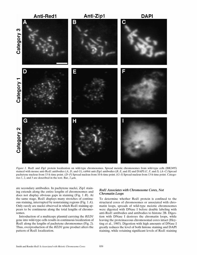
In wild-type cells, Red1 staining is generally not contin-uous along the lengths of pachytene chromosomes. Thoughcontinuous linear stretches of Red1 localization are ob-served, frequent gaps in staining are also seen. This pat-tern contrasts with the continuous localization of the Zip1protein along pachytene chromosomes (Sym et al., 1993).To compare directly the anti-Red1 and anti-Zip1 stainingpatterns, wild-type meiotic chromosomes were double-labeledwith mouse anti-Red1 antibodies and rabbit anti-Zip1 an-tibodies, which were subsequently detected with appropri-
Smith and Roeder
Red1 Is Associated with Meiotic Chromosome Cores
959
ate secondary antibodies. In pachytene nuclei, Zip1 stain-ing extends along the entire lengths of chromosomes anddoes not display obvious gaps in staining (Fig. 1
B
). Atthe same stage, Red1 displays many stretches of continu-ous staining, interrupted by nonstaining regions (Fig. 1
A
).Only rarely are nuclei observed in which Red1 staining ap-pears to be continuous along the total lengths of chromo-somes.
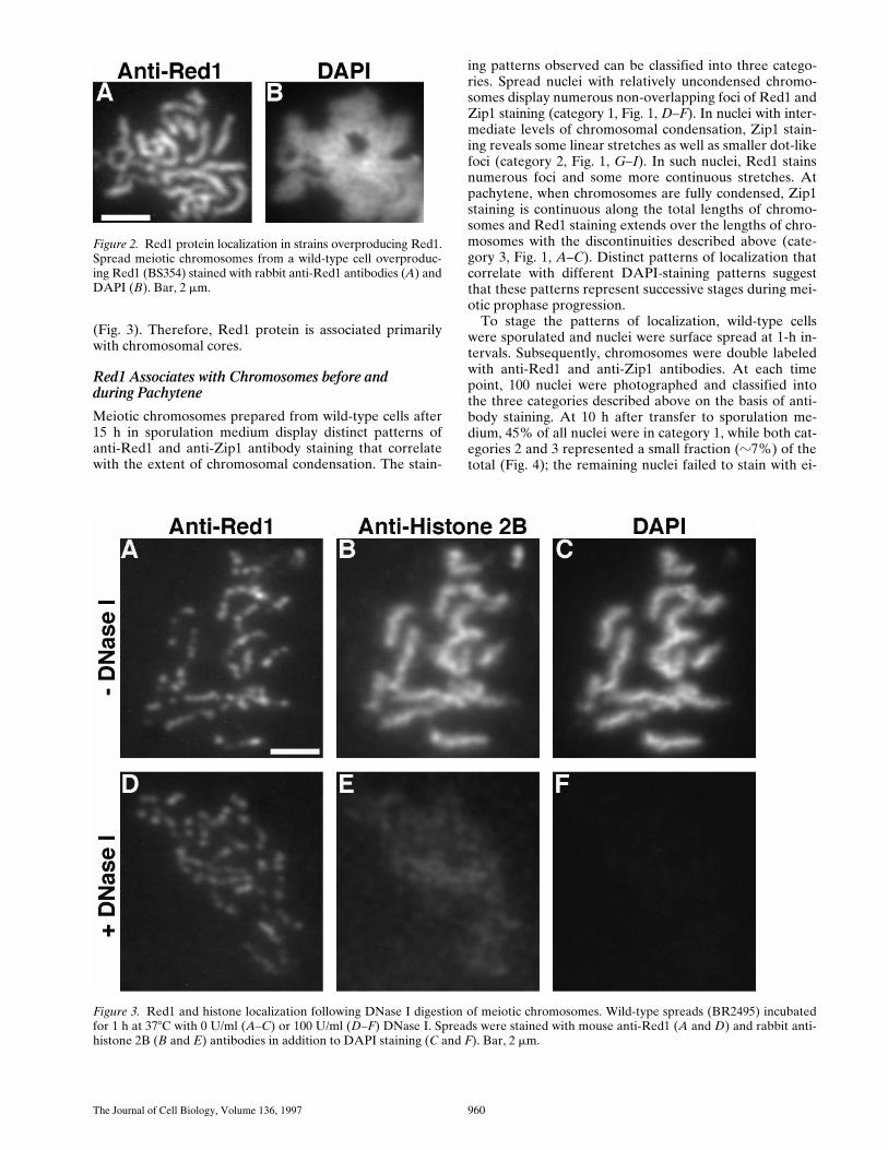
Introduction of a multicopy plasmid carrying the
RED1
gene into wild-type cells results in continuous localization ofRed1 along the lengths of pachytene chromosomes (Fig. 2).Thus, overproduction of the
RED1
gene product alters thepattern of Red1 localization.
Red1 Associates with Chromosome Cores, Not Chromatin Loops
To determine whether Red1 protein is confined to thestructural cores of chromosomes or associated with chro-matin loops, spreads of wild-type meiotic chromosomeswere digested with DNase I before double labeling withanti-Red1 antibodies and antibodies to histone 2B. Diges-tion with DNase I destroys the chromatin loops, whileleaving the proteinaceous chromosomal cores intact (Hey-ting et al., 1985). Digestion with high amounts of DNase Igreatly reduces the level of both histone staining and DAPIstaining, while retaining significant levels of Red1 staining
Figure 1. Red1 and Zip1 protein localization on wild-type chromosomes. Spread meiotic chromosomes from wild-type cells (BR2495)stained with mouse anti-Red1 antibodies (A, D, and G), rabbit anti-Zip1 antibodies (B, E, and H) and DAPI (C, F, and I). (A–C) Spreadpachytene nucleus from 15-h time point. (D–F) Spread nucleus from 10-h time point. (G–I) Spread nucleus from 13-h time point. Catego-ries 1, 2, and 3 are described in the text. Bar, 2 mm.
The Journal of Cell Biology, Volume 136, 1997 960
(Fig. 3). Therefore, Red1 protein is associated primarilywith chromosomal cores.
Red1 Associates with Chromosomes before andduring Pachytene
Meiotic chromosomes prepared from wild-type cells after15 h in sporulation medium display distinct patterns ofanti-Red1 and anti-Zip1 antibody staining that correlatewith the extent of chromosomal condensation. The stain-
ing patterns observed can be classified into three catego-ries. Spread nuclei with relatively uncondensed chromo-somes display numerous non-overlapping foci of Red1 andZip1 staining (category 1, Fig. 1,
D–F
). In nuclei with inter-mediate levels of chromosomal condensation, Zip1 stain-ing reveals some linear stretches as well as smaller dot-likefoci (category 2, Fig. 1,
G–I
). In such nuclei, Red1 stainsnumerous foci and some more continuous stretches. Atpachytene, when chromosomes are fully condensed, Zip1staining is continuous along the total lengths of chromo-somes and Red1 staining extends over the lengths of chro-mosomes with the discontinuities described above (cate-gory 3, Fig. 1,
A–C
). Distinct patterns of localization thatcorrelate with different DAPI-staining patterns suggestthat these patterns represent successive stages during mei-otic prophase progression.
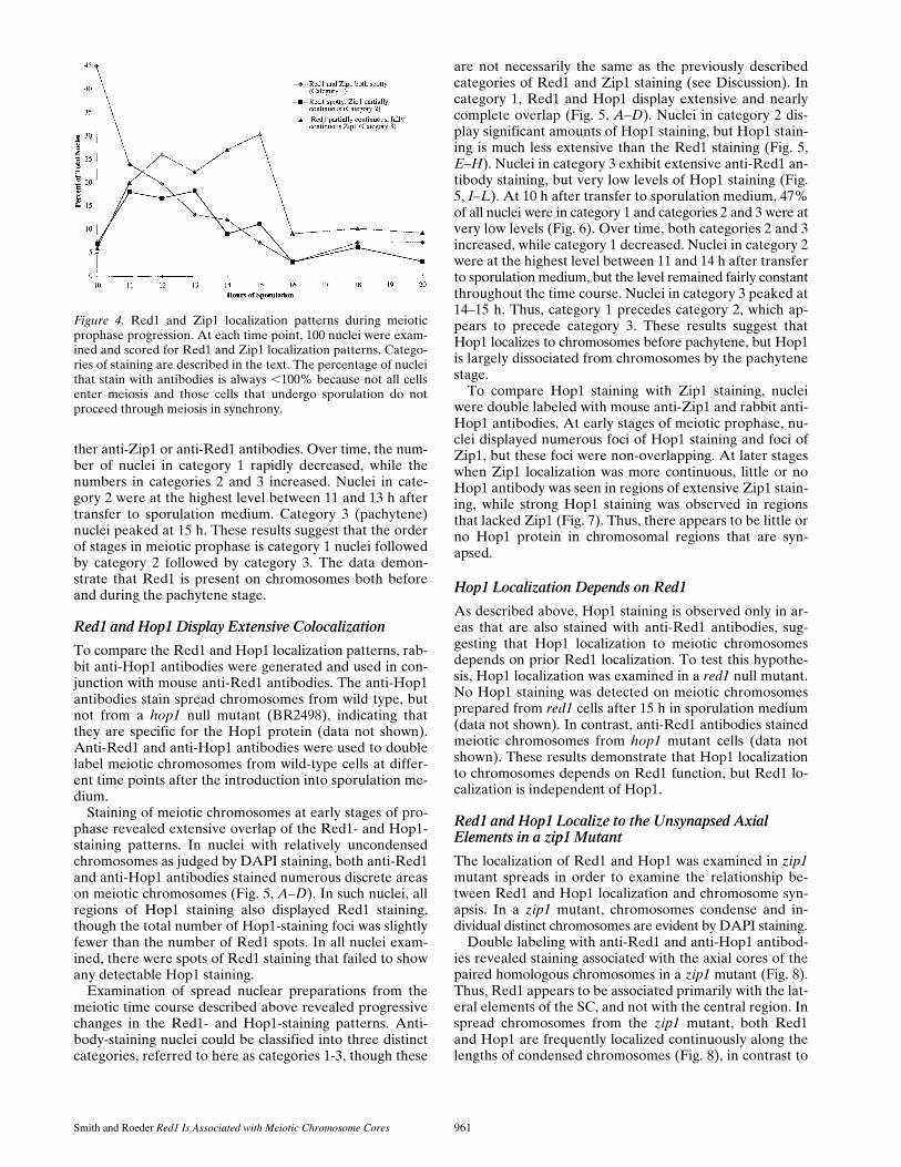
To stage the patterns of localization, wild-type cellswere sporulated and nuclei were surface spread at 1-h in-tervals. Subsequently, chromosomes were double labeledwith anti-Red1 and anti-Zip1 antibodies. At each timepoint, 100 nuclei were photographed and classified intothe three categories described above on the basis of anti-body staining. At 10 h after transfer to sporulation me-dium, 45% of all nuclei were in category 1, while both cat-egories 2 and 3 represented a small fraction (
z
7%) of thetotal (Fig. 4); the remaining nuclei failed to stain with ei-
Figure 2. Red1 protein localization in strains overproducing Red1.Spread meiotic chromosomes from a wild-type cell overproduc-ing Red1 (BS354) stained with rabbit anti-Red1 antibodies (A) andDAPI (B). Bar, 2 mm.
Figure 3. Red1 and histone localization following DNase I digestion of meiotic chromosomes. Wild-type spreads (BR2495) incubatedfor 1 h at 378C with 0 U/ml (A–C) or 100 U/ml (D–F) DNase I. Spreads were stained with mouse anti-Red1 (A and D) and rabbit anti-histone 2B (B and E) antibodies in addition to DAPI staining (C and F). Bar, 2 mm.
Smith and Roeder
Red1 Is Associated with Meiotic Chromosome Cores
961
ther anti-Zip1 or anti-Red1 antibodies. Over time, the num-ber of nuclei in category 1 rapidly decreased, while thenumbers in categories 2 and 3 increased. Nuclei in cate-gory 2 were at the highest level between 11 and 13 h aftertransfer to sporulation medium. Category 3 (pachytene)nuclei peaked at 15 h. These results suggest that the orderof stages in meiotic prophase is category 1 nuclei followedby category 2 followed by category 3. The data demon-strate that Red1 is present on chromosomes both beforeand during the pachytene stage.
Red1 and Hop1 Display Extensive Colocalization
To compare the Red1 and Hop1 localization patterns, rab-bit anti-Hop1 antibodies were generated and used in con-junction with mouse anti-Red1 antibodies. The anti-Hop1antibodies stain spread chromosomes from wild type, butnot from a
hop1
null mutant (BR2498), indicating thatthey are specific for the Hop1 protein (data not shown).Anti-Red1 and anti-Hop1 antibodies were used to doublelabel meiotic chromosomes from wild-type cells at differ-ent time points after the introduction into sporulation me-dium.
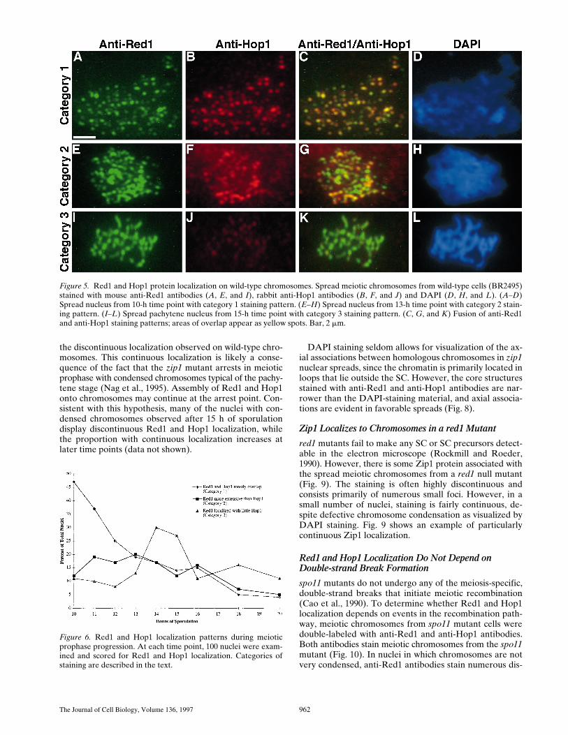
Staining of meiotic chromosomes at early stages of pro-phase revealed extensive overlap of the Red1- and Hop1-staining patterns. In nuclei with relatively uncondensedchromosomes as judged by DAPI staining, both anti-Red1and anti-Hop1 antibodies stained numerous discrete areason meiotic chromosomes (Fig. 5,
A–D
). In such nuclei, allregions of Hop1 staining also displayed Red1 staining,though the total number of Hop1-staining foci was slightlyfewer than the number of Red1 spots. In all nuclei exam-ined, there were spots of Red1 staining that failed to showany detectable Hop1 staining.
Examination of spread nuclear preparations from themeiotic time course described above revealed progressivechanges in the Red1- and Hop1-staining patterns. Anti-body-staining nuclei could be classified into three distinctcategories, referred to here as categories 1-3, though these
are not necessarily the same as the previously describedcategories of Red1 and Zip1 staining (see Discussion). Incategory 1, Red1 and Hop1 display extensive and nearlycomplete overlap (Fig. 5,
A–D
). Nuclei in category 2 dis-play significant amounts of Hop1 staining, but Hop1 stain-ing is much less extensive than the Red1 staining (Fig. 5,
E–H
). Nuclei in category 3 exhibit extensive anti-Red1 an-tibody staining, but very low levels of Hop1 staining (Fig.5, I–L). At 10 h after transfer to sporulation medium, 47%of all nuclei were in category 1 and categories 2 and 3 were atvery low levels (Fig. 6). Over time, both categories 2 and 3increased, while category 1 decreased. Nuclei in category 2were at the highest level between 11 and 14 h after transferto sporulation medium, but the level remained fairly constantthroughout the time course. Nuclei in category 3 peaked at14–15 h. Thus, category 1 precedes category 2, which ap-pears to precede category 3. These results suggest thatHop1 localizes to chromosomes before pachytene, but Hop1is largely dissociated from chromosomes by the pachytenestage.
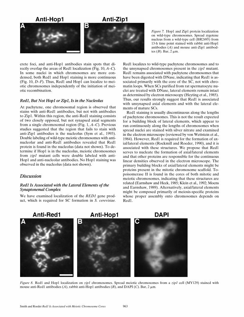
To compare Hop1 staining with Zip1 staining, nucleiwere double labeled with mouse anti-Zip1 and rabbit anti-Hop1 antibodies. At early stages of meiotic prophase, nu-clei displayed numerous foci of Hop1 staining and foci ofZip1, but these foci were non-overlapping. At later stageswhen Zip1 localization was more continuous, little or noHop1 antibody was seen in regions of extensive Zip1 stain-ing, while strong Hop1 staining was observed in regionsthat lacked Zip1 (Fig. 7). Thus, there appears to be little orno Hop1 protein in chromosomal regions that are syn-apsed.
Hop1 Localization Depends on Red1
As described above, Hop1 staining is observed only in ar-eas that are also stained with anti-Red1 antibodies, sug-gesting that Hop1 localization to meiotic chromosomesdepends on prior Red1 localization. To test this hypothe-sis, Hop1 localization was examined in a red1 null mutant.No Hop1 staining was detected on meiotic chromosomesprepared from red1 cells after 15 h in sporulation medium(data not shown). In contrast, anti-Red1 antibodies stainedmeiotic chromosomes from hop1 mutant cells (data notshown). These results demonstrate that Hop1 localizationto chromosomes depends on Red1 function, but Red1 lo-calization is independent of Hop1.
Red1 and Hop1 Localize to the Unsynapsed Axial Elements in a zip1 Mutant
The localization of Red1 and Hop1 was examined in zip1mutant spreads in order to examine the relationship be-tween Red1 and Hop1 localization and chromosome syn-apsis. In a zip1 mutant, chromosomes condense and in-dividual distinct chromosomes are evident by DAPI staining.
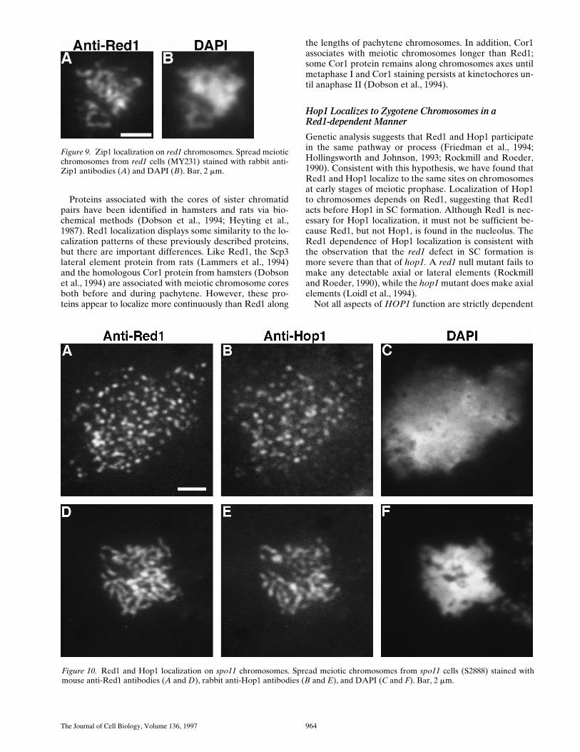
Double labeling with anti-Red1 and anti-Hop1 antibod-ies revealed staining associated with the axial cores of thepaired homologous chromosomes in a zip1 mutant (Fig. 8).Thus, Red1 appears to be associated primarily with the lat-eral elements of the SC, and not with the central region. Inspread chromosomes from the zip1 mutant, both Red1and Hop1 are frequently localized continuously along thelengths of condensed chromosomes (Fig. 8), in contrast to
Figure 4. Red1 and Zip1 localization patterns during meioticprophase progression. At each time point, 100 nuclei were exam-ined and scored for Red1 and Zip1 localization patterns. Catego-ries of staining are described in the text. The percentage of nucleithat stain with antibodies is always ,100% because not all cellsenter meiosis and those cells that undergo sporulation do notproceed through meiosis in synchrony.
The Journal of Cell Biology, Volume 136, 1997 962
the discontinuous localization observed on wild-type chro-mosomes. This continuous localization is likely a conse-quence of the fact that the zip1 mutant arrests in meioticprophase with condensed chromosomes typical of the pachy-tene stage (Nag et al., 1995). Assembly of Red1 and Hop1onto chromosomes may continue at the arrest point. Con-sistent with this hypothesis, many of the nuclei with con-densed chromosomes observed after 15 h of sporulationdisplay discontinuous Red1 and Hop1 localization, whilethe proportion with continuous localization increases atlater time points (data not shown).
DAPI staining seldom allows for visualization of the ax-ial associations between homologous chromosomes in zip1nuclear spreads, since the chromatin is primarily located inloops that lie outside the SC. However, the core structuresstained with anti-Red1 and anti-Hop1 antibodies are nar-rower than the DAPI-staining material, and axial associa-tions are evident in favorable spreads (Fig. 8).
Zip1 Localizes to Chromosomes in a red1 Mutant
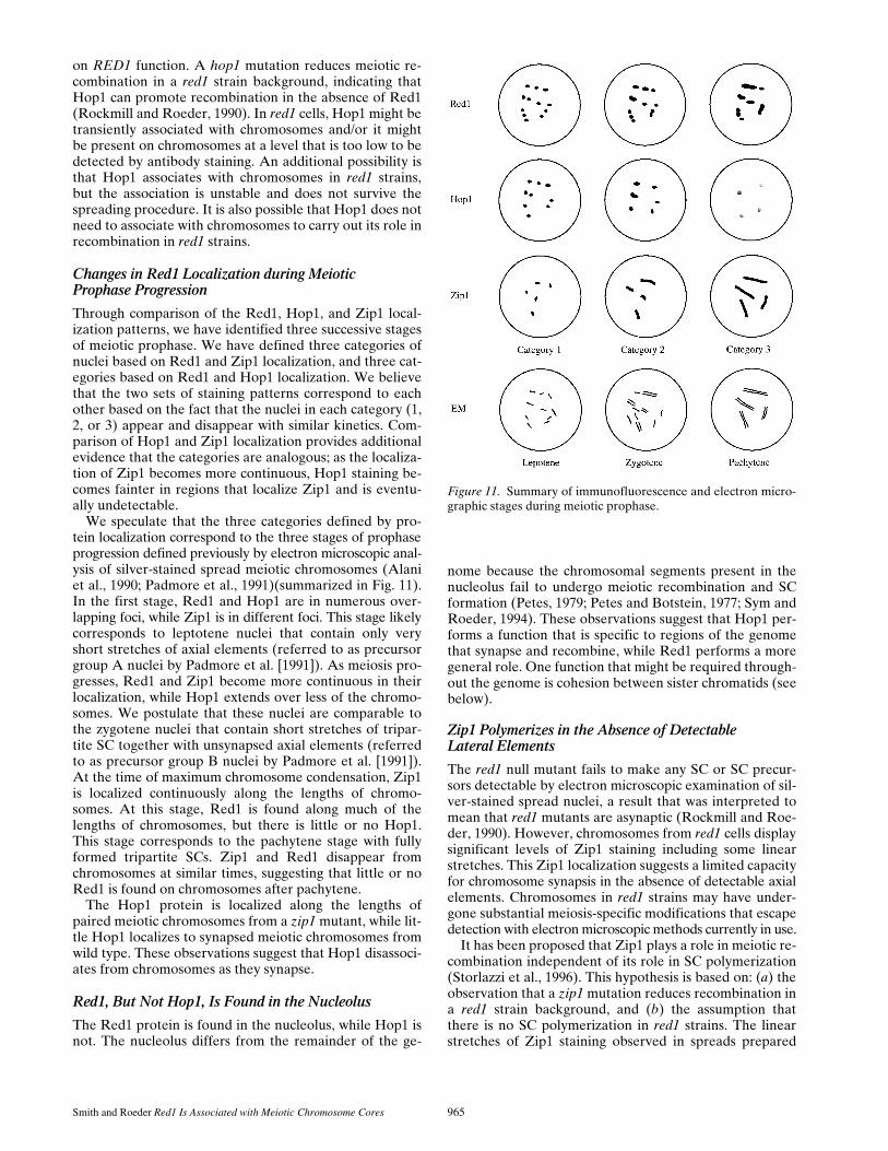
red1 mutants fail to make any SC or SC precursors detect-able in the electron microscope (Rockmill and Roeder,1990). However, there is some Zip1 protein associated withthe spread meiotic chromosomes from a red1 null mutant(Fig. 9). The staining is often highly discontinuous andconsists primarily of numerous small foci. However, in asmall number of nuclei, staining is fairly continuous, de-spite defective chromosome condensation as visualized byDAPI staining. Fig. 9 shows an example of particularlycontinuous Zip1 localization.
Red1 and Hop1 Localization Do Not Depend on Double-strand Break Formation
spo11 mutants do not undergo any of the meiosis-specific,double-strand breaks that initiate meiotic recombination(Cao et al., 1990). To determine whether Red1 and Hop1localization depends on events in the recombination path-way, meiotic chromosomes from spo11 mutant cells weredouble-labeled with anti-Red1 and anti-Hop1 antibodies.Both antibodies stain meiotic chromosomes from the spo11mutant (Fig. 10). In nuclei in which chromosomes are notvery condensed, anti-Red1 antibodies stain numerous dis-
Figure 5. Red1 and Hop1 protein localization on wild-type chromosomes. Spread meiotic chromosomes from wild-type cells (BR2495)stained with mouse anti-Red1 antibodies (A, E, and I), rabbit anti-Hop1 antibodies (B, F, and J) and DAPI (D, H, and L). (A–D)Spread nucleus from 10-h time point with category 1 staining pattern. (E–H) Spread nucleus from 13-h time point with category 2 stain-ing pattern. (I–L) Spread pachytene nucleus from 15-h time point with category 3 staining pattern. (C, G, and K) Fusion of anti-Red1and anti-Hop1 staining patterns; areas of overlap appear as yellow spots. Bar, 2 mm.
Figure 6. Red1 and Hop1 localization patterns during meioticprophase progression. At each time point, 100 nuclei were exam-ined and scored for Red1 and Hop1 localization. Categories ofstaining are described in the text.
Smith and Roeder Red1 Is Associated with Meiotic Chromosome Cores 963
crete foci, and anti-Hop1 antibodies stain spots that di-rectly overlap the areas of Red1 localization (Fig. 10, A–C).In some nuclei in which chromosomes are more con-densed, both Red1 and Hop1 staining is more continuous(Fig. 10, D–F). Thus, Red1 and Hop1 can localize to mei-otic chromosomes independently of the initiation of mei-otic recombination.
Red1, But Not Hop1 or Zip1, Is in the Nucleolus
At pachytene, one chromosomal region is observed thatstains with anti-Red1 antibodies, but not with antibodiesto Zip1. Within this region, the anti-Red1 staining consistsof two closely opposed, but not synapsed axial segmentsfrom a single chromosomal region (Fig. 1, A–C). Previousstudies suggested that the region that fails to stain withanti-Zip1 antibodies is the nucleolus (Sym et al., 1993).Double labeling of wild-type meiotic chromosomes with anti-nucleolar and anti-Red1 antibodies revealed that Red1protein is found in the nucleolus (data not shown). To de-termine if Hop1 is in the nucleolus, meiotic chromosomesfrom zip1 mutant cells were double labeled with anti-Hop1 and anti-nucleolar antibodies. No Hop1 staining wasobserved in the nucleolus (data not shown).
Discussion
Red1 Is Associated with the Lateral Elements of the Synaptonemal Complex
We have examined localization of the RED1 gene prod-uct, which is required for SC formation in S. cerevisiae.
Red1 localizes to wild-type pachytene chromosomes and tothe unsynapsed chromosomes present in the zip1 mutant.Red1 remains associated with pachytene chromosomes thathave been digested with DNase, indicating that Red1 is as-sociated primarily with the core of the SC, not with chro-matin loops. When SCs purified from rat spermatocyte nu-clei are treated with DNase, lateral elements remain intactas determined by electron microscopy (Heyting et al., 1985).Thus, our results strongly suggest that Red1 is associatedwith unsynapsed axial elements and with the lateral ele-ments of mature SCs.
Red1 staining is usually discontinuous along the lengthsof pachytene chromosomes. This is not the result expectedfor a building block of lateral elements, which appear torun continuously along the lengths of chromosomes whenspread nuclei are stained with silver nitrate and examinedin the electron microscope (reviewed by von Wettstein et al.,1984). However, Red1 is required for the formation of ax-ial/lateral elements (Rockmill and Roeder, 1990), and it isassociated with these structures. We propose that Red1serves to nucleate the formation of axial/lateral elementsand that other proteins are responsible for the continuouslinear densities observed in the electron microscope. Theprimary building blocks of axial/lateral elements might beproteins present in the mitotic chromosome scaffold. To-poisomerase II is found in the cores of both mitotic andmeiotic chromosomes, indicating that these structures arerelated (Earnshaw and Heck, 1985; Klein et al., 1992; Moensand Earnshaw, 1989). Alternatively, axial/lateral elementsmight be composed primarily of meiosis-specific proteinswhose proper assembly onto chromosomes depends onRed1.
Figure 7. Hop1 and Zip1 protein localizationon wild-type chromosomes. Spread zygotenenucleus from a wild-type cell (BR2495) from13-h time point stained with rabbit anti-Hop1antibodies (A) and mouse anti-Zip1 antibod-ies (B). Bar, 2 mm.
Figure 8. Red1 and Hop1 localization on zip1 chromosomes. Spread meiotic chromosomes from a zip1 cell (MY129) stained withmouse anti-Red1 antibodies (A), rabbit anti-Hop1 antibodies (B), and DAPI (C). Bar, 2 mm.
The Journal of Cell Biology, Volume 136, 1997 964
Proteins associated with the cores of sister chromatidpairs have been identified in hamsters and rats via bio-chemical methods (Dobson et al., 1994; Heyting et al.,1987). Red1 localization displays some similarity to the lo-calization patterns of these previously described proteins,but there are important differences. Like Red1, the Scp3lateral element protein from rats (Lammers et al., 1994)and the homologous Cor1 protein from hamsters (Dobsonet al., 1994) are associated with meiotic chromosome coresboth before and during pachytene. However, these pro-teins appear to localize more continuously than Red1 along
the lengths of pachytene chromosomes. In addition, Cor1associates with meiotic chromosomes longer than Red1;some Cor1 protein remains along chromosomes axes untilmetaphase I and Cor1 staining persists at kinetochores un-til anaphase II (Dobson et al., 1994).
Hop1 Localizes to Zygotene Chromosomes in aRed1-dependent Manner
Genetic analysis suggests that Red1 and Hop1 participatein the same pathway or process (Friedman et al., 1994;Hollingsworth and Johnson, 1993; Rockmill and Roeder,1990). Consistent with this hypothesis, we have found thatRed1 and Hop1 localize to the same sites on chromosomesat early stages of meiotic prophase. Localization of Hop1to chromosomes depends on Red1, suggesting that Red1acts before Hop1 in SC formation. Although Red1 is nec-essary for Hop1 localization, it must not be sufficient be-cause Red1, but not Hop1, is found in the nucleolus. TheRed1 dependence of Hop1 localization is consistent withthe observation that the red1 defect in SC formation ismore severe than that of hop1. A red1 null mutant fails tomake any detectable axial or lateral elements (Rockmilland Roeder, 1990), while the hop1 mutant does make axialelements (Loidl et al., 1994).
Not all aspects of HOP1 function are strictly dependent
Figure 9. Zip1 localization on red1 chromosomes. Spread meioticchromosomes from red1 cells (MY231) stained with rabbit anti-Zip1 antibodies (A) and DAPI (B). Bar, 2 mm.
Figure 10. Red1 and Hop1 localization on spo11 chromosomes. Spread meiotic chromosomes from spo11 cells (S2888) stained withmouse anti-Red1 antibodies (A and D), rabbit anti-Hop1 antibodies (B and E), and DAPI (C and F). Bar, 2 mm.
Smith and Roeder Red1 Is Associated with Meiotic Chromosome Cores 965
on RED1 function. A hop1 mutation reduces meiotic re-combination in a red1 strain background, indicating thatHop1 can promote recombination in the absence of Red1(Rockmill and Roeder, 1990). In red1 cells, Hop1 might betransiently associated with chromosomes and/or it mightbe present on chromosomes at a level that is too low to bedetected by antibody staining. An additional possibility isthat Hop1 associates with chromosomes in red1 strains,but the association is unstable and does not survive thespreading procedure. It is also possible that Hop1 does notneed to associate with chromosomes to carry out its role inrecombination in red1 strains.
Changes in Red1 Localization during MeioticProphase Progression
Through comparison of the Red1, Hop1, and Zip1 local-ization patterns, we have identified three successive stagesof meiotic prophase. We have defined three categories ofnuclei based on Red1 and Zip1 localization, and three cat-egories based on Red1 and Hop1 localization. We believethat the two sets of staining patterns correspond to eachother based on the fact that the nuclei in each category (1,2, or 3) appear and disappear with similar kinetics. Com-parison of Hop1 and Zip1 localization provides additionalevidence that the categories are analogous; as the localiza-tion of Zip1 becomes more continuous, Hop1 staining be-comes fainter in regions that localize Zip1 and is eventu-ally undetectable.
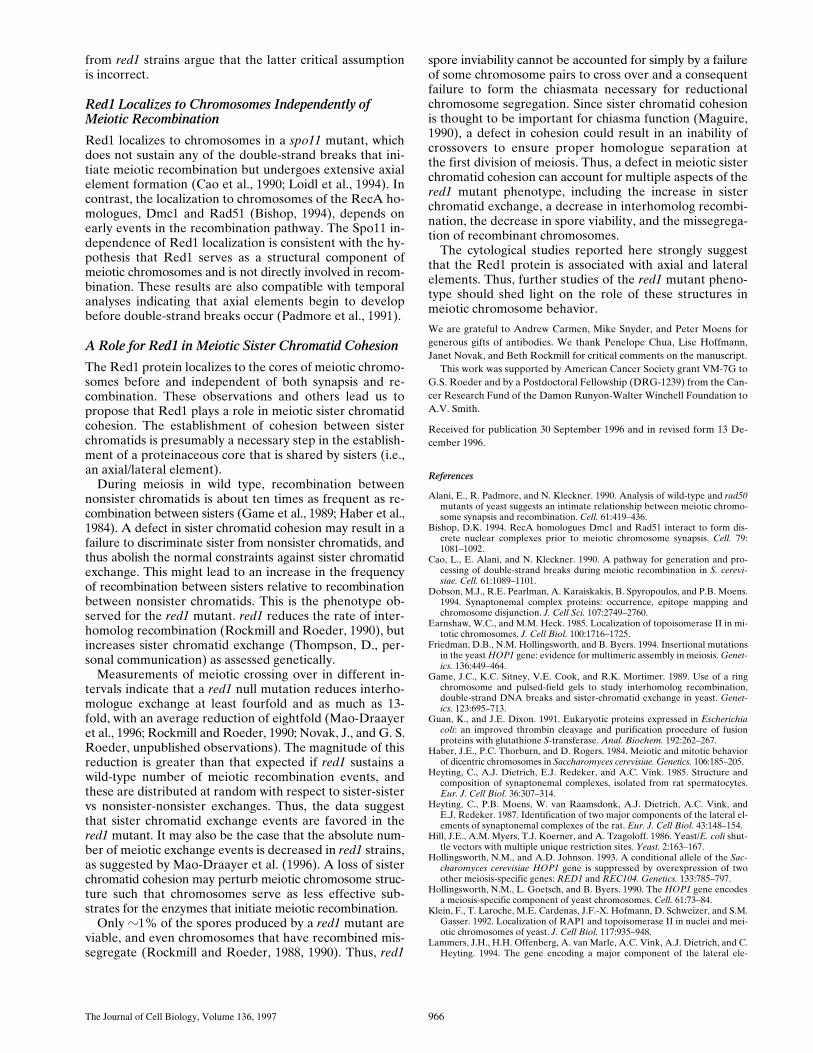
We speculate that the three categories defined by pro-tein localization correspond to the three stages of prophaseprogression defined previously by electron microscopic anal-ysis of silver-stained spread meiotic chromosomes (Alaniet al., 1990; Padmore et al., 1991)(summarized in Fig. 11).In the first stage, Red1 and Hop1 are in numerous over-lapping foci, while Zip1 is in different foci. This stage likelycorresponds to leptotene nuclei that contain only veryshort stretches of axial elements (referred to as precursorgroup A nuclei by Padmore et al. [1991]). As meiosis pro-gresses, Red1 and Zip1 become more continuous in theirlocalization, while Hop1 extends over less of the chromo-somes. We postulate that these nuclei are comparable tothe zygotene nuclei that contain short stretches of tripar-tite SC together with unsynapsed axial elements (referredto as precursor group B nuclei by Padmore et al. [1991]).At the time of maximum chromosome condensation, Zip1is localized continuously along the lengths of chromo-somes. At this stage, Red1 is found along much of thelengths of chromosomes, but there is little or no Hop1.This stage corresponds to the pachytene stage with fullyformed tripartite SCs. Zip1 and Red1 disappear fromchromosomes at similar times, suggesting that little or noRed1 is found on chromosomes after pachytene.
The Hop1 protein is localized along the lengths ofpaired meiotic chromosomes from a zip1 mutant, while lit-tle Hop1 localizes to synapsed meiotic chromosomes fromwild type. These observations suggest that Hop1 disassoci-ates from chromosomes as they synapse.
Red1, But Not Hop1, Is Found in the Nucleolus
The Red1 protein is found in the nucleolus, while Hop1 isnot. The nucleolus differs from the remainder of the ge-
nome because the chromosomal segments present in thenucleolus fail to undergo meiotic recombination and SCformation (Petes, 1979; Petes and Botstein, 1977; Sym andRoeder, 1994). These observations suggest that Hop1 per-forms a function that is specific to regions of the genomethat synapse and recombine, while Red1 performs a moregeneral role. One function that might be required through-out the genome is cohesion between sister chromatids (seebelow).
Zip1 Polymerizes in the Absence of DetectableLateral Elements
The red1 null mutant fails to make any SC or SC precur-sors detectable by electron microscopic examination of sil-ver-stained spread nuclei, a result that was interpreted tomean that red1 mutants are asynaptic (Rockmill and Roe-der, 1990). However, chromosomes from red1 cells displaysignificant levels of Zip1 staining including some linearstretches. This Zip1 localization suggests a limited capacityfor chromosome synapsis in the absence of detectable axialelements. Chromosomes in red1 strains may have under-gone substantial meiosis-specific modifications that escapedetection with electron microscopic methods currently in use.
It has been proposed that Zip1 plays a role in meiotic re-combination independent of its role in SC polymerization(Storlazzi et al., 1996). This hypothesis is based on: (a) theobservation that a zip1 mutation reduces recombination ina red1 strain background, and (b) the assumption thatthere is no SC polymerization in red1 strains. The linearstretches of Zip1 staining observed in spreads prepared
Figure 11. Summary of immunofluorescence and electron micro-graphic stages during meiotic prophase.
The Journal of Cell Biology, Volume 136, 1997 966
from red1 strains argue that the latter critical assumptionis incorrect.
Red1 Localizes to Chromosomes Independently of Meiotic Recombination
Red1 localizes to chromosomes in a spo11 mutant, whichdoes not sustain any of the double-strand breaks that ini-tiate meiotic recombination but undergoes extensive axialelement formation (Cao et al., 1990; Loidl et al., 1994). Incontrast, the localization to chromosomes of the RecA ho-mologues, Dmc1 and Rad51 (Bishop, 1994), depends onearly events in the recombination pathway. The Spo11 in-dependence of Red1 localization is consistent with the hy-pothesis that Red1 serves as a structural component ofmeiotic chromosomes and is not directly involved in recom-bination. These results are also compatible with temporalanalyses indicating that axial elements begin to developbefore double-strand breaks occur (Padmore et al., 1991).
A Role for Red1 in Meiotic Sister Chromatid Cohesion
The Red1 protein localizes to the cores of meiotic chromo-somes before and independent of both synapsis and re-combination. These observations and others lead us topropose that Red1 plays a role in meiotic sister chromatidcohesion. The establishment of cohesion between sisterchromatids is presumably a necessary step in the establish-ment of a proteinaceous core that is shared by sisters (i.e.,an axial/lateral element).
During meiosis in wild type, recombination betweennonsister chromatids is about ten times as frequent as re-combination between sisters (Game et al., 1989; Haber et al.,1984). A defect in sister chromatid cohesion may result in afailure to discriminate sister from nonsister chromatids, andthus abolish the normal constraints against sister chromatidexchange. This might lead to an increase in the frequencyof recombination between sisters relative to recombinationbetween nonsister chromatids. This is the phenotype ob-served for the red1 mutant. red1 reduces the rate of inter-homolog recombination (Rockmill and Roeder, 1990), butincreases sister chromatid exchange (Thompson, D., per-sonal communication) as assessed genetically.
Measurements of meiotic crossing over in different in-tervals indicate that a red1 null mutation reduces interho-mologue exchange at least fourfold and as much as 13-fold, with an average reduction of eightfold (Mao-Draayeret al., 1996; Rockmill and Roeder, 1990; Novak, J., and G. S.Roeder, unpublished observations). The magnitude of thisreduction is greater than that expected if red1 sustains awild-type number of meiotic recombination events, andthese are distributed at random with respect to sister-sistervs nonsister-nonsister exchanges. Thus, the data suggestthat sister chromatid exchange events are favored in thered1 mutant. It may also be the case that the absolute num-ber of meiotic exchange events is decreased in red1 strains,as suggested by Mao-Draayer et al. (1996). A loss of sisterchromatid cohesion may perturb meiotic chromosome struc-ture such that chromosomes serve as less effective sub-strates for the enzymes that initiate meiotic recombination.
Only z1% of the spores produced by a red1 mutant areviable, and even chromosomes that have recombined mis-segregate (Rockmill and Roeder, 1988, 1990). Thus, red1
spore inviability cannot be accounted for simply by a failureof some chromosome pairs to cross over and a consequentfailure to form the chiasmata necessary for reductionalchromosome segregation. Since sister chromatid cohesionis thought to be important for chiasma function (Maguire,1990), a defect in cohesion could result in an inability ofcrossovers to ensure proper homologue separation atthe first division of meiosis. Thus, a defect in meiotic sisterchromatid cohesion can account for multiple aspects of thered1 mutant phenotype, including the increase in sisterchromatid exchange, a decrease in interhomolog recombi-nation, the decrease in spore viability, and the missegrega-tion of recombinant chromosomes.
The cytological studies reported here strongly suggestthat the Red1 protein is associated with axial and lateralelements. Thus, further studies of the red1 mutant pheno-type should shed light on the role of these structures inmeiotic chromosome behavior.
We are grateful to Andrew Carmen, Mike Snyder, and Peter Moens forgenerous gifts of antibodies. We thank Penelope Chua, Lise Hoffmann,Janet Novak, and Beth Rockmill for critical comments on the manuscript.
This work was supported by American Cancer Society grant VM-7G toG.S. Roeder and by a Postdoctoral Fellowship (DRG-1239) from the Can-cer Research Fund of the Damon Runyon-Walter Winchell Foundation toA.V. Smith.
Received for publication 30 September 1996 and in revised form 13 De-cember 1996.
References
Alani, E., R. Padmore, and N. Kleckner. 1990. Analysis of wild-type and rad50mutants of yeast suggests an intimate relationship between meiotic chromo-some synapsis and recombination. Cell. 61:419–436.
Bishop, D.K. 1994. RecA homologues Dmc1 and Rad51 interact to form dis-crete nuclear complexes prior to meiotic chromosome synapsis. Cell. 79:1081–1092.
Cao, L., E. Alani, and N. Kleckner. 1990. A pathway for generation and pro-cessing of double-strand breaks during meiotic recombination in S. cerevi-siae. Cell. 61:1089–1101.
Dobson, M.J., R.E. Pearlman, A. Karaiskakis, B. Spyropoulos, and P.B. Moens.1994. Synaptonemal complex proteins: occurrence, epitope mapping andchromosome disjunction. J. Cell Sci. 107:2749–2760.
Earnshaw, W.C., and M.M. Heck. 1985. Localization of topoisomerase II in mi-totic chromosomes. J. Cell Biol. 100:1716–1725.
Friedman, D.B., N.M. Hollingsworth, and B. Byers. 1994. Insertional mutationsin the yeast HOP1 gene: evidence for multimeric assembly in meiosis. Genet-ics. 136:449–464.
Game, J.C., K.C. Sitney, V.E. Cook, and R.K. Mortimer. 1989. Use of a ringchromosome and pulsed-field gels to study interhomolog recombination,double-strand DNA breaks and sister-chromatid exchange in yeast. Genet-ics. 123:695–713.
Guan, K., and J.E. Dixon. 1991. Eukaryotic proteins expressed in Escherichiacoli: an improved thrombin cleavage and purification procedure of fusionproteins with glutathione S-transferase. Anal. Biochem. 192:262–267.
Haber, J.E., P.C. Thorburn, and D. Rogers. 1984. Meiotic and mitotic behaviorof dicentric chromosomes in Saccharomyces cerevisiae. Genetics. 106:185–205.
Heyting, C., A.J. Dietrich, E.J. Redeker, and A.C. Vink. 1985. Structure andcomposition of synaptonemal complexes, isolated from rat spermatocytes.Eur. J. Cell Biol. 36:307–314.
Heyting, C., P.B. Moens, W. van Raamsdonk, A.J. Dietrich, A.C. Vink, andE.J. Redeker. 1987. Identification of two major components of the lateral el-ements of synaptonemal complexes of the rat. Eur. J. Cell Biol. 43:148–154.
Hill, J.E., A.M. Myers, T.J. Koerner, and A. Tzagoloff. 1986. Yeast/E. coli shut-tle vectors with multiple unique restriction sites. Yeast. 2:163–167.
Hollingsworth, N.M., and A.D. Johnson. 1993. A conditional allele of the Sac-charomyces cerevisiae HOP1 gene is suppressed by overexpression of twoother meiosis-specific genes: RED1 and REC104. Genetics. 133:785–797.
Hollingsworth, N.M., L. Goetsch, and B. Byers. 1990. The HOP1 gene encodesa meiosis-specific component of yeast chromosomes. Cell. 61:73–84.
Klein, F., T. Laroche, M.E. Cardenas, J.F.-X. Hofmann, D. Schweizer, and S.M.Gasser. 1992. Localization of RAP1 and topoisomerase II in nuclei and mei-otic chromosomes of yeast. J. Cell Biol. 117:935–948.
Lammers, J.H., H.H. Offenberg, A. van Marle, A.C. Vink, A.J. Dietrich, and C.Heyting. 1994. The gene encoding a major component of the lateral ele-
Smith and Roeder Red1 Is Associated with Meiotic Chromosome Cores 967
ments of synaptonemal complexes of the rat is related to X-linked lympho-cyte-regulated genes. Mol. Cell. Biol. 14:1137–1146.
Loidl, J., F. Klein, and H. Scherthan. 1994. Homologous pairing is reduced butnot abolished in asynaptic mutants of yeast. J. Cell Biol. 125:1191–1200.
Maguire, M.P. 1990. Sister chromatid cohesiveness: vital function, obscuremechanism. Biochem. Cell Biol. 68:1231–1242.
Mao-Draayer, Y., A.M. Galbraith, D.L. Pittman, M. Cool, and R.E. Malone.1996. Analysis of meiotic recombination pathways in the yeast Saccharomy-ces cerevisiae. Genetics. 144:71–86.
Moens, P.B., and W.C. Earnshaw. 1989. Anti-topoisomerase II recognizes mei-otic chromosome cores. Chromosoma. 98:317–322.
Nag, D.K., H. Scherthan, B. Rockmill, J. Bhargava, and G.S. Roeder. 1995.Heteroduplex DNA formation and homolog pairing in yeast meiotic mu-tants. Genetics. 141:75–86.
Padmore, R., L. Cao, and N. Kleckner. 1991. Temporal comparison of recombi-nation and synaptonemal complex formation during meiosis in S. cerevisiae.Cell. 66:1239–1256.
Petes, T.D. 1979. Meiotic mapping of yeast ribosomal deoxyribonucleic acid onchromosome XII. J. Bacteriol. 138:185–192.
Petes, T.D., and D. Botstein. 1977. Simple Mendelian inheritance of the reiter-ated ribosomal DNA of yeast. Proc. Natl. Acad. Sci. USA. 74:5091–5095.
Rockmill, B., and G.S. Roeder. 1988. RED1: a yeast gene required for the seg-regation of chromosomes during the reductional division of meiosis. Proc.Natl. Acad. Sci. USA. 85:6057–6061.
Rockmill, B., and G.S. Roeder. 1990. Meiosis in asynaptic yeast. Genetics. 126:563–574.
Rockmill, B., J. Engebrecht, H. Scherthan, J. Loidl, and G.S. Roeder. 1995a.The yeast MER2 gene is required for meiotic recombination and chromo-some synapsis. Genetics. 141:49–59.
Rockmill, B., M. Sym, H. Scherthan, and G.S. Roeder. 1995b. Roles for twoRecA homologs in promoting meiotic chromosome synapsis. Genes Dev. 9:2684–2695.
Roeder, G.S. 1995. Sex and the single cell: meiosis in yeast. Proc. Natl. Acad.Sci. USA. 92:10450–10456.
Sherman, F., G.R. Fink, and J.B. Hicks. 1986. Methods in Yeast Genetics: ALaboratory Manual. Cold Spring Harbor Laboratory, Cold Spring Harbor,New York.
Smith, D.B., and K.S. Johnson. 1988. Single-step purification of polypeptidesexpressed in Escherichia coli as fusions with glutathione S-transferase. Gene(Amst.). 67:31–40.
Snyder, M. 1989. The SPA2 protein of yeast localizes to sites of cell growth. J.Cell Biol. 108:1419–1429.
Storlazzi, A., L. Xu, A. Schwacha, and N. Kleckner. 1996. Synaptonemal com-plex (SC) component Zip1 plays a role in meiotic recombination indepen-dent of SC polymerization along the chromosomes. Proc. Natl. Acad. Sci.USA. 93:9043–9048.
Sym, M., and G.S. Roeder. 1994. Crossover interference is abolished in the ab-sence of a synaptonemal complex protein. Cell. 79:283–292.
Sym, M., and G.S. Roeder. 1995. Zip1-induced changes in synaptonemal com-plex structure and polycomplex assembly. J. Cell Biol. 128:455–466.
Sym, M., J. Engebrecht, and G.S. Roeder. 1993. ZIP1 is a synaptonemal com-plex protein required for meiotic chromosome synapsis. Cell. 72:365–378.
Thompson, E.A., and G.S. Roeder. 1989. Expression and DNA sequence ofRED1, a gene required for meiosis I chromosome segregation in yeast. Mol.Gen. Genet. 218:293–301.
von Wettstein, D., S.W. Rasmussen, and P.B. Holm. 1984. The synaptonemalcomplex in genetic segregation. Annu. Rev. Genet. 18:331–413.