Embed Size (px)
Citation preview
REVIEW
The virulence of Streptococcus mutans and the abilityto form biofilms
W. Krzyściak & A. Jurczak & D. Kościelniak &
B. Bystrowska & A. Skalniak
Received: 4 September 2013 /Accepted: 2 October 2013 /Published online: 24 October 2013# The Author(s) 2013. This article is published with open access at Springerlink.com
Abstract In some diseases, a very important role is played bythe ability of bacteria to form multi-dimensional complexstructure known as biofilm. The most common disease of theoral cavity, known as dental caries, is a top leader. Streptococcusmutans , one of the many etiological factors of dental caries, is amicroorganismwhich is able to acquire new properties allowingfor the expression of pathogenicity determinants determining itsvirulence in specific environmental conditions. Through themechanism of adhesion to a solid surface, S. mutans is capableof colonizing the oral cavity and also of forming bacterialbiofilm. Additional properties enabling S. mutans to colonizethe oral cavity include the ability to survive in an acidicenvironment and specific interaction with other microorganismscolonizing this ecosystem. This review is an attempt to establishwhich characteristics associated with biofilm formation—virulence determinants of S. mutans—are responsible for thedevelopment of dental caries. In order to extend the knowledgeof the nature of Streptococcus infections, an attempt to face thefollowing problems will be made: Biofilm formation as acomplex process of protein–bacterium interaction. To whatextent do microorganisms of the cariogenic flora exemplified
by S. mutans differ in virulence determinants “expression”from microorganisms of physiological flora? How does theenvironment of the oral cavity and its microorganismsaffect the biofilm formation of dominant species? How doselected inhibitors affect the biofilm formation of cariogenicmicroorganisms?
Introduction
In the 18th century, it was demonstrated that microorganismslive not only in a single-cell form, but are also capable offorming clusters suspended in a mucilaginous extracellularsubstance. The pathogenicity of certain microbial species suchas Streptococcus mutans , Staphylococcus epidermidis ,Legionella pneumophila or Pseudomonas aeruginosa isinseparably associated with their ability to form biofilms onsolid surfaces, e.g., tissues, catheters or implants [1–4]. Thisfeature allows microorganisms to form three-dimensionalstructures in which cells become more resistant to antibioticsand changing environmental conditions, among others,through changes occurring as a result of interbacterialinteractions and the presence of an exopolysaccharide matrixprotecting the entire structure [5, 6].
Microorganism pathogenicity
Interactions observed between pathogen and host havebeen the subject of research and discussion for many years.The historical approach to the problem of microorganismpathogenicity postulated by, amongst others, Koch, puts thepathogen or host in themain position by this featured affiliationto one of them. The term ‘pathogenicity determinants’ isrelated to the features which determine a microorganism’sability to cause disease, but which themselves are not required
W. Krzyściak (*)Department of Medical Diagnostics, Faculty of Pharmacy, MedicalCollege, Jagiellonian University, UJCM 9 Medyczna St.,30-688 Krakow, Polande-mail: [email protected]
A. Jurczak :D. KościelniakDepartment of Pediatric Dentistry, Institute of Dentistry, MedicalCollege, Jagiellonian University, Krakow, Poland
B. BystrowskaDepartment of Toxicity, Faculty of Pharmacy, Medical College,Jagiellonian University, Krakow, Poland
A. SkalniakGenetics Laboratory, Department of Endocrinology, MedicalCollege, Jagiellonian University, Krakow, Poland
Eur J Clin Microbiol Infect Dis (2014) 33:499–515DOI 10.1007/s10096-013-1993-7

for its survival [7]. Henderson et al. defined pathogenicitydeterminants as pathogen components which cause damagein a host organism; this may include factors vitally importantfor the microorganisms [8]. These definitions do not, however,take into account the role of host susceptibility to infection,indicating only that pathogen properties are responsible fordisease development. According to these definitions, onlythose microorganisms which cause diseases in healthypeople are pathogens and not opportunistic or commensalmicroorganisms which are only able to infect the hosts withimmune system disorders.
Casadevall and Pirofski [9] proposed a new definitionfor pathogenicity and pathogenicity determinants of themicroorganisms taking into account the state of hostimmunological defense. A given microorganism pathogenicityis expressed as a range of damage which is caused by themicroorganism itself and by the immune system as a responseto a pathogen. The state of a host’s immunological defense isthe main determinant for bacterial pathogens and it determinesthe infection course and cure [9].
The determination of microorganism pathogenicitydeterminants according to the classification proposed byCasadevall and Pirofski (six classes) poses some interpretationproblems in class I, since it seems that the key role in theseinfections is played by a host’s condition. The infected humanconstitutes a complex ecosystem in which homeostasis inthe field of the bacteria-immune system is observed in thephysiological state. The development of bacteremiaoccurs in cases of disturbances in this system, usually asa result of immunity deficiency or, more rarely, as a resultof an overexpression of the features determining bacteriapathogenicity. There are, however, some bacteria, e.g., S.salivarius or most bacteria colonizing the oral cavity,which live in the human environment but are rarely describedas pathogens, even in those patients with an impaired immunesystem. This suggests that even pathogens of a low virulencehave to possess a minimum set of features determining theirpathogenicity, which would allow them to penetrate andproliferate in a host organism.
In the study by Kreikemeyer et al. [10] concerning biofilmstructure, the determinants of Streptococcus pathogenicity arerelated to the discovery of long filamentous structures similarto the pilus observed on bacteria surfaces. These structuresexhibit adhesive properties and may play a key role inadhering to host cells and tissues, as well as in biofilmformation by pathogens of the Streptococcus species. Thestudy demonstrated that the described pilus in S. pyogenesare responsible for bacteria adhesion and the formation ofmicrocolonies on host cell surfaces, and also for aggregationitself, especially when influenced by human saliva. Also, inthe case of S. agalactiae , these structures are engaged inpathogen interaction with host cells, e.g., adherence andbypassing the epithelium barrier. They are also responsible
for bacteria clustering in a biofilm-like structure. In thepathogenesis of the diseases caused by S. pneumoniae , as inthe case of other Streptococcus species, these structures areengaged in adhesion and colonization processes; however,compared to other Streptococcus species, the frequency ofpilus occurrence is lower (less than 30 %) [11].
There are studies suggesting that S. mutans isolates have agreater ability to form biofilm than the isolates of otherStreptococcus species, which colonize the human oral cavityenvironment [12, 13]. The studies focused on S. mutans cells,which form biofilm and proved that they exhibit a differentexpression of some proteins in comparison to planktoniccultures, e.g., an increase in exopolyphosphatase expressionand a decrease of lactate dehydrogenase or pyruvate kinaseexpression [14]. Increased virulence of cells forming biofilmcan also be associated with a higher tolerance to low pH, ascompared to planktonic cultures.
The main components of the biofilm formed on the surfaceof teeth include: glucan (10–20 % of dry weight), fructan(1–2 % of dry weight) and proteins (40 % dry matter).Moreover, it differs from the surrounding saliva in termsof the levels of lipids, calcium, magnesium, fluorine, andphosphorus. In situ, in 80 % it consists of water [6]. Thus,an amorphous membrane formed in such conditions providesideal conditions for bacterial survival and determines thevirulence of the biofilm structure. Physical and biochemicalmatrix features allow for the adhesion of microorganisms,promote cohesion (the aggregation of cells), and act as a sourceof energy reservoirs. Moreover, limited diffusion to and fromthe biofilm helps to focus the ions and other nutrients in themicroenvironment, such as biofilm; however, it hinders thepenetration of substances from outside, including antibiotics[6]. It can be observed that bacteria in the biofilm resembleother organisms for which the clustering was also anevolutionary adaptation for survival.
The ability of bacteria of the S. mutans species to formbiofilms is significant from a clinical point of view, mainly inthe context of carries etiology; however, there are also singlecasuistic cases of infective endocarditis (IE) with theinvolvement of this bacteria [15, 16]. The development of IEis observed when endocardial damage occurs followed by theformation of a very small blood clot, in which platelets play acrucial role. If, at the same time, microorganisms enter thebloodstream, they may use these favorable growth conditionsfor deposition and biofilm formation.
Such microclots are formed at the interface of the mitraland tricuspid valves from the side of atria and from the side ofchambers at the aortic and pulmonary valves [17]. Thesurvival of S. mutans in the bloodstream, where it is veryrarely observed after dental surgery, is associated with thepresence of several virulence factors on the surface of bacteria,and these have been described in different cases of IE causedby this microorganism. First are those factors responsible for
500 Eur J Clin Microbiol Infect Dis (2014) 33:499–515

the increased resistance of proteins to phagocytosis, namely,the fibronectin-binding protein (so-called autolysin A, AtlA)[18] and serotype-specific polysaccharide [19]. Next, C antigensincreased platelet aggregation, initiating the coagulation cascadeprocess and, consequently, often leading to hypercoagulablestates [20]. An equally important role is attributed to thecollagen-binding protein (Cnm) [21], so that the S. mutansbacterium can adhere to the heart tissue and penetrate theendothelium of the coronary arteries, consequently leadingto endocarditis [22].
Biofilm formation as a complex processof protein–bacterium interaction
The most common oral cavity infectious disease in which animportant causative role is played by biofilms formed bymicroorganisms on the teeth and gums surface is dental caries.One of the main etiological factors of the above-mentioneddisease is S. mutans [23–25].
Caries is the most common childhood illness. It is estimatedto occur five times more often than the second most commonchildhood illness, asthma [26]. The Decayed, Missing andFilled Teeth (DMFT) index rate was observed for Bolivia,Guatemala, Bosnia andHerzegovina, and Philippines. Directlybehind them in contrast, the lowest DMFT index rate wasobserved for Russia, Europe’s Eastern Bloc including Poland,and most South American countries [27, 28].
Comparing the DMFT index rate for European countries,one can observe that the epidemiological situation in Poland isnot ideal. In 2012, the DMFT index rate for 12-year-olds wasestimated at 4.4, whereas the lowest observed in Holland wasestimated at 0.9 and the highest in Latvia was estimated at 7.7.In terms of 6-year-olds, the situation deteriorates because,among the countries under investigation, the highest DMFTindex rate equal to 5.1 was recorded in Poland. It is estimatedthat, in Poland, dental caries is observed in 35–50 % ofchildren aged 2–3 years and in 50–60 % of children aged 3–4 years [27, 29]. According to alarming reports from theWorld Health Organization (WHO) and the National Instituteof Public Health (PZH), dental caries is observed in almost100 % of children aged 6–7 years. Caries in permanent teethstarts just after the dentition of the first molar teeth and hasbeen reported in approximately 90 % of Polish 12-year-oldchildren [30, 31].
In the view of the above data, it is extremely important tounderstand more fully the mechanisms of cariogenic strainactivity on the basis of the biofilm formed by them, whichmay be applied in the prevention and early diagnostics ofdental caries in children.
Biofilms present in the oral cavity are three-dimensionalstructures, consisting of bacterial strains anchored to solidsurfaces such as tooth enamel, tooth roots or dental implants.
They are embedded in an exopolysaccharidematrix [32]. Over700 different bacterial species incorporating into biofilms havebeen identified so far [33, 34]. The structure and composition ofthe exopolysaccharide matrix is determined by the conditionsexisting in the oral cavity and change over time. Theextracellular polysaccharides (EPS) also affect the physicaland biochemical properties of the biofilm [6, 35].
The primary sources of EPS are glucosyltransferase (GTF)and fructosyltransferase (Ftf), products of interaction withsucrose and starch hydrolysates. Exopolymers contained inthe polysaccharide matrix form its stability and providepossibilities of binding bacterial cells [6, 35, 36]. Researchon the composition of the polysaccharide matrix revealed thepresence of components as shown in Fig. 1.
The process of biofilm formation begins with the coating ofthe tooth surface through the salivary pellicle [6, 32]. Thispellicle is formed by salivary components (such as proline-rich proteins, amylase, lysozyme, histatin, peroxidase, mucin,and bacterial components, e.g., Ftf, Gtf, lipoteichoic acid)specifically adsorbed to the acquired enamel pellicle (AEP)[6, 37]. AEP is the basis for microorganism-induced biofilmformation colonizing the oral cavity [37]. Single S. mutanscells or their aggregates fuse with pellicles via two independentmechanisms: sucrose-dependent and sucrose-independent [23,25, 32].
The sucrose-dependent mechanism
Glucosyltransferases (GtfS)
The sucrose-dependent mechanism of plaque formation isbased on glucosyltransferases (GTFB, -C, and-D) producedby S. mutans in combination with glucan-binding proteins(GBPs) [6, 25, 38]. Glucosyltransferases play critical roles in
Fig. 1 The percentage composition of polysaccharide matrix (the figurewas prepared based on the data published by Bowen and Koo [6])
Eur J Clin Microbiol Infect Dis (2014) 33:499–515 501

virulent dental plaque development and are responsiblefor glucans formation from sucrose. The synthesizedglucans provide the possibility of both bacterial adhesionto the tooth enamel and microorganisms to each other. Thanksto this process, microcolonies are formed which favor theformation of biofilm. Each of the three types of Gtf plays adifferent, though similar role in biofilm formation and,therefore, the loss or mutation of one of them impairs thewhole process [6, 38].
In vivo, GtfS very rapidly (approximately 1 min) adsorbsinto the hydroxyapatite surface (sHA) of the enamel-coatedsalivary pellicle. The highest affinity for the sHA is reportedfor GTFC (formally known as GtfSI), which is a hydrophiliccompound and produces a mixture of soluble (with mostly α-1,6-linkages) and insoluble glucans. However, it has ahydrophobic domain which is related to its affinity for dentalplaque. This enables the interaction with saliva proteins in thepellicle, such as lysozyme or α-amylase. GtfB (known asGtfI), a glucosyltransferase, is primarily responsible for theinteraction with other S. mutans bacteria which mainlysynthesize insoluble glucan rich in α-1,3-linkages. It isresponsible for the formation of highly differentiatedmicrocolonies forming the structure of biofilm [39]. Itsactivity significantly increases when there is a glucose in theenvironment, which is not a typical situation and is observedextremely rarely [6, 40].
GtfD (GtfS) predominantly forms soluble, quicklymetabolizable polysaccharides and acts as a primer for GtfB.
Glucosyltransferases also interact with the components ofsaliva: amylase, in consequence blocking the activity andadsorption to hydroxyapatite; lysozyme, which decreases theactivity of GtfB, but does not affect the characteristics ofglucans formed by the enzyme; peroxidase, which inhibitsthe activity of all three glycosyltransferases.
GtfS also have the ability to bind to other bacterialcells, even if these are not bacteria synthesizing their ownglycosyltransferases. Therefore, cooperation between Gtfsand microorganisms allows for the building of a plate stronglyattached to the teeth surface with stable bonds betweenbacterial cells [6, 35].
Genes encodingGtfB and GtfC lie close to each other, havea very similar amino acid composition (95 % homology), andare subject to the same regulatory processes. These genes areexpressed in response to acidification of the environment or insituations of glucose or sucrose excess in the environment.Many factors additionally influence the physiologicalexpression of these genes. One of them is catabolism andfactors like RegM, an inhibitory regulator of S. mutans [6].The protein regulating the catabolism in Streptococcus andStaphylococcus bacteria, RegM, is also known as catabolitecontrol protein A (CCPA) [41]. Its regulatory function is basedon the inhibition of genes involved in the utilization ofalternative carbon sources and the activation of the expression
of genes whose products are involved in the elimination ofexcess carbon from the cell [42–44]. Inactivation of the RegMof S. mutans results in a very strong decrease in the promoterexpression of gtfBC [6]. Other factors are the products ofgenes: luxS (AI-2 autoinducer-coding synthesis), whichaffects the expression of gtfB and gtfC [6, 25, 45], and alsoropA (encoding for the trigger factor) regulating theproduction of GtfB and GtfD [6, 23]. Also, the VicRK signaltransduction system affects the physiological expression ofGtfs. In a mutant strain of S. mutans which does not have thissystem, a significant decrease in gtfD gene expression, as wellas increased expression of the gtfB gene, was observed.
The gtfD gene lies distant from other genes, is characterizedby lesser homology (50 %) and is subject to different regulatorymechanisms. Its expression is specifically induced by copperions [6]. Apart from Gtfs, the synthesis and structure of glucansis also affected by enzymes such as mutanase and α-1,6-glucosidase. Therefore, the structure of glucans in the biofilmmatrix is changeable, and in mature dental plaque, water-insoluble polysaccharides dominate [6, 46, 47].
Glucans binding of proteins (Gbps)
Another component of the sucrose-dependent mechanism isGbps mediating the binding of bacteria to glucans. Four typesof this protein are known: GbpA, -B, -C, and -D [6, 25, 38].The GBPC protein (and probablyGbpB) is associatedwith thebacterial cell wall and, therefore, acts as a specific receptor forglucan. All four types of proteins play a role in microorganismadhesion and biofilm formation; however, the GbpD proteinseems to play a key role [6]. Research on the utility of GS5(deletion of the gbpB gene) and UACA2 (a strain of the gbpBgene expression encoding antisense RNA) of S. mutansstrains showed that the absence or mutation of the geneencoding GbpB results in a change of cell shape and a slowingdown of its growth [38]. This disables the appropriatedevelopment of biofilm, which, instead of a diverse, denseformation, becomes a product of non-regular cell clusterssurrounded by a matrix of unusual structure.
The expression patterns of GbpB which have been studiedby Fujita et al. [40] may have some relationships with S.mutans virulence. Attention was paid to the relationship ofthe profiles and biological activity of the above-mentionedproteins with the virulence of isolated proteins. GbpBexpression analysis isolates revealed the existence of severalexpression patterns. Strains that showed single and multiplebonding were classified as S and M strains, while strainswithout explicit GbpB expression were classified as Ntype. The GbpB expression pattern distribution was foundto be different for the Japanese and Finnish clinical strainssimultaneously.
For the Japanese strains, the highest frequency wasreported for S (81 %), followed by the M (15 %) and N types
502 Eur J Clin Microbiol Infect Dis (2014) 33:499–515

(4 %), while in the Finnish population, the frequencies wereestimated at 42 % for the M type, 37 % for the S type, and21 % for the N type without the explicit expression of GbpB.Studies show a clear differentiation of each type depending onthe origin of clinical isolates. Geographic variation seems tobe crucial in determining the virulence of the strains, which,depending on the environment, do or do not acquire specificvirulence factors, constituting a kind of adaptation whichallows them to colonize a new ecological niche. Anexplanation of the distribution and specificity of GbpBexpression patterns may be useful for the evaluation of S.mutans virulence in individual patients [40].
The sucrose-independent mechanism
The sucrose-independent mechanism is not relevant in thevirulence of S. mutans . In the second mechanism of adhesion(sucrose-independent), an interaction is observed between theadhesive particles of S. mutans and the AEP. Agglutininsfound in saliva are involved in the process of adhesion andaggregation of S. mutans thanks to interaction with the I/IIantigen, which is a multifunctional PI adhesin (also known asAgB, SpaP, or Pac1 adhesin) anchored in the bacterial cellwall, and encoded by the spaP gene [23–25, 48].
The protein family of Ag I/II, represented by SpaP, SspA,or SspB, is identified not only on the surface of S. mutans , butalso on other microorganisms, such as Streptococcuspyogenes , Streptococcus agalactiae , or Streptococcus suis[48]. Genetic sequences encoding Ag I/II comprise six distinctregions. The most important of these are the A region rich inalanine and the P region rich in proline. Region V, locatedbetween them, is composed in the majority of differentsequences found in individual strains. The A and V regionsencode adhesive epitopes appearing on the surface of bacterialcells (so-called adhesive types) responsible for affinity to thesalivary glycoproteins [49]. The contribution of the A, P, andV regions to the adhesion has been confirmed by studies usingmutant strains [48]. None of them had the ability to adhere tosolid surfaces coated with the salivary pellicle. The expressionand biological activity of the P1 protein in S. mutans is alsodependent on multiple gene products, i.e., luxS, ropA, andsrtA genes (encoding the enzyme responsible for theattachment of P1 adhesin to the cell wall) [22, 50].
The SpaP protein and other proteins of the Ag I/II familyspecifically interact with glycoprotein-340 (gp-340) found insaliva. It is interesting that the gp-340 dissolved in the liquidphase of saliva plays a role in the aggregation of bacterial cellsand, thus, purifies the oral cavity. However, if gp-340 isadsorbed on the surface of the teeth or gums, it acts as areceptor for surface bacterial adhesins initiating the adhesionprocess [22, 48] (Fig. 2). The Ag I/II protein family is alsoinvolved in the interaction between microorganisms, e.g.,
Streptococcus gordonii and Porphyromonas gingivalis , andin the aggregation of cells in the absence of gp-340 [48].
A key role in the interactions between S. mutans and salivaagglutinins is played by the actual structure and location of P1protein, as has been proven through the use of mutant strains(spaP and srtA) of this microorganism. Additionally, it hasbeen shown that the lack of expression of the gene encodingsortase A enzyme (SrtA) results not only in an abnormallocation of the P1 adhesin, but also of other surface proteins,which is not negligible in the context of bacterial aggregationability [23]. The survival of S. mutans inside biofilms, at avery low pH inside microcolonies of other bacterial species(as observed in the oral cavity), should be considered as threemechanism-dependent phenomena which include: theupregulation system of F1F0-ATPase, biosynthesis ofmembrane fatty acids (for instance, FabM), and branched-chain amino acid (BCAA) (for instance, IIvC). This processmay be the main driving force in the survival of S. mutansinside a mixed biofilm (Fig. 3); thus, innovative therapiesaimed at inhibiting biofilm formation should be oriented alongthese three mechanisms and should be directed at all threesimultaneously and not just in one of them [51].
Salivary agglutinins
In the process of biofilm formation, a role equally important asthat of surface bacterial adhesins is played by salivaryagglutinins. This has been confirmed in vitro [23, 24].Researchers have also used mutant bacterial strains of themajor genes encoding the P1 protein to study the differencesand interactions during biofilm formation. The results clearlyindicate a less satisfactory process in the absence of salivaryagglutinins, as well as significantly less sufficiency in themutant strains compared to the parental strain [22, 23].It has also been proven that the initial stage of adhesionand biofilm formation of S. mutans may be stimulatedby salivary agglutinins and other salivary proteins (e.g.,mucin of high molecular mass or acidic proline-rich proteins)(Fig. 2) [23, 52].
Under specific conditions, the expression of the virulencefactors of S. mutans and biofilm formation in the oral cavitymay be modulated in two ways: through the environment inwhich bacterial growth was reported as well as by thepresence of other microorganisms, and the interactionsbetween them [25].
Culturing S. mutans strains under aerobic conditionsinduces an 80% reduction of bacterial ability to form biofilms[23]. Oxygen availability is the causative agent of variations inbacterial cell surface composition and modification of theproduction of autolysins and, specifically, in the signaltransduction system of VicRK (Fig. 2). The production of AtlAautolysin is conditioned by the SMu0629 gene expression ofoxidoreductase activity. Under aerobic conditions, increased
Eur J Clin Microbiol Infect Dis (2014) 33:499–515 503
expression of this gene is being observed and, thus, theoverproduction of AtlA autolysin, which inhibits the formationof biofilm. Vick kinase transducer is a system regulating theexpression and activity of AtlA autolysin. In order to confirmoxygen-induced alterations in autolysin, studies on mutantstrains for SMu0629 and vicK genes were conducted. Strainsfrom which the above-mentioned genes were removed adaptedbetter to aerobic conditions and showed a greater ability to formbiofilm as compared to the UA159 parental strain [52–54].
Diet habits
Diet is another factor influencing the process of biofilmformation. The occurrence of dental caries is stronglycorrelated to diet. Meal cariogenicity is affected not only by
the content of carbohydrates but also by the frequency of theirconsumption. Particular carbohydrates differ in their ability tocause caries. Saccharose is definitely the most cariogeniccarbohydrate [55]. It constitutes the main center of S. mutansmetabolism. These bacteria exhibit not only the ability todecompose this carbohydrate, but also produce glucans whichare significant in interactions between tooth enamel andcariogenic bacteria [6].
The presence of high levels of carbohydrates in children’sdiets, especially in the drinks given to children immediatelybefore sleep or during the night, constitutes a significant factorin ECC development [56]. Falling asleep with a sweetenednipple or bottle in the mouth stimulates the formation ofcarious lesions. Saliva excretion decreases during sleep, whichaccelerates the cariogenic strength of the activity of acids
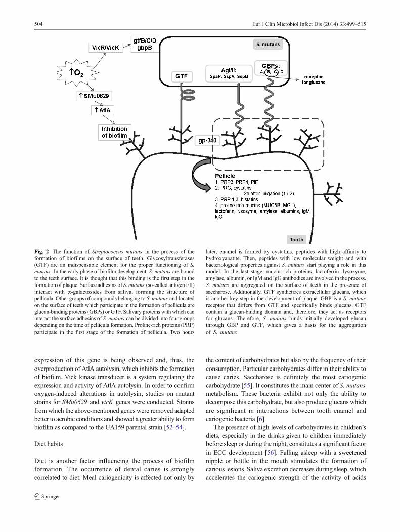
Fig. 2 The function of Streptococcus mutans in the process of theformation of biofilms on the surface of teeth. Glycosyltransferases(GTF) are an indispensable element for the proper functioning of S.mutans . In the early phase of biofilm development, S. mutans are boundto the teeth surface. It is thought that this binding is the first step in theformation of plaque. Surface adhesins of S. mutans (so-called antigen I/II)interact with α-galactosides from saliva, forming the structure ofpellicula. Other groups of compounds belonging to S. mutans and locatedon the surface of teeth which participate in the formation of pellicula areglucan-binding proteins (GBPs) or GTF. Salivary proteins with which caninteract the surface adhesins of S. mutans can be divided into four groupsdepending on the time of pellicula formation. Proline-rich proteins (PRP)participate in the first stage of the formation of pellicula. Two hours
later, enamel is formed by cystatins, peptides with high affinity tohydroxyapatite. Then, peptides with low molecular weight and withbacteriological properties against S. mutans start playing a role in thismodel. In the last stage, mucin-rich proteins, lactoferrin, lysozyme,amylase, albumin, or IgM and IgG antibodies are involved in the process.S. mutans are aggregated on the surface of teeth in the presence ofsaccharose. Additionally, GTF synthetizes extracellular glucans, whichis another key step in the development of plaque. GBP is a S. mutansreceptor that differs from GTF and specifically binds glucans. GTFcontain a glucan-binding domain and, therefore, they act as receptorsfor glucans. Therefore, S. mutans binds initially developed glucanthrough GBP and GTF, which gives a basis for the aggregationof S. mutans
504 Eur J Clin Microbiol Infect Dis (2014) 33:499–515

derived from carbohydrate metabolism. The opinionsconcerning the effect of feeding with milk on dental cariesdevelopment still remain controversial. According toresearchers, cow milk does not contain high amounts oflactose and, moreover, no significant relationship withcarious lesion formation has been demonstrated. Human milk,in turn, despite having higher amounts of lactose, alsocontains case in which adsorbs to the tooth surface and retardscaries development. Moreover, it contains considerableamounts of calcium and phosphates, which additionallystimulate enamel remineralization [57]. Also, the significantinfluence of sparkling drinks and fruit juice on dentalcaries development in children and teenagers has beendemonstrated. Acids common in sparkling drinks, such ascitric or phosphoric acids, considerably lower the oral cavitypH and stimulate an exchange of calcium ions present in theenamel on hydrons. It has also been demonstrated thatdrinking through a straw elongates and increases liquidcontact with the tooth surface, which unprofitably affects theirstructure [58].
It is not the level of carbohydrates in ameal but the frequencyof consumption of meals with high carbohydrate levels that is a
significant factor [59]. Also, time plays a substantial role in toothmineralization. It has been demonstrated that alternate processesof enamel demineralization and remineralization occur duringthe day. The loss of hydroxyapatite mineral components isrelated to the period when bacteria fermenting carbohydratesmay freely develop and synthesize acids into carbohydratesmetabolism byproducts. Time acts in favor of cariogenicpathogens which adapt to low pH values, which favor theprogress of the cariogenic process. The length of change in theperiod on the enamel surface is especially significant in cases ofearly childhood caries characterized by an aggressive course.Noticing the first changes on the upper incisor surface isextremely important, since carious lesions quickly spread onmolar teeth [60]. Moreover, the changes in the daily rhythm ofsaliva excretion play a significant role in caries exposure. Salivasecretion is decreased during the night hours, which limits itsability to protect against enamel demineralization [58].
Currently, studies on the development of products toprevent dental caries development are being conducted. Someare aimed at inhibiting the development of biofilm through theinfluence on Gtfs. There are a number of synthetic and naturalglycosyltransferase inhibitors, i.e., hop components, green tea,
Fig. 3 Production of lactic acidby Streptococcus mutans .Metabolism of variouscarbohydrates (including glucoseand fructose) by bacterial biofilm.Production and secretion of asignificant amount of lactic acid,which can cause demineralizationof teeth structure that can finallyresult in the development of decay
Eur J Clin Microbiol Infect Dis (2014) 33:499–515 505
medicinal plant extracts, plant polyphenols of high molecularmass (e.g., curcumin), cardiolipin, putrescine, cadaverine, orhypochlorite compounds or metal ions (Fe2+, Zn2+, Cu2+). Inorder to increase their effectiveness, there is a tendency todevelop formulations for oral hygiene which best use thepotential of the above-mentioned substances [6, 61, 62].
Biofilm structure
The formation of biofilm is a multistep and very complicatedprocess. A number of relevant factors and conditions arerequired in the oral cavity for the process to run correctly.
Interactions occurring between agglutinins of saliva andbacteria, and simultaneously between microorganisms, mightcause the formation of fur composed of cells beginning thecolonization: Actinomyces species, Streptococcus species,Lactobacillus species, Candida species. They transform intodifferent types of biofilm in the first layer of subgingivalplaque. Biofilm maturation is followed by the aggregation ofsubsequent bacteria and their growth. After 7 days, thenumber of Streptococcus bacteria decreases, but the numberof Fusobacterium nucleatum increases. After 3 weeks, intactsubgingival plaque begins to resemble morphologicallysupragingival plaque [32].
In the architecture of supragingival biofilm, four layers canbe distinguished. The first layer of biofilm consists of cellsdisplaying little fluorescence relative to cells in the top layer.This indicates the presence of Actinomyces sp. In this layer,physiologically inactive or dead cells can also be found. In theintermediate layer, many spindle-shaped cells are found,whose fluorescence indicates Fusobacterium nucleatum andTannerella sp., mostly T. forsythia . These bacteria can benefitfrom the proximal location of dead cells (e.g., they acquiresugar building their cell wall). The third layer of biofilm andthe intermediate layer mainly consist of a bacterial clustertermed the Cytophaga–Flavobacterium–Bacteroides (CFB),which consists mainly of Gram-negative bacteria Tannerellasp., Prevotella sp., and Bacteroidetes sp.
Apart from this, long cigar-like Synergistetes bacteria fromthe A group forming palisade-like stroma are also found inthis layer. These bacteria are in direct contact with the cells ofthe host immune system resembling polynuclear leukocytes,which, thereby, suggests their important role in theinteractions occurring between host and biofilm. Observationsof the top biofilm layer have shown that Spirochaetes bacteriaare the most abundant. Among them, bacterial aggregatescalled rough and fine brushes are found [32].
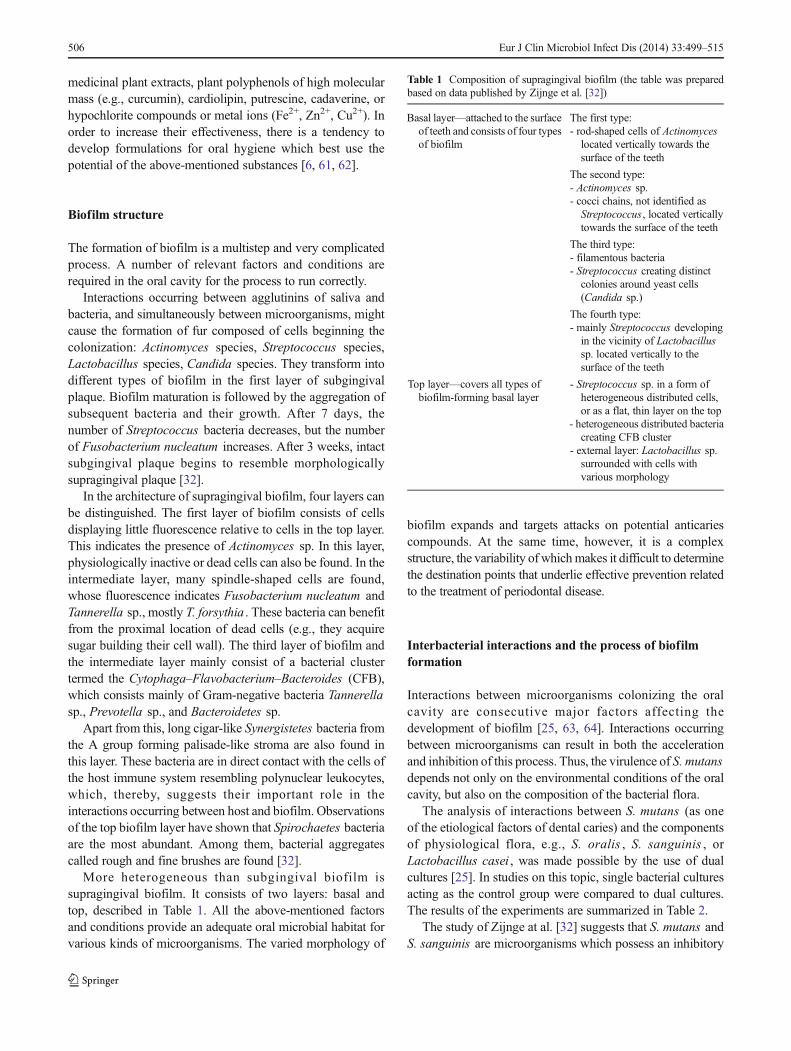
More heterogeneous than subgingival biofilm issupragingival biofilm. It consists of two layers: basal andtop, described in Table 1. All the above-mentioned factorsand conditions provide an adequate oral microbial habitat forvarious kinds of microorganisms. The varied morphology of
biofilm expands and targets attacks on potential anticariescompounds. At the same time, however, it is a complexstructure, the variability of whichmakes it difficult to determinethe destination points that underlie effective prevention relatedto the treatment of periodontal disease.
Interbacterial interactions and the process of biofilmformation
Interactions between microorganisms colonizing the oralcavity are consecutive major factors affecting thedevelopment of biofilm [25, 63, 64]. Interactions occurringbetween microorganisms can result in both the accelerationand inhibition of this process. Thus, the virulence of S. mutansdepends not only on the environmental conditions of the oralcavity, but also on the composition of the bacterial flora.
The analysis of interactions between S. mutans (as oneof the etiological factors of dental caries) and the componentsof physiological flora, e.g., S. oralis , S. sanguinis , orLactobacillus casei , was made possible by the use of dualcultures [25]. In studies on this topic, single bacterial culturesacting as the control group were compared to dual cultures.The results of the experiments are summarized in Table 2.
The study of Zijnge at al. [32] suggests that S. mutans andS. sanguinis are microorganisms which possess an inhibitory
Table 1 Composition of supragingival biofilm (the table was preparedbased on data published by Zijnge et al. [32])
Basal layer—attached to the surfaceof teeth and consists of four typesof biofilm
The first type:- rod-shaped cells of Actinomyceslocated vertically towards thesurface of the teeth
The second type:- Actinomyces sp.- cocci chains, not identified asStreptococcus , located verticallytowards the surface of the teeth
The third type:- filamentous bacteria- Streptococcus creating distinctcolonies around yeast cells(Candida sp.)
The fourth type:- mainly Streptococcus developingin the vicinity of Lactobacillussp. located vertically to thesurface of the teeth
Top layer—covers all types ofbiofilm-forming basal layer
- Streptococcus sp. in a form ofheterogeneous distributed cells,or as a flat, thin layer on the top
- heterogeneous distributed bacteriacreating CFB cluster
- external layer: Lactobacillus sp.surrounded with cells withvarious morphology
506 Eur J Clin Microbiol Infect Dis (2014) 33:499–515

effect on biofilm formation ability. Thus, taking into accountthis observation, researchers have decided to isolate andanalyze proteins produced by S. salivarius [63]. Based onthese studies, they found that substances responsible forbiofilm formation inhibition are: fructosyltransferase (Ftf)and exo-β-D-fructosidase (FruA).
Ftf is an enzyme involved in the metabolism of sucrose tofructose characterized as an extracellular homopolymer, levan(fructan). Next, the enzyme FruA performs a hydrolysisreaction of the formed fructan to fructose. Fructosidase isencoded by the FruA gene, which is identified in manydifferent species of streptococci and in fungi. The mechanismof action of both enzymes allows them to effectively inhibitthe sucrose-dependent adhesion of the S. mutans pathway[63]. Authors have confirmed the inhibitory effect of FruAon the process of biofilm formation by S. mutans . To confirmthis, a fructanase mixture (the mixture used was derived froma culture of Aspergillus niger and showed significant FruAactivity) containing exo- and endo-inulinase was addedfor the cultivation of this bacterium. It was shown thatthese enzymes did not affect bacterial growth inhibition;however, they strongly inhibited their ability to formbiofilm [63]. In addition, the ability of FruA fructosidaseto use sucrose from the oral environment causes a lack ofavailability of this carbohydrate for S. mutans . This phenomenonis observed because FruA digests sucrose before S. mutansglucosyltransferases manage to produce the glucans necessaryfor biofilm formation [63].
Another microorganism strongly affecting S. mutans -induced biofilm in the oral cavity is Lactobacillus spp. (LB).LB bacteria are commensal microorganisms colonizing,among others, the human oral cavity. LB is strongly associatedwith the development of dental caries in the dentine, because itferments sugars into acidic products, which decrease the pH inthe oral cavity, and promote the development of biofilm(Fig. 3) [65]. On the other hand, low pH and antibacterialagents, such as hydrogen peroxide or bacteriocins producedby LB microorganisms, favor the purification of the oralcavity from microorganisms which are non-adaptive to suchenvironmental conditions, for instance, Porphyromonasgingivalis [65]. Thanks to such properties, LB bacteria canbe widely used as probiotics [34, 65–68]. LBmicroorganisms,
by interacting with other microorganisms and the productionof antimicrobial proteins, contribute to maintaining thebalance in the natural flora of many ecosystems, e.g., the oralcavity [66].
Research conducted on various Lactobacil lusmicroorganisms has confirmed the inhibitory effect of manyof them on cariogenic microorganisms colonizing the oralcavity, among others S. mutans . Among the most activemicroorganisms are: L. paracasei , L. plantarum , L.salivarius , and L. rhamnosus [66]. It is also known that theabove-mentioned probiotics do not affect the adhesive abilityof S. mutans ; however, they are able to inhibit the proliferationof the pathogen, and, thus, retard biofilm formation anddevelopment [68, 69]. Unfortunately, the mechanism ofinteraction between Lactobacillus and S. mutans has not yetbeen sufficiently investigated. Detailed in vitro studiesfocused on Lactobacillus reuteri [34, 65] have shown thatdifferent strains of this species interact with S. mutans withdifferent forces. These interactions are dependent on the pH ofthe environment and the ability of probiotics to producehydrogen peroxide and reuterin, an antibacterial proteinformed from glycerol, resistant to proteolytic and lipolyticenzymes [24, 68]. The strains also differed in terms of theirability to adhere to surfaces coated with saliva. This feature isvery important because bacteria unconjugated to the solidphase are quickly removed by swallowing [63]. Similardifferences in activity against S. mutans have been observedamong different strains of Lactobacillus salivarius [66].
The role of Lactobacillus bacteria to sustain oral purity isalso a result of their specific coaggregation with S. mutanscells. This relationship applies to L. paracasei and L.rhamnosus [67]. The fusion of microorganisms in commonaggregates prevents the adhesion of S. mutans to the surfaceof teeth and gums. This allows the removal of the pathogenfrom the oral cavity with saliva before the first stage of biofilmformation begins, that is, adhesion [67].
Tahmourespour et al. showed that a biosurfactantderivative produced by probiotic bacterium, Lactobacillusfermentum , inhibits biofilm formation. In the presence of S.mutans , the above-mentioned ability to form a derivative wasinhibited, probably due to the inhibition of two key enzymes,GtfB and GtfC. A decrease in the gene expression encoding
Table 2 The results obtained in a study with the use of dual cultures (the table was prepared on the basis of data published by Wen et al. [25])
Dual culture Biofilm formation Expression of pathogenic factors of S. mutans
spaP gtfB gbpB luxS
S. mutans + S. oralis Not a relevant change 30-fold decrease No decrease 30-fold decrease 15-fold decrease
S. mutans + S. sanguinis Significantly important decrease Small decrease Small decrease Small decrease Change not statistically significant
S. mutans + L. casei Small increase(around 2-fold) 40-fold decrease 40-fold decrease 40-fold decrease 7-fold decrease
Eur J Clin Microbiol Infect Dis (2014) 33:499–515 507

the mentioned glucosyltransferases in the presence of abiosurfactant derivative produced by L. fermentum [70, 71]has been reported. Ogawa et al. noted that S. salivariuscan inhibit biofilm formation. Exo-beta-D-fructosidasesynthesized by S. salivarius blocks the synthesis ofpolysaccharides, which reduces the amount of extracellularmatrix produced by S. mutans [63].
Interactions occurring between S. mutans and othermicroorganisms in the oral cavity may contribute to both thedevelopment as well as the inhibition of biofilm formation.Through ongoing research, we have obtained new data onthese interactions which enables the search for and use ofnovel, more specific and effective measures for the preventionof and fight against dental caries. In this fight, it is important toutilize, apart from synthetic products, the potential of naturalproducts, such as probiotics and specific antimicrobialproteins. These substances exhibit a significantly lowernumber of side effects than artificially made specimens andare suitable for use under specific environmental conditions,which allows a high level of activity of the specimens to bemaintained over a long period of time.
Inhibition of biofilm formation—treatment perspectivesin dental caries
First, second, and third-generation medicines (phenoliccompounds, chlorhexidine, delmopinol)
The growing problem of strain resistance to antibioticscompels us to seek other methods which deal with pathogensthat occur in the oral cavity. Nowadays, we know of a numberof substances with effective antimicrobial activity inhibitingbiofilm development, for instance, chlorhexidine [72],delmopinol [73], or phenolic compounds [74]. Unfortunately,most of these substances cause side effects such as vomiting,diarrhea, addiction, or teeth discoloration. Therefore,alternative substances of antibacterial activity are being soughtwhich would be safe for the users [33, 75].
“Liquid enamel”—calcium phosphate
For many years, the primary role in the fight against dentalcaries has been played by calcium phosphate, which possessesremineralization properties [76–78]. A special calciumphosphate resin, which was designed for the gradual releaseof large amounts of these elements in places that requirereconstruction, was established very quickly. Later, thiscompound gained the form of a nanoparticle of amorphouscalcium phosphate composite (NACP), which was also aswidely used in dentistry as its precursor. The advantages ofthe nanocomposite are:
– Improved mechanical features for its use in dentistry ascompared to the classic combination of elements;
– Increased release of ions in acidic environments favoringthe formation of voids and dental caries;
– Fast neutralization and raising of low pH to a safe level(about 6).
[76, 77].
Nanoparticles of quaternary ammonium salts (QAS)
In the search for a substance which, apart from remineralizationproperties, would possess antibacterial ability, NACP wasenriched with quaternary ammonium salts (QAS). A specificapplication was found for a quaternary ammonium calleddimethacrylate (QADM). This compound has two activeantibacterial domains at its ends, which increase the desiredproperties, and, additionally, mixes with other dental mediaeasily [77]. Thanks to this change, a new composite of strongantibacterial activity was found. Its application results inshortening microorganism viability both as a planktonic formand in biofilms among others (Streptococcus sp. andLactobacillus sp.), decreasing acid production by bacteria anda reduction of microorganism metabolic activity [76, 77]. Thisis possible thanks to the amphiphilicity of QAS compounds,which allow for these substances to react with the lipid part ofthe cell membrane and interfere with its function. As a result, itindirectly affects the activity of enzymes involved in thetransport of substances through the membrane lipid, and, thus,alters the metabolic activity of bacterial cells [79].
Antibacterial nanoemulsions
Lethal QAS properties against a broad spectrum ofmicroorganisms have also been used in antibacterialnanoemulsions. In this context, cetylpyridinium chloride (CPC)was used [80]. Antibacterial nanoemulsion is characterized asa dispersing substance, namely, water and a lipid substancecomposed of surfactant, which forms nanoemulsion droplets.It is not toxic to humans or animals; however, it exhibitsantibacterial, antifungal, and antiviral activity. Antibacterialproperties of the emulsion result from the activity ofnanodrops on bacterial cell membranes destabilizing theintegration of its lipids [80].
Cetylpyridinium chloride (CPC)
CPChas the ability to inhibit Ftfs enzymes (fructosyltransferases),which play an important role in microorganism-inducedbiofilm formation in the oral cavity. Thanks to this property,CPC plays the role of an antibacterial substance (affecting thecell membrane of a microorganism) and a substance inhibitingthe development of biofilm. Increasing the efficiency of
508 Eur J Clin Microbiol Infect Dis (2014) 33:499–515
nanoemulsions through CPC enrichment has led to their widerapplication in products for oral hygiene, such as toothpastesand mouthwashes, and in dental materials, e.g., varnishes anddental fillings [80].
12-methacryloyloxydodecylpyridinium bromide (MDPB)
Among QAS, 12-methacryloyloxydodecylpyridiniumbromide (MDPB) has also found an application in the fightagainst dental caries [81, 82]. The properties ofMDPB and thepossibility of its application have attracted the attention ofresearch groups [82]. In experiments, the authors haveinvestigated the effect of MDPB on the bacterial flora of theoral cavity, interactions with dental materials, and thepossibility of a synergistic effect of this compound withnanoparticles of silver (NAg).
MDPB is a monomer characterized by antibacterialactivity against aerobic and anaerobic bacteria isolatedfrom translucent zones (i.e., Actinomyces , S. mutans ) andantifungal activity (e.g., Candida albicans ) [82]. Stiffenedby polymerization of the filling bonding layer, it remainsactive against pathogens and, at the same time, does notnegatively affect either human cells or the binding capacityof the filling material [82].
Silver nanoparticles
Silver compounds became the research objective for Chenget al. [81], who drew attention to their application inconjunction with QADM. Silver is known for its lethalactivity against a wide spectrum of bacteria, viruses, andfungi. Its antibacterial activity is due to the ability of cellmembrane disintegration, internal penetration, and destructionof intracellular organelles. An additional mechanism allowingfor the inactivation of bacterial enzymes by inhibitingbacterial DNA replication capability enhances the efficacy of
silver compounds. Low toxicity and long-term antibacterialactivity due to the gradual release and lower resistance ofbacteria against these compounds in relation to antibioticsare other advantages in the fight against microorganisms[81–83]. The sheer number of mechanisms and propertiesdescribing the activity of substances containing silver particleslead to the inability for microorganisms to sustain fullprotection against their effects. An additional difficulty is therole of silver compounds as catalysts, rather than substancesparticipating in chemical reactions [84].
Silver nanoparticles have a huge active surface, and,therefore, their level in dental composites is estimated at only0.05 to 0.1 % by weight. This proportion provides botheffective antibacterial activity as well as a lack of effect onthe mechanical filling properties [81].
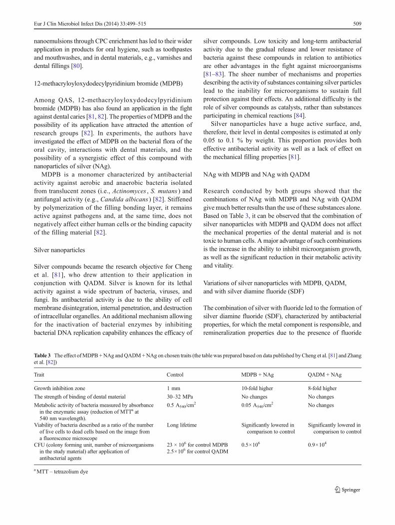
NAg with MDPB and NAg with QADM
Research conducted by both groups showed that thecombinations of NAg with MDPB and NAg with QADMgivemuch better results than the use of these substances alone.Based on Table 3, it can be observed that the combination ofsilver nanoparticles with MDPB and QADM does not affectthe mechanical properties of the dental material and is nottoxic to human cells. A major advantage of such combinationsis the increase in the ability to inhibit microorganism growth,as well as the significant reduction in their metabolic activityand vitality.
Variations of silver nanoparticles with MDPB, QADM,and with silver diamine fluoride (SDF)
The combination of silver with fluoride led to the formation ofsilver diamine fluoride (SDF), characterized by antibacterialproperties, for which the metal component is responsible, andremineralization properties due to the presence of fluoride
Table 3 The effect ofMDPB +NAg andQADM+NAg on chosen traits (the table was prepared based on data published byCheng et al. [81] and Zhanget al. [82])
Trait Control MDPB + NAg QADM + NAg
Growth inhibition zone 1 mm 10-fold higher 8-fold higher
The strength of binding of dental material 30–32 MPa No changes No changes
Metabolic activity of bacteria measured by absorbancein the enzymatic assay (reduction of MTTa at540 nm wavelength).
0.5 A540/cm2 0.05 A540/cm
2 No changes
Viability of bacteria described as a ratio of the numberof live cells to dead cells based on the image froma fluorescence microscope
Long lifetime Significantly lowered incomparison to control
Significantly lowered incomparison to control
CFU (colony forming unit, number of microorganismsin the study material) after application ofantibacterial agents
23 × 106 for control MDPB2.5×106 for control QADM
0.5×10⁶ 0.9×10⁴
aMTT – tetrazolium dye
Eur J Clin Microbiol Infect Dis (2014) 33:499–515 509
[85]. It has been shown that SDF can be used as an element ofdental caries prevention [86], as well as a substance inhibitingthe development of disease by reducing the amount ofcariogenic microorganisms in the oral cavity and that it has apositive influence in zones of tooth enamel demineralization[85]. During studies on the described compound, thesensitivity of S. mutans and Actinomyces naeslundii to itsfunction was also demonstrated [87].
However, studies examining the effects of SDF on dual-species biofilm formed by S. mutans and Lactobacillusacidophilus illustrate much better the effectiveness of thiscompound in the natural oral environment, in which biofilmis characterized as a structure of multi- and not single species.Mei et al. [72] showed that, in dual-species biofilm, theantibacterial activity of silver ions is much lower than in thebiofilm of single species; however, it does not disappearcompletely. This is confirmed by the ratio of dead and livecells, which, for the control, was equal to 0.02, whereas for thesamples with SDF addition, it was 6.74. These studies, apartfrom confirming SDF antibacterial activity against S. mutansand L. acidophilus , also proved that this substance slows theprocess of enamel demineralization and protects collagenfrom damage. This dual action of SDF may contribute to awider use in oral hygiene products and to clinical success inthe fight against dental caries.
Natural inhibitors: chitosan
Antibacterial substances which could be included in theformulations of oral hygiene products and would not exertside effects are still being researched. One such substance ischitosan, a polysaccharide formed by the N-acetylationreaction of chitin [88]. Both compounds are present in theplant, fungi, and animal worlds, and have natural antibacterialand antifungal properties. A limitation in the use of chitosanfor oral hygiene products, however, was proven by itsinsolubility in water, because the compound is dissolved onlyin acids. Attempts have been made to modify this property,and as a result of the Maillard reaction or sugar modification,water-soluble chitosan of unchanged antibacterial propertieswas obtained. It has been shown [88] that such modifiedchitosan exhibits the highest antibacterial activity againstmicroorganisms isolated from the oral cavity in an environmentwith a pH range between 5 (for Klebsiella pneumoniae ,Streptococcus mutans , Staphylococcus aureus) and 8 (forEnterobacter gergoviae , Lactobacillus brevis , Staphylococcussaprophyticus).
The optimum temperature for chitosan activity, in whichthe antibacterial activity remained at the level of 50–96%,wasestimated at 37 °C in relation to the above-mentioned bacterialspecies. For K. pneumoniae , L. brevis , and S. saprophyticus ,the minimum bactericidal concentration of chitosan (MBC)was 500 μg/ml, whereas for other species, it was 400 μg/ml. It
was also shown that 5 s is sufficient for chitosan to exhibitantibacterial activity in relation to the above-mentionedmicroorganisms species at the level of 99.6 %, and 20s wasrequired to reach the level of 99.9 %.
Thus, it was proven that water-soluble chitosan has acomparable efficacy in the fight against dental caries inrelation to commonly used antibacterial mouthwashes for oralhygiene. A very strong argument for the use of chitosan insuch formulations is also its much lower toxicity than theconventionally used alcohols, chlorhexidines, or cetylpyridiniumchloride [88], which are commonly used in products for oralcavity disinfection in diseases of the upper respiratory tract,e.g., Tantum Verde.
Natural inhibitors—antimicrobial peptides (AMPs):chrisofsin-1, D-Nal-Pac-525
Another safe and potential solution to the problem of dentalcaries in humans are antimicrobial peptides (AMPs). Theyexhibit a lethal effect against many microorganisms but donot cause side effects for humans [33].
AMPs represent a family of short polypeptides, typicallyassociated with congenital human and other organism immunesystems. An example of such a protein is chrisofsin-1amphipathic, an α-helical protein of broad activity spectrumagainst both Gram-negative and Gram-positive bacteria.Chrisofsin-1 possesses a hydrophilic domain at the C-terminalend of the polypeptide chain.
Via electrostatic interactions with negatively-chargedphospholipids of bacterial cell membranes, it covers themembrane tightly, and then the hydrophilic domain penetratesinto the structure of the membrane, forming a number of poresinside. Wang et al. observed the first changes in cell shapeduring their studies [33] using chrisofsin-1 at concentrationsof 2–4 mg/ml (depending on the bacteria species). Thus,chrisofsin-1 exhibits potent destructive action against lipidcomponents of microorganism cell membranes, such as S.mutans , S. sanguinis , S. sobrinus , S. gordonii , Actinomycessp., and Lactobacillus sp. and, thus, it is able to inducedisintegration and bacterial lysis.
This contributes to reduced viability of pathogens, both asplanktonic forms as well as those grouped in biofilm.Compared to S. mutans , chrisofsin-1 exhibits lethal activityafter 30 min with the use of an 8 times higher concentrationthan the minimum inhibitory concentration (MIC). Forcomparison, E. faecalis was destroyed after 5 min and L.fermentis after 60 min with the use of 12 times the MIC.Among the strains under investigation, only A. viscosus wasobserved to be more resistant than S. mutans . Its eradicationrequired the application of 8 times the MIC for 60 min. Thisshows the different effects of chrisofsin-1 in relation toparticular bacterial species [32].
510 Eur J Clin Microbiol Infect Dis (2014) 33:499–515
Microorganisms remaining in the biofilm structure are 10–1,000 times more resistant than those in the planktonic phase.For this reason, AMPs could be used most effectively in theearly stages of biofilm formation. It would prevent its growthand the colonization of the oral cavity by other pathogens. Inthe phase of biofilm maturation, AMPs would only delay thecariogenic process [33].
The D-Nal-Pac-525 peptide appeared to be anotherexample of an AMP with high antibacterial activity. The highanticaries activity of D-Nal-Pac-525 peptide is associated withthe tryptophan conversion in the tryptophan-rich (Trp-rich)Pac-525 peptide fragment to the D-β-naphthylalaninesfragment [62]. Studies with the use of scanning andtransmission electron microscopes have shown that the D-Nal-Pac-525 peptide causes morphological changes and cellmembrane damages of S. mutans with a MIC of 4 ug/ml,while the inhibition of biofilm formation was observed underanMIC of 2 ug/ml. The effective anti-biofilm protection of theD-Nal-Pac-525 peptide was highlighted in the first stage of itsformation (planktonic and colonization phase of bacteria)(Fig. 2), with no effect on pre-existing bacterial biofilms.The above-mentioned results are comparable to beta-defensin (6 mM) action in human saliva. Data suggest theuse of AMPs exemplified by D-Nal-Pac-525 as newtherapeutic agents which combat the problems caused by theoccurrence of bacterial strains resistant to antibiotics [62].
The problem of infectious diseases is inherently associatedwith the growing number of microorganisms becomingresistant to a broader spectrum of antibiotics (multidrugresistance). Therefore, the attention of scientists hasincreasingly been drawn to antibacterial, antifungal, andantiviral substances of natural origin. Many metabolites ofplant origin, such as polyphenols, alkaloids, tannin,terpenoids, steroids, or flavonoids, are known for suchproperties [61, 89, 90]. Because of their proven antibacterialactivity, these compounds act as supplements in oral hygieneproducts.
Most of the above-mentioned substances have foundan application in dentistry. They are included incomposites and dental restorative resins, the propertiesof which allow for the reduction of biofilm formationand reduce the probability of dental caries episodes atthe same place. They have been introduced into treatmentbecause commonly used methods of treating developingdental caries usually do not allow for the removal of bacteriafrom the mineralized tooth tissues. In such situations, it issuggested that antibacterial agents should be applied onalready developed dentin [91]. These are usually fillingswhich release ions [92] in the form of dicalcium phosphateanhydrous nanoparticles (DPCA) or tetracalcium phosphate(TTCP) often enriched with fluorine [92], or modifications ofsuch fillings, e.g., with the addition of QAS or silvernanoparticles [76, 77, 81, 82].
Iodine compounds
Studies on substances allowing for the reduction of dentalcaries incidence also include iodine compounds such aspotassium iodide or povidone–iodine (PVP-I). PVP-I is acomplex of iodide and polyvinylpyrrolidone, which is aspecific “reservoir” of free iodine ions forming an activedomain of the whole structure. Iodine slowly released fromthe complex is able to penetrate the bacterial membrane andenter into the cytosol, where it deactivates the key proteins,fatty acids, and nucleotides in the metabolism of the cell [93].Additionally, the slow release of iodine reduces its toxiceffects for human cells [94] and its short-term use doesnot irritate even damaged oral mucosa. No side effects,such as discoloration of teeth, tongue, or taste change, havebeen reported [95].
Hosaka et al. [95] decided to test the antibacterial activityof PVP-I in relation to the two microorganisms associatedwith oral cavity diseases, Porphyromonas gingivalis andFusobacterium nucleatum . They showed that PVP-I, at aconcentration of 7 %, is able to reach the activity allowing100 % of P. gingivalis species to die after 3 min, and anidentical effectiveness against F. nucleatum with the use of5 % of PVP-I was observed after 30 s. The results obtained fordual-species biofilm formed by the microorganisms underinvestigation indicated that this biofilm is 200 times moreresistant to the activity of the tested substance than mono-species cultures, although the activity of 5 % PVP-I allowsbacteria which form this biofilm to be killed.
Unfortunately, it has also been shown that the concentrationsof PVP-I (0.23–0.47 %) used in common mouthwashes are notsufficient for a significant reduction of P. gingivalis and F.nucleatum biofilm formation ability during a time of 30 s to1 min, which was estimated as the average time ofmouthwashing by the public [95]. Based on these reports, onemay conclude that other bacterial species will also exhibit adifferent sensitivity to PVP-I activity, and a multi-speciesbiofilm will be much more resistant to both planktonic cells aswell as structures formed by a lower number ofmicroorganisms.
The antibacterial activity of PVP-I has also been tested forS. mutans strains isolated from children with early childhoodcaries [96, 97]. It has been proven that the application of 10 %PVP-I on healthy teeth and on teeth with dental caries every3 months for 1 year significantly reduces the number of thesemicroorganisms in the oral cavity as compared to baselinelevels [96, 98]. This reduced number of cariogenic bacteria is,therefore, likely to reduce the probability of childhood dentalcaries. Similar conclusions can be drawn from studiesconducted on a group of 172 children, which showed thatcombination therapy of dental caries through the applicationof 10 % PVP-I and dental varnish containing 5 % sodiumfluoride reduces the incidence of new carious lesions by 31 %in relation to the application of the varnish alone [86].
Eur J Clin Microbiol Infect Dis (2014) 33:499–515 511

Ursolic acid
Composite resins containing ursolic acid which inhibited thebiofilm formation of S. mutans at a concentration of 0.1–0.5 wt.% [99] have been proposed as another solution to theproblem of dental caries. This compound has similarproperties to those tested in clinical trials: triterpenoids ofrecorded anticancer, antiwrinkle, and antibacterial activity.
Aromatic amino acid pathways
There is little information concerning the effect of amino acidson the ability of S. mutans to form biofilms. Kolodkin-Galet al. conducted a study on biofilm structure decomposition onthe example of Bacillus subtilis . This microorganismproduced a factor which inhibited biofilm formation and nextinduced structure disassembly. Analysis of this factor’scomposition demonstrated that it is a mixture of four D-amino acids: leucine, methionine, tyrosine, and tryptophan[100]. A similar inhibitory effect of the D-amino acidmixture was observed in the case of biofilm formation byStaphylococcus aureus [101] and Pseudomonas aeruginosa[102]. Recent studies have indicated that increased levels of L-tryptophan itself may also give the signal for biofilmdecomposition; a suitable experiment was conducted on theexample of biofilm formed by E. coli [103]. Our studyindicates that another aromatic L-amino acid, i.e., tyrosine,has similar properties and may inhibit biofilm formation byS. mutans (paper submitted to the editorial board). It needs tobe verified as to whether other aromatic amino acids mayexhibit anti-biofilm formation activity for S. mutans species,and also whether inhibition caused by these compounds islimited to ex vivo models, as well as whether and to whatdegree they may be applied in clinical situations (proposedanimal model).
The increasingly broader range of substances protecting usfrom the occurrence of carious lesions while not causing sideeffects creates hope for the design of oral hygiene productsallowing for the effective fight against this disease and for thecomplete protection of children from the development of earlycaries lesions in primary teeth.
Summary
Studies on biofilm formed by S. mutans clearly show thatthe virulence of S. mutans strains is dependent onenvironmental conditions, and, thus, on in vivo modelhost-dependent characteristics. On the other hand, to forma biofilm structure, it is not sufficient to ensure appropriateenvironmental conditions. Microorganisms alone must possesscharacteristics which, in terms of the above-mentioned
favorable conditions, will allow the adhesion and formationof microcolonies [6, 35, 36].
Cariogenic strains of S. mutans exhibit large variation dueto the occupation and expression of characteristics consideredas virulence determinants: (A) most S. mutans strains exhibitthe ability to survive and reproduce in acidic pH, withouteliminating the possibility to survive in alkaline mediumlevels (described in case studies isolated cases of bacteremia),(B) most S. mutans strains possess the activity of specificproteins (enzymes) associated with biofilm formation,depending on a number of biochemical properties of the host(EPS production, the condition of the host immune system).
Biofilm formation in the oral cavity is a complex process,dependent not only on EPS produced by S. mutans and thehost, but also on other species colonizing the oral cavity. Theoccurrence of potentially non-pathogenic species in the oralcavity environment seems to be the key point.
This study is an introduction to the research which aims toconfirm or exclude the virulence of selected Streptococcusstrains in clinical material, as well as shedding new light onthe development of potential compounds blocking themetabolism of Streptococcus mutans through influence onspecific proteins involved in the formation of the complexstructure which is a biofilm.
Conflict of interest All authors confirm that there is no conflict ofinterest.
Open AccessThis article is distributed under the terms of the CreativeCommons Attribution License which permits any use, distribution, andreproduction in any medium, provided the original author(s) and thesource are credited.
References
1. Huang L, Xu QA, Liu C, Fan MW, Li YH (2013) Anti-caries DNAvaccine-induced secretory immunoglobulin A antibodies inhibitformation of Streptococcus mutans biofilms in vitro. ActaPharmacol Sin 34(2):239–246
2. Nomura H, Isshiki Y, Sakuda K, SakumaK, Kondo S (2013) Effectsof oakmoss and its components on biofilm formation of Legionellapneumophila. Biol Pharm Bull 36(5):833–837
3. Wojtyczka RD, Kępa M, Idzik D, Kubina R, Kabała-Dzik A,Dziedzic A, Wąsik TJ (2013) In vitro antimicrobial activity ofethanolic extract of Polish propolis against biofilm formingStaphylococcus epidermidis strains. Evid Based ComplementAlternat Med 2013:590703
4. Zhao K, TsengBS, Beckerman B, Jin F, GibianskyML,Harrison JJ,Luijten E, Parsek MR, Wong GC (2013) Psl trails guide explorationand microcolony formation in Pseudomonas aeruginosa biofilms.Nature 497(7449):388–391. doi:10.1038/nature12155
5. Welin-Neilands J, Svensäter G (2007) Acid tolerance of biofilmcells of Streptococcus mutans. Appl Environ Microbiol 73(17):5633–5638
6. Bowen WH, Koo H (2011) Biology of Streptococcus mutans-derived glucosyltransferases: role in extracellular matrix formationof cariogenic biofilms. Caries Res 45(1):69–86
512 Eur J Clin Microbiol Infect Dis (2014) 33:499–515
7. Sørensen UB, Poulsen K, Ghezzo C, Margarit I, Kilian M (2010)Emergence and global dissemination of host-specific Streptococcusagalactiae clones. MBio 1(3). pii: e00178-10
8. Henderson B, Poole S, Wilson M (1996) Bacterial modulins: anovel class of virulence factors which cause host tissue pathologyby inducing cytokine synthesis. Microbiol Rev 60(2):316–341
9. Casadevall A, Pirofski LA (1999) Host–pathogen interactions:redefining the basic concepts of virulence and pathogenicity.Infect Immun 67(8):3703–3713
10. Kreikemeyer B, GámezG,Margarit I, Giard JC, Hammerschmidt S,Hartke A, Podbielski A (2011) Genomic organization, structure,regulation and pathogenic role of pilus constituents in majorpathogenic Streptococci and Enterococci. Int J Med Microbiol301(3):240–251
11. Bagnoli F, Moschioni M, Donati C, Dimitrovska V, Ferlenghi I,Facciotti C, Muzzi A, Giusti F, Emolo C, Sinisi A, HilleringmannM, Pansegrau W, Censini S, Rappuoli R, Covacci A, Masignani V,Barocchi MA (2008) A second pilus type in Streptococcuspneumoniae is prevalent in emerging serotypes and mediatesadhesion to host cells. J Bacteriol 190:5480–5492
12. Jaykus LA,Wang HH, Schlesinge LS (2009) Food-borne microbes:shaping the host ecosystem. ASM Press, Washington, p 124
13. Tamura S, YonezawaH,MotegiM,NakaoR, Yoneda S,WatanabeH,Yamazaki T, Senpuku H (2009) Inhibiting effects of Streptococcussalivarius on competence-stimulating peptide-dependent biofilmformation by Streptococcus mutans. Oral Microbiol Immunol 24(2):152–161
14. Svensäter G, Welin J, Wilkins JC, Beighton D, Hamilton IR (2001)Protein expression by planktonic and biofilm cells of Streptococcusmutans. FEMS Microbiol Lett 205(1):139–146
15. Jung CJ, Yeh CY, Shun CT, Hsu RB, Cheng HW, Lin CS, Chia JS(2012) Platelets enhance biofilm formation and resistance ofendocarditis-inducing streptococci on the injured heart valve. JInfect Dis 205(7):1066–1075
16. Takahashi Y, Urano-Tashiro Y, Konishi K (2013) Adhesins of oralstreptococci. Nihon Saikingaku Zasshi 68(2):283–293
17. Habib G, Hoen B, Tornos P, Thuny F, Prendergast B, Vilacosta I,Moreillon P, de Jesus Antunes M, Thilen U, Lekakis J, Lengyel M,Müller L, Naber CK, Nihoyannopoulos P, Moritz A, Zamorano JL;ESC Committee for Practice Guidelines (2009) Guidelines on theprevention, diagnosis, and treatment of infective endocarditis (newversion 2009): the Task Force on the Prevention, Diagnosis, andTreatment of Infective Endocarditis of the European Society ofCardiology (ESC). Endorsed by the European Society of ClinicalMicrobiology and Infectious Diseases (ESCMID) and theInternational Society of Chemotherapy (ISC) for Infection andCancer. Eur Heart J 30(19):2369–2413
18. Jung CJ, Zheng QH, Shieh YH, Lin CS, Chia JS (2009)Streptococcus mutans autolysin AtlA is a fibronectin-bindingprotein and contributes to bacterial survival in the bloodstream andvirulence for infective endocarditis. Mol Microbiol 74(4):888–902
19. Nakano K, Ooshima T (2009) Serotype classification ofStreptococcus mutans and its detection outside the oral cavity.Future Microbiol 4:891–902
20. Matsumoto-Nakano M, Tsuji M, Inagaki S, Fujita K, Nagayama K,Nomura R, Ooshima T (2009) Contribution of cell surface proteinantigen c of Streptococcus mutans to platelet aggregation. OralMicrobiol Immunol 24:427–430
21. Abranches J, Miller JH, Martinez AR, Simpson-Haidaris PJ, BurneRA, Lemos JA (2011) The collagen-binding protein Cnm is requiredfor Streptococcus mutans adherence to and intracellular invasion ofhuman coronary artery endothelial cells. Infect Immun 79:2277–2284
22. Abranches J, Zeng L, Bélanger M, Rodrigues PH, Simpson-HaidarisPJ, Akin D, Dunn WA Jr, Progulske-Fox A, Burne RA (2009)Invasion of human coronary artery endothelial cells by Streptococcusmutans OMZ175. Oral Microbiol Immunol 24:141–145
23. Ahn SJ, Ahn SJ, Wen ZT, Brady LJ, Burne RA (2008)Characteristics of biofilm formation by Streptococcus mutans inthe presence of saliva. Infect Immun 76(9):4259–4268
24. KhanAU, IslamB, Khan SN, AkramM (2011) A proteomic approachfor exploring biofilm in Streptococcus mutans. Bioinformation 5(10):440–445
25. Wen ZT, Yates D, Ahn SJ, Burne RA (2010) Biofilm formation andvirulence expression by Streptococcus mutans are altered whengrown in dual-species model. BMC Microbiol 10:111
26. Peterson SN, Snesrud E, Liu J, OngAC, KilianM, Schork NJ, BretzW (2013) The dental plaque microbiome in health and disease.PLoS One 8(3):e58487. doi:10.1371/journal.pone.0058487
27. Pawka B, Dreher P, Herda J, Szwiec I, KrasickaM (2010) Próchnicazębów u dzieci problemem społecznym. Probl Hig Epidemiol91(1):5–7
28. Krzyściak W, Pluskwa KK, Jurczak A, Kościelniak D (2013) Thepathogenicity of the Streptococcus genus. Eur J Clin MicrobiolInfect Dis (in press)
29. Składnik-Jankowska J, Kaczmarek U (2010) Istotny WskaźnikPróchnicy u 12-letnich dzieci z różnych środowisk województwadolnośląskiego. Czas Stomatol 63(3):166–173
30. World Health Organization (WHO) (2011) Poland: health systemreview. Health systems in transition, Vol. 13, No. 8
31. PZH Raports (2007–2012) Informacje o zachorowaniach nachoroby zakaźne i zatruciach w Polsce oraz szczepieniachochronnych w latach 2007–2012 dostępne na stronie internetowej.Available online at: http://www.pzh.gov.pl/oldpage/epimeld/index_p.html#01
32. Zijnge V, van Leeuwen MB, Degener JE, Abbas F, Thurnheer T,Gmür R, Harmsen HJ (2010) Oral biofilm architecture on naturalteeth. PLoS One 5(2):e9321
33. Wang W, Tao R, Tong Z, Ding Y, Kuang R, Zhai S, Liu J, Ni L(2012) Effect of a novel antimicrobial peptide chrysophsin-1on oral pathogens and Streptococcus mutans biofilms. Peptides33(2):212–219
34. Kang MS, Oh JS, Lee HC, Lim HS, Lee SW, Yang KH, Choi NK,Kim SM (2011) Inhibitory effect of Lactobacillus reuteri onperiodontopathic and cariogenic bacteria. J Microbiol 49(2):193–199
35. Xiao J, Klein MI, Falsetta ML, Lu B, Delahunty CM, Yates JR 3rd,Heydorn A,KooH (2012) The exopolysaccharidematrixmodulatesthe interaction between 3D architecture and virulence of a mixed-species oral biofilm. PLoS Pathog 8(4):e1002623
36. Schwab C, Walter J, Tannock GW, Vogel RF, Gänzle MG (2007)Sucrose utilization and impact of sucrose on glycosyltransferaseexpression in Lactobacillus reuteri. Syst Appl Microbiol 30(6):433–443
37. Siqueira WL, Bakkal M, Xiao Y, Sutton JN, Mendes FM (2012)Quantitative proteomic analysis of the effect of fluoride on theacquired enamel pellicle. PLoS One 7(8):e42204
38. Duque C, Stipp RN,Wang B, Smith DJ, Höfling JF, Kuramitsu HK,Duncan MJ, Mattos-Graner RO (2011) Downregulation of GbpB, acomponent of the VicRK regulon, affects biofilm formation and cellsurface characteristics of Streptococcus mutans. Infect Immun79(2):786–796
39. Fujita K, Matsumoto-Nakano M, Inagaki S, Ooshima T (2007)Biological functions of glucan-binding protein B of Streptococcusmutans. Oral Microbiol Immunol 22(5):289–292
40. Fujita K, Takashima Y, Inagaki S, Nagayama K, Nomura R, ArdinAC, Grönroos L, Alaluusua S, Ooshima T, Matsumoto-Nakano M(2011) Correlation of biological properties with glucan-bindingprotein B expression profile in Streptococcus mutans clinicalisolates. Arch Oral Biol 56(3):258–263. doi:10.1016/j.archoralbio.2010.09.018
41. Zeng L, Choi SC, Danko CG, Siepel A, Stanhope MJ, Burne RA(2013) Gene regulation by CcpA and catabolite repression explored
Eur J Clin Microbiol Infect Dis (2014) 33:499–515 513
by RNA-Seq in Streptococcus mutans. PLoS One 8(3):e60465. doi:10.1371/journal.pone.0060465
42. Giammarinaro P, Paton JC (2002) Role of RegM, a homologue ofthe catabolite repressor protein CcpA, in the virulence ofStreptococcus pneumoniae. Infect Immun 70(10):5454–5461
43. Zeng L, Burne RA (2013) Comprehensive mutational analysis ofsucrose-metabolizing pathways in Streptococcus mutans revealsnovel roles for the sucrose phosphotransferase system permease. JBacteriol 195(4):833–843
44. Cai J, Tong H, Qi F, Dong X (2012) CcpA-dependent carbohydratecatabolite repression regulates galactose metabolism in Streptococcusoligofermentans. J Bacteriol 194(15):3824–3832
45. Tsang P, Merritt J, Shi W, Qi F (2006) IrvA-dependent and IrvA-independent pathways for mutacin gene regulation in Streptococcusmutans. FEMS Microbiol Lett 261(2):231–234
46. Koo H, Xiao J, Klein MI, Jeon JG (2010) Exopolysaccharidesproduced by Streptococcus mutans glucosyltransferases modulatethe establishment of microcolonies within multispecies biofilms. JBacteriol 192(12):3024–3032
47. KleinMI, Xiao J, Lu B, Delahunty CM, Yates JR 3rd, Koo H (2012)Streptococcus mutans protein synthesis during mixed-speciesbiofilm development by high-throughput quantitative proteomics.PLoS One 7(9):e45795. doi:10.1371/journal.pone.0045795
48. Brady LJ, Maddocks SE, Larson MR, Forsgren N, Persson K,Deivanayagam CC, Jenkinson HF (2010) The changing faces ofStreptococcus antigen I/II polypeptide family adhesins. MolMicrobiol 77(2):276–286
49. Edberg SC (2009) Does the possession of virulence factor genesmean that those genes will be active? J Water Health 7(Suppl 1):S19–S28
50. Crowley PJ, Seifert TB, Isoda R, van TilburgM, Oli MW, RobinetteRA, McArthur WP, Bleiweis AS, Brady LJ (2008) Requirements forsurface expression and function of adhesin P1 from Streptococcusmutans. Infect Immun 76(6):2456–2468
51. McLean JS, Fansler SJ, Majors PD, McAteer K, Allen LZ, ShirtliffME, Lux R, Shi W (2012) Identifying low pH active and lactate-utilizing taxa within oral microbiome communities from healthychildren using stable isotope probing techniques. PLoS One 7(3):e32219
52. Ahn SJ, Burne RA (2007) Effects of oxygen on biofilm formationand the AtlA autolysin of Streptococcus mutans. J Bacteriol189(17):6293–6302
53. Gibbons RJ, Hay DI (1989) Adsorbed salivary acidic proline-richproteins contribute to the adhesion of Streptococcus mutans JBP toapatitic surfaces. J Dent Res 68:1303–1307
54. Yamada A, Tamura H, Kato H (2009) Identification andcharacterization of an autolysin gene, atlg, from Streptococcussobrinus. FEMS Microbiol Lett 291(1):17–23. doi:10.1111/j.1574-6968.2008.01426.x
55. Aires CP, Del Bel Cury AA, Tenuta LM, Klein MI, Koo H, Duarte S,Cury JA (2008) Effect of starch and sucrose on dental biofilm formationand on root dentine demineralization. Caries Res 42(5):380–386
56. Bhalla S, Tandon S, Satyamoorthy K (2010) Salivary proteins andearly childhood caries: a gel electrophoretic analysis. Contemp ClinDent 1(1):17–22
57. Kaczmarek U (2004) Aspekt bakteryjny próchnicy zębówmlecznych. Dent Med Probl 41(3):509–514
58. Taji S, Seow WK (2010) A literature review of dental erosion inchildren. Aust Dent J 55(4):358–367
59. Moynihan PJ (2005) The role of diet and nutrition in the etiologyand prevention of oral diseases. Bull World Health Organ 83(9):694–699
60. Selwitz RH, Ismail AI, Pitts NB (2007) Dental caries. Lancet369(9555):51–59
61. Hu P, Huang P, Chen MW (2013) Curcumin reducesStreptococcus mutans biofilm formation by inhibiting sortase
A activity. Arch Oral Biol 58(10):1343–1348. doi:10.1016/j.archoralbio.2013.05.004
62. Li H, Cheng JW, Yu HY, Xin Y, Tang L, Ma Y (2013)Effect of the antimicrobial peptide D-Nal-Pac-525 on the growth ofStreptococcus mutans and its biofilm formation. J MicrobiolBiotechnol 3(8):1070–1075
63. Ogawa A, Furukawa S, Fujita S, Mitobe J, Kawarai T, Narisawa N,Sekizuka T, Kuroda M, Ochiai K, Ogihara H, Kosono S, Yoneda S,Watanabe H, Morinaga Y, Uematsu H, Senpuku H (2011) Inhibitionof Streptococcus mutans biofilm formation by Streptococcussalivarius FruA. Appl Environ Microbiol 77(5):1572–1580
64. Redanz S, Standar K, Podbielski A, Kreikemeyer B (2011) A five-species transcriptome array for oral mixed-biofilm studies. PLoSOne 6(12):e27827. doi:10.1371/journal.pone.0027827
65. Jalasvuori H, Haukioja A, Tenovuo J (2012) Probiotic Lactobacillusreuteri strains ATCC PTA 5289 and ATCC 55730 differ in theircariogenic properties in vitro. Arch Oral Biol 57(12):1633–1638
66. Teanpaisan R, Piwat S, Dahlén G (2011) Inhibitory effect of oralLactobacillus against oral pathogens. Lett Appl Microbiol 53(4):452–459
67. Lang C, Böttner M, Holz C, VeenM, Ryser M, Reindl A, PompejusM, Tanzer JM (2010) Specific Lactobacillus/Mutans Streptococcusco-aggregation. J Dent Res 89(2):175–179
68. Söderling EM, Marttinen AM, Haukioja AL (2011) Probioticlactobacilli interfere with Streptococcus mutans biofilm formationin vitro. Curr Microbiol 62(2):618–622
69. Samot J, Lebreton J, Badet C (2011) Adherence capacities of orallactobacilli for potential probiotic purposes. Anaerobe 17(2):69–72.doi:10.1016/j.anaerobe.2011.04.001
70. Tahmourespour A, Salehi R, Kermanshahi RK, Eslami G (2011)The anti-biofouling effect of Lactobacillus fermentum-derivedbiosurfactant against Streptococcus mutans. Biofouling 27(4):385–392
71. Kim do K, Kim KH, Cho EJ, Joo SJ, Chung JM, Son BY, Yum JH,Kim YM, Kwon HJ, Kim BW, Kim TH, Lee EW (2013) Genecloning and characterization ofMdeA, a novel multidrug efflux pumpin Streptococcus mutans. J Microbiol Biotechnol 23(3):430–435
72. Mei ML, Chu CH, Low KH, Che CM, Lo EC (2013) Cariesarresting effect of silver diamine fluoride on dentine carious lesionwith S. mutans and L. acidophilus dual-species cariogenic biofilm.Med Oral Patol Oral Cir Bucal (in press)
73. García-Godoy F, Hicks MJ (2008) Maintaining the integrity of theenamel surface: the role of dental biofilm, saliva and preventiveagents in enamel demineralization and remineralization. J Am DentAssoc 139(Suppl):25S–34S
74. Goyal D, Sharma S, Mahmood A (2013) Inhibition ofdextransucrase activity in Streptococcus mutans by plant phenolics.Indian J Biochem Biophys 50(1):48–53
75. Melo MA, Guedes SF, Xu HH, Rodrigues LK (2013)Nanotechnology-based restorative materials for dental cariesmanagement. Trends Biotechnol 31(8):459–467. doi:10.1016/j.tibtech.2013.05.010
76. Cheng L, Weir MD, Zhang K, Xu SM, Chen Q, Zhou X, Xu HHK(2012) Antibacterial nanocomposite with calcium phosphate andquaternary ammonium. J Dent Res 91:460–466
77. Cheng L, Weir MD, Zhang K, Wu EJ, Xu SM, Zhou X, Xu HH(2012) Dental plaque microcosm biofilm behavior on calciumphosphate nanocomposite with quaternary ammonium. DentMater 28(8):853–862
78. van der Waal SV, van der Sluis LW (2012) Potential of calcium toscaffold an endodontic biofilm, thus protecting the micro-organismsfrom disinfection. Med Hypotheses 79(1):1–4. doi:10.1016/j.mehy.2012.03.012
79. Obłak E, Gamian A (2010) The biological activity of quaternaryammonium salts (QASs). Postepy Hig Med Dosw (Online) 64:201–211
514 Eur J Clin Microbiol Infect Dis (2014) 33:499–515
80. Lee VA, Karthikeyan R, Rawls HR, Amaechi BT (2010) Anti-cariogenic effect of a cetylpyridinium chloride-containingnanoemulsion. J Dent 38(9):742–749
81. Cheng L, Zhang K, Weir MD, Liu H, Zhou X, Xu HH (2013)Effects of antibacterial primers with quaternary ammonium andnano-silver on Streptococcus mutans impregnated in human dentinblocks. Dent Mater 29(4):462–472
82. Zhang K, Cheng L, Imazato S, Antonucci JM, Lin NJ, Lin-GibsonS, Bai Y, Xu HH (2013) Effects of dual antibacterial agents MDPBand nano-silver in primer on microcosm biofilm, cytotoxicity anddentine bond properties. J Dent 1(5):464–474
83. Kim YJ, Lee DY, Lee JY, Lim YK (2012) The effect of silver ion-releasing elastomers on mutans streptococci in dental plaque.Korean J Orthod 42(2):87–93
84. You C, Han C, Wang X, Zheng Y, Li Q, Hu X, Sun H(2012) The progress of silver nanoparticles in the antibacterialmechanism, clinical application and cytotoxicity. Mol Biol Rep39(9):9193–9201
85. Mei ML, Chu CH, Lo EC, Samaranayake LP (2013) Preventingroot caries development under oral biofilm challenge in an artificialmouth. Med Oral Patol Oral Cir Bucal 18(4):e557–e563
86. MilgromPM, Tut OK,Mancl LA (2011) Topical iodine and fluoridevarnish effectiveness in the primary dentition: a quasi-experimentalstudy. J Dent Child (Chic) 78(3):143–147
87. Chu CH, Mei L, Seneviratne CJ, Lo EC (2012) Effects of silverdiamine fluoride on dentine carious lesions induced byStreptococcus mutans and Actinomyces naeslundii biofilms. Int JPaediatr Dent 22:2–10
88. Chen CY, Chung YC (2012) Antibacterial effect of water-solublechitosan on representative dental pathogens Streptococcus mutansand Lactobacilli brevis. J Appl Oral Sci 20(6):620–627
89. Shafiei Z, Shuhairi NN, Md Fazly Shah Yap N, Harry SibungkilCA, Latip J (2012) Antibacterial activity of myristica fragransagainst oral pathogens. Evid Based Complement Alternat Med2012:825362
90. Noumedem JA, Mihasan M, Lacmata ST, Stefan M, Kuiate JR,Kuete V (2013) Antibacterial activities of the methanol extracts often Cameroonian vegetables against gram-negative multidrug-resistant bacteria. BMC Complement Alternat Med 13(1):26
91. Łukomska-Szymańska M, Olbert-Sieroszewska V, Żurawska-Olszewska J, Szczerba I, Krzemiński Z, Sokołowski J (2010)Antibacterial properties of 6th generation bonding systems. DentMed Probl 47(3):304–308
92. Chen MH (2010) Update on dental nanocomposites. J Dent Res89(6):549–560
93. Durani P, Leaper D (2008) Povidone–iodine: use in handdisinfection, skin preparation and antiseptic irrigation. Int WoundJ 5:376–387
94. Anderson MJ, Horn ME, Lin YC, Parks PJ, Peterson ML (2010)Efficacy of concurrent application of chlorhexidine gluconate andpovidone iodine against six nosocomial pathogens. Am J InfectControl 38(10):826–831
95. Hosaka Y, Saito A, Maeda R, Fukaya C, Morikawa S, Makino A,Ishihara K, Nakagawa T (2012) Antibacterial activity of povidone–iodine against an artificial biofilm of Porphyromonas gingivalis andFusobacterium nucleatum. Arch Oral Biol 57(4):364–368
96. Simratvir M, Singh N, Chopra S, Thomas AM (2010) Efficacy of10% povidone iodine in children affected with early childhoodcaries: an in vivo study. J Clin Pediatr Dent 34(3):233–238
97. Berkowitz RJ, Koo H, McDermott MP, Whelehan MT, Ragusa P,Kopycka-Kedzierawski DT, Karp JM, Billings R (2009)Adjunctive chemotherapeutic suppression of mutans streptococciin the setting of severe early childhood caries: an exploratory study.J Public Health Dent 69(3):163–167. doi:10.1111/j.1752-7325.2009.00118.x
98. Berkowitz RJ, Amante A, Kopycka-Kedzierawski DT, Billings RJ,Feng C (2011) Dental caries recurrence following clinical treatmentfor severe early childhood caries. Pediatr Dent 33(7):510–514
99. Kim S, Song M, Roh BD, Park SH, Park JW (2013) Inhibition ofStreptococcus mutans biofilm formation on composite resinscontaining ursolic acid. Restor Dent Endod 38(2):65–72. doi:10.5395/rde.2013.38.2.65
100. Kolodkin-Gal I, Romero D, Cao S, Clardy J, Kolter R, Losick R(2010) D-amino acids trigger biofilm disassembly. Science328(5978):627–629
101. Hochbaum AI, Kolodkin-Gal I, Foulston L, Kolter R, Aizenberg J,Losick R (2011) Inhibitory effects of D-amino acids onStaphylococcus aureus biofilm development. J Bacteriol 193(20):5616–5622
102. McDougald D, Rice SA, Barraud N, Steinberg PD, Kjelleberg S(2012) Should we stay or should we go: mechanisms and ecologicalconsequences for biofilm dispersal. Nat Rev Microbiol 10:39–50
103. Shimazaki J, Furukawa S, Ogihara H, Morinaga Y (2012) L-Tryptophan prevents Escherichia coli biofilm formation andtriggers biofilm degradation. Biochem Biophys Res Commun419(4):715–718
Eur J Clin Microbiol Infect Dis (2014) 33:499–515 515