Embed Size (px)
Citation preview

The Value of Long-Chain Fatty Acid Analysis, Randomly Amplified Polymorphic DNA and Electrophoretic Karyotyping for the Characterization of Wine Yeast Strains T.J. van der Westhuizenl*, O.P.H. Augustynl and I.S. Pretorius2
1) ARC-Fruit, Vine and Wine Research Institute, Nietvoorbij Centre for Vine and Wine, Private Bag X5026, 7599 Stellenbosch, South Africa
2) Institute for Wine Biotechnology and Department of Viticulture & Oenology, University of Stellenbosch, Private Bag Xl, 7602 Matieland (Stellenbosch), South Africa
Submitted for publication: July 1998 Accepted for publication: November 1998 Key words: Wine yeasts, long-chain fatty acid analysis, randomly amplified polymorphic DNA (RAPD), electrophoretic karyotyping (CHEF)
Wine yeast strains of Saccharomyces had previously been classified into several different species or varieties. This classification system was based mainly on sugar fermentation and assimilation patterns. Subsequently, most of these species were reclassified as Saccharomyces cerevisiae. The assignment of the majority of wine yeast strains to a single species does, however, not imply that all stains of S. cerevisiae are equally suitable for wine fermentation. These physiological strains of S. cerevisiae differ significantly in their fermentation performance and their ability to contribute to the final bouquet and quality of the various types of wine and distillates. Therefore, to ensure strain authenticity, security and proper strain management, it is of cardinal importance to have reliable taxonomic techniques available to identify and characterize individual strains of commercial cultures. In this study, 18 commercial wine yeast strains were characterized in order to evaluate and compare three taxonomic techniques, namely long-chain fatty acid analysis, randomly amplified polymorphic DNA (RAPD) and electrophoretic karyotyping. As a single identification technique, electrophoretic karyotyping seems to be the most useful method for routine fingerprinting of wine yeast strains. However, we propose that the combined use of these three techniques provides the most reliable means of differentiating amongst commercial wine yeast strains. -
Many of the traditional taxonomic criteria (e.g., sugar fermentation and assimilation patterns) used for the speciation of yeasts were derived from the analysis of a small portion of the genome. These phenotypic characteristics still serve a useful purpose in classification, since not all of them are unstable and insignificant. Phenotypic traits, however, do not necessarily reflect genetic relatedness, since the same phenotype may be a result of convergent evolution. Conversely, the phylogenetic relationships should be reflected in similarities at the level of base composition of deoxyribonucleic acid (DNA) and DNA sequence homology as well as ribosomal RNA/DNA sequence relatedness (ribotypes) in different yeasts (Pretorius & Van der Westhuizen, 1991; Kurtzman & Fell, 1998).
Wine yeast strains of the genus Saccharomyces were traditionally classified into several different species or varieties, including S. bayanus, S. beticus, S. capensis, S. ellipsoideus, S.fermentati, S. oviformis and S. vini (Lodder & Kreger-van Rij, 1952; Lodder, 1970). A strong wine-type/-taxonomic relationship for the various wine yeasts was believed to exist. For example, strains of S. beticus and S. capensis were preferred for producing flor sherry because of their superior film-forming ability and desirable oxidative metabolism. Also, strains of S. ellipsoideus were generally preferred for making table (dry, still) wines, whereas strains of S. bayanus were chosen for making sparkling wines, because the latter strains were often more tolerant to alcohol and could
*Present address: Anchor Yeast, PO Box 14,7475 Eppindust (Cape Town), South Africa_
ferment to a lower residual sugar concentration (Henschke, 1997). Using genetic taxonomic techniques, most of these strains were reclassified as Saccharomyces cerevisiae (Kreger-van Rij, 1984; Kurtzman & Fell, 1998). However, the assignment of most of these wine yeast strains to a single species does not imply that all strains of S. cerevisiae are equally suitable for wine fermentation. It is well known that these physiological strains of S. cerevisiae differ significantly in their fermentation performance and their ability to contribute to the final bouquet and quality of the various types of wine and distillates. Characterization and identification of wine yeasts to the strain level is therefore, of key importance to ensure strain authenticity, security and proper strain management.
Due to the complexity of the vinification process and the widespread practice of seeding must with dry commercial wine yeast cultures with favourable characteristics, it has become increasingly important to use an identifiable yeast strain, so as to ensure consistency of wine type, style and quality. The difficulty of identifying yeasts by standard microbiological methods prompted the development of a large number of different identification techniques. However, these techniques are not universally adept at differentiating amongst strains of the same species. As a result, a variety of additional methods of strain identification are in use i.e. fingerprinting of industrial strains by protein profiles (Van Vuuren & Van der Meer, 1987; Degre et aI., 1989; Van der
Acknowledgements: The authors thank the South African wine industry (Winetech), the National Research Foundation (NRF) and Anchor Yeast for financial support_
S. Afr. J. Enol. Vitie., Vol. 20, No.1, 1999
3

4 Characterization of Wine Yeast Strains
Westhuizen & Pretorius, 1989, 1992), restrIctIOn analysis of genomic and mitochondrial DNA (Querol et at., 1992; Van der Westhuizen & Pretorius, 1992; Versavaud et at., 1995), electrophoretic karyotyping (Degre et aI., 1989; Yamamoto et aI., 1991; Bidenne et at., 1992; Van der Westhuizen & Pretorius, 1992; Naumov, Naumova & Gaillardin, 1993; Grando & Calato, 1994; Kishimoto, Soma & Goto, 1994), randomly amplified polymorphic DNA analysis (Huffman, Molina & Jong, 1992; Ness et at., 1993; Lalvallee et at., 1994; De Barros Lopes et at., 1995; Quesada & Cenis, 1995) and gas-liquid chromatographic analysis of the cellular fatty acids (Tredoux et at., 1987; Augustyn, 1989; Augustyn & Kock, 1989; Rozes et at., 1992). However, despite the variety of strain identification methods in use, there is no single method which is sufficiently reliable to consistently differentiate amongst strains of the same yeast species. It is therefore of key importance to compare and evaluate the various techniques in terms of their usefulness as routine aids for proper management and security of commercial wine yeast cultures.
This paper describes the characterization of 18 commercial wine yeast strains by comparing the three most commonly used differentiation techniques in the wine industry, viz. long-chain fatty acid analysis, randomly amplified polymorphic DNA (RAPD) and electrophoretic karyotyping. It is proposed that the use of these three techniques in combination gives a reliable method for yeast strain identification.
MATERIALS AND METHODS
Yeast strains: The commercial wine yeast strains used in the study are listed in Table 1. Despite their considerable phenotypic differences, all these strains are considered to be physiological strains of S. cerevisiae. According to the yeast manufacturers, strains N96, Maurivin PDM and Zymaflore FlO are referred to as S. bayanus. However, it must be kept in mind that, according to the latest classification (Kurtzman & Fell, 1998), some of these strains might in fact be S. cerevisiae strains.
Fatty acid analysis: The yeast strains were cultivated, harvested and lyophilized, their long chain cellular fatty acids recovered, methylated and analyzed according to the techniques described by Augustyn & Kock (1989). The yeast strains were cultivated on a rotary shaker at 30°C in flasks equipped with a side arm to facilitate direct reading of the optical density in a Klett apparatus equipped with a 640-nm filter. Culture medium consisted of 80 gIL glucose and 6,7 gIL yeast nitrogen base (Difco). The preculture consisted of 40 mL culture medium (250-mL flasks) and organisms were cultivated for 16 h (minimum Klett reading of 190-200). Slow-growing yeast strains were left in the preculture until the required Klett reading was reached. For the second stage of the cultivation, 10 mL preculture was added to 300 mL culture medium in a I-litre flask. Cultivation then proceeded for 48 h to ensure that organisms were harvested in the stationary phase. Growth was continually monitored on the Klett apparatus. Strains that had not entered stationary phase were left until that stage was reached. Cells were harvested by centrifugation at 8 000 x g for 15 min at 4°C, the sediment washed once with cold saline solution and the lyophilized·cells stored in glass bottles in a desiccator at -8 to -10°C. The lyophilized yeast cells (0,12 g) were mixed with 5 mL, 2,5% KOH in 50% CH30H/H20 and placed in a
TABLE 1
Commercial yeast strains used in this study.
Strain Yeast manufacturer/distributor
VIN7 Anchor Yeast (Warren Chern)
VIN13 Anchor Yeast (Warren Chern)
WE14 Anchor Yeast (Warren Chern)
N96 Anchor Yeast (Warren Chern)
228 Anchor yeast (Warren Chern)
WE372 Anchor Yeast (Warren Chern)
Levuline BRG Groupe Oeno France
Fermol bouquet Pascal Biotech (AEB Africa)
Maurivin AWRI 796 Maurivin (CJ Pedro Chemicals)
Fermivin cryo Gist-brocades (Chemserve)
Actiflore Killer F5 Laffort (Vintec)
Maurivin PDM Maurivin (CJ Pedro Chemicals)
Blastosel kappa Perdomini Spa
Zymaflore FlO Laffort (Vintec)
Lalvin 71B Lallemand (Pro tea chemicals)
Fermol Killer Pascal Biotech (AEB Africa)
Ferrnirouge Gist-brocades (Chemserve)
Enoferm Bordeaux Red Lallemand (Protea chemicals)
screwcapped (Teflon-lined) glass tube. After saturating the contents with N2, the tube was sealed and heated for 1 h at 100°C with occasional shaking. After cooling to room temperature, nonsaponifiable material was extracted by shaking with two successive 5-mL aliquots of 1:4 CHCI3/C6H14 and the extracts discarded. The reaction mixture was then acidified by the addition of 1,06 mL 32% HCI and free acids extracted by shaking with two successive 5-mL aliquots of 1:4 CHCliC6H14. Combined extracts were evaporated to dryness in a clean glass tube, 2 mL 20% BF3/CH30H added, the contents of the tube saturated with N2 and the tube sealed and heated at 100°C for 5 min. After cooling to room temperature, 4 mL saturated NaCI solution was added and methyl esters extracted with three successive 2-mL aliquots of 1:4 CHC1iC6H14. Combined extracts were dried over anhydrous MgS04 (1 h) in a refrigerator and the liquid then decanted into a graduated centrifuge tube containing 1 mL
s. Afr. J. Enol. Vitic., Vol. 20, No.1, 1999
Characterization of Wine Yeast Strains 5
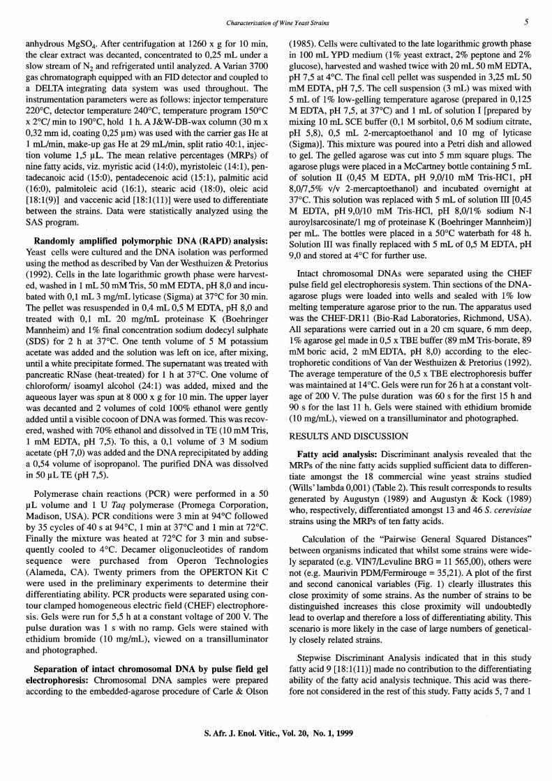
anhydrous MgS04. After centrifugation at 1260 x g for 10 min, the clear extract was decanted, concentrated to 0,25 mL under a slow stream of N2 and refrigerated until analyzed. A Varian 3700 gas chromatograph equipped with an FID detector and coupled to a DELTA integrating data system was used throughout. The instrumentation parameters were as follows: injector temperature 220°C, detector temperature 240°C, temperature program 150°C x 2°C/ min to 190°C, hold 1 h. A J&W-DB-wax column (30 m x 0,32 mm id, coating 0,25 Jlm) was used with the carrier gas He at 1 mL/min, make-up gas He at 29 mL/min, split ratio 40: 1, injection volume 1,5 JlL. The mean relative percentages (MRPs) of nine fatty acids, viz. myristic acid (14:0), myristoleic (14:1), pentadecanoic acid (15:0), pentadecenoic acid (15:1), palmitic acid (16:0), palmitoleic acid (16:1), stearic acid (18:0), oleic acid [18: 1(9)] and vaccenic acid [18: 1(11)] were used to differentiate between the strains. Data were statistically analyzed using the SAS program.
Randomly amplified polymorphic DNA (RAPD) analysis: Yeast cells were cultured and the DNA isolation was performed using the method as described by Van der Westhuizen & Pretorius (1992). Cells in the late logarithmic growth phase were harvested, washed in 1 mL 50 mM Tris, 50 mM EDTA, pH 8,0 and incubated with 0,1 mL 3 mg/mL lyticase (Sigma) at 37°C for 30 min. The pellet was resuspended in 0,4 mL 0,5 M EDTA, pH 8,0 and treated with 0,1 mL 20 mg/mL proteinase K (Boehringer Mannheim) and 1 % final concentration sodium dodecyl sulphate (SDS) for 2 h at 37°C. One tenth volume of 5 M potassium acetate was added and the solution was left on ice, after mixing, until a white precipitate formed. The supernatant was treated with pancreatic RNase (heat-treated) for 1 h at 37°C. One volume of chloroform/ isoamyl alcohol (24:1) was added, mixed and the aqueous layer was spun at 8 000 x g for 10 min. The upper layer was decanted and 2 volumes of cold 100% ethanol were gently added until a visible cocoon of DNA was formed. This was recovered, washed with 70% ethanol and dissolved in TE (10 mM Tris, 1 mM EDTA, pH 7,5). To this, a 0,1 volume of 3 M sodium acetate (pH 7,0) was added and the DNAreprecipitated by adding a 0,54 volume of isopropanol. The purified DNA was dissolved in 50 JlL TE (pH 7,5).
Polymerase chain reactions (peR) were performed in a 50 JlL volume and 1 U Taq polymerase (Promega Corporation, Madison, USA). PCR conditions were 3 min at 94°C followed by 35 cycles of 40 s at 94°C, 1 min at 37°C and 1 min at noc. Finally the mixture was heated at 72°C for 3 min and subsequently cooled to 4°C. Decamer oligonucleotides of random sequence were purchased from Operon Technologies (Alameda, CA). Twenty primers from the OPERTON Kit C were used in the preliminary experiments to determine their differentiating ability. PCR products were separated using contour clamped homogeneous electric field (CHEF) electrophoresis. Gels were run for 5,5 h at a constant voltage of 200 V. The pulse duration was 1 s with no ramp. Gels were stained with ethidium bromide (10 mg/mL), viewed on atransilluminator and photographed.
Separation of intact chromosomal DNA by pulse field gel electrophoresis: Chromosomal DNA samples were prepared according to the embedded-agarose procedure of Carle & Olson
(1985). Cells were cultivated to the late logarithmic growth phase in 100 mL YPD medium (1 % yeast extract, 2% peptone and 2% glucose), harvested and washed twice with 20 mL 50 mM EDTA, pH 7,5 at 4°C. The final cell pellet was suspended in 3,25 mL 50 mM EDTA, pH 7,5. The cell suspension (3 mL) was mixed with 5 mL of 1 % low-gelling temperature agarose (prepared in 0,125 M EDTA, pH 7,5, at 37°C) and 1 mL of solution I [prepared by mixing 10 mL SCE buffer (0,1 M sorbitol, 0,6 M sodium citrate, pH 5,8), 0,5 mL 2-mercaptoethanol and 10 mg of lyticase (Sigma)]. This mixture was poured into a Petri dish and allowed to gel. The gelled agarose was cut into 5 mm square plugs. The agarose plugs were placed in a McCartney bottle containing 5 mL of solution II (0,45 M EDTA, pH 9,0/10 mM Tris-HCl, pH 8,0/7,5% v/v 2-mercaptoethanol) and incubated overnight at 37°C. This solution was replaced with 5 mL of solution ill [0,45 M EDTA, pH 9,0/10 mM Tris-HCl, pH 8,0/1 % sodium N-l auroylsarcosinate/l mg of proteinase K (Boehringer Mannheim)] per mL. The bottles were placed in a 50°C waterbath for 48 h. Solution ill was finally replaced with 5 mL of 0,5 M EDTA, pH 9,0 and stored at 4°C for further use.
Intact chromosomal DNAs were separated using the CHEF pulse field gel electrophoresis system. Thin sections of the DNAagarose plugs were loaded into wells and sealed with 1 % low melting temperature agarose prior to the run. The apparatus used was the CHEF-DRll (Bio-Rad Laboratories, Richmond, USA). All separations were carried out in a 20 cm square, 6 mm deep, 1 % agarose gel made in 0,5 x TBE buffer (89 mM Tris-borate, 89 mM boric acid, 2 mM EDTA, pH 8,0) according to the electrophoretic conditions of Van derWesthuizen & Pretorius (1992). The average temperature of the 0,5 x TBE electrophoresis buffer was maintained at 14°C. Gels were run for 26 h at a constant voltage of 200 V. The pulse duration was 60 s for the first 15 h and 90 s for the last 11 h. Gels were stained with ethidium bromide (10 mg/mL), viewed on a transilluminator and photographed.
RESULTS AND DISCUSSION
Fatty acid analysis: Discriminant analysis revealed that the MRPs of the nine fatty acids supplied sufficient data to differentiate amongst the 18 commercial wine yeast strains studied (Wills' lambda 0,001) (Table 2). This result corresponds to results generated by Augustyn (1989) and Augustyn & Kock (1989) who, respectively, differentiated amongst 13 and 46 S. cerevisiae strains using the MRPs of ten fatty acids.
Calculation of the "Pairwise General Squared Distances" between organisms indicated that whilst some strains were widely separated (e.g. VIN7ILevuline BRG = 11 565,00), others were not (e.g. Maurivin PDM/Fermirouge = 35,21). A plot of the first and second canonical variables (Fig. 1) clearly illustrates this close proximity of some strains. As the number of strains to be distinguished increases this close proximity will undoubtedly lead to overlap and therefore a loss of differentiating ability. This scenario is more likely in the case of large numbers of genetically closely related strains.
Stepwise Discriminant Analysis indicated that in this study fatty acid 9 [18: 1 (11)] made no contribution to the differentiating ability of the fatty acid analysis technique. This acid was therefore not considered in the rest of this study. Fatty acids 5, 7 and 1
S. Afr. J. Enol. Vitic., Vol. 20, No.1, 1999

6 Characterization of Wine Yeast Strains
TABLE 2 Mean* relative percentages of nine fatty acids in 18 commercial wine yeast strains.
Strain
1 2 3
14:0 14:1(9) 15:0
VIN7 2,05 0,45 0,45
VIN13 0,50 0,20 0,20
WE14 1,00 0,40 0,20
N96 0,80 0,20 0,45
228 0,65 0,25 0,30
WE372 0,70 0,20 0,20
Levuline BRG 0,50 0,10 0,10
Fermol bouquet 1,05 0,40 0,20
Maurivin A WRI 796 0,80 0,20 0,20
Fermivin cryo 0,80 0,25 0,25
Actiflore Killer F5 0,55 0,30 0,10
Maurivin PDM 0,75 0,45 0,15
Blastosel kappa 0,95 0,60 0,10
Zymaflore FlO 1,10 0,30 0,Q1
Lalvin 71B 1,00 0,80 0,40
Fermol Killer 0,85 0,50 0,01
Fermirouge 0,90 0,50 0,20
Enoferm Bordeaux Red 0,90 0,50 0,20
*Mean of two replicates per strain.
made the greatest contribution to the differentiating ability of the technique. Upon reclassification of the duplicates per strain using "Linear Discriminant Functions" calculated from data for only fatty acids 5, 7 and 1, only one of the duplicates for Lalvin 71B and WE14 exhibited a probability of less than 0,700 for reclassification as itself. In all other instances probability for correct reclassification exceeded 0,920 while for 22 of the 36 duplicates the probability was 1,000.
In general, the fatty acid analysis technique is reliable but time-
Fatty acids
4 5 6 7 8 9
15:1(9) 16:0 16:1(9) 18:0 18:1(9) 18:1(11)
0,20 16,90 41,10 4,25 31,85 1,85
0,20 7,70 43,55 6,65 37,80 1,85
0,20 7,60 45,70 5,15 37,25 1,35
0,20 12,75 52,45 5,15 34,60 2,15
0,25 9,55 44,40 6,95 34,25 1,85
0,10 10,20 42,30 6,65 36,80 1,75
0,10 6,50 30,60 9,40 48,70 1,90
0,25 10,35 43,95 6,20 32,00 1,90
0,10 12,60 42,70 6,70 32,20 2,00
0,15 10,30 40,55 4,50 39,20 1,20
0,20 7,45 37,40 5,25 46,25 1,30
0,20 6,95 46,90 5,10 36,40 1,25
0,20 5,70 43,60 4,85 41,50 0,90
0,10 9,15 37,00 4,70 41,20 1,10
0,45 7,30 50,15 4,75 33,00 1,20
0,Q1 6,40 38,05 5,50 43,75 1,25
0,25 7,45 48,25 5,05 33,35 1,15
0,30 7,10 44,60 4,00 40,15 0,95
consuming which makes it unsuitable as a routine characterization technique. However, this technique is still useful to verify ambiguous results obtained with other characterization techniques.
RAPD-PCR banding patterns: Only some of the primers used in this study gave satisfactory results in amplification reactions with all the yeast strains studied. The amplification banding patterns obtained when using primer OPC-09 for the different strains are shown in Fig. 2A and B. A considerable number of
S. Afr. J. Enol. Vitic., Vol. 20, No.1, 1999

Characterization of Wine Yeast Strains
20
A
15 0 A -
0
0 N
10 - 0 G N
V X G N X CJ.) V H ..c 5 - H ro L.. p ro > p
ro 0 -U
C 0
C c ro W C
0 -5 -W
E 8 L E Q
Q -10 - ~L
M M
-15 ~
F
-20 I I I I I I
-60 -40 -20 0 20 40 60 80
Canonical Variable 1 FIGURE 1
Canonical plot. Code for canonical plot: A = Maurivin AWRI 796; B = Levuline BRG; C = Fermivin cryo; D = Lalvin 7IB; E = Enoferm Bordeaux Red; F = Zymaflore FlO; G = Fermol bouquet; H = Fermirouge; K = Blastocel kappa; L = Actiflore killer F5; M = Fermol killer; N = N96; 0 = 228; P = Maurivin PDM; Q = VIN 7; V = Yin 13; W = WE 14; X = WE 372.
s. Afr. J. Enol. Vitic., Vol. 20, No.1, 1999
7

8
bp
8576 7427 6106 4899 3639 2799
1953 1882 1515 1482 1184 992
710
492
359
bp
8576 7427 6106 4899 3639 2799
1953 lB82 1515 1482 1184 992
710
492
359
A
c
:> .... Q)
-I: ,... C\l Z :2 :>
:> .... Q)
-I: ,... z C\l
:2 :>
M "<t .... .... <D Z W Ol 00
:> $: z N N
M "<t .... ,... z W <D 00 Ol ;;: $: Z
N N
Characterization a/Wine Yeast Strains
(!) Ol
Q) ,... c:: C,!) ;:j
c::: 0-
~ ;:j 0) 0 :> .0 Q) C .... N ,S (5 'S: Q) ,...
'C -I: M '"5 E ;:j W > ... C\l C\l
$: Q) Q) :2 :2 ....J U.
B
<D Ol
:> .... Q)
-I: C\l :2
Q) ,...
C,!) ;:j c:: c::: 0- ~ ;:j 0) 0 Q) .0 C
N ,S (5 'S: ,... M '"5 E 'C
W > ;:j C\l
$: ~ Q)
:2 U.
o
FIGURE 2
"C Q)
c::: X
LO ;:j
u. C\l C\l .... :2 0.. 0 Q)
0 ,:g 0.. .... .... "E c:- O ~ u. ,:g Q) 0 <.> :;z 0- 0) 0) I!! :;z Ol C I!! C Q)
0 T"" ;:j
E 'S: 'S: If) ,... e 0 .9 c;:: (5
'§ ~
'C C\l C
E '§ J£ ;:j If) E 'S: 0 Q) C\l C\l
~ ~ Q) Q) C u. « :2 CO u. u. W
"C Q)
c::: )(
LO ;:j
U. C\l C\l .... :2 0.. 0 ~ ~ ~ 0.. .... ....
0 C\l U. ~ Q) 0 <.> 2 0- ~
I!! 0) 2 Ol 0)
C C Q) .... ;:j
E 'S: I!! 'S: .9 0 ,...
(5 e c;:: '§ 0 'C C\l C '§ J£ c;:: ;:j If) E ~ E :o::l C\l C\l 0 Q) .<;i ~ C\l Q) Q) C u. :2 CO ....J U. U. W
Patterns of amplified DNA obtained with different wine yeast strains in PCR. Primer OPC-09 (5'-CTCACCGTCC-3') was used in A and B, whilst primer OPC-13 (5'-AAGCCTGTCC-3') was used in C and D, Molecular weight marker VII (Boehringer Mannheim) was used as standard,
S. Afr. J. Enol. Vitic., Vol. 20, No.1, 1999

Characterization of Wine Yeast Strains
shared bands were generated for the different strains. It was apparent that one could not distinguish amongst the South African yeast strains, VIN13, WE14, N96, 228 and WE372, using this primer. No differences in their DNA banding patterns were found between Maurivin PDM and Blastosel kappa. Different RAPD-PCR profiles were generated for the rest of the strains. Results obtained with the OPC-13 primer indicated that strains Maurivin PDM and Blastosel kappa had different profiles (Fig. 2C and D). Strains VIN13, WE14, N96, 228 and WE372 also displayed different banding patterns when using the OPC-13 primer. However, where it was possible to differentiate between strains Fermol bouquet and Maurivin AWRI 796, using OPC-09, this was not possible when using OPC-13. It is interesting to see that both the PCR-profiles obtained for strains Blastosel kappa and Fermol killer were different, but the electrophoretic karyotypes were similar.
RAPD-PCR is a rapid technique that does not require prior knowledge of the genome sequence and reveals more polymorphisms. The main disadvantage is the time and effort needed to select an appropriate primer for differentiating a new strain from a group of previously characterized strains. Another problem of RAPD-PCR frequently cited is the poor reproducibility of some bands. There are many variables in the reaction that may contribute to this problem, including the different time-temperature responses of various types of thermal cyclers, the specific activity of commercial DNA-polymerase preparations, the concentration of DNA and primers, and the composition of the reaction buffer. However, with the correct standardization of the reaction variables, the problem can be minimized. The effect of some of these variables on the reproducibility of the bands was reported by MacPherson et al. (1993). It is therefore imperative to standardize the reaction parameters and perform replications of the same reaction on different days. The separation of the PCR-generated DNA fragments by CHEF pulse field gel electrophoresis is another important factor that contributes to clear differences between RAPD banding patterns thereby enhancing the differentiating power of this technique.
Electrophoretic karyotyping: The chromosomal banding patterns of the different yeast strains are depicted in Fig. 3. Almost all the strains had different karyotypes and the variations were apparent in the number, position and intensity of the bands. Identical profiles were obtained for only two of the strains, Blastosel kappa and Fermol killer. It was, however, possible to differentiate between these yeasts by means ofRAPD-PCR (both primers) and long-chain cellular fatty acid analysis.
The chromosome numbers of the 18 commercial wine yeast strains varied from 12 to 20. It is known that Saccharomyces strains contain the most chromosomes of all the yeast genera. This unique chromosome complexity of S. cerevisiae is advantageous from the viewpoint of this species being amendable to identification and fingerprinting by comparing chromosomal banding patterns. Although time-consuming, electrophoretic karyotyping is highly reliable and widely used to differentiate amongst wine yeast strains.
CONCLUSIONS
e E 0 ()
e -c: 0 ()
('") .q-".... ..... ..-z Z L1J :> :> $:
IS") u.. .... :::2: 0 g c:- O
u 2 0... c:
~ c: .s; .s; 0
1~ ·c co:: ::::J
~ co Q) :::2: u..
9
(0 0>
Q) i'-
e:> ::::J 0:: et:: C"
~ ::::J to 0 Q) .0 c:
C\I .s (5 .s; i'-('") "3 E ·c
c.o co L1J > ::::J 0) C\I $: Q) Q) co Z N ....J u.. :::2:
"C Q)
a::: x ::::J
co co c. Q) c. .... "E co g Q) 0 .::.:. CO CO ~ 2 Ol
Q) ..... ::::J E /J) 0 ".... (5 e .9 co:: co c: ·E ~ .s; E /J) E co (ij ... ... 0
iIi >- Q) Q) c: N ....J u.. u.. L1J
FIGURE 3 From practical experience, when using these three identification techniques and comparing them to one another in terms of Electrophoretic karyotypes of commercial wine yeast strains.
s. Afr. J. Enol. Vitic., Vol. 20, No.1, 1999

10 Characterization a/Wine Yeast Strains
accuracy, clarity and ease of interpreting results, reliability and reproducibility, time and cost effectiveness, as well as appropriateness for differentiating between a wide variety of commercial wine yeast strains, the following conclusions can be made:
(i) The combined use of these three techniques provides the most reliable means of differentiating amongst commercial wine yeast strains.
(ii) As a single identification technique, electrophoretic karyotyping seems to be the most useful method for routine fingerprinting of wine yeast strains and should therefore be used as the primary means of differentiating between these yeast strains and confirming their authenticity.
(iii) In cases of uncertainty, RAPD-PCR and/or long-chain fatty acid analysis could be used as back-up methods to verify the results obtained by electrophoretic karyotyping.
LITERATURE CITED
AUGUSTYN. O.P.H .• 1989. Differentiation between yeast species. and strains within a species. by cellular fatty acid analysis. 2. Saccharomyces cerevisiae. S. Afr. J. Enol. Vitic. 10, 8-17.
AUGUSTYN, O.P.H. & KOCK, J.F.L., 1989. Differentiation of yeast species, and strains within a species, by cellular fatty acid analysis. 1. Application of an adapted technique to differentiate between strains of Saccharomyces cerevisiae. J. Microbial. Meth. 10, 9-23.
BIDENNE, C., BLONDIN, B., DEQUlN, S. & VEZINHET. F.. 1992. Analysis of the chromosomal DNA polymorphisms of wine yeast strains of Saccharomyces cerevisiae. Curro Genet. 22, 1-7.
CARLE, G.F. & OLSON, M.V., 1985. An electrophoretic karyotype for yeast. Proc. Nat!. Acad. Sci. USA 82, 3756-3760.
DE BARROS LOPES, M., SODEN, A., LANGRIDGE, P. & HENSCHKE, P.A., 1995, Identification of wine yeast. Proc. 7th Int. Confer. Yeast Genet. Mol. Bioi., Lisbon, Portugal. (Yeast 11, 14-5JB Special Issue).
DEGRE, R., THOMAS, D.Y., ASH, J., MAILHIOT, K., MARIN, A. & DUBORD, C., 1989. Wine yeast strain identification. Am. J. Eno/. Vitic. 40, 309-315.
GRANDO, M.S. & COLATO, L., 1994. Polimorfismo del cariotipo electroforetico in lieviti Saccharomyces cerevisiae di interesse enologico. Vignivini 5, 57-61.
HENSCHKE, P.A., 1997. Wine yeast. In: ZIMMERMANN, F.K. & ENTIAN, K.-D. (eds). Yeast sugar metabolism. Technomic Publishing Co., Lancaster, Pennsylvania. pp. 527-560.
HUFFMAN, J.L., MOLINA, F.1. & JONG, S-C, 1992. Authentication of ATCC strains in the Saccharomyces cerevisiae complex by PCR fingerprinting. Expel'. Mycol.16, 316-319.
KISHIMOTO, M., SOMA, E. & GOTO, S., 1994. Classification of cryophilic wine yeasts based on electrophoretic karyotype, G + C content and DNA similarity. J. Gen. App/. Microbia!. 40, 83-94.
KREGER-VAN RIJ, N.J.W., 1984. The yeasts, a taxonomic study (3rd ed.). Elsevier Science Publishers, Amsterdam.
KURTZMAN, CP. & FELL, J.w., 1998. The yeasts, a taxonomic study (4th ed.) Elsevier Science Publisher, Amsterdam.
LODDER, J. L. & KREGER-VAN RIJ, NJ.W., 1952. The yeasts, a taxonomic study (1st ed.). North-Holland Pub!. Co., Amsterdam.
LODDER, J.L., 1970. The yeast, a taxonomic study (2nd ed.). North-Holland Publ. Co., Amsterdam.
LALVALLEE, F., SALVAS, Y., LAMY, S., THOMAS, D.Y., DEGRE, R. & DULAU, L., 1994. PCR and DNA fingerprinting used as quality control in production of wine yeast strains. Am. 1. E1101. Vitic. 45, 86-9 I.
MACPHERSON, J.M., EKSTEIN, P.E., SCOLES, G.J. & GAJADHAR, A.A., 1993. Variability of the randomly amplified polymorphic DNA assay among thermal cyclers, and effects of primer and DNA concentration. Mol. Cell. Probes 7,293-299.
NAUMOV, G., NAUMOVA, E. & GAILLARDIN, C, 1993. Genetic and karyotypic identification of wine Saccharomyces bayanus yeasts isolated in France and Italy. System. App/. Microbia!. 16, 274-279.
NESS, F., LAVALLEE, F., DUBOURDIEU, D., AIGLE, M. & DULAU, L.,1993. Identification of yeast strains using polymerase chain reaction. J. Sci. Food Agric. 62, 89-94.
PRETORIUS, LS. & VAN DER WESTHUIZEN, TJ., 1991. The impact of yeast genetics and recombinant DNA technology on the wine industry: A Review. S. Afi: J. Eno!. Vitic. 12, 3-31.
QUEROL, A., BARRIO, E., HUERTA, T. & RAMON, D., 1992. Molecular monitoring of wine fermentations conducted by active dried yeast strains. Appl. Environ. Microbial. 58, 2948-2953.
QUESADA, M.P. & CENIS, J.L., 1995. Use of randomly amplified polymorphic DNA CRAPD-PCR) in the characterization of wine yeasts. Am. J. Eno/. Vitic. 46, 204-208.
ROZES, N., GARCIA-JONES, C, LARUE, F. & LONVAUD-FUNEL, A., 1992. Differentiation between fermenting and spoilage yeasts in wine by total free fatty acid analysis . .T. Sci. Food Agric. 59, 351-357.
TREDOUX, H.G., KOCK, J.F.L., LATEGAN, P.M. & MULLER, H.B., 1987. A rapid identification technique to differentiate between Saccharomyces cerevisiae strains and other yeast species in the wine industry. Am. J. Eno!. Vitic. 38, 161-164.
VAN DER WESTHUIZEN, T.J. & PRETORIUS, I.S., 1989. Genetic characterization and breeding of wine yea,ts, S. Afr. Soc. Microbial. Congo 6th, Stellenbosch CPp 15.15).
VAN DER WESTHUIZEN, T.J. & PRETORIUS, I.S., 1992. The value of electrophoretic fingerprinting and karyotyping in wine yeast breeding programmes. Ant. V. Leeuwen. 61, 249-257.
VAN VUUREN, H.J,J. & VAN DER MEER, L., 1987. Fingerprinting of yeasts by protein electrophoresis. Am. J. Eno!. Vitic. 38, 49-53.
VERSAVAUD, A., COURCOUX, P., ROULLAND, C., DULAU, L. & HALLET, J.-N., 1995. Genetic diversity and geographical distribution of wild Saccharomyces cerevisiae strains from the wine-producing area of Chanrentes, France. Appl. Eviron, Microbial. 61, 3521-3529.
YAMAMOTO, N., YAMAMOTO, N., AMEMIYA, H., YOKOMORI, Y., SHIMIZU, K. & TOTSUKA, A., 1991. Electrophoretic karyotypes of wine yeasts. Am J. Enol. Vitic. 42, 358-363.
S. Afr. J. Enol. Vitic., Vol. 20, No.1, 1999