Embed Size (px)
Citation preview
1
The use of Thoracodorsal Artery
Perforator Flap in Oncoplastic
Procedures
A thesis presented by
Tarek Mohamed Hashem
MBBch, Master of General Surgery
For partial fulfillment of the MD degree in Surgical Oncology
Under supervision of
Prof. Dr Ayman A. Amin
Professor of Surgical Oncology
National Cancer Institute Cairo University
Prof. Dr Mohammed A. Rifaat
FRCS, FEBOPRAS
Assistant Professor of Surgical Oncology
(Consultant Plastic and Reconstructive surgeon)
National Cancer Institute Cairo University
Dr. Ahmed Farahat
Lecturer of Surgical Oncology
National Cancer Institute Cairo University
Cairo University
2015
2
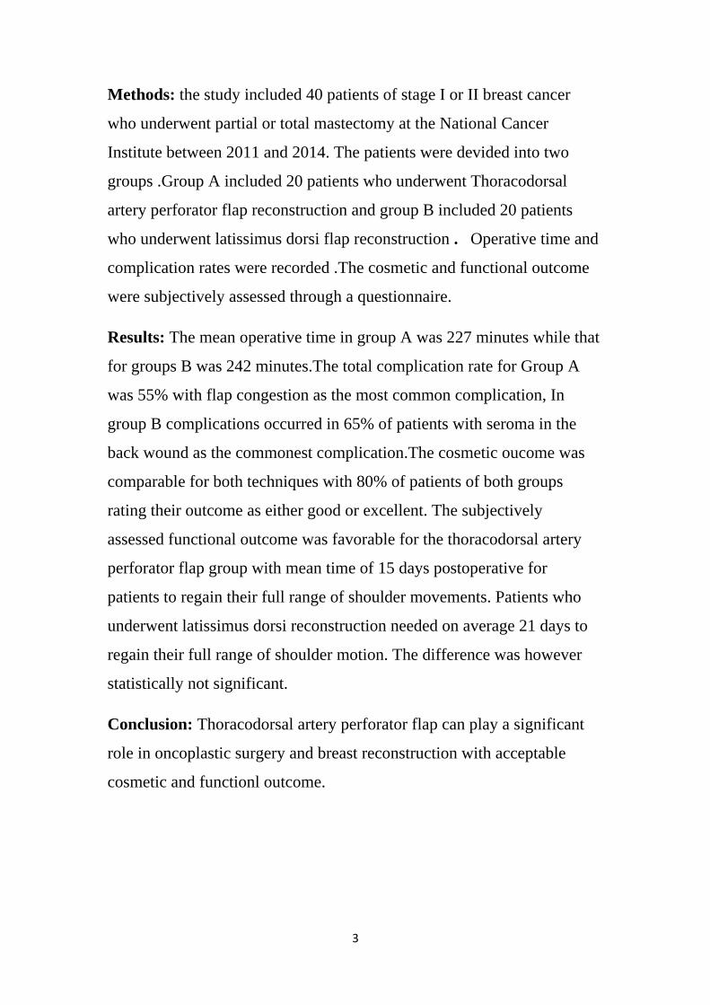
Keywords
Breast cancer, Breast conserving surgery, oncoplastic breast surgery,
breast reconstruction, perforator flaps
Abstract
Objective: To evaluate the outcome of thoracodorsal artery perforator
flap in oncoplastic procedures regarding operative time, post operative
complications and cosmetic outcome.
Background: The advent of oncoplastic surgery in the early 1990s has
revolutionized the concepts of breast reconstruction for breast cancer.This
has been paralleled by a steep evolution of our understanding of the
vascular anatomy of the various flaps used for reconstruction. Earlier
when mastectomies where prevailing, it made perfect sense to look for
flaps with large volumes of tissue and muscle bulk such as the TRAM or
the conventional LD flaps. The harvest of these flaps often left significant
morbidities such as the abdominal wall weakness and the seroma in the
back.
Nowadays the breast surgeon is more than often faced with smaller
defects for which such bulky flaps offer a surplus of tissue with
unacceptable morbidities compared to the smaller defects these flaps have
to reconstruct. Moreover the aesthetic result is often jeopardized by the
bulge of the muscle. The advent of perforator flaps has enabled us to
reconstruct these smaller breast defects with more limited flaps based on
perforating branches of the main thoracodorsal pedicle. Thus the major
part of the muscle with its main pedicle is saved for potential further
reconstruction in case the patient develops recurrence requiring a total
mastectomy.
3
Methods: the study included 40 patients of stage I or II breast cancer
who underwent partial or total mastectomy at the National Cancer
Institute between 2011 and 2014. The patients were devided into two
groups .Group A included 20 patients who underwent Thoracodorsal
artery perforator flap reconstruction and group B included 20 patients
who underwent latissimus dorsi flap reconstruction . Operative time and
complication rates were recorded .The cosmetic and functional outcome
were subjectively assessed through a questionnaire.
Results: The mean operative time in group A was 227 minutes while that
for groups B was 242 minutes.The total complication rate for Group A
was 55% with flap congestion as the most common complication, In
group B complications occurred in 65% of patients with seroma in the
back wound as the commonest complication.The cosmetic oucome was
comparable for both techniques with 80% of patients of both groups
rating their outcome as either good or excellent. The subjectively
assessed functional outcome was favorable for the thoracodorsal artery
perforator flap group with mean time of 15 days postoperative for
patients to regain their full range of shoulder movements. Patients who
underwent latissimus dorsi reconstruction needed on average 21 days to
regain their full range of shoulder motion. The difference was however
statistically not significant.
Conclusion: Thoracodorsal artery perforator flap can play a significant
role in oncoplastic surgery and breast reconstruction with acceptable
cosmetic and functionl outcome.
4
Introduction
The significant developments in the surgical management of breast
cancer have been paralleled by similar advancements in reconstructive
surgery. Improvements in our knowledge of the vascular anatomy have
enabled the design of a new type of fasciocutaneous flaps which are
based on perforating vessels only.
Koshima and Soeda coined the terminology “perforator flaps” in 1989.
In two cases, the authors had used a paraumbilical skin and fat island
based on a muscular perforator to reconstruct the groin and the tongue.
In 1995 Angigiani et al first described the Thoracodorsal Artery
Perforator flap (TDAP). However, Hamdi et al were the first to describe
the use of TDAP in breast reconstruction.
Aim of Work
The aim of this study is to define the role of thoracodorsal artery
perforator flap in oncoplastic breast surgery and explore the
reconstructive options that this flap can offer to breast cancer patients. In
order to outline this role, the TDAP flap will be compared to the
latissimus dorsi myocutaneous flap , which is not only one of the most
frequently utilized flaps in breast reconstruction but also bears the same
anatomical donor site .
Patients and Methods
The study included 40 patients who underwent partial or total
mastectomy for breast cancer or Phylloides tumour and had
reconstruction using either TDAP flap or LD flap. Patients were divided
into two groups. Group A included 20 patients who underwent TDAP
flap reconstruction. This group was compared to another group of 20
patients who had breast reconstruction using the latissimus dorsi
myocutaneous flap, which was designated as group B.The patients were
operated upon between the years 2011 - 2014 at the National Cancer
Institute of Cairo University.
Inclusion criteria
- Pathologically proven breast cancer cases of stage I or II who will
undergo partial or total matsectomies and who will seek
reconstruction and who will consent to a dorsal donor site.
5
- Breast cancer patients of stage III who will be downstaged by
neoadjuvant chemotherapy to stage I or II.
- Pathologically proven phylloides tumour patients who will require
partial mastectomy and reconstruction
- Patients requiring skin excision such as those :
- With tumours close or attached to but not infiltrating the skin.
- With misplaced scars of previous open biopsies,who needed wider
excision due to inadequate or infiltrated margins.
- Patients who needed excision of 20% or more of their breast
volume.
- Patients younger than 60 years of age and free of medical
comorbidities.
Exclusion criteria
- Patients who will need excision of less than 20% of breast
volume
- Patients who will not need skin excision (and still with a
resultant volume loss of less than 20%) i.e small lesions away
from skin.
- Inflammatory breast cancer.
- Surgically inoperable and metastatic breast cancer.
- Severe uncontrolled medical comorbidities.
- Smokers.
Methods All forty patients of both groups underwent:
- History and physical examination
- Routine labs
- Metastatic work up for breast carcinoma patients
- Preoperative counseling session by the operating surgeon to
explain the operative procedure and expected complications.
Patients undergoing TDAP flap were also explained that if
perforators were found insufficient intraoperatively, a conventional
latissimus dorsi myocutaneous flap might be harvested.
6
- Patients who had TDAP flap planned underwent a handheld
Doppler mapping and marking of the thoracodorsal artery
perforators on the night before surgery by the operating surgeon.
- Preoperative marking of the area to be resected and the area of flap
harvest with dimensions being recorded.
- Preoperative photographing with a digital camera in three views:
front or anteroposterior, oblique and lateral with arms to the sides
and elevated.
- Intraoperatively during TDAP flap harvest it was recorded if the
perforators found were compatible in number and distribution to
those marked by the preoperative Doppler mapping or not.
- Operative time was recorded.
- Postoperatively during hospital stay flaps were followed up for
colour, temperature and capillary circulation and drains for colour
and amount of output and early complications were recorded.
- All patients were discharged with their drains.
- Patients were reviewed by operating surgeon one week then two
weeks postoperatively and postoperative photographs were taken in
three views and all complications that have developed were
recorded and dealt with.
- Drains were removed when their output was equal to or less than
50 cc.
- Patients with breast cancer were then referred to receive their
adjuvant treatment according to their final pathology report.
- After finishing their adjuvant treatment patients were invited again
to be reviewed by the operating surgeon where they were
photographed in three views and were asked to answer a five scale
subjective questionnaire evaluating the cosmetic outcome of their
reconstructive procedure. This was graded as: excellent (5), good
(4),fair (3),poor (2) or very poor (1). The criteria they were asked
to evaluate were symmetry, colour match, consistency of the flap,
the appearance of their scars and overall satisfaction.
- The functional effects of both techniques were generally assessed
in a subjective manner through asking the patients in the same
questionnaire about the time elapsed until they regained the full
range of motion of their shoulder movements.
7
- The preoperative and postoperative pictures for each patient were
shown on a computer screen to a panel composed of a breast
surgeon, a radiotherapist and a nurse. The panel was asked to
evaluate the cosmetic outcome of each case and give it a grade on a
scale of 5 similar to the questionnaire answered by the patients.
Again the criteria the panel had to evaluate were symmetry in
shape and size, visibility of scars (raised or depressed, hyper or
hypopigmentation, narrow or wide), symmetry of both
inframammary folds and overall appearance.
- Patients were then followed up oncologically every 3 months for
the first 3 years then every 6 months thereafter for the occurrence
of local or distant relapse.
- Data were statistically described in terms of mean standard
deviation ( SD), and, or frequencies (number of cases) and
percentages when appropriate. Comparison of numerical variables
between the study groups was done using Student t test for
independent samples in normally distributed data and Mann
Whitney U test for independent samples in not normal data. For
comparing categorical data, Chi square (2) test was performed.
Exact test was used instead when the expected frequency is less
than 5. p values less than 0.05 was considered statistically
significant. All statistical calculations were done using computer
program SPSS (Statistical Package for the Social Science; SPSS
Inc., Chicago, IL, USA) release 15 for Microsoft Windows (2006).
Results
Group A (TDAP flap)
Technical results
Operative time:
The mean operative time was 227 minutes or three hours and forty seven
minutes (range 310- 180 minutes).The operative time included excision
of breast tumour, flap harvest and insetting. It is noteworthy that there
was a trend towards a less operative time by time of completion of this
study.
8
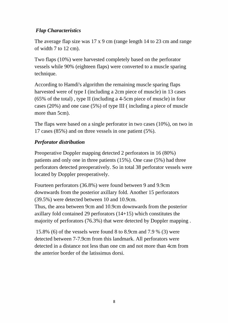
Flap Characteristics
The average flap size was 17 x 9 cm (range length 14 to 23 cm and range
of width 7 to 12 cm).
Two flaps (10%) were harvested completely based on the perforator
vessels while 90% (eighteen flaps) were converted to a muscle sparing
technique.
According to Hamdi's algorithm the remaining muscle sparing flaps
harvested were of type I (including a 2cm piece of muscle) in 13 cases
(65% of the total) , type II (including a 4-5cm piece of muscle) in four
cases (20%) and one case (5%) of type III ( including a piece of muscle
more than 5cm).
The flaps were based on a single perforator in two cases (10%), on two in
17 cases (85%) and on three vessels in one patient (5%).
Perforator distribution
Preoperative Doppler mapping detected 2 perforators in 16 (80%)
patients and only one in three patients (15%). One case (5%) had three
perforators detected preoperatively. So in total 38 perforator vessels were
located by Doppler preoperatively.
Fourteen perforators (36.8%) were found between 9 and 9.9cm
downwards from the posterior axillary fold. Another 15 perforators
(39.5%) were detected between 10 and 10.9cm.
Thus, the area between 9cm and 10.9cm downwards from the posterior
axillary fold contained 29 perforators (14+15) which constitutes the
majority of perforators (76.3%) that were detected by Doppler mapping .
15.8% (6) of the vessels were found 8 to 8.9cm and 7.9 % (3) were
detected between 7-7.9cm from this landmark. All perforators were
detected in a distance not less than one cm and not more than 4cm from
the anterior border of the latissimus dorsi.
9
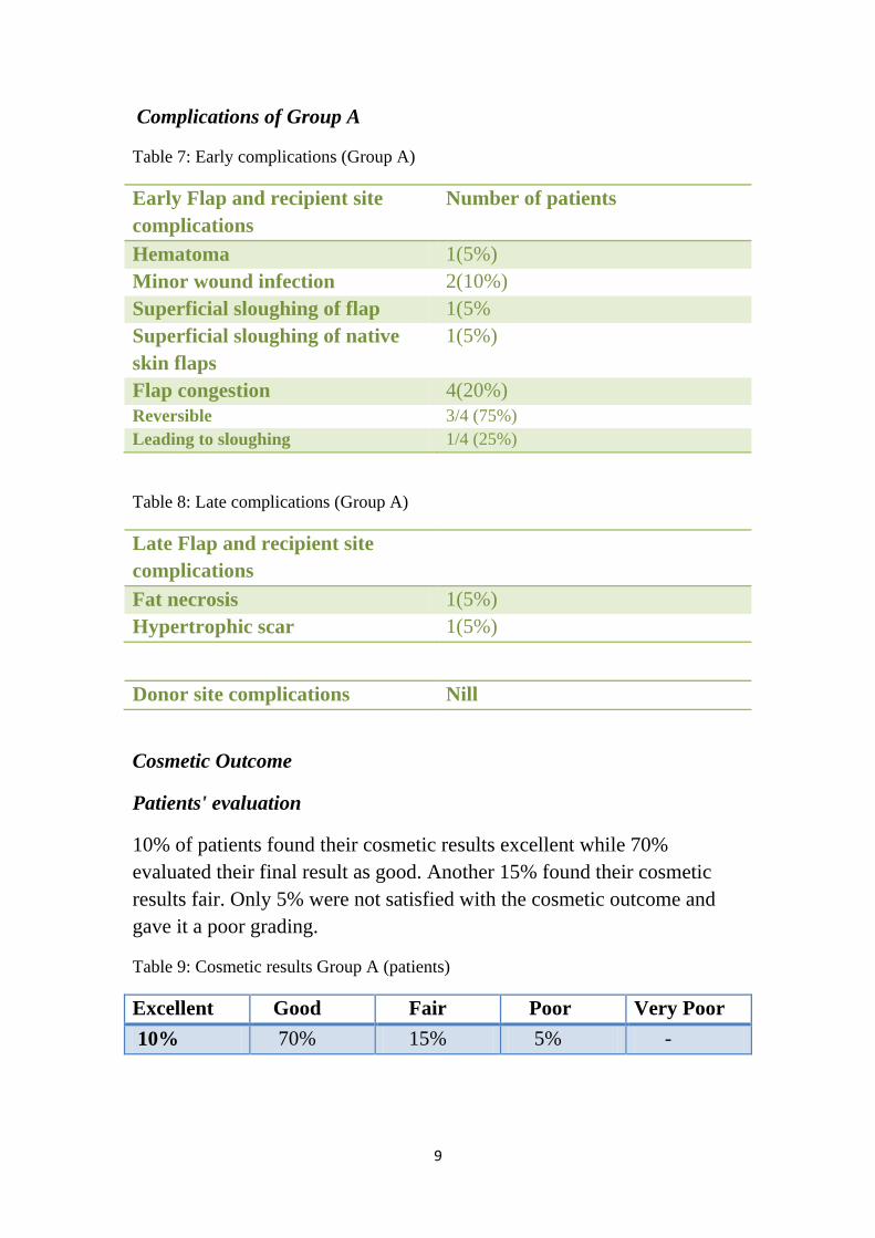
Complications of Group A
Table 7: Early complications (Group A)
Early Flap and recipient site
complications
Number of patients
Hematoma 1(5%)
Minor wound infection 2(10%)
Superficial sloughing of flap 1(5%
Superficial sloughing of native
skin flaps
1(5%)
Flap congestion 4(20%)
Reversible 3/4 (75%)
Leading to sloughing 1/4 (25%)
Table 8: Late complications (Group A)
Late Flap and recipient site
complications
Fat necrosis 1(5%)
Hypertrophic scar 1(5%)
Donor site complications Nill
Cosmetic Outcome
Patients' evaluation
10% of patients found their cosmetic results excellent while 70%
evaluated their final result as good. Another 15% found their cosmetic
results fair. Only 5% were not satisfied with the cosmetic outcome and
gave it a poor grading.
Table 9: Cosmetic results Group A (patients)
Excellent Good Fair Poor Very Poor
10% 70% 15% 5% -
11
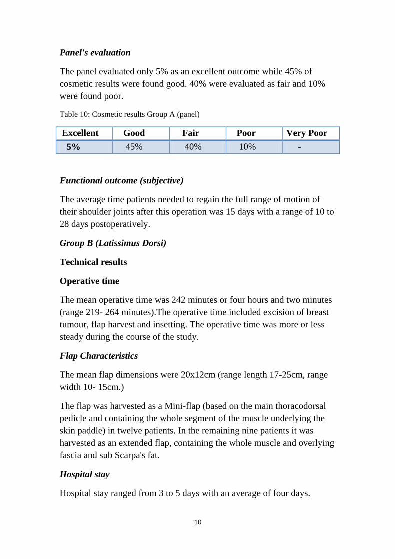
Panel's evaluation
The panel evaluated only 5% as an excellent outcome while 45% of
cosmetic results were found good. 40% were evaluated as fair and 10%
were found poor.
Table 10: Cosmetic results Group A (panel)
Excellent Good Fair Poor Very Poor
5% 45% 40% 10% -
Functional outcome (subjective)
The average time patients needed to regain the full range of motion of
their shoulder joints after this operation was 15 days with a range of 10 to
28 days postoperatively.
Group B (Latissimus Dorsi)
Technical results
Operative time
The mean operative time was 242 minutes or four hours and two minutes
(range 219- 264 minutes).The operative time included excision of breast
tumour, flap harvest and insetting. The operative time was more or less
steady during the course of the study.
Flap Characteristics
The mean flap dimensions were 20x12cm (range length 17-25cm, range
width 10- 15cm.)
The flap was harvested as a Mini-flap (based on the main thoracodorsal
pedicle and containing the whole segment of the muscle underlying the
skin paddle) in twelve patients. In the remaining nine patients it was
harvested as an extended flap, containing the whole muscle and overlying
fascia and sub Scarpa's fat.
Hospital stay
Hospital stay ranged from 3 to 5 days with an average of four days.
11
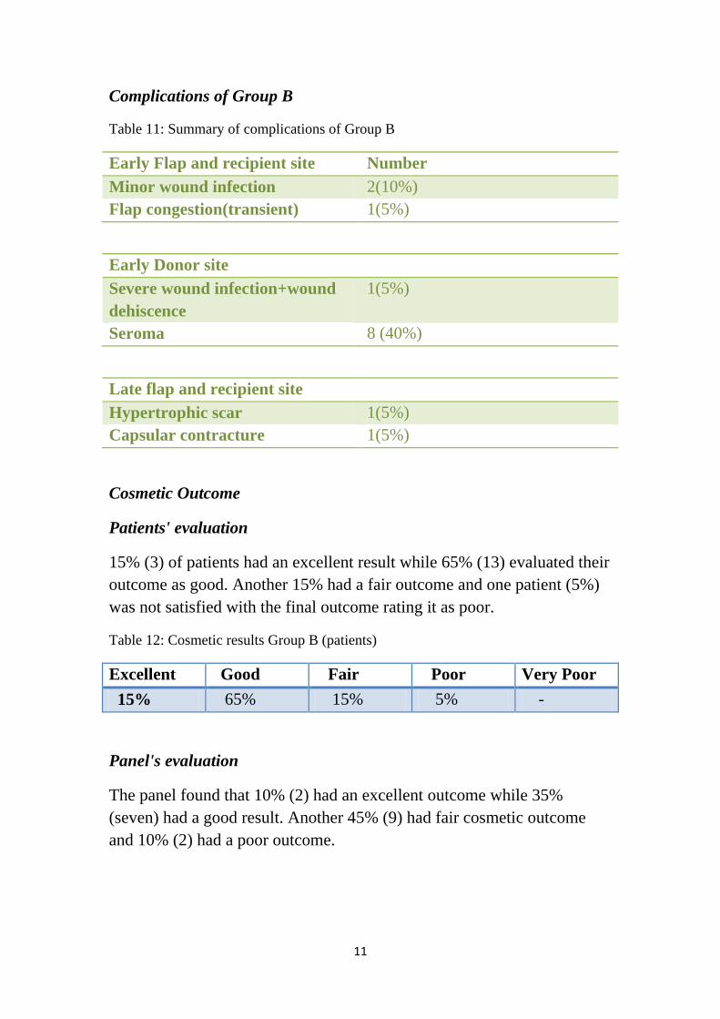
Complications of Group B
Table 11: Summary of complications of Group B
Early Flap and recipient site Number
Minor wound infection 2(10%)
Flap congestion(transient) 1(5%)
Early Donor site
Severe wound infection+wound
dehiscence
1(5%)
Seroma 8 (40%)
Late flap and recipient site
Hypertrophic scar 1(5%)
Capsular contracture 1(5%)
Cosmetic Outcome
Patients' evaluation
15% (3) of patients had an excellent result while 65% (13) evaluated their
outcome as good. Another 15% had a fair outcome and one patient (5%)
was not satisfied with the final outcome rating it as poor.
Table 12: Cosmetic results Group B (patients)
Excellent Good Fair Poor Very Poor
15% 65% 15% 5% -
Panel's evaluation
The panel found that 10% (2) had an excellent outcome while 35%
(seven) had a good result. Another 45% (9) had fair cosmetic outcome
and 10% (2) had a poor outcome.
12
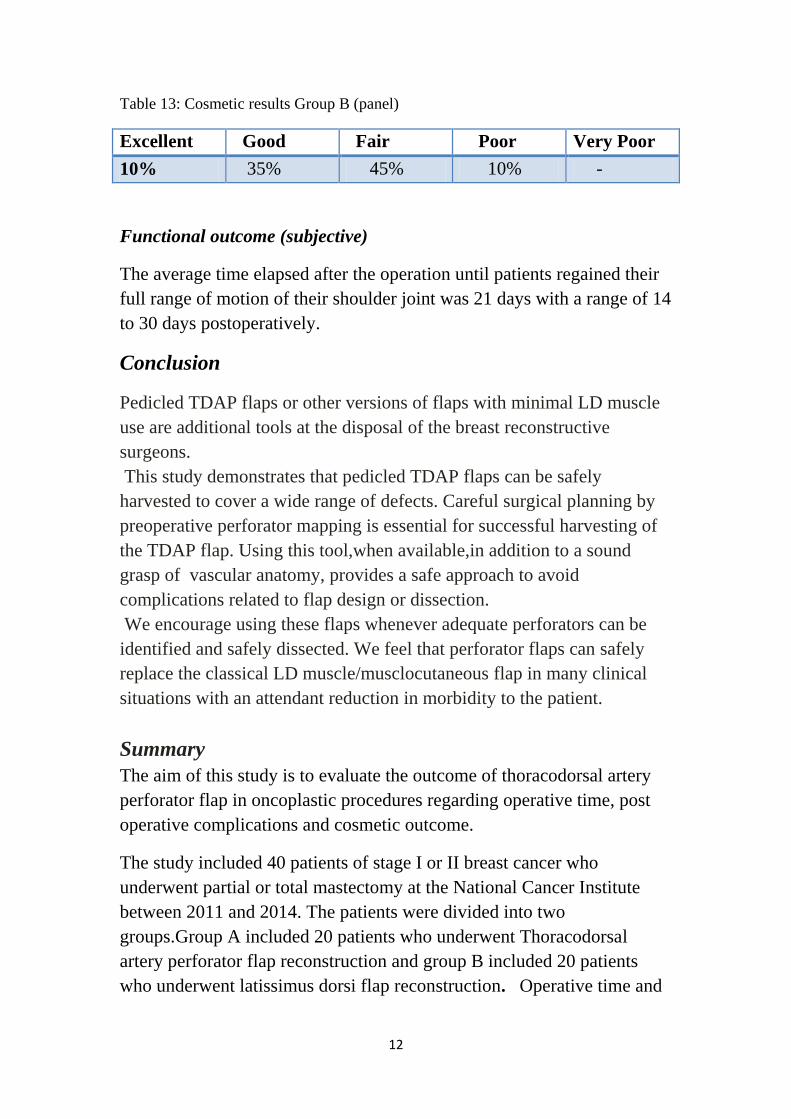
Table 13: Cosmetic results Group B (panel)
Excellent Good Fair Poor Very Poor
10% 35% 45% 10% -
Functional outcome (subjective)
The average time elapsed after the operation until patients regained their
full range of motion of their shoulder joint was 21 days with a range of 14
to 30 days postoperatively.
Conclusion
Pedicled TDAP flaps or other versions of flaps with minimal LD muscle
use are additional tools at the disposal of the breast reconstructive
surgeons.
This study demonstrates that pedicled TDAP flaps can be safely
harvested to cover a wide range of defects. Careful surgical planning by
preoperative perforator mapping is essential for successful harvesting of
the TDAP flap. Using this tool,when available,in addition to a sound
grasp of vascular anatomy, provides a safe approach to avoid
complications related to flap design or dissection.
We encourage using these flaps whenever adequate perforators can be
identified and safely dissected. We feel that perforator flaps can safely
replace the classical LD muscle/musclocutaneous flap in many clinical
situations with an attendant reduction in morbidity to the patient.
Summary
The aim of this study is to evaluate the outcome of thoracodorsal artery
perforator flap in oncoplastic procedures regarding operative time, post
operative complications and cosmetic outcome.
The study included 40 patients of stage I or II breast cancer who
underwent partial or total mastectomy at the National Cancer Institute
between 2011 and 2014. The patients were divided into two
groups.Group A included 20 patients who underwent Thoracodorsal
artery perforator flap reconstruction and group B included 20 patients
who underwent latissimus dorsi flap reconstruction. Operative time and
13
complication rates were recorded .The cosmetic and functional outcome
were subjectively assessed through a questionnaire.
The results showed a mean operative time in group A which was 227
minutes while that for groups B was 242 minutes. The total complication
rate for Group A was 55% with flap congestion as the most common
complication; In group B complications occurred in 65% of patients with
seroma in the back wound as the commonest complication. The cosmetic
oucome was comparable for both techniques with 80% of patients of both
groups rating their outcome as either good or excellent. The subjectively
assessed functional outcome was favorable for the thoracodorsal artery
perforator flap group with mean time of 15 days postoperative for
patients to regain their full range of shoulder movements. Patients who
underwent latissimus dorsi reconstruction needed on average 21 days to
regain their full range of shoulder motion. The difference was however
statistically not significant.
14
الملخص العربى
فى الدراسة هى دراسة بحثية الغرض منها تقييم استخدامات سديلة الصدر الخلفية هذه ان
فى التجميلية.وتعد هذه السديلة احدث التطورات الجراحيةجراحات اورام الثدى التحفظية
تكون هذه عمليات اعادة البناء بصفة عامة و فى عمليات اعادة بناء الثدى بصفة خاصة حيث ت
ماد قبل ذلك السديلة فقط من الجلد و الدهون الموجودة بمنطقة الصدر الخلفية حيث كان يتم االعت
مع ما كان يتسبب فى حدوث مضاعفات ابرزها تجعلى سديلة عضلية من عضالت الظهرم
ربعين سوائل بمنطقة الظهر و تأثر للحركة بمفصل الكتف.و قد اشتملت هذه الدراسة على ا
تحفظى مريضة بالمراحل المبكرة لسرطان الثدى و التى عادة ما يتم فيها استئصال جزئى او
نة من زء المستأصل فى مجموعة مكوللثدى و قد تم استخدام السديلة الجديدة العادة بناء الج
عشرين مريضة ومقارنة نتائج هذا االسلوب الجراحى الجديد مع مجموعة اخرى مكونة من
دة على عشرين مريضة اخرى تم لديهم اعادة البناء الجزئى للثدى باستخدام السديلة المعتم
ادة ن من خالله اععضلة الظهرو تبين من خالل النتائج ان هذا االسلوب الجراحى الجديد يمك
تبيان بناء الجزء المستاصل من الثدى بصورة مرضية من الناحية الجمالية حيث تم عمل اس
دى بعد للمرضى من كلتا المجموعتين لبيان مدى رضاء كل مريضة عن النتيجة الجمالية للث
نسبة يناعادة البناء و جائت نتائج كلتا المجموعتين متشابهة و حقق كال االسلوبين الجراحي
اكبر نسبة كبيرة من رضاء المرضى عن النتيجة الجمالية.اما بالنسبة للمضاعفات فقد سجلت
من الحاالت %65مضاعفات فى المجموعة التى استخدمت فيها سديلة عضلة الظهر بنسبة
ت فى كان اكثره شيوعا هو حدوث تجمع لسوائل بمنطقة الظهربينما بلغت نسبة المضاعفا
و لم يحدث فى اى من الحاالت تجمع الى سوائل بالظهر. %55لصدر الخلفية مجموعة سديلة ا
15
أستخدامات سديلة الصدر الخلفية فى جراحات أورام
الثدى التحفظية التجميلية
رسالة مقدمة من
محمد هاشم طارق
معهد االورام-مدرس مساعد جراحة االورام
توطئة للحصول على درجة الدكتوراة فى جراحة االورام
تحت اشراف
د أيمن عبد الوهاب أمين.ا
معهد االورام-جراحة االورام ذاستا
جامعة القاهرة
د محمد رفعت.ا
معهد االورام-جراحة االورام مساعد ذاستا
جامعة القاهرة
أحمد فرحات.د
معهد االورام-مدرس جراحة االورام
جامعة القاهرة
جامعة القاهرة
5102