Embed Size (px)
Citation preview
REVIEW
The use of PRP injections in the management of knee osteoarthritis
Brendan O’Connell1 & Nicholas Martin Wragg2& Samantha Louise Wilson2
Received: 3 May 2018 /Accepted: 11 January 2019 /Published online: 13 February 2019# The Author(s) 2019
AbstractOsteoarthritis (OA) is a degenerative disease involving joint damage, an inadequate healing response and progressive deterio-ration of the joint architecture that commonly affects the knee and/or hip joints. It is a major world public health problem and ispredicted to increase rapidly with an ageing population and escalating rate of obesity. Autologous blood-derived products possessmuch promise in the repair and regeneration of tissue and have important roles in inflammation, angiogenesis, cell migration andmetabolism in pathological conditions, including OA. Utilising platelet-rich plasma (PRP) to treat tendon, ligament and skeletalmuscle has shown variable results across many studies with the current evidence base for the efficacy of PRP in treating sportsinjuries remaining inconclusive. More uniformly positive results have been observed by various studies for PRP in OA knee incomparison to hyaluronic acid, other intra-articular injections and placebo than in other musculoskeletal tissue. However,methodological concerns as well as satisfactory PRP product classification prevent the true characterisation of this treatment.Thus, further research is required to investigate how leukocyte inclusion, activation and platelet concentration affect therapeuticefficacy. Furthermore, the optimisation of timing, dosage, volume, frequency and rehabilitation strategies need to be ascertained.For knee OA management, these concerns must be addressed before this promising treatment can be widely implemented.
Keywords Platelet-rich plasma . Osteoarthritis . Intra-articular injection . Knee . Repair and regeneration
Introduction
Osteoarthritis (OA) is a serious degenerative joint diseaseresulting from the degradation of articular cartilage, degrada-tion and proliferative reformation of subchondral bone and alow degree of synovitis that leads to a reduced quality of life(QoL). It is a major cause of pain and disability in the elderlypopulation (> 70 years) (Neogi and Zhang 2013). OA altersthe normal joint metabolism favouring increased catabolismand decreased anabolism (Dhillon et al. 2017). Inflammationand vascular pathology, in combination with cell death,meniscal changes, bone remodelling and subchondral sclero-sis, produces a vicious cycle of progressive joint degeneration.This can be exacerbated by excessive mechanical stress and
oxidative damage (Wruck et al. 2011). Moreover, under con-ditions of metabolic or cytotoxic stress, such as in ageing,autophagy can be upregulated, further decompensating ho-meostatic mechanisms (Lotz and Caramés 2011).
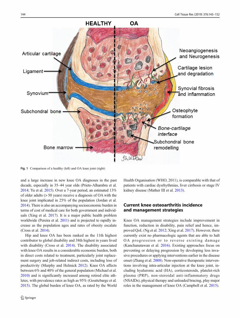
In OA knees (Fig. 1), chondrocyte senescence and lossof cartilage integrity are major features. There is an in-crease in the water content of hyaline cartilage, accompa-nied by corresponding decreases in proteoglycan concen-tration, length and aggregation, causing reduced cartilagestiffness and fibrillation of the cartilage surface. Fromthis, cartilage proceeds to erode and deep clefts may form.Concurrently, morphological changes in subchondral boneare found. As synovial fluid infiltrates, the formation ofsubarticular cysts in the subchondral bone also occurs.Osteophytes (bony projections) are characteristic featuresof knee OA in non-pressure areas, caused by the flatten-ing of bone from pressure in high-wear areas (Adatia et al.2012).
Many interacting factors have a role in indicating the poten-tial for the development of knee OA, although age is typicallyhighlighted (Blagojevic et al. 2010;Michael et al. 2010; Heidari2011; Silverwood et al. 2015; Driban et al. 2017) (Table 1).Primary care databases from a variety of countries haveshown a higher incidence of knee OA than hip or hand OA
* Samantha Louise [email protected]
1 National Centre for Sport and Exercise Medicine, School of Sport,Exercise and Health Sciences, Loughborough University,Loughborough, Leicestershire, UK
2 Centre for Biological Engineering, Wolfson School of Mechanical,Electrical and Manufacturing Engineering, LoughboroughUniversity, Epinal Way, Loughborough, Leicestershire LE11 3TU,UK
Cell and Tissue Research (2019) 376:143–152https://doi.org/10.1007/s00441-019-02996-x
and a large increase in new knee OA diagnoses in the pastdecade, especially in 35–44 year olds (Prieto-Alhambra et al.2014; Yu et al. 2015). Over a 7-year period, an estimated 13%of older adults (> 50 years) receive a diagnosis of OAwith theknee joint implicated in 25% of the population (Jordan et al.2014). There is also an accompanying socioeconomic burden interms of cost of medical care for both government and individ-uals (Xing et al. 2017). It is a major public health problemworldwide (Pereira et al. 2011) and is projected to rapidly in-crease as the population ages and rates of obesity escalate(Cross et al. 2014).
Hip and knee OA has been ranked as the 11th highestcontributor to global disability and 38th highest in years livedwith disability (Cross et al. 2014). The disability associatedwith knee OA results in a considerable economic burden, bothin direct costs related to treatment, particularly joint replace-ment surgery and job-related indirect costs, including loss ofproductivity (Murphy and Helmick 2012). Knee OA affectsbetween 6% and 40% of the general population (Michael et al.2010) and is significantly increased among retired elite ath-letes, with prevalence rates as high as 95% (Gouttebarge et al.2015). The global burden of knee OA, as rated by the World
Health Organisation (WHO, 2011), is comparable with that ofpatients with cardiac dysrhythmias, liver cirrhosis or stage IVkidney disease (Mather III et al. 2013).
Current knee osteoarthritis incidenceand management strategies
Knee OA management strategies include improvement infunction, reduction in disability, pain relief and hence, im-proved QoL (Ng et al. 2012; Xing et al. 2017). However, therecurrently exist no pharmacologic agents that are able to haltOA progress ion or to reverse ex i s t ing damage(Kanchanatawan et al. 2016). Existing approaches focus onpreventing or delaying progression by developing less inva-sive procedures or applying interventions earlier in the diseaseonset (Zhang et al. 2008). Non-operative therapeutic interven-tions involving intra-articular injection at the knee joint, in-cluding hyaluronic acid (HA), corticosteroids, platelet-richplasma (PRP), non-steroidal anti-inflammatory drugs(NSAIDs), physical therapy and unloaded bracing, play majorroles in the management of knee OA (Campbell et al. 2015).
Fig. 1 Comparison of a healthy (left) and OA knee joint (right)
144 Cell Tissue Res (2019) 376:143–152
Platelet-rich plasma for inducingregeneration
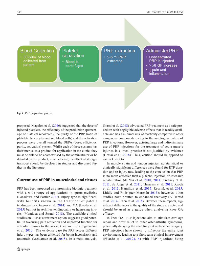
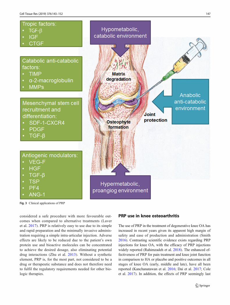
PRP is an autologous mixture of highly concentrated plateletsand associated growth factors and other bioactive componentsproduced by centrifugal separation of whole blood (Fig. 2)that is used in orthopaedic and sports medicine practices totreat bone, tendon and ligament injuries (Fig. 3) (Sundmanet al. 2014). The growth factors released by PRP have beendiscussed in great detail within the literature (Lubkowska et al.2012; Pavlovic et al. 2016; Fernandes and Yang 2016; Parrishand Roides 2018) and have been shown to promote cellrecruitment, proliferation and angiogenesis resulting in a re-duction in the critical regulators of the inflammatory processand a decrease in the expression of inflammatory enzymes(Table 2) (van Buul et al. 2011). PRP may induce aregenerative response by improving the metabolicfunctions of damaged structures (Ficek et al. 2011; Chenet al. 2018) and has been shown to have a positive effect onchondrogenesis and mesenchymal stem cell proliferation(Kabiri et al. 2014).
In clinical practice, PRP is used to enable the application ofautologous plasma and platelet-derived proteins to a desiredlocation with the use of an appropriate scaffold to assist in therepair of the injured tissue (Marx 2001). The rationale for PRPin scaffolds is to take advantage of the huge amount of growthfactors contained in platelets to promote cartilage regenera-tion; however, the use of PRP-augmented scaffolds is still ina preliminary state, with a low scientific level of power (Konet al. 2013). In application to chondrocytes, growth factorspromote matrix synthesis, cell growth and migration and fa-cilitate protein transcription. The supra-physiological releaseof platelet-derived factors directly at the site of cartilage dis-ease, particularly with interest to knee OA, may stimulate thenatural regenerative signalling cascade and enhance thehealing of tissue with further mediation of the anti-inflammatory response (Mascarenhas et al. 2014). In OAjoints, PRP has been shown to affect local and infiltratingcells, mainly synovial cells, endothelial cells, those cells in-volved in innate immunity (such as macrophages) and carti-lage and bone cellular components (Mifune et al. 2013;Dhillon et al. 2017). Additionally, PRP can affect inflamma-tion and angiogenic processes and anabolism and catabolismbalance in cartilage formation and alter the existing microen-vironment during disease progression (Andia and Maffulli2013).
The combined effects of PRP make it a potential optionfor management of knee OA, especially as a primary anal-gesic agent (Meheux et al. 2016). This is due to an increasein proliferation of tenocytes, osteoblasts, mesenchymalstem cells resulting in decreased pain levels postoperative-ly (Ogino et al. 2006). Despite encouraging preclinical re-sults and increasing clinical interest, there remain multiplequestions regarding the clinical application and efficacy ofPRP, not least in the production of PRP, which can causewildly varying characteristics. A simple classification wasfirst proposed in 2009 following the fibrin architecture andcell content (pure PRP: leukocyte-poor PRP, leukocyte-and platelet-rich plasma; pure platelet-rich fibrin:leukocyte-poor platelet-rich fibrin, leukocyte- andplatelet-rich fibrin). Each of these classifications had dif-ferent growth factor release profiles (time, concentration)but studies found additional biological signatures andmechanisms within each family (Dohan Ehrenfest et al.2014). A further classification can be included in the meth-od of application (e.g., injections, glues) and the area ofapplication (e.g., oral/maxillofacial surgery, skin woundhealing). A more recent classification system called PAW(platelet, activation, white blood cells) is based upon abso-lute number of platelets, activation method and presence orabsence of white blood cells (DeLong et al. 2012).According to the authors, the specific determination ofthe components of the PRP is vital in allowing compari-sons between studies. In 2016, a further classification was
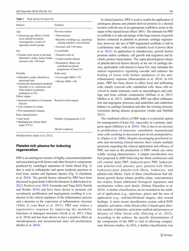
Table 1 Risk factors for knee OA
Intrinsic Extrinsic
Age Previous trauma
• Advancing age effects of bothsexes altered locomotor(stereognostic) control ofopposing muscle groups
• Macrotrauma
• Repetitive loading (e.g., squatting)causing repetitive microtrauma
• Increases risk 3.86 times
Sex Co-morbidity
• Females more prone to developHeberden’s nodes, being femaleincreases risk 1.84 times
• Diseases such as:
▪ Cardiovascular disease▪ Respiratory illness can
contribute/accelerateprogression of knee OA
Heredity Body mass
• Heberden’s nodes inherited asautosomal trait
•Genetically determined metabolicdisorders (e.g., ochronosis andEhler-Danlos syndrome)predispose to OA
• Overweight (BMI > 25)
• Obesity (BMI > 30)
Ethnic origins
• Increased incidence in Europeandescent
Resective joint surgery
• Less common in Asians
• Post-menopausal changes
Knee characteristics:
▪ Joint laxity Number of pregnancies (> 6)▪ Malalignment (femur-tibial
angle (FTA))
Muscle weakness
Modified from Adatia et al. (2012)
Cell Tissue Res (2019) 376:143–152 145
proposed. Magalon et al. (2016) suggested that the dose ofinjected platelets, the efficiency of the production (percent-age of platelets recovered), the purity of the PRP (ratio ofplatelets, leucocytes and red blood cells) and the activationprocess were overall termed the DEPA (dose, efficiency,purity, activation) system. Whilst each of these systems hastheir merits, as a product for application in the clinic, theymust be able to be characterised by the administrator or bedetailed on the product, in which case, the effect of storage/transport should be disclosed in studies and discussed fur-ther in the literature.
Current use of PRP in musculoskeletal tissues
PRP has been proposed as a promising biologic treatmentwith a wide range of applications in sports medicine(Lansdown and Fortier 2017). Injury type is significant,wi th benef i ts shown in the t rea tment of pate l latendinopathy (Dragoo et al. 2014) and OA (Laudy et al.2015) but not in Achilles tendinopathy or hamstring inju-ries (Manduca and Straub 2018). The available clinicalstudies on PRP as a treatment option suggest a good poten-tial in favouring pain reduction and improved function forarticular injuries to the ankle, knee and hip (Engebretsenet al. 2010). The evidence base for PRP across differentinjury types has been criticised for being inconsistent anduncertain (McNamee et al. 2018). In a meta-analysis,
Grassi et al. (2018) advocated PRP treatment as a safe pro-cedure with negligible adverse effects that is readily avail-able and has a minimal risk of reactivity compared to otherexogenous compounds owing to the autologous nature ofPRP injections. However, existing large and indiscriminateuse of PRP injections for the treatment of acute muscleinjuries in clinical practice is not justified by evidence(Grassi et al. 2018). Thus, caution should be applied touse in knee OA.
In muscle strain and tendon injuries, no statistical orclinically significant differences were found for RTP dura-tion and re-injury rate, leading to the conclusion that PRPis no more effective than a placebo injection or intensiverehabilitation (de Vos et al. 2010, 2014; Creaney et al.2011; de Jonge et al. 2011; Thanasas et al. 2011; Kroghet al. 2013; Hamilton et al. 2015; Reurink et al. 2015;Liddle and Rodríguez-Merchán 2015); however, otherstudies have pointed to enhanced recovery (A Hamidet al. 2014; Chen et al. 2018). Between these reports, sig-nificant differences in the quality of the study are noted andshould be used as a guide when analysing knee OAefficacy.
In knee OA, PRP injections aim to stimulate cartilagerepair and offer relief to other osteoarthritic symptoms,potentially delaying the need for joint replacement surgery.PRP injections have shown to influence the entire jointenvironment, leading to a short-term clinical improvement(Filardo et al. 2012a, b) with PRP injections being
Fig. 2 PRP preparation process
146 Cell Tissue Res (2019) 376:143–152
considered a safe procedure with more favourable out-comes when compared to alternative treatments (Laveret al. 2017). PRP is relatively easy to use due to its simpleand rapid preparation and the minimally invasive adminis-tration requiring a simple intra-articular injection. Adverseeffects are likely to be reduced due to the patient’s ownprotein use and bioactive molecules can be concentratedto achieve the desired dosage, also eliminating potentialdrug interactions (Zhu et al. 2013). Without a syntheticelement, PRP is, for the most part, not considered to be adrug or therapeutic substance and does not therefore needto fulfil the regulatory requirements needed for other bio-logic therapies.
PRP use in knee osteoarthritis
The use of PRP in the treatment of degenerative knee OA hasincreased in recent years given its apparent high margin ofsafety and ease of production and administration (Smith2016). Contrasting scientific evidence exists regarding PRPinjections for knee OA, with the efficacy of PRP injectionswidely reported (Rahimzadeh et al. 2018). The enhanced ef-fectiveness of PRP for pain treatment and knee joint functionin comparison to HA or placebo and positive outcomes in allstages of knee OA (early, middle and late), have all beenreported (Kanchanatawan et al. 2016; Dai et al. 2017; Coleet al. 2017). In addition, the effects of PRP seemingly last
Fig. 3 Clinical applications of PRP
Cell Tissue Res (2019) 376:143–152 147
longer and are superior in comparison with intramuscular in-jection therapies (Prieto-Alhambra et al. 2014). Comparisonsbetween intra-articular injection of PRP and placebo and HAtherapy in mild and moderate knee OA have generally shownhigher clinical outcome scores with PRP use (Filardo et al.2012a, b). Similarly, using meta-analysis to compare the effi-cacy of PRP injections against placebo or other therapeuticmeans for the treatment of knee OA (Bennell et al. 2017)has reported greater pain reduction (Laudy et al. 2015) andfunctional improvement (Chang et al. 2014) with the use ofPRP. However, this is at the expense of an increase in nonspe-cific adverse events (Khoshbin et al. 2013).
PRP use has been advocated as a treatment option in allstages of knee OA. Intra-articular PRP injections in activepatients with knee OA show significant improvements in painreduction, improved symptoms and QoL (Gobbi et al. 2012).This could be due to the immediate and sustained release ofgrowth factors over a prolonged period, which enhanceshealing resulting in sustained clinical effects (Dhillon et al.2017). Symptomatic relief for up to 12 months with increasedbenefits to patients with early knee degenerative changes hasbeen found (Campbell et al. 2015) with significant improve-ments in function and reductions in pain with three injectionsper month yielding significantly better outcomes in the short-term (Huang et al. 2017). Improved pain outcomes after3 months with a greater effect in lower OA grades have beenreported (Montañez-Heredia et al. 2016). In moderate kneeOA, functional status and pain have improved with a mini-mum of two injections (Kavadar et al. 2015). In late-stageknee OA, it may be that only a single PRP intra-articularinjection is required to provide effective pain relief, thus
improving activities of daily living and QoL (Joshi Jubertet al. 2017).
Research into the efficacy of PRP has focused on compar-ing the effects of intra-articular PRP injections to other injec-tion therapies. In many studies, PRP injections have improvedfunctional outcomes when compared to HA and placebo con-trols and appear more efficacious in reducing symptoms andimproving QoL (Raeissadat et al. 2015; Kanchanatawan et al.2016). Kon et al. (2011) examined three homogenous groupsof patients treated with three injections of PRP, low molecularweight HA and high molecular weight HA and concluded thatautologous PRP injections have longer efficacy than HA in-jections and enhance articular function. The results showedimproved outcomes for the PRP group at 6 months with youn-ger and more active patients achieving better results with alow degree of cartilage degeneration (Meheux et al. 2016).Conversely, PRP causes a significantly greater acute inflam-matory response and an increase in synoviocyte cell death(Braun et al. 2014) and induces more transient reactions thanHA (Riboh et al. 2016). Spaková et al. (2012) compared threePRP injections with three HA injections in a randomised con-trolled trial (RCT) on 120 patients and discovered betterWestern Ontario and McMaster Universities Arthritis Index(WOMAC) scores and Numerical Rating Scale (NRS) in thePRP group compared to the HA group (Table 3). In a separateRCTwith 120 patients, Cerza et al. (2012) compared four PRPinjections at 1-week intervals with low molecular weight HAand observed better improvement of WOMAC scores at24 weeks in the PRP group. No correlations with the gradeof OA were found in either study. Additionally, betterWOMAC scores were achieved at 24 weeks using PRP by
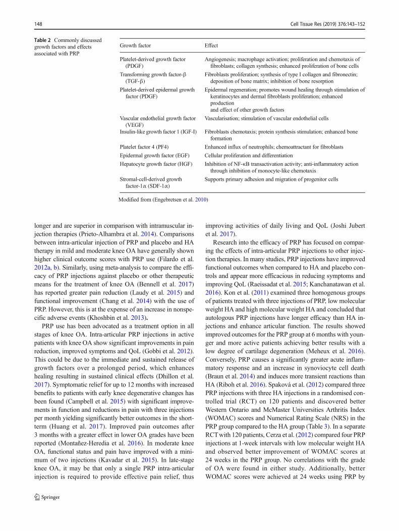
Table 2 Commonly discussedgrowth factors and effectsassociated with PRP
Growth factor Effect
Platelet-derived growth factor(PDGF)
Angiogenesis; macrophage activation; proliferation and chemotaxis offibroblasts; collagen synthesis; enhanced proliferation of bone cells
Transforming growth factor-β(TGF-β)
Fibroblasts proliferation; synthesis of type I collagen and fibronectin;deposition of bone matrix; inhibition of bone resorption
Platelet-derived epidermal growthfactor (PDGF)
Epidermal regeneration; promotes wound healing through stimulation ofkeratinocytes and dermal fibroblasts proliferation; enhancedproductionand effect of other growth factors
Vascular endothelial growth factor(VEGF)
Vascularisation; stimulation of vascular endothelial cells
Insulin-like growth factor 1 (IGF-I) Fibroblasts chemotaxis; protein synthesis stimulation; enhanced boneformation
Platelet factor 4 (PF4) Enhanced influx of neutrophils; chemoattractant for fibroblasts
Epidermal growth factor (EGF) Cellular proliferation and differentiation
Hepatocyte growth factor (HGF) Inhibition of NF-κB transactivation activity; anti-inflammatory actionthrough inhibition of monocyte-like chemotaxis
Stromal-cell-derived growthfactor-1α (SDF-1α)
Supports primary adhesion and migration of progenitor cells
Modified from (Engebretsen et al. 2010)
148 Cell Tissue Res (2019) 376:143–152

Sánchez et al. (2012) who examined 126 patients in a RCTwith different grades of OA and compared three PRP injec-tions at 1-week intervals with HA. Similarly, better outcomeshave been documented when comparing PRP to HA groups at6 months (Li et al. 2011; Say et al. 2013). Patel et al. (2013)compared normal saline with PRP and demonstrated that PRPsignificantly improved WOMAC scores following 6 monthsin comparison to the placebo groups, with patients experienc-ing benefits as early as 18 days. In a follow-up study, Patel andDhillon (2014) hypothesised that the anti-inflammatory effectand chondral remodelling induced by PRP could be the reasonfor the improved clinical effects.
Limitations and recommendations for furtherresearch
Despite the apparent positivity in the use of PRP for treatmentof knee OA, methodological concerns and considerable het-erogeneity between studies are evident (Rodriguez-Merchan2013b). Large RCTs are needed to further assess the efficacyand duration of PRP treatment for patients with knee OA(Rodriguez-Merchan 2013a; Lai et al. 2015). When planningor analysing treatments, frequency and number of injections,
as well as the activation methods (in the case of anticoagulatedPRP), storage aspects, time from plasma isolation and accom-panying therapy should be considered as at present they varywidely between groups. The greatest limiting factor for PRPuse is the lack of standardisation with further research requiredto investigate how leukocyte inclusion, activation and plateletconcentration affect therapeutic efficacy (Chen et al. 2018).Potential classification systems are discussed in depth byDohan Ehrenfest et al. (2014), Lana et al. (2017) and Alvesand Grimalt (2018). The cost-effectiveness of PRP, the demo-graphic most likely to benefit and the optimal PRP protocolmust all be researched further (Bennell et al. 2017). To thisend, optimisation is still required regarding timing, dosage,volume, frequency, composition and post-injection rehabilita-tion (Engerbresten et al. 2010) and a unified classificationneeds to be agreed before this promising treatment can bewidely implemented.
Compliance with ethical standards
Conflict of interest The authors declare that they have no conflict ofinterest.
Open Access This article is distributed under the terms of the CreativeCommons At t r ibut ion 4 .0 In te rna t ional License (h t tp : / /
Table 3 Methods to assessimpact of clinical intervention Outcome measure Function
Western Ontario and McMasterUniversities OsteoarthritisIndex (WOMAC)
Assesses pain, stiffness and physical function. Consists of 24 itemsdivided into 3 subscales:
• Pain (5 items): during walking, using stairs, in bed, sitting or lyingand standing
• Stiffness (2 items): following waking and later in the day
• Physical function (17 items): stair use, rising from sitting, standing,bending, walking, getting in/out of a car, shopping,putting on removing socks, rising from bed, lying in bed, gettingin/out of bath, sitting, getting on/off toilet, heavy household duties,light household duties
Lequesne Assesses the effectiveness of therapeutic interventions.
Sections for index:
1. Pain or discomfort
2. Maximum distance walked
3. Activities of daily living
EQ-VAS Patients’ self-evaluation of overall health. 0 (worst possible health)to 100 (best possible health)
International Knee DocumentationCommittee (IKDC) Questionnaire
Subjective overall function score. Questionnaire looks at 3 categories:
• Symptoms
• Sports activity
• Knee function
Symptom subscale assists evaluation of pain, stiffness, swelling andgiving-way of the knee. Sports activity subscale focuses on functionssuch as ascending and descending stairs, rising from a chair,squatting and jumping.
Cell Tissue Res (2019) 376:143–152 149
creativecommons.org/licenses/by/4.0/), which permits unrestricted use,distribution and reproduction in any medium, provided you give appro-priate credit to the original author(s) and the source, provide a link to theCreative Commons license and indicate if changes were made.
Publisher’s note Springer Nature remains neutral with regard to jurisdic-tional claims in published maps and institutional affiliations.
References
A Hamid MS, Mohamed Ali MR, Yusof A et al (2014) Platelet-richplasma injections for the treatment of hamstring injuries. Am JSports Med 42:2410–2418
Adatia A, Rainsford KD, Kean WF (2012) Osteoarthritis of the knee andhip. Part I: aetiology and pathogenesis as a basis for pharmacother-apy. J Pharm Pharmacol 64:617–625
Alves R, Grimalt R (2018) A review of platelet-rich plasma: history,biology, mechanism of action, and classification. Ski AppendageDisord 4:18–24
Andia I, Maffulli N (2013) Platelet-rich plasma for managing pain andinflammation in osteoarthritis. Nat Rev Rheumatol 9:721–730
Bennell KL, Hunter DJ, Paterson KL (2017) Platelet-rich plasma for themanagement of hip and knee osteoarthritis. Curr Rheumatol Rep 19:24
BlagojevicM, Jinks C, Jeffery A, Jordan KP (2010) Risk factors for onsetof osteoarthritis of the knee in older adults: a systematic review andmeta-analysis. Osteoarthr Cartil 18:24–33
Braun HJ, Kim HJ, Chu CR, Dragoo JL (2014) The effect of platelet-richplasma formulations and blood products on human synoviocytes.Am J Sports Med 42:1204–1210
Campbell KA, Saltzman BM, Mascarenhas R et al (2015) Does intra-articular platelet-rich plasma injection provide clinically superioroutcomes compared with other therapies in the treatment of kneeosteoarthritis? A systematic review of overlapping meta-analyses.Arthrosc J Arthrosc Relat Surg 31:2213–2221
Cerza F, Carnì S, Carcangiu A et al (2012) Comparison betweenhyaluronic acid and platelet-rich plasma, intra-articular infiltrationin the treatment of gonarthrosis. Am J Sports Med 40:2822–2827
Chang K-V, Hung C-Y, Aliwarga F et al (2014) Comparative effective-ness of platelet-rich plasma injections for treating knee joint carti-lage degenerative pathology: a systematic review and meta-analysis.Arch Phys Med Rehabil 95:562–575
Chen X, Jones IA, Park C, Vangsness CT (2018) The efficacy of platelet-rich plasma on tendon and ligament healing: a systematic review andmeta-analysis with bias assessment. Am J Sports Med 46:2020–2032
Cole BJ, Karas V, Hussey K et al (2017) Hyaluronic acid versus platelet-rich plasma: a prospective, double-blind randomized controlled trialcomparing clinical outcomes and effects on intra-articular biology forthe treatment of knee osteoarthritis. Am J Sports Med 45:339–346
Creaney L, Wallace A, Curtis M, Connell D (2011) Growth factor-basedtherapies provide additional benefit beyond physical therapy in re-sistant elbow tendinopathy: a prospective, single-blind, randomisedtrial of autologous blood injections versus platelet-rich plasma in-jections. Br J Sports Med 45:966–971
Cross M, Smith E, Hoy D et al (2014) The global burden of hip and kneeosteoarthritis: estimates from the global burden of disease 2010study. Ann Rheum Dis 73:1323–1330
Dai WL, Zhou AG, Zhang H, Zhang J (2017) Efficacy of platelet-richplasma in the treatment of knee osteoarthritis: a meta-analysis ofrandomized controlled trials. Arthrosc J Arthrosc Relat Surg 33:659–670.e1
de Jonge S, de Vos RJ,Weir A et al (2011) One-year follow-up of platelet-rich plasma treatment in chronic Achilles tendinopathy. Am J SportsMed 39:1623–1630
de Vos RJ, Weir A, van Schie HTM et al (2010) Platelet-rich plasmainjection for chronic Achilles tendinopathy. JAMA 303:144
de Vos R-J, Windt J, Weir A (2014) Strong evidence against platelet-richplasma injections for chronic lateral epicondylar tendinopathy: asystematic review. Br J Sports Med 48:952–956
DeLong JM,Russell RP,MazzoccaAD (2012) Platelet-rich plasma: the PAWclassification system. Arthrosc J Arthrosc Relat Surg 28:998–1009
Dhillon MS, Patel S, John R (2017) PRP in OA knee – update, currentconfusions and future options. Sicot-J 3:27
Dohan Ehrenfest DM, Andia I, Zumstein MA et al (2014) Classificationof platelet concentrates (platelet-rich plasma-PRP, platelet-rich fi-brin-PRF) for topical and infiltrative use in orthopedic and sportsmedicine: current consensus, clinical implications and perspectives.Muscles Ligaments Tendons J 4:3–9
Dragoo JL, Wasterlain AS, Braun HJ, Nead KT (2014) Platelet-rich plas-ma as a treatment for patellar tendinopathy. Am J Sports Med 42:610–618
Driban JB, McAlindon TE, Amin M, Price LL, Eaton CB, Davis JE, LuB, Lo GH, Duryea J, Barbe MF (2017) Risk factors can classifyindividuals who develop accelerated knee osteoarthritis: data fromthe osteoarthritis initiative. J Orthop Res 36:876–880
Engebretsen L, Steffen K, Alsousou J, Anitua E, Bachl N, Devilee R,Everts P, Hamilton B, Huard J, Jenoure P and Kelberine F, (2010)IOC consensus paper on the use of platelet-rich plasma in sportsmedicine. British Journal of Sports Medicine 44(15):1072–1081
Fernandes G, Yang S (2016) Application of platelet-rich plasma with stemcells in bone and periodontal tissue engineering. Bone Res 4:16036
Ficek K, Kamiński T, Wach E, Cholewiński J, Cięszczyk P (2011)Application of platelet rich plasma in sports medicine. J HumKinet 30:85–97
Filardo G, Kon E, Di Martino A, Di Matteo B, Merli ML, Cenacchi A,Fornasari PM, Marcacci M (2012a) Platelet-rich plasma vshyaluronic acid to treat knee degenerative pathology: study designand preliminary results of a randomized controlled trial. BMCMusculoskelet Disord 13:229
Filardo G, Di Matteo B, Di Martino A, Merli ML, Cenacchi A, FornasariP, Marcacci M, Kon E (2012b) Platelet-rich plasma intra-articularknee injections show no superiority versus viscosupplementation: arandomized controlled trial. Am J Sports Med 43:1575–1582
Gobbi A, Karnatzikos G, Mahajan V, Malchira S (2012) Platelet-richplasma treatment in symptomatic patients with knee osteoarthritis.Sport Health A Multidiscip Approach 4:162–172
Gouttebarge V, Inklaar H, Backx F, Kerkhoffs G (2015) Prevalence ofosteoarthritis in former elite athletes: a systematic overview of therecent literature. Rheumatol Int 35:405–418
Grassi A, Napoli F, Romandini I, Samuelsson K, Zaffagnini S, CandrianC, Filardo G (2018) Is platelet-rich plasma (PRP) effective in thetreatment of acute muscle injuries? A systematic review and meta-analysis. Sport Med 48:971–989
Hamilton B, Tol JL, Almusa E, Boukarroum S, Eirale C, Farooq A,Whiteley R, Chalabi H (2015) Platelet-rich plasma does not enhancereturn to play in hamstring injuries: a randomised controlled trial. BrJ Sports Med 49:943–950
Heidari B (2011) Knee osteoarthritis prevalence, risk factors, pathogene-sis and features: part I. Casp J Intern Med 2:205–212
Huang PH, Wang CJ, Chou WY, Wang JW, Ko JY (2017) Short-termclinical results of intra-articular PRP injections for early osteoarthri-tis of the knee. Int J Surg 42:117–122
Jordan KP, JöudA, Bergknut C, Croft P, Edwards JJ, Peat G, Petersson IF,Turkiewicz A, Wilkie R, Englund M (2014) International compari-sons of the consultation prevalence of musculoskeletal conditionsusing population-based healthcare data from England and Sweden.Ann Rheum Dis 73:212–218
150 Cell Tissue Res (2019) 376:143–152
Joshi Jubert N, Rodríguez L, Reverté-Vinaixa MM, Navarro A (2017)Platelet-rich plasma injections for advanced knee osteoarthritis: aprospective, randomized, double-blinded clinical trial. Orthop JSport Med 5:232596711668938
Kabiri A, Esfandiari E, Esmaeili A, Hashemibeni B, Pourazar A,MardaniM (2014) Platelet-rich plasma application in chondrogenesis. AdvBiomed Res 3:138
Kanchanatawan W, Arirachakaran A, Chaijenkij K, Prasathaporn N,Boonard M, Piyapittayanun P, Kongtharvonskul J (2016) Short-term outcomes of platelet-rich plasma injection for treatment of os-teoarthritis of the knee. Knee Surgery, Sport Traumatol Arthrosc 24:1665–1677
Kavadar G, Demircioglu DT, Celik MY, Emre TY (2015) Effectiveness ofplatelet-rich plasma in the treatment of moderate knee osteoarthritis: arandomized prospective study. J Phys Ther Sci 27:3863–3867
Khoshbin A, Leroux T, Wasserstein D, Marks P, Theodoropoulos J,Ogilvie-Harris D, Gandhi R, Takhar K, Lum G, Chahal J (2013)The efficacy of platelet-rich plasma in the treatment of symptomaticknee osteoarthritis: a systematic review with quantitative synthesis.Arthrosc J Arthrosc Relat Surg 29:2037–2048
Kon E, Filardo G, Di Martino A, Marcacci M (2011) Platelet-rich plasma(PRP) to treat sports injuries: evidence to support its use. KneeSurgery, Sport Traumatol Arthrosc 19:516–527
Kon E, Filardo G, Di Matteo B, Perdisa F, Marcacci M (2013) PRP-augmented scaffolds for cartilage regeneration: a systematic review.Oper Tech Sports Med 21:108–115
Krogh TP, Fredberg U, Stengaard-Pedersen K, Christensen R, Jensen P,Ellingsen T (2013) Treatment of lateral epicondylitis with platelet-rich plasma, glucocorticoid, or saline: a randomized, double-blind,placebo-controlled trial. Am J Sports Med 41:625–635
Lai LP, Stitik TP, Foye PM, Georgy JS, Patibanda V, Chen B (2015) Useof platelet-rich plasma in intra-articular knee injections for osteoar-thritis: a systematic review. PM&R 7:637–648
Lana JFSD, Purita J, Paulus C, Huber SC, Rodrigues BL, Rodrigues AA,Santana MH, Madureira JL Jr, Malheiros Luzo AC, Belangero WD,Annichino-Bizzacchi JM (2017) Contributions for classification ofplatelet rich plasma – proposal of a new classification: MARSPILL.Regen Med 12:565–574
Lansdown DA, Fortier LA (2017) Platelet-rich plasma: formulations,preparations, constituents, and their effects. Oper Tech Sports Med25:7–12
Laudy ABM, Bakker EWP, Rekers M, Moen MH (2015) Efficacy ofplatelet-rich plasma injections in osteoarthritis of the knee: a system-atic review and meta-analysis. Br J Sports Med 49:657–672
Laver L, Marom N, Dnyanesh L, Mei-Dan O, Espregueira-Mendes J,Gobbi A (2017) PRP for degenerative cartilage disease: a systematicreview of clinical studies. Cartilage 8:341–364
Li M, Zhang C, Ai Z, Yuan T, Feng Y, Jia W (2011) Therapeutic effec-tiveness of intra-knee-articular injection of platelet-rich plasma onknee articular cartilage degeneration. Zhongguo Xiu Fu Chong JianWai Ke Za Zhi 25:1192–1196
Liddle AD, Rodríguez-Merchán EC (2015) Platelet-rich plasma in thetreatment of patellar tendinopathy. Am J Sports Med 43:2583–2590
Lotz MK, Caramés B (2011) Autophagy and cartilage homeostasis mech-anisms in joint health, aging and OA. Nat Rev Rheumatol 7:579–587
Lubkowska A, Dolegowska B, Banfi G (2012) Growth factor content inPRP and their applicability in medicine. J Biol Regul HomeostAgents 26:3–22
Magalon J, Chateau AL, Bertrand B, Louis ML, Silvestre A, Giraudo L,Veran J, Sabatier F (2016) DEPA classification: a proposal forstandardising PRP use and a retrospective application of availabledevices. BMJ Open Sport Exerc Med 2:e000060
Manduca ML, Straub SJ (2018) Effectiveness of PRP injection in reduc-ing recovery time of acute hamstring injury: a critically appraisedtopic. J Sport Rehabil 27:480–484
Marx RE (2001) Platelet-rich plasma (PRP): what is PRP and what is notPRP? Implant Dent 10:225–228
Mascarenhas R, Saltzman B, Fortier L, Cole B (2014) Role of platelet-rich plasma in articular cartilage injury and disease. J Knee Surg 28:003–010
Mather RC III, Koenig L, Kocher MS, Dall TM, Gallo P, Scott DJ, BachBR Jr, Spindler KP (2013) Societal and economic impact of anteriorcruciate ligament tears. J Bone Joint Surg Am 95:1751–1759
McNameeMJ, Coveney CM, Faulkner A, Gabe J (2018) Ethics, evidencebased sports medicine, and the use of platelet rich plasma in theEnglish premier league. Health Care Anal 26:344–361
Meheux CJ, McCulloch PC, Lintner DM, Varner KE, Harris JD (2016)Efficacy of intra-articular platelet-rich plasma injections in knee os-teoarthritis: a systematic review. Arthrosc J Arthrosc Relat Surg 32:495–505
Michael JW-P, Schlüter-Brust KU, Eysel P (2010) The epidemiology,etiology, diagnosis, and treatment of osteoarthritis of the knee.Dtsch Aerzteblatt Online 107:152–162
Mifune Y, Matsumoto T, Takayama K, Ota S, Li H, Meszaros LB, UsasA, Nagamune K, Gharaibeh B, Fu FH, Huard J (2013) The effect ofplatelet-rich plasma on the regenerative therapy of muscle derivedstem cells for articular cartilage repair. Osteoarthr Cartil 21:175–185
Montañez-Heredia E, Irízar S, Huertas PJ, Otero E, del Valle M, Prat I,Díaz-Gallardo MS, Perán M, Marchal JA, Hernandez-Lamas MD(2016) Intra-articular injections of platelet-rich plasma versushyaluronic acid in the treatment of osteoarthritic knee pain: a ran-domized clinical trial in the context of the Spanish national healthcare system. Int J Mol Sci 17:1064
Murphy L, Helmick CG (2012) The impact of osteoarthritis in the UnitedStates. Orthop Nurs 31:85–91
Neogi T, Zhang Y (2013) Epidemiology of osteoarthritis. Rheum DisClin N Am 39:1–19
Ng NTM, Heesch KC, Brown WJ (2012) Strategies for managing oste-oarthritis. Int J Behav Med 19:298–307
Ogino Y, Ayukawa Y, Kukita T, Koyano K (2006) The contribution ofplatelet-derived growth factor, transforming growth factor-β1, andinsulin-like growth factor-I in platelet-rich plasma to the prolifera-tion of osteoblast-like cells. Oral Surgery, OralMedOral Pathol OralRadiol Endodontol 101:724–729
Parrish WR, Roides B (2018) Platelet rich plasma in osteoarthritis: morethan a growth factor therapy. Musculoskelet Regen 3:e1518
Patel S, Dhillon MS (2014) The anti-inflammatory and matrix restorativemechanisms of platelet-rich plasma in osteoarthritis: letter to theeditor. Am J Sports Med 42:NP30–NP31
Patel S, Dhillon MS, Aggarwal S, Marwaha N, Jain A (2013) Treatmentwith platelet-rich plasma is more effective than placebo for kneeosteoarthritis. Am J Sports Med 41:356–364
Pavlovic V, Ciric M, Jovanovic V, Stojanovic P (2016) Platelet rich plas-ma: a short overview of certain bioactive components. Open Med11:242–247
Pereira D, Peleteiro B, Araujo J, Branco J, Santos RA, Ramos E (2011)The effect of osteoarthritis definition on prevalence and incidenceestimates: a systematic review. Osteoarthr Cartil 19:1270–1285
Prieto-Alhambra D, Judge A, Javaid MK, Cooper C, Diez-Perez A, ArdenNK (2014) Incidence and risk factors for clinically diagnosed knee,hip and hand osteoarthritis: influences of age, gender and osteoarthri-tis affecting other joints. Ann Rheum Dis 73(9):1659–1664
Raeissadat SA, Rayegani SM, Hassanabadi H, Fathi M, Ghorbani E,Babaee M, Azma K (2015) Knee osteoarthritis injection choices:platelet- rich plasma (prp) versus hyaluronic acid (a one-year ran-domized clinical trial). Clin Med Insights Arthritis MusculoskeletDisord 8:CMAMD.S17894
Rahimzadeh P, Imani F, Faiz SH, Entezary SR, Zamanabadi MN,Alebouyeh MR (2018) The effects of injecting intra-articular plate-let-rich plasma or prolotherapy on pain score and function in kneeosteoarthritis. Clin Interv Aging 13:73–79
Cell Tissue Res (2019) 376:143–152 151
Reurink G, Goudswaard GJ, Moen MH, Weir A, Verhaar JA, Bierma-Zeinstra SM, Maas M, Tol JL (2015) Rationale, secondary outcomescores and 1-year follow-up of a randomised trial of platelet-richplasma injections in acute hamstring muscle injury: the Dutch ham-string injection therapy study. Br J Sports Med 49:1206–1212
Riboh JC, Saltzman BM, Yanke AB, Fortier L, Cole BJ (2016) Effect ofleukocyte concentration on the efficacy of platelet-rich plasma in thetreatment of knee osteoarthritis. Am J Sports Med 44:792–800
Rodriguez-Merchan EC (2013a) Intra-articular injections of hyaluronicacid and other drugs in the knee joint. HSS J 9:180–182
Rodriguez-Merchan EC (2013b) Intraarticular injections of platelet-richplasma (prp) in the management of knee osteoarthritis. Arch Bone JtSurg 1:5–8
SánchezM, FizN, Azofra J, Usabiaga J, Recalde EA,GutierrezAG,AlbillosJ, Gárate R,Aguirre JJ, Padilla S, OriveG (2012)A randomized clinicaltrial evaluating plasma rich in growth factors (PRGF-Endoret) versushyaluronic acid in the short-term treatment of symptomatic knee oste-oarthritis. Arthrosc J Arthrosc Relat Surg 28:1070–1078
Say F, Gürler D, Yener K, Bülbül M, Malkoc M (2013) Platelet-richplasma injection is more effective than hyaluronic acid in the treat-ment of knee osteoarthritis. Acta Chir Orthop Traumatol Cechoslov80:278–283
Silverwood V, Blagojevic-Bucknall M, Jinks C, Jordan JL, Protheroe J,Jordan KP (2015) Current evidence on risk factors for knee osteo-arthritis in older adults: a systematic review and meta-analysis.Osteoarthr Cartil 23:507–515
Smith PA (2016) Intra-articular autologous conditioned plasma injectionsprovide safe and efficacious treatment for knee osteoarthritis. Am JSports Med 44:884–891
Spaková T, Rosocha J, Lacko M, Harvanová D, Gharaibeh A (2012)Treatment of knee joint osteoarthritis with autologous platelet-richplasma in comparisonwith hyaluronic acid. Am J PhysMed Rehabil91:411–417
Sundman EA, Cole BJ, Karas V, Della Valle C, Tetreault MW,Mohammed HO, Fortier LA (2014) The anti-inflammatory and ma-trix restorative mechanisms of platelet-rich plasma in osteoarthritis.Am J Sports Med 42:35–41
Thanasas C, Papadimitriou G, Charalambidis C, Paraskevopoulos I,Papanikolaou A (2011) Platelet-rich plasma versus autologouswhole blood for the treatment of chronic lateral elbow epicondylitis.Am J Sports Med 39:2130–2134
van Buul GM, Koevoet WL, Kops N, Bos PK, Verhaar JA, Weinans H,Bernsen MR, van Osch GJ (2011) Platelet-rich plasma releasateinhibits inflammatory processes in osteoarthritic chondrocytes. AmJ Sports Med 39:2362–2370
Wruck CJ, Fragoulis A, Gurzynski A, Brandenburg LO, Kan YW, ChanK, Hassenpflug J, Freitag-Wolf S, Varoga D, Lippross S, Pufe T(2011) Role of oxidative stress in rheumatoid arthritis: insights fromthe Nrf2-knockout mice. Ann Rheum Dis 70:844–850
Xing D, Wang B, Zhang W, Yang Z, Hou Y, Chen Y, Lin J (2017) Intra-articular platelet-rich plasma injections for knee osteoarthritis: anoverview of systematic reviews and risk of bias considerations. IntJ Rheum Dis 20:1612–1630
Yu D, Peat G, Bedson J, Jordan KP (2015) Annual consultation incidenceof osteoarthritis estimated from population-based health care data inEngland. Rheumatology 54:2051–2060
Zhang W, Moskowitz RW, Nuki G, Abramson S, Altman RD, Arden N,Bierma-Zeinstra S, Brandt KD, Croft P, Doherty M, Dougados M(2008) OARSI recommendations for the management of hip andknee osteoarthritis, part ii: OARSI evidence-based, expert consensusguidelines. Osteoarthr Cartil 16:137–162
Zhu Y, Yuan M, Meng HY, Wang AY, Guo QY, Wang Y, Peng J (2013)Basic science and clinical application of platelet-rich plasma forcartilage defects and osteoarthritis: a review. Osteoarthr Cartil 21:1627–1637 QSA
152 Cell Tissue Res (2019) 376:143–152